hall.pdf - the ancient history bulletin
TRANSCRIPT
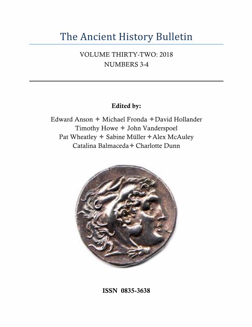
TheAncientHistoryBulletinVOLUME THIRTY-TWO: 2018
NUMBERS 3-4
Edited by:
Edward Anson ò Michael Fronda òDavid Hollander Timothy Howe ò John Vanderspoel
Pat Wheatley ò Sabine MülleròAlex McAuley Catalina BalmacedaòCharlotte Dunn
ISSN 0835-3638
ANCIENT HISTORY BULLETIN Volume 32 (2018) Numbers 3-4
Edited by: Edward Anson, Catalina Balmaceda, Michael Fronda, David Hollander, Alex McAuley, Sabine Müller, John Vanderspoel, Pat Wheatley Senior Editor: Timothy Howe Assistant Editor: Charlotte Dunn Editorial correspondents
Elizabeth Baynham, Hugh Bowden, Franca Landucci Gattinoni, Alexander Meeus, Kurt Raaflaub, P.J. Rhodes, Robert Rollinger, Victor Alonso Troncoso
Contents of volume thirty-two
Numbers 3-4 72 Fabrizio Biglino, Early Roman Overseas Colonization 95 Catherine Rubincam, How were Battlefield Dead Counted in Greek Warfare? 106 Katherine Hall, Did Alexander the Great Die from Guillain-Barré Syndrome? 129 Bejamin Keim, Communities of Honor in Herodotus’ Histories
NOTESTOCONTRIBUTORSANDSUBSCRIBERSThe Ancient History Bulletin was founded in 1987 by Waldemar Heckel, Brian Lavelle, and JohnVanderspoel. The board of editorial correspondents consists of Elizabeth Baynham (University ofNewcastle), Hugh Bowden (Kings College, London), Franca Landucci Gattinoni (Università Cattolica,Milan), Alexander Meeus (University of Mannhiem), Kurt Raaflaub (Brown University), P.J. Rhodes(DurhamUniversity),RobertRollinger(UniversitätInnsbruck),VictorAlonsoTroncoso(UniversidadedaCoruña)AHB is currently edited by: TimothyHowe (Senior Editor: [email protected]), Edward Anson, CatalinaBalmaceda, Michael Fronda, David Hollander, Alex McAuley, Sabine Müller, John Vanderspoel, PatWheatleyandCharlotteDunn.AHBpromotesscholarlydiscussioninAncientHistoryandancillaryfields(suchasepigraphy,papyrology,andnumismatics)bypublishingarticlesandnotesonanyaspectoftheancientworldfromtheNearEastto Late Antiquity. Submitted articles should normally be in English, but the journal will considercontributionsinFrench,German,ItalianorSpanish.SUBMISSIONGUIDELINESAHBadherestotheusualNorthAmericaneditorialpoliciesinthesubmissionandacceptanceofarticlesbut imposes no House Style. Authors are, however, asked to use the abbreviations of L’Annéephilologique(APh)forjournals,andoftheThesauruslinguaelatinae(TLL)forLatinauthors.Pleasesendsubmissions to the editor most closely identified with your field of enquiry or, in case of doubt, toTimothy Howe ([email protected]). Articles must be submitted in electronic format, preferablygeneratedbyMSWord.GreekfontorotherspecialcharactersmustconvertsuchtoUnicodeandshouldbeaccompaniedbyaPDFversion.AuthorswillreceivePDFoffprintsoftheircontributions.Copyrightisretainedbytheauthor.BooksforreviewsandenquiriesconcerningbookreviewsshouldbedirectedtoJosephRoisman([email protected]).SUBSCRIPTIONINFORMATIONThe subscription rate for individual and institutional subscribers is USD 25.00. Detailed instructionsabout subscriptions and access to digital content can be found on the AHB website:http://ancienthistorybulletin.org PAYMENTPaymentmaybemadeviathesubscriptionportalontheAHBwebsite:http://www.ancienthistorybulletin.org/subscribed-users-area/membership-levels/
CoverimagecourtesyofTheNickleArtsMuseum,UniversityofCalgary
AHB 32 (2018): 106-128 Page 106
Did Alexander the Great Die from Guillain-Barré Syndrome? Katherine Hall
Abstract : The most striking feature of Alexander the Great’s death is that, despite being extremely unwell, he was reported to have remained compos mentis until just before his death. Combined with evidence that he developed a progressive, symmetrical, ascending paralysis, it is argued that he died from a sub-type of the autoimmune disorder, Guillain-Barré Syndrome (GBS), most likely induced from a Campylobacter pylori infection. GBS could also account for the reported lack of decay of his body, and his death may be the most famous case of pseudothanatos, or false diagnosis of death, ever recorded.
Introduction
Previous medical explanations for the death of Alexander the Great have been varied and many, but none have been entirely satisfactory and all have had cogent counter-arguments put forward.1 What has been held in common by almost all of these papers is an emphasis on either one, or both, of two very common symptoms which occurred in his illness; fever and abdominal pain. In comparison, the very unusual description of an extremely unwell man who remains compos mentis until moments before he is declared dead has received barely any attention. In addition, there is also the impression given from reading the accounts of his death that Alexander may have developed a progressive, symmetrical and ascending paralysis which first rendered him unable to walk, then unable to move his arms and finally took his ability to speak. This type of paralysis only rarely occurs and the most common reason for it today is a condition called Guillain-Barré Syndrome (GBS). Only Oldbach et. al.2 have given this symptom any significance in their paper ‘A Mysterious Death’, but this was not explored fully. They were not able, due to when it was written, to take into account all the developments in our understanding of GBS over the past thirty years. I will argue that they were correct in drawing attention to this symptom. Further, I will argue that the type of acute, flaccid paralysis documented in Alexander’s death is, in fact, GBS. Unlike them, I will conclude that the precipitant cause of his GBS was not typhoid, but much more likely to be a gastrointestinal cause, Campylobacter jejuni.3 This would also adequately account for his other symptoms, including a combination of fever and abdominal pain, thus avoiding having to give preference to one historiographic tradition over another.4 In addition, there has also been the mystery (largely dismissed as mythology or romantic legend) of the lack of decay in Alexander’s corpse once he had died.
1 Whilst there is no review paper evaluating all of these, Schep et. al. (2014) contains a reasonably up-
to-date but not entirely complete overview; it is nevertheless useful. 2 Oldbach et. al. (1998). 3 Oldbach et. al. dismissed this as a cause due to the lack of diarrhoea, but I will present evidence that
diarrhoea can be absent in over 50% of cases of Campylobacter jejuni infection. 4 Readers coming to this paper via medicine may not be aware that the historiographic paradoxes of
the varying accounts have been a source of much academic discussion in Classics. Examples of literature on this issue includes: Bosworth (1971), Bosworth (1988) especially pages 171-3, and Anson (2009).
Katherine Hall
Page 107
Oldbach et. al. briefly mention this was due to a premature diagnosis of death: I will expand upon this extensively in support of this idea. I will discuss the difficulties of knowing when someone has actually died and why this could have been particularly problematic in Alexander’s case. I will explain how this example (and possibly the most famous example) of pseudothanatos, or the false diagnosis of death, could occur making the diagnosis of GBS as the cause for his death both feasible and plausible.
There are four, major, historical accounts for Alexander’s death: Arrian, Plutarch, Diodorus and Curtius.5 None of these were contemporary with his life and all of them have issues. I do not intend to enter this debate, as my argument can encompass the variations between these accounts. Medicine is very fond of applying Occam’s Razor,6 and if all symptoms can be explained by a single diagnosis, doctors argue that this is the most likely conclusion. GBS does provide such a unifying ‘diagnosis’ to these varying stories.
Alexander’s Death
Arrian7 and Plutarch8 record similar versions of Alexander’s death; both authors state they base their accounts on the Royal Journal9 of Alexander’s court. He became unwell following a drinking party and developed a fever, which was initially mild and intermittent. He continued his duties but increasingly needed to be carried rather than walk. By the fourth night the fever was constant. By the seventh night he was seriously ill and two days later he was extremely ill, but he remained alert and aware; ‘When the officers came in he knew them, but said no more; he was speechless.’10 The following day the army filed past him, and being unable to speak, ‘he greeted one and all, raising his head, though with difficulty and making a sign to them with his eyes.’11 After this ‘he died.’12 Plutarch’s account agrees essentially with this, making it clearer that Alexander died after eleven days of illness (from the 18th to the 28th of the month called Daesius).13 Diodorus’ account of Alexander’s death is the shortest; after a drinking party Alexander was stricken with a severe pain and died sometime thereafter.14 Curtius’ account is fragmentary—some parts are missing—but he reiterates that Alexander acknowledged his troops as they filed past him with very little movement; ‘he maintained the same posture … until the last salute … by now, even his voice had started to fail.’15 Curtius uniquely records that Alexander’s body did not putrefy
5 To be specific this analysis is based on Arrian’s Anabasis, Plutarch’s Life of Alexander, Diodorus
Siculus’ Library of History and Quintus Curtius Rufus’ The History of Alexander. The earliest account is that of Diodorus (c.80-20 BC) and the latest Arrian (c.86-172 AD). Alexander died in 323 BC.
6 For brief outline of Occam’s Razor in medicine see Smith (2013). 7 Arr. Anab. 7.25-26. 8 Plut. Alex. 76-7. 9 Hammond (1988). 10 Arr. Anab. 7.25.6. 11 Arr. Anab. 7.26.1. 12 Arr. Anab. 7.26.3. The Greek used is the aorist infinitive ἀποθανεῖν. 13 Plut. Alex. 76. 14 Diod. 17.117.1-5. The Metz Epitome 99-100 also describes Alexander clutching his stomach,
suggesting this pain was abdominal. 15 Curt. 10.5.3.
Did Alexander the Great Die from Guillain-Barré Syndrome?
Page 108
for six days despite the Mesopotamian heat.16 Other, minor accounts also exist, mainly supporting the idea that he was poisoned.17
Guillain-Barré Syndrome
GBS is the most common cause of acute (i.e. reaching the peak severity of symptoms within four weeks of onset), flaccid (the muscles are relaxed, not tight), ascending form of paralysis in the post-polio world.18 It is, however, quite rare with an overall incidence of 0.6 to 4 cases per 100,000 people per year but can still be fatal; even with modern treatment the mortality rate is between 4% and 15%.19 GBS is named after two of three authors—George Guillain and Jean-Alexandre Barré—who published a paper in 1916.20 Originally in French, a translation is available in English.21 In this paper, they described two soldiers who developed a sudden onset of paralysis, abnormal sensation and loss of reflexes, but who recovered within a month. Until the 1980s, GBS was thought to be a single type of disease, but now it is known that there are three major variants of this syndrome.22 These variants are reactions to a precipitating cause but can vary significantly in terms of the likely aetiology, geographical distribution, mechanism of injury to the nerves and the symptoms;23 this is very relevant to understanding the possible relationship between GBS and Alexander’s death. All three variants are associated with damage to the peripheral nervous system giving rise to a symmetrical,24 flaccid and ascending paralysis and loss of reflexes beginning with difficulty in walking. Sometimes the autonomic nervous system is involved which results in problems in the body’s control of heart rate, blood pressure, and temperature. If the paralysis rises high enough, speech can be lost, pupils dilate, and respiratory arrest can occur.25
GBS is an auto-immune disorder where the patient’s own immune system has become confused in differentiating between an invading organism, such as a bacteria, virus, or (very rarely) vaccine products, and the patient’s own body.26 Approximately two-thirds of patients have clear-cut evidence for a preceding infection,27 although this can be as high as
16 Curt. 10.10.9-13. 17 These include Ephippus (FGrH 126 F3 = Athen. 10.434a-b), Nicobule (FGrH 127 F1 & 2 = Athen 10.434c
&12.537d), Aelian (Varia Historia 3.23), Valerius Maximus (5.1, ext. 1b), Justin (12.13.6-16.1), Orosius (3.20.4), Pseudo-Callisthenes (3.31-33), and the unknown author(s) of the Metz Epitome (99-100).
18 Eldar and Chapman (2014). 19 Hughes and Cornblath (2005). 20 Kusunoki (2016). The third author was André Strohl. Octave Landry had first described cases of
ascending paralysis in 1869 (Wijdicks and Klein (2017), but failed to gain the eponym. 21 Guillain et. al. (1968). 22 There are several minor, very rare subtypes such as Miller Fisher Syndrome and Bickerstaff
brainstem encephalitis but these are not relevant to the discussion here, as their symptoms differ from those experienced by Alexander.
23 Willison et. al. (2016). 24 Rarely, it can be asymmetrical. 25 Hauser and Asbury (2005). 26 Wakerley and Yuki (2013). 27 Wakerley and Yuki (2013).
Katherine Hall
Page 109
88% in some studies.28 The three major variants are: acute inflammatory demyelinating polyradiculoneuropathy (AIDP), acute motor axonal neuropathy (AMAN), and acute and sensory axonal neuropathy (AMSAN). AIDP has a different mechanism of injury from the other two variants in that the nerves’ protective covering, the myelin sheath, is affected leading to delayed or absent conduction along both motor (which control movement) and sensory nerves. With both AMAN and AMSAN, the nodes of Ranvier which sit at regular intervals along the nerves and concentrate and facilitate the conduction of electrical impulses, are affected; the difference between the two is that with AMAN only motor nerves are affected, and with AMSAN both motor and sensory nerves are affected.29 Most of the medical literature is predicated upon the AIDP variant as this is the most common form seen in Europe and in America, with AMAN and AMSAN only contributing to 5% of cases in these regions.30 Alexander’s symptoms of ascending, symmetrical and flaccid paralysis fit best with AMAN, but with this being a rare cause of a rare disease, could this be a diagnosis too far?
Recently there has developed good cause to consider this may be so. AMAN was only first described in 1986.31 Until then, medical understanding of GBS had been entirely based on AIDP, which has a slower onset of four to six weeks after the precipitating illness and combined motor and sensory symptoms. In contrast, AMAN has a very rapid onset of within one to six days of contracting the precipitating illness.32 It usually only has motor symptoms such as paralysis:33 sensory symptoms such as pins and needles, pain, or numbness do not occur or are rare and mild.34 Whilst the AMAN variant is rare in Western Europe and America, it is appears to be more common35 the further east one travels towards Eastern Europe and the Near and Middle East; for example, 7% of GBS cases in the Western Balkans,36 13% in North-West Greece,37 9-24% in Iran,38 39% in Pakistan,39 and 44% in India.40 The cause of these variations in the relative frequency of GBS variants is unknown,41 but it is thought this relates to differing rates of local exposure to the organisms that cause the precipitating illness, rather than any hereditary susceptibility. In other words, Alexander’s chances of contracting GBS would not be related to his Greek ancestry but to where he was at the time; i.e. Babylon in ancient Persia, or modern day Iraq.
In Iraq, where Alexander died in Babylon, the measured incidence of GBS (in a paediatric population) is 1.33 cases per 100,000 people aged less than fifteen years,42 rising
28 Koga et. al. (2001). 29 Hauser and Asbury (2005). 30 Hughes and Cornblath (2005). 31 Feasby et. al. (1986). 32 McKhann et. al. (1991). 33 Hughes and Cornblath (2005). 34 Yadegari et. al. (2014); Ruts et. al. (2008). 35 It is very difficult to get accurate figures for GBS in many countries; see Sejvar et. al. (2011). 36 Peric (2014). 37 Markoula et. al. (2007) 38 Arami et. al. (2006); Yadegari et. al. (2014). 39 Yadegari et. al. (2014). 40 Shrivastava et. al. (2017). 41 Kanda (2016). 42 Jasem et. al. (2013).
Did Alexander the Great Die from Guillain-Barré Syndrome?
Page 110
to an extraordinarily high 22.9 in 100,000.43 GBS accounts for 52.5% of cases of acute flaccid paralysis in Iraq.44 Unlike Western studies, Iraqi GBS was often present with fever (55.4%) and in 97% of cases the time of onset to maximum paralysis was within four days.45 This short time course is highly suggestive of a high incidence of AMAN. The percentage of GBS of AMAN and AMSAN combined is 18%.46 Generally the group in the Iraqi and Iranian regions who contract AMAN are more likely to be male (approximately a 2:1 ratio to females),47 are aged significantly younger than people getting AIDP in the West (one study showing an average age of 32 years), and are more likely to develop the syndrome in late spring or summer.48 It is striking how well this profile matches that of Alexander, as he was male, 32 years old, and died at the start of summer.49
Other conditions which can mimic GBS include: poliomyelitis, botulism, electrolyte disturbance, myasthenia gravis, acute myopathy, diphtheria, porphyria, tick paralysis and vasculitis.50 Of these, poliomyelitis is the strongest possible alternative diagnosis. It is strongly associated with fever, but the paralysis that occurs usually does so within 24 hours of onset of symptoms and is most commonly asymmetrical. If extensive paralysis does occur, it is usually following an initial phase with headache, and a lowered level of consciousness or coma due to an associated meningitis.51 The time course of the illness does not fit with Alexander’s illness, nor does the way he remained cognitively intact even at the end of his illness. Botulism does cause a symmetrical paralysis but this descends, not ascends, from the head to the extremities.52 Electrolyte disturbances, which can occur after excessive alcoholic intake (and Alexander had indulged just prior to his illness), are unlikely causes as they also result in a reduced level of consciousness and are not associated with fever.53 Myasthenia gravis is strongly associated with fatigue of muscles with recurrent use, and a waxing and waning course over several years before diagnosis, all of which makes this to be an unlikely cause for Alexander’s death.54 Acute myopathy due to alcohol intoxication causes seizures and coma, neither of which was present in Alexander’s case.55 Diphtheria causes a severe sore throat,56 which was absent in Alexander’s case. Porphyria is an inherited disorder of the metabolism of heme, a by-product of haemoglobin
43 Al-Dabbas (2016). This was also in a paediatric population, but is per 100,000 hospital admissions. 44 Jasem et. al. (2014). In ancient times poliomyelitis is likely to have been a much more common cause. 45 Jasem et. al. (2013). Western medical textbooks, based on the high proportion of AIDP in their
countries, often discount fever as a symptom of GBS. For example, Hauser and Asbury (2005), page 2513, write ‘Fever and constitutional symptoms are absent at the onset [of paralysis], and, if present, cast doubts on the diagnosis.’ Hasan et. al. (2014) found the mean time from onset of paralysis to peak paralysis to be 5.4 days in a paediatric population in Iraq.
46 Hasan et. al. (2014). 47 Benamer and Bredan, (2014). 48 Yadegari et. al., (2014); Abdul-Kareem et. al. (2004). Al-Dabbas (2016) found a bimodal distribution
with peaks in winter and summer (including June, the time of Alexander’s death). 49 Alexander died on 11 June, 323 BC; for discussion of this date, see Depuydt (1997). 50 Eldar and Chapman (2014); Hughes and Cornblath (2005). 51 Cohen (2005a). 52 Abrutyn (2005). 53 Singer and Brenner (2005). 54 Drachman (2005). 55 Brown and Mendell (2005), 2539. 56 Holmes (2005).
Katherine Hall
Page 111
degradation; it can cause an acute crisis, especially after alcohol ingestion, but is rarely fatal and is always associated with a fever.57 Alexander regularly drank and there was nothing to suggest previous porphyric crises in the past (porphyria has previously been suggested58 and repudiated as a cause for Alexander’s death59). Tick paralysis is extremely rare, almost always occurs in children, and there is no fever.60 Vasculitis is a usually long-term, complex disorder primarily affecting the connective tissues and causing symptoms of muscle and joint pain.61 The symptoms and time course of Alexander’s illness make this diagnosis unlikely. If Alexander did suffer from acute ascending, symmetrical and flaccid paralysis, GBS is the most likely diagnosis.
Precipitant Causes of Guillain-Barré Syndrome
GBS can be associated with a number of infections preceding its onset. These include: Campylobacter jejuni,62 Cytomegalovirus,63 Salmonella typhi (typhoid fever) and paratyphi,64 Epstein-Barr (glandular fever) virus,65 Haemophilus influenzae,66 Mycoplasma pneumoniae,67 Varicella zoster (chicken pox),68 Influenza A and B and Parainfluenza 1 viruses,69 Adenovirus,70 Herpes simplex,71 Hepatitis E,72 Dengue fever,73 Chikungunya virus,74 Leprosy or Hansen’s disease,75 West Nile Virus,76 and the Zika virus.77 But association does not necessarily mean causation and some of these possible causes can be eliminated by looking at whether the chance of getting GBS is higher with these illnesses than in the general population: on this basis infections with Haemophilus influenzae, Influenza A, B and Parainfluenza 1 viruses, Adenovirus, Herpes simplex virus, and Varicella zoster virus, do not occur any more frequently in GBS patients than expected in the general population
57 Desnick (2005). 58 Schnorf (1998). 59 Schep et. al. (2014). 60 Edlow and McGillicuddy (2008). 61 Sneller et. al. (2005). 62 Wijdicks and Klein, (2017). 63 Orlikowski et. al. (2011). 64 Wakerley and Yuki (2013). 65 Tam et. al. (2007). 66 Yuki and Shahrizaila (2011). 67 Eldar and Chapman (2014). 68 Yadegari et. al. (2014). 69 Kutlesa et. al. (2010), Tam et. al. (2007), Jacobs et. al. (1998). 70 Jacobs et. al. (1998). 71 Jacobs et. al. (1998). 72 Geurtsvankessel et. al. (2013), Van Den Berg et. al. (2014). 73 Solomon et. al (2000), Chen and Lee (2017). 74 Agarwal et. al. (2017), Mehta et. al. (2018). 75 Mittal et. al. (2017). 76 Ahmed et. al. (2000), Marr and Calisher (2003). 77 Musso et. al. (2015), Brasil et. al. (2016).
Did Alexander the Great Die from Guillain-Barré Syndrome?
Page 112
and are unlikely to be true causes.78 On the same basis, Campylobacter jejuni, and Cytomegalovirus and Epstein-Barr viruses are highly likely to be a cause of GBS, and Mycoplasma moderately so.79
Further possibilities may be eliminated by considering what symptoms and signs Alexander did not have, although here is where the difficulty of dealing with the historical sources becomes relevant: is a symptom absent from the record because it did not occur, or did it occur but was either not recognised or not recorded?80 There was no report he suffered any respiratory symptoms, enlarged glands or rash. Mycoplasma causes a pneumonia-like disease. Cytomegalovirus is associated with sneezing and a sore throat, especially in the early days of the illness, and with a rash (although this is less common in adults).81 Epstein-Barr virus causes a sore throat in 75% of patients, widespread enlarged lymph nodes in 95%, and pharyngitis or tonsillitis in 82%.82 These are unlikely precipitants. The sources do not mention that Alexander became jaundiced, although this symptom was well described by ancient doctors,83 and the lack of this symptom would make Hepatitis E unlikely.84 Dengue fever is characterised by the sudden onset of a high fever (not the slow onset of an increasingly severe fever as Alexander suffered), together with headache, back pain, a rash and red eyes.85 Alexander was not recorded as having any of these symptoms. Zika is unlikely to have been present in ancient times; it was only first identified in 1947, and is usually very mild and lasts two to five days, again with headache and red eyes.86 West Nile Virus has previously been suggested as the cause for Alexander’s death,87 but again its symptomatology of confusion accompanying the paralysis and usually only being fatal in patients over 75 tells against it.88 Chikungunya virus was first described in 1824, and starts with a sudden high fever, headache and severe arthritis.89 Leprosy is an ancient disease, having been first described in 6th century BC Indian texts; it takes years to slowly develop, has marked, characteristic skin and limb changes, is not associated with a sudden fever, and is not fatal.90 These, too, are unlikely precipitants.
This leaves the two most likely contenders: Campylobacter infection and Salmonella (typhi and paratyphi). Campylobacter is a world-wide bacteria which causes a gastroenteritis and was first described as being associated with GBS in 1982;91 it most
78 Jacobs et. al. (1998). 79 The other causes were not analysed because they were not known to be associated with GBS at the
time of publication of this paper in 1998. 80 These questions are unanswerable, but to make any meaningful progress in my argument I have
made the assumption that the absence of a symptom or sign from the historical record means it was absent from Alexander’s illness.
81 Jacobs et. al. (1998). 82 Cohen (2005b). 83 Nutton (2013), 29. 84 Dienstag and Isselbacher (2005), 1831. 85 Peters (2005), 1164. 86 Paixão et. al. (2016). 87 Marr and Calisher (2003). 88 Schep et. al. (2014) 89 Kucharz and Cebula-Byrska (2012). 90 Gelber (2005). 91 Rhodes and Tattersfield (1982).

Katherine Hall
Page 113
frequently results in the AMAN variant.92 Identification of Campylobacter species in archaeological studies on ancient dental plaque show that this family of bacteria has been with us for millennia.93 It is the most common cause for GBS worldwide,94 and is the most common cause for the AMAN variant,95 especially in the region where Iran borders Iraq—the very place where Alexander died.96 Approximately one in 3000 cases of Campylobacter infection is associated with GBS.97 GBS can begin as quickly as within one day of infection, although the mean duration between the precipitating infection and the onset of paralysis is ten days with this cause.98 Although GBS is a rare complication of Campylobacter infection,99 the associated risk of acquiring GBS can vary and appears related to the serotypes (subtypes) of the bacteria,100 and also to the severity of the initial infection.101 Campylobacter-induced GBS is most commonly sporadic with only isolated cases occurring, although some epidemics have also been documented.102 It is a waterborne disease and can also be transmitted by animals, especially in milk from affected cows and chickens.103 It most commonly causes a diarrhoeal illness with fever and abdominal pain, lasting from one to two weeks, but infections can occur without these symptoms; in one series only 24% had diarrhoea, 38% abdominal pain and 14% fever.104 Thus, whether or not the sources record Alexander having or not having diarrhoea is irrelevant to making this diagnosis. Although his fever would be unusual, this could have been related to the severity of his infection.
Salmonella typhi, which causes typhoid fever, is still endemic in Iraq and an outbreak was reported there as recently as 2014.105 It, and Salmonella paratyphi, cause similar illnesses although paratyphi infections tend to be milder; in the pre-antibiotic era, S. typhi had a mortality rate of 16.1% versus 1.5% for paratyphi for those aged 31 to 40 years.106 Both organisms can cause typhoid fever, which begins with cough, headache, rigors and sore throat (none of which Alexander was reported having), progressing onto high, sustained fever in 75% and abdominal pain in 40%; 30% also develop a distinctive rash by the end of the first week of symptoms.107 Later complications, usually in weeks three and four, include gastrointestinal perforation and peritonitis; only extremely rarely does this occur in week
92 Willison et. al. (2016). 93 Warriner et. al. (2014). 94 Moore et. al. (2005). 95 Wakerley and Yuki (2013), Kusunoki (2016), Wijdicks et. al. (2017). 96 Barzegar et. al. (2008). 52% of patients with AMAN or AMSAN had had a preceding Campylobacter
infection. 97 Moore et. al. (2005). 98 Takahashi et. al. (2005). 99 McCarthy et. al (1999), Tam et. al. (2006). 100 Takahashi et. al. (2005). 101 Baker et. al. (2012). 102 McKhann et. al. (1991). 103 Young et. al. (2007). 104 Ho et. al. (1995). This shows Oldbach et. al. (1998) is incorrect in saying that the infection could not
have been Campylobacter due to the lack of diarrhoea. 105 Dworkin et. al. (2014). 106 Gadeholt and Madsen (1963). 107 Lesser and Miller (2005).
Did Alexander the Great Die from Guillain-Barré Syndrome?
Page 114
one.108 Alexander’s fever was not sustained but slowly worsened; in addition, if he had developed peritonitis and with it septic shock, he would have rapidly developed a ‘muttering delirium’109 typical of these cases. Typhoid fever also requires human-to-human transmission as there are no animal vectors and it is rare for isolated cases to occur. No epidemics were reported concurrently with Alexander’s death (although the significance of his death could well have outshone those of the hoi polloi, no matter how many).
Typhoid fever has extremely rarely been associated with GBS. The first case was reported in 1969 in a 27-year-old male.110 Since then, it has also been reported in a 15-year-old female,111 a 47-year-old woman,112 a 53-year-old man,113 a 41-year-old man,114 a 43-year-old man,115 as well as two paediatric cases.116 Of the adult cases, four had pure motor loss indicative of AMAN, and two mixed motor and sensory losses. The paucity of cases must call into question whether typhoid fever is actually a cause, or merely an association.117 In addition, death is usually by gastrointestinal perforation in the third to fourth week of the illness.118 Data comparing outcomes in the pre-antibiotic era of 1,119 cases of typhoid fever and 1,528 cases of paratyphoid fever show that in the first week of the illness only 0.7% of typhoid patients developed this complication and no cases of this occur with paratyphoid illness. Those who died from typhoid did so 6 to 76 days after the start of their illness, and with paratyphoid this was between 11 and 66 days. Whilst GBS was not specifically mentioned in this paper, polyneuritis (inflammation of multiple nerves causing motor and/or sensory disturbance which could be cases of GBS) occurred in 0.4% of typhoid and 0.2% for paratyphoid cases and gives some idea of the frequency of GBS; far lower than Campylobacter which from this analysis remains the most likely cause for GBS in Alexander’s case.
Proving Death in the Ancient Greek World.
It may seem counter-intuitive but death can be difficult to prove even in modern times.119 Celsus comments that Democritus had studied the diagnosis of death and had collected evidence of people coming back to life after having been declared dead, so much so that Democritus could ‘not admit that there could be any sure signs of approaching death.’120
108 Gadeholt and Madsen (1963) report gastrointestinal perforation and peritonitis in the pre-antibiotic era occurring in only 0.7% of patients with typhoid fever in the first week.
109 Lesser and Miller (2005), 899. Chanmugam and Waniganetti (1969) also describe a ‘typhoid stupor’. 110 Chanmugam D and Waniganetti (1963). 111 Samantray (1977). 112 Aldrey et. al. (1999). 113 Berger et. al. (1986). 114 Donoso et. al. (1988). 115 Subrata (2012). 116 Mehndiratta et. al. (2012), Kapoor et. al. (2014). 117 Jacobs et. al. (1998) did not included typhoid fever in their statistical analysis, but if such analysis
was applied it would be doubtful that it would reach statistical likelihood for causation. 118 Schep et. al. (2014). 119 Two anaesthetists called for further review of this as recently as 2013 (see
http://www.bbc.com/news/health-22730360 accessed 17 April, 2018). 120 Cels. Med. 2.13-8.
Katherine Hall
Page 115
Even up until the eighteenth century, the action of the heart and pulsation was still considered subordinate to respiration,121 probably because of the commonplace observation that when the breathing stopped, the heart usually then followed, whereas the reverse was harder to observe. Until the 19th century AD, it was generally ‘concluded that advanced putrefaction of the entire body was the only valid single test to exclude apparent death.’122 It is not surprising then that the embalmers were reluctant to commence working on Alexander’s body when it showed no signs of decomposition.123
There is very little extant evidence as to how death was actually determined in the Ancient Greek world. While the Hippocratic corpus, particularly in Prognosis, does discuss what was termed a ‘sign of death’ (τὸ σημεῖον Φανατῶδες),124 this actually refers to a sign of impending death, not diagnosing actual death.125 It has been argued that in the ancient Greek world (not specifically but including the time of Alexander), the presence or absence of a heartbeat was considered the sine qua non for determining death.126 But at the time of Alexander’s death this was almost certainly not the case. He died at a time when pulsation or ‘throbbing’ was considered ‘an unnatural and mostly random phenomenon, a sign of disease simply by virtue of its existence … as opposed to natural phenomena such as respiration, urine and sleep.’127 Certainly, the Ancient Egyptians had identified the heart as the central organ of the cardiovascular system,128 and some believe that the Egyptians also linked the heart with the pulse.129 Aristotle noted that the heart was the first organ to form and the last to stop.130 But Hippocrates did not advocate taking the pulse from the wrist (although he writes of observing temporal artery pulsating in fever),131 and the link between peripheral pulses and heartbeat was poorly understood by Greeks, including Aristotle.132 He did connect the heart with respiration and pulsation, but believed the peripheral vascular system had its own active, intrinsic pulsation rather than passively distending in response to cardiac pulsation.133 After the time of Alexander’s death Praxagoras identified that only arteries pulsated,134 but five centuries would then pass before Rufus of Ephesus correctly linked peripheral pulsation to the heart, not to the
121 Cooper et. al. (2006). 122 Powner et. al. (1996), 1220. Pernick’s chapter (1988) also gives an informative overview of the
history of how death was diagnosed (or not) over the ages. 123 Curt. 10.10.13. 124 Hp. Prog. 2.22. Also see Hp. Aph. 7. end fragment (page 219 of Loeb edition), which lists such signs
as a retracted right testicle, blackened nails, cold toes, livid fingertips, dark, swollen lips, and swollen, puffed up bowels as signs of death.
125 Bondeson (2001), 18. 126 Bondeson (2001), Powner et. al. (1996), Pernick (1988). The first two sources appear to make
Pernick the basis for their claim. Pernick cites Wiener PP (ed.), Dictionary of the History of Ideas, 1968, New York, Scribner, not the primary Ancient Greek sources.
127 Lewis (2015), 346. 128 Loukas et. al. (2016). 129 Loukas et. al. (2007). 130 Harris (1973), 173. 131 Cheng (2001). 132 Harris (1973), 124-135. 133 Shoja et. al. (2008). 134 Ghasemzadeh and Zafari (2011).
Did Alexander the Great Die from Guillain-Barré Syndrome?
Page 116
vessels themselves.135 The relevancy and importance of the pulse in deciding if life was extinct was not truly appreciated until Galen’s seminal works on the subject.136 At the time of Alexander’s death, therefore, the absence of pulsation was unlikely to be considered a sign of death.
Given this relative silence of medical texts on the determinants of diagnosing death, non-medical literature was also reviewed.137 Here, too, the evidence remains generally scanty. Vermeule’s analysis of death and its relationship to early Greek Art and Poetry fails to consider the aspect of determining when one is dead,138 as does Christiane Sourvinou-Inwood in her book Reading Greek Death.139 Religious piety and convention generally restricted the visual act of killing onstage in tragedies (although the process of dying could be presented),140 and prior to either Sophocles or Euripides there were few depictions of death-bed scenes.141 Two rare examples are Eurydice’s suicide which mentions changes in the eyes; ‘By the altar, with a sharp-whetted sword, she struck until her eyes went slack and dark,’142 and the death of Sophocles’ Ajax in the eponymous play which is less subtle. Having fallen on his sword, he subsequently ‘spurt[s] the darkened gore of his self-inflicted slaughter up his nostrils and out of the bloody gash.’143 Death from acute trauma is usually self-evident and non-problematic, which may be why, despite describing 240 cases of death in The Iliad, Homer did not specifically mention any means by which the diagnosis of death was confirmed.144 In contrast, one Hippocratic aphorism states ‘in the case of acute diseases to predict either death or recovery is not quite safe.’145 The clearest example in literature for the determination of death is that of Socrates in Phaedo: ‘the attendant uncovered him [Socrates]; his eyes were fixed. And when Crito saw it, closed his mouth and eyes. Such was the end.’146
Other references for signs of death included the extinguishing of thymos, which was most commonly thought to reside in the chest, and a loosening of the limbs.147 In Hippocrates’ Aphorisms it states that ‘the boundary of death is passed when the heat of the soul has risen above the navel to the part above the diaphragm.’148 Another more self-
135 Bedford (1951). 136 Horine (1941). See especially pages 220-221. 137 This presents a synopsis of what is relatively easily found, not a full and complete account. This
would be fascinating but beyond the scope of the current article and possibly frustrating; Garland (1985),16, states that such descriptions of the moments of death itself are ‘in both art and literature extremely rare.’
138 Vermeule (1979), 5. 139 Sourvinou-Inwood (1995). 140 Sommerstein (2010), chapter 2. 141 Vermeule (1979), 10. 142 Soph. Ant. 1301. More strictly this refers to eyelids, rather than eyes, as in the original it reads: ‘ἡ δ᾽
ὀξυθήκτῳ βωμία περὶ ξίφει λύει κελαινὰ βλέφαρα,…’. 143 Soph. Aj. 918-9. 144 Garland (1981). 145 Hp. Aph. 2.19. The Greek reads: ‘Τῶν ὀξέων νοσημάτων οὐ πάμπαν ἀσφαλέες αί προαγορεύσιες,
οὔτε τοῦ θανάτου, οὔτε τῆς ὑγιείης.’ 146 Plat. Phaedo 118a. reads; 'ὁ ἄνθρωπος ἐξεκάλυψεν αὐτόν, καὶ ὃς τὰ ὄμματα ἔστησεν· ἰδὼν δὲ ὁ
Κρίτων συνέλαβε τὸ στόμα καὶ τοὺς ὀφθαλμούς. Ἥδε ἡ τελευτή.' 147 Garland (1981). 148 Hp. Aph. 7.(end fragment).26-9. The Greek reads: ‘ὅρος δὲ θανάτου· ἐπειδὰν τὸ τῆς ψυχῆς θερμὸν
ἐπανέλθῃ ὑπὲρ τοῦ ὀμφαλοῦ ἐς τὸ ἄνω τῶν φρενῶν.’
Katherine Hall
Page 117
evident sign of death than the cessation of cardiac activity is the cessation of respiratory effort. As Alcestis lies dying on behalf of her husband, it is commented that ‘she has scarcely any breath within her.’149 Homer refers to the psyche being released or leaving the body at death, most commonly via the mouth or the wound.150 Despite his remarks on the primacy of the heart (see first paragraph this section), Aristotle also stated that breath ‘evidently has control over life and death, for it results synchronously that when respiring animals are unable to breathe they perish.’151 The large philosophical and medical literature concerning pneuma underscores its conceptual importance to both these fields, with a full analysis being beyond the scope of this article. But given pneuma’s pre-eminence in the thinking in both these fields, it could be surmised that this would have been the early sign of death.
Unlike in modern times, the determination of being dead was probably not done by doctors, but more likely performed by family, relatives or non-medical attendants. There is no advice in the Hippocratic corpus that a doctor needed or should be involved in declaring death.152 No medical certificate was required, although one was in Roman Egypt where bureaucratic processes required such a certificate to be produced to avoid posthumous taxation.153 Whilst death from trauma would often be very obvious, death from an internal cause with an outwardly intact body could be far more problematic. For Alexander, only Diodorus mentions physicians being present early in his illness154 and neither he, Arrian, Plutarch, Diodorus or Curtius mention any at his death. The priests of Serapis, who may also have had a doctoring role,155 perhaps wisely stayed away in their temple.
With proving death being problematic, and often left to the layperson, perhaps it is not surprising that stories of people reviving after having been declared dead have been recorded in classical literature.156 The stories of the Ephesian Tale, Metamorphoses of Apuleius and the anonymously authored Apollonius, Prince of Tyre all employ a character who is mistaken for being dead.157 Then there is the tale of Er in Plato’s Republic, an Armenian soldier who wakes up on the funeral pyre ten days after being found and thought dead on the battle-field, which clearly describes a case of non-putrefaction of a ‘corpse’.158 Pliny the Elder, in his Natural History, had a whole section in Book Seven entitled ‘Persons Who Have Come To Life Again After Being Laid Out For Burial.’159 He described several instances of people being placed on funeral pyres only to waken, including Acilius Aviola, Lucius Aelius Lamia and Gaieus Aelius Tubero. Although the date for the death of the consul Acilius
149 Eur. Alc. 205. The Greek reads: ‘καίπερ σμικρὸν ἐμπνέουσ᾽ ἔτι.’ 150 Garland (1981). 151 Aristot. Resp. 11.472b. 152 Bondeson (2001), 18, argues that within the Hippocratic corpus that there are specific instructions
for a doctor to withdraw when a patient is considered terminally ill, but there is no citation and it is not clear to which section is being referred. In Hippocrates’ The Art (ΠΕΡΙ ΤΕΧΝΗΣ) 8, the author discusses whether doctors should ‘undertake desperate cases’ but rather than being abandoned, the author argues that attempts should not be started in the first place.
153 Garland (1985), 17. 154 Diod. 17.117.2-3. 155 Geller (2010), see chapter two: ‘Who Did What to Whom’. 156 Amundsen (1974). 157 Amundsen (1974). 158 Plat. Rep. 10.614-21. 159 Plin. Nat. 7.53
Did Alexander the Great Die from Guillain-Barré Syndrome?
Page 118
Aviola is not absolutely certain,160 he probably died in Pliny’s childhood, and the story of this would certainly have had a strong impression on a growing lad. Lucius Lamia was accorded ‘a censorian funeral’,161 so that if Pliny was correct, Lamia’s subsequent reanimation and final death (the pyre was burning too fiercely for him to be rescued) would have been a public event, witnessed by thousands, possibly including a young Pliny. These, in addition to several other more plebeian cases recorded by him, led Pliny to rather gloomily conclude: ‘we cannot be sure of anything, no, not even that a person is dead.’ These stories have parallels with the disthanees, the mortals who visit Hades and yet still return.162 A much later case of misdiagnosed death was also reported from mediaeval Persia.163
Guillain-Barre Syndrome as a Cause for Pseudothanatos
Thus in the Ancient World, determining when a patient was dead could be very problematic, especially if the cause was not related to trauma, and especially since often it was lay people who made this judgement. Whilst there is no evidence in the ancient literature for GBS being a cause for misdiagnosed death or pseudothanatos, since 1979 there have been a number of cases in the modern medical literature which show GBS can cause particular difficulties in the diagnosis of death, even with modern medical technology. A small number of cases of GBS164 have been reported developing a deep coma, unresponsive to sight, sound or painful stimuli, with no voluntary respiratory effort. Pupils become dilated and unresponsive to light and no reflexes can be elicited. Formal brain-stem death testing shows that death—as defined by a non-functioning brain-stem—has occurred. Fortunately for these people, one of the essential criteria for brain-death before removing ventilator and intensive care support is to be certain of the cause for the brain-stem non-function, and that reversible causes are eliminated. Due to uncertainty about the cause and suspicions of GBS being present, intensive care support was not withdrawn, and further diagnostic tests were done which confirmed GBS was indeed present. Of the 15 cases so far described, 12 (80%) have been male with an average age of 47 years. What is perhaps more extraordinary is that despite being thus afflicted, the cerebral effects proved to be fully reversible with 13 out of 15 cases who survived regaining their brain function intact, albeit with no memories of the time they were so unwell.165 Of course, these cases involve patients who are in an intensive care unit, and who have their respiration, circulation and temperature closely monitored and treated continuously. How could someone like Alexander, completely or near-completely paralysed, possibly survive any length of time?
160 Gallivan (1978). 161 Tac. Ann. 6.27. 162 Vermeule (1979), 132. 163 Agutter et. al. (2013). 164 The following details are a summary of Carroll and Mastaglia (1979), Coad and Byrne (1990),
Hassan and Mumford (1991), Vargas et. al. (2000), Friedman et. al. (2003), and Joshi et. al. (2008). 165 This is not to be confused with the ‘Lazarus Syndrome’ where patients are declared dead after
having a cardiac arrest and failed cardiopulmonary resuscitation only to be found to be alive some time later. These GBS patients had uninterrupted cardiac function. For further details on the Lazarus Syndrome see Braun, Herff, and Paal (2011) and Sahni (2016).
Katherine Hall
Page 119
To grasp Alexander’s condition it is necessary to understand two determinants of survival: the oxygen needs of the body, and the ability of the body to absorb oxygen without any muscular effort. The oxygen needs of the body are determined by the energy needs. These comprise three broad categories: the basal metabolic rate (BMR) or resting energy expenditure (REE) which is the energy required for involuntary, basic cellular survival associated with functions like respiration and circulation; the energy associated with voluntary physical activity; and the energy that is required to absorb and digest one’s food.166 Normally the BMR/REE accounts for 70% of all energy needs with physical activity and digestion accounting for 20% and 10% respectively. When paralysed and not being fed, as would be the case for Alexander, his energy expenditure would automatically be reduced by 30%. In addition, there would be a further reduction in his BMR/REE due to the paralysis of all his muscles, which would be in a flaccid, relaxed state and rapidly wasting from not being innervated. There have not been any studies in the BMR/REE of patients with GBS, but in studies with patients who have had a high spinal cord lesion resulting in paralysis of all four limbs (tetraplegia) there is a marked drop in the BMR.167 Assuming a pre-illness weight of 75kg, at age 32 his BMR would have been about 1734+/-167 kcal/day before his illness.168 When paralysed in all four limbs his BMR would have dropped to 1324 kcal/day169; it may have been even lower as no studies have ever been done in the acute phase of the first month of spinal cord injury. Given a premorbid total daily energy expenditure of 2476 kcal/day,170 having his energy requirements reduced to 1324 kcal/day represents a reduction of 54% of his energy requirements. Furthermore, apart from paralysis, there is a second mechanism by which Alexander’s metabolism could have been reduced further: hypothermia. Normally one regulates one’s temperature within fairly tight parameters. GBS can lead to destabilisation of this by affecting the autonomic nervous system. Despite having a fever earlier in the illness, once Alexander had reached the stage of being extensively paralysed his GBS could have caused his temperature to subsequently fall. Abnormal sweating171 and poor regulation of body temperature can occur with GBS and is more likely to occur if the symptoms are severe.172 Hypothermia does have its advantages; it causes more blood to be shunted to the vital organs of heart, brain and lungs away from the limbs and it also protects the brain by slowing metabolism.173 Put simply, he would need less oxygen to exist.174
The second factor to consider concerning Alexander’s miraculous non-putrefaction is how oxygen is absorbed into the body.175 Although mechanical effort in the form of respiratory muscles contracting is needed to shift large volumes of air in and out of the lungs, oxygen is absorbed into the body by diffusing down a concentration gradient; it
166 Prentice (2014). 167 Cox et. al. (1985) and Nevin et. al. (2016). 168 See Table 6.5 in Prentice (2014). 169 This BMR estimation comes for Cox et. al. (1985), only one of two studies investigating BMR/REE in
patients with spinal cord lesions within six months of injury. 170 Calculated from a premorbid BMR/REE of 1734 kcal/day representing 70% of his energy needs. 171 Eldar and Chapman (2014). 172 Asahina et. al. (2002). 173 Gauci et. al. (2015). 174 Mirzakhani and Nozari (2011), see especially page 152. 175 Arroyo and Schweickert (2015). For a physiology textbook it is remarkable for its accessibility and
ease of reading, even for a layperson.
Did Alexander the Great Die from Guillain-Barré Syndrome?
Page 120
flows from an area of high concentration (the air) across the lung membrane to an area of lower concentration (the blood). This movement is entirely passive and driven by the degree of difference in the two concentrations. It is not related to actual chest wall movements at all.176 Under normal circumstances, the oxygen concentration in the lungs needs to be maintained and refreshed by breathing to ensure an adequate concentration gradient and flow of oxygen into the blood. Even when fully paralysed, this oxygen concentration gradient remains from mouth to alveoli and continues to move passively (but in a far diminished amount) into the circulation. If this situation is combined with the condition of low oxygen demand due to reduced metabolic need, theoretically prolonged survival may be possible with very reduced respiratory effort.177
As the paralysis progressively ascended his body, Alexander’s respiratory muscles would have become progressively paralysed. This would have reduced the size of his breaths (tidal volume) and the visible use of his muscles. Eventually he would have lost the function of his chest wall muscles and become dependent on his diaphragm and neck muscles to shift air into his lungs. This type of breathing can be very difficult to see, especially if the breaths are small and infrequent. With a reduced metabolism, he would make less carbon dioxide which is the primary driver for breathing, and this would reduce his respiratory rate further. His body temperature autoregulation may have failed and, despite the heat of a Babylonian summer, his body temperature may have dropped, reducing his metabolism yet again.
As Alexander lay critically ill his staff, more used to dealing with death due to battle carnage, trauma, and exsanguination, would not be well trained to diagnose the moment of his passing. His GBS could have resulted in a situation where he was only breathing infrequently and with very small tidal volumes, unable to be seen by the naked eye easily. They would be unlikely to have checked for a pulse. His body could have cooled somewhat, again suggesting death, and his pupils could have become fixed and dilated again mimicking these signs of death. His miraculous bodily preservation, rather than being due to his innate divinity, could well have been because of the far more prosaic reason that he was not dead (yet). The embalmers were correct in hesitating, although it is very likely Alexander was in a deep coma by this stage and would have had no awareness when they began their task.
Conclusion
By concentrating on the very unusual combination of bilateral, flaccid, ascending paralysis with the absence of coma, confusion or unconsciousness, it is both feasible and plausible that GBS was the cause of Alexander’s demise. It is most likely that if this were the case, that he had the AMAN variant, which produced a paralysis without associated sensory symptoms. The AMAN variant fits best with Alexander’s recorded symptoms and is supported by modern day epidemiological evidence. The most likely cause for his GBS
176 This is why patients can be kept well oxygenated during brain-stem death testing when the
ventilator is stopped to see if they can breathe on their own, simply by passing a catheter into the trachea (windpipe) and entraining 6 litres of oxygen per minute.
177 I am extrapolating from the data for survival after drowning. Not surprisingly, it is difficult to find volunteers for the necessary experiments to prove this and ethics committee approval would probably be problematic too.
Katherine Hall
Page 121
would be Campylobacter jejuni infection given its ubiquitous world-wide distribution, evidence of existence contemporaneously with Alexander’s time, and being the most frequent cause for GBS world-wide. His death was further complicated by the difficulties in diagnosing death in ancient times, a problem which can still occur in modern times. This, along with the nature of his illness and lowered oxygen demands which would result there from, explain his otherwise miraculous preservation of his body. The elegance of this diagnosis for the cause of his death is that it explains so many of otherwise diverse elements and renders them into a coherent whole.
KATHERINE HALL OTAGO UNIVERSITY
Bibliography
Abdul-Kareem AM, Al-Hamdani HA and Al-Rikabi MB. The Seasonal Variation of Guillain-Barre Syndrome in Iraq. Iraqi Journal of Medical Science, 2004; 3(2): 167-170.
Abrutyn E. Botulism. In: Harrison’s Principle of Internal Medicine, 16th edition, DL Kaspar, E Braunwald, AS Fauci et.al. (eds.), New York, McGraw-Hill, 2005, 842-845.
Agarwal A, Vibha D, Kumar A, et. al. Guillain-Barré syndrome complicating Chikungunya Virus Infection. Journal of Neurovirology, 2017: 23(3): 504-507.
Agutter PS, Shoja MM, Tubbs RS, et.al. Hysterical Paralysis and Premature Burial: A Medieval Persian Case, Fear and Fascination in the West, and Modern Practice. Journal of Forensic and Legal Medicine, 20: 2013, 133-135.
Ahmed S, Libman RB, Wesson K, et. al. Guillain-Barré Syndrome: An unusual presentation of West Nile virus infection. Neurology, 2000; 55(1): 144-146.
Al-Dabbas NBS. Guillain Barré Syndrome in a Sample of Iraqi Children: seasonal and sex variation. Journal of the Faculty of Medicine, Baghdad, 2016; 58(1): 8-12.
Aldrey JM, Fernández-Rial A, López-González FJ et. al. Guillain-Barré Syndrome as First Manifestation of Typhoid Fever. Clinical Infectious Diseases, 1999; 28(5): 1171-1172.
Amundsen, DW. Romanticizing The Ancient Medical Profession: The Characterization of the Physician in the Graeco-Roman Novel. Bulletin of the History of Medicine,1974; 48 (3): 320-337.
Anson EM. Alexander the Great in Current Scholarship. History Compass, 2009; 7(3): 981-992.
Arami MA, Yazdchi M, and Khandaghi R. Epidemiology and Characteristics of Guillain-Barre Syndrome in the Northwest of Iran. Annals of Saudi Medicine, 2006; 26(1): 22-26.
Arroyo JP and Schweickert AJ. Back to Basics in Physiology: O2 and CO2 in the Respiratory and Cardiovascular Systems. Amsterdam, Academic Press, 2015.
Asahina M, Kuwabara S, Suzuki A and Hattori T. Autonomic Function in Demyelinating and Axonal Subtypes of Guillain-Barré Syndrome. Acta Neurologica Scandinavica, 2002; 105: 44-50.
Baker MG, Kvalsvig A, Zhang J, et. al. Declining Guillain-Barré Syndrome After Campylobacteriosis Control, New Zealand, 1988-2010. Emerging Infectious Diseases, 2012; 18(2): 226-33.
Did Alexander the Great Die from Guillain-Barré Syndrome?
Page 122
Barzegar M, Alizadeh A, Toopchizadeh V, et. al. Association of Campylobacter Jejuni Infection and Guillain-Barré Syndrome: a cohort study in the northwest of Iran. Turkish Journal of Pediatrics, 2008; 50: 443-448.
Bedford DE. The Ancient Art of Feeling the Pulse. British Heart Journal 1951; 13:423-437.
Benamer HTS and Bredan A. Guillain-Barré Syndrome in Arab Countries: a systematic review. Journal of the Neurological Sciences, 2014; 343: 221-223.
Berger JR, Ayyar DR, and Kaszovitz B. Guillain-Barré Syndrome Complicating Typhoid Fever. Annals of Neurology, 1986; 20(5): 649-650.
Bondeson J. Buried Alive: The Terrifying History of Our Most Primal Fear, New York, Norton, 2001.
Bosworth AB. The Death of Alexander the Great: rumour and propaganda. The Classical Quarterly, 1971; 21(1): 112-136.
Bosworth AB. Conquest and Empire: The Reign of Alexander the Great. Cambridge, Cambridge University Press, 1988.
Brasil P, Sequeira PC, Freitas AD, et. al. Guillain-Barré Syndrome Associated with Zika Virus Infection. Lancet, 2016; 387(10026): 1482-1482.
Braun P, Herff H and Paal P. The Lazarus Phenomenon: false positive death certifications and auto-resuscitation cases covered in lay press. Resuscitation, 2011; 82: 1363-1364.
Brown RH and Mendell JR. Muscular Dystrophies and Other Muscle Diseases. In: Harrison’s Principle of Internal Medicine, 16th edition, DL Kaspar, E Braunwald, AS Fauci et.al. (eds.), New York, McGraw-Hill, 2005, 2527-2540.
Carroll WM and Mastaglia FL. ‘Locked-In Coma’ in Postinfective Polyneuropathy. Archives of Neurology, 1979; 36: 46-47.
Chanmugam D and Waniganetti A. Guillain-Barre Syndrome Associated with Typhoid Fever. British Medical Journal, 1969; 1: 95-96.
Cheng TO. Hippocrates and Cardiology. American Heart Journal, 2001; 141(2): 173-183.
Chen T-Y and Lee C-T. Guillain-Barré Syndrome Following Dengue Fever. Annals of Emergency Medicine, 2007; Vol.50(1): 94-95.
Coad NR and Byrne AJ. Guillain-Barré Syndrome Mimicking Brainstem Death. Anaesthesia, 1990; 45: 456-457.
Cohen JI. Enteroviruses and Reoviruses. In: Harrison’s Principle of Internal Medicine, 16th edition, DL Kaspar, E Braunwald, AS Fauci et.al. (eds.), New York McGraw-Hill, 2005a, 1143-1148.
Cohen JI. Epstein-Barr Virus Infections, including Infectious Mononucleosis. In: Harrison’s Principle of Internal Medicine, 16th edition, DL Kaspar, E Braunwald, AS Fauci et.al. (eds.), New York, McGraw-Hill, 2005b, 1046-1049.
Cooper JA, Cooper JD, and Cooper JM. Cardiopulmonary Resuscitation: history, current practice and future direction. Circulation, 2006; 114(25): 2839-2849.
Cox SAR, Weiss SM, Posuniak EA et. al. Energy Expenditure after Spinal Cord Injury: an evaluation of stable rehabilitating patients. Journal of Trauma, 1985; 25(5): 419-423.
Katherine Hall
Page 123
Desnick RJ. The Porphyrias. In; Harrison’s Principle of Internal Medicine, 16th edition, DL Kaspar, E Braunwald, AS Fauci et.al. (eds.), New York, McGraw-Hill, 2005, 2303- 2308.
Depuydt L. The Time of Death of Alexander the Great: 11 June 323 (-322), ca. 4:00-5:00pm. Die Welt des Orients, 1997; 28:117-135.
Dienstag JL and Isselbacher KJ. Acute Viral Hepatitis. In Harrison’s Principle of Internal Medicine, 16th edition, DL Kaspar, E Braunwald, AS Fauci et.al. (eds.), New York, McGraw-Hill, 2005, 1822-1838.
Donoso R. Guillain-Barre Syndrome Following Typhoid Syndrome. Annals of Neurology, 1988; 23(6): 627.
Drachman DB. Myasthenia Gravis and Other Diseases of the Neuromuscular Junction. In; Harrison’s Principle of Internal Medicine, 16th edition, DL Kaspar, E Braunwald, AS Fauci et.al. (eds.), New York, McGraw-Hill, 2005, 2518-2523.
Dworkin J, Saeed R, Hawar M, et. al. Burden of Typhoid Fever in Sulaimania, Iraqi Kurdistan. International Journal of Infectious Diseases, 2014; 27: 70-73.
Edlow JA and McGillicuddy DC. Tick Paralysis. Infectious Disease Clinics of North America, 2008; 22 (3): 397-413.
Eldar AH and Chapman J. Guillain-Barré Syndrome and Other Immune Mediated Neuropathies: diagnosis and classification. Autoimmunity Reviews, 2014; 13: 525-530.
Feasby TE, Gilbert JJ, Brown WF et. al. An Acute Axonal Form of Guillain-Barré Polyneuropathy. Brain: A Journal of Neurology, 1986; 109 (6): 1115-26.
Friedman Y, Lee L, Wherrett JR et. al. Simulation of Brain Death from Fulminant De-efferentation. Canadian Journal of Neurological Sciences, 2003; 30(4): 397-404.
Gadeholt H and Madsen ST. Clinical Course, Complications and Mortality in Typhoid Fever as Compared with Paratyphoid B; a survey of 2,647 cases. Acta Medica Scandinavica, 1963; 174(6): 753-760.
Gallivan P. Who was Acilius? Historia: Zeitschrift für Alte Geschichte, 1978; 27(4): 621-625.
Garland R. The Causation of Death in the “Iliad”: A Theological and Biological Investigation. Bulletin of the Institute of Classical Studies, 1981; 28: 43-60.
Garland R. The Greek Way of Death. London, Duckworth,1985.
Gauci J, Cassar K A, Mercieca S A, Joslin J, Borg N. The Role of Accidental and Therapeutic Hypothermia in Non-Fatal Drowning. European Journal of Case Reports in Medicine 2015; 2: doi: 10.12890/2015_000195. (Accessed 27 April, 2018).
Gelber RH. Leprosy (Hansen’s Disease). In Harrison’s Principle of Internal Medicine, 16th edition, DL Kaspar, E Braunwald, AS Fauci et.al. (eds.), New York, McGraw-Hill, 2005, 966-972.
Geller MJ. Ancient Babylonian Medicine: Theory and Practice. Chichester, Wiley-Blackwell, 2010.
Geurtsvankessel CH, Islam Z, Mohammad QD, et. al. Hepatitis E and Guillain-Barré Syndrome. Clinical Infectious Diseases, 2013; 57(9): 1369-70.
Ghasemzadeh N and Zafari AM. A Brief Journey into the History of the Arterial Pulse. Cardiology Research and Practice, 2011, doi:10.4061/2011/164832.
Did Alexander the Great Die from Guillain-Barré Syndrome?
Page 124
Guillain G, Barré JA and Strohl A. Concerning A Syndrome of Radicular Neuritis with Hyperalbuminosis of the Cerebrospinal Fluid without Cellular Reaction. Notes on the clinical and graphic characteristics of the tendon reflexes. Archives of Neurology, 1968; 18(4): 450-452.
Hammond NGL. The Royal Journal of Alexander. Historia: Zeitschrift fur Alte Geschichte, 1988; 37(2): 129-150.
Harris CRS. The Heart and the Vascular System in Ancient Greek Medicine: From Alcmaeon to Galen. Oxford, Clarendon Press, 1973.
Hasan ZN, Kadhim SI, Ghufran KS, et. al. Clinical and Paraclinical Predictors of Mechanical Ventilation in Guillain Barré Syndrome. Iraqi Journal of Medical Science, 2014; 12(3): 216-221.
Hassan T and Mumford C. Guillain-Barré Syndrome Mistaken for Brain Stem Death. Postgraduate Medical Journal, 1991; 67: 280-281.
Hauser SL and Asbury AK. Guillain-Barré Syndrome and Other Immune-Mediated Neuropathies. In Harrison’s Principle of Internal Medicine, 16th edition, DL Kaspar, E Braunwald, AS Fauci et.al. (eds.), New York, McGraw-Hill, 2005; 2513-2518.
Ho TW, Mishu B, Li CY, et. al. Guillain-Barre Syndrome in Northern China: relationship to Campylobacter jejuni and anti-glycolipid antibodies. Brain, 1995; 118: 597-605.
Holmes RK. Diphtheria and Other Corynebacterial Infections. In: Harrison’s Principle of Internal Medicine, 16th edition, DL Kaspar, E Braunwald, AS Fauci et.al. (eds.), New York, McGraw-Hill, 2005, 832-837.
Horine E. An Epitome of Pulse Lore. Bulletin of the History of Medicine, 1941; 10: 209-249.
Hughes RAC and Cornblath DR. Guillain-Barré Syndrome. Lancet, 2005; 366: 1653-1666.
Jacobs BC, Rothbarth HP, Van Der Meché GA, et. al. The Spectrum of Antecedent Infections in Guillain-Barré Syndrome: A case-control study. Neurology, 1998; 51(4): 1110-1115.
Jasem J, Marof K, Nawar A, et. al. Guillain-Barre as a Cause of Acute Flaccid Paralysis in Iraqi Children: a result of 15 years of nation-wide study. BMC Neurology, 2013; 13: 195-201.
Jasem JA, Marof K, Nawar A, et. al. An Epidemiological Analysis of Acute Flaccid Paralysis and its Surveillance System in Iraq, 1997-2011. BMC Infectious Diseases, 2014; 14: 448457.
Joshi MC, Azim A, Gupta GL et. al. Guillain-Barré Syndrome with Absent Reflexes: a report of two cases. Anaesthesia and Intensive Care, 2008; 36: 867-869.
Kanda T. One Hundred Years of Guillain-Barré Syndrome. Clinical and Experimental Neuroimmunology, 2016; 7: 301-302.
Kapoor K, Sumidha J, Jajoo M, et. al. A Rare Neurological Complication of Typhoid Fever: Guillain-Barre syndrome. Journal of Pediatric Neurosciences, 2014; 9: 148-149.
Koga M, Yuki M and Hirata K. Antecedent Symptoms in Guillain-Barré Syndrome: an important indicator for clinical and serological subgroups. Acta Neurologica Scandinavica, 2001; 103: 278-287.
Kucharz EJ and Cebula-Byrska I. Chikungunya Fever. European Journal of Internal Medicine, 2012; 23(4): 325-329.
Katherine Hall
Page 125
Kusunoki S. History of Guillain-Barré Syndrome. Clinical and Experimental Neuroimmunology, 2016; 7: 305-311.
Kutlesa M, Santini M, Krajinovic V, et. al. Acute Motor Axonal Neuropathy Associated with Pandemic H1N1 Influenza A Infection. Neurocritical Care, 2010; 13(1): 98-100.
Lesser CF and Miller SI. Salmonellosis. In Harrison’s Principle of Internal Medicine, 16th edition, DL Kaspar, E Braunwald, AS Fauci et.al. (eds.), New York, McGraw-Hill, 2005, 897-902.
Lewis O. The Practical Application of Ancient Pulse-Lore and its Influence on the Patient-Doctor Interaction. In: Homo Patiens: Approaches to the Patient in the Ancient World, (eds.) by G Petridou and C Thumiger, Brill, 2015. ProQuest Ebook Central, http://ebookcentral.proquest.com/lib/otago/detail.action?docID=4397582. (Accessed 25 April, 2018).
Loukas M, Tubbs RS, Louise Jnr. RG, Pinyard J, Vaid S and Curry B. The Cardiovascular System in the Pre-Hippocratic Era. International Journal of Cardiology, 2007; 120: 145-149.
Loukas M, Gielecki J, Walocha J, Natsis K and Tubbs RS. History of Cardiac Anatomy: a comprehensive review from the Egyptians to today. Clinical Anatomy, 2016; 29: 270-284.
Markoula S, Giannopoulos S, Sarmas I, et. al. Guillain-Barré Syndrome in Northwest Greece. Acta Neurologica Scandinavica, 2007; 115: 167-173.
Marr JS and Calisher CH. Alexander the Great and West Nile Virus Encephalitis. Emerging Infectious Diseases, 2003; 9: 1599-1603.
McCarthy N, Andersson Y, Jormanainen V, et. al. The Risk of Guillain-Barré Syndrome Following Infection with Campylobacter Jejuni. Epidemiology and Infection, 1999; 122(1): 15-17.
McKhann GM, Cornblath DR, Ho T et. al. Clinical and Electrophysiological Aspects of Acute Paralytic Disease of Children and Young Adults in Northern China. The Lancet, 1991; 338(8767): 593-597.
Mehndiratta SK, Rajesh K and Dubey AP. Guillain-Barré Syndrome as a Complication of Typhoid Fever in a Child. Neurology India, 2012; 60(4): 433-435.
Mehta R, Gerardin P, De Brito CAA, et. al. The Neurological Complications of Chikungunya Virus: A systematic review. Reviews in Medical Virology, 2018; e1978– e2002.
Mirzakhani H and Nozari A. Chapter 10 Ischemia. In Chemistry and Biochemistry of Oxygen Therapeutics: from transfusion to artificial blood. A Mozzarelli and S Bettati (eds.), 2011, John Wiley, DOI: 10.1002/9781119975427 (accessed 13 March 2018).
Mittal SH, Rakshith K, Misri Z, et. al. Guillain–Barré Syndrome: A Rare Manifestation of Hansen's Disease. Neurology India, 2017; 65(1): 179-180.
Moore JE, Corcoran D, Dooley JSG, et. al. Campylobacter. Veterinary Research, 2005; 36(3): 351-382.
Musso D, Cao-Lormeau VM and Gubler DJ. Zika Virus: following the path of Dengue and Chikungunya? Lancet, 2015; 386: 243-244.
Did Alexander the Great Die from Guillain-Barré Syndrome?
Page 126
Nevin AN, Steenson J, Vivanti A et.al. Investigation of measured and predicted resting energy needs in adults after spinal cord injury: a systematic review. Spinal Cord, 2016; 54: 248-253.
Nutton V. Ancient Medicine (2nd ed.). London, Routledge, 2013.
Oldbach DW, Richard RE, Borza EN, Benitez RM. New England Journal of Medicine 1998; 388(24): 1764-1769.
Orlikowski D, Porcher R, Sivadon-Tardy V, et. al. Guillain-Barré Syndrome Following Primary Cytomegalovirus Infection: a prospective cohort study. Clinical Infectious Diseases, 2011; 52(7): 837-44.
Paixão ES, Barreto F, Teixeira MDG, et. al. History, Epidemiology, and Clinical Manifestations of Zika: a systematic review. American Journal of Public Health, 2016; 106(4): 606-12.
Peric S, Milosevic V, Berisavac I et. al. Clinical and Epidemiological Features of Guillain-Barré Syndrome in the Western Balkans. Journal of the Peripheral Nervous System, 2014; 19: 317-321.
Pernick MS. Back from the Grave: Recurring Controversies over Defining Death and Diagnosing Death in History. In: Death: Beyond Whole-Brain Criteria, RM Zaner (ed.) 1988, Dordrecht, Springer Netherlands: 17-74.
Peters CJ. Infections Caused by Arthropod- and Rodent-Borne Viruses. In Harrison’s Principle of Internal Medicine, 16th edition, DL Kaspar, E Braunwald, AS Fauci et. al. (eds.), New York, McGraw-Hill, 2005, 1161-1174.
Powner DJ, Ackerman BM and Grenvik A. Medical Diagnosis of Death in Adults: historical contributions to current controversies. Lancet, 1996; 348: 1219-23.
Prentice AM. Energy. In: Essentials of Human Nutrition, J Mann and S Truswell (eds.), Oxford University Press, 2014, ProQuest Ebook Central, docID=1591378. (Accessed 27 April, 2018).
Rhodes KM and Tattersfield AE. Guillain-Barré Syndrome Associated with Campylobacter Infection. British Medical Journal, 1982; 285(6336): 173-174.
Ruts L, Rico R, Van Koningsveld R et. al. Pain Accompanies Pure Motor Guillain‐Barré Syndrome. Journal of the Peripheral Nervous System, 2008; 13(4): 305-306.
Sahni V. The Lazarus Phenomenon. Journal of the Royal Society of Medicine, 2016; 7(8): 1-6.
Samantray SK. Landry-Guillain-Barré-Strohl Syndrome in Typhoid Fever. Australian and New Zealand Journal of Medicine, 1977; 7: 307-308.
Schep L, Slaughter RJ, Vale A and Wheatley PV. Was the Death of Alexander the Great Due to Poisoning? Was It Veratrum Album? Clinical Toxicology, 2014; 52(1): 72-77.
Schnorf H. A Mysterious Death. New England Journal of Medicine, 1998; 339: 1248-1249.
Sejvar JJ, Baughman AL, Wise M and Morgan O. Population Incidence of Guillain-Barré Syndrome: A Systematic Review and Meta-Analysis. Neuroepidemiology, 2011; 36(2): 123-133.
Shoja MM, Tubbs RS, Loukas M and Ardalan MR. The Aristotelian Account of “Heart and Veins”. International Journal of Cardiology, 2008; 125: 304-310.
Katherine Hall
Page 127
Shrivastava M, Nehal S and Seema N. Guillain-Barré Syndrome: demographics, clinical profile and seasonal variation in a tertiary care centre of central India. Indian Journal of Medical Research, 2017; 145: 203-208.
Singer GG and Brenner BM. Fluid and Electrolyte Disturbances. In: Harrison’s Principle of Internal Medicine, 16th edition, DL Kaspar, E Braunwald, AS Fauci et.al. (eds.), New York, McGraw-Hill, 2005, 252-263.
Smith M. Staying Sharp with Occam's Razor. EMS World, 2013; 42(7): 18.
Sneller MC, Langford CA and Fauci AS. The Vasculitic Syndromes. In: Harrison’s Principle of Internal Medicine, 16th edition, DL Kaspar, E Braunwald, AS Fauci et.al. (eds.), New York, McGraw-Hill, 2005, 2002-2014.
Solomon T, Dung NM, Vaughn DW, et. al. Neurological Manifestations of Dengue Infection. The Lancet, 2000; 355(9209): 1053-1059.
Sommerstein AH. The Tangled Ways of Zeus and Other Stories In and Around Greek Tragedy. Oxford, Oxford University Press, 2010.
Sourvinou-Inwood C. ‘Reading’ Greek Death: to the End of the Classical Period. Oxford, Clarendon, 1995.
Subrata C. Guillain-Barré Syndrome in a Case of Typhoid Fever: a less common scenario. Journal of Health and Research, 2015; 2(2): 176.
Takahashi M, Koga M, Yokoyama K, et. al. Epidemiology of Campylobacter jejuni Isolated from Patients with Guillain-Barré and Fisher Syndromes in Japan. Journal of Clinical Microbiology, 2005; 43(1): 335.
Tam CC, Rodrigues LC, Petersen I, et. al. Incidence of Guillain-Barré Syndrome among Patients with Campylobacter Infection: a general practice research database study. Journal of Infectious Diseases, 2006; 194(1): 95-97.
Tam CC, O’Brien SJ, Petersen I, et. al. Guillain-Barré Syndrome and Preceding Infection with Campylobacter, Influenza and Epstein-Barr virus in the General Practice Research Database. PLoS One, 2007; 2(4): e344-e350.
Vargas F, Hilbert G, Gruson D et. al. Fulminant Guillain-Barré Syndrome Mimicking Cerebral Death: case report and literature review. Intensive Care Medicine, 2000; 26: 623-627.
Vermeule E. Aspects of Death in Early Greek Art and Roman Poetry. London, University of California, 1979.
Wakerley BR and Yuki N. Infectious and Noninfectious Triggers in Guillain-Barré Syndrome. Expert Review of Clinical Immunology, 2013; 9(7): 627-639.
Warriner C, Rodrigues JFM, Vyas C, et. al. Pathogens and Host Immunity in the Ancient Oral Cavity. Nature Genetics, 2014; 46(4): 336-346.
Wijdicks EFM and Klein CJ. Guillain-Barré Syndrome. Mayo Clinic Proceedings, 2017; 92(3): 467-469.
Willison HJ, Jacobs BC and Van Doorn PA. Guillain-Barré Syndrome. The Lancet, 2016; 388: 717-727.
Yadegari S, Kazemi N and Nafissi S. Clinical and Electrophysiological Features of Guillain-Barré Syndrome in Iran. Journal of Clinical Neuroscience, 2014; 21: 1554-1557.
Did Alexander the Great Die from Guillain-Barré Syndrome?
Page 128
Van Den Burg B, Van Der Eijk AA, Pas S, et. al. Guillain-Barré syndrome Associated with Preceding Hepatitis E Virus Infection. Neurology, 2014; 82: 491-497.
Young KT, Davis L and DiRita VJ. Campylobacter Jejuni: molecular biology and pathogenesis. Nature Reviews, 2007; 5: 665-679.
Yuki N and Shahrizaila N. How Do We Identify Infectious Agents that Trigger Guillain-Barré Syndrome, Fisher Syndrome and Bickerstaff Brainstem Encephalitis? Journal of Neurological Sciences, 2011; 302: 1-5.