growth factor release from tissue engineering scaffolds
TRANSCRIPT

Review ArticleJournal of
Pharmacy and Pharmacology
JPP 2001, 53: 1427–1437# 2001 The AuthorsReceived March 27, 2001Accepted June 8, 2001ISSN 0022-3573 Growth factor release from tissue engineering
scaffolds
M. J. Whitaker, R. A. Quirk, S. M. Howdle and K. M. Shakesheff
Abstract
Synthetic scaffold materials are used in tissue engineering for a variety of applications,
including physical supports for the creation of functional tissues, protective gels to aid in
wound healing and to encapsulate cells for localized hormone-delivery therapies. In order to
encourage successful tissue growth, these scaffold materials must incorporate vital growth
factors that are released to control their development. A major challenge lies in the requirement
for these growth factor delivery mechanisms to mimic the in-vivo release profiles of factors
produced during natural tissue morphogenesis or repair. This review highlights some of the
major strategies for creating scaffold constructs reported thus far, along with the approaches
taken to incorporate growth factors within the materials and the benefits of combining tissue
engineering and drug delivery expertise.
Introduction
The recent intensive interest in the field of tissue engineering has generated a large
number of strategies for the growth of functional tissues in-vitro or the enhanced
repair of damaged tissues in-vivo. Examples of current goals in tissue engineering
include the regeneration of skeletal tissues (bone and cartilage), neural tissue
(peripheral and central), muscle tissue (cardiac, skeletal and smooth), liver tissue
and skin.
There are strong scientific links between tissue engineering and drug delivery
because successful tissue growth is often dependent on the delivery of growth
factors to cells within regenerating tissues. Increasingly, there is an awareness of the
need to develop more sophisticated growth factor delivery mechanisms to mimic
the endogenous profiles of growth factor production during natural tissue
morphogenesis or regeneration. This review highlights recent advances at the
interface between tissue engineering and drug delivery.
Mimicking endogenous growth factor production
During natural regeneration of tissues, for example skin regeneration after wound
formation or liver regeneration after a toxic injury, a complex and orchestrated
delivery of numerous growth factors to cells occurs. Taking skin regeneration as an
example, growth factors and cytokines, such as fibroblast growth factor (FGF),
keratinocyte growth factor, interleukin 1α and vascular endothelial growth factor
(VEGF) are released within the wound bed to trigger cell proliferation, macrophage
activation and angiogenesis. The timing of growth factor release is precisely
controlled and is triggered by the progress of repair. Similar complex patterns of
School of PharmaceuticalSciences, The University ofNottingham, University Park,Nottingham, NG7 2RD, UK
M. J. Whitaker, K. M. Shakesheff
Regentec Ltd, The University ofNottingham, University Park,Nottingham, NG7 2RD, UK
R. A. Quirk
School of Chemistry, TheUniversity of Nottingham,University Park, Nottingham,NG7 2RD, UK
S. M. Howdle
Correspondence: K. M.Shakesheff, School ofPharmaceutical Sciences, TheUniversity of Nottingham,University Park, Nottingham,NG7 2RD, UK.
1427

1428 M. J. Whitaker et al
soluble molecule release can be traced during the forma-
tion or repair of every tissue within the body.
The spatial and temporal complexity of endogenous
growth factor release provides a significant new drug
delivery challenge. Mimicking the endogenous release
profiles within in-vitro tissue engineering scaffolds re-
quires the following:
E maintenance of function of proteins, glycoproteins andother biological molecules during the fabrication of scaf-folds,
E precise control of the kinetics of growth factor release,E potentially independent control of two or more molecule
types,E potentially targeted delivery to specific cell populations.
The role of the scaffold
The biomaterials that regularly feature in tissue en-
gineering applications are either solid synthetics or
hydrogel-type substances. Hydrogels that are able to
form in-situ have increased in popularity owing to the
emergence of minimally invasive surgical procedures.
Polymers tailored for these applications typically display
a poorly adherent interface (thus reducing unwanted
cell–protein adhesions), and may be used to deliver
proteins and growth factors locally to aid wound healing
(Hubbell 1996). Additionally, therapeutic strategies in-
volving the transplantation of hormone-secreting en-
capsulated cells often employ natural gel substances
(e.g. alginate ; Rowley et al 1999). Solid synthetics are
commonly used as supportive structures in the quest for
systems that aid the development of cell populations
into tissue structures suitable for transplantation. In
this way, the scaffold is used to mimic the native extra-
cellular matrix (ECM), an endogenous substance that
surrounds cells, binds them into tissues and provides
signals that aid cellular development and morphogen-
esis. To optimize the scaffold performance, additional
considerations such as surface–peptide derivitization (to
provide integrin receptor-mediated cell responses) (Hern
and Hubbell 1998; Quirk et al 2001), soluble protein
delivery (for growth-factor induced developmental
events), sufficient surface area (for seeding adequate cell
densities), porosity (for efficient gas–nutrient exchange)
and acceptable biocompatibility (Ferber 1999) need to
be addressed. Several points need to be considered when
incorporating growth factors for release from such scaf-
folds (Maquet and Jerome 1997) :
E loading capacity – defined as the amount of growth factorthat can be mixed into the scaffold,
E load distribution – the growth factor needs to be dispersedevenly throughout the scaffold,
E binding affinity – defined as how tightly the growth factorbinds the scaffold; this binding affinity must be sufficientlylow to allow release,
E release kinetics – need to be controlled to allow the ap-propriate dose of growth factor to reach the cells over agiven period of time,
E long-term stability – the stability of the growth factor whenincorporated within the scaffold at physiological tempera-ture; growth factors need to maintain their structure andactivity over a prolonged period of time.
The material selected for use as a support device for
developing cells varies widely depending on the ap-
plication. Some examples of reported scaffold materials
are shown in Table 1. By far the most common are based
on poly(α-hydroxyacid)s such as poly(lactic acid)
(PLA), poly(glycolic acid) (PGA) and their poly(lactide-
co-glycolide) copolymers (PLGA). These materials are
described as degradable because they resorb by hy-
drolysis and are distinguished from biodegradable
materials suchas fibrin and collagen,whichare degraded
actively by cells. Poly(α-hydroxyacid)s already have
applications in controlled drug delivery (Uhrich et al
1999) and have found relevance for use as tissue en-
gineering scaffolds as their resorption results in a natural
replacement tissue without the long-term complications
associated with foreign implants (Babensee et al 1998;
Anderson and Langone 1999). Unfortunately, the man-
ner in which these materials degrade results in the
generation of acidic species, which potentially create
local inflammation in tissues (Bostman et al 1989) and
initiate enzyme hydrolysis (Fu et al 2000). Despite these
drawbacks, FDA approval and an established history of
use ensure these synthetic polymers remain a recurring
feature in tissue engineering strategies.
The rate of degradation of these polyesters can be
controlled in a variety of ways, over a period of months
to years, and this can be used to synchronize resorption
with appropriate developmental stages in tissue growth.
Factors affecting this rate are the degree of crystallinity
(as amorphous regions allow easier access for water and
therefore degrade faster) (Li et al 1990b), molecular
weight (Li et al 1990a), copolymer ratios (Schmitt et al
1994), stress factors (Suuronen et al 1992), and the site
of implantation (Matsusue et al 1992). Being thermo-
plastics, these polymers can be easily formed into desired
shapes by moulding, extrusion or solvent processing.
However, it is the creation of porous sponges or fibrous
constructs that is required for tissue engineering appli-
cations in order to generate scaffolds with high surface
area to volume ratios (Lu & Mikos 1996; Ferber 1999)
(Table 1). This is essential for both effective cell seeding
onto the support and efficient nutrient acquisition so
that rapid proliferation and sufficient physiological ac-

1429Tissue engineering scaffolds
Table 1 Methods of scaffold manufacture.
Material Scaffold fabrication method Reference
Poly(α-hydroxy acid)s (e.g. PLA,
PGA, PLGA, PLA-ε-caprolactone)
Fibre mesh Vacanti et al (1991, 1994) ; Cao et al (1994) ; Freed
et al (1994) ; Fujisato et al (1996) ; Zund et al
(1998) ; Kim et al (1999)
Particulate leaching den Dunnen et al (1996) ; Mooney et al (1996a) ;
Kaufmann et al (1997) ; Ishaug-Riley et al (1998) ;
Murphy et al (2000a)
Extrusion and particulate leaching Evans et al (1999)
Microsphere leaching Thomson et al (1995)
Emulsion freeze-drying Whang et al (1998)
Particulate leaching and gas foaming Mooney et al (1996a) ; Falk et al (1997) ; Kim et al
(1999)
Supercritical carbon dioxide Hile et al (2000) ; Howdle et al (2001)
3D printing Kim et al (1998) ; Park et al (1998)
PEG hydrogels Photopolymerization Drumheller et al (1994) ; Cruise et al (1998) ; Hern
and Hubbell (1998) ; An & Hubbell (2000)
Lactic acid-PEG hydrogels Photopolymerization Han & Hubbell (1996, 1997) ; Metters et al (2000)
Poly--lactic acid-co--aspartic
acid hydrogels
Photopolymerization Elisseeff et al (1997)
Fibrin Enzymatic polymerization Sakiyama-Elbert & Hubbell (2000b) ; Ye et al (2000)
Polypropylene fumarate Photopolymerization Yaszemski et al (1996) ; Peter et al (1997) ; Suggs et
al (1997, 1998a, b, 1999)
PVA Sponges Davis & Vacanti (1996) ; Tokiwa & Kodama (1997) ;
Li et al (1998) ; Kneser et al (1999a, b)
Polyethylene terepthalate Fibre mesh Ma et al (2000) ; Mayer et al (2000)
Natural substances (e.g. alginates,
collagen, chitosan, gelatin)
Sponges, gels Boyne et al (1997) ; Rowley et al (1999) ; Uludag et
al (1999) ; Chenite et al (2000) ; Glicklis et al
(2000) ; Kuijpers et al (2000) ; Kuo & Ma (2001)
tivity are achieved. Polyester scaffolds can be fabricated
using various techniques (Agrawal et al 1997), but all
result in different characteristics and, therefore, the most
suitable method may vary depending on the application.
Perhaps one of the more straightforward strategies is
to use polymer fibres in a mesh form to provide a
scaffold with a large surface area (Cima et al 1991).
However, their lack of structural stability means that
they are not ideal for many in-vivo applications. A fibre-
bonding technique has been reported to overcome this
drawback (Mooney et al 1996c), whereby PGA fibres
are sprayed with poly(-lactic acid) (P-L-LA) in chloro-
form. The P-L-LA solidifies following evaporation of
the solvent, producing a physically cross-linked PGA
mesh.
The problems associated with fibre constructs have
also been challenged using a solvent-casting particulate
leaching approach (Freed et al 1993). This procedure
involves pouring a polymer solution (e.g. PLA in chloro-
form) onto a bed of salt particles of defined size. The
solvent is evaporated under vacuum, resulting in the
polymer solidifying around the salt particles. The en-
trapped salt can then be leached out of the scaffold by
numerous rinses in distilled water, thus creating a de-
fined pore structure. Introducing growth factors within
the polymer–solvent solution before casting results in
their physical entrapment throughout the subsequent
scaffold material. Unlike the aforementioned fibre
method, particulate leaching can be used to control
material porosity, surface area and pore size. Unfortu-
nately, the solvent-casting process means that scaffold
thickness is generally limited to a few millimetres (Lu
and Mikos 1996), but the leaching strategy has found
application in the creation of porous tubular conduits
for peripheral nerve regeneration (Evans et al 1999).
Other common approaches in the creation of bio-
compatible scaffold constructs are those of phase sep-
aration, where the polymer is precipitated from a liquid–
liquid system (Schugens et al 1996; Nam & Park 1999),
and gel-casting, which uses solvent exchange techniques
to precipitate the polymer from solution (Coombes et al
1997). A recently reported strategy is that of solid free-
form fabrication, which is essentially a two-dimensional
printing process that is used to create complex three-
dimensional structures by building up printed layers of
polymer (Park et al 1998). The technique works by using
1430 M. J. Whitaker et al
a CAD}CAM program to control an inkjet of chloro-
form that binds the polymer in desired areas across a
surface. This strategy can generate micro-architectured
scaffolds with complex internal features and regionally
selective cell-interactive properties for spatial cell de-
velopment.
Examples of growth factors
Growth factors can be used to promote or prevent cell
proliferation, differentiation, migration, motility and
adhesion. A growth factor may be produced by a variety
of different cell types and the same growth factor can act
on different cell types with a diverse range of effects. The
effects exerted by growth factors are concentration-
dependent and this determines whether there will be a
resultant up- or down-regulation in the synthesis of
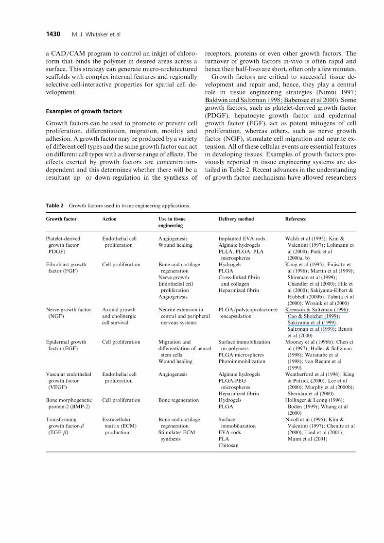
Table 2 Growth factors used in tissue engineering applications.
Growth factor Action Use in tissue
engineering
Delivery method Reference
Platelet-derived
growth factor
PDGF)
Endothelial cell
proliferation
Angiogenesis
Wound healing
Implanted EVA rods
Alginate hydrogels
PLLA, PLGA, PLA
microspheres
Walsh et al (1995) ; Kim &
Valentini (1997) ; Lohmann et
al (2000) ; Park et al
(2000a, b)
Fibroblast growth
factor (FGF)
Cell proliferation Bone and cartilage
regeneration
Nerve growth
Endothelial cell
proliferation
Angiogenesis
Hydrogels
PLGA
Cross-linked fibrin
and collagen
Heparinized fibrin
Kang et al (1995) ; Fujisato et
al (1996) ; Martin et al (1999) ;
Shireman et al (1999) ;
Chandler et al (2000) ; Hile et
al (2000) ; Sakiyama-Elbert &
Hubbell (2000b) ; Tabata et al
(2000) ; Wissink et al (2000)
Nerve growth factor
(NGF)
Axonal growth
and cholinergic
cell survival
Neurite extension in
central and peripheral
nervous systems
PLGA}poly(caprolactone)
encapsulation
Krewson & Saltzman (1996) ;
Cao & Shoichet (1999) ;
Sakiyama et al (1999) ;
Saltzman et al (1999) ; Benoit
et al (2000)
Epidermal growth
factor (EGF)
Cell proliferation Migration and
differentiation of neural
stem cells
Wound healing
Surface immobilization
on polymers
PLGA microspheres
Photoimmobilization
Mooney et al (1996b) ; Chen et
al (1997) ; Haller & Saltzman
(1998) ; Watanabe et al
(1998) ; von Recum et al
(1999)
Vascular endothelial
growth factor
(VEGF)
Endothelial cell
proliferation
Angiogenesis Alginate hydrogels
PLGA-PEG
microspheres
Heparinized fibrin
Weatherford et al (1996) ; King
& Patrick (2000) ; Lee et al
(2000) ; Murphy et al (2000b) ;
Sheridan et al (2000)
Bone morphogenetic
protein-2 (BMP-2)
Cell proliferation Bone regeneration Hydrogels
PLGA
Hollinger & Leong (1996) ;
Boden (1999) ; Whang et al
(2000)
Transforming
growth factor-β(TGF-β)
Extracellular
matrix (ECM)
production
Bone and cartilage
regeneration
Stimulates ECM
synthesis
Surface
immobilazation
EVA rods
PLA
Chitosan
Nicoll et al (1995) ; Kim &
Valentini (1997) ; Chenite et al
(2000) ; Lind et al (2001) ;
Mann et al (2001)
receptors, proteins or even other growth factors. The
turnover of growth factors in-vivo is often rapid and
hence their half-lives are short, often only a few minutes.
Growth factors are critical to successful tissue de-
velopment and repair and, hence, they play a central
role in tissue engineering strategies (Nimni 1997;
Baldwin and Saltzman 1998; Babensee et al 2000). Some
growth factors, such as platelet-derived growth factor
(PDGF), hepatocyte growth factor and epidermal
growth factor (EGF), act as potent mitogens of cell
proliferation, whereas others, such as nerve growth
factor (NGF), stimulate cell migration and neurite ex-
tension. All of these cellular events are essential features
in developing tissues. Examples of growth factors pre-
viously reported in tissue engineering systems are de-
tailed in Table 2. Recent advances in the understanding
of growth factor mechanisms have allowed researchers

1431Tissue engineering scaffolds
to make recombinant growth factors (e.g. recombinant
bone morphogenetic protein-2; rhBMP-2), purify
growth factors from cell extracts and use gene therapy
to induce local growth factor production.
Emerging strategies in the fabrication and useof controlled-release scaffolds
Hydrogel systems
Hydrogel matrices are swollen materials fabricated by
physically or chemically cross-linking networks of
water-soluble polymers. These systems offer a viable
approach for the delivery of biologically active mole-
cules to regenerating tissues as, in addition to the reduced
biofouling of hydrophilic polymers, hydrogels are more
porous than solid polymers and therefore display rapid
release profiles. An example of this approach is the
delivery of a bone-inducing growth factor preparation
from thermally sensitive chitosan–polyol salt com-
binations (Chenite et al 2000). Proteins are introduced
to the material in a liquid state and, after a temperature
increase to 37°C, subsequently become entrappedwithin
the created gel network. The resultant system demon-
strated de-novo bone and cartilage formation following
ectopic implantation.
Hubbell and co-workers have reported a highly ver-
satile polyethylene glycol (PEG)-containing hydrogel
system with localized drug-delivery applications. This
approach uses acrylate end-groups to photopolymerize
water-soluble monomers in-situ, thus minimizing the
need for invasive surgical procedures (Drumheller et al
1994; Cruise et al 1998). Using this system, interfacial
polymerization of the monomer has been demonstrated
by adsorbing the photo-initiator (e.g. eosin Y) directly
to a tissue surface. After UV exposure, the created gel
adheres to the cellular interface as a result of inter-
penetration of the liquid monomer into the textured
tissue (Hill-West et al 1994; Hern and Hubbell 1998).
The external hydrogel surface is then able to prevent cell
and protein adhesions (that may lead to thrombus
formation) to the coated area. This system has found
application in wound healing for the prevention of
postoperative adhesions, a process termed ‘‘gel paving’’
(Slepian and Hubbell 1997).
Acrylated PEG hydrogels have been modified for
tissue engineering applications by covalently attaching
adhesion peptides (e.g. RGD; Hern and Hubbell 1998)
and growth factors (transforming growth factor-β ;
TGF-β ; Mann et al 2001). Despite enhancing cell ad-
hesion and spreading, ECM production has been ob-
served to diminish when cells are attached to materials
displaying grafted adhesion peptide motifs (Mann et al
1999). TGF-β has been shown to increase ECM se-
cretion under such circumstances, thus ensuring the
developing tissue structure has suitable mechanical pro-
perties. This growth factor retains its ability to stimulate
matrix production following attachment to the hydro-
gel, and significantly increases production compared
with an equivalent concentration of unbound TGF-β.
One important variation on the PEG hydrogel system
is the development of degradable materials that in-
corporate lactic acid units into the monomer structure
(Han & Hubbell 1996, 1997). Enzymes and growth
factors can be entrapped within these materials by
polymerizing the hydrogel in the presence of the active
molecule. Depending on the molecular weight of the
incorporated protein, release from these hydrogel net-
works may be controlled by either diffusive or degra-
dative processes. Further improvements to the wound-
healing methodology described above have been
achieved by incorporating fibrinolytic agents, such as
ancrod (Chowdhury and Hubbell 1996), tissue plas-
minogen activator and urokinase plasminogen activator
(Hubbell 1996), into lactide-based PEG hydrogels.
These local deliveries were shown to result in marked
reductions of postoperative adhesions, while enabling
low systemic dosing with the enzymes. Recently, Molina
et al (2001) have illustrated that the release kinetics of
proteins from physically crosslinked poly(lactic acid)}poly(ethylene oxide) hydrogels depends greatly on the
compatibility of the gel and protein. This was high-
lighted by the compatible BSA–hydrogel system re-
sulting in a concentration-dependent protein release,
whereas the gel network created a reservoir-type release
for the phase-separating substrate fibrinogen.
Heparin-binding growth factor delivery
Fibrin-based biomaterial scaffolds have increased in
popularity in recent years as fibrin already acts as a
natural matrix for tissue regeneration, is non-toxic and
biodegradable, and can be created using a patient’s own
blood supply. Systems based on this natural product
have already been used in peripheral nerve (Sakiyama et
al 1999; Schense et al 2000) and cardiovascular (Ye et al
2000) regeneration studies. The performance of these
scaffolds in the area of peripheral nerve regeneration
has been enhanced by the incorporation of basic FGF
(bFGF) (Sakiyama-Elbert & Hubbell 2000a). This
growth factor was used to actively promote neurite
extensionby employingaheparin-baseddelivery system.
Bi-domain peptides, containing factor XIIIa and
heparin-binding sequences, are covalently cross-linked
to fibrin during the coagulation process. Heparin is then
1432 M. J. Whitaker et al
introduced and electrostatically associates with the
heparin-binding domain, after which heparin-binding
growth factors (e.g. bFGF) can be attached. These
materials are then able to act as reservoir devices,
releasing factors both passively and in response to fibrin
and heparin degradation, which may be augmented by
enzymes (e.g. plasmin) secreted by migrating cells. Con-
trolling the initial relative concentrations of heparin and
bFGF within the matrix, thus ensuring that growth-
factor delivery is predominantly cell-mediated, can min-
imize passive release. This concept of controlled release
via the cleavage of susceptible peptide sequences has
previously been demonstrated in the area of chemo-
therapy. Here, preselected oligopeptides were incorpor-
ated into synthetic polymer structures, their enzymatic
degradation resulting in the release of the active mol-
ecule within targeted cells (Putnam et al 1996). The
fabrication of biomaterial scaffolds exhibiting such cell-
mediated release mechanisms holds great potential for
tissue engineering applications as they enable long-term
growth-factor delivery owing to localized release con-
fined to the area in which they are required for cell
growth and migration.
In addition to bFGF, growth factors unable to bind
heparin effectively, such as β-NGF, brain-derived
growth factor and neurotrophin-3, have also been im-
mobilized and released from modified fibrin materials
(Sakiyama-Elbert & Hubbell 2000b). This is made poss-
ible by the presence of basic domains along their native
structures that, to some degree, mimic heparin-binding
sites. By displaying a large excess of heparin within the
matrix, diffusion of these neurotrophins is sufficiently
slowed to enable a controlled release profile. Following
both the bFGF and non-heparin-binding neurotrophin
modifications, neurite extensions from dorsal root gan-
glia have been shown to increase by up to around 100%
relative to unmodified fibrin. Unbound growth factors
present within the matrices did not enhance neurite
extension.
Encapsulation by double emulsion methods
The encapsulation of growth factors in microspheres
involves first generating an initial water-in-oil (w}o)
emulsion. The growth factor is dissolved in the water
phase and the polymer, typically a polyester, is dissolved
in the organic phase (ethyl acetate or methylene chlor-
ide), before the two solutions are mixed at an appro-
priate ratio. A second emulsion (w}o)}w is next formed
bydispersion in anaqueousphaseusinghomogenization
or sonication. This emulsion is then stirred to evaporate
the solvent, thus forming microspheres that can be
subsequently isolated by centrifugation or filtration.
This approach has been used by a number of groups to
produce degradable microspheres that incorporate the
growth factors NGF, EGF, PDGF and VEGF (see
Table 2). Microsphere morphology studies of these
delivery systems, typically determined by scanning elec-
tron microscopy, reveal either a hollow core surrounded
by a porous surface or scattered internal pores depend-
ing on the precise method of production and the starting
material used.
The harsh conditions of this technique, namely ex-
posure to organic solvents, may have unfavourable
effects on the integrity of the growth factor and may
result in deactivation during the encapsulation pro-
cedure (Crotts & Park 1997; Fu et al 1999) or ag-
gregation at the solvent–water interface (van de Weert
et al 2000). For example, carbonic anhydrase forms
non-covalent aggregates on exposure to the oil–water
interface, leading to a significant loss in activity.
The enhanced retention of growth factors within
microspheres prepared using double emulsion tech-
niques may be achieved by the addition of stabilizers
that reduce surface aggregation during manufacture
(Fu et al 1999; Sturesson & Carlfors 2000; van de Weert
et al 2000). Stabilizers, such as gelatin, poly(vinyl al-
cohol) (PVA), sucrose, poloxamer 407 or trehalose, may
be added to either or both of the emulsion steps,
although studies by Bezemer et al (2000) have shown
that these have the greatest effect when introduced
during the formation of the first emulsion where up to a
third of the biological activity may be lost.
Supercritical carbon dioxide processing
Supercritical fluid technology has traditionally been
used for chemical extraction and synthesis (McHugh &
Krukonis 1994). The properties of supercritical fluids, in
that they possesses densities and solvating powers simi-
lar to those of liquids, but have the diffusivity and
viscosity similar to those of gases, make them ideal
media for such chemical reactions. In the mid-1980s the
field of supercritical fluids was applied by several groups
to bio-catalytic reactions, allowing control of such pro-
cesses by simply altering either temperature or pressure
(Hammond et al 1985; Randolph et al 1985; Nakamura
et al 1986). Supercritical fluid technology was used to
develop protein powders of defined size that could be
utilized for drug delivery without the use of solvents
(Tom et al 1993; Winters et al 1996). Furthermore, there
was no loss of protein activity within these powders
(Yeo et al 1993; Johnston et al 1996). Advances in the
design of biodegradable polymers for drug delivery
applications led to theuseof supercritical carbondioxide

1433Tissue engineering scaffolds
(scCO2) to create porous polymer scaffolds that entrap
growth factors (Mooney et al 1996a; Howdle et al 2001).
CO2 becomes supercritical by raising the temperature
and pressure above its critical point (72 bar and 31.1°C).
Under these conditions, CO2 has enhanced solvent pro-
perties, thus enabling the glass transition temperature
(Tg) of an exposed polymer to be depressed. By choosing
a polymer with a relatively low Tg (such as poly(,-
lactide) ; Tg¯ 50–60°C, depending on molecular
weight), it will become plasticized below physiological
temperatures.Depression of the Tg increases the motility
of the polymer chains as the gas enters the polymer
phase. Therefore, physical mixing may simply incor-
porate growth factors throughout the polymer material.
On depressurization, nucleation of gas bubbles occurs
as the gas attempts to escape from the polymer phase.
The foamed structure is preserved, entrapping the
growth factor, as the polymer rises above its Tg. This
method does not require the use of organic solvents or
thermal processing.
Two methods have been described. In the first, a gas-
foaming process using either supercritical or non-super-
critical CO2 is used in addition to the traditional method
of salt leaching described above (Mooney et al 1996a;
Hile et al 2000). In the second, Howdle et al (2001) have
incorporated several proteins, at high loadings (up to
70%, w}w), into poly(,-lactide) scaffolds in a one-
step process. The enzyme ribonuclease was shown to
retain full activity after processing and its release was
monitored for 80 days.
DNA delivery
A novel alternative to growth factor encapsulation is the
proposed replacement of the actual protein in the poly-
mer matrix with plasmid DNA encoding the growth
factor of interest. Ideally, the plasmid encoding the
growth factor is incorporated into the DNA of regener-
ating cells and is expressed at physiological levels with-
out any further need for systemic delivery of growth
factor. Local gene delivery has been used in wound
healing (Bonadio et al 1999) and bone repair (Chandler
et al 2000), where a gene-activated matrix consisting of
the plasmid DNA, collagen and poly(α-hydroxyacid)s
was used to deliver plasmid encoding cytokine, PDGF
and human parathyroid hormone. Recently, Shea et al
(1999) have introduced plasmid DNA encoding for the
PDGF gene into PLGA matrices by gas-foaming, with
efficiencies of up to 60%. The PDGF plasmid DNA was
subsequently released and transfected cells in both in-
vitro and in-vivo studies, leading to enhanced vascu-
larization of tissue. Unfortunately, the delivery of plas-
mid DNA in-vivo is typically associated with low trans-
fection rates. It remains tobe seenwhether this technique
will be viable for large vascular tissue demands such as
that required for liver regeneration.
Conclusions and future directions
The controlled release of growth factors is essential in
many tissue engineering applications because co-
ordinated cell responses, such as proliferation, dif-
ferentiation and angiogenesis, are required to regenerate
functional tissues. Many current technologies in this
field have been borrowed from conventional concepts of
controlled release of small drug molecules. However,
there is an increasing appreciation that the molecular
nature of growth factors and the demands for more
complex release kinetics will necessitate the design of
new delivery devices. In the future, the rate of progress
in tissue engineering may be determined by the avail-
ability of controlled release systems that exceed current
capabilities.
References
Agrawal, C. M., Athanasiou, K. A., Heckman, J. D. (1997) Bio-
degradable PLA-PGA polymers for tissue engineering in ortho-
paedics. Mater. Sci. Forum 250 : 115–128
An, Y. J., Hubbell, J. A. (2000) Intraarterial protein delivery via
intimally-adherent bilayer hydrogels. J. Control. Rel. 64 : 205–215
Anderson, J. M., Langone, J. J. (1999) Issues and perspectives on the
biocompatibility and immunotoxicity evaluation of implanted con-
trolled release systems. J. Control. Rel. 57 : 107–113
Babensee, J. E.,Anderson, J. M.,McIntire, L. V.,Mikos,A. G. (1998)
Host response to tissue engineered devices. Adv. Drug Del. Rev. 33 :
111–139
Babensee, J. E., McIntire, L. V., Mikos, A. G. (2000) Growth factor
delivery for tissue engineering. Pharm. Res. 17 : 497–504
Baldwin, S. P., Saltzman, W. M. (1998) Materials for protein delivery
in tissue engineering. Adv. Drug Del. Rev. 33 : 71–86
Benoit, J. P., Faisant, N., Venier-Julienne, M. C., Menei, P. (2000)
Development of microspheres for neurological disorders : from
basics to clinical applications. J. Control. Rel. 65 : 285–296
Bezemer, J. M., Radersma, R., Grijpma, D. W., Dijkstra, P. J., van
Blitterswijk, C. A., Feijen, J. (2000) Microspheres for protein de-
livery prepared from amphiphilic multiblock copolymers. 1. Influ-
ence of preparation techniques on particle characteristics and
protein delivery. J. Control. Rel. 67 : 233–248
Boden, S. D. (1999) Bioactive factors for bone tissue engineering.
Clin. Orthop. 367 (Suppl.) : S84–S94
Bonadio, J., Smiley, E., Patil, P., Goldstein, S. (1999) Localized, direct
plasmid gene delivery in vivo: prolonged therapy results in repro-
ducible tissue regeneration. Nat. Med. 5 : 753–759
Bostman, O., Hirvensalo, E., Vainionpaa, S., Makela, A., Vihtonen,
K., Tormala, P., Rokkanen, P. (1989) Ankle fractures treated using
biodegradable internal fixation. Clin. Orthop. 238 : 195–203
Boyne, P. J., Marx, R. E., Nevins, M., Triplett, G., Lazaro, E., Lilly,
L. C., Alder, M., Nummikoski, P. (1997) A feasibility study evalu-
ating rhBMP-2}absorbable collagen sponge for maxillary sinus
floor augmentation. Int. J. Periodon. Rest. Dent. 17 : 10–25
1434 M. J. Whitaker et al
Cao, X. D., Shoichet, M. S. (1999) Delivering neuroactive molecules
from biodegradable microspheres for application in central nervous
system disorders. Biomaterials 20 : 329–339
Cao, Y., Vacanti, J. P., Ma, X., Paige, K. T., Upton, J., Chowanski,
Z., Schloo, B., Langer, R., Vacanti, C. A. (1994) Generation of neo-
tendon using synthetic polymers seeded with tenocytes. Transplant.
Proc. 26 : 3390–3392
Chandler, L. A., Doukas, J., Gonzalez, A. M., Hoganson, D. K., Gu,
D. L., Ma, C. L., Nesbit, M., Crombleholme, T. M., Herlyn, M.,
Sosnowski, B. A., Pierce, G. F. (2000) FGF2-targeted adenovirus
encoding platelet-derived growth factor-B enhances de novo tissue
formation. Mol. Ther. 2 : 153–160
Chen, G. P., Ito, Y., Imanishi, Y. (1997) Photo-immobilization of
epidermal growth factor enhances its mitogenic effect by artificial
juxtacrine signaling. Biochim. Biophys. Acta Mol. Cell Res. 1358 :
200–208
Chenite, A., Chaput, C., Wang, D., Combes, C., Buschmann, M. D.,
Hoemann, C. D., Leroux, J. C., Atkinson, B. L., Binette, F.,
Selmani, A. (2000) Novel injectable neutral solutions of chitosan
form biodegradable gels in situ. Biomaterials 21 : 2155–2161
Chowdhury, S. M., Hubbell, J. A. (1996) Adhesion prevention with
ancrod released via a tissue-adherent hydrogel. J. Surg. Res. 61 :
58–64
Cima, L. G., Vacanti, J. P., Vacanti, C., Ingber, D., Mooney, D.,
Langer, R. (1991) Tissue engineering by cell transplantation using
degradable polymer substrates. J. Biomech. Eng. 113 : 143–149
Coombes, A. G. A., Tasker, S., Lindbald, M., Holmgren, J., Hoste,
K., Toncheva, V., Schacht, E., Davies, M. C., Illum, L., Davis,
S. S. (1997) Biodegradable polymeric microparticles for drug
delivery and vaccine formulation: the surface attachment of
hydrophilic species using the concept of poly(ethylene glycol)
anchoring segments. Biomaterials 18 : 1153–1161
Crotts, G., Park, T. G. (1997) Stability and release of bovine serum
albumin encapsulated within poly(,-lactide-co-glycolide) micro-
particles. J. Control. Rel. 44 : 123–134
Cruise, G. M., Scharp, D. S., Hubbell, J. A. (1998) Characterization
of permeability and network structure of interfacially photo-
polymerized poly(ethylene glycol) diacrylate hydrogels. Biomateri-
als 19 : 1287–1294
Davis, M., Vacanti, J. P. (1996) Toward development of an im-
plantable tissue engineered liver. Biomaterials 17 : 365–372
den Dunnen, W. F. A., Stokroos, I., Blaauw, E. H., Holwerda, A.,
Pennings, A. J., Robinson, P. H., Schakenraad, J. M. (1996) Light-
microscopic and electron-microscopic evaluation of short-term
nerve regeneration using a biodegradable poly(-lactide-ε-capro-
lacton) nerve guide. J. Biomed. Mater. Res. 31 : 105–115
Drumheller, P. D., Elbert, D. L., Hubbell, J. A. (1994) Multi-
functional poly(ethylene glycol) semi-interpenetrating polymer net-
works as highly selective adhesive substrates for bioadhesive peptide
grafting. Biotechnol. Bioeng. 43 : 772–780
Elisseeff, J., Anseth, K., Langer, R., Hrkach, J. S. (1997) Synthesis
and characterization of photo-gross-linked polymers based on
poly(-lactic acid-co--aspartic acid) Macromolecules 30 : 2182–
2184
Evans, G. R. D., Brandt, K., Widmer, M. S., Lu, L., Meszlenyi,
R. K., Gupta, P. K., Mikos, A. G., Hodges, J., Williams, J.,
Gurlek, A., Nabawi, A., Lohman, R., Patrick, C. W. (1999)
In vivo evaluation of poly(-lactic acid) porous conduits for
peripheral nerve regeneration. Biomaterials 20 : 1109–1115
Falk, R., Randolph, T. W., Meyer, J. D., Kelly, R. M., Manning,
M. C. (1997) Controlled release of ionic compounds from poly(-
lactide) microspheres produced by precipitation with a compressed
antisolvent. J. Control. Rel. 44 : 77–85
Ferber, D. (1999) Lab-grown organs begin to take shape. Science 284 :
422–423
Freed, L. E., Marquis, J. C., Nohria, A., Emmanual, J., Mikos, A. G.,
Langer, R. (1993) Neocartilage formation in vitro and in vivo using
cells cultured on synthetic biodegradable polymers. J. Biomed.
Mater. Res. 27 : 11–23
Freed, L. E., Vunjak-Novakovic, G., Biron, R. J., Eagles, D. B.,
Lesnoy, D. C., Barlow, S. K., Langer, R. (1994) Biodegradable
polymer scaffolds for tissue engineering. Biotechnology 12 : 689–693
Fu, K., Griebenow, K., Hsieh, L., Klibanov, A. M., Langer, R. (1999)
FTIR characterization of the secondary structure of proteins encap-
sulated within PLGA microspheres. J. Control. Rel. 58 : 357–366
Fu, K., Pack, D. W., Klibanov, A. M., Langer, R. (2000) Visual
evidence of acidic environment within degrading poly(lactic-co-
glycolic acid) (PLGA) microspheres. Pharm. Res. 17 : 100–106
Fujisato, T., Sajiki, T., Liu, Q., Ikada, Y. (1996) Effect of basic
fibroblast growth factor on cartilage regeneration in chondrocyte-
seeded collagen sponge scaffold. Biomaterials 17 : 155–162
Glicklis, R., Shapiro, L., Agbaria, R., Merchuk, J. C., Cohen, S.
(2000) Hepatocyte behavior within three-dimensional porous
alginate scaffolds. Biotechnol. Bioeng. 67 : 344–353
Haller, M. F., Saltzman, W. M. (1998) Localized delivery of proteins
in the brain: can transport be customized? Pharm. Res. 15 : 377–385
Hammond, D. A., Karel, M., Klibanov, A. M., Krukonis, V. J. (1985)
Enzymatic-reactions in supercritical gases. Appl. Biochem. Bio-
technol. 11 : 393–400
Han, D. K., Hubbell, J. A. (1996) Lactide-based poly(ethylene glycol)
polymer networks for scaffolds in tissue engineering. Macro-
molecules 29 : 5233–5235
Han, D. K., Hubbell, J. A. (1997) Synthesis of polymer network
scaffolds from -lactide and poly(ethylene glycol) and their inter-
actions with cells. Macromolecules 30 : 6077–6083
Hern, D. L., Hubbell, J. A. (1998) Incorporation of adhesion peptides
into nonadhesive hydrogels useful for tissue resurfacing. J. Biomed.
Mater. Res. 39 : 266–276
Hile, D. D., Amirpour, M. L., Akgerman, A., Pishko, M. V. (2000)
Active growth factor delivery from poly(,-lactide-co-glycolide)
foams prepared in supercritical CO2. J. Control. Rel. 66 : 177–185
Hill-West, J. L., Chowdhury, S. M., Slepian, M. J., Hubbell, J. A.
(1994) Inhibition of thrombosis and intimal thickening by in-situ
photopolymerization of thin hydrogel barriers. Proc. Natl Acad.
Sci. USA 91 : 5967–5971
Hollinger, J. O., Leong, K. (1996) Poly(alpha-hydroxy acids) : carriers
for bone morphogenetic proteins. Biomaterials 17 : 187–194
Howdle, S. M.,Watson,M. S.,Whitaker,M. J., Popov,V. K.,Davies,
M. C., Mandel, F. S., Wang, J. D., Shakesheff, K. M. (2001) Super-
critical fluid mixing: preparation of thermally sensitive polymer
composites containing bioactive materials. Chem. Commun. 109–
110
Hubbell, J. A. (1996) Hydrogel systems for barriers and local drug
delivery in the control of wound healing. J. Control. Rel. 39 :
305–313
Ishaug-Riley, S. L., Crane-Kruger, G. M., Yaszemski, M. J., Mikos,
A. G. (1998) Three-dimensional culture of rat calvarial osteoblasts
in porous biodegradable polymers. Biomaterials 19 : 1405–1412
Johnston, K. P., Harrison, K. L., Clarke, M. J., Howdle, S. M., Heitz,
M. P., Bright, F. V., Carlier, C., Randolph, T. W. (1996) Water in
carbon dioxide microemulsions : an environment for hydrophiles
including proteins. Science 271 : 624–626
Kang, S. S., Gosselin, C., Ren, D. W., Greisler, H. P. (1995) Selective

1435Tissue engineering scaffolds
stimulation of endothelial-cell proliferation with inhibition of
smooth-muscle cell-proliferation by fibroblast growth-factor-1 plus
heparin delivered from fibrin glue suspensions. Surgery 118 : 280–
287
Kaufmann, P. M., Heimrath, S., Kim, B. S., Mooney, D. J. (1997)
Highly porous polymer matrices as a three-dimensional culture
system for hepatocytes. Cell Transplant. 6 : 463–468
Kim, H. D., Valentini, R. F. (1997) Human osteoblast response in
vitro to platelet-derived growth factor and transforming growth
factor-beta delivered from controlled-release polymer rods. Bio-
materials 18 : 1175–1184
Kim, S. S., Utsunomiya, H., Koski, J. A., Wu, B. M., Cima, M. J.,
Sohn, J., Mukai, K., Griffith, L. G., Vacanti, J. P. (1998) Survival
and function of hepatocytes on a novel three- dimensional synthetic
biodegradable polymer scaffold with an intrinsic network of
channels. Ann. Surg. 228 : 8–13
Kim, B. S., Nikolovski, J., Bonadio, J., Smiley, E., Mooney, D. J.
(1999) Engineered smooth muscle tissues : regulating cell phenotype
with the scaffold. Exp. Cell Res. 251 : 318–328
King, T. W., Patrick, C. W. (2000) Development and in vitro charac-
terization of vascular endothelial growth factor (VEGF)-loaded
poly(-lactic-co-glycolic acid)}poly(ethylene glycol) microspheres
using a solid encapsulation}single emulsion}solvent extraction
technique. J. Biomed. Mater. Res. 51 : 383–390
Kneser, U., Kaufmann, P. M., Fiegel, H. C., Pollok, J. M., Kluth, D.,
Herbst, H., Rogiers, X. (1999a) Heterotopic hepatocyte trans-
plantation utilizing pancreatic islet cotransplantation for hepato-
trophic stimulation: morphologic and morphometric evaluation.
Pediatr. Surg. Int. 15 : 168–174
Kneser, U., Kaufmann, P. M., Fiegel, H. C., Pollok, J. M., Kluth, D.,
Herbst, H., Rogiers, X. (1999b) Long-term differentiated function
of heterotopically transplanted hepatocytes on three-dimensional
polymer matrices. J. Biomed. Mater. Res. 47 : 494–503
Krewson, C. E., Saltzman, W. M. (1996) Transport and elimination
of recombinant human NGF during long-term delivery to the
brain. Brain Res. 727 : 169–181
Kuijpers, A. J., Engbers, G. H. M., Meyvis, T. K. L., de Smedt,
S. S. C., Demeester, J., Krijgsveld, J., Zaat, S. A. J., Dankert, J.,
Feijen, J. (2000) Combined gelatin-chondroitin sulfate hydrogels
for controlled release of cationic antibacterial proteins. Macro-
molecules 33 : 3705–3713
Kuo, C. K., Ma, P. X. (2001) Ionically crosslinked alginate hydrogels
as scaffolds for tissue engineering: part 1. Structure, gelation rate
and mechanical properties. Biomaterials 22 : 511–521
Lee, K. Y., Peters, M. C., Anderson, K. W., Mooney, D. J. (2000)
Controlled growth factor release from synthetic extracellular matri-
ces. Nature 408 : 998–1000
Li, R. H., White, M., Williams, S., Hazlett, T. (1998) Poly(vinyl
alcohol) synthetic polymer foams as scaffolds for cell encapsulation.
J. Biomater. Sci. Polym. Ed. 9 : 239–258
Li, S. M., Garreau, H., Vert, M. (1990a) Structure property relation-
ships in the case of the degradation of massive aliphatic poly-
(alpha-hydroxy acids) in aqueous-media.1. Poly(-lactic acid). J.
Mater. Sci. Mater. Med. 1 : 123–130
Li, S. M., Garreau, H., Vert, M. (1990b) Structure-property relation-
ships in the case of the degradation of massive poly(alpha-hydroxy
acids) in aqueous-media. 3. Influence of the morphology of poly(-
lactic acid). J. Mater. Sci. Mater. Med. 1 : 198–206
Lind, M., Overgaard, S., Glerup, H., Soballe, K., Bunger, C. (2001)
Transforming growth factor-beta 1 adsorbed to tricalciumphos-
phate coated implants increases peri-implant bone remodeling.
Biomaterials 22 : 189–193
Lohmann, C. H., Schwartz, Z., Niederauer, G. G., Carnes, D. L.,
Dean,D. D., Boyan, B. B. (2000) Pretreatment with platelet derived
growth factor-BB modulates the ability of costochondral resting
zone chondrocytes incorporated into PLA}PGA scaffolds to form
new cartilage in vivo. Biomaterials 21 : 49–61
Lu, L. C., Mikos, A. G. (1996) The importance of new processing
techniques in tissue engineering. MRS Bull. 21 : 28–32
Ma, T., Li, Y., Yang, S. T., Kniss, D. A. (2000) Effects of pore size in
3-D fibrous matrix on human trophoblast tissue development.
Biotechnol. Bioeng. 70 : 606–618
Mann, B. K., Tsai, A. T., Scott-Burden, T., West, J. L. (1999) Modi-
fication of surfaces with cell adhesion peptides alters extracellular
matrix deposition. Biomaterials 20 : 2281–2286
Mann, B. K., Schmedlen, R. H., West, J. L. (2001) Tethered-TGF-
beta increases extracellular matrix production of vascular smooth
muscle cells. Biomaterials 22 : 439–444
Maquet, V., Jerome, R. (1997) Design of macroporous biodegradable
polymer scaffolds for cell transplantation. Mater. Sci. 250 : 15–42
Martin, I., Vunjak-Novakovic, G., Yang, J., Langer, R., Freed, L. E.
(1999) Mammalian chondrocytes expanded in the presence of
fibroblast growth factor 2 maintain the ability to differentiate and
regenerate three-dimensional cartilaginous tissue. Exp. Cell Res.
253 : 681–688
Matsusue, Y., Yamamuro, T., Oka, M., Shikinami, Y., Hyon, S. H.,
Ikada, Y. (1992) In vitro and in vivo studies on bioabsorbable
ultrahigh-strength poly(-lactide) rods. J. Biomed. Mater. Res. 26 :
1553–1567
Mayer, J., Karamuk, E.,Akaike, T.,Wintermantel, E. (2000) Matrices
for tissue engineering-scaffold structure for a bioartificial liver
support system. J. Control. Rel. 64 : 81–90
McHugh, M. A., Krukonis, V. J. (1994) Supercritical fluid extraction :
principles and practice. Butterworth, London
Metters, A. T., Anseth, K. S., Bowman, C. N. (2000) Fundamental
studies of a novel, biodegradable PEG-b-PLA hydrogel. Polymer
41 : 3993–4004
Molina, I., Li, S. M., Martinez, M. B., Vert, M. (2001) Protein release
from physically crosslinked hydrogels of the PLA}PEO}PLA tri-
block copolymer-type. Biomaterials 22 : 363–369
Mooney, D. J., Baldwin, D. F., Suh, N. P., Vacanti, L. P., Langer, R.
(1996a) Novel approach to fabricate porous sponges of poly(,-
lactic-co-glycolic acid) without the use of organic solvents. Bio-
materials 17 : 1417–1422
Mooney, D. J., Kaufmann, P. M., Sano, K., Schwendeman, S. P.,
Majahod, K., Schloo, B., Vacanti, J. P., Langer, R. (1996b)
Localised delivery of epidermal growth factor improves the survival
of transplanted hepatocytes. Biotechnol. Bioeng. 50 : 422–429
Mooney, D. J., Mazzoni, C. L., Breuer, C., McNamara, K., Hern, D.,
Vacanti, J. P., Langer, R. (1996c) Stabilized polyglycolic acid fibre-
based tubes for tissue engineering. Biomaterials 17 : 115–124
Murphy, W. L., Kohn, D. H., Mooney, D. J. (2000a) Growth of
continuous bonelike mineral within porous poly(lactide-co-glyco-
lide) scaffolds in vitro. J. Biomed. Mater. Res. 50 : 50–58
Murphy, W. L., Peters, M. C., Kohn, D. H., Mooney, D. J. (2000b)
Sustained release of vascular endothelial growth factor from min-
eralized poly(lactide-co-glycolide) scaffolds for tissue engineering.
Biomaterials 21 : 2521–2527
Nakamura, K., Chi, Y. M., Yamada, Y., Yano, T. (1986) Lipase
activity and stability in supercritical carbon dioxide. Chem. Eng.
Commun. 45 : 207–212
Nam, Y. S., Park, T. G. (1999) Porous biodegradable polymeric
scaffolds prepared by thermally induced phase separation. J. Bio-
med. Mater. Res. 47 : 8–17

1436 M. J. Whitaker et al
Nicoll, S. B., Denker, A. E., Tuan, R. S. (1995) In vitro charac-
terization of transforming growth factor-beta 1 loaded composites
of biodegradable polymer and mesenchymal cells. Cells Mater. 5 :
231–244
Nimni, M. E. (1997) Polypeptide growth factors : targeted delivery
systems. Biomaterials 18 : 1201–1225
Park, A., Wu, B., Griffith, L. G. (1998) Integration of surface modi-
fication and 3D fabrication techniques to prepare patterned poly(-
lactide) substrates allowing regionally selective cell adhesion. J.
Biomater. Sci. Polym. Ed. 9 : 89–110
Park, Y. J., Lee, Y. M., Lee, J. Y., Seol, Y. J., Chung, C. P., Lee,
S. J. (2000a) Controlled release of platelet-derived growth factor-
BB from chondroitin sulfate-chitosan sponge for guided bone
regeneration. J. Control. Rel. 67 : 385–394
Park, Y. J., Lee, Y. M., Park, S. N., Sheen, S. Y., Chung, C. P., Lee,
S. J. (2000b) Platelet derived growth factor releasing chitosan
sponge for periodontal bone regeneration.Biomaterials 21 : 153–159
Peter, S. J., Nolley, J. A., Widmer, M. S., Merwin, J. E., Yaszemski,
M. J., Yasko, A. W., Engel, P. S., Mikos, A. G. (1997) In vitro
degradation of a poly(propylene fumarate)}beta-tricalcium phos-
phate composite orthopaedic scaffold. Tissue Eng. 3 : 207–215
Putnam, D. A., Shiah, J. G., Kopecek, J. (1996) Intracellularly bio-
recognizable derivatives of 5-fluorouracil – implications for site-
specific delivery in the human condition. Biochem. Pharmacol. 52 :
957–962
Quirk, R. A., Chan, W. C., Davies, M. C., Tendler, S. J. B.,
Shakesheff, K. M. (2001) Poly(-lysine)-GRGDS as a biomimetic
surface modifier for poly(lactic acid) Biomaterials 22 : 865–872
Randolph,T. W.,Blanch,H. W., Prausnitz, J. M.,Wilke,C. R. (1985)
Enzymatic catalysis in a supercritical fluid. Biotechnol. Lett. 7 :
325–328
Rowley, J. A., Madlambayan, G., Mooney, D. J. (1999) Alginate
hydrogels as synthetic extracellular matrix materials. Biomaterials
20 : 45–53
Sakiyama, S. E., Schense, J. C., Hubbell, J. A. (1999) Incorporation
of heparin-binding peptides into fibrin gels enhances neurite ex-
tension: an example of designer matrices in tissue engineering.
FASEB J. 13 : 2214–2224
Sakiyama-Elbert, S. E., Hubbell, J. A. (2000a) Controlled release of
nerve growth factor from a heparin-containing fibrin-based cell
ingrowth matrix. J. Control. Rel. 69 : 149–158
Sakiyama-Elbert, S. E., Hubbell, J. A. (2000b) Development of fibrin
derivatives for controlled release of heparin-binding growth factors.
J. Control. Rel. 65 : 389–402
Saltzman, W. M., Mak, M. W., Mahoney, M. J., Duenas, E. T.,
Cleland, J. L. (1999) Intracranial delivery of recombinant nerve
growth factor : release kinetics and protein distribution for three
delivery systems. Pharm. Res. 16 : 232–240
Schense, J. C., Bloch, J., Aebischer, P., Hubbell, J. A. (2000) En-
zymatic incorporation of bioactive peptides into fibrin matrices
enhances neurite extension. Nat. Biotechnol. 18 : 415–419
Schmitt, E. A., Flanagan, D. R., Linhardt, R. J. (1994) Importance of
distinct water environments in the hydrolysis of poly(-lactide-co-
glycolide) Macromolecules 27 : 743–748
Schugens, C., Maquet, V., Grandfils, C., Jerome, R., Teyssie, P.
(1996) Polylactide macroporous biodegradable implants for cell
transplantation. 2. Preparation of polylactide foams by liquid-
liquid phase separation. J. Biomed. Mater. Res. 30 : 449–461
Shea, L. D., Smiley, E., Bonadio, J., Mooney, D. J. (1999) DNA
delivery from polymer matrices for tissue engineering. Nat. Bio-
technol. 17 : 551–554
Sheridan, M. H., Shea, L. D., Peters, M. C., Mooney, D. J. (2000)
Bioadsorbable polymer scaffolds for tissue engineering capable of
sustained growth factor delivery. J. Control. Rel. 64 : 91–102
Shireman, P. K., Hampton, B., Burgess, W. H., Greisler, H. P. (1999)
Modulation of vascular cell growth kinetics by local cytokine
delivery from fibrin glue suspensions. J. Vasc. Surg. 29 : 852–861
Slepian, M. J., Hubbell, J. A. (1997) Polymeric endoluminal gel
paving: hydrogel systems for local barrier creation and site-specific
drug delivery. Adv. Drug Del. Rev. 24 : 11–30
Sturesson, C., Carlfors, J. (2000) Incorporation of protein in PLG-
microspheres with retention of bioactivity. J. Control. Rel. 67 :
171–178
Suggs, L. J., Payne, R. G., Yaszemski, M. J., Alemany, L. B., Mikos,
A. G. (1997) Synthesis and characterization of a block copolymer
consisting of poly(propylene fumarate) and poly(ethylene glycol)
Macromolecules 30 : 4318–4323
Suggs, L. J., Kao, E. Y., Palombo, L. L., Krishnan, R. S., Widmer,
M. S., Mikos, A. G. (1998a) Preparation and characterization of
poly(propylene fumarate-co-ethylene glycol) hydrogels. J. Bio-
mater. Sci. Polym. Ed. 9 : 653–666
Suggs, L. J., Krishnan, R. S., Garcia, C. A., Peter, S. J., Anderson,
J. M., Mikos, A. G. (1998b) In vitro and in vivo degradation of
poly(propylene fumarate-co-ethylene glycol) hydrogels. J. Biomed.
Mater. Res. 42 : 312–320
Suggs, L. J., West, J. L., Mikos, A. G. (1999) Platelet adhesion on a
bioresorbable poly(propylene fumarate-co-ethylene glycol) copoly-
mer. Biomaterials 20 : 683–690
Suuronen, R., Pohjonen, T., Taurio, R., Tormala, P., Wessman, L.,
Ronkko, K., Vainionpaa, S. (1992) Strength retention of self-
reinforced poly--lactide screws and plates – an in vivo and in vitro
study. J. Mater. Sci. Mater. Med. 3 : 426–431
Tabata, Y., Miyao, M., Inamoto, T., Ishii, T., Hirano, Y., Yamaoki,
Y., Ikada, Y. (2000) De novo formation of adipose tissue by
controlled release of basic fibroblast growth factor. Tissue Eng. 6 :
279–289
Thomson, R. C., Yaszemski, M. J., Powers, J. M., Mikos, A. G.
(1995) Fabrication of biodegradable polymer scaffolds to engineer
trabecular bone. J. Biomater. Sci. Polym. Ed. 7 : 23–38
Tokiwa, T., Kodama, M. (1997) Multilayer aggregate culture of rat
hepatocytes on a porous material : three dimensional cellular organ-
ization and maintenance of hepatic functions. Porous Mater. Tissue
Eng. 250 : 97–103
Tom, J. W., Lim,G. B.,Debenedetti, P. G., Prudhomme,R. K. (1993)
Applications of supercritical fluids in the controlled release of
drugs. ACS Symp. Ser. 514 : 238–257
Uhrich, K. E., Cannizzaro, S. M., Langer, R. S., Shakesheff, K. M.
(1999) Polymeric systems for controlled drug release. Chem. Rev.
99 : 3181–3198
Uludag, H., D’Augusta, D., Palmer, R., Timony, G., Wozney, J.
(1999) Characterization of rhBMP-2 pharmacokinetics implanted
with biomaterial carriers in the rat ectopic model. J. Biomed. Mater.
Res. 46 : 193–202
Vacanti, C. A., Langer, R., Schloo, B., Vacanti, J. P. (1991) Synthetic-
polymers seeded with chondrocytes provide a template for new
cartilage formation. Plast. Reconstr. Surg. 88 : 753–759
Vacanti, C. A., Vacanti, J. P., Langer, R. (1994) Tissue engineering
using synthetic biodegradable polymers. Polymers of biological
biomedical significance. ACS Symp. Ser. 16–34
van de Weert, M., Hoechstetter, J., Hennink, W. E., Crommelin,
D. J. A. (2000) The effect of a water}organic solvent interface on
the structural stability of lysozyme. J. Control. Rel. 68 : 351–359

1437Tissue engineering scaffolds
von Recum, H., Kikuchi, A., Yamato, M., Sakurai, Y., Okano, T.,
Kim, S. W. (1999) Growth factor and matrix molecules preserve
cell function on thermally responsive culture surfaces. Tissue Eng.
5 : 251–265
Walsh, W. R., Kim, H. D., Jong, Y. S., Valentini, R. F. (1995) Con-
trolled-release of platelet-derived growth-factor using ethylene-
vinyl acetate copolymer (evac) coated on stainless-steel wires.
Biomaterials 16 : 1319–1325
Watanabe, Y., Ajioka, I., Akaike, T. (1998) Gene transfection of
multicellular speroid of hepatocytes on an artificial substrate.
Cytotechnology 26 : 65–78
Weatherford, D. A., Sackman, J. E., Reddick, T. T., Freeman, M. B.,
Stevens, S. L., Goldman, M. H. (1996) Vascular endothelial growth
factor and heparin in a biologic glue promotes human aortic
endothelial cell proliferation with aortic smooth muscle cell in-
hibition. Surgery 120 : 433–439
Whang, K., Tsai, D. C., Nam, E. K., Aitken, M., Sprague, S. M.,
Patel, P. K., Healy, K. E. (1998) Ectopic bone formation via
rhBMP-2 delivery from porous bioabsorbable polymer scaffolds. J.
Biomed. Mater. Res. 42 : 491–499
Whang, K., Goldstick, T. K., Healy, K. E. (2000) A biodegradable
polymer scaffold for delivery of osteotropic factors. Biomaterials
21 : 2545–2551
Winters, M. A., Knutson, B. L., Debenedetti, P. G., Sparks, H. G.,
Przybycien, T. M., Stevenson, C. L., Prestrelski, S. J. (1996) Pre-
cipitation of proteins in supercritical carbon dioxide. J. Pharm. Sci.
85 : 586–594
Wissink, M. J. B., Beernink, R., Scharenborg, N. M., Poot, A. A.,
Engbers, G. H. M., Beugeling, T., van Aken, W. G., Feijen, J.
(2000) Endothelial cell seeding of (heparinized) collagen matrices :
effects of bFGF pre-loading on proliferation (after low density
seeding) and pro-coagulant factors. J. Control. Rel. 67 : 141–155
Yaszemski, M. J., Payne, R. G., Hayes, W. C., Langer, R., Mikos,
A. G. (1996) In vitro degradation of a poly(propylene fumarate)-
based composite material. Biomaterials 17 : 2127–2130
Ye, Q., Zund, G., Benedikt, P., Jockenhoevel, S., Hoerstrup, S. P.,
Sakyama, S., Hubbell, J. A., Turina, M. (2000) Fibrin gel as a three
dimensional matrix in cardiovascular tissue engineering. Eur. J.
Cardiothorac. Surg. 17 : 587–591
Yeo, S. D., Lim, G. B., Debenedetti, P. G., Bernstein, H. (1993)
Formationofmicroparticulate proteinpowders using a supercritical
fluid antisolvent. Biotechnol. Bioeng. 41 : 341–346
Zund, G., Hoerstrup, S. P., Schoeberlein, A., Lachat, M., Uhlschmid,
G., Vogt, P. R., Turina, M. (1998) Tissue engineering: a new
approach in cardiovascular surgery; Seeding of human fibroblasts
followed by human endothelial cells on resorbable mesh. Eur. J.
Cardiothorac. Surg. 13 : 160–164