electrospun sulfated silk fibroin nanofibrous scaffolds for vascular tissue engineering
TRANSCRIPT
lable at ScienceDirect
Biomaterials 32 (2011) 3784e3793
Contents lists avai
Biomaterials
journal homepage: www.elsevier .com/locate/biomateria ls
Electrospun sulfated silk fibroin nanofibrous scaffolds for vascular tissueengineering
Haifeng Liu a,*, Xiaoming Li a, Gang Zhou a, Hongbin Fan b, Yubo Fan a,**
aKey Laboratory for Biomechanics and Mechanobiology of Ministry of Education, School of Biological Science and Medical Engineering, Beihang University, Beijing 100191,People’s Republic of Chinab Institute of Orthopaedics and Traumatology, Xijing Hospital, The Fourth Military Medical University, Xi’an 710032, People’s Republic of China
a r t i c l e i n f o
Article history:Received 1 December 2010Accepted 1 February 2011Available online 3 March 2011
Keywords:Sulfated silk fibroinElectrospinningAnticoagulant activitySmall-diameterVascular grafts
* Corresponding author. Tel./fax: þ86 10 82338456** Corresponding author. Tel./fax: þ86 10 82339428
E-mail addresses: [email protected] (H.(Y. Fan).
0142-9612/$ e see front matter � 2011 Elsevier Ltd.doi:10.1016/j.biomaterials.2011.02.002
a b s t r a c t
One of the major downfalls of tissue-engineered small-diameter vascular grafts is the inability to obtaina confluent endothelium on the lumenal surface. Loosely attached endothelial cells (ECs) are easilyseparated from the vessel wall when exposed to the in vivo vascular system. Thus any denuded areas onthe lumenal surface of vascular grafts may lead to thrombus formation via platelet deposition andactivation. If the denuded areas could express anticoagulant activity until the endothelial cell lining isfully achieved, it may greatly improve the chances of successful vascular reconstruction. In this study, wefabricate sulfated silk fibroin nanofibrous scaffolds (S-silk scaffolds) and assess the anticoagulant activityand cytocompatibility of S-silk scaffolds in vitro in order to improve the antithrombogenicity and getsome insights into its potential use for vascular tissue engineering. Sulfated silk fibroin was prepared byreaction with chlorosulphonic acid in pyridine, and then was developed to form an S-silk scaffold byelectrospinning technique. FTIR analyses identified the successful incorporation of sulfate groups in silkfibroin molecules. It was found that the anticoagulant activity of S-silk scaffolds was significantlyenhanced compared with silk fibroin nanofibrous scaffolds (Silk scaffolds). Vascular cells, including ECsand smooth muscle cells (SMCs), demonstrated strong attachment to S-silk scaffolds and proliferatedwell with higher expression of some phenotype-related marker genes and proteins. Overall, the data inthis study suggest the suitability of S-silk scaffolds used along with vascular cells for the development oftissue-engineered vascular grafts.
� 2011 Elsevier Ltd. All rights reserved.
1. Introduction
Diseases of the vascular system are themain causes of death anddisability in people with diabetes [1]. Unfortunately, many patientswho require arterial bypass procedures do not have suitable vesselsfor use. The development of blood vessel substitutes has rapidlyprogressed in the past decade in response to the clinical need forimproved vascular grafts in surgical procedures. Whereas vasculargrafts have been used successfully to replace large-diameter (innerdiameter >6 mm) blood vessels, the long-term patency of small-diameter (inner diameter <6 mm) vascular grafts is still disap-pointing, mostly due to thrombus formation [2]. Tissue engineeringoffers an alternative approach to address the need for small-
.
.Liu), [email protected]
All rights reserved.
diameter vascular grafts through the design of non-thrombogenicinterface. Various approaches for this aim have been attemptedover the years. However, successful clinical results have not yetbeen reported till now.
Silk fibroin has been established as an attractive biomaterial forscaffolding [3]. As a biomaterial, silk fibroin features excellentbiocompatibility, adaptable biodegradability, good oxygen/watervapor permeability, and remarkable mechanical properties [4e6].Silk fibroin has been electrospun into nanofibrous tubular scaffoldsfor small-diameter vascular grafting applications [7]. Nanofibrousscaffolds created by electrospinning technique have enormouspotential for tissue engineering since they can mimic the structureand function of native extracellular matrix (ECM). Moreover, theelectrospun nanofibers have large surface area to volume ratio,which allows for the direct attachment of ECM ligands, growthfactors, and other biomolecules onto fiber surfaces to locallymodulate cell and tissue function and to enhance tissue regenera-tion [8,9]. The in vitro cytocompatibility results have demonstratedthat the electrospun silk fibroin nanofibrous scaffolds support
H. Liu et al. / Biomaterials 32 (2011) 3784e3793 3785
vascular cell viability, maintain cell phenotype, and promote cellreorganization [10]. However, an ideal scaffold for small-diametervascular tissue engineering should be able to interact favorably notonly with vascular cells but also with blood [11]. Hemocompati-bility, which frequently is seen as the most critical aspect ofbiocompatibility, is primarily determined by the lumenal surface ofthe blood vessel [12]. One of the major downfalls of small-diametertissue-engineered vascular grafts is the inability to obtaina confluent endothelium on the lumenal surface [13]. Looselyattached ECs are easily separated from the vessel wall whenexposed to shear stress caused by blood flow in the vascular system[14]. This implies that the amount of available ECs is usually notsufficient to produce a confluent coverage of the vascular graft,leaving large parts of the scaffold surface exposed to blood. Theplasma proteins will immediately deposit on the denuded areasand subsequently make the foreign materials attractive for plateletadhesion and aggregation [15,16]. Thus any denuded areas on thelumenal surface of vascular graft are probable sites for thrombusformation. Occlusive thrombus can quickly lead to graft failure andpotentially catastrophic downstream consequences includingmyocardial infarction and limb ischemia [17,18]. On the other hand,if the denuded areas could have anticoagulant activity to preventplatelet adhesion and aggregation before the endothelial conflu-ence is achieved once again, it may greatly improve the chances ofsuccessful vascular reconstruction. Therefore, a successful tissue-engineered vascular graft must have the two fundamental biolog-ical qualities: one is to express anticoagulant activity until theendothelial cell lining is fully achieved and the other is to supportcell growth and expansion.
It is notable that the anticoagulant activity of silk fibroin can besignificantly increased by reaction with chlorosulphonic acid inpyridine [19,20]. Therefore, in the present study, we fabricateda sulfated silk fibroin nanofibrous scaffold for improving itsantithrombogenicity and explored its potential application fortissue engineering of small-diameter vascular grafts.
2. Materials and methods
2.1. Preparation of sulfated silk fibroin
Raw Bombyx mori silk fibers were supplied from Zhejiang Cathaya InternationalCo. Ltd. Silk fibroin solution was obtained from raw silk fibers that were degummedin a 0.1% (w/v) Na2CO3 solution, dissolved in CaCl2eCH3CH2OHeH2O (moleratio ¼ 1: 2: 8) at 78 � 2 �C with continuous stirring and subsequently dialyzedagainst distilled water using a Snake Skin Pleated Dialysis Tubing (PIERCE, MWCO3500). Finally, the silk fibroin solution was freeze-dried for 24 h to form silk fibroinsponges and was kept in a vacuum drying desiccator for future use.
Sulfated silk fibroin was prepared according to procedures previously described[20,21]. The silk fibroin sponges were treated in a glass beaker with a solutionprepared by gradually adding 10 mL of chlorosulfonic acid (Sigma, USA) to 60 mL ofpyridine (Sigma, USA) in an ice bath. The reaction system was gradually heated to80 �C in a thermostatically controlled bath and kept at constant temperature for 1 hwith stirring. After reaction, 200 mL of distilled water was added to the system.Subsequently, the solution was neutralized by equivalent molar NaOH solution. Theinsoluble portion was removed by vacuum filtration; the soluble portion wasprecipitated with 500 mL of ethanol. The precipitate was harvested by centrifuga-tion and was dissolved with a small amount of water. And then, it was dialyzedagainst distilled water using a SnakeSkin Pleated Dialysis Tubing (PIERCE, MWCO3500) for desalting. After freeze drying, sulfated silk fibroin was stored in a vacuumdrying dessicator until use.
2.2. Electrospining of sulfated silk fibroins
Sulfated silk fibroin was dissolved in hexafluoro-2-propanol (HFIP; FlukaChemie GmBH, Germany) to generate a 10% (wt/v) solution. Electrospinning wasperformed with a steel capillary tube with a 1.5 mm inside diameter tip mounted onan adjustable, electrically insulated stand. The capillary tube was maintained ata high electric potential for electrospinning and was mounted in the parallel plategeometry. The capillary tube was connected to a syringe filled with the sulfated silkfibroin/HFIP solution. A constant volume flow rate of 0.8 mL/h was maintained usinga syringe pump. High voltage of 10 kV was applied when the solution was drawn
into fibers and was collected on an aluminum foil covered collection plate kept ata distance of 13 cm from the needle tip. And then, the electrospun nanofibrousscaffolds were treated with 100% methanol for 10 min to induce a b-sheet confor-mational transition, which resulted in insolubility in water. Silk fibroins were alsoelectrospun to form a nanfibrous scaffold as a control. For convenience, silk fibroinand sulfated silk fibroin nanfibrous scaffolds were abbreviated as Silk scaffolds andS-silk scaffolds in the present research, respectively.
2.3. Scaffold characterization
Fourier Transform Infrared (FTIR) spectroscopic analysis of electrospun nano-fibrous scaffolds was performed using a Nicolet AVATAR 360 FTIR spectrophotom-eter (Nicolet, USA) in the region from 550 to 4000 cm�1 (spectral resolution 4 cm�1,32 scans per spectrum).
The surface morphology of Silk and S-silk scaffolds were viewed under a scan-ning electron microscope (SEM; JEOL JSM-5600LV, Japan), with an acceleratedvoltage of 10 kV. All samples were coated with a sputter coater (BAL-TEC Inc)equipped with a gold target to increase electrical conductivity. The average diameterand diameter distribution were obtained by analyzing SEM images using a customcode image analysis program.
Static water surface contact angle measurements were performed on thenanofibrous scaffolds using VCA optima Surface Analysis System (AST Products, Inc.,Billerica, MA).
The anticoagulant activities were evaluated by in vitro coagulation time tests,including thrombin time (TT), prothrombin time (PT), and activated partial throm-boplastin time (APTT) [22]. Briefly, the tested scaffolds (1 cm� 1 cm� 0.1 mm)wereincubated with healthy human blood plasma in a transparent plastic tube, and thereagents for each coagulation time test were added to the tube immediately. Theclotting times were measured by a photo-optical clot detection instrument Coag-A-MateR-XM (Organon Teknika, USA). Each experiment was repeated three times withreproducible results.
2.4. Isolation and culture of endothelial cells and smooth muscle cells
Endothelial cells (ECs) and smooth muscle cells (SMCs) were obtained fromporcine thoracic aorta under aseptic conditions according to a published method[23]. Briefly, after removing the external adventitia, the aorta was perfused with 1%type I collagenase (Sigma, USA) in serum-free Dulbecco’s modified Eagle’s medium(DMEM; Invitrogen, CA) and was incubated at 37 �C for 15 min. ECs were isolatedand were seeded on 75-cm2 plastic flasks pre-coated with 0.2% porcine gelatin. Thecells were cultured in M199 medium (Invitrogen, CA) supplemented with 20% fetalbovine serum (FBS; HyClone, Logan, UT), 100 U/100 mg/mL Pen/Strep (Invitrogen,CA), 2 mM L-glutamine (Gibco), 25 mg/mL sodium heparin (Sigma, USA), and 25 mg/mL endothelial cell growth supplement (Sigma, USA). Following the first digestion,the aorta was cut into small pieces and SMCs were released by incubation in a 0.5%solution of type II collagenase (Sigma, USA) in serum-free Dulbecco’s modifiedEagle’s medium (DMEM; Invitrogen, CA) in a moist atmosphere (5% CO2 and 95% air)at 37 �C overnight. Free cells were recovered by centrifugation, resuspended bygentle flushing with a pipette, and seeded on 75-cm2 plastic flasks. SMCs werecultured in DMEM supplemented with 10% FBS (HyClone, Logan, UT), 2 mM L-glutamine (Gibco), penicillin (100 U/mL), and streptomycin (100 mg/mL).
When primary ECs and SMCs became nearly confluent, theywere detached fromthe flasks with trypsineEDTA solution (Invitrogen, CA), and were expanded ata splitting rate of 1:3. The second passage cells were then used for further study. Allcells were maintained at 37 �C in an incubator with 5% humidified CO2. The cultureswere replenished with flash medium at 37 �C every 3 days.
2.5. In vitro culture of vascular cells on S-silk scaffolds
For cell seeding, the nanofibrous scaffolds were collected on small glass cover-slips (length �width; 1 cm � 1 cm). Then the scaffolds were treated with methanoland were sterilized by autoclave. Porcine ECs and SMCs were seeded at a density of2.5�105 cells/scaffold onto each of the scaffolds placing in thewells of a non-treated6-well culture plate (NUNC, Denmark). After pipetting 100 mL of cell mediumsuspension onto the scaffolds, the plates were incubated for 3 h to allow cellattachment. And then, cell-free mediumwas added to bring the total well volume to1000 mL. The cell-seeded scaffolds were grown in vitro in a 5% CO2 incubator at 37 �Cfor 2 weeks, with the medium being replaced every 3 days. Cellular morphologycharacterization of cell-seeded Silk and S-silk scaffolds was accomplished using SEM(JEOL JSM-5600LV, Japan) on day 1, 7, and 14.
2.6. DNA content and metabolism activity assay
The DNA content was measured with Hoechst 33258 (Invitrogen, CA) to assesscell proliferation. Briefly, cells were harvested from cell-scaffold constructs byincubating with a 0.05% solution of trypsin. And then, the cells were collected bycentrifugation and were lysed in cell lysis buffer. Cell lysates were diluted 10 timesand were incubated with equal volume of 0.1 mg/mL Hoechest 33258 solution for10 min at room temperature in 96-well black plates. Fluorescence was determined
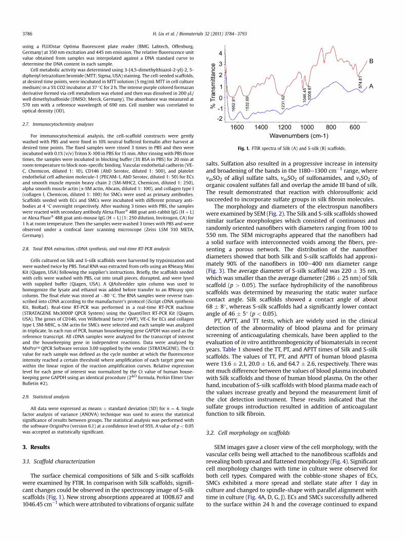
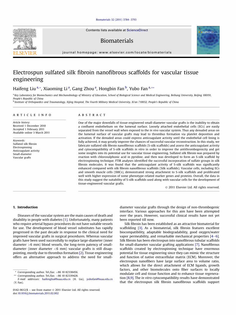
Fig. 1. FTIR spectra of Silk (A) and S-silk (B) scaffolds.
H. Liu et al. / Biomaterials 32 (2011) 3784e37933786
using a FLUOstar Optima fluorescent plate reader (BMG Labtech, Offenburg,Germany) at 350 nm excitation and 445 nm emission. The relative fluorescence unitvalue obtained from samples was interpolated against a DNA standard curve todetermine the DNA content in each sample.
Cell metabolic activity was determined using 3-[4,5-dimethylthiazol-2-yl]-2, 5-diphenyl tetrazolium bromide (MTT; Sigma, USA) staining. The cell-seeded scaffolds,at desired time points, were incubated in MTT solution (5 mg/mL MTT in cell culturemedium) in a 5% CO2 incubator at 37 �C for 2 h. The intense purple colored formazanderivative formed via cell metabolismwas eluted and then was dissolved in 200 mL/well dimethylsulfoxide (DMSO; Merck, Germany). The absorbance was measured at570 nm with a reference wavelength of 690 nm. Cell number was correlated tooptical density (OD).
2.7. Immunocytochemisty analyses
For immunocytochemical analysis, the cell-scaffold constructs were gentlywashed with PBS and were fixed in 10% neutral buffered formalin after harvest atdesired time points. The fixed samples were rinsed 3 times in PBS and then wereincubatedwith 0.1% (v/v) Triton X-100 in PBS for 15min. After rinsing with PBS threetimes, the samples were incubated in blocking buffer (3% BSA in PBS) for 20 min atroom temperature to block non-specific binding. Vascular endothelial cadherin (VE-C, Chemicon, diluted 1: 10), CD146 (AbD Serotec, diluted 1: 500), and plateletendothelial cell adhesion molecule-1 (PECAM-1, AbD Serotec, diluted 1: 50) for ECsand smooth muscle myosin heavy chain 2 (SM-MHC2, Chemicon, diluted 1: 250),alpha smooth muscle actin (a-SM actin, Abcam, diluted 1: 100), and collagen type I(collagen I, Chemicon, diluted 1: 100) for SMCs were used as primary antibodies.Scaffolds seeded with ECs and SMCs were incubated with different primary anti-bodies at 4 �C overnight respectively. After washing 3 times with PBS, the sampleswere reacted with secondary antibody Alexa Fluor� 488 goat anti-rabbit IgG (H þ L)or Alexa Fluor� 488 goat anti-mouse IgG (H þ L) (1: 250 dilution, Invitrogen, CA) for1 h at room temperature. Then the samples were washed 3 times with PBS and wereobserved under a confocal laser scanning microscope (Zeiss LSM 510 META,Germany).
2.8. Total RNA extraction, cDNA synthesis, and real-time RT-PCR analysis
Cells cultured on Silk and S-silk scaffolds were harvested by trypsinization andwere washed twice by PBS. Total RNAwas extracted from cells using an RNeasyMiniKit (Qiagen, USA) following the supplier’s instructions. Briefly, the scaffolds seededwith cells were washed with PBS, cut into small pieces, disrupted, and were lysedwith supplied buffer (Qiagen, USA). A QIAshredder spin column was used tohomogenize the lysate and ethanol was added before transfer to an RNeasy spincolumn. The final elute was stored at �80 �C. The RNA samples were reverse tran-scribed into cDNA according to the manufacturer’s protocol (iScript cDNA synthesisKit, BioRad). Real-time RT-PCR was performed in a real-time RT-PCR machine(STRATAGENE Mx3000P QPCR System) using the QuantiTect RT-PCR Kit (Qiagen,USA). The genes of CD146, von Willebrand factor (vWF), VE-C for ECs and collagentype I, SM-MHC, a-SM actin for SMCs were selected and each sample was analyzedin triplicate. In each run of PCR, human housekeeping gene GAPDH was used as thereference transcript. All cDNA samples were analyzed for the transcript of interestand the housekeeping gene in independent reactions. Data were analyzed byMxPro� QPCR Software version 3.00 supplied by the vendor (STRATAGENE). The Ctvalue for each sample was defined as the cycle number at which the fluorescenceintensity reached a certain threshold where amplification of each target gene waswithin the linear region of the reaction amplification curves. Relative expressionlevel for each gene of interest was normalized by the Ct value of human house-keeping gene GAPDH using an identical procedure (2DCt formula, Perkin Elmer UserBulletin #2).
2.9. Statistical analysis
All data were expressed as means � standard deviation (SD) for n ¼ 4. Singlefactor analysis of variance (ANOVA) technique was used to assess the statisticalsignificance of results between groups. The statistical analysis was performed withthe software OriginPro (version 6.1) at a confidence level of 95%. A value of p < 0.05was accepted as statistically significant.
3. Results
3.1. Scaffold characterization
The surface chemical compositions of Silk and S-silk scaffoldswere examined by FTIR. In comparison with Silk scaffolds, signifi-cant changes could be observed in the spectroscopy image of S-silkscaffolds (Fig. 1). New strong absorptions appeared at 1008.67 and1046.45 cm�1 whichwere attributed to vibrations of organic sulfate
salts. Sulfation also resulted in a progressive increase in intensityand broadening of the bands in the 1180e1300 cm�1 range, wherevasSO2 of alkyl sulfate salts, vasSO2 of sulfonamides, and vsSO2 oforganic covalent sulfates fall and overlap the amide III band of silk.The result demonstrated that reaction with chlorosulfonic acidsucceeded to incorporate sulfate groups in silk fibroin molecules.
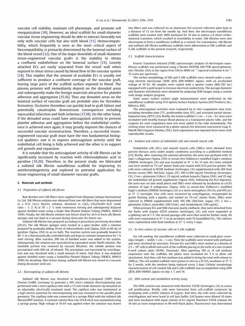
The morphology and diameters of the electrospun nanofiberswere examined by SEM (Fig. 2). The Silk and S-silk scaffolds showedsimilar surface morphologies which consisted of continuous andrandomly oriented nanofibers with diameters ranging from 100 to550 nm. The SEM micrographs appeared that the nanofibers hada solid surface with interconnected voids among the fibers, pre-senting a porous network. The distribution of the nanofiberdiameters showed that both Silk and S-silk scaffolds had approxi-mately 90% of the nanofibers in 100e400 nm diameter range(Fig. 3). The average diameter of S-silk scaffold was 220 � 35 nm,which was smaller than the average diameter (286 � 25 nm) of Silkscaffold (p > 0.05). The surface hydrophilicity of the nanofibrousscaffolds was determined by measuring the static water surfacecontact angle. Silk scaffolds showed a contact angle of about68 � 8�, whereas S-silk scaffolds had a significantly lower contactangle of 46 � 5� (p < 0.05).
PT, APTT, and TT tests, which are widely used in the clinicaldetection of the abnormality of blood plasma and for primaryscreening of anticoagulating chemicals, have been applied to theevaluation of in vitro antithrombogenicity of biomaterials in recentyears. Table 1 showed the TT, PT, and APTT times of Silk and S-silkscaffolds. The values of TT, PT, and APTT of human blood plasmawere 13.6 � 2.1, 20.0 � 1.6, and 64.7 � 2.6, respectively. There wasnot much difference between the values of blood plasma incubatedwith Silk scaffolds and those of human blood plasma. On the otherhand, incubation of S-silk scaffolds with blood plasmamade each ofthe values increase greatly and beyond the measurement limit ofthe clot detection instrument. These results indicated that thesulfate groups introduction resulted in addition of anticoagulantfunction to silk fibroin.
3.2. Cell morphology on scaffolds
SEM images gave a closer view of the cell morphology, with thevascular cells being well attached to the nanofibrous scaffolds andrevealing both spread and flattenedmorphology (Fig. 4). Significantcell morphology changes with time in culture were observed forboth cell types. Compared with the cobble-stone shapes of ECs,SMCs exhibited a more spread and stellate state after 1 day inculture and changed to spindle-shape with parallel alignment withtime in culture (Fig. 4A, D, G, J). ECs and SMCs successfully adheredto the surface within 24 h and the coverage continued to expand
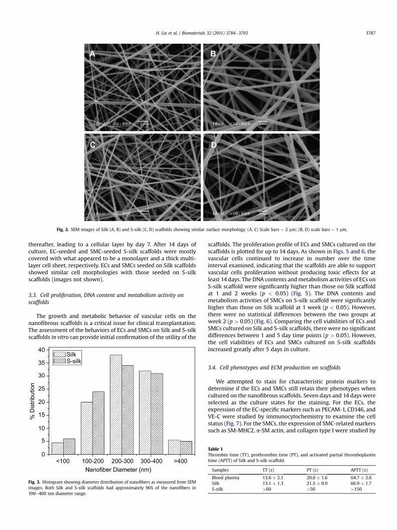
Fig. 2. SEM images of Silk (A, B) and S-silk (C, D) scaffolds showing similar surface morphology. (A, C) Scale bars ¼ 2 mm; (B, D) scale bars ¼ 1 mm.
H. Liu et al. / Biomaterials 32 (2011) 3784e3793 3787
thereafter, leading to a cellular layer by day 7. After 14 days ofculture, EC-seeded and SMC-seeded S-silk scaffolds were mostlycovered with what appeared to be a monolayer and a thick multi-layer cell sheet, respectively. ECs and SMCs seeded on Silk scaffoldsshowed similar cell morphologies with those seeded on S-silkscaffolds (images not shown).
3.3. Cell proliferation, DNA content and metabolism activity onscaffolds
The growth and metabolic behavior of vascular cells on thenanofibrous scaffolds is a critical issue for clinical transplantation.The assessment of the behaviors of ECs and SMCs on Silk and S-silkscaffolds in vitro can provide initial confirmation of the utility of the
Fig. 3. Histogram showing diameter distribution of nanofibers as measured from SEMimages. Both Silk and S-silk scaffolds had approximately 90% of the nanofibers in100e400 nm diameter range.
scaffolds. The proliferation profile of ECs and SMCs cultured on thescaffolds is plotted for up to 14 days. As shown in Figs. 5 and 6, thevascular cells continued to increase in number over the timeinterval examined, indicating that the scaffolds are able to supportvascular cells proliferation without producing toxic effects for atleast 14 days. The DNA contents andmetabolism activities of ECs onS-silk scaffold were significantly higher than those on Silk scaffoldat 1 and 2 weeks (p < 0.05) (Fig. 5). The DNA contents andmetabolism activities of SMCs on S-silk scaffold were significantlyhigher than those on Silk scaffold at 1 week (p < 0.05). However,there were no statistical differences between the two groups atweek 2 (p > 0.05) (Fig. 6). Comparing the cell viabilities of ECs andSMCs cultured on Silk and S-silk scaffolds, there were no significantdifferences between 1 and 5 day time points (p > 0.05). However,the cell viabilities of ECs and SMCs cultured on S-silk scaffoldsincreased greatly after 5 days in culture.
3.4. Cell phenotypes and ECM production on scaffolds
We attempted to stain for characteristic protein markers todetermine if the ECs and SMCs still retain their phenotypes whencultured on the nanofibrous scaffolds. Seven days and 14 days wereselected as the culture states for the staining. For the ECs, theexpression of the EC-specific markers such as PECAM-1, CD146, andVE-C were studied by immunocytochemistry to examine the cellstatus (Fig. 7). For the SMCs, the expression of SMC-relatedmarkerssuch as SM-MHC2, a-SM actin, and collagen type I were studied by
Table 1Thrombin time (TT), prothrombin time (PT), and activated partial thromboplastintime (APTT) of Silk and S-silk scaffold.
Samples TT (s) PT (s) APTT (s)
Blood plasma 13.6 � 2.1 20.0 � 1.6 64.7 � 2.6Silk 13.1 � 1.3 21.5 � 0.9 66.9 � 1.7S-silk >60 >50 >150
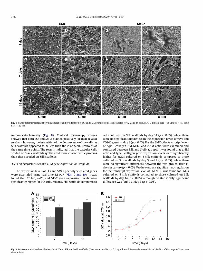
Fig. 4. SEM photomicrographs showing adherence and proliferation of ECs and SMCs cultured on S-silk scaffolds for 1, 7, and 14 days. (A-C, G-I) Scale bars ¼ 50 mm; (D-F, J-L) scalebars ¼ 20 mm.
H. Liu et al. / Biomaterials 32 (2011) 3784e37933788
immunocytochemistry (Fig. 8). Confocal microscopy imagesshowed that both ECs and SMCs stained positively for their relatedmarkers, however, the intensities of the fluorescence of the cells onSilk scaffolds appeared to be less than those on S-silk scaffolds atthe same time points. The results indicated that the vascular cellsseeded on S-silk scaffolds synthesized more characteristic proteinsthan those seeded on Silk scaffolds.
3.5. Cell-characteristics and ECM gene expression on scaffolds
The expression levels of ECs and SMCs phenotype-related geneswere quantified using real-time RT-PCR (Figs. 9 and 10). It wasfound that CD146, vWF, and VE-C gene expression levels weresignificantly higher for ECs cultured on S-silk scaffolds compared to
Fig. 5. DNA content (A) and metabolism (B) of ECs on Silk and S-silk scaffolds. (Data in meantime points).
cells cultured on Silk scaffolds by day 14 (p < 0.05), while therewere no significant differences in the expression levels of vWF andCD146 genes at day 3 (p > 0.05). For the SMCs, the transcript levelsof type I collagen, SM-MHC, and a-SM actin were examined andcompared between Silk and S-silk groups. It was found that a-SMactin and type I collagen gene expression levels were significantlyhigher for SMCs cultured on S-silk scaffolds compared to thosecultured on Silk scaffolds by day 3 and 7 (p < 0.05), while therewere no significant differences between the two groups after 14days in culture (p> 0.05). On the contrary, significant up-regulationfor the transcript expression level of SM-MHC was found for SMCscultured on S-silk scaffolds compared to those cultured on Silkscaffolds by day 14 (p < 0.05), although no statistically significantdifference was found at day 3 (p > 0.05).
�SD, n ¼ 6, * significant difference between Silk and S-silk scaffolds at p<0.05 at same
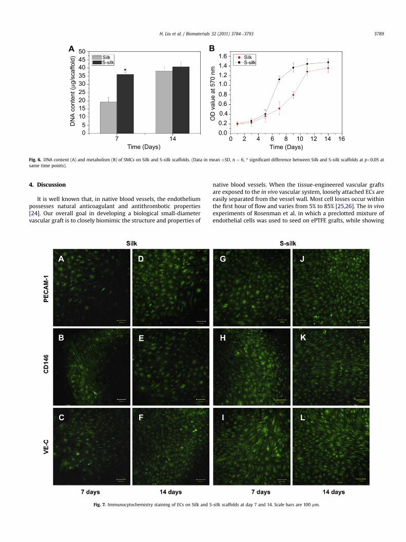
Fig. 6. DNA content (A) and metabolism (B) of SMCs on Silk and S-silk scaffolds. (Data in mean �SD, n ¼ 6, * significant difference between Silk and S-silk scaffolds at p<0.05 atsame time points).
H. Liu et al. / Biomaterials 32 (2011) 3784e3793 3789
4. Discussion
It is well known that, in native blood vessels, the endotheliumpossesses natural anticoagulant and antithrombotic properties[24]. Our overall goal in developing a biological small-diametervascular graft is to closely biomimic the structure and properties of
Fig. 7. Immunocytochemistry staining of ECs on Silk and S
native blood vessels. When the tissue-engineered vascular graftsare exposed to the in vivo vascular system, loosely attached ECs areeasily separated from the vessel wall. Most cell losses occur withinthe first hour of flow and varies from 5% to 85% [25,26]. The in vivoexperiments of Rosenman et al. in which a preclotted mixture ofendothelial cells was used to seed on ePTFE grafts, while showing
-silk scaffolds at day 7 and 14. Scale bars are 100 mm.
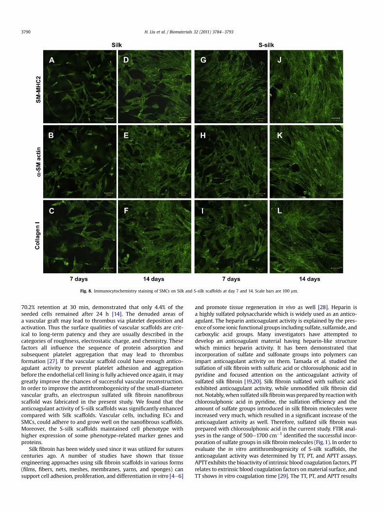
Fig. 8. Immunocytochemistry staining of SMCs on Silk and S-silk scaffolds at day 7 and 14. Scale bars are 100 mm.
H. Liu et al. / Biomaterials 32 (2011) 3784e37933790
70.2% retention at 30 min, demonstrated that only 4.4% of theseeded cells remained after 24 h [14]. The denuded areas ofa vascular graft may lead to thrombus via platelet deposition andactivation. Thus the surface qualities of vascular scaffolds are crit-ical to long-term patency and they are usually described in thecategories of roughness, electrostatic charge, and chemistry. Thesefactors all influence the sequence of protein adsorption andsubsequent platelet aggregation that may lead to thrombusformation [27]. If the vascular scaffold could have enough antico-agulant activity to prevent platelet adhesion and aggregationbefore the endothelial cell lining is fully achieved once again, it maygreatly improve the chances of successful vascular reconstruction.In order to improve the antithrombogenicity of the small-diametervascular grafts, an electrospun sulfated silk fibroin nanofibrousscaffold was fabricated in the present study. We found that theanticoagulant activity of S-silk scaffolds was significantly enhancedcompared with Silk scaffolds. Vascular cells, including ECs andSMCs, could adhere to and grow well on the nanofibrous scaffolds.Moreover, the S-silk scaffolds maintained cell phenotype withhigher expression of some phenotype-related marker genes andproteins.
Silk fibroin has been widely used since it was utilized for suturescenturies ago. A number of studies have shown that tissueengineering approaches using silk fibroin scaffolds in various forms(films, fibers, nets, meshes, membranes, yarns, and sponges) cansupport cell adhesion, proliferation, and differentiation in vitro [4e6]
and promote tissue regeneration in vivo as well [28]. Heparin isa highly sulfated polysaccharide which is widely used as an antico-agulant. The heparin anticoagulant activity is explained by the pres-ence of some ionic functional groups including sulfate, sulfamide, andcarboxylic acid groups. Many investigators have attempted todevelop an anticoagulant material having heparin-like structurewhich mimics heparin activity. It has been demonstrated thatincorporation of sulfate and sulfonate groups into polymers canimpart anticoagulant activity on them. Tamada et al. studied thesulfation of silk fibroin with sulfuric acid or chlorosulphonic acid inpyridine and focused attention on the anticoagulant activity ofsulfated silk fibroin [19,20]. Silk fibroin sulfated with sulfuric acidexhibited anticoagulant activity, while unmodified silk fibroin didnot. Notably,when sulfated silkfibroinwasprepared by reactionwithchlorosulphonic acid in pyridine, the sulfation efficiency and theamount of sulfate groups introduced in silk fibroin molecules wereincreased very much, which resulted in a significant increase of theanticoagulant activity as well. Therefore, sulfated silk fibroin wasprepared with chlorosulphonic acid in the current study. FTIR anal-yses in the range of 500e1700 cm�1 identified the successful incor-poration of sulfate groups in silk fibroinmolecules (Fig.1). In order toevaluate the in vitro antithrombogenicity of S-silk scaffolds, theanticoagulant activity was determined by TT, PT, and APTT assays.APTTexhibits the bioactivity of intrinsic blood coagulation factors, PTrelates to extrinsic blood coagulation factors onmaterial surface, andTT shows in vitro coagulation time [29]. The TT, PT, and APTT results
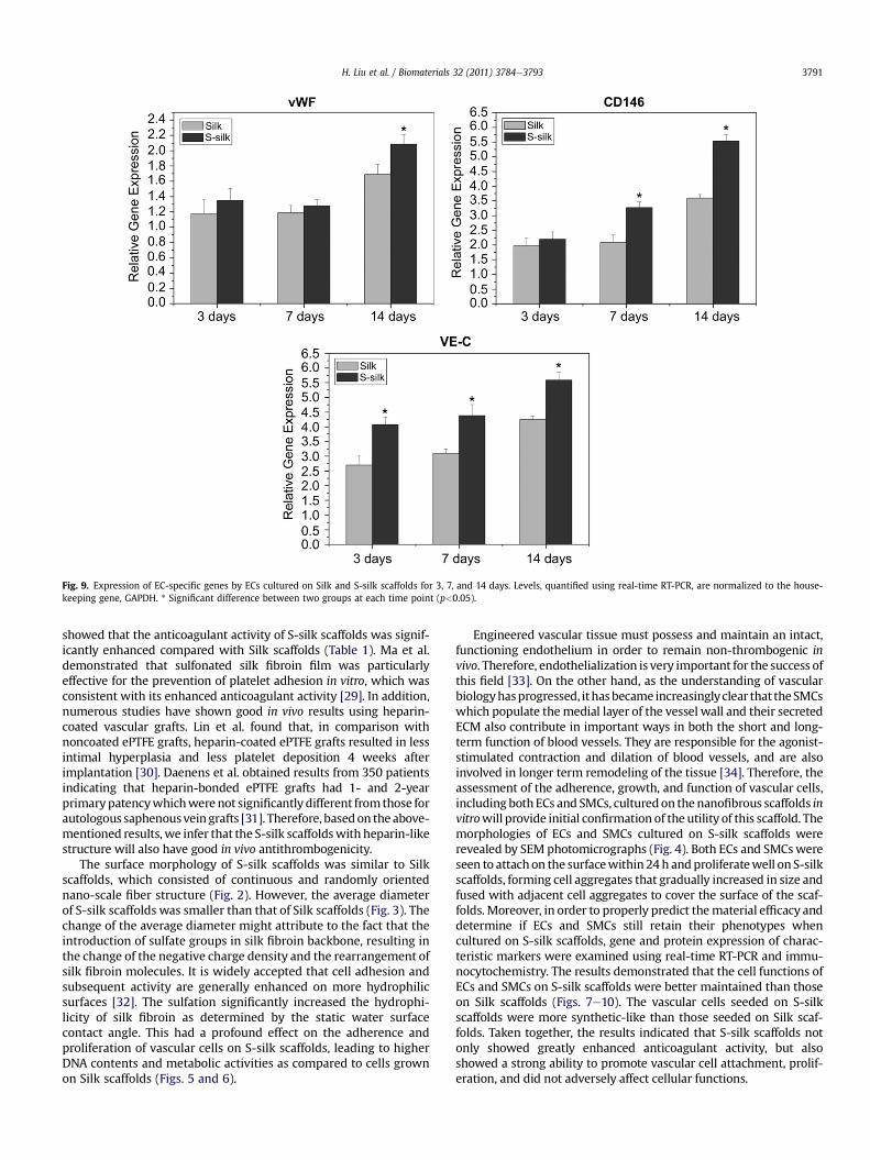
Fig. 9. Expression of EC-specific genes by ECs cultured on Silk and S-silk scaffolds for 3, 7, and 14 days. Levels, quantified using real-time RT-PCR, are normalized to the house-keeping gene, GAPDH. * Significant difference between two groups at each time point (p<0.05).
H. Liu et al. / Biomaterials 32 (2011) 3784e3793 3791
showed that the anticoagulant activity of S-silk scaffolds was signif-icantly enhanced compared with Silk scaffolds (Table 1). Ma et al.demonstrated that sulfonated silk fibroin film was particularlyeffective for the prevention of platelet adhesion in vitro, which wasconsistent with its enhanced anticoagulant activity [29]. In addition,numerous studies have shown good in vivo results using heparin-coated vascular grafts. Lin et al. found that, in comparison withnoncoated ePTFE grafts, heparin-coated ePTFE grafts resulted in lessintimal hyperplasia and less platelet deposition 4 weeks afterimplantation [30]. Daenens et al. obtained results from 350 patientsindicating that heparin-bonded ePTFE grafts had 1- and 2-yearprimarypatencywhichwerenot significantly different fromthose forautologous saphenousveingrafts [31]. Therefore, basedon theabove-mentioned results,we infer that the S-silk scaffoldswithheparin-likestructure will also have good in vivo antithrombogenicity.
The surface morphology of S-silk scaffolds was similar to Silkscaffolds, which consisted of continuous and randomly orientednano-scale fiber structure (Fig. 2). However, the average diameterof S-silk scaffolds was smaller than that of Silk scaffolds (Fig. 3). Thechange of the average diameter might attribute to the fact that theintroduction of sulfate groups in silk fibroin backbone, resulting inthe change of the negative charge density and the rearrangement ofsilk fibroin molecules. It is widely accepted that cell adhesion andsubsequent activity are generally enhanced on more hydrophilicsurfaces [32]. The sulfation significantly increased the hydrophi-licity of silk fibroin as determined by the static water surfacecontact angle. This had a profound effect on the adherence andproliferation of vascular cells on S-silk scaffolds, leading to higherDNA contents and metabolic activities as compared to cells grownon Silk scaffolds (Figs. 5 and 6).
Engineered vascular tissue must possess and maintain an intact,functioning endothelium in order to remain non-thrombogenic invivo. Therefore, endothelialization is very important for the success ofthis field [33]. On the other hand, as the understanding of vascularbiologyhasprogressed, ithasbecame increasinglyclear that theSMCswhich populate themedial layer of the vessel wall and their secretedECM also contribute in important ways in both the short and long-term function of blood vessels. They are responsible for the agonist-stimulated contraction and dilation of blood vessels, and are alsoinvolved in longer term remodeling of the tissue [34]. Therefore, theassessment of the adherence, growth, and function of vascular cells,including bothECs and SMCs, culturedon thenanofibrous scaffolds invitrowill provide initial confirmation of the utility of this scaffold. Themorphologies of ECs and SMCs cultured on S-silk scaffolds wererevealed by SEM photomicrographs (Fig. 4). Both ECs and SMCswereseen to attachon the surfacewithin24h andproliferatewell on S-silkscaffolds, forming cell aggregates that gradually increased in size andfused with adjacent cell aggregates to cover the surface of the scaf-folds. Moreover, in order to properly predict thematerial efficacy anddetermine if ECs and SMCs still retain their phenotypes whencultured on S-silk scaffolds, gene and protein expression of charac-teristic markers were examined using real-time RT-PCR and immu-nocytochemistry. The results demonstrated that the cell functions ofECs and SMCs on S-silk scaffolds were better maintained than thoseon Silk scaffolds (Figs. 7e10). The vascular cells seeded on S-silkscaffolds were more synthetic-like than those seeded on Silk scaf-folds. Taken together, the results indicated that S-silk scaffolds notonly showed greatly enhanced anticoagulant activity, but alsoshowed a strong ability to promote vascular cell attachment, prolif-eration, and did not adversely affect cellular functions.
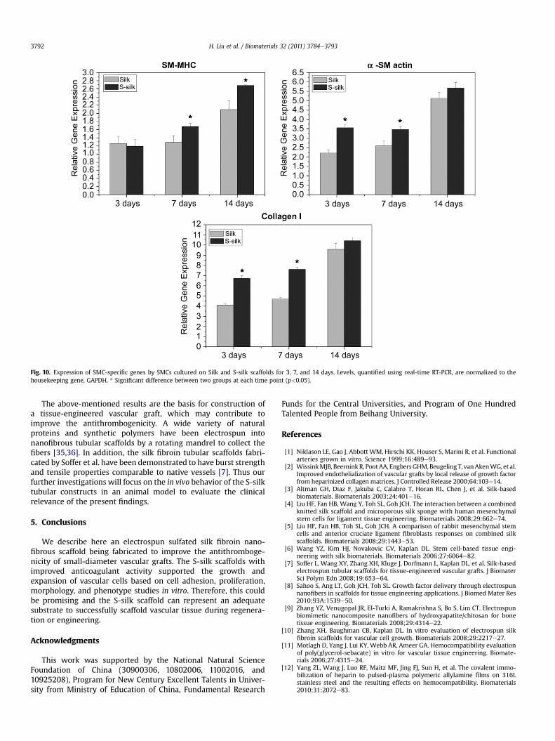
Fig. 10. Expression of SMC-specific genes by SMCs cultured on Silk and S-silk scaffolds for 3, 7, and 14 days. Levels, quantified using real-time RT-PCR, are normalized to thehousekeeping gene, GAPDH. * Significant difference between two groups at each time point (p<0.05).
H. Liu et al. / Biomaterials 32 (2011) 3784e37933792
The above-mentioned results are the basis for construction ofa tissue-engineered vascular graft, which may contribute toimprove the antithrombogenicity. A wide variety of naturalproteins and synthetic polymers have been electrospun intonanofibrous tubular scaffolds by a rotating mandrel to collect thefibers [35,36]. In addition, the silk fibroin tubular scaffolds fabri-cated by Soffer et al. have been demonstrated to have burst strengthand tensile properties comparable to native vessels [7]. Thus ourfurther investigations will focus on the in vivo behavior of the S-silktubular constructs in an animal model to evaluate the clinicalrelevance of the present findings.
5. Conclusions
We describe here an electrospun sulfated silk fibroin nano-fibrous scaffold being fabricated to improve the antithromboge-nicity of small-diameter vascular grafts. The S-silk scaffolds withimproved anticoagulant activity supported the growth andexpansion of vascular cells based on cell adhesion, proliferation,morphology, and phenotype studies in vitro. Therefore, this couldbe promising and the S-silk scaffold can represent an adequatesubstrate to successfully scaffold vascular tissue during regenera-tion or engineering.
Acknowledgments
This work was supported by the National Natural ScienceFoundation of China (30900306, 10802006, 11002016, and10925208), Program for New Century Excellent Talents in Univer-sity from Ministry of Education of China, Fundamental Research
Funds for the Central Universities, and Program of One HundredTalented People from Beihang University.
References
[1] Niklason LE, Gao J, Abbott WM, Hirschi KK, Houser S, Marini R, et al. Functionalarteries grown in vitro. Science 1999;16:489e93.
[2] WissinkMJB, Beernink R, PootAA, EngbersGHM,BeugelingT, vanAkenWG, et al.Improved endothelialization of vascular grafts by local release of growth factorfrom heparinized collagen matrices. J Controlled Release 2000;64:103e14.
[3] Altman GH, Diaz F, Jakuba C, Calabro T, Horan RL, Chen J, et al. Silk-basedbiomaterials. Biomaterials 2003;24:401e16.
[4] Liu HF, Fan HB, Wang Y, Toh SL, Goh JCH. The interaction between a combinedknitted silk scaffold and microporous silk sponge with human mesenchymalstem cells for ligament tissue engineering. Biomaterials 2008;29:662e74.
[5] Liu HF, Fan HB, Toh SL, Goh JCH. A comparison of rabbit mesenchymal stemcells and anterior cruciate ligament fibroblasts responses on combined silkscaffolds. Biomaterials 2008;29:1443e53.
[6] Wang YZ, Kim HJ, Novakovic GV, Kaplan DL. Stem cell-based tissue engi-neering with silk biomaterials. Biomaterials 2006;27:6064e82.
[7] Soffer L, Wang XY, Zhang XH, Kluge J, Dorfmann L, Kaplan DL, et al. Silk-basedelectrospun tubular scaffolds for tissue-engineered vascular grafts. J BiomaterSci Polym Edn 2008;19:653e64.
[8] Sahoo S, Ang LT, Goh JCH, Toh SL. Growth factor delivery through electrospunnanofibers in scaffolds for tissue engineering applications. J Biomed Mater Res2010;93A:1539e50.
[9] Zhang YZ, Venugopal JR, El-Turki A, Ramakrishna S, Bo S, Lim CT. Electrospunbiomimetic nanocomposite nanofibers of hydroxyapatite/chitosan for bonetissue engineering. Biomaterials 2008;29:4314e22.
[10] Zhang XH, Baughman CB, Kaplan DL. In vitro evaluation of electrospun silkfibroin scaffolds for vascular cell growth. Biomaterials 2008;29:2217e27.
[11] Motlagh D, Yang J, Lui KY, Webb AR, Ameer GA. Hemocompatibility evaluationof poly(glycerol-sebacate) in vitro for vascular tissue engineering. Biomate-rials 2006;27:4315e24.
[12] Yang ZL, Wang J, Luo RF, Maitz MF, Jing FJ, Sun H, et al. The covalent immo-bilization of heparin to pulsed-plasma polymeric allylamine films on 316Lstainless steel and the resulting effects on hemocompatibility. Biomaterials2010;31:2072e83.
H. Liu et al. / Biomaterials 32 (2011) 3784e3793 3793
[13] Boccafoschi F, Habermehl J, Vesentini S, Mantovani D. Biological performanceof collagen-based scaffolds for vascular tissue engineering. Biomaterials2005;26:7410e7.
[14] Rosenman JE, Kempczinski RF, Pearce WH, Silberstein EB. Kinetics of endo-thelial cell seeding. J Vasc Surg 1985;6:778e84.
[15] Roald HE, Barstad RM, Bakken IJ, Roald B, Lyberg T, Sakariassen KS. Initial inter-actionsofplatelets andplasmaproteins inflowingnon-anticoagulatedbloodwiththe artificial surfaces Dacron and PTFE. Blood Coagul Fibrinol 1994;5:355e63.
[16] Baier RE. Selected methods of investigation for blood-contact surfaces. AnnNY Acad Sci 1987;516:68e77.
[17] Parsons TI, Haycraft DL, Hoak JC, Sage H. Interaction of platelets and purifiedcollagens in a laminar flow model. Thromb Res 1986;43:435e43.
[18] Klinger RY, Niklason LE. Tissue-engineered blood vessels. In: Vunjak-Novakovic G, Freshney RI, editors. Culture of cells for tissue engineering. JohnWiley & Sons, Inc; 2006. p. 293e322.
[19] Tamada Y. Sulfation of silk fibroin by sulfuric acid and anticoagulant activity.J Appl Polym Sci 2003;87:2377e82.
[20] Tamada Y. Sulfation of silk fibroin by chlorosulfonic acid and the anticoagu-lant activity. Biomaterials 2004;25:377e83.
[21] Taddei P, Arosio C, Monti P, Tsukada M, Arai T, Freddi G. Chemical and physicalproperties of sulfated silk fibrics. Biomacromolecules 2007;8:1200e8.
[22] Gu JW, Yang XL, Zhu HS. Surface sulfonation of silk fibroin film by plasma treat-ment and in vitro antithrombogenicity study.Mater Sci Eng C 2002;20:199e202.
[23] Wu HC, Wang TW, Kang PL, Tsuang YH, Sun JS, Lin FH. Coculture of endo-thelial and smooth muscle cells on a collagen membrane in the developmentof a small-diameter vascular graft. Biomaterials 2007;28:1385e92.
[24] Michiels C. Endothelial cell functions. J Cell Physiol 2003;196:430e43.[25] Schneider PA, Hanson SR, Price TM. Durability of confluent endothelial cell
monolayers on small calibre vascular prostheses in vitro. Surgery 1988;103:456e62.
[26] Campbell JB, Lundgren C, Herring MB. Attachment and retention ofIndium-11 I-labelled endothelial cells onto polyester elastomer. ASAIO J1985;8:113e7.
[27] Joist JH, Pennington DC. Platelet reactions with artificial surfaces. Trans AmSoc Artif Intern Organs 1987;33:341e4.
[28] Fan HB, Liu HF, Toh SL, Goh JCH. In vivo study of anterior cruciate ligamentregeneration using mesenchymal stem cells and silk scaffold. Biomaterials2008;29:3324e37.
[29] Ma XL, Cao CB, Zhu HS. The biocompatibility of silk fibroin films containingsulfonated silk fibroin. J Biomed Mater Res 2006;78B:89e96.
[30] Lin PH, Chen C, Bush RL, Yao Q, Lumsden AB, Hanson SR. Small-caliberheparin-coated ePTFE grafts reduce platelet deposition and neointimalhyperplasia in a baboon model. J Vasc Surg 2004;39(6):1322e8.
[31] Daenens K, Schepers S, Fourneau I, Houthoofd S, Nevelsteen A. Hep-arin-bonded ePTFE grafts compared with vein grafts in femoropoplitealand femorocrural bypasses: 1- and 2-year results. J Vasc Surg 2009;49:1210e6.
[32] Wilson CJ, Clegg RE, Leavesley DI, Pearcy MJ. Mediation of biomaterial-cellinteraction by adsorbed proteins: a review. Tissue Eng 2005;11:1e18.
[33] Zhang X, Thomas V, Xu YY, Bellis SL, Vohra YK. An in vitro regeneratedfunctional human endothelium on a nanofibrous electrospun scaffold.Biomaterials 2010;31:4376e81.
[34] Stegemann JP, Rowe SL, Nerem RM. Engineered blood vessel substitutes. In:Ma PX, Elisseeff JH, editors. Scaffolding in tissue engineering. Taylor & Francis;2005. p. 371e84.
[35] Boland ED, Matthews JA, Pawlowski KJ, Simpson DG, Wnek GE, Bowlin GL.Electrospinning collagen and elastin: preliminary vascular tissue engineering.Front Biosci 2004;9:1422e32.
[36] Matthews JA, Wnek GE, Simpson DG, Bowlin GL. Electrospinning of collagennanofibers. Biomacromolecules 2002;3:232e8.