glutamate receptors in preclinical research on alzheimer's disease: update on recent advances
TRANSCRIPT

�������� ����� ��
Glutamate receptors in preclinical research on Alzheimer’s disease: update onrecent advances
Neng-Wei Hu, Tomas Ondrejcak, Michael J. Rowan
PII: S0091-3057(11)00123-7DOI: doi: 10.1016/j.pbb.2011.04.013Reference: PBB 71175
To appear in: Pharmacology
Received date: 23 December 2010Revised date: 5 April 2011Accepted date: 15 April 2011
Please cite this article as: Hu Neng-Wei, Ondrejcak Tomas, Rowan Michael J., Gluta-mate receptors in preclinical research on Alzheimer’s disease: update on recent advances,Pharmacology (2011), doi: 10.1016/j.pbb.2011.04.013
This is a PDF file of an unedited manuscript that has been accepted for publication.As a service to our customers we are providing this early version of the manuscript.The manuscript will undergo copyediting, typesetting, and review of the resulting proofbefore it is published in its final form. Please note that during the production processerrors may be discovered which could affect the content, and all legal disclaimers thatapply to the journal pertain.
ACC
EPTE
D M
ANU
SCR
IPT
ACCEPTED MANUSCRIPT
Glutamate receptors in preclinical research on Alzheimer’s disease:
update on recent advances
Neng-Wei Hua,b,$, Tomas Ondrejcak a,b,$ and Michael J Rowan a,b,*
aDepartment of Pharmacology and Therapeutics, Biotechnology Building,
Trinity College, Dublin 2, Ireland
bTrinity College Institute of Neuroscience, Trinity College, Dublin 2, Ireland
$These authors contributed equally to this work.
*Correspondence to: Michael J Rowan
Department of Pharmacology and Therapeutics, Biotechnology Building,
Trinity College, Dublin 2, Ireland
e-mail: [email protected]
Telephone: ++353 1 8961567
Fax: ++353 1 8961466
Key Words: Dementia; synaptic plasticity; long-term potentiation; long-term
depression; L-glutamate receptor trafficking; learning
Abbreviations Footnote:
A , amyloid beta; β AD, Alzheimer’s disease; hAPP, human amyloid precursor protein;
AMPA, -amino-3-hydroxy-5-methyl-4-isoxazolepropionate; BDNF, brain-derivedα
neurotrophic factor; CaMKII, calcium/calmodulin- dependent protein kinase II;
EphB2, ephrin type-B receptor 2; EPSC, excitatory postsynaptic current; EPSP,
1

ACC
EPTE
D M
ANU
SCR
IPT
ACCEPTED MANUSCRIPT
excitatory postsynaptic potential; GSK3, glycogen synthase kinase 3; LTD, long-term
depression; LTP, long-term potentiation; MAPK, mitogen-activated protein kinase;
mGlu, metabotropic glutamate; mTOR, mammalian target of rapamycin; NMDA, N-
Methyl-D-Aspartate; PKC, protein kinase C; PSD-95, postsynaptic density protein 95;
Src, protein-tyrosine kinase; STEP, striatal-enriched phosphatase; TNF- , tumorα
necrosis factor-alpha; trkB, tropomyosin-related kinase B
2

ACC
EPTE
D M
ANU
SCR
IPT
ACCEPTED MANUSCRIPT
Abstract
The cognitive and related symptoms of Alzheimer’s disease are mainly attributable to
synaptic failure. Here we review recent research on how the Alzheimer’s disease
amyloid ß-protein (Aß) affects glutamate receptors and fast excitatory synaptic
transmission and plasticity of that transmission. L-glutamate, the main excitatory
neurotransmitter in the brain, has long been implicated in causing NMDA receptor-
mediated excitotoxicity leading to neurodegeneration in the late stages of the disease.
However there is now extensive evidence that soluble Aß oligomers disrupt synaptic
transmission and especially synaptic plasticity via non-excitotoxic glutamatergic
mechanisms. New data highlight the relatively selective involvement of certain
glutamate receptor subtypes including GluN2B (NR2B) subunit-containing NMDA
receptors and mGlu5 receptors. Aß exerts direct and indirect effects on synaptic
plasticity-related glutamate receptor signaling and trafficking between different
neuronal compartments. For example, Aß-induced ectopic NMDA and mGlu
receptor-mediated signaling coupled with caspase-3 activation may cause inhibition
of long-term potentiation and facilitation of long-term depression. Intriguingly, some
of the disruptive synaptic actions of Aß have been found to be dependent on
endogenous tau located in dendrites or spines. Given the role of glutamatergic
transmission in regulating Aß production and release, future therapies targeting
glutamate offer the opportunity to remedy both mis-processing of Aß and cellular
mechanisms of synaptic failure in early AD.
3

ACC
EPTE
D M
ANU
SCR
IPT
ACCEPTED MANUSCRIPT
Numerous reviews that address the role of glutamate receptors and related
synaptic mechanisms in preclinical research on Alzheimer’s disease (AD) and other
neurodegenerative disorders have been published recently (Johnson et al. , 2009, Lau
and Tymianski, 2010, Luscher and Huber, 2010, Ondrejcak et al. , 2010, Palop and
Mucke, 2010a, b, Randall et al. , 2010). The present review provides an update on
some of the recent findings in this rapidly advancing area of research.
AD is the main cause of dementia and can now be diagnosed years before
clinically severe symptoms arise (Perrin et al. , 2009). Several different animal
models are currently used to elucidate the mechanisms of AD, especially in its early
stages (Jucker, 2010). Because a high proportion of familial forms of AD are caused
by misprocessing of amyloid precursor protein (APP) and patients with trisomy 21
(Down syndrome) develop cerebral pathology and dementia characteristic of AD,
transgenic over-expression of human APP (hAPP) is commonly studied in mice.
Many of these transgenic lines display cognitive impairment but little or no
neurodegeneration. In sporadic forms of AD, although there is evidence for increased
activity of ß-secretase, the enzyme that cleaves APP prior to amyloid ß-protein (Aß)
production, it is likely that reduced clearance of Aß is a critical factor. Genetic or
pharmacological disruption of Aß clearance is therefore of great interest, but most
commonly the effects of exogenously applied Aß are examined. Many different forms
of synthetic and animal/human-derived Aß have been investigated, with particular
emphasis on water soluble, non-fibrillar aggregates of Aß. These range from low-n
oligomers to large soluble protofibrils (O'Nuallain et al. , 2010). Much recent
discussion has focused on the prion-like propagation of Aß (Eisele et al., 2010).
Because hyperphosphorylated and aggregated forms of the microtubule-
associated tau protein are present in AD brain and cerebrospinal fluid, much recent
4
1 Introduction

ACC
EPTE
D M
ANU
SCR
IPT
ACCEPTED MANUSCRIPT
attention has been devoted to investigating tau in animals. Like Aß, there is a
growing realization that pre-fibrillar aggregates of tau may be most culpable in AD
(Hoover et al. , 2010, Zempel et al. , 2010).
A major still unresolved issue is the relative role of “loss” versus “gain” of
function in mediating the actions of Aß and tau in AD. Thus tau and Aß may have
physiological roles that are usurped when these proteins aggregate or get
misprocessed leading to loss of function in addition to the more widely accepted view
that abnormally aggregated proteins interact with novel targets to cause a toxic “gain”
in function.
In addition to Aß and tau pathology, key factors influencing the onset and
progression of AD including ageing, cerebrovascular dysfunction, pro-inflammatory,
and cellular and behavioural stress mechanisms have been the focus of research. Such
factors are likely to promote or trigger Aß and tau pathogenic mechanisms but may
also interact as additive, independent causes of dementia (Bishop et al. , 2010,
Pimplikar et al. , 2010).
In structural terms synaptic loss rather than frank neurodegeneration is more
relevant to decline of cognitive and other functions in clinical dementia. Thus,
understanding the relationship between pathological factors such as Aß and tau and
disruption of synapses is of paramount importance. Given the early loss of
glutamatergic neurons in AD in vulnerable pathways such as the medial temporal
lobe/hippocampal network the role of irreversible excitotoxic mechanisms has long
been hypothesized (Greenamyre and Young, 1989). More recently synaptic
transmission and plasticity of this transmission, before detectible loss of synapses,
have been found to be disrupted in several models, increasing the possibility of
targeting disease mechanisms at a potentially reversible stage. Interestingly, Aß
5

ACC
EPTE
D M
ANU
SCR
IPT
ACCEPTED MANUSCRIPT
accumulates relatively selectively at certain (Deshpande et al. , 2009) and is released
at synapses in a use-dependent manner (Bordji et al. , 2010, Hoey et al. , 2009).
2 AMPA ( -amino-3-hydroxy-5-methyl-4-isoxazolepropionate) receptor-α
mediated transmission (see also Fig. 1)
Most investigators to date have reported that acute exogenous application of
sub-micromolar concentrations of Aß has little or no acute effects on AMPA receptor-
mediated transmission. However, a possible physiological role of sub-nanomolar
concentrations of endogenous rodent Aß in the facilitation of activity-dependent
presynaptic vesicular release of glutamate was reported recently (Abramov et al. ,
2009). Thus, lowering extracellular concentration of Aß at cultured hippocampal
neurons reduced synaptic facilitation whereas inhibition of Aß metabolism caused a
rapid increase in the frequency, but not amplitude, of AMPA receptor-mediated
miniature EPSCs. The increase in release probability was associated with reduced
paired-pulse facilitation such that excitatory synapses behaved like low-pass filters,
facilitating low frequency activation of AMPA receptors in hippocampal slices. How
this presynaptic facilitatory action relates to the putative negative feedback
postsynaptic actions of endogenously-generated human Aß (Hsieh et al. , 2006, Wei
et al. , 2010) is not clear. If confirmed, caution will be needed in the use of anti-Aß
therapies that might interfere with such physiological processes.
The loss of AMPA-receptor-mediated transmission in AD is likely to be at least
partly caused by the generation of non-physiological assemblies of Aß. Li et al (2009)
reported that cell-derived oligomeric human Aß acutely increased extracellular
glutamate concentrataion in hippocampal slices. Whereas low nanomolar
concentrations of these oligomers reduced the amplitude of AMPA receptor-mediated
6
ACC
EPTE
D M
ANU
SCR
IPT
ACCEPTED MANUSCRIPT
evoked EPSCs there was no change in AMPA receptor-mediated field EPSPs or
paired-pulse facilitation. Somewhat similarly, certain oligomer-enriched preparations
of synthetic Aß1-42 can potently and rapidly trigger a reduction in evoked AMPA
receptor-mediated EPSCs and/or field EPSPs with no significant change in paired
pulse facilitation (Cerpa et al. , 2010, Kessels et al. , 2010, Ronicke et al. , 2010).
Although there are many possible explanations, Li et al (2009) found that an agent
that blocks AMPA receptor desensitization, cylothiazide, prevented the oligomer-
induced reduction of EPSCs, consistent with Aß acting by inhibiting glutamate
uptake. Indeed, micromolar concentrations of synthetic Aß1-42 oligomers, especially
in the presence of cyclothiazide, apparently can rapidly trigger AMPA receptor-
dependent inward currents and delayed neurodegeneration in cultured cortical neurons
(Alberdi et al. , 2010). Taken together, these findings indicate that agents designed to
directly boost AMPA receptor function in AD may have a relatively narrow
therapeutic window.
Evidence for a more direct interaction between AMPA receptors and Aß
oligomers was provided in a recent paper that confirmed the ability of high nanomolar
concentrations of Aß oligomers to preferentially bind to excitatory dendritic spines in
cultured hippocampal neurons (Zhao et al. , 2010). Ca2+ impermeable AMPA
receptors containing the GluA2 (also named GluR2) subunit were particularly
implicated in Aß binding and certain AMPA receptor antagonists prevented this
binding. Importantly, the binding was associated with the rapid, clathrin-dependent,
endocytosis of AMPA receptors via activation of calcineurin (protein phosphatase 2B)
and dynamin. Moreover Aß oligomers were also internalized in a calcineurin-
dependent manner. Further support for the importance of GluA2-containing AMPA
receptors was independently provided by Liu et al (2010) who reported that
7

ACC
EPTE
D M
ANU
SCR
IPT
ACCEPTED MANUSCRIPT
micromolar Aß oligomers induced a protein kinase C-dependent phosphorylation and
internalization of these receptors (Liu et al. , 2010). GluA1-containing receptors are
also internalized in response to treatment with micromolar Aß oligomers in cortical
slice cultures, consistent with the predominant heteromeric assembly of AMPA
receptors (Gu et al. , 2009). The removal of AMPA receptors was associated with a
selective and delayed (>1h, <24h) reduction in AMPA receptor-mediated synaptic
currents and a redistribution of Ca2+/calmodulin- dependent protein kinase II
(CaMKII) away from the synapse. Indeed loss of hippocampal dendritic spines,
reduced GluA1-containing AMPA receptors, decreased AMPA receptor-mediated
synaptic transmission, and memory impairments were all attributable to Aß oligomer-
induced caspase-3 activation in young hAPP transgenic mice (D'Amelio et al. , 2011).
The authors provided evidence that these morphological, molecular, cellular and
behavioural deficits are due to a reversible oxidative stress-induced increase in
caspase-3 cleavage of calcineurin (thereby increasing its activity), which in turn
promotes dephosphorylation of postsynaptic AMPA receptors thereby triggering their
internalization.
There is growing evidence that tau may mediate synaptic dysfunction in AD.
Thus, accumulation of mis-sorted hyperphosphorylated tau in dendritic spines has
been implicated in disruption of AMPA (and NMDA) receptor trafficking and
anchoring (Hoover et al., 2010). Indeed micromolar Aß oligomers, like glutamate, can
cause rapid mis-sorting of several proteins including phosphorylated tau into dendrites
and local depletion of mitochondria and subsequent loss of spines in cultured
hippocampal neurons (Zempel et al., 2010). Furthermore, prolonged exposure to
soluble AD brain extracts that contain nanomolar Aß oligomers cause synaptic loss in
a tau-dependent manner (Jin et al. , 2011). Interestingly, cortical spine loss associated
8

ACC
EPTE
D M
ANU
SCR
IPT
ACCEPTED MANUSCRIPT
with soluble Aß was detected in vivo in hAPP-tau transgenic mice with confocal
imaging only in spines that accumulated hyperphosphorylated tau (Bittner et al. ,
2010). Furthermore, abnormal increased hippocampal excitatory synaptic
transmission in hAPP transgenic mice is absent when these mice are crossed with tau-
deficient mice (Roberson et al. , 2011).
An emerging view is that the most pathologically relevant concentrations and
assembly states of Aß acutely can indirectly increase extracellular glutamate levels
with relatively subtle immediate effects on AMPA receptor-mediated transmission.
Prolonged exposure or higher acute concentrations of Aß oligomers cause AMPA
receptor endocytosis and may consequently trigger synaptic loss, probably as a result
of non-apoptotic activation of certain caspases.
3 NMDA (N-methyl-D-aspartate) receptor-mediated transmission (see also
Fig. 2)
An Aß-mediated increase in extracellular glutamate levels would be expected to
modulate NMDA receptor- as well as AMPA receptor-mediated transmission. Indeed,
Li et al. (2009) reported that in hippocampal slices the peak amplitude of NMDA
receptor-mediated EPSCs was rapidly reduced by low nanomolar cell-derived Aß
oligomers but the total charge transfer was not significantly affected due to
prolongation of the EPSC. Interestingly, the relative contribution of different NMDA
receptors to transmission was changed. Thus pharmacological evidence was provided
that following Aß oligomer treatment the relative contribution of GluN2B (also
known as NR2B) subunit-containing NMDA receptors, and extrasynaptic NMDA
receptors increased markedly. In a separate study (Cerpa et al., 2010), a high
nanomolar oligomer-enriched preparation of synthetic Aß1-42 also caused a rapid and
9

ACC
EPTE
D M
ANU
SCR
IPT
ACCEPTED MANUSCRIPT
relatively large reduction in the amplitude of evoked NMDA receptor-mediated
EPSCs in hippocampal slices. However these authors did not report the kinetics of the
currents or the total charge transfer. In contrast, in cultured cortical neurons
micromolar Aß1-42 oligomers either rapidly triggered inward currents that were
NMDA receptor-dependent (Alberdi et al. , 2010) or had no significant effect on
NMDA-evoked currents (Gu et al., 2009). Furthermore, and unlike AMPA receptor-
mediated EPSCs, there was no change in NMDA receptor-mediated EPSCs in cortical
; Goussakov et al. , 2010) or hippocampal neurons (D'Amelio et al., 2011) from
young hAPP transgenic mice. However electrically evoked NMDA receptor-triggered
increases in dendritic Ca2+ concentration were greatly enhanced in cortical neuron
spines and dendrites of these mice due to aberrant calcium induced calcium release
from ryanodine receptor-regulated intracellular stores (Goussakov et al., 2010). A
putative mechanism proposed for this synergism is formation of a complex between
GluN2B subunits and ryanodine receptors (Seeber et al. , 2004).
Indeed high nanomolar Aß oligomer-induced delayed reduction in AMPA
receptor-mediated transmission and synaptic pruning in hippocampal neurons is also
GluN2B-dependent, being ameliorated by selective antagonists for NMDA receptors
containing this subunit (Ronicke et al., 2010). This deleterious effect was related to a
GluN2B-dependent translocation to the nucleus of a signaling protein termed Jacob
and subsequent activation of the CREB shut-off pathway. Interestingly, low
nanomolar concentration of cell-derived Aß oligomers also increase the activity of a
tyrosine phosphatase, striatal-enriched phosphatase 61 (STEP) which promotes the
endocytosis of GluN2B-containing NMDA receptors in cortical slices via
dephosphorylation of the Src kinase Fyn and GluN2B at Y1472 (Kurup et al., 2010).
Furthermore, genetic knockout of STEP prevented a reduction in hippocampal
10
ACC
EPTE
D M
ANU
SCR
IPT
ACCEPTED MANUSCRIPT
synaptic GluN2 content and cognitive impairment in hAPP and hAPP-tau transgenic
mice (Zhang et al., 2010).
Remarkably, although tau, the other key player in AD pathology, is normally
considered to be principally involved in the stable assembly of microtubules, a key
role in regulating the phosphorylation of GluN2B subunits by the Src kinase Fyn has
been reported (Ittner et al., 2010). Tau may traffic Fyn into dendritic spines thereby
enabling Fyn to phosphorylate Y1472 in the extreme C terminus of GluN2B subunits.
Phosphorylation at this site facilitates the interaction of the GluN2B subunit with the
postsynaptic density (PSD) protein PSD95. This interaction somehow couples NMDA
receptors to pro-convulsant downstream signaling, but apparently does not directly
affect NMDA (or AMPA) receptor-mediated fast excitatory synaptic transmission.
Importantly, genetic ablation of tau, or peptide (Tat-NR2B9c)-mediated inhibition of
NMDA receptor association with PSD-95, reduced the increased seizure
susceptibility, T-maze errors and premature death seen in hAPP transgenic mice.
Furthermore, the protective effects of Tat-NR2B9c persisted for 4 months after
ceasing intracerebroventricular infusion in these mice.
Also implicating a role for GluN2B subunit-containing NMDA receptors, the
activity-dependent synaptic localization and binding of Aß oligomers in the
hippocampus has been reported to be ifenprodil-sensitive (Deshpande et al., 2009).
Whether or not Aß binds less efficiently to synapses lacking GluN2B subunits or if
the accumulation of larger aggregates leads to more profound synaptic dysfunction
remains to be elucidated.
Interestingly, Aß1-42 fibrils, but not oligomers, slightly depolarized cortical and
hippocampal neurons, similar to membrane depolarization found in hAPP transgenic
mice (Minkeviciene et al., 2009). Such depolarization would be expected to partially
11

ACC
EPTE
D M
ANU
SCR
IPT
ACCEPTED MANUSCRIPT
relieve NMDA receptors from their Mg2+ block. Indeed fibrillar Aß1-42 was recently
reported to enhance NMDA evoked firing via ß1 integrin and Src kinase in the
hippocampus in vivo (Uhasz et al., 2010).
Overall there is a growing consensus that acute and delayed synaptic effects of
pathophysiologically relevant concentrations of Aß oligomers are dependent on
activation of GluN2B-containing NMDA receptors. This activation may be
accompanied by modulation of NMDA receptor-mediated synaptic transmission and
neuronal excitability. A putative role of synaptic tau is a matter of ongoing intensive
research.
4 MGlu receptor trafficking and related signaling (see also Fig. 3)
Clearly, Aß-induced excessive extracellular glutamate concentration will cause
increased activation of metabotropic as well as ionotropic glutamate receptors.
However, evidence of a more direct link between mGlu receptors and Aß’s synaptic
actions was recently reported (Renner et al., 2010). Using antibodies Renner et al.
(2010) discovered that relatively low nanomolar Aß oligomer binding to cultured
hippocampal neuron synapses was dependent on mGlu5 but not AMPA receptors and
confirmed a dependence on NMDA receptors and cellular prion protein, which were
non-additive contributors to binding. They found that A 1-42 oligomers rapidly bindβ
to neuronal membrane, diffuse laterally and then gradually accumulate in clusters at
excitatory synapses. These clusters altered the distribution and reduced the mobility of
associated mGlu5 receptors. The slower lateral mobility of these receptors impaired
their exchange between synaptic and extrasynaptic locations, which in turn increased
synaptic mGlu5 receptors. Consequently, aberrant activation of mGlu5 receptors in
ectopic signaling platforms promoted an increased intracellular Ca2+ and the removal
12

ACC
EPTE
D M
ANU
SCR
IPT
ACCEPTED MANUSCRIPT
of NMDA receptors from synapses (Renner et al., 2010). mGlu5 and NMDA
receptors are closely associated signaling partners, e.g. activation of mGlu5 receptors
potentiates NMDA receptor function (Niswender and Conn, 2010) and the authors
concluded that NMDA receptor-dependent synaptic effects are downstream of mGlu5
receptors.
Interestingly, by using astrocyte-rich cultures, Casley et al. (Casley et al. , 2009)
found that A can also enhance the magnitude of the intracellular calciumβ
mobilization induced by the mGlu5 receptor activation. Whether or not similar
mechanisms to those described for receptor clustering in neurons applies to glia
requires further study.
5 Plasticity of AMPA receptor-mediated synaptic transmission (see also Fig.
4)
Synaptic plasticity mechanisms underlie cognitive functions including memory,
and are exquisitely sensitive to Aß oligomers, which potently enhance long-term
depression (LTD) and inhibit long-term potentiation (LTP) (Li et al., 2009). Since the
expression of LTP often requires increased insertion and enhanced function of AMPA
receptors in the postsynaptic membrane whereas conversely, the expression of LTD
requires increased removal and decreased function of these receptors, AD-related
reductions in postsynaptic AMPA receptor function and number may be caused by
disruption of synaptic plasticity mechanisms.
In support of this view, some studies on the mechanisms of Aß-triggered
reduction of baseline AMPA receptor-mediated synaptic transmission have confirmed
parallels with the mechanisms underlying electrically or chemically induced long-
13

ACC
EPTE
D M
ANU
SCR
IPT
ACCEPTED MANUSCRIPT
term depression (LTD), reviewed by Collingridge et al. (2010). Both NMDA and
mGlu receptor-dependent forms of LTD are facilitated by Aß (Kim et al., 2001, Li et
al., 2009). In the case of Aß-facilitated NMDA receptor-dependent LTD these authors
reported a requirement for activation of glycogen synthase kinase 3 (GSK3) and
calcineurin but not p38 MAP kinase or intracellular Ca2+ stores. As noted in Section 2
above, baseline reductions in AMPA receptors induced by Aß oligomers are
calcineurin-dependent (D'Amelio et al., 2011, Liu et al., 2010, Zhao et al., 2010).
Moreover, a role for GSK3 was implicated in Aß-mediated inhibition of baseline and
chemically –induced rapid membrane insertion of AMPA receptors in cultured
hippocampal neuron spines using the a selective inhibitor (Rui et al., 2010).
Interestingly, spines associated with mitochondria, and showing surface accumulation
of AMPA receptors, tended to be more resistant to A -mediated inhibition of AMPAβ
receptor trafficking.
Inhibitors of GSK3 also can prevent the inhibition of high frequency
stimulation-induced LTP in hAPP transgenic mice (Ma et al., 2010). Furthermore,
genetic deletion of FK506-binding protein 12 prevented disruption of LTP by Aß1-42
oligomers. The protective effect of these interventions was attributed by these authors
to reversing Aß-mediated inhibition of activity of the Ser/Thr protein kinase
mammalian target of rapamycin (mTOR) signaling pathway. Consistent with this, the
neurotrophin BDNF, which acts through trkB receptors partly via the mTOR signaling
pathway, is critical for synaptic AMPA receptor expression and delivery (Li and
Keifer, 2008) and has been found to prevent A 1-42-mediated reduction of LTP inβ
hippocampal slices, and LTP-associated CaMKII activation and AMPA receptor
phosphorylation at a CaMKII-dependent site (Zeng et al., 2010).
Further support for a key role of GSK3 in mediating disruption of synaptic
14

ACC
EPTE
D M
ANU
SCR
IPT
ACCEPTED MANUSCRIPT
plasticity in hippocampal slices was recently reported by Shipton et al, (2011). These
authors implicated downstream GSK3-mediated phosphorylation of tau since the
deficit in LTP was not triggered in slices from tau deficient mice. Remarkably, the
acute inhibition of LTP by Aß in hippocampal slices can be reversed by treatment
with a selective GSK3 inhibitor even after the application of Aß (Jo et al., 2011). In a
very elegant set of experiments strong evidence was provided that the disruption of
LTP by Aß was caused by relatively specific activation of caspase 3, which in turn
cleaved Akt, a key negative regulator of GSK3 kinase activity (Jo et al., 2011).
Additional, more indirect, mechanisms have been hypothesized to mediate the
inhibition of NMDA receptor-dependent LTP by Aß. For example, a reduction in
synaptic NMDA receptor function has been proposed to underlie this impairment of
plasticity (Cisse et al., 2011). Aß oligomers were found to potently bind to the
postsynaptic protein tyrosine kinase EphB2, which in turn was internalized and
cleaved. Loss of EphB2, which regulates NMDA receptor trafficking, triggered the
removal of synaptic NMDA receptors. Consistent with the hypothesis, the impairment
of LTP by Aß and similar LTP deficits and learning impairments in hAPP transgenic
mice were ameliorated by genetic overexpression of EphB2. Given the potential
redundancy of synaptic NMDA receptors it will be important to determine if the
observed reduction in NMDA receptors was sufficient on its own to disrupt synaptic
plasticity. Another related putative mechanism for the inhibition of LTP by Aß
involves increased activation of STEP with consequent endocytosis of GluN2B
subunits (Kurup et al., 2010). Genetic knockout of STEP enhanced LTP in hAPP-tau
transgenic mice (Zhang et al., 2010). However, removal of STEP in controls also
enhanced LTP and it was unclear if there was an LTP deficit in the transgenic mice.
Given the ability of the clinically used NMDA receptor antagonist memantine to
15

ACC
EPTE
D M
ANU
SCR
IPT
ACCEPTED MANUSCRIPT
partially prevent the inhibition of LTP in vivo (Klyubin et al., 2011), we (Hu et al.,
2009) recently compared the activity of subtype selective antagonists. Doses of
GluN2B selective antagonists that did not significantly affect control LTP, completely
prevented the disruptive effect of Aß. Similar treatment of animals with relatively low
doses of the antagonists with greater preference for GluN2A,C,D subunits had no
significant effect on the inhibition of LTP. Although, as discussed above, Aß
oligomer-mediated inhibition of glutamate uptake can lead to inappropriate activation
of extrasynaptic NMDA receptors incorporating GluN2B subunits (Li et al., 2009),
we found evidence for a role of the cytokine TNF in mediating the action of Aß.α
Indeed, a GluN2B antagonist also abrogated a similar plasticity disrupting action of
TNF . It is possible that pro-inflammatory actions of Aß triggers the release of TNFα α
which in turn may promote increased spillover of glutamate.
6 Conclusions
Collectively, the above reports support a model of early disease pathogenesis in
which low concentrations of A oligomers initially preferentially and inappropriatelyβ
boost activation of certain glutamate receptors, including mGlu5 and GluN2B
subunit-containing NMDA receptors. Such activation disrupts synaptic plasticity,
promoting LTD and inhibiting LTP of AMPA receptor-mediated synaptic
transmission. The associated persistent reduction in the number of functional synaptic
AMPA receptors reduces fast excitatory transmission and eventually triggers spine
retraction and synaptic loss.
Future studies are likely to probe more deeply into the relationship between
rapid and delayed effects of Aß at pre- and post-synaptic sites on glutamatergic
synapses. Already it is known that Aß can be released in an activity-dependent
manner from both axons and dendrites to disrupt chemically-induced structural
16
ACC
EPTE
D M
ANU
SCR
IPT
ACCEPTED MANUSCRIPT
plasticity and initiate spine loss in hippocampal slice culture within 1-3 days (Wei et
al., 2010). Similarly, the relative importance of synaptic versus extrasynaptic
glutamate receptors and their trafficking between different compartments in mediating
Aß action needs to be integrated with our growing knowledge of their roles in
cognitive decline in other neurodegenerative diseases such as Huntington’s disease
(Hardingham and Bading, 2010). How, and at what stage, the many different neuronal
and non-neuronal (e.g. glial and vascular) binding sites for Aß oligomers contribute to
synaptic and non-synaptic mechanisms mediating cognitive impairment is only
beginning to be understood.
Of particular interest is how behavioural factors affect Aß -induced changes in
glutamate receptor function and distribution. Intriguingly, the profile of changes in
synaptic plasticity observed in hAPP transgenic mice are determined by prior training
in a learning task (Middei et al., 2010). Thus, an impairment in LTP persistence was
only detected in transgenic animals that had undergone water maze training.
As noted in the Introduction, glutamate receptors are not only involved in the
process of A -mediated synaptic dysfunction but also play important roles in Aβ β
production (see also Figs. 2 and 3). Recently it was reported that whereas synaptic
NMDA receptor activation promotes non-amyloidogenic -secretase processing ofα
APP and thereby decreases A production, extrasynaptic receptor activation increasesβ
A production β (Bordji et al., 2010, Hoey et al., 2009). Furthermore, activation of
NMDA receptors by endogenously released glutamate in the presence of glycine can
reduce intraneuronal A levels β (Tampellini et al., 2009). Similarly group I and group
II mGlu receptor subtype selective agents exert differential actions on Aß production
and release (Kim et al., 2010). These findings raise many questions such as how
endogenous glutamate receptor stimulation affects neuronal A production fromβ
17

ACC
EPTE
D M
ANU
SCR
IPT
ACCEPTED MANUSCRIPT
healthy and diseased tissue. Given the role of glutamatergic transmission in regulating
Aß production and release future therapies targeting glutamate offer the opportunity
to remedy both mis-processing of Aß and cellular mechanisms of synaptic failure in
early AD. More research is needed to answer these questions and to clarify the
potential therapeutic value of selectively targeting specific glutamate receptor
subtypes and associated signaling mechanisms.
Acknowledgements
We wish to acknowledge the support of Science Foundation Ireland and the Health
Research Board of Ireland.
References
Abramov E, Dolev I, Fogel H, Ciccotosto GD, Ruff E, Slutsky I. Amyloid-beta as a
positive endogenous regulator of release probability at hippocampal synapses. Nat
Neurosci. 2009;12:1567-76.
Alberdi E, Sanchez-Gomez MV, Cavaliere F, Perez-Samartin A, Zugaza JL, Trullas
R, et al. Amyloid beta oligomers induce Ca2+ dysregulation and neuronal death
through activation of ionotropic glutamate receptors. Cell Calcium. 2010;47:264-72.
Bishop NA, Lu T, Yankner BA. Neural mechanisms of ageing and cognitive decline.
Nature. 2010;464:529-35.
Bittner T, Fuhrmann M, Burgold S, Ochs SM, Hoffmann N, Mitteregger G, et al.
Multiple events lead to dendritic spine loss in triple transgenic Alzheimer's disease
mice. PLoS One. 2010;5:e15477.
Bordji K, Becerril-Ortega J, Nicole O, Buisson A. Activation of extrasynaptic, but not
18

ACC
EPTE
D M
ANU
SCR
IPT
ACCEPTED MANUSCRIPT
synaptic, NMDA receptors modifies amyloid precursor protein expression pattern and
increases amyloid-ss production. J Neurosci. 2010;30:15927-42.
Casley CS, Lakics V, Lee HG, Broad LM, Day TA, Cluett T, et al. Up-regulation of
astrocyte metabotropic glutamate receptor 5 by amyloid-beta peptide. Brain Res.
2009.
Cerpa W, Farias GG, Godoy JA, Fuenzalida M, Bonansco C, Inestrosa NC. Wnt-5a
occludes Abeta oligomer-induced depression of glutamatergic transmission in
hippocampal neurons. Mol Neurodegener. 2010;5:3.
Cisse M, Halabisky B, Harris J, Devidze N, Dubal DB, Sun B, et al. Reversing EphB2
depletion rescues cognitive functions in Alzheimer model. Nature. 2011;469:472.
Collingridge GL, Peineau S, Howland JG, Wang YT. Long-term depression in the
CNS. Nat Rev Neurosci. 2010;11:459-73.
D'Amelio M, Cavallucci V, Middei S, Marchetti C, Pacioni S, Ferri A, et al. Caspase-
3 triggers early synaptic dysfunction in a mouse model of Alzheimer's disease. Nat
Neurosci. 2011;14:69-76.
Deshpande A, Kawai H, Metherate R, Glabe CG, Busciglio J. A role for synaptic zinc
in activity-dependent Abeta oligomer formation and accumulation at excitatory
synapses. J Neurosci. 2009;29:4004-15.
Eisele YS, Obermuller U, Heilbronner G, Baumann F, Kaeser SA, Wolburg H, et al.
Peripherally applied Abeta-containing inoculates induce cerebral beta-amyloidosis.
Science. 2010;330:980-2.
Goussakov I, Miller MB, Stutzmann GE. NMDA-mediated Ca(2+) influx drives
aberrant ryanodine receptor activation in dendrites of young Alzheimer's disease
mice. J Neurosci. 2010;30:12128-37.
Greenamyre JT, Young AB. Excitatory amino acids and Alzheimer's disease.
19

ACC
EPTE
D M
ANU
SCR
IPT
ACCEPTED MANUSCRIPT
Neurobiol Aging. 1989;10:593-602.
Gu Z, Liu W, Yan Z. {beta}-Amyloid impairs AMPA receptor trafficking and
function by reducing Ca2+/calmodulin-dependent protein kinase II synaptic
distribution. J Biol Chem. 2009;284:10639-49.
Hardingham GE, Bading H. Synaptic versus extrasynaptic NMDA receptor
signalling: implications for neurodegenerative disorders. Nat Rev Neurosci.
2010;11:682-96.
Hoey SE, Williams RJ, Perkinton MS. Synaptic NMDA receptor activation stimulates
alpha-secretase amyloid precursor protein processing and inhibits amyloid-beta
production. J Neurosci. 2009;29:4442-60.
Hoover BR, Reed MN, Su J, Penrod RD, Kotilinek LA, Grant MK, et al. Tau
mislocalization to dendritic spines mediates synaptic dysfunction independently of
neurodegeneration. Neuron. 2010;68:1067-81.
Hsieh H, Boehm J, Sato C, Iwatsubo T, Tomita T, Sisodia S, et al. AMPAR removal
underlies Abeta-induced synaptic depression and dendritic spine loss. Neuron.
2006;52:831-43.
Hu NW, Klyubin I, Anwyl R, Rowan MJ. GluN2B subunit-containing NMDA
receptor antagonists prevent Abeta-mediated synaptic plasticity disruption in vivo.
Proc Natl Acad Sci U S A. 2009;106:20504-9.
Ittner LM, Ke YD, Delerue F, Bi M, Gladbach A, van Eersel J, et al. Dendritic
function of tau mediates amyloid-beta toxicity in Alzheimer's disease mouse models.
Cell. 2010;142:387-97.
Jin M, Shepardson N, Yang T, Chen G, Walsh D, Selkoe DJ. Soluble amyloid {beta}-
protein dimers isolated from Alzheimer cortex directly induce Tau
hyperphosphorylation and neuritic degeneration. Proc Natl Acad Sci U S A. 2011.
20

ACC
EPTE
D M
ANU
SCR
IPT
ACCEPTED MANUSCRIPT
Jo J, Whitcomb DJ, Olsen KM, Kerrigan TL, Lo SC, Bru-Mercier G, et al. Abeta(1-
42) inhibition of LTP is mediated by a signaling pathway involving caspase-3, Akt1
and GSK-3beta. Nat Neurosci. 2011; in press: doi:10.1038/nn.2785.
Johnson KA, Conn PJ, Niswender CM. Glutamate receptors as therapeutic targets for
Parkinson's disease. CNS Neurol Disord Drug Targets. 2009;8:475-91.
Jucker M. The benefits and limitations of animal models for translational research in
neurodegenerative diseases. Nat Med. 2010;16:1210-4.
Kessels HW, Nguyen LN, Nabavi S, Malinow R. The prion protein as a receptor for
amyloid-beta. Nature. 2010;466:E3-4; discussion E-5.
Kim JH, Anwyl R, Suh YH, Djamgoz MB, Rowan MJ. Use-dependent effects of
amyloidogenic fragments of (beta)-amyloid precursor protein on synaptic plasticity in
rat hippocampus in vivo. J Neurosci. 2001;21:1327-33.
Kim SH, Fraser PE, Westaway D, St George-Hyslop PH, Ehrlich ME, Gandy S.
Group II metabotropic glutamate receptor stimulation triggers production and release
of Alzheimer's amyloid(beta)42 from isolated intact nerve terminals. J Neurosci.
2010;30:3870-5.
Klyubin I, Wang Q, Reed MN, Irving EA, Upton N, Hofmeister J, et al. Protection
against Abeta-mediated rapid disruption of synaptic plasticity and memory by
memantine. Neurobiol Aging. 2011;32:614.
Kurup P, Zhang Y, Xu J, Venkitaramani DV, Haroutunian V, Greengard P, et al.
Abeta-mediated NMDA receptor endocytosis in Alzheimer's disease involves
ubiquitination of the tyrosine phosphatase STEP61. J Neurosci. 2010;30:5948-57.
Lau A, Tymianski M. Glutamate receptors, neurotoxicity and neurodegeneration.
Pflugers Arch. 2010;460:525-42.
Li S, Hong S, Shepardson NE, Walsh DM, Shankar GM, Selkoe D. Soluble oligomers
21

ACC
EPTE
D M
ANU
SCR
IPT
ACCEPTED MANUSCRIPT
of amyloid Beta protein facilitate hippocampal long-term depression by disrupting
neuronal glutamate uptake. Neuron. 2009;62:788-801.
Li W, Keifer J. Coordinate action of pre- and postsynaptic brain-derived neurotrophic
factor is required for AMPAR trafficking and acquisition of in vitro classical
conditioning. Neuroscience. 2008;155:686-97.
Liu SJ, Gasperini R, Foa L, Small DH. Amyloid-beta decreases cell-surface AMPA
receptors by increasing intracellular calcium and phosphorylation of GluR2. J
Alzheimers Dis. 2010;21:655-66.
Luscher C, Huber KM. Group 1 mGluR-dependent synaptic long-term depression:
mechanisms and implications for circuitry and disease. Neuron. 2010;65:445-59.
Ma T, Hoeffer CA, Capetillo-Zarate E, Yu F, Wong H, Lin MT, et al. Dysregulation
of the mTOR pathway mediates impairment of synaptic plasticity in a mouse model
of Alzheimer's disease. PLoS One. 2010;5. pii: e12845.
Middei S, Roberto A, Berretta N, Panico MB, Lista S, Bernardi G, et al. Learning
discloses abnormal structural and functional plasticity at hippocampal synapses in the
APP23 mouse model of Alzheimer's disease. Learn Mem. 2010;17:236-40.
Minkeviciene R, Rheims S, Dobszay MB, Zilberter M, Hartikainen J, Fulop L, et al.
Amyloid beta-induced neuronal hyperexcitability triggers progressive epilepsy. J
Neurosci. 2009;29:3453-62.
Niswender CM, Conn PJ. Metabotropic glutamate receptors: physiology,
pharmacology, and disease. Annu Rev Pharmacol Toxicol. 2010;50:295-322.
O'Nuallain B, Freir DB, Nicoll AJ, Risse E, Ferguson N, Herron CE, et al. Amyloid
beta-protein dimers rapidly form stable synaptotoxic protofibrils. J Neurosci.
2010;30:14411-9.
Ondrejcak T, Klyubin I, Hu NW, Barry AE, Cullen WK, Rowan MJ. Alzheimer's
22

ACC
EPTE
D M
ANU
SCR
IPT
ACCEPTED MANUSCRIPT
disease amyloid beta-protein and synaptic function. Neuromolecular Med.
2010;12:13-26.
Palop JJ, Mucke L. Amyloid-beta-induced neuronal dysfunction in Alzheimer's
disease: from synapses toward neural networks. Nat Neurosci. 2010a;13:812-8.
Palop JJ, Mucke L. Synaptic depression and aberrant excitatory network activity in
Alzheimer's disease: two faces of the same coin? Neuromolecular Med. 2010b;12:48-
55.
Perrin RJ, Fagan AM, Holtzman DM. Multimodal techniques for diagnosis and
prognosis of Alzheimer's disease. Nature. 2009;461:916-22.
Pimplikar SW, Nixon RA, Robakis NK, Shen J, Tsai LH. Amyloid-independent
mechanisms in Alzheimer's disease pathogenesis. J Neurosci. 2010;30:14946-54.
Randall AD, Witton J, Booth C, Hynes-Allen A, Brown JT. The functional
neurophysiology of the amyloid precursor protein (APP) processing pathway.
Neuropharmacology. 2010;59:243-67.
Renner M, Lacor PN, Velasco PT, Xu J, Contractor A, Klein WL, et al. Deleterious
effects of amyloid beta oligomers acting as an extracellular scaffold for mGluR5.
Neuron. 2010;66:739-54.
Roberson ED, Halabisky B, Yoo JW, Yao J, Chin J, Yan F, et al. Amyloid-beta/Fyn-
induced synaptic, network, and cognitive impairments depend on tau levels in
multiple mouse models of Alzheimer's disease. J Neurosci. 2011;31:700-11.
Ronicke R, Mikhaylova M, Ronicke S, Meinhardt J, Schroder UH, Fandrich M, et al.
Early neuronal dysfunction by amyloid beta oligomers depends on activation of
NR2B-containing NMDA receptors. Neurobiol Aging. 2010; in press:
doi:10.1016/j.neurobiolaging.2010.01.011.
Rui Y, Gu J, Yu K, Hartzell HC, Zheng JQ. Inhibition of AMPA receptor trafficking
23
ACC
EPTE
D M
ANU
SCR
IPT
ACCEPTED MANUSCRIPT
at hippocampal synapses by beta-amyloid oligomers: the mitochondrial contribution.
Mol Brain. 2010;3:10.
Seeber S, Humeny A, Herkert M, Rau T, Eschenhagen T, Becker CM. Formation of
molecular complexes by N-methyl-D-aspartate receptor subunit NR2B and ryanodine
receptor 2 in neonatal rat myocard. J Biol Chem. 2004;279:21062-8.
Shipton OA, Leitz JR, Dworzak J, Acton CE, Tunbridge EM, Denk F, Dawson HN,
Vitek MP, Wade-Martins R, Paulsen O, Vargas-Caballero M. Tau protein is required
for amyloid {beta}-induced impairment of hippocampal long-term potentiation. J
Neurosci. 2011;3:1688.
Tampellini D, Rahman N, Gallo EF, Huang Z, Dumont M, Capetillo-Zarate E, et al.
Synaptic activity reduces intraneuronal Abeta, promotes APP transport to synapses,
and protects against Abeta-related synaptic alterations. J Neurosci. 2009;29:9704-13.
Uhasz GJ, Barkoczi B, Vass G, Datki Z, Hunya A, Fulop L, et al. Fibrillar Abeta (1-
42) enhances NMDA receptor sensitivity via the integrin signaling pathway. J
Alzheimers Dis. 2010;19:1055-67.
Wei W, Nguyen LN, Kessels HW, Hagiwara H, Sisodia S, Malinow R. Amyloid beta
from axons and dendrites reduces local spine number and plasticity. Nat Neurosci.
2010;13:190-6.
Zempel H, Thies E, Mandelkow E, Mandelkow EM. Abeta oligomers cause localized
Ca(2+) elevation, missorting of endogenous Tau into dendrites, Tau phosphorylation,
and destruction of microtubules and spines. J Neurosci. 2010;30:11938-50.
Zeng Y, Zhao D, Xie CW. Neurotrophins enhance CaMKII activity and rescue
amyloid-beta-induced deficits in hippocampal synaptic plasticity. J Alzheimers Dis.
2010;21:823-31.
Zhang Y, Kurup P, Xu J, Carty N, Fernandez SM, Nygaard HB, et al. Genetic
24

ACC
EPTE
D M
ANU
SCR
IPT
ACCEPTED MANUSCRIPT
reduction of striatal-enriched tyrosine phosphatase (STEP) reverses cognitive and
cellular deficits in an Alzheimer's disease mouse model. Proc Natl Acad Sci U S A.
2010;107:19014-9.
Zhao WQ, Santini F, Breese R, Ross D, Zhang XD, Stone DJ, et al. Inhibition of
calcineurin-mediated endocytosis and alpha-amino-3-hydroxy-5-methyl-4-
isoxazolepropionic acid (AMPA) receptors prevents amyloid beta oligomer-induced
synaptic disruption. J Biol Chem. 2010;285:7619-32.
25

ACC
EPTE
D M
ANU
SCR
IPT
ACCEPTED MANUSCRIPT
Figure Legends
Figure 1. AMPA receptors and Alzheimer’s disease (AD) Aß-mediated synaptic
dysfunction
Changes in postsynaptic AMPA receptor function and number (through a process of
synaptic trafficking) may play a crucial role in AD pathogenesis. A oligomer (Aßo)-β
induced synaptic dysfunction has been attributed to AMPA receptor removal and
trafficking defects leading to synaptic inhibition.
In dendritic spines, oligomeric A binding occurs at the synapses that express mainlyβ
GluA2 subunit-containing AMPA receptors, which are calcium-impermeable. An
Aß-triggered, caspase-3-dependent and calcineurin-mediated dephosphorylation of
GluA1 subunit or a protein kinase C (PKC)-mediated phosphorylation of GluA2
subunit may be responsible for a rapid internalization of surface AMPA receptor
subunits.
A oligomers were also reported to reduce both protein levels of BDNF and synapticβ
pool of Ca2+/calmodulin-dependent protein kinase (CaMKII). Since they are
necessary for maintaining AMPA receptors in the postsynaptic membrane, their
reduction by Aß may trigger internalization of GluA1 subunit-containing receptors,
presumably including GluA2-lacking receptors. See text for references.
Figure 2. NMDA receptors and Aß-mediated synaptic dysfunction.
Aberrant enhancement of glutamate caused by A initially may activate synapticβ
NMDA receptors including GluN2A/GluN2B (formerly NR2A/B)-containing
receptors and further activate peri- and/or extrasynaptic NMDA receptors which also
are GluN2B-containing. The activation of both synaptic and peri/extrasynaptic
NMDA receptors leads to a rise of intracellular Ca2+ concentration which may trigger
26

ACC
EPTE
D M
ANU
SCR
IPT
ACCEPTED MANUSCRIPT
an aberrant calcium release from endoplasmic reticulum through ryanodine receptor-
regulated stores. Also, A may directly co-localize with GluN2B enriched NMDAβ
receptors.
A can also directly bind the extracellular region of EphB2 and this binding leads toβ
degradation of EphB2 in the proteasome. Since EphB2 modulates NMDA receptors
by tyrosine phosphorylation and may be involved in NMDA receptor recruitment, the
depletion of EphB2 reduces surface NMDA receptor levels by endocytosis.
Fyn, a Src kinase, can phosphorylate GluN2B after it trafficks to the spine in a tau-
dependent manner. This phosphorylation enhances the interaction between NMDA
receptors and the scaffolding protein PSD-95 thereby increasing the linkage between
NMDA receptors and downstream pro-convulsant signaling.
Activation of synaptic NMDA receptors decreases A production while activation ofβ
extrasynaptic NMDA receptors increases A production. See text for references.β
Figure 3. mGlu receptors and synaptic dysfunction induced by Aß.
Apart from increasing extracellular glutamate concentration A forms clusters atβ
excitatory synaptic plasma membranes, which may trigger the redistribution of mGlu5
receptor and cause an increase of synaptic mGlu5 receptors. Aberrant activation of
ectopic clusters of mGlu5 receptor may increase intracellular Ca2+ directly or
indirectly via NMDA receptors.
Activation of both group I and group II mGlu receptors may increase A productionβ
but the mechanisms are not fully understood. See text for references.
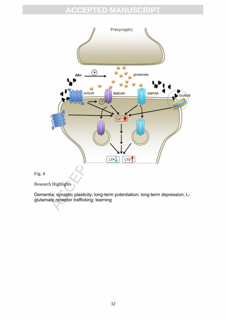
Figure 4. Overall roles of glutamate receptors in synaptic plasticity disruption.
Pathologically elevated A increases extracellular glutamate concentration whichβ
27

ACC
EPTE
D M
ANU
SCR
IPT
ACCEPTED MANUSCRIPT
activates synaptic and peri-/extrasynaptic glutamate receptors. This aberrant
activation of receptors causes an abnormal increase in intracellular Ca2+ and
internalization of both AMPA and NMDA receptors. As a result, these changes may
modify synaptic function by inhibiting LTP and facilitating LTD. Prolonged
pathological synaptic actions of Aß and tau would eventually cause synaptic silencing
and pruning. See text for references.
28

ACC
EPTE
D M
ANU
SCR
IPT
ACCEPTED MANUSCRIPT
Fig. 1
29

ACC
EPTE
D M
ANU
SCR
IPT
ACCEPTED MANUSCRIPT
Fig. 2
30

ACC
EPTE
D M
ANU
SCR
IPT
ACCEPTED MANUSCRIPT
Fig. 3
31
ACC
EPTE
D M
ANU
SCR
IPT
ACCEPTED MANUSCRIPT
Fig. 4
Research Highlights
Dementia; synaptic plasticity; long-term potentiation; long-term depression; L-glutamate receptor trafficking; learning
32