glucose6-phosphate dehydrogenase-derived nadph fuels superoxide production in the failing heart
TRANSCRIPT
Journal of Molecular and Cellular Cardiology 41 (2006) 340–349www.elsevier.com/locate/yjmcc
Original article
Glucose-6-phosphate dehydrogenase-derived NADPH fuels superoxideproduction in the failing heart
Sachin A. Gupte a,⁎, Robert J. Levine a, Rakhee S. Gupte a, Martin E. Young b,Vincenzo Lionetti a,c, Volodymyr Labinskyy a,c, Beverly C. Floyd a, Caroline Ojaimi a,
Michelle Bellomo a, Michael S. Wolin a, Fabio A. Recchia a,c
a Department of Physiology, BSB Room 626, New York Medical College, Valhalla, NY 10595, USAb USDA/ARS Children's Nutrition Research Center, Baylor College of Medicine, Houston, TX, USA
c Sector of Medicine, Scuola Superiore S. Anna, Pisa, Italy
Received 18 February 2006; received in revised form 1 May 2006; accepted 5 May 2006
Abstract
In the failing heart, NADPH oxidase and uncoupled NO synthase utilize cytosolic NADPH to form superoxide. NADPH is supplied principallyby the pentose phosphate pathway, whose rate-limiting enzyme is glucose 6-phosphate dehydrogenase (G6PD). Therefore, we hypothesized thatcardiac G6PD activation drives part of the excessive superoxide production implicated in the pathogenesis of heart failure. Pacing-induced heartfailure was performed in eight chronically instrumented dogs. Seven normal dogs served as control. End-stage failure occurred after 28 ± 1 daysof pacing, when left ventricular end-diastolic pressure reached 25 mm Hg. In left ventricular tissue homogenates, spontaneous superoxidegeneration measured by lucigenin (5 μM) chemiluminescence was markedly increased in heart failure (1338 ± 419 vs. 419 ± 102 AU/mg protein,P < 0.05), as were NADPH levels (15.4 ± 1.5 vs. 7.5 ± 1.5 μmol/gww, P < 0.05). Superoxide production was further stimulated by the addition ofNADPH. The NADPH oxidase inhibitor gp91ds-tat (50 μM) and the NO synthase inhibitor L-NAME (1 mM) both significantly lowered superoxidegeneration in failing heart homogenates by 80% and 76%, respectively. G6PD was upregulated and its activity higher in heart failure compared tocontrol (0.61 ± 0.10 vs. 0.24 ± 0.03 nmol/min/mg protein, P < 0.05), while superoxide production decreased to normal levels in the presence ofthe G6PD inhibitor 6-aminonicotinamide. We conclude that the activation of myocardial G6PD is a novel mechanism that enhances NADPHavailability and fuels superoxide-generating enzymes in heart failure.© 2006 Elsevier Inc. All rights reserved.
Keywords: Heart failure; NADPH oxidase; Glucose-6-phosphate; Pentose phosphate pathway
1. Introduction
Excessive production of reactive oxygen species (ROS) isimplicated in the pathogenesis of several cardiovasculardiseases, including heart failure [1]. A large number of clinicaland experimental studies have shown increased oxidative stressin the failing heart, with detrimental consequences that rangefrom endothelial dysfunction to contractile impairment andventricular remodeling [2]. The main sources of superoxideanion (O2
−), a major precursor of reactive oxygen species,
⁎ Corresponding author. Fax: +1 914 594 4018.E-mail address: [email protected] (S.A. Gupte).
0022-2828/$ − see front matter © 2006 Elsevier Inc. All rights reserved.doi:10.1016/j.yjmcc.2006.05.003
include the mitochondrial respiratory chain [3], xanthineoxidase [4], and NADPH oxidase [5]. The relative contributionof these enzymes to O2
− production in the failing heart is not welldefined, yet. Recent studies have suggested important roles forboth xanthine oxidase [6], and NADPH oxidase as sources ofcardiac O2
− in heart failure patients [7,8]. The latter enzymeutilizes both cytosolic NADPH or NADH as donors of reducingequivalents to transfer one electron to molecular oxygen,thereby producing O2
−. The increased activity of NADPHoxidase in response to mediators such as angiotensin II,norepinephrine, and TNFα is presently recognized as a chiefmechanism of vascular disease [9], but its potential impact onoxidative stress of the failing myocardium requires furtherelucidation. In addition, when constitutive NO synthase is
341S.A. Gupte et al. / Journal of Molecular and Cellular Cardiology 41 (2006) 340–349
uncoupled from essential cofactors, as found in the failing heart,it transfers electrons from NADPH to oxygen, thus becoming anadditional source of O2
− [10].The generation of O2
− from NADPH oxidase and uncoupledNO synthase depends not only on enzyme activation, but alsoon the availability of reducing equivalents. It is conceivable,therefore, that, in the presence of hyperactive NADPH oxidaseand uncoupled NO synthase, alterations of the metabolicpathways accounting for NAD+ and NADP+ reduction canpotentially affect the rate of O2
− production. Other investigatorsand we have previously reported profound alterations of fattyacid and carbohydrate metabolism in the failing heart [11]. Inparticular, myocardial glucose uptake and oxidation areincreased [12,13], although the glycolytic pathway and themitochondrial capacity to oxidize carbohydrates appear down-regulated, as opposed to upregulated [14,15]. A small fractionof total glucose in the cytosol can also be oxidized to ribose bythe pentose phosphate pathway. Moreover, it has beenpreviously shown by us and others that NADPH oxidasepreferentially utilizes NADPH derived from the pentosephosphate pathway [16,17]. In the light of these findings, wehypothesized that, in the failing heart, more glucose is oxidizedthrough the pentose phosphate pathway, with a consequentincrease in electron donors available to fuel O2
− generatingenzymes. This hypothesis was tested in myocardial tissueharvested from dogs with pacing-induced heart failure, anestablished model of dilated cardiomyopathy.
2. Methods
2.1. Surgical instrumentation and in vivo protocol
Fifteen dogs were chronically instrumented and heartfailure was induced in eight by pacing the left ventricle at210 beats/min for 3 weeks and at 240 beats/min thereafter. Theremaining seven dogs were used as a control. Hemodynamicsrecordings and two-dimensional and M-mode echocardiogra-phy were performed at baseline and during terminaldecompensation. Dogs were considered in end-stage heartfailure when left ventricular end-diastolic pressure reached25 mm Hg and clinical signs of severe decompensation wereobserved. At this point, the dogs were anesthetized with30 mg/kg i.v. of sodium pentobarbital to harvest and snap-freeze left ventricular tissue samples. These methods havebeen fully described by us [18] and the protocol was approvedby the Institutional Animal Care and Use Committee of theNew York Medical College.
2.2. Cardiac tissue
Left ventricular tissue was pulverized in liquid nitrogen andhomogenates were prepared in MOPS (50 mmol/l)-Sucrose(250 mmol/l) buffer at pH 7.4, as previously described [17,19].Only freshly prepared homogenates were used for biochemicalassays.
To identify the source of O2−, the homogenates were brought
to a final volume of 50 μl and pretreated for 30 min with: (1) the
xanthine oxidase inhibitors allopurinol (100 μmol/l) [20] andoxypurinol (100 μmol/l) [21]; (2) the cofactor tetrahydrobiop-terin (10 μmol/l) [22], and the inhibitor L-NAME (1 mmol/l)[10] to prevent O2
− generation from NO synthase; (3) thespecific NADPH oxidase inhibitor gp91ds-tat (50 μmol/l) [23];(4) 6-aminonicotinamide (5 mmol/l), an inhibitor of glucose-6-phosphate dehydrogenase [17,24]. Time-matched controlsamples were incubated for 30 minutes at 37 °C without addingany drug. Incubated samples (20 μl) were placed in plasticscintillation minivials containing 5 μM lucigenin for thedetection of O2
− in a final volume of 1 ml of air-equilibratedKrebs solution buffered with 10 mmol/l HEPES-NaOH (pH7.4), as previously described [17]. In a separate set ofexperiments, lucigenin chemiluminescence was measuredfrom normal and heart failure homogenate samples withoutand with NADPH (50-μmol/l). Lucigenin assays were done inthe presence and absence of O2
− scavenger, superoxidedismutase (SOD).
2.3. Hydrogen peroxide and peroxynitrite production
Hydrogen peroxide levels were measured by chemilumines-cence methods as previously described [25]. Briefly, hydrogenperoxide was estimated by incubating homogenate samples(10 μl) in luminol (10 μmol/l) and horseradish peroxidase(100 nmol/l) with and without catalase (200 U/ml), and luminolchemiluminescence was measured. To measure peroxynitriteproduction, tissue homogenate samples (10 μl) were incubatedin luminol (100 μmol/l) and luminol chemiluminesence wasused after a brief modification as previously described [26].Luminol assays were done in the presence and absence of H2O2
scavenger, catalase.
2.4. Glutathione concentration and glutathione reductaseactivity
Frozen samples were homogenized in 50 mmol/l potassiumphosphate buffer, pH 6–7, 1 mmol/l EDTA and then centrifugedat 10,000 × g for 15 min at 4 °C. The supernatant was used formeasurement of oxidized glutathione (GSSG) after deproteinat-ing the sample with equal amounts of 5 g/50 ml metaphosphoricacid. The samples were centrifuged at 2000 × g for at least2 min, and the supernatant was used for GSSG measurementafter neutralization with 4 mol/l triethanolamine. Afterseparation by centrifugation and neutralization, the sampleswere treated with 1 mol/l 2-vinylpyridine. GSSG concentrationwas measured using a commercially available assay kit(Cayman Chemical) [27]. Also, glutathione reductase activityin myocardial homogenates was determined by a commerciallyavailable kit (Cayman Chemical).
2.5. Superoxide dismutase activity
We measured superoxide dismutase (SOD) activity as anindicator of oxidative stress induced by O2
− [28]. Cardiac tissuehomogenates was assayed with a commercially available kit(Cayman Chemicals).
342 S.A. Gupte et al. / Journal of Molecular and Cellular Cardiology 41 (2006) 340–349
2.6. Lipid peroxidation
Malondialdehyde and 4-hydroxyalkenals, end products ofperoxidation of polyunsaturated fatty acids with related esters,were measured in cardiac tissue extract with a colorimetricmethod by employing a commercially available assay kit(Calbiochem) [18].
2.7. Oxidized and reduced pyridine nucleotide concentrations
NADPH was determined by HPLC using previouslydescribed methods [17]. Briefly, to measure NADPH, frozentissues were homogenized in an extraction medium containingNaOH (0.02 N) and cysteine (0.5 mmol/l) at 0 °C. The extractsthen were heated at 60 °C for 10 min and neutralized with 2 mlof 0.25 M glycylglycine buffer, pH 7.6. The neutralizedextracts were centrifuged at 10,000×g for 10 min, thesupernatants were passed through 0.45 μm Millipore filters,and the filtered solutions were used to measure NADPH byHPLC. NADPH were eluted from a reverse-phase HPLCcolumn (4.6× 250 mm; Supelco C18, Sigma) at roomtemperature using HP 1100 Series (Agilent Technologies,DE) with a buffer system consisting of 100 mmol/l potassiumphosphate (pH 6.0) (buffer A), and 100 mmol/l potassiumphosphate (pH 6.0) containing 5% methanol (buffer B). Thecolumn was eluted with 100% buffer A from 0 to 8.5 min, 80%buffer A plus 20% buffer B from 8.5 to 14.5 min, and 100%buffer B from 14.5 to 40 min. The flow rate was 1.0 ml/min,and the ultraviolet absorbance was monitored at 260 nm.NADPH standards were used to calibrate the HPLC. Internalstandards containing 2 nmol of NADPH, NADH, NADP+, andNAD+ were used to verify the quantitative recovery of theextraction procedure and HPLC retention time in the presenceand absence of tissue samples.
2.8. Glucose-6-phosphate dehydrogenase activity
The activity glucose-6-phosphate dehydrogenase (G6PD),the rate limiting enzyme in the pentose phosphate pathway,was measured in myocardial tissue homogenates by followingthe reduction of NADP+ to NADPH [17]. NADPH fluores-cence was detected at 340 nm (Ex) and 460 nm (Em) using aFlx800 microplate fluorescence detector, BioTek Instruments,Winooski, Vermont, USA.
Table 1Primer and probe sequences used in real time quantitative RT-PCR
Gene Primer/Probe Se
g6pd Forward 5′Reverse 5′Probe 5′
nox2 Forward 5′Reverse 5′Probe 5′
nox4 Forward 5′Reverse 5′Probe 5′
2.9. 6-Phosphogluconate concentration
6-Phosphogluconate, a product of the oxidative branch of thepentose phosphate pathway, was measured in myocardial tissueby the method of Kauffman et al. [29]. The reduction of NADP+
(0.02 mmol/l) to NADPH was catalyzed by 6-phosphogluco-nate dehydrogenase (0.5 μg/ml) at 37 °C for 20 min in Tris–HCl(0.05 mol/l) buffer (pH 8.0) containing EDTA (0.1 mmol/l),dithiothreitol (0.1 mmol/l), and ammonium acetate (0.03 mol/l).NADPH concentration was then detected fluorometrically aspreviously described by us [17].
2.10. Real-time polymerase chain reaction
The expression of selected genes was quantitated by real-time quantitative polymerase chain reaction (PCR) as describedpreviously [18]. Analysis was focused on G6PD and theNADPH subunits Nox-2 and Nox-4, while the traditionalhousekeeping gene ribosomal 18 S was used for normalization.We chose to measure Nox-4 and Nox-2 expression, since boththese oxidases are expressed in vascular and myocardial tissue[7,17]. Specific quantitative assays were designed from dogsequences available in GenBank, except in the cases of Nox-2and Nox-4 for which the dog sequences were not available. Incontrast, these genes have been cloned for the human, mouse,and rat. Multiple assays were then designed using the humansequence for these genes, in a region that was highly conservedbetween the three species. These human-specific assays weresubsequently tested for compatibility in the dog. Primers andprobes were designed to be isoform specific, spanning siteswhere two exons join (splice sites) when such sites are known(preventing recognition of the assay to any potential contam-inating genomic DNA). Sequences of these primers and probesare given in Table 1. Standard RNAwas made for all assays bythe T7 polymerase method (Ambion), using total RNA isolatedfrom the dog heart. Transcript levels are expressed as the numberof molecules of mRNA per nanogram of total RNA (asmeasured by UV spectrophotometry).
2.11. Western immunoblot analysis
Protein was extracted from frozen tissue as previouslydescribed [17] and its concentration measured spectrophoto-metrically with a colorimetric assay (Bio-Rad DC reagent).
quence
-GACCACTACCTGGGCAAGG-3′-CGATGTTGTCTCGGTTCCA-3′-FAM-ATCCTGTTGGCAAACCTCAGCACCAT-TAMRA-3′-AGCCAGATGCAGGAAAGGA-3′-TCATCATGGTGCACAGCAAA-3′-FAM-ATTGGCCTGAGACTCATCCCAGCCA-TAMRA-3′-CGTCCTCGGTGGAAACTTTT-3′-GTGAATTGGGTCCACAACAGA-3′-FAM-CACCAACTGTTTTTCCTCTGTTATATTTTGCTATTTCA-TAMRA-3′
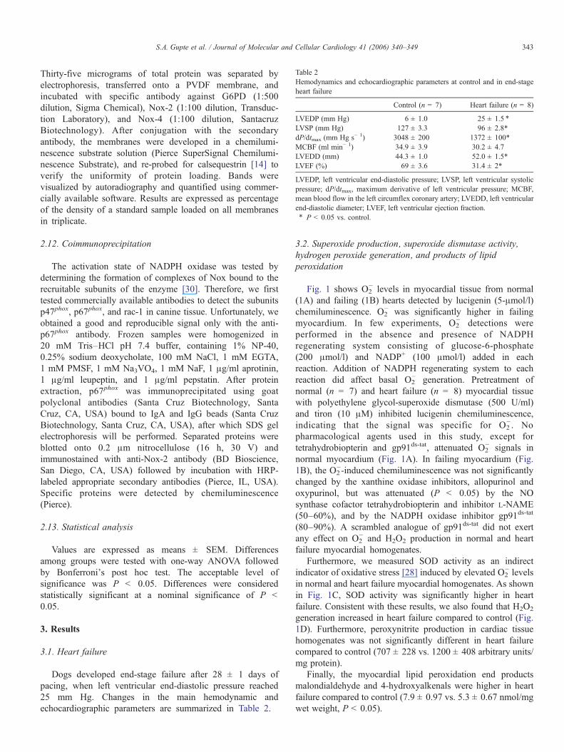
Table 2Hemodynamics and echocardiographic parameters at control and in end-stageheart failure
Control (n = 7) Heart failure (n = 8)
LVEDP (mm Hg) 6 ± 1.0 25 ± 1.5 ⁎
LVSP (mm Hg) 127 ± 3.3 96 ± 2.8*dP/dtmax (mm Hg s− 1) 3048 ± 200 1372 ± 100*MCBF (ml min− 1) 34.9 ± 3.9 30.2 ± 4.7LVEDD (mm) 44.3 ± 1.0 52.0 ± 1.5*LVEF (%) 69 ± 3.6 31.4 ± 2*
LVEDP, left ventricular end-diastolic pressure; LVSP, left ventricular systolicpressure; dP/dtmax, maximum derivative of left ventricular pressure; MCBF,mean blood flow in the left circumflex coronary artery; LVEDD, left ventricularend-diastolic diameter; LVEF, left ventricular ejection fraction.⁎ P < 0.05 vs. control.
343S.A. Gupte et al. / Journal of Molecular and Cellular Cardiology 41 (2006) 340–349
Thirty-five micrograms of total protein was separated byelectrophoresis, transferred onto a PVDF membrane, andincubated with specific antibody against G6PD (1:500dilution, Sigma Chemical), Nox-2 (1:100 dilution, Transduc-tion Laboratory), and Nox-4 (1:100 dilution, SantacruzBiotechnology). After conjugation with the secondaryantibody, the membranes were developed in a chemilumi-nescence substrate solution (Pierce SuperSignal Chemilumi-nescence Substrate), and re-probed for calsequestrin [14] toverify the uniformity of protein loading. Bands werevisualized by autoradiography and quantified using commer-cially available software. Results are expressed as percentageof the density of a standard sample loaded on all membranesin triplicate.
2.12. Coimmunoprecipitation
The activation state of NADPH oxidase was tested bydetermining the formation of complexes of Nox bound to therecruitable subunits of the enzyme [30]. Therefore, we firsttested commercially available antibodies to detect the subunitsp47phox, p67phox, and rac-1 in canine tissue. Unfortunately, weobtained a good and reproducible signal only with the anti-p67phox antibody. Frozen samples were homogenized in20 mM Tris–HCl pH 7.4 buffer, containing 1% NP-40,0.25% sodium deoxycholate, 100 mM NaCl, 1 mM EGTA,1 mM PMSF, 1 mM Na3VO4, 1 mM NaF, 1 μg/ml aprotinin,1 μg/ml leupeptin, and 1 μg/ml pepstatin. After proteinextraction, p67phox was immunoprecipitated using goatpolyclonal antibodies (Santa Cruz Biotechnology, SantaCruz, CA, USA) bound to IgA and IgG beads (Santa CruzBiotechnology, Santa Cruz, CA, USA), after which SDS gelelectrophoresis will be performed. Separated proteins wereblotted onto 0.2 μm nitrocellulose (16 h, 30 V) andimmunostained with anti-Nox-2 antibody (BD Bioscience,San Diego, CA, USA) followed by incubation with HRP-labeled appropriate secondary antibodies (Pierce, IL, USA).Specific proteins were detected by chemiluminescence(Pierce).
2.13. Statistical analysis
Values are expressed as means ± SEM. Differencesamong groups were tested with one-way ANOVA followedby Bonferroni's post hoc test. The acceptable level ofsignificance was P < 0.05. Differences were consideredstatistically significant at a nominal significance of P <0.05.
3. Results
3.1. Heart failure
Dogs developed end-stage failure after 28 ± 1 days ofpacing, when left ventricular end-diastolic pressure reached25 mm Hg. Changes in the main hemodynamic andechocardiographic parameters are summarized in Table 2.
3.2. Superoxide production, superoxide dismutase activity,hydrogen peroxide generation, and products of lipidperoxidation
Fig. 1 shows O2− levels in myocardial tissue from normal
(1A) and failing (1B) hearts detected by lucigenin (5-μmol/l)chemiluminescence. O2
− was significantly higher in failingmyocardium. In few experiments, O2
− detections wereperformed in the absence and presence of NADPHregenerating system consisting of glucose-6-phosphate(200 μmol/l) and NADP+ (100 μmol/l) added in eachreaction. Addition of NADPH regenerating system to eachreaction did affect basal O2
− generation. Pretreatment ofnormal (n = 7) and heart failure (n = 8) myocardial tissuewith polyethylene glycol-superoxide dismutase (500 U/ml)and tiron (10 μM) inhibited lucigenin chemiluminescence,indicating that the signal was specific for O2
−. Nopharmacological agents used in this study, except fortetrahydrobiopterin and gp91ds-tat, attenuated O2
− signals innormal myocardium (Fig. 1A). In failing myocardium (Fig.1B), the O2
−-induced chemiluminescence was not significantlychanged by the xanthine oxidase inhibitors, allopurinol andoxypurinol, but was attenuated (P < 0.05) by the NOsynthase cofactor tetrahydrobiopterin and inhibitor L-NAME(50–60%), and by the NADPH oxidase inhibitor gp91ds-tat
(80–90%). A scrambled analogue of gp91ds-tat did not exertany effect on O2
− and H2O2 production in normal and heartfailure myocardial homogenates.
Furthermore, we measured SOD activity as an indirectindicator of oxidative stress [28] induced by elevated O2
− levelsin normal and heart failure myocardial homogenates. As shownin Fig. 1C, SOD activity was significantly higher in heartfailure. Consistent with these results, we also found that H2O2
generation increased in heart failure compared to control (Fig.1D). Furthermore, peroxynitrite production in cardiac tissuehomogenates was not significantly different in heart failurecompared to control (707 ± 228 vs. 1200 ± 408 arbitrary units/mg protein).
Finally, the myocardial lipid peroxidation end productsmalondialdehyde and 4-hydroxyalkenals were higher in heartfailure compared to control (7.9 ± 0.97 vs. 5.3 ± 0.67 nmol/mgwet weight, P < 0.05).
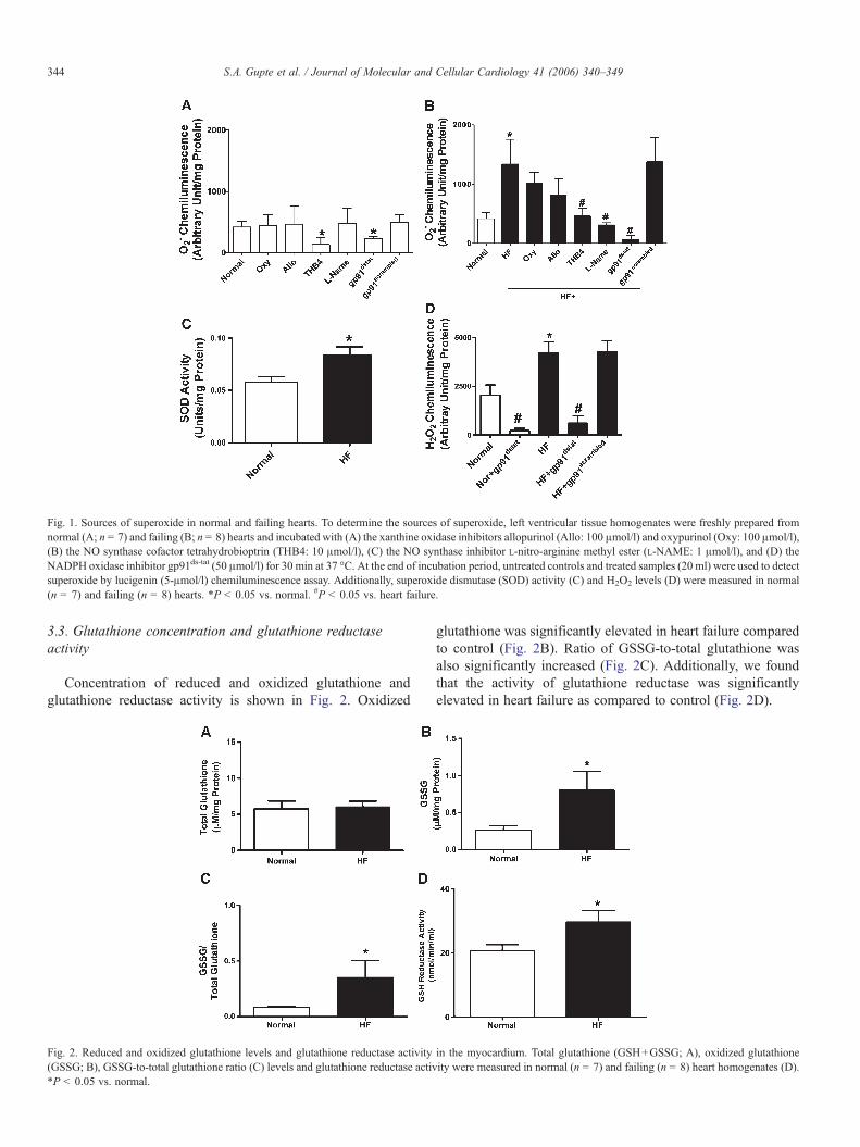
Fig. 1. Sources of superoxide in normal and failing hearts. To determine the sources of superoxide, left ventricular tissue homogenates were freshly prepared fromnormal (A; n = 7) and failing (B; n = 8) hearts and incubated with (A) the xanthine oxidase inhibitors allopurinol (Allo: 100 μmol/l) and oxypurinol (Oxy: 100 μmol/l),(B) the NO synthase cofactor tetrahydrobioptrin (THB4: 10 μmol/l), (C) the NO synthase inhibitor L-nitro-arginine methyl ester (L-NAME: 1 μmol/l), and (D) theNADPH oxidase inhibitor gp91ds-tat (50 μmol/l) for 30 min at 37 °C. At the end of incubation period, untreated controls and treated samples (20 ml) were used to detectsuperoxide by lucigenin (5-μmol/l) chemiluminescence assay. Additionally, superoxide dismutase (SOD) activity (C) and H2O2 levels (D) were measured in normal(n = 7) and failing (n = 8) hearts. *P < 0.05 vs. normal. #P < 0.05 vs. heart failure.
344 S.A. Gupte et al. / Journal of Molecular and Cellular Cardiology 41 (2006) 340–349
3.3. Glutathione concentration and glutathione reductaseactivity
Concentration of reduced and oxidized glutathione andglutathione reductase activity is shown in Fig. 2. Oxidized
Fig. 2. Reduced and oxidized glutathione levels and glutathione reductase activity(GSSG; B), GSSG-to-total glutathione ratio (C) levels and glutathione reductase activ*P < 0.05 vs. normal.
glutathione was significantly elevated in heart failure comparedto control (Fig. 2B). Ratio of GSSG-to-total glutathione wasalso significantly increased (Fig. 2C). Additionally, we foundthat the activity of glutathione reductase was significantlyelevated in heart failure as compared to control (Fig. 2D).
in the myocardium. Total glutathione (GSH+GSSG; A), oxidized glutathioneity were measured in normal (n = 7) and failing (n = 8) heart homogenates (D).
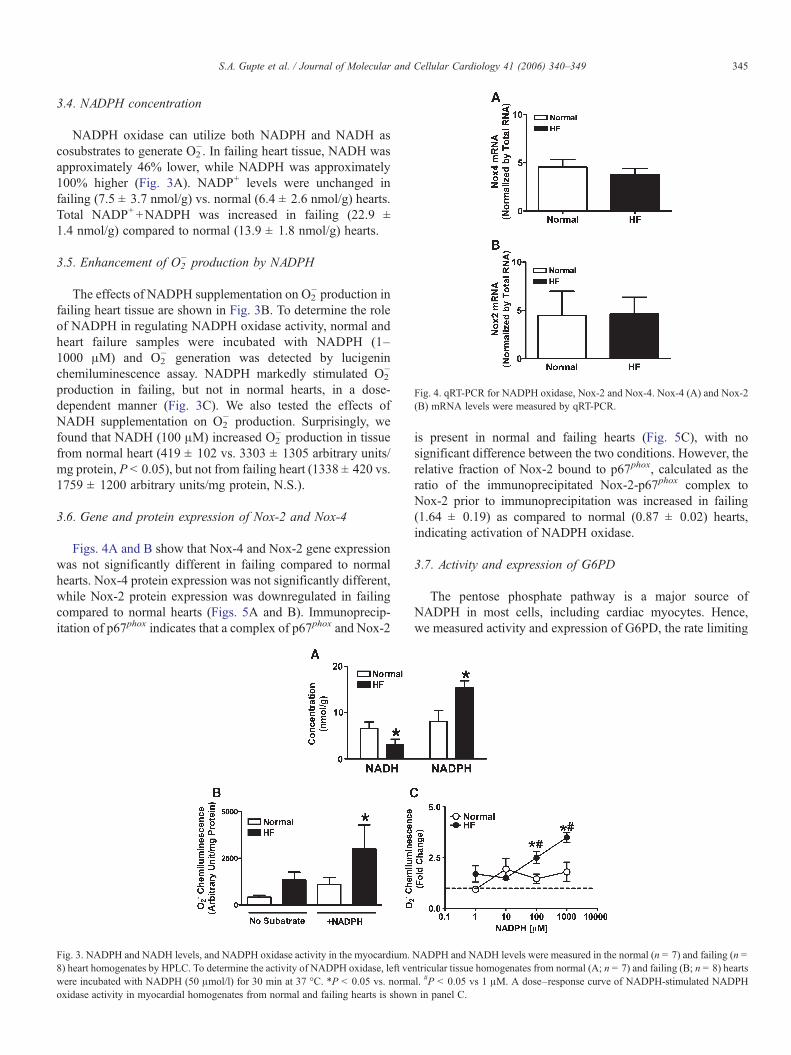
Fig. 4. qRT-PCR for NADPH oxidase, Nox-2 and Nox-4. Nox-4 (A) and Nox-2(B) mRNA levels were measured by qRT-PCR.
345S.A. Gupte et al. / Journal of Molecular and Cellular Cardiology 41 (2006) 340–349
3.4. NADPH concentration
NADPH oxidase can utilize both NADPH and NADH ascosubstrates to generate O2
−. In failing heart tissue, NADH wasapproximately 46% lower, while NADPH was approximately100% higher (Fig. 3A). NADP+ levels were unchanged infailing (7.5 ± 3.7 nmol/g) vs. normal (6.4 ± 2.6 nmol/g) hearts.Total NADP++NADPH was increased in failing (22.9 ±1.4 nmol/g) compared to normal (13.9 ± 1.8 nmol/g) hearts.
3.5. Enhancement of O2− production by NADPH
The effects of NADPH supplementation on O2− production in
failing heart tissue are shown in Fig. 3B. To determine the roleof NADPH in regulating NADPH oxidase activity, normal andheart failure samples were incubated with NADPH (1–1000 μM) and O2
− generation was detected by lucigeninchemiluminescence assay. NADPH markedly stimulated O2
−
production in failing, but not in normal hearts, in a dose-dependent manner (Fig. 3C). We also tested the effects ofNADH supplementation on O2
− production. Surprisingly, wefound that NADH (100 μM) increased O2
− production in tissuefrom normal heart (419 ± 102 vs. 3303 ± 1305 arbitrary units/mg protein, P < 0.05), but not from failing heart (1338 ± 420 vs.1759 ± 1200 arbitrary units/mg protein, N.S.).
3.6. Gene and protein expression of Nox-2 and Nox-4
Figs. 4A and B show that Nox-4 and Nox-2 gene expressionwas not significantly different in failing compared to normalhearts. Nox-4 protein expression was not significantly different,while Nox-2 protein expression was downregulated in failingcompared to normal hearts (Figs. 5A and B). Immunoprecip-itation of p67phox indicates that a complex of p67phox and Nox-2
Fig. 3. NADPH and NADH levels, and NADPH oxidase activity in the myocardium.8) heart homogenates by HPLC. To determine the activity of NADPH oxidase, left vewere incubated with NADPH (50 μmol/l) for 30 min at 37 °C. *P < 0.05 vs. normaoxidase activity in myocardial homogenates from normal and failing hearts is show
is present in normal and failing hearts (Fig. 5C), with nosignificant difference between the two conditions. However, therelative fraction of Nox-2 bound to p67phox, calculated as theratio of the immunoprecipitated Nox-2-p67phox complex toNox-2 prior to immunoprecipitation was increased in failing(1.64 ± 0.19) as compared to normal (0.87 ± 0.02) hearts,indicating activation of NADPH oxidase.
3.7. Activity and expression of G6PD
The pentose phosphate pathway is a major source ofNADPH in most cells, including cardiac myocytes. Hence,we measured activity and expression of G6PD, the rate limiting
NADPH and NADH levels were measured in the normal (n = 7) and failing (n =ntricular tissue homogenates from normal (A; n = 7) and failing (B; n = 8) heartsl. #P < 0.05 vs 1 μM. A dose–response curve of NADPH-stimulated NADPHn in panel C.
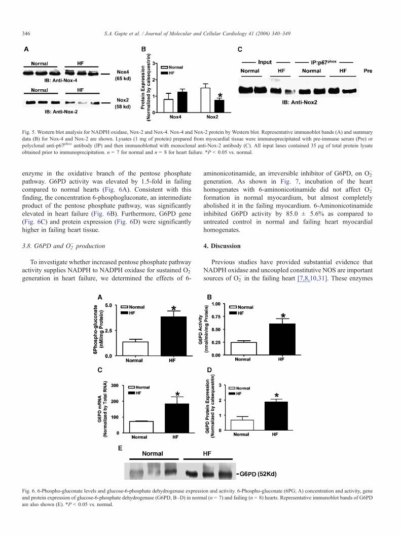
Fig. 5. Western blot analysis for NADPH oxidase, Nox-2 and Nox-4. Nox-4 and Nox-2 protein by Western blot. Representative immunoblot bands (A) and summarydata (B) for Nox-4 and Nox-2 are shown. Lysates (1 mg of protein) prepared from myocardial tissue were immunoprecipitated with pre-immune serum (Pre) orpolyclonal anti-p67phox antibody (IP) and then immunoblotted with monoclonal anti-Nox-2 antibody (C). All input lanes contained 35 μg of total protein lysateobtained prior to immunoprecipitation. n = 7 for normal and n = 8 for heart failure. *P < 0.05 vs. normal.
346 S.A. Gupte et al. / Journal of Molecular and Cellular Cardiology 41 (2006) 340–349
enzyme in the oxidative branch of the pentose phosphatepathway. G6PD activity was elevated by 1.5-fold in failingcompared to normal hearts (Fig. 6A). Consistent with thisfinding, the concentration 6-phosphogluconate, an intermediateproduct of the pentose phosphate pathway, was significantlyelevated in heart failure (Fig. 6B). Furthermore, G6PD gene(Fig. 6C) and protein expression (Fig. 6D) were significantlyhigher in failing heart tissue.
3.8. G6PD and O2− production
To investigate whether increased pentose phosphate pathwayactivity supplies NADPH to NADPH oxidase for sustained O2
−
generation in heart failure, we determined the effects of 6-
Fig. 6. 6-Phospho-gluconate levels and glucose-6-phosphate dehydrogenase expressiand protein expression of glucose-6-phosphate dehydrogenase (G6PD, B–D) in normare also shown (E). *P < 0.05 vs. normal.
aminonicotinamide, an irreversible inhibitor of G6PD, on O2−
generation. As shown in Fig. 7, incubation of the hearthomogenates with 6-aminonicotinamide did not affect O2
−
formation in normal myocardium, but almost completelyabolished it in the failing myocardium. 6-Aminonicotinamideinhibited G6PD activity by 85.0 ± 5.6% as compared tountreated control in normal and failing heart myocardialhomogenates.
4. Discussion
Previous studies have provided substantial evidence thatNADPH oxidase and uncoupled constitutive NOS are importantsources of O2
− in the failing heart [7,8,10,31]. These enzymes
on and activity. 6-Phospho-gluconate (6PG; A) concentration and activity, geneal (n = 7) and failing (n = 8) hearts. Representative immunoblot bands of G6PD
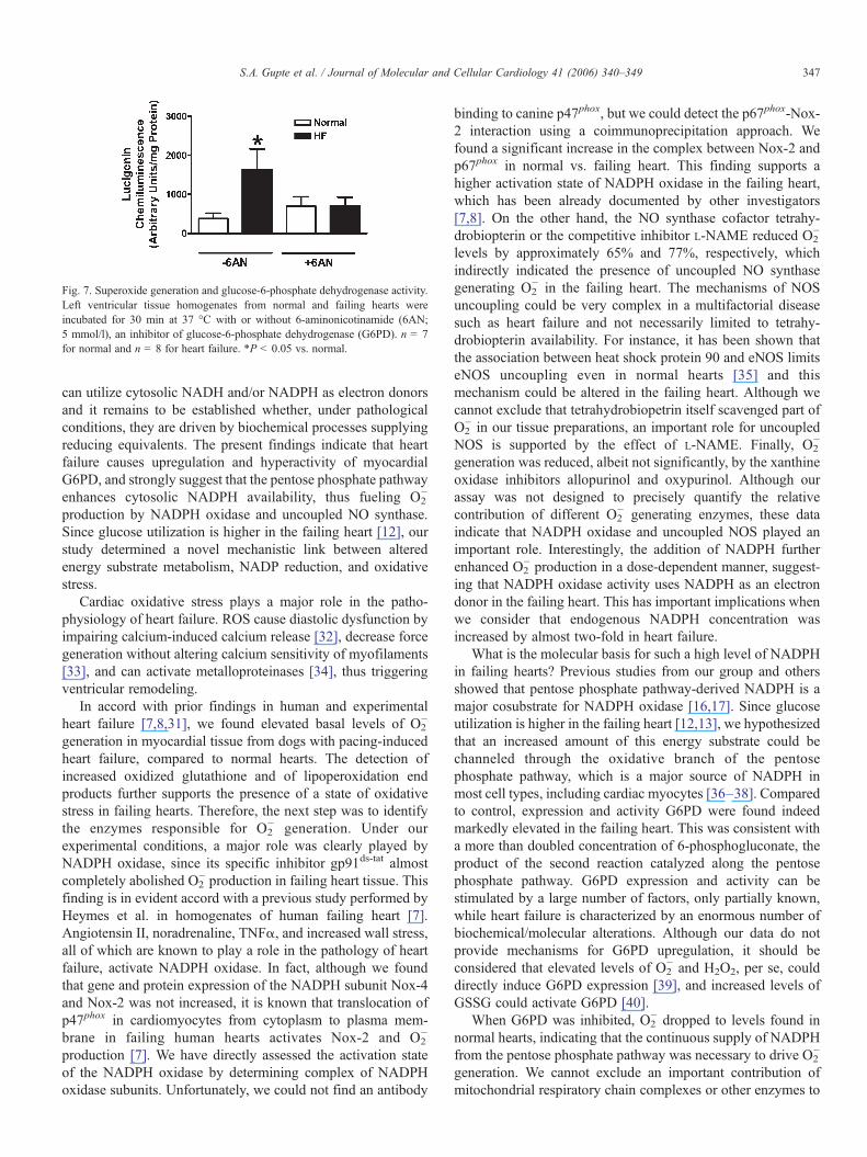
Fig. 7. Superoxide generation and glucose-6-phosphate dehydrogenase activity.Left ventricular tissue homogenates from normal and failing hearts wereincubated for 30 min at 37 °C with or without 6-aminonicotinamide (6AN;5 mmol/l), an inhibitor of glucose-6-phosphate dehydrogenase (G6PD). n = 7for normal and n = 8 for heart failure. *P < 0.05 vs. normal.
347S.A. Gupte et al. / Journal of Molecular and Cellular Cardiology 41 (2006) 340–349
can utilize cytosolic NADH and/or NADPH as electron donorsand it remains to be established whether, under pathologicalconditions, they are driven by biochemical processes supplyingreducing equivalents. The present findings indicate that heartfailure causes upregulation and hyperactivity of myocardialG6PD, and strongly suggest that the pentose phosphate pathwayenhances cytosolic NADPH availability, thus fueling O2
−
production by NADPH oxidase and uncoupled NO synthase.Since glucose utilization is higher in the failing heart [12], ourstudy determined a novel mechanistic link between alteredenergy substrate metabolism, NADP reduction, and oxidativestress.
Cardiac oxidative stress plays a major role in the patho-physiology of heart failure. ROS cause diastolic dysfunction byimpairing calcium-induced calcium release [32], decrease forcegeneration without altering calcium sensitivity of myofilaments[33], and can activate metalloproteinases [34], thus triggeringventricular remodeling.
In accord with prior findings in human and experimentalheart failure [7,8,31], we found elevated basal levels of O2
−
generation in myocardial tissue from dogs with pacing-inducedheart failure, compared to normal hearts. The detection ofincreased oxidized glutathione and of lipoperoxidation endproducts further supports the presence of a state of oxidativestress in failing hearts. Therefore, the next step was to identifythe enzymes responsible for O2
− generation. Under ourexperimental conditions, a major role was clearly played byNADPH oxidase, since its specific inhibitor gp91ds-tat almostcompletely abolished O2
− production in failing heart tissue. Thisfinding is in evident accord with a previous study performed byHeymes et al. in homogenates of human failing heart [7].Angiotensin II, noradrenaline, TNFα, and increased wall stress,all of which are known to play a role in the pathology of heartfailure, activate NADPH oxidase. In fact, although we foundthat gene and protein expression of the NADPH subunit Nox-4and Nox-2 was not increased, it is known that translocation ofp47phox in cardiomyocytes from cytoplasm to plasma mem-brane in failing human hearts activates Nox-2 and O2
−
production [7]. We have directly assessed the activation stateof the NADPH oxidase by determining complex of NADPHoxidase subunits. Unfortunately, we could not find an antibody
binding to canine p47phox, but we could detect the p67phox-Nox-2 interaction using a coimmunoprecipitation approach. Wefound a significant increase in the complex between Nox-2 andp67phox in normal vs. failing heart. This finding supports ahigher activation state of NADPH oxidase in the failing heart,which has been already documented by other investigators[7,8]. On the other hand, the NO synthase cofactor tetrahy-drobiopterin or the competitive inhibitor L-NAME reduced O2
−
levels by approximately 65% and 77%, respectively, whichindirectly indicated the presence of uncoupled NO synthasegenerating O2
− in the failing heart. The mechanisms of NOSuncoupling could be very complex in a multifactorial diseasesuch as heart failure and not necessarily limited to tetrahy-drobiopterin availability. For instance, it has been shown thatthe association between heat shock protein 90 and eNOS limitseNOS uncoupling even in normal hearts [35] and thismechanism could be altered in the failing heart. Although wecannot exclude that tetrahydrobiopetrin itself scavenged part ofO2− in our tissue preparations, an important role for uncoupled
NOS is supported by the effect of L-NAME. Finally, O2−
generation was reduced, albeit not significantly, by the xanthineoxidase inhibitors allopurinol and oxypurinol. Although ourassay was not designed to precisely quantify the relativecontribution of different O2
− generating enzymes, these dataindicate that NADPH oxidase and uncoupled NOS played animportant role. Interestingly, the addition of NADPH furtherenhanced O2
− production in a dose-dependent manner, suggest-ing that NADPH oxidase activity uses NADPH as an electrondonor in the failing heart. This has important implications whenwe consider that endogenous NADPH concentration wasincreased by almost two-fold in heart failure.
What is the molecular basis for such a high level of NADPHin failing hearts? Previous studies from our group and othersshowed that pentose phosphate pathway-derived NADPH is amajor cosubstrate for NADPH oxidase [16,17]. Since glucoseutilization is higher in the failing heart [12,13], we hypothesizedthat an increased amount of this energy substrate could bechanneled through the oxidative branch of the pentosephosphate pathway, which is a major source of NADPH inmost cell types, including cardiac myocytes [36–38]. Comparedto control, expression and activity G6PD were found indeedmarkedly elevated in the failing heart. This was consistent witha more than doubled concentration of 6-phosphogluconate, theproduct of the second reaction catalyzed along the pentosephosphate pathway. G6PD expression and activity can bestimulated by a large number of factors, only partially known,while heart failure is characterized by an enormous number ofbiochemical/molecular alterations. Although our data do notprovide mechanisms for G6PD upregulation, it should beconsidered that elevated levels of O2
− and H2O2, per se, coulddirectly induce G6PD expression [39], and increased levels ofGSSG could activate G6PD [40].
When G6PD was inhibited, O2− dropped to levels found in
normal hearts, indicating that the continuous supply of NADPHfrom the pentose phosphate pathway was necessary to drive O2
−
generation. We cannot exclude an important contribution ofmitochondrial respiratory chain complexes or other enzymes to
348 S.A. Gupte et al. / Journal of Molecular and Cellular Cardiology 41 (2006) 340–349
the generation of O2− in vivo; however, taken together, our data
provide compelling evidence that failing heart tissue maintainselevated levels of NADPH and has the molecular machinerycapable of enhancing glucose flux through the pentosephosphate pathway, thus supplying large amounts of NADPHthat drive O2
− production. Interestingly, the increased availabil-ity of NADPH in failing hearts appeared to have a moredominant effect on promoting superoxide production comparedto its antioxidant metabolic effect of maintaining high levels ofreduced glutathione. A possible explanation is that, while innormal hearts G6PD-derived NADPH is critical to preservecytosolic antioxidant redox systems such as GSH, [37,38] in thepresence of pathologically enhanced superoxide-generatingsystems sharing the same type of electron donor, the NADPHoxidase has a greater affinity for the available electron donor.Consistent with this interpretation, other authors have recentlyreported that, in a pathological condition induced by angioten-sin infusion, G6PD deficiency limits the deleterious effects ofvascular superoxide production possibly by reducing theavailability of the substrate for NADPH oxidase [16].
In severe heart failure, cardiac oxidation of free fatty acid isimpaired, leading to augmented uptake and oxidation of thealternative substrate glucose. However, other authors and uspreviously found molecular alterations suggestive of a de-pressed, rather than increased, glycolytic pathway [14,15]. Thisis part of the global mitochondrial impairment that characterizesheart failure [11]. It is possible, therefore, that a larger fractionof glucose enters the upregulated pentose phosphate pathway.
In conclusion, our study provides evidence for elevatedNADPH in the failing heart due to G6PD upregulation. Thisbiochemical alteration drives superoxide production byNADPH oxidase and uncoupled NO synthase. Thus, the fluxof glucose metabolism through the pentose phosphate pathwaymay be playing an important role in fueling the oxidative stressseen in heart failure.
Acknowledgments
This study was supported by an AHA Scientist Developmentgrant (S.A. Gupte), and by HL-31069 and HL-66331 (MSW),and P01 HL-74237 (F.A. Recchia). Authors would like to thankDr. Pagano, Henry Ford Hospital, Detroit, MI, for kindlyproviding NADPH oxidase inhibitor gp91ds-tat.
References
[1] Li JM, Shah AM. Endothelial cell superoxide generation: regulation andrelevance for cardiovascular pathophysiology. Am J Physiol, Regul IntegrComp Physiol 2004;287:R1014–30.
[2] Dhalla NS, Temsah RM, Netticadan T. Role of oxidative stress incardiovascular diseases. J Hypertens 2000;18:655–73.
[3] Ballinger SW. Mitochondrial dysfunction in cardiovascular disease. FreeRadic Biol Med 2005;38:1278–95.
[4] Berry CE, Hare JM. Xanthine oxidoreductase and cardiovascular disease:molecular mechanisms and pathophysiological implications. J Physiol2004;555:589–606.
[5] Griendling KK, Sorescu D, Ushio-Fukai M. NAD(P)H oxidase: role incardiovascular biology and disease. Circ Res 2000;86:494–501.
[6] Cappola TP, Kass DA, Nelson GS, Berger RD, Rosas GO, Kobeissi ZA, et
al. Allopurinol improves myocardial efficiency in patients with idiopathicdilated cardiomyopathy. Circulation 2001;104:2407–11.
[7] Heymes C, Bendall JK, Ratajczak P, Cave AC, Samuel JL, Hasenfuss G,et al. Increased myocardial NADPH oxidase activity in human heartfailure. J Am Coll Cardiol 2003;41:2164–71.
[8] Maack C, Kartes T, Kilter H, Schafers HJ, Nickenig G, Bohm M, et al.Oxygen free radical release in human failing myocardium is associatedwith increased activity of rac1-GTPase and represents a target for statintreatment. Circulation 2003;108:1567–74.
[9] Wolin MS, Gupte SA, Oeckler RA. Superoxide in the vascular system.J Vasc Res 2002;39:191–207.
[10] Dixon LJ, Morgan DR, Hughes SM, McGrath LT, El-Sherbeeny NA,Plumb RD, et al. Functional consequences of endothelial nitric oxidesynthase uncoupling in congestive cardiac failure. Circulation 2003;107:1725–8.
[11] Stanley WC, Recchia FA, Lopaschuk GD. Myocardial substrate metabo-lism in the normal and failing heart. Physiol Rev 2005;85:1093–129.
[12] Osorio JC, Stanley WC, Linke A, Castellari M, Diep QN, Panchal AR, etal. Impaired myocardial fatty acid oxidation and reduced proteinexpression of retinoid X receptor-alpha in pacing-induced heart failure.Circulation 2002;106:606–12.
[13] Davila-Roman VG, Vedala G, Herrero P, de las Fuentes L, Rogers JG,Kelly DP, et al. Altered myocardial fatty acid and glucose metabolism inidiopathic dilated cardiomyopathy. J Am Coll Cardiol 2002;40:271–7.
[14] Lei B, Lionetti V, Young ME, Chandler MP, d'Agostino C, Kang E, et al.Paradoxical downregulation of the glucose oxidation pathway despiteenhanced flux in severe heart failure. J Mol Cell Cardiol 2004;36:567–76.
[15] Razeghi P, Young ME, Alcorn JL, Moravec CS, Frazier OH, TaegtmeyerH. Metabolic gene expression in fetal and failing human heart. Circulation2001;104:2923–31.
[16] Matsui R, Xu S, Maitland KA, Hayes A, Leopold JA, Handy DE, et al.Glucose-6 phosphate dehydrogenase deficiency decreases the vascularresponse to angiotensin II. Circulation 2005;112:257–63.
[17] Gupte SA, Kaminski PM, Floyd B, Agarwal R, Ali N, Ahmad M, et al.Cytosolic NADPH may regulate differences in basal Nox oxidase-derivedsuperoxide generation in bovine coronary and pulmonary arteries. Am JPhysiol, Heart Circ Physiol 2005;288:H13–21.
[18] Lionetti V, Linke A, Chandler MP, Young ME, Penn MS, Gupte S, et al.Carnitine palmitoyl transferase-I inhibition prevents ventricular remodel-ing and delays decompensation in pacing-induced heart failure. CardiovascRes 2005;66:454–61.
[19] Mohazzab KM, Wolin MS. Sites of superoxide anion production detectedby lucigenin in calf pulmonary artery smooth muscle. Am J Physiol1994;267:L815–22.
[20] Ungvari Z, Csiszar A, Edwards JG, Kaminski PM, Wolin MS, Kaley G, etal. Increased superoxide production in coronary arteries in hyperhomo-cysteinemia: role of tumor necrosis factor-alpha, NAD(P)H oxidase, andinducible nitric oxide synthase. Arterioscler Thromb Vasc Biol2003;23:418–24.
[21] Kinugawa S, Huang H, Wang Z, Kaminski PM, Wolin MS, Hintze TH. Adefect of neuronal nitric oxide synthase increases xanthine oxidase-derivedsuperoxide anion and attenuates the control of myocardial oxygenconsumption by nitric oxide derived from endothelial nitric oxidesynthase. Circ Res 2005;96:355–62.
[22] Chalupsky K, Cai H. Endothelial dihydrofolate reductase: critical for nitricoxide bioavailability and role in angiotensin II uncoupling of endothelialnitric oxide synthase. Proc Natl Acad Sci U S A 2005;102:9056–61.
[23] Rey FE, Cifuentes ME, Kiarash A, Quinn MT, Pagano PJ. Novelcompetitive inhibitor of NAD(P)H oxidase assembly attenuates vascular O(2)(−) and systolic blood pressure in mice. Circ Res 2001;89:408–14.
[24] Kohler E, Barrach H, Neubert D. Inhibition of NADP dependentoxidoreductases by the 6-aminonicotinamide analogue of NADP. FEBSLett 1970;6:225–8.
[25] Mohazzab-H KM, Agarwal R, Wolin MS. Influence of glutathioneperoxidase on coronary artery responses to alterations in PO2 and H2O2.Am J Physiol 1999;276:H235–41.
[26] Radi R, Cosgrove TP, Beckman JS, Freeman BA. Peroxynitrite-inducedluminol chemiluminescence. J Biochem 1993;290(Pt 1):51–7.
349S.A. Gupte et al. / Journal of Molecular and Cellular Cardiology 41 (2006) 340–349
[27] Gupte SA, Arshad M, Viola S, Kaminski PM, Ungvari Z, Rabbani G, et al.Pentose phosphate pathway coordinates multiple redox-controlled relaxingmechanisms in bovine coronary arteries. Am J Physiol, Heart Circ Physiol2003;285:H2316–26.
[28] Blum J, Fridovich I. Superoxide, hydrogen peroxide, and oxygen toxicityin two free-living nematode species. Arch Biochem Biophys 1983;222:35–43.
[29] Kauffman FC, Brown JG, Passonneau JV, Lowry OH. Effects of changesin brain metabolism on levels of pentose phosphate pathway intermediates.J Biol Chem 1969;244:3647–53.
[30] Gupte RS, Weng Y, Liu L, Lee MY. The second subunit of the replicationfactor C complex (RFC40) and the regulatory subunit (RIalpha) of proteinkinase A form a protein complex promoting cell survival. Cell Cycle2005;4:323–9.
[31] Mollnau H, Oelze M, August M, Wendt M, Daiber A, Schulz E, et al.Mechanisms of increased vascular superoxide production in an experi-mental model of idiopathic dilated cardiomyopathy. Arterioscler ThrombVasc Biol 2005.
[32] Gao WD, Liu Y, Marban E. Selective effects of oxygen free radicals onexcitation–contraction coupling in ventricular muscle. Implications for themechanism of stunned myocardium. Circulation 1996;94:2597–604.
[33] MacFarlane NG, Miller DJ. Depression of peak force without alteringcalcium sensitivity by the superoxide anion in chemically skinned cardiacmuscle of rat. Circ Res 1992;70:1217–24.
[34] Siwik DA, Pagano PJ, Colucci WS. Oxidative stress regulates collagensynthesis and matrix metalloproteinase activity in cardiac fibroblasts. Am JPhysiol, Cell Physiol 2001;280:C53–60.
[35] Shi Y, Baker JE, Zhang C, Tweddell JS, Su J, Pritchard Jr KA. Chronichypoxia increases endothelial nitric oxide synthase generation of nitricoxide by increasing heat shock protein 90 association and serinephosphorylation. Circ Res 2002;91:300–6.
[36] Gupte SA, Tateyama M, Okada T, Oka M, Ochi R. Epiandrosterone, ametabolite of testosterone precursor, blocks L-type calcium channels ofventricular myocytes and inhibits myocardial contractility. J Mol CellCardiol 2002;34:679–88.
[37] Jain M, Brenner DA, Cui L, Lim CC, Wang B, Pimentel DR, et al.Glucose-6-phosphate dehydrogenase modulates cytosolic redox statusand contractile phenotype in adult cardiomyocytes. Circ Res 2003;93:e9–16.
[38] Jain M, Cui L, Brenner DA, Wang B, Handy DE, Leopold JA, et al.Increased myocardial dysfunction after ischemia–reperfusion in micelacking glucose-6-phosphate dehydrogenase. Circulation 2004;109:898–903.
[39] Kletzien RF, Harris PK, Foellmi LA. Glucose-6-phosphate dehydrogenase:a “housekeeping” enzyme subject to tissue-specific regulation byhormones, nutrients, and oxidant stress. FASEB J 1994;8:174–81.
[40] Eggleston LV, Krebs HA. Regulation of the pentose phosphate cycle.Biochem J 1974;138:425–35.