genomic analyses lead to novel secondary metabolites
TRANSCRIPT
Abstract Genomic analyses of Amycolatopsis orientalisATCC 43491 strain, deposited as a vancomycin producer,revealed the presence of genetic loci for the production ofat least 10 secondary metabolites other than vancomycin.One of these gene clusters, which contained a type Ipolyketide synthase, was predicted to direct the synthesis ofnovel class of compound, a glycosidic polyketide ECO-0501 (1). Screening of culture extracts for a compoundwith the predicted physicochemical properties of theproduct from this locus, led to the isolation of the 13-O-glucuronide of 13-hydroxy-2,12,14,16,22-pentamethyl-28-(N-methyl-guanidino)-octacosa-2,4,6,8,10,14,20,24-octaenoic acid (2-hydroxy-5-oxo-cyclopent-1-enyl)-amide(ECO-0501, 1). The structure, confirmed by spectralanalyses including MS, and 1D and 2D NMR experiments,were in accord with that predicted by genomic analyses.ECO-0501 possessed strong antibacterial activity against aseries of Gram-positive pathogens including several strainsof methicillin-resistant Staphylococcus aureus (MRSA) andvancomycin-resistant Enterococci (VRE). ECO-0501 waschemically modified by esterification (1a�1c), N-acetylation (1d) and hydrogenation (1e) in order to explorestructure activity relationships (SAR).
Keywords Amycolatopsis orientalis, ECO-0501,antibacterial, PKS I
Introduction
Drug-resistant bacterial infections are a growing healthconcern. Resistance has been developed to every majorclass of antibiotics on the market, and an increasingnumber of pathogenic bacteria are becoming resistant tomultiple classes of antibiotics, thereby limiting treatmentoptions. Hence, there is a renewed urgency for thediscovery of new classes of antibiotics for the treatment ofdrug resistant bacterial infections. To accelerate thediscovery of such potential antibacterial candidates fromnatural resources a new, fast and efficient technology isneeded. The genomics of secondary metabolite biosynthesisrecently evolved to the point where analysis of the genomeof an organism can define its biosynthetic capabilities forsecondary metabolites. A genome scanning technique thathas been developed in our laboratories, and used with ourDECIPHER® technology to analyze the genomes ofactinomycetes for their secondary metabolite biosyntheticgenes, greatly reduces the amount of sequencing requiredto define this capability [1, 2]. This approach not onlyascertains the potential of a producing organism, but itprovides a handle to detect, isolate and structurally define aspecific metabolite. We have demonstrated this approach inthe isolation and structural determination of an antifungal
Genomic Analyses Lead to Novel Secondary MetabolitesPart 3† ECO-0501, a Novel Antibacterial of a New Class
Arjun H. Banskota, James B. McAlpine, Dan Sørensen, Ashraf Ibrahim, Mustapha Aouidate,Mahmood Piraee, Anne-Marie Alarco, Chris M. Farnet, Emmanuel Zazopoulos
Dedicated to the memory of Professor Kenneth Rinehart
Received: June 1, 2006 / Accepted: September 8, 2006© Japan Antibiotics Research Association
J. Antibiot. 59(9): 533–542, 2006
ORIGINAL ARTICLETHE JOURNAL OF
ANTIBIOTICS
J. B. McAlpine (Corresponding author), A. H. Banskota, D.Sørensen, A. Ibrahim, M. Aouidate, M. Piraee, A.-M. Alarco,C. M. Farnet, E. Zazopoulos: Ecopia BioSciences Inc., 7290Frederick-Banting, Montréal, Québec, H4S 2A1, Canada, E-mail: [email protected]
† References 3 and 4 are considered as Parts 1 and 2, respectively,of this series.
[_ J
agent, ECO-02301 from Streptomyces aizunensis [3] andthree 5-alkenyl-3,3(2H)-furanones from two differentStreptomyces species [4].
In this article, we are describing the use of the genomescanning technique [5, 6] to identify and isolate a novelclass of antibacterial (ECO-0501) from the Amycolatopsisorientalis ATCC 43491 strain, which was deposited as a vancomycin producer. ECO-0501 possessed strongantibacterial activity against several, broadly resistant,Gram-positive pathogens.
Results and Discussion
A. orientalis ATCC 43491 was obtained from the AmericanType Culture Collection where it has been deposited as a vancomycin producer. Genomic analysis of thisorganism identified at least 10 gene clusters responsible for the biosynthesis of secondary metabolites other thanvancomycin. Here we chose one of these to express andcharacterize the product; viz. a locus dominated by a type I
polyketide synthase.This locus spans approximately 100,000 base pairs of
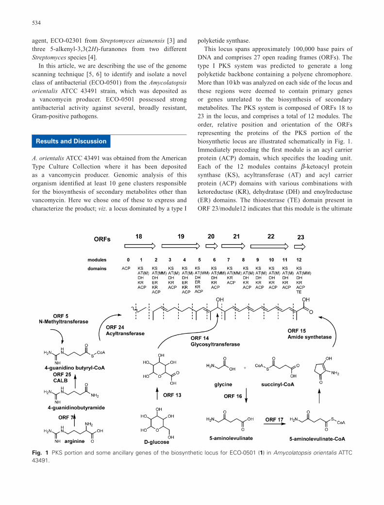
DNA and comprises 27 open reading frames (ORFs). Thetype I PKS system was predicted to generate a longpolyketide backbone containing a polyene chromophore.More than 10 kb was analyzed on each side of the locus andthese regions were deemed to contain primary genes or genes unrelated to the biosynthesis of secondarymetabolites. The PKS system is composed of ORFs 18 to23 in the locus, and comprises a total of 12 modules. Theorder, relative position and orientation of the ORFsrepresenting the proteins of the PKS portion of thebiosynthetic locus are illustrated schematically in Fig. 1.Immediately preceding the first module is an acyl carrierprotein (ACP) domain, which specifies the loading unit.Each of the 12 modules contains b -ketoacyl proteinsynthase (KS), acyltransferase (AT) and acyl carrier protein (ACP) domains with various combinations withketoreductase (KR), dehydratase (DH) and enoylreductase(ER) domains. The thioesterase (TE) domain present inORF 23/module12 indicates that this module is the ultimate
534
Fig. 1 PKS portion and some ancillary genes of the biosynthetic locus for ECO-0501 (1) in Amycolatopsis orientalis ATTC43491.
ORFs
modules
domains
ORF 5 N-Methyltransferase
18
::::=====~> 0 2
19 20
::::=====~> c::> 3 4 5 6
21
::::====> 7 8
22 23
~--> c::::::> 9 10 11 12
ACP KS KS KS KS KS KS KS KS KS KS KS KS AT(M) AT(MM) AT(M) AT(M) AT(MM) AT(MM) AT(MM) AT(M) AT(M) AT(M) AT(M) AT(MM) DH DH DH DH DH DH KR DH DH DH DH DH KR ER KR ER ER KR ACP KR KR KR KR KR ACP KR ACP KR KR ACP ACP ACP ACP ACP ACP
ACP ACP ACP TE
OH OH ' ' t.!. /--· -,(./" ,, ' ' ' ' ' '
' ' ' ' ' 0
Acyltransferase ORF14 Glycosyltransferase
:~ ORF15 Amide synthetase
,oho,o HOAO~
OH
t ORF 13
HOhOH
HOAO~
OH D-glucose
+
glycine
ORF 16 i 0
H2N~OH
0
5-aminolevulinate
0
CoA,s½o
OH
succinyl-CoA
ORF 17
5-am i nolevul i nate-CoA
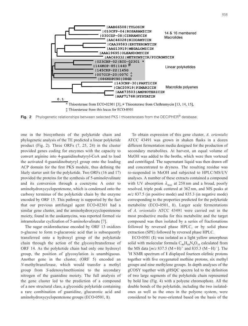
one in the biosynthesis of the polyketide chain andphylogenetic analysis of the TE predicted a linear polyketideproduct (Fig. 2). Three ORFs (7, 25, 24) in the clusterprovided genes coding for enzymes with the capacity toconvert arginine into 4-guanidinobutyryl-CoA and to loadthe activated 4-guanidinobutyryl group onto the loadingACP domain for the first PKS module, thus defining thelikely starter unit for the polyketide. Two ORFs (16 and 17)provided the proteins for the synthesis of 5-aminolevulinateand its conversion through a coenzyme A ester toaminohydroxycyclopentenone, which is condensed onto thecarboxy terminus of the polyketide chain by the enzymeencoded by ORF 15. This pathway is supported by the factthat our previous antifungal agent ECO-02301 had asimilar gene cluster, and the aminohydroxycyclopentenonemoiety, found in the asukamycins, was reported formed viaintramolecular cyclization of 5-aminolevulinate [7].
The sugar oxidoreductase encoded by ORF 13 oxidizesD-glucose to form D-glucuronic acid that is subsequentlytransferred onto a hydroxyl group of the polyketide chain through the action of the glycosyltransferase of ORF 14. As the polyketide chain had only one hydroxylgroup, the position of glycosylation is unambiguous.Another gene in the cluster, (ORF 5) encoded an N-methyltransferase, which would transfer a methyl group from S-adenosylmethionine to the secondarynitrogen of the guanidine moiety. The full analysis of the gene cluster led to the prediction of a compound of a new structural class, a glycosidic polyketide containinga rare combination of guanidine, glucuronic acid andaminohydroxycyclopentenone groups (ECO-0501, 1).
To obtain expression of this gene cluster, A. orientalisATCC 43491 was grown in shaken flasks in a dozendifferent fermentation media designed for the production ofsecondary metabolites. At harvest, an equal volume ofMeOH was added to the broths, which were then vortexedand centrifuged. The supernatant liquid was then drawn offand concentrated to dryness. The resulting residue was re-suspended in MeOH and subjected to HPLC/MS/UVanalyses. A number of these extracts contained a compoundwith UV absorption lmax at 258 nm and a broad, poorly resolved, triple peak centered at 362 nm, and MS peaks atm/z 837.5 (in positive mode) and 835.3 (in negative mode)corresponding to the properties predicted for the polyketidemetabolite (ECO-0501, 1). Larger scale fermentations of A. orientalis ATCC 43491 were carried out in the most productive media for this metabolite and the targetcompound was then isolated by a series of fractionationsfollowed by reversed phase HPLC, or by solid phaseextraction (SPE) followed by reversed phase HPLC.
ECO-0501 (1) was isolated as a light yellow amorphoussolid with molecular formula C46H68N4O10 calculated fromthe MS data [m/z 837.5 (M�H)� and 835.3 (M�H)�]. The1H NMR spectrum of 1 displayed fourteen olefinic protonstogether with five oxygenated methine protons, six methylgroups and nine methylene groups. In depth analyses of thegCOSY together with gHSQC spectra led to the definitionof two large segments of the polyketide chain representedby bold line (Fig. 4) with a polyene chromophore. All thedouble bonds of the polyketide, including the two isolated-ones as well as the ones in the polyene system, wereconsidered to be trans-oriented based on the basis of the
535
Fig. 2 Phylogenetic relationships between selected PKS I thioesterases from the DECIPHER® database.
IAAB665081TYLOSIN 1010CFP-04IROSARAMICIN
1030CZP-08ICIRRAMYCIN ._ ________ IAAC460281NIDDAMYCIN
-----1=:::::::::::ICAA395831ERYTHROMYCIN IAAG13919IMEGALOMICIN
IAAA19695IOLEANDOMYCIN
14 & 16 membered Macrolides
- .. -~::::--:::~~:-:.~~-~-~- ~- ~- ~- ~- ~----- IAAC693321METHYMYCIN/PICROMYCIN 1023CBP-02IECO-02301 *
---- I 144KOP-85 i 144D # 1145CKP-22 1145G
r-------- 1007CCP-20 1007C t Linear polyketides
0.1
1086KDPC901086D ------- 1143CNP-30IPARTICIN
I CAC20919 I PIMARICIN Macrolide polyenes IAAK735031AMPHOTERICIN IAAF717681NYSTATIN
* Thioesterase from ECO-02301 [3], # Thioesterase from Clethramycin [13, 14, 15],
t Thioesterase from this locus for ECO-0501
536
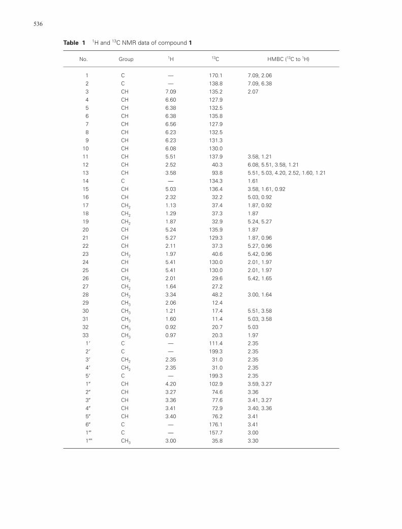
Table 1 1H and 13C NMR data of compound 1
No. Group 1H 13C HMBC (13C to 1H)
1 C — 170.1 7.09, 2.062 C — 138.8 7.09, 6.383 CH 7.09 135.2 2.074 CH 6.60 127.95 CH 6.38 132.56 CH 6.38 135.87 CH 6.56 127.98 CH 6.23 132.59 CH 6.23 131.3
10 CH 6.08 130.011 CH 5.51 137.9 3.58, 1.2112 CH 2.52 40.3 6.08, 5.51, 3.58, 1.2113 CH 3.58 93.8 5.51, 5.03, 4.20, 2.52, 1.60, 1.2114 C — 134.3 1.6115 CH 5.03 136.4 3.58, 1.61, 0.9216 CH 2.32 32.2 5.03, 0.9217 CH2 1.13 37.4 1.87, 0.9218 CH2 1.29 37.3 1.8719 CH2 1.87 32.9 5.24, 5.2720 CH 5.24 135.9 1.8721 CH 5.27 129.3 1.87, 0.9622 CH 2.11 37.3 5.27, 0.9623 CH2 1.97 40.6 5.42, 0.9624 CH 5.41 130.0 2.01, 1.9725 CH 5.41 130.0 2.01, 1.9726 CH2 2.01 29.6 5.42, 1.6527 CH2 1.64 27.228 CH2 3.34 48.2 3.00, 1.6429 CH3 2.06 12.430 CH3 1.21 17.4 5.51, 3.5831 CH3 1.60 11.4 5.03, 3.5832 CH3 0.92 20.7 5.0333 CH3 0.97 20.3 1.971� C — 111.4 2.352� C — 199.3 2.353� CH2 2.35 31.0 2.354� CH2 2.35 31.0 2.355� C — 199.3 2.351� CH 4.20 102.9 3.59, 3.272� CH 3.27 74.6 3.363� CH 3.36 77.6 3.41, 3.274� CH 3.41 72.9 3.40, 3.365� CH 3.40 76.2 3.416� C — 176.1 3.411� C — 157.7 3.001�� CH3 3.00 35.8 3.30
537
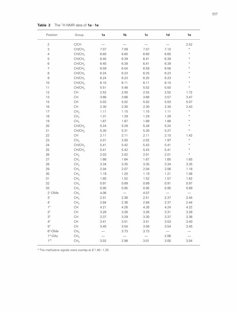
Table 2 The 1H NMR data of 1a�1e
Position Group 1a 1b 1c 1d 1e
2 C/CH — — — — 2.523 CH/CH2 7.07 7.09 7.07 7.10 *4 CH/CH2 6.60 6.60 6.60 6.60 *5 CH/CH2 6.40 6.39 6.41 6.39 *6 CH/CH2 6.40 6.39 6.41 6.39 *7 CH/CH2 6.59 6.54 6.59 6.56 *8 CH/CH2 6.24 6.23 6.25 6.23 *9 CH/CH2 6.24 6.23 6.25 6.23 *
10 CH/CH2 6.10 6.11 6.11 6.10 *11 CH/CH2 5.51 5.48 5.52 5.50 *12 CH 2.52 2.50 2.52 2.52 1.7213 CH 3.66 3.66 3.66 3.57 3.4715 CH 5.03 5.02 5.02 5.03 5.0716 CH 2.30 2.30 2.30 2.30 2.4317 CH2 1.11 1.15 1.15 1.11 *18 CH2 1.31 1.29 1.29 1.29 *19 CH2 1.87 1.87 1.89 1.88 *20 CH/CH2 5.24 5.29 5.28 5.24 *21 CH/CH2 5.30 5.31 5.30 5.27 *22 CH 2.11 2.11 2.11 2.10 1.4223 CH2 2.01 2.00 2.02 1.97 *24 CH/CH2 5.41 5.42 5.43 5.41 *25 CH/CH2 5.41 5.42 5.43 5.41 *26 CH2 2.03 2.02 2.01 2.01 *27 CH2 1.66 1.64 1.67 1.65 1.6528 CH2 3.34 3.35 3.35 3.34 3.3529 CH3 2.04 2.07 2.04 2.06 1.1830 CH3 1.19 1.20 1.19 1.21 1.0831 CH3 1.60 1.52 1.52 1.57 1.6232 CH3 0.91 0.89 0.89 0.91 0.9733 CH3 0.95 0.95 0.95 0.95 0.892�-OMe CH3 4.06 — 4.07 — —3� CH2 2.51 2.36 2.51 2.37 2.444� CH2 2.84 2.36 2.84 2.37 2.441� CH 4.21 4.26 4.26 4.24 4.222� CH 3.28 3.26 3.26 3.31 3.283� CH 3.37 3.29 3.30 3.37 3.364� CH 3.41 3.51 3.51 3.53 3.405� CH 3.40 3.54 3.56 3.54 3.456�-OMe CH3 — 3.73 3.73 — —1�-OAc CH3 — — — 2.06 —1�� CH3 3.02 2.98 3.01 3.00 3.04
* The methylene signals were overlap at d 1.40�1.20.
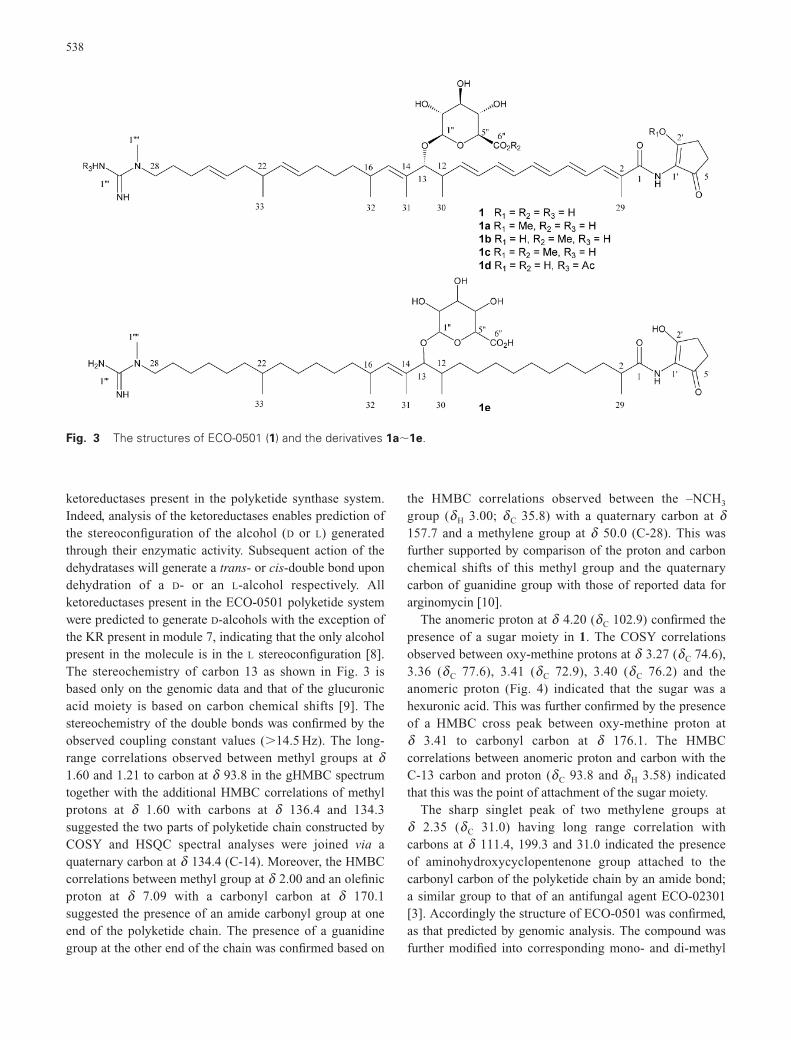
ketoreductases present in the polyketide synthase system.Indeed, analysis of the ketoreductases enables prediction ofthe stereoconfiguration of the alcohol (D or L) generatedthrough their enzymatic activity. Subsequent action of thedehydratases will generate a trans- or cis-double bond upondehydration of a D- or an L-alcohol respectively. Allketoreductases present in the ECO-0501 polyketide systemwere predicted to generate D-alcohols with the exception ofthe KR present in module 7, indicating that the only alcoholpresent in the molecule is in the L stereoconfiguration [8].The stereochemistry of carbon 13 as shown in Fig. 3 isbased only on the genomic data and that of the glucuronicacid moiety is based on carbon chemical shifts [9]. Thestereochemistry of the double bonds was confirmed by theobserved coupling constant values (�14.5 Hz). The long-range correlations observed between methyl groups at d1.60 and 1.21 to carbon at d 93.8 in the gHMBC spectrumtogether with the additional HMBC correlations of methylprotons at d 1.60 with carbons at d 136.4 and 134.3suggested the two parts of polyketide chain constructed byCOSY and HSQC spectral analyses were joined via aquaternary carbon at d 134.4 (C-14). Moreover, the HMBCcorrelations between methyl group at d 2.00 and an olefinicproton at d 7.09 with a carbonyl carbon at d 170.1suggested the presence of an amide carbonyl group at oneend of the polyketide chain. The presence of a guanidinegroup at the other end of the chain was confirmed based on
the HMBC correlations observed between the –NCH3
group (dH 3.00; dC 35.8) with a quaternary carbon at d157.7 and a methylene group at d 50.0 (C-28). This wasfurther supported by comparison of the proton and carbonchemical shifts of this methyl group and the quaternarycarbon of guanidine group with those of reported data forarginomycin [10].
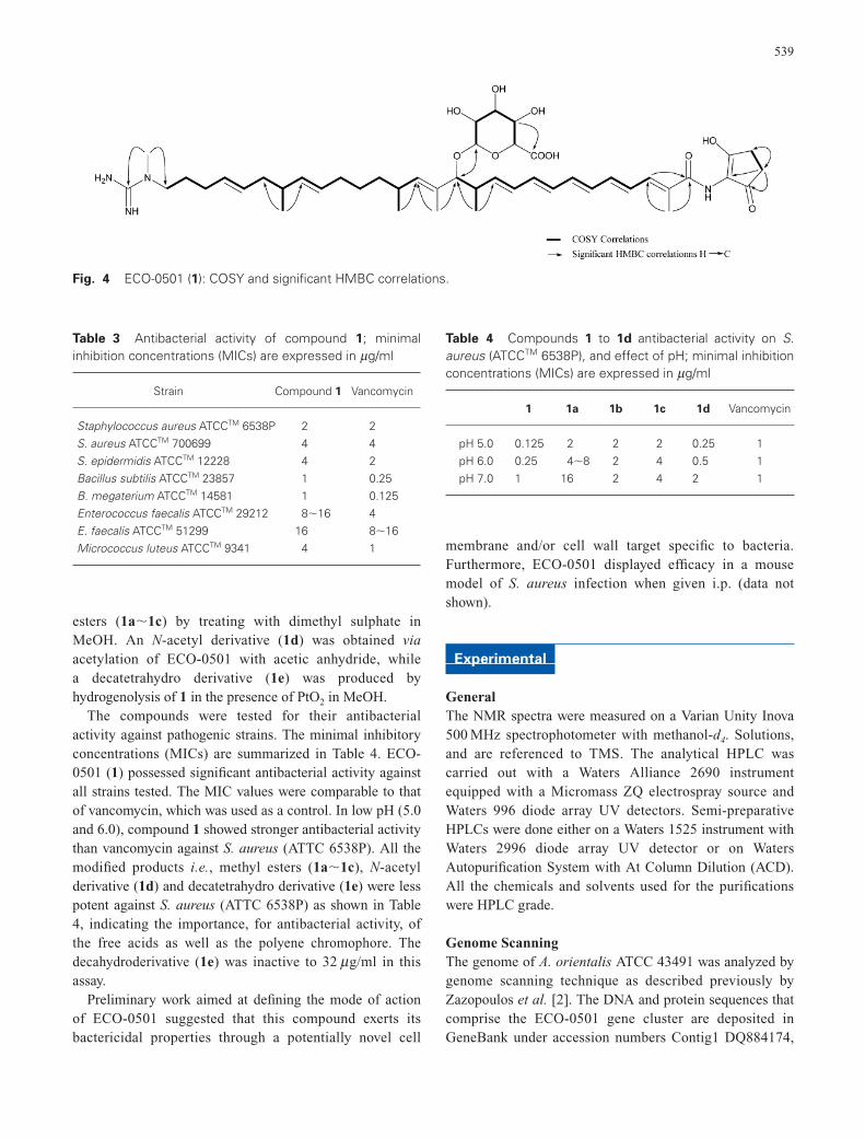
The anomeric proton at d 4.20 (dC 102.9) confirmed thepresence of a sugar moiety in 1. The COSY correlationsobserved between oxy-methine protons at d 3.27 (dC 74.6),3.36 (dC 77.6), 3.41 (dC 72.9), 3.40 (dC 76.2) and theanomeric proton (Fig. 4) indicated that the sugar was ahexuronic acid. This was further confirmed by the presenceof a HMBC cross peak between oxy-methine proton at d 3.41 to carbonyl carbon at d 176.1. The HMBCcorrelations between anomeric proton and carbon with theC-13 carbon and proton (dC 93.8 and dH 3.58) indicatedthat this was the point of attachment of the sugar moiety.
The sharp singlet peak of two methylene groups at d 2.35 (dC 31.0) having long range correlation withcarbons at d 111.4, 199.3 and 31.0 indicated the presenceof aminohydroxycyclopentenone group attached to thecarbonyl carbon of the polyketide chain by an amide bond;a similar group to that of an antifungal agent ECO-02301[3]. Accordingly the structure of ECO-0501 was confirmed,as that predicted by genomic analysis. The compound wasfurther modified into corresponding mono- and di-methyl
538
Fig. 3 The structures of ECO-0501 (1) and the derivatives 1a�1e.
l ""
16 I
22 N 28 R3HNY
1"'
NH 33 32
l""
I
H2~,y N 28 22 16
NH 33 32
14
31
14
13
30 R - R3 = H 1 R1 = 2 - R = R3 = H 1a R = Me, 2 R = H
1 - H R = Me, 3 1b R1 - ' 2 R = H
R = Me, 3 1cR1= 2 R =Ac 1d R1 = R2 = H, 3
HO DOH OH
6" C02H 0 0
12
13
31 30 1e
29
H04, 2 0 N 1'1 5'
1 H 0
29
esters (1a�1c) by treating with dimethyl sulphate inMeOH. An N-acetyl derivative (1d) was obtained viaacetylation of ECO-0501 with acetic anhydride, while a decatetrahydro derivative (1e) was produced byhydrogenolysis of 1 in the presence of PtO2 in MeOH.
The compounds were tested for their antibacterialactivity against pathogenic strains. The minimal inhibitoryconcentrations (MICs) are summarized in Table 4. ECO-0501 (1) possessed significant antibacterial activity againstall strains tested. The MIC values were comparable to thatof vancomycin, which was used as a control. In low pH (5.0and 6.0), compound 1 showed stronger antibacterial activitythan vancomycin against S. aureus (ATTC 6538P). All themodified products i.e., methyl esters (1a�1c), N-acetylderivative (1d) and decatetrahydro derivative (1e) were lesspotent against S. aureus (ATTC 6538P) as shown in Table4, indicating the importance, for antibacterial activity, ofthe free acids as well as the polyene chromophore. Thedecahydroderivative (1e) was inactive to 32 mg/ml in thisassay.
Preliminary work aimed at defining the mode of actionof ECO-0501 suggested that this compound exerts itsbactericidal properties through a potentially novel cell
membrane and/or cell wall target specific to bacteria.Furthermore, ECO-0501 displayed efficacy in a mousemodel of S. aureus infection when given i.p. (data notshown).
Experimental
GeneralThe NMR spectra were measured on a Varian Unity Inova500 MHz spectrophotometer with methanol-d4. Solutions,and are referenced to TMS. The analytical HPLC wascarried out with a Waters Alliance 2690 instrumentequipped with a Micromass ZQ electrospray source andWaters 996 diode array UV detectors. Semi-preparativeHPLCs were done either on a Waters 1525 instrument withWaters 2996 diode array UV detector or on WatersAutopurification System with At Column Dilution (ACD).All the chemicals and solvents used for the purificationswere HPLC grade.
Genome ScanningThe genome of A. orientalis ATCC 43491 was analyzed bygenome scanning technique as described previously byZazopoulos et al. [2]. The DNA and protein sequences thatcomprise the ECO-0501 gene cluster are deposited inGeneBank under accession numbers Contig1 DQ884174,
539
Fig. 4 ECO-0501 (1): COSY and significant HMBC correlations.
Table 3 Antibacterial activity of compound 1; minimalinhibition concentrations (MICs) are expressed in mg/ml
Strain Compound 1 Vancomycin
Staphylococcus aureus ATCCTM 6538P 2 2S. aureus ATCCTM 700699 4 4S. epidermidis ATCCTM 12228 4 2Bacillus subtilis ATCCTM 23857 1 0.25B. megaterium ATCCTM 14581 1 0.125Enterococcus faecalis ATCCTM 29212 8�16 4E. faecalis ATCCTM 51299 16 8�16Micrococcus luteus ATCCTM 9341 4 1
Table 4 Compounds 1 to 1d antibacterial activity on S.aureus (ATCCTM 6538P), and effect of pH; minimal inhibitionconcentrations (MICs) are expressed in mg/ml
1 1a 1b 1c 1d Vancomycin
pH 5.0 0.125 2 2 2 0.25 1pH 6.0 0.25 4�8 2 4 0.5 1pH 7.0 1 16 2 4 2 1
- COSY Con-elations
---- Significant HMBC corrclationns H -.c
Contig2 DQ884175, Contig3 DQ884176.
FermentationA. orientalis ATCC 43491, which was obtained from theAmerican Type Culture Collection (P.O. Box 1549,Manassas, VA 20108, USA), was cultivated on agar platesof ISP2 medium (Difco). To prepare a vegetative culture, A.orientalis ATCC 43491 was grown on ISP2 agar (Difco)for 5 to 7 days, and the surface growth from the agar platewas homogenized and transferred to a 125 ml flaskcontaining three glass beads (5 mm diameter), and 25 ml ofsterile medium prepared from trypticase soy broth (Bacto)30 g, yeast extract 3 g, MgSO4 2 g, glucose 5 g, maltose 4 g,to which one liter distilled water was added. This vegetativeculture was incubated at 28°C for about 60 hours on ashaker with a 2.5 cm throw and set at 250 rpm.
The vegetative culture (10 ml aliquots) was used toinoculate 2 liter baffled flasks each containing 500 ml ofsterile production medium prepared from glucose 10 g,glycerol 5 g, corn steep liquor 3 g, beef extract 3 g, maltextract 3 g, yeast extract 3 g, calcium carbonate 2 g,thiamine 0.1 g made up to one liter with distilled water[11]. The medium was adjusted at pH 7.0, and then 1 ml ofsilicon defoamer-oil (Chem Service) was added to eachflask before sterilization. The fermentation batches wereincubated aerobically on a shaker (200 rpm) at 28°C for aperiod of 4 days.
Isolation of ECO-0501 (1)The mycelia and broth of the culture media (12�500 ml)was separated by centrifugation (3000 rpm, 20 min). Themycelial cake was extracted consecutively with methanol(200 ml/liter broth) and acetone (200 ml/liter) to produce anorganic cell extract. The organic extract was used forfurther purification by two different methods.
Method AThe combined organic extract was dried under vacuum, and further suspended in a mixture of MeOH/aqueousNH4HCO3 solution adjusted to pH 10 with NH4OH (3 : 2,100 ml/liter original broth volume) and consecutivelyextracted by CHCl3 (100 ml/liter) and n-BuOH (100 ml/liter).The BuOH fraction was concentrated, and the residue wasdissolved in a minimal amount of DMSO/MeOH (3 : 1) andsubjected for HPLC purification after filtering through a0.45 mm 13 mm Acrodisc GHP syringe filter. The HPLCwas performed on a Waters Autopurification System withACD using a Waters Xterra MS C18 column (5 m ,19�150 mm), and a gradient of 10 mM aqueous NH4HCO3
(pH 10)/acetonitrile 85 : 15 to 25 : 75 over 30 minutes at19 ml/minute, UV detector set at 261 nm. The semi-purified
ECO-0501 (1, 1.04 g), eluting at 11.8�12.1 minutes, wascollected.
ECO-0501 (1, 37.4 mg/liter) was purified by repeatedHPLC on a Waters Autopurification System with ACDusing a Waters RCM Column (Novapak C-18, 6 m ,40�200 mm) with a gradient of 10 mM aqueous NH4OAcadjusted to pH 5 with glacial AcOH/acetonitrile from80 : 20 v/v to 20 : 80 over 25 minutes at 35 ml/minute.
Method BThe combined organic extract, which was dried undervacuum was suspended in a mixture of MeOH and aqueousNH4HCO3 solution adjusted to pH 10 with NH4OH (3 : 2,100 ml/liter) and extracted with hexane (3�100 ml/literoriginal broth volume) to remove fatty substances. Theaqueous methanolic fraction was then adsorbed (slurry-mode) on Diaion HP-20 resin (30 ml/liter of fermentationbroth) and applied to SPE on a Startat C-18 Cartridge(Phenomenex) with a precolumn of Diaion HP-20 resin(70 ml). The column was subsequently eluted with a stepgradient of EtOH/aqueous NH4HCO3 buffer pH 10 tocollect one 500 ml fraction and then seven fractions (200 mleach) i.e., 1 : 9 (fraction 1); 1 : 4 (fraction 2); 3 : 7 (fraction3); 2 : 3 (fraction 4); 1 : 1 (fraction 5); 3 : 2 (fraction 6); 8 : 1(fraction 7) and EtOH (fraction 8). Fractions 4�7 werepooled, concentrated and the residue was subjected forHPLC (Waters Autopurification System with ACD), usinga Symmetry C18 column (5 m , 30�100 mm) with agradient of 10 mM aqueous NH4OAc, (adjusted to pH 5with glacial AcOH)/acetonitrile 74 : 26 v/v to 50 : 50 over20 minutes at 39 ml/minute. The collection was triggeredby UV absorption at 261 nm (PDA). The sample wasloaded as a suspension in DMSO : MeOH (3 : 1). ECO-0501, which eluted at 14.9�15.2 minutes, was pure.
1: UV (lmax) 258 and 362 nm; MS (ESI in positivemode) m/z 837.5 (M�H)�, 823.5 (M�H�CH3)
�; MS (ESI in negative mode) m/z 835.3 (M�H)�, 821.5(M�H�CH3)
�; HRMS 837.5018 calcd for C46H69N4O10
(M�H)� 837.5014. The 1H and 13C NMR data are in Table1.
Synthesis of 1a�1cA solution of 1 (20 mg) in MeOH (2.0 ml) was stirred witha mixture of 0.1 mM NaOH (Fisher Chemicals) solution inMeOH (334 m l) and dimethyl sulfate (5.68 m l, Sigma) atroom temperature for 24 hours. The reagents weresuccessively added to the reaction mixture after 24 and 48hours (NaOH solution 300 and 400 m l; dimethyl sulphate10 and 15 m l). The reaction was monitored by TLC (MerckSilica gel 60 F254, eluted with 7% methanol in chloroform,visualized under UV) and stopped after 72 hours. The
540
reaction mixture was purified by HPLC on a Waters Auto-Purification System using a Symmetry column (C-18, 5 m ,30�100 mm) with 10 mM NH4OAc in water/MeCNgradient (74 : 26 v/v to 50 : 50 in 20 minutes, 40 ml/minute).The monomethyl derivatives 1b (0.53 mg), 1a (5.36 mg)and a dimethyl derivative 1c (4.04 mg) were eluted at 9.4,11.5 and 15.5 minutes, respectively.
1a: UV (lmax) 258 and 362 nm; MS (ESI in positivemode) m/z 852.03 (M�H)�; MS (ESI in negative mode)m/z 849.97 (M�H�). The 1H and 13C NMR data are inTable 2.
1b: UV (lmax) 258 and 362 nm; MS (ESI in positivemode) m/z 852.03 (M�H)�; MS (ESI in negative mode)m/z 849.98 (M�H)�. The 1H and 13C NMR data are inTable 2.
1c: UV (lmax) 258 and 362 nm; MS (ESI in positivemode) m/z 866.06 (M�H)�; MS (ESI in negative mode)m/z 863.89 (M�H)�; The 1H and 13C NMR data are inTable 2.
Synthesis of 1dA solution of 1 (20 mg) in MeOH (2 ml) was stirred withacetic anhydride (20 m l) at room temperature for 24 hours.Additional acetic anhydride (20 m l) was added to thereaction mixture at 24 and 48 hours. The reaction wasmonitored by TLC (Merck Silica gel 60 F254, eluted with7% MeOH in chloroform, visualized under UV) andstopped at 72 hours.
The reaction mixture was purified by HPLC on a WatersAuto-Purification System using a Symmetry (C-18, 5 m ,30�100 mm) column with 10 mM NH4OAc in wateradjusted to pH 5 with glacial AcOH/acetonitrile gradientsystem (74 : 26 v/v to 50 : 50 in 20 minutes, 40 ml/minute).Compound 1d (6.43 mg) was obtained as a single product.
1d: UV (lmax) 258 and 362 nm; MS (ESI in positivemode) m/z 880.03 (M�H)�; MS (ESI in negative mode)m/z 877.98 (M�H)�; The 1H and 13C NMR data are inTable 2.
Synthesis of 1eA solution of 1 (20 mg) in MeOH (2 ml) was stirred underhydrogen gas overnight at room temperature in the presenceof PtO2 (10 mg) as a catalyst. The reaction mixture wasfiltered and the filtrate was concentrated to obtain thedecatetrahydro derivative (1e, 18.7 mg).
1e: UV (lmax) 258 nm; MS (ESI in positive mode) m/z853.03 (M�H)�; MS (ESI in negative mode) m/z 851.08(M�H)�. The 1H and 13C NMR data are in Table 2.
Antibacterial ActivityAntibacterial activity of the isolated compounds were
measured by determining the minimal inhibitoryconcentrations (MIC) against eight pathogenic strains,namely Staphylococcus aureus (ATCC 6538P),Staphylococcus aureus MRS3 (TM 700699), Staphylococcusepidermidis (ATCC 12228), Bacillus subtilis (ATCC23857), Bacillus megaterium (ATCC 14581), Enterococcusfaecalis VRE-1 (ATCC 29212), Enterococcus faecalisVRE-2 (ATCC 51299) and Micrococcus luteus (ATCC9341). The antibacterial experiments were performedaccording to the National Committee for ClinicalLaboratory Standards (NCCLS) guideline M7-A5 [12].
The stock solutions of the tested compounds wereprepared in DMSO (100�) and diluted with Mueller-Hinton test medium as two-fold series over 11 points from3.2 mg/ml to 0.003 mg/ml. An aliquot of each stocksolution was diluted 50-fold in test medium describedbelow to give a set of eleven 2� solutions. Fifty microlitersof each of the eleven 2� solutions were aliquoted into thecorresponding wells of a 12-well row, with the final wellreserved for a medium-alone control. Vancomycin (Sigma)used as positive control, which was prepared as 2� stocksolutions in Mueller-Hinton test medium ranging from64 mg/ml to 0.06 mg/ml (a two-fold dilution series over 11points). An aliquot of 50 m l of each concentration (at 2�)was then transferred to 96-well microplates to obtain aseries of eleven two-fold dilutions.
An isolated colony of each of the eight indicator strainswas used to inoculate tubes containing 2 ml of test medium.Mueller-Hinton test medium was used for S. aureus (ATCC6538P), S. aureus MRS3 (ATCC 700699), S. epidermidis(ATCC 12228), B. subtilis (ATCC 23857), B. megaterium(ATCC 14581) and M. luteus (ATCC 9341) indicatorstrains, and Brain Heart Infusion test medium was used forE. faecalis VRE-1 (ATCC 29212) and E. faecalis VRE-2(ATCC 51299) indicator strains. Cells were grownovernight at 35°C with shaking. Inoculum density for eachindicator strain was adjusted to OD600�0.1 in 5 ml 0.85%saline, then further diluted 1/100 in appropriate medium.50 m l of the final dilution (in test medium) of each indicatorstrain was added to each well of a 12-well row. This bringsthe final dilution of the test compound or control compoundin solution to 1�. The final inoculum has approximately5�105 CFU/ml.
The indicator strains were incubated with 11concentrations of each of test compounds, vancomycin(Sigma) control and one medium-alone control. For MICdetermination, assay plates were incubated at 35°C for 16to 20 hours. The MIC for each indicator was assessed asthe lowest concentration of the compound resulting in totalabsence of growth and is shown in Table 3.
541
References
1. Zazopoulos E, Huang K, Staffa A, Liu W, Bachmann BO,Nonaka K, Ahlert J, Thorson JS, Shen B, Farnet CM. Agenomics-guided approach for discovering and expressingcryptic metabolic pathways. Nat Biotechnol 21: 187–190(2003).
2. Zazopoulos E, Farnet CM. Improving drug discovery fromMicroorganisms. Natural Products: Drug Discovery andTherapeutic Medicine; Zhang, Demain eds., pp. 95–106(2005)
3. McAlpine JB, Bachmann BO, Piraee M, Tremblay S, AlarcoAM, Zazopoulos E, Farnet CM. Microbial genomics as aguide to drug discovery and structural elucidation: ECO-02301, a novel antifungal agent, as an example. J Nat Prod68: 493–496 (2005)
4. Banskota AH, McAlpine JB, Sørensen D, Aouidate M,Piraee M, Alarco AM, Omura S, Shiomi K, Farnet CM,Zazopoulos E. Isolation and identification of three new 5-Alkenyl-3,3(2H)-furanones from two Streptomyces speciesusing a genomic screening approach. J Antibiot 59: 168–176(2006)
5. Sørensen D, McAlpine JB, Piraee M, Farnet CM,Zazopoulos E. Genome scanning technology reveals anantibacterial compound (ECO-0501) of a new structuralclass from the vancomycin-producer Amycolatopsis orientalis.44th ICAAC: No. F-720a, Washington, DC (2004)
6. McAlpine JB, Zazopoulos E, Sørensen D, Piraee M, IbrahimA, Aouidate M, Farnet CM. The power of genomic analysisin the discovery of novel secondary metabolites. 46thAnnual Meeting of American Society of Pharmacognosy:No. O-21, Corvallis (2005)
7. Nakagawa A, Wu TS, Keller PJ, Lee JP, Omura S, Floss HG.Biosynthesis of asukamycin. Formation of the 2-
aminocyclopentenol-3-one moiety. J Chem Soc ChemCommun 519–521 (1985)
8. Caffrey P. Conserved amino acid residues correlating withketoreductase stereospecificity in modular polyketidesynthases. Chembiochem 4: 654–657 (2003)
9. Block K, Pedersen C. Carbon 13 nuclear magneticresonance spectroscopy of monosaccharides. In Advances inCarbohydrate Chemistry and Biochemistry, Vol. 41, pp.27–66 (1983) Academic Press
10. Argoudelis AD, Baczynskyj L, Kuo MT, Laborde AL, SebekOK, Truesdell SE, Shilliday FB. Arginomycin: production,isolation, characterization and structure. J Antibiot 11:750–760 (1987)
11. Kanzaki H, Wada K, Nitoda T, Kawazu K. Novel bioactiveoxazolomycin isomers by Streptomyces albus JA3453.Biosci Biotechnol Biochem 62: 438–442 (1998)
12. Methods for Dilution Antimicrobial Susceptibility Tests forBacteria That Grow Aerobically; Approved Standard-FifthEdition. (NCCLS document M7-A5, ISBN 1-56238-394-9;NCCLS, 940 West Valley Road, Suite 1400, Wayne,Pennsylvania 19087-1898 USA)
13. Yamakawa T, Furumai T, Yoshida R, Igarashi Y. Clethramycin,a new inhibitor of pollen tube growth with antifungalactivity from Streptomyces hygroscopicus TP-A0623 I.Screening, taxonomy, fermentation, isolation and biologicalproperties. J Antibiot 56: 700–704 (2003)
14. Igarashi Y, Iwashita T, Fujita T, Naoki H, Yamakawa T, Yoshida R, Furumai T. Clethramycin, a new inhibitor of pollen tube growth with antifungal activity fromStreptomyces hygroscopicus TP-A0623. II Physico-chemicalproperties and structure determination. J Antibiot 56:705–708 (2003)
15. Clethramycin was isolated and identified independently atEcopia Biosciences and correlated with its biosyntheticlocus (data not shown)
542