genome‐wide association meta‐analysis reveals novel
TRANSCRIPT
Genome-Wide Association Meta-Analysis Reveals NovelJuvenile Idiopathic Arthritis Susceptibility Loci
Laura A. McIntosh,1 Miranda C. Marion,2 Marc Sudman,3 Mary E. Comeau,2 Mara L. Becker,4
John F. Bohnsack,5 Tasha E. Fingerlin,6 Thomas A. Griffin,7 J. Peter Haas,8 Daniel J. Lovell,1
Lisa A. Maier,6 Peter A. Nigrovic,9 Sampath Prahalad,10 Marilynn Punaro,11 Carlos D. Ros�e,12
Carol A. Wallace,13 Carol A. Wise,14 Halima Moncrieffe,1 Timothy D. Howard,15
Carl D. Langefeld,15 and Susan D. Thompson1
Objective. Juvenile idiopathic arthritis (JIA) isthe most common childhood rheumatic disease and hasa strong genomic component. To date, JIA genetic associ-ation studies have had limited sample sizes, used hetero-geneous patient populations, or included only candidateregions. The aim of this study was to identify new associ-ations between JIA patients with oligoarticular diseaseand those with IgM rheumatoid factor (RF)�negativepolyarticular disease, which are clinically similar andthe most prevalent JIA disease subtypes.
Methods. Three cohorts comprising 2,751 patientswith oligoarticular or RF-negative polyarticular JIA weregenotyped using the Affymetrix Genome-Wide SNP Array6.0 or the Illumina HumanCoreExome-12+ Array. Over-all, 15,886 local and out-of-study controls, typed on theseplatforms or the Illumina HumanOmni2.5, were used
for association analyses. High-quality single-nucleotidepolymorphisms (SNPs) were used for imputation to 1000Genomes prior to SNP association analysis.
Results. Meta-analysis showed evidence of asso-ciation (P < 1 3 10�6) at 9 regions: PRR9_LOR (P =5.12 3 10�8), ILDR1_CD86 (P = 6.73 3 10�8), WDFY4(P = 1.79 3 10�7), PTH1R (P = 1.87 3 10�7), RNF215(P = 3.09 3 10�7), AHI1_LINC00271 (P = 3.48 310�7), JAK1 (P = 4.18 3 10�7), LINC00951 (P = 5.803 10�7), and HBP1 (P = 7.29 3 10�7). Of these,PRR9_LOR, ILDR1_CD86, RNF215, LINC00951, andHBP1 were shown, for the first time, to be autoimmunedisease susceptibility loci. Furthermore, associatedSNPs included cis expression quantitative trait loci forWDFY4, CCDC12, MTP18, SF3A1, AHI1, COG5, HBP1,and GPR22.
Conclusion. This study provides evidence of bothunique JIA risk loci and risk loci overlapping betweenJIA and other autoimmune diseases. These newly asso-ciated SNPs are shown to influence gene expression,
Supported in part by the Cincinnati Children's ResearchFoundation and its Cincinnati Genomic Control Cohort. Recruitmentand DNA preparation in the US were supported by the NIH(National Institute of Arthritis and Musculoskeletal and Skin Dis-eases [NIAMS] grants N01-AR-42272, P01-AR-048929, P30-AR-473639, P30-AR-070549, and P30-AR-070253, National Heart, Lung,and Blood Institute grant R01-HL-11487, National Institute of Envi-ronmental Health Sciences grant P0-ES-0118101, and National Insti-tute for Research Resources grant UL1-RR-025780), the Fundaci�onBechara, the PhRMA Foundation, and the Rheumatology ResearchFoundation. Genotyping of JIA and control collections in the US wassupported by the NIH (NIAMS grant RC1-AR-058587). Recruitmentand DNA preparation in Germany were supported by the BMBF(grants 01GM0907 and 01 ZZ 0403).
1Laura A. McIntosh, BA, Daniel J. Lovell, MD, MPH, Hal-ima Moncrieffe, PhD, Susan D. Thompson, PhD: Cincinnati Chil-dren’s Hospital Medical Center and University of Cincinnati,Cincinnati, Ohio; 2Miranda C. Marion, MA, Mary E. Comeau, MA:Wake Forest University School of Medicine, Winston-Salem, NorthCarolina; 3Marc Sudman, BA: Cincinnati Children’s Hospital MedicalCenter, Cincinnati, Ohio; 4Mara L. Becker, MD: Children’s Mercy–Kansas City, Kansas City, Missouri; 5John F. Bohnsack, MD: Univer-sity of Utah, Salt Lake City; 6Tasha E. Fingerlin, PhD, Lisa A. Maier,MD, MSPH, FCCP: National Jewish Health and University of Col-orado, Denver; 7Thomas A. Griffin, MD, PhD: Levine Children’s
Specialty Center, Charlotte, North Carolina; 8J. Peter Haas, MD:German Center for Pediatric and Adolescent Rheumatology, Gar-misch-Partenkirchen, Germany; 9Peter A. Nigrovic, MD: Boston Chil-dren’s Hospital and Brigham and Women’s Hospital and HarvardMedical School, Boston, Massachusetts; 10Sampath Prahalad, MD,MSc: Emory University School of Medicine, Atlanta, Georgia;11Marilynn Punaro, MD: Texas Scottish Rite Hospital for Childrenand UT Southwestern Medical Center, Dallas; 12Carlos D. Ros�e,MD: DuPont Children’s Hospital, Wilmington, Delaware; 13Carol A.Wallace, MD: Seattle Children’s Hospital and Research Institute,Seattle, Washington; 14Carol A. Wise, PhD: Texas Scottish RiteHospital for Children, McDermott Center for Human Growth andDevelopment, and UT Southwestern Medical Center, Dallas;15Timothy D. Howard, PhD, Carl D. Langefeld, PhD: University ofCincinnati, Cincinnati, Ohio.
Address correspondence to Susan D. Thompson, PhD,Cincinnati Children’s Hospital Medical Center and University ofCincinnati, Cincinnati, Ohio. E-mail: [email protected].
Submitted for publication April 13, 2017; accepted in revisedform July 13, 2017.
2222
ARTHRITIS & RHEUMATOLOGYVol. 69, No. 11, November 2017, pp 2222–2232DOI 10.1002/art.40216© 2017, American College of Rheumatology
and their bounding regions tie into molecular pathwaysof immunologic relevance. Thus, they likely representregions that contribute to the pathology of oligoarticularJIA and RF-negative polyarticular JIA.
Juvenile idiopathic arthritis (JIA) is the mostcommon childhood rheumatic disease, with a preva-lence of ~1 per 1,000 children (1). It is a disabling, com-plex disorder characterized by inflammation of thejoints and other tissues that persists for at least6 weeks. Although there is ethnic diversity in JIA, it is adisease that predominately affects children of Europeandescent (2). There is a strong genetic component toJIA, with numerous established susceptibility loci (3�5)and a sibling recurrence risk ratio (ks) of 11.6 (6). Addi-tionally, the prevalence of other autoimmune diseases isincreased in relatives of patients with JIA (7).
The International League of Associations forRheumatology (ILAR) categorizes JIA into 7 subtypes(8). Two of these subtypes, oligoarticular JIA (persis-tent and extended forms) and IgM rheumatoid factor(RF)�negative polyarticular JIA, account for ~70% ofcases of JIA and are the focus of the current study.These 2 subtypes present similarly in the clinic, aredistinguished only by the number of affected jointsafter a disease duration of 6 months, and share HLAassociations (9,10).
Genome-wide association studies (GWAS), whilewidely used across autoimmune diseases, have been some-what limited in scope for JIA. Previous high-densityarray�based studies in patients with oligoarticular JIA orRF-negative polyarticular JIA have provided evidence ofassociation for single-nucleotide polymorphisms (SNPs)corresponding to loci near or including PTPN22, PTPN2,IL2RA, TNFAIP3, COG6, ADAD1/IL2/IL21, STAT4,chromosome 3q13 within C3orf1 and near CD80, andchromosome 10q21 near JMJD1C (4,5). Other GWASincluded all JIA subtypes and showed associations withTRAF1/C5 or VTCN1 loci but were limited by either smallsample sizes (11) or the number of markers assayed (12).
More recently, the Juvenile Arthritis Consortiumfor Immunochip studied patients and controls of Euro-pean ancestry, using the Illumina Infinium Immunochipgenotyping array, which provides dense SNP coveragein the HLA region and is limited to 186 non-HLAregions identified in 12 early studies of autoimmunedisease association (not including JIA) (3). Therefore,it does not reflect the current catalog of autoimmunedisease findings. Results from the Immunochip analysesprovided convincing evidence of association for a num-ber of JIA risk loci that are also risk loci for other
autoimmune diseases, including the HLA region (10)and 27 non-HLA loci (3). However, a significant pro-portion of JIA heritability risk remains unexplained.Although there is a partial overlap between the patientand control samples used in this study and the Immu-nochip studies (3,10), the current study extends findingsto a genome-wide level to further delineate JIA geneticrisk factors and allows the findings to be translated toJIA disease mechanisms.
PATIENTS AND METHODS
Subjects. Three cohorts comprising 2,751 JIA patientsof European ancestry with oligoarticular disease or RF-negative polyarticular disease and 15,886 controls (cohort I,814 cases and 3,058 controls; cohort II, 1,057 cases and11,843 controls; cohort III, 880 cases and 985 controls) wereused for association analyses. Subjects in cohort I were pri-marily recruited from the Cincinnati Children’s HospitalMedical Center (CCHMC) or as part of a National Instituteof Arthritis and Musculoskeletal and Skin Diseases�supported registry of JIA-affected sibpairs. Collaborating cen-ters including Children’s Hospital of Wisconsin, SchneiderChildren’s Hospital, and Children’s Hospital of Philadelphiaprovided additional samples (4,5). JIA patients in cohort IIhave been described previously as a validation cohort (4,5).Clinics enrolling JIA patients for cohort III were located inCincinnati, OH, Atlanta, GA, Charlotte, NC, Columbus, OH,Little Rock, AR, Long Island, NY, Chicago, IL, Salt LakeCity, UT, Cleveland, OH, Nashville, TN, and Charleston, SC.Additional DNA samples, split between cohorts II and III,were collected in Cincinnati, OH (n = 105) or were collectedas part of or obtained from the Gene Expression in PediatricArthritis Study (National Institute of Arthritis and Musculos-keletal and Skin Diseases [NIAMS] grant P01-AR-048929)(n = 117), Children’s Mercy Hospital, Kansas City (n = 75),the Improved Understanding of the Biology and Use of TNFInhibition in Children with JIA Study (ClinicalTrials.gov iden-tifier: NCT00792233) (n = 40), Nemours/Alfred I. duPontHospital for Children (n = 38), the Boston Children’s Hospi-tal JIA Registry (n = 26), the Trial of Early Aggressive Ther-apy in JIA Study (TREAT) (ClinicalTrials.gov identifier:NCT00443431) (n = 25), Emory University School of Medi-cine (n = 19), and Cohen Children’s Medical Center (n = 4).Members of the consortia are shown in Appendix A.
All cases met the ILAR or American College ofRheumatology (13) classification criteria for JIA or juvenilerheumatoid arthritis (RA). Both regional and out-of-studycontrols obtained from the dbGaP database were included inthe control cohorts. Controls for cohort I included regionalcontrols recruited from the geographic region served byCCHMC (4,5) and 2,400 out-of-study controls from theMolecular Genetics of Schizophrenia nonGAIN Sample(MGS_nonGAIN; phs000167.v1vp1). Controls for cohort IIincluded “Texas,” “Utah,” and “German” regional controls(4,5), 7,324 controls from the Atherosclerosis Risk in Com-munities Study cohort (phs000280.v3.p1), 2,555 controls fromthe Genetic Association Information Network (GAIN;phs000021.v3.p2 and phs000017.v3.p1), and 1,792 controlsfrom the Cooperative Health Research in the Region
NEWLY IDENTIFIED JIA SUSCEPTIBILITY LOCI 2223
Augsburg (KORA) study (14). Cohort III contained only regio-nal controls recruited in Denver. Use of these DNA collectionshas been approved by the Institutional Review Board at allparticipating centers, and participants or their parents providedwritten consent prior to study enrollment. Cohorts I and IIhave been used in previous association studies (4,5), and over-all, ~65% of the JIA samples from the current study were usedfor Immunochip analyses (3,10).
Genotyping and quality control. Cohorts I and II weregenotyped using the Affymetrix Genome-Wide Human SNPArray 6.0. Cohort I was genotyped at the Affymetrix ServiceCenter, while cohort II was genotyped at Expression Analysis/Quintiles; cohort III was genotyped with the IlluminaHumanCoreExome-12+ Array (Exome Array), which included2,508 custom SNP assays derived from initial analyses incohort I, at CCHMC. All out-of-study controls were geno-typed on SNP Array 6.0, with the exception of the KORAcohort, which was genotyped on the HumanOmni2.5 Bead-Chip (Infinium). Samples were excluded if their call rateswere <98% across the SNPs that passed quality control filters.Duplicates and first-degree relatives were identified using thesoftware package KING (15), retaining the sample with thehighest call rate. Self-reported and genetically determined sexwere compared using chromosome X genotype data. The pro-gram ADMIXTURE (16) was used to compute admixtureestimates from a subset of SNPs that met quality control cri-teria and were pruned to have low linkage disequilibrium(LD) (r2 < 0.2). This study was limited to individuals whoself-reported European ancestry, and individuals whoseadmixture estimates were outliers were removed. Primaryinference was based on SNPs that showed no significant evi-dence of departure from expectation in Hardy-Weinberg equilib-rium proportions (P < 1 9 10�6 and P < 0.01 in cases andcontrols, respectively), significant differential missingnessbetween cases and controls (P < 0.05), a minor allele frequencyof ≥0.01, and a call rate of >95%.
Statistical analysis. Imputation. Because the 3 cohortswere genotyped on 2 different arrays at 3 different times,imputation was performed separately for each cohort, usingIMPUTE2 with the 1000 Genomes phase 1 integrated refer-ence panel (17). Imputed SNPs were retained if their infor-mation score was >0.5 and their confidence score was >0.9.To validate imputation, a representative subset of subjects (96subjects from each of the 3 JIA cohorts and 96 control sub-jects) were genotyped for 16 SNPs, including 7 of the 9 SNPsshown in Table 1, using TaqMan SNP Genotyping Assays(Life Technologies) and evaluated for concordance betweenimputed and TaqMan-generated genotypes. Reactions wereperformed on a ViiA 7 real-time polymerase chain reactionsystem (Applied Biosystems).
Tests of association. Tests of association were per-formed on the imputed data using SNPTEST under a logisticmodel, taking imputed genotype uncertainty into account (16).Admixture proportions were included as covariates. Aweighted inverse normal meta-analysis was conducted tocombine results across cohorts. Due to the discrepancy in thecontrol-to-case ratio across cohorts, evidence was weighted bysize of the case-only sample. For each cohort, a SNP wasincluded in the meta-analysis if it passed the quality controlcriteria described previously and the additional requirement of30 and 10 homozygotes for the minor allele for the additiveand recessive models, respectively. For the dominant model, a
total of 10 minor allele genotypes (heterozygote or homozy-gote) was required. Inference was based on the set of SNPsfor which the meta-analysis contained data from at least 2 ofthe 3 cohorts and where the direction of the effect was consis-tent across contributing cohorts. This additional requirementreduces the Type I error rate.
Functional annotation analysis. The functional poten-tial of the SNPs in the region of association (r2 ≥ 0.8) wereexamined using HaploReg version 2 (18) and RegulomeDB, adatabase that annotates SNPs with known and predicted regu-latory elements, expression quantitative trait loci (eQTLs),DNase hypersensitivity, and binding sites of transcriptionfactors in the intergenic regions of the human genome (19).Histone data were evaluated using the positions of the originalSNPs as well as proxy SNPs in LD (r2 ≥ 0.8). Three well-studiedepigenetic marks (H3K4me1, H3K4me3, and H3K27ac) fromthe ENCODE and Roadmap Epigenomic projects were evalu-ated. For the ENCODE data, tables for the 3 marks for each ofthe tier 1 cell lines (GM12878 cells, H1-hESC cells, humanskeletal muscle myoblasts [HSMMs], human umbilical veinendothelial cells [HUVECs], K562 cells, normal HEK cells,and normal human lung fibroblasts [HLFs]) were downloadedfrom the UCSC Genome Browser (genome.ucsc.edu) usingSNP positions. The Roadmap Epigenomic data were down-loaded similarly, using the EpiGenome Browser (www.epigenomebrowser.org) for available cell types with probable rele-vance to JIA (CD14, CD15, CD19, primary peripheral bloodmononuclear cells [PBMCs], CD3, primary memory CD4 cells(CD4M), primary CD4-naive cells (CD4N), and primaryCD4+CD25+CD127� Treg cells [CCCTreg]).
Pathway analysis. Relationships between gene productswere analyzed using IPA (Qiagen; www.qiagen.com/ingenuity).The genes analyzed were from 2 sources: all genes from thecurrent JIA GWAS (P < 1 9 10�6) and all genes associatedwith oligoarticular and RF-negative polyarticular JIA usingthe Immunochip (P < 5 9 10�8) (3). Genes included in theanalysis were JAK1, PRR9, LOR, PTH1R, CD86, LINC00951(FLJ41649), AHI1, LINC00271, HBP1, WDFY4, RNF215,HLA�DRB1, PTPN22, ATP8B2, IL6R, STAT4, IL2, IL21,ERAP2, LNPEP, C5orf56, IRF1, IL2RA, PRR5L, COG6,PTPN2, ANKRD55, TYK2, SH2B3, ATXN2, UBE2L3,RUNX1, IL2RB, FAS, ZFP36L1, and LTBR. LINC00951(FLJ41649) was not mapped using IPA at the time of analysis(version release date: December 2016). Only experimentallyvalidated interactions were considered.
RESULTS
Demographics. The 3 JIA cohorts in this studywere restricted to patients with either oligoarticular orRF-negative polyarticular disease, in order to reducephenotypic heterogeneity. Across cohorts, 2,751patients met individual-level quality control criteria (seeSupplementary Table 1, available on the Arthritis &Rheumatology web site at http://onlinelibrary.wiley.com/doi/10.1002/art.40216/abstract). Of these, 1,581patients had a diagnosis of oligoarticular JIA, and 1,170patients had a diagnosis of RF-negative polyarticularJIA. Overall, there were 629 male JIA patients (22.9%)
2224 McINTOSH ET AL
and 2,122 female JIA patients (77.1%). The mean � SDage at the onset of JIA was 4.37 � 3.59 years in femalepatients with oligoarticular JIA, 6.59 � 4.51 years infemale patients with RF-negative polyarticular JIA, 5.93� 3.71 years in male patients with oligoarticular JIA,and 7.35 � 4.08 years in male patients with RF-negativepolyarticular JIA. A group of 15,886 genetically well-matched controls was used, which included local controlsfrom the US and Germany as well as out-of-study controls(see Patients and Methods) (details are shown in Supple-mentary Table 1). The current study is powered to detectassociations with odds ratios (ORs) of 1.20, assuming anallele frequency between 0.30 and 0.40 (see Supplemen-tary Figure 1, available on the Arthritis & Rheumatologyweb site at http://onlinelibrary.wiley.com/doi/10.1002/art.40216/abstract).
Inferential SNP data set. The association analy-sis accounted for imputation uncertainty and includedadmixture proportions in the logistic model as covari-ates. For comparability, scaling the genomic inflationfactors (k) to the equivalent of 1,000 cases and 1,000controls within each cohort yielded k1,000 values of1.04, 1.09, and 1.03, respectively, for the 3 cohorts. Atotal of 622,740 SNPs (SNP Array 6.0) for cohort I,535,078 SNPs (SNP Array 6.0) for cohort II, and256,455 SNPs (Exome Array) for cohort III passed thegenotyping quality control measures described inPatients and Methods. Including the HLA region,4,710,143 SNPs passed imputation meta-analysis qualitycontrol filtering.
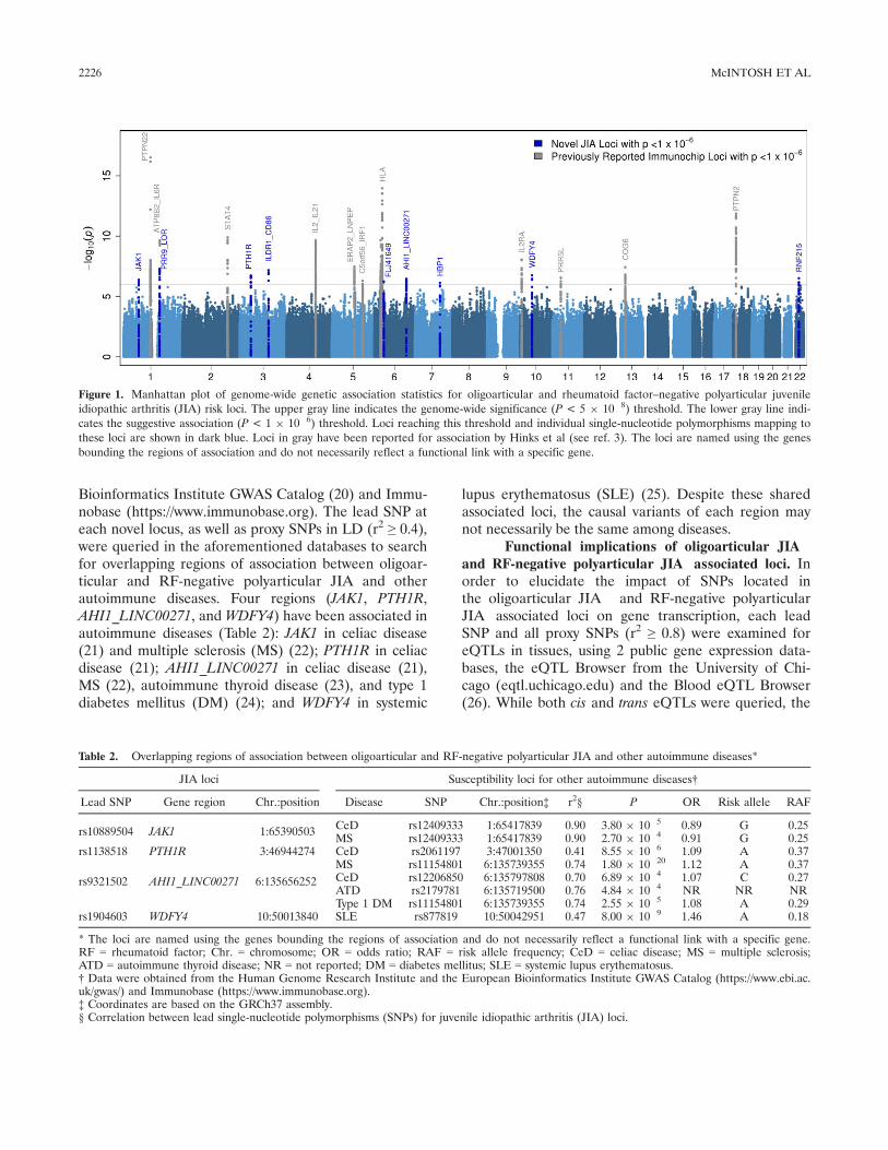
Discovery of new oligoarticular and RF-negativepolyarticular JIA loci. The association results (P < 1 9
10�6) of the meta-analysis of 3 cohorts (2,751 patients and15,886 controls) are shown in Figure 1. The 9 newly
identified oligoarticular and RF-negative polyarticular JIAloci include JAK1, PRR9_LOR, PTH1R, ILDR1_CD86, LINC00951 (FLJ41649), AHI1_LINC00271, HBP1,WDFY4, and RNF215. Regional plots of association(LocusZoom) are shown in Supplementary Figure 2, avail-able on the Arthritis & Rheumatology web site at http://onlinelibrary.wiley.com/doi/10.1002/art.40216/abstract.Lead SNPs representing novel associations with oligoar-ticular and RF-negative polyarticular JIA are shown inTable 1. For each region shown in Table 1, no signalremained after conditioning on the lead SNP. Thestrongest associations included PRR9_LOR (rs873234[P = 5.12 9 10�8, OR 1.43, 95% CI 1.25�1.63]) andILDR1_CD86 (rs111700762 [P = 6.73 9 10�8, OR 1.45,95% CI 1.26�1.66]). In addition, 36 previously unidenti-fied loci achieved suggestive levels of significance (1 9
10�6 < P < 1 9 10�5) in the meta-analysis and are shownin Supplementary Table 2 (available on the Arthritis &Rheumatology web site at http://onlinelibrary.wiley.com/doi/10.1002/art.40216/abstract).
A subset of imputed SNPs from Table 1 and Sup-plementary Table 2 were technically validated by directgenotyping (n = 16). Concordance of >98% betweenimputed and genotyped SNPs was observed in all SNPsevaluated (>99% concordance in 11 of the SNPs). Allremaining SNPs meeting a false discovery rate�cor-rected P value less than 0.05 (n = 1,782) are shown inSupplementary Table 3 (available on the Arthritis &Rheumatology web site at http://onlinelibrary.wiley.com/doi/10.1002/art.40216/abstract).
Autoimmune disease loci overlap. Support forthe 9 newly associated oligoarticular and RF-negativepolyarticular JIA loci was gathered using the NationalHuman Genome Research Institute and the European
Table 1. SNPs representing top statistical associations (P < 1 9 10�6) with oligoarticular JIA and RF-negative polyarticular JIA*
Lead SNP (gene region) Chr.:position† Minor allele
MAF
P Model OR (95% CI) SNP positionCases Controls
rs10889504 (JAK1) 1:65390503 C 0.11 0.13 4.18 9 10�7 Add 0.78 (0.71�0.86) Intronrs873234 (PRR9_LOR) 1:153227177 A 0.40 0.37 5.12 9 10�8 Rec 1.43 (1.25�1.63) Intergenicrs1138518 (PTH1R)‡ 3:46944274 T 0.42 0.37 1.87 9 10�7 Add 1.23 (1.14�1.34) Coding (synonymous)rs111700762 (ILDR1_CD86) 3:121780807 A 0.08 0.06 6.73 9 10�8 Dom 1.45 (1.26�1.66) Intronrs10807228 (LINC00951) 6:40188351 T 0.36 0.33 5.80 9 10�7 Rec 1.42 (1.23�1.65) Intergenicrs9321502 (AHI1_LINC00271) 6:135656252 C 0.43 0.40 3.48 9 10�7 Add 1.18 (1.11�1.26) Intronrs111865019 (HBP1) 7:106812246 G 0.24 0.27 7.29 9 10�7 Add 0.84 (0.78�0.90) Intronrs1904603 (WDFY4) 10:50013840 G 0.29 0.25 1.79 9 10�7 Dom 1.27 (1.16�1.39) Intronrs5753109 (RNF215) 22:30777888 C 0.31 0.28 3.09 9 10�7 Add 1.19 (1.11�1.28) Intron
* The 3 cohorts comprised 2,751 cases and 15,886 controls. The loci are named using the genes bounding the regions of association and do notnecessarily reflect a functional link with a specific gene. SNPs = single-nucleotide polymorphisms; JIA = juvenile idiopathic arthritis; RF =rheumatoid factor; MAF = minor allele frequency; Chr. = chromosome; OR = odds ratio; 95% CI = 95% confidence interval; Add = additive;Rec = recessive; Dom = dominant.† Coordinates are based on the GRCh37 assembly.‡ Because rs1138518 did not pass the quality control criteria in all cohorts, the numbers of cases and controls were 1,937 and 12,828, respectively.
NEWLY IDENTIFIED JIA SUSCEPTIBILITY LOCI 2225
Bioinformatics Institute GWAS Catalog (20) and Immu-nobase (https://www.immunobase.org). The lead SNP ateach novel locus, as well as proxy SNPs in LD (r2 ≥ 0.4),were queried in the aforementioned databases to searchfor overlapping regions of association between oligoar-ticular and RF-negative polyarticular JIA and otherautoimmune diseases. Four regions (JAK1, PTH1R,AHI1_LINC00271, and WDFY4) have been associated inautoimmune diseases (Table 2): JAK1 in celiac disease(21) and multiple sclerosis (MS) (22); PTH1R in celiacdisease (21); AHI1_LINC00271 in celiac disease (21),MS (22), autoimmune thyroid disease (23), and type 1diabetes mellitus (DM) (24); and WDFY4 in systemic
lupus erythematosus (SLE) (25). Despite these sharedassociated loci, the causal variants of each region maynot necessarily be the same among diseases.
Functional implications of oligoarticular JIA�and RF-negative polyarticular JIA�associated loci. Inorder to elucidate the impact of SNPs located inthe oligoarticular JIA� and RF-negative polyarticularJIA�associated loci on gene transcription, each leadSNP and all proxy SNPs (r2 ≥ 0.8) were examined foreQTLs in tissues, using 2 public gene expression data-bases, the eQTL Browser from the University of Chi-cago (eqtl.uchicago.edu) and the Blood eQTL Browser(26). While both cis and trans eQTLs were queried, the
Table 2. Overlapping regions of association between oligoarticular and RF-negative polyarticular JIA and other autoimmune diseases*
JIA loci Susceptibility loci for other autoimmune diseases†
Lead SNP Gene region Chr.:position Disease SNP Chr.:position‡ r2§ P OR Risk allele RAF
rs10889504 JAK1 1:65390503 CeD rs12409333 1:65417839 0.90 3.80 9 10�5 0.89 G 0.25MS rs12409333 1:65417839 0.90 2.70 9 10�4 0.91 G 0.25
rs1138518 PTH1R 3:46944274 CeD rs2061197 3:47001350 0.41 8.55 9 10�6 1.09 A 0.37MS rs11154801 6:135739355 0.74 1.80 9 10�20 1.12 A 0.37
rs9321502 AHI1_LINC00271 6:135656252 CeD rs12206850 6:135797808 0.70 6.89 9 10�4 1.07 C 0.27ATD rs2179781 6:135719500 0.76 4.84 9 10�4 NR NR NRType 1 DM rs11154801 6:135739355 0.74 2.55 9 10�5 1.08 A 0.29
rs1904603 WDFY4 10:50013840 SLE rs877819 10:50042951 0.47 8.00 9 10�9 1.46 A 0.18
* The loci are named using the genes bounding the regions of association and do not necessarily reflect a functional link with a specific gene.RF = rheumatoid factor; Chr. = chromosome; OR = odds ratio; RAF = risk allele frequency; CeD = celiac disease; MS = multiple sclerosis;ATD = autoimmune thyroid disease; NR = not reported; DM = diabetes mellitus; SLE = systemic lupus erythematosus.† Data were obtained from the Human Genome Research Institute and the European Bioinformatics Institute GWAS Catalog (https://www.ebi.ac.uk/gwas/) and Immunobase (https://www.immunobase.org).‡ Coordinates are based on the GRCh37 assembly.§ Correlation between lead single-nucleotide polymorphisms (SNPs) for juvenile idiopathic arthritis (JIA) loci.
Figure 1. Manhattan plot of genome-wide genetic association statistics for oligoarticular and rheumatoid factor–negative polyarticular juvenileidiopathic arthritis (JIA) risk loci. The upper gray line indicates the genome-wide significance (P < 5 9 10�8) threshold. The lower gray line indi-cates the suggestive association (P < 1 9 10�6) threshold. Loci reaching this threshold and individual single-nucleotide polymorphisms mapping tothese loci are shown in dark blue. Loci in gray have been reported for association by Hinks et al (see ref. 3). The loci are named using the genesbounding the regions of association and do not necessarily reflect a functional link with a specific gene.
2226 McINTOSH ET AL
results identified evidence of cis eQTLs only. StrongeQTLs were identified for 5 oligoarticular and RF-negative polyarticular JIA loci: PTH1R SNPs forCCDC12; AHI1_LINC00271 SNPs for AHI1; HBP1SNPs for HBP1, COG5, and GPR22; WDFY4 SNPs forWDFY4; and RNF215 SNPs for MTP18 and SF3A1(Table 3).
Next, the novel oligoarticular JIA� and RF-negative polyarticular JIA�associated loci were analyzedfor histone modifications. Using the 7 cell types in theENCODE database for which the desired data wereavailable (GM12878 cells, H1-hESC cells, HSMMs,HUVECs, K562 cells, normal HEK cells, and normalHLFs) and immunologically relevant cell types from theEpiGenome Browser (CD14, CD15, CD19, PBMCs,CD3, CD4M, CD4N, and CCCTreg [see Patients andMethods]), the SNP set described above for the eQTLanalysis was evaluated for H3K4me1, H3K4me3, andH3K27ac modifications. H3K4me1 modifications tendto mark enhancer regions, whereas H3K4me3 modifica-tions tend to mark promoter regions. Regions that aretranscriptionally activated are associated with H3K27acmarks (27). Supplementary Table 4 (available on theArthritis & Rheumatology web site at http://onlinelibrary.wiley.com/doi/10.1002/art.40216/abstract) summarizes thehistone modification mark data for each of the 9 noveloligoarticular JIA and RF-negative polyarticular JIAassociations, indicated by the enrichment score (number
of sequences) centered on a 25-bp window size. The max-imum enrichment score among cell lines was determined,and the top 5 values for each histone mark were identi-fied. Several SNPs were located in regions with enrichedhistone marks, mostly in isolated cell types. In addition,some SNPs were in regions with multiple marks (e.g.,rs72922282 and rs10511408), indicating a high likelihoodthat these SNPs are in functionally active chromatinregions.
Comparison of RA- and type 1 DM�associatedloci with oligoarticular and RF-negative polyarticularJIA. Many of the risk loci identified for oligoarticular JIAand RF-negative polyarticular JIA are shared with otherautoimmune diseases, particularly RA and type 1 DM(3). However, individual variants identified may varybetween diseases. To date, association studies have identi-fied 101 loci for RA (28) and 50 loci for type 1 DM (24).Supplementary Tables 5 and 6 (available on the Arthritis &Rheumatology web site at http://onlinelibrary.wiley.com/doi/10.1002/art.40216/abstract) show the findings fromthis study for each SNP reported for RA and type 1 DM,respectively. Within the oligoarticular JIA and RF-nega-tive polyarticular JIA data set, 86 RA and 39 type 1 DMSNPs met quality control metrics. In total, 9 RA and 6type 1 DM SNPs, or only 10�15%, reached a Bonferroni-corrected P value less than 5.8 9 10�4 (calculated basedon 86 tests) or P < 1.2 9 10�3 (calculated based on 39tests), respectively. These significantly associated SNPs inoligoarticular and RF-negative polyarticular JIA areshown in Supplementary Tables 5 and 6.
Additionally, a power analysis for each of theseRA-associated SNPs (see Supplementary Table 5)and type 1 DM�associated SNPs (see SupplementaryTable 6) was computed, assuming the OR reported forRA or type 1 DM, the allele frequency in the JIA con-trols, the JIA patient sample size, and the Type I errorrate defined by the Bonferroni corrections describedabove. The sum of the power across the SNPs is thestatistical expectation of the number of associations acohort of the size observed in this meta-analysis woulddetect. If the effect sizes in the JIA cohort were consis-tent with the RA or type 1 DM effect sizes for therespective SNPs, then the expected number of associa-tions the JIA cohort should detect for RA is 25.9 (95%confidence interval [95% CI] 16.5�32.7) and for type 1DM is 26.1 (95% CI 21.1�31.1).
DISCUSSION
This study includes the largest JIA cohort ana-lyzed on genome-wide platforms to date (2,751 patientswith oligoarticular JIA or RF-negative polyarticular
Table 3. Expression of quantitative trait locus (eQTL) genes in noveloligoarticular- and RF-negative polyarticular JIA�associated regions*
Lead SNP (gene region),eQTL SNP† Chr.:position‡ r2§ eQTL gene
rs1138518 (PTH1R)rs2242116 3:46941116 1.00 CCDC12
rs9321502 (AHI1_LINC00271)rs2614276 6:135681704 0.83 AHI1
rs111865019 (HBP1)rs7790080 7:107031322 0.82 COG5rs2301801 7:106870746 0.82 HBP1rs2237659 7:106847492 0.82 GPR22
rs1904603 (WDFY4)rs2940707 10:49989792 0.95 WDFY4
rs5753109 (RNF215)rs757870 22:30776419 0.91 MTP18rs4820003 22:30711624 0.91 SF3A1
* The loci are named using the genes bounding the regions of asso-ciation and do not necessarily reflect a functional link with a specificgene. RF = rheumatoid factor; JIA = juvenile idiopathic arthritis;Chr. = chromosome.† The single-nucleotide polymorphism (SNP) with the highest eQTLscore for each eQTL gene is shown; data were obtained from theUniversity of Chicago (eqtl.uchicago.edu) and Blood eQTL Browser(26).‡ Coordinates are based on the GRCh37 assembly.§ Correlation with lead SNP.
NEWLY IDENTIFIED JIA SUSCEPTIBILITY LOCI 2227
JIA and 15,886 controls) and identifies new oligoarticu-lar and RF-negative polyarticular JIA associations (Fig-ure 1). Nine of the 28 loci detected in oligoarticularand RF-negative polyarticular JIA Immunochip studies(3) remained significant (P < 5 9 10�8) in the currentgenome-wide analysis (PTPN22, ATP8B2_IL6R, STAT4,IL2_IL21, ERAP2_LNPEP, HLA, IL2RA, COG6, andPTPN2); 2 additional regions, C5orf56_IRF1 andPRR5L, achieved suggestive levels of association (P < 19 10�6) (Figure 1). The higher-density SNP coverageprovided by the Immunochip platform compared togenome-wide arrays may account for differences in thefindings. However, there are comparable magnitudes ofeffect (Spearman’s correlation coefficient = 0.89)between the OR reported for the most significant SNPsidentified in the Immunochip study (3) and the currentGWAS. Use of the Exome Array for the genotyping ofcohort III was economically driven, but the lack ofSNP inclusion in this array may contribute to these dif-ferential findings.
Although ~70% of the patient samples used forthis study have been used in previous association stud-ies (3�5), by using genome-wide data sets, we nowextend the number of genetic loci associated with JIA.In spite of these efforts, the newly reported associationsdo not meet the generally used, but somewhat arbi-trary, threshold of P < 5 9 10�8. Genome-wide thresh-olds assume 1 million independent tests and a genome-wide Type I error rate of 0.05. Yet, it is difficult todetermine how many independent tests of associationare computed in a GWAS, because LD pruning usingr2 < 0.4 reduces the number of loci in this Europeanancestral cohort to <1 million, and even these remaincorrelated. Nonetheless, the need remains to validatethese findings in other cohorts, which are difficult toacquire for a rare disease affecting young children.Indeed, the patient cohorts reported were collectedover the course of several decades.
Four of the newly identified oligoarticular JIAand RF-negative polyarticular JIA loci (JAK1, PTH1R,AHI1_LINC00271, and WDFY4) (see Table 2) reportedhere have been described as being associated withanother autoimmune disease, according to the GWASCatalog and ImmunoBase, suggesting common patho-physiologic mechanisms. Although the databases cata-loging GWAS and Immunochip findings are incomplete,they begin to allow the findings of this study to be putin the context of other autoimmune diseases. Only asmall percentage of the reported lead RA (28) and type1 DM (24) SNPs reaching the Bonferroni-correctedP value cutoffs in our data set (see SupplementaryTables 5 and 6, available on the Arthritis &
Rheumatology web site at http://onlinelibrary.wiley.com/doi/10.1002/art.40216/abstract). This is markedlyless than expected and well outside the confidenceintervals. Given that the RA- and type 1 DM�associ-ated SNPs were discovered in predominately Europeanancestral groups, it is unlikely that this deficit is merelydue to differences in LD among the different diseasepopulations. Rather, these results support the notionthat although JIA (at least the oligoarticular and RF-negative polyarticular subtypes) shares some risk loci, itis genetically distinct from seropositive RA (despite sim-ilar clinical presentations) and type 1 DM (anotherautoimmune disease with childhood onset). The remain-ing 5 unique newly identified oligoarticular and RF-negative polyarticular JIA loci reveal additional regionsof the genome that will require further investigation tofully delineate their importance in JIA and autoimmunediseases in general.
The PRR9_LOR locus was the most stronglyassociated novel region, approaching genome-widesignificance (P = 5.12 9 10�8). PRR9 encodes a proteinof unknown function, with the highest expression levelsin the skin (29), while LOR encodes loricrin, a compo-nent of the cornified cell envelope in terminally differ-entiated epidermal cells. Interestingly, associationanalyses in patients with psoriasis, an inflammatory skindisorder, have identified PRR9 as a susceptibility locus(30,31). Although this may represent overlap of diseaserisk loci between JIA and psoriasis, there is also thepossibility that the JIA cohorts include psoriasispatients, given that ~20�25% of patients present witharthritis before skin disease, which is difficult to definein younger children.
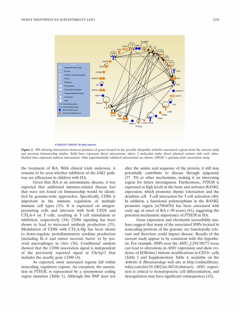
Biologic pathways that feature gene products sug-gested by the results of this study and previous Immuno-chip studies (3,10) may offer insight into diseasepathophysiology (see Figure 2). While some of the asso-ciated regions observed in the current study may nothave direct functional relationships with other loci, locisuch as CD86 and JAK1 appear to be central to key sig-naling pathways involving T cell differentiation and pro-liferation. JAK1 encodes a tyrosine kinase that interactswith the common c-chain to elicit signals from cytokines,such as interleukin-2 (IL-2) and IL-21, to induce inter-feron-c, IL-17, and IL-6 production (32). Further bioin-formatic analysis revealed the presence of H3K4me1histone modifications near JAK1 in CD14+ cells andCD15+ cells, suggesting the presence of an enhancerregion at this locus (see Supplementary Table 4, avail-able on the Arthritis & Rheumatology web site at http://onlinelibrary.wiley.com/doi/10.1002/art.40216/abstract).Notably, a JAK inhibitor, tofacitinib, is already used for
2228 McINTOSH ET AL
the treatment of RA. With clinical trials underway, itremains to be seen whether inhibitors of the JAK1 path-way are efficacious in children with JIA.
Given that JIA is an autoimmune disease, it wasexpected that additional immune-related disease locithat were not found on Immunochip would be identi-fied by genome-wide approaches. Specifically, CD86 isimportant in the immune regulation of multipleimmune cell types (33). It is expressed on antigen-presenting cells and interacts with both CD28 andCTLA-4 on T cells, resulting in T cell stimulation orinhibition, respectively (34). CD86 signaling has beenshown to lead to increased antibody production (35).Modulation of CD86 with CTLA-4Ig has been shownto down-regulate proinflammatory cytokine production(including IL-6 and tumor necrosis factor a) by syn-ovial macrophages in vitro (36). Conditional analysisshowed that the CD86 association signal is independentof the previously reported signal at Chr3q13 thatincludes the nearby gene CD80 (4).
As expected, most associated regions fall withinnoncoding, regulatory regions. An exception, the associa-tion in PTH1R, is represented by a synonymous codingregion mutation (Table 1). Although this SNP does not
alter the amino acid sequence of the protein, it still maypotentially contribute to disease through epigenetic(37�39) or other mechanisms, making it an interestingregion for future investigation. Furthermore, PTH1R isexpressed at high levels in the bone and activates RANKLexpression, which promotes thymic tolerization and thedendritic cell�T cell interaction for T cell activation (40).In addition, a functional polymorphism in the RANKLpromoter region (rs7984870) has been associated withearly age at onset of RA (~50 years) (41), suggesting thepotential mechanistic importance of PTH1R in JIA.
Gene expression and chromatin accessibility ana-lyses suggest that many of the associated SNPs located innoncoding portions of the genome are functionally rele-vant and therefore could impact disease. Results of thecurrent study appear to be consistent with this hypothe-sis. For example, SNPs near the AHI1_LINC00271 locuscan lead to alterations in AHI1 expression and show evi-dence of H3K4me1 histone modifications in CD14+ cells(Table 3 and Supplementary Table 4, available on theArthritis & Rheumatology web site at http://onlinelibrary.wiley.com/doi/10.1002/art.40216/abstract). AHI1 expres-sion is critical to hematopoietic cell differentiation, anddysregulation may have significant consequences (42).
Figure 2. IPA showing interactions between products of genes located in the juvenile idiopathic arthritis–associated regions from the current studyand previous Immunochip studies. Solid lines represent direct interactions, where 2 molecules make direct physical contact with each other.Dashed lines represent indirect interactions. Only experimentally validated interactions are shown. GWAS = genome-wide association study.
NEWLY IDENTIFIED JIA SUSCEPTIBILITY LOCI 2229
Other mechanisms are also probable. For exam-ple, several East Asian GWAS showed associations ofWDFY4 with SLE (43,44). Functional studies usingperipheral blood mononuclear cells revealed decreasedmessenger RNA expression of WDFY4 in SLE patientswhen compared to controls. This may be due to thefact that intronic SNP rs877819 has a decreased bind-ing affinity to transcription factor Yin Yang 1 (YY1).Binding of YY1 to this intronic region was shown todirectly influence WDFY4 expression (45). This SNP ismarginally associated with JIA (P = 1.3 9 10�3 [geno-typed in cohorts I and II]) but is in LD with the leadJIA SNP in the WDFY4 region (rs1904603; r2 = 0.47).Although the functional relevance of these eQTLs andhistone analyses is limited to the cell and tissue typesavailable in public data sets, the information providedis, nonetheless, important to prioritizing future func-tional studies.
Expression QTL analysis also showed that SNPvariations near HBP1 and RNF215 are related to theexpression levels of nearby genes COG5 and MTP18/SF3A1, respectively (Table 3 and SupplementaryTable 4). Intriguingly, COG5, like COG6, is a memberof the conserved oligomeric Golgi (COG) complex thatregulates protein glycosylation and Golgi trafficking.We previously reported COG6 genetic associations inJIA, and associations with the COG6 region have alsobeen described in RA and SLE (3,5,46). Thus, thefindings that risk variants for multiple COG proteinsin JIA were identified, that COG6 associations havebeen reported for other autoimmune disease, and thatCOG defects result in systemic pathologies (47) sug-gests that Golgi complex pathways may be importantin autoimmune disease. In addition, results of theeQTL analysis also implicate MTP18, which representsa plausible candidate because it is a mitochondrialprotein downstream of phosphatidylinositol 3-kinasesignaling, potentially playing a role in the induction ofapoptosis. Although there is no direct support for theassociated region nearest LINC00951 (FLJ41649), thegenetic data, including numerous directly genotypedSNPs, support inclusion as a JIA risk locus (see Sup-plementary Figure 2, available on the Arthritis &Rheumatology web site at http://onlinelibrary.wiley.com/doi/10.1002/art.40216/abstract).
In conclusion, the results of this study furtheremphasize the role of common genetic variation andadd to the understanding of the genomic architectureinfluencing the risk of oligoarticular and RF-negativepolyarticular JIA. As seen in other autoimmune dis-eases, the majority of these JIA-associated SNPs arelocated in regulatory regions, supporting the notion
that JIA is also a disease of disordered gene regulation.Further work, including evaluation of chromatin inter-actions and regulatory regions, is essential to under-standing the contributions of associated SNPs todisease risk and the genomics influencing oligoarticularand RF-negative polyarticular JIA (48). There is alsoevidence of interplay among associated regions, sug-gesting dysregulated pathways as potential targets inclinical care.
ACKNOWLEDGMENTS
We would like to thank the Wake Forest School ofMedicine Center for Public Health Genomics for providingcomputational resources and data analytics support.
AUTHOR CONTRIBUTIONS
All authors were involved in drafting the article or revising itcritically for important intellectual content, and all authors approvedthe final version to be published. Dr. Thompson had full access to allof the data in the study and takes responsibility for the integrity ofthe data and the accuracy of the data analysis.Study conception and design. Langefeld, Thompson.Acquisition of data. McIntosh, Sudman, Becker, Bohnsack, Fingerlin,Griffin, Haas, Lovell, Maier, Nigrovic, Prahalad, Punaro, Ros�e, Wal-lace, Wise, Langefeld, Thompson.Analysis and interpretation of data. McIntosh, Marion, Sudman,Comeau, Moncrieffe, Howard, Langefeld, Thompson.
REFERENCES
1. Petty R, Cassidy J. Chronic arthritis in childhood. Philadelphia:Elsevier; 2011.
2. Saurenmann RK, Rose JB, Tyrrell P, Feldman BM, Laxer RM,Schneider R, et al. Epidemiology of juvenile idiopathic arthritisin a multiethnic cohort: ethnicity as a risk factor. ArthritisRheum 2007;56:1974–84.
3. Hinks A, Cobb J, Marion MC, Prahalad S, Sudman M, Bowes J,et al. Dense genotyping of immune-related disease regions identi-fies 14 new susceptibility loci for juvenile idiopathic arthritis. NatGenet 2013;45:664–9.
4. Thompson SD, Marion MC, Sudman M, Ryan M, Tsoras M,Howard TD, et al. Genome-wide association analysis of juvenileidiopathic arthritis identifies a new susceptibility locus at chromo-somal region 3q13. Arthritis Rheum 2012;64:2781–91.
5. Thompson SD, Sudman M, Ramos PS, Marion MC, Ryan M,Tsoras M, et al. The susceptibility loci juvenile idiopathic arthritisshares with other autoimmune diseases extend to PTPN2, COG6,and ANGPT1. Arthritis Rheum 2010;62:3265–76.
6. Prahalad S, Zeft AS, Pimentel R, Clifford B, McNally B, MineauGP, et al. Quantification of the familial contribution to juvenileidiopathic arthritis. Arthritis Rheum 2010;62:2525–9.
7. Prahalad S, Shear ES, Thompson SD, Giannini EH, Glass DN.Increased prevalence of familial autoimmunity in simplex andmultiplex families with juvenile rheumatoid arthritis. ArthritisRheum 2002;46:1851–6.
8. Petty RE, Southwood TR, Baum J, Bhettay E, Glass DN,Manners P, et al. Revision of the proposed classification criteriafor juvenile idiopathic arthritis: Durban, 1997. J Rheumatol1998;25:1991–4.
2230 McINTOSH ET AL
9. Hollenbach JA, Thompson SD, Bugawan TL, Ryan M, SudmanM, Marion M, et al. Juvenile idiopathic arthritis and HLA class Iand class II interactions and age-at-onset effects. Arthritis Rheum2010;62:1781–91.
10. Hinks A, Bowes J, Cobb J, Ainsworth HC, Marion MC, ComeauME, et al. Fine-mapping the MHC locus in juvenile idiopathicarthritis (JIA) reveals genetic heterogeneity corresponding to dis-tinct adult inflammatory arthritic diseases. Ann Rheum Dis2016;76:765–72.
11. Behrens EM, Finkel TH, Bradfield JP, Kim CE, Linton L,Casalunovo T, et al. Association of the TRAF1–C5 locus onchromosome 9 with juvenile idiopathic arthritis. Arthritis Rheum2008;58:2206–7.
12. Hinks A, Barton A, Shephard N, Eyre S, Bowes J, Cargill M,et al. Identification of a novel susceptibility locus for juvenileidiopathic arthritis by genome-wide association analysis. ArthritisRheum 2009;60:258–63.
13. Brewer EJ Jr, Bass J, Baum J, Cassidy JT, Fink C, Jacobs J, et al.Current proposed revision of JRA criteria. Arthritis Rheum1977;20 Suppl 2:195–9.
14. Holle R, Happich M, Lowel H, Wichmann HE, MONICA/KORAStudy Group. KORA: a research platform for population basedhealth research. Gesundheitswesen 2005;67 Suppl 1:S19–25.
15. Manichaikul A, Mychaleckyj JC, Rich SS, Daly K, Sale M, ChenWM. Robust relationship inference in genome-wide associationstudies. Bioinformatics 2010;26:2867–73.
16. Alexander DH, Novembre J, Lange K. Fast model-based estimationof ancestry in unrelated individuals. Genome Res 2009;19:1655–64.
17. 1000 Genomes Project Consortium, Abecasis GR, Auton A,Brooks LD, DePristo MA, Durbin RM, et al. An integrated mapof genetic variation from 1,092 human genomes. Nature 2012;491:56–65.
18. Ward LD, Kellis M. HaploReg: a resource for exploring chro-matin states, conservation, and regulatory motif alterations withinsets of genetically linked variants. Nucleic Acids Res 2012;40(database issue):D930–4.
19. Boyle AP, Hong EL, Hariharan M, Cheng Y, Schaub MA,Kasowski M, et al. Annotation of functional variation in personalgenomes using RegulomeDB. Genome Res 2012;22:1790–7.
20. Welter D, MacArthur J, Morales J, Burdett T, Hall P, Junkins H,et al. The NHGRI GWAS catalog, a curated resource of SNP-trait associations. Nucleic Acids Res 2014;42(database issue):D1001–6.
21. Trynka G, Hunt KA, Bockett NA, Romanos J, Mistry V, SzperlA, et al. Dense genotyping identifies and localizes multiple com-mon and rare variant association signals in celiac disease. NatGenet 2011;43:1193–201.
22. International Multiple Sclerosis Genetics Consortium, BeechamAH, Patsopoulos NA, Xifara DK, Davis MF, Kemppinen A, et al.Analysis of immune-related loci identifies 48 new susceptibilityvariants for multiple sclerosis. Nat Genet 2013;45:1353–60.
23. Cooper JD, Simmonds MJ, Walker NM, Burren O, Brand OJ,Guo H, et al. Seven newly identified loci for autoimmune thyroiddisease. Hum Mol Genet 2012;21:5202–8.
24. Onengut-Gumuscu S, Chen WM, Burren O, Cooper NJ, QuinlanAR, Mychaleckyj JC, et al. Fine mapping of type 1 diabetes sus-ceptibility loci and evidence for colocalization of causal variantswith lymphoid gene enhancers. Nat Genet 2015;47:381–6.
25. Yang W, Tang H, Zhang Y, Tang X, Zhang J, Sun L, et al. Meta-analysis followed by replication identifies loci in or nearCDKN1B, TET3, CD80, DRAM1, and ARID5B as associatedwith systemic lupus erythematosus in Asians. Am J Hum Genet2013;92:41–51.
26. Westra HJ, Peters MJ, Esko T, Yaghootkar H, Schurmann C,Kettunen J, et al. Systematic identification of trans eQTLs as putativedrivers of known disease associations. Nat Genet 2013;45:1238–43.
27. Shlyueva D, Stampfel G, Stark A. Transcriptional enhancers:from properties to genome-wide predictions. Nat Rev Genet2014;15:272–86.
28. Okada Y, Wu D, Trynka G, Raj T, Terao C, Ikari K, et al. Genet-ics of rheumatoid arthritis contributes to biology and drug discov-ery. Nature 2014;506:376–81.
29. GTEx Consortium. The Genotype-Tissue Expression (GTEx) pro-ject. Nat Genet 2013;45:580–5.
30. Munir S, ber Rahman S, Rehman S, Saba N, Ahmad W, NilssonS, et al. Association analysis of GWAS and candidate gene lociin a Pakistani population with psoriasis. Mol Immunol 2015;64:190–4.
31. Chen H, Toh TK, Szeverenyi I, Ong RT, Theng CT, McLean WH,et al. Association of skin barrier genes within the PSORS4 locusis enriched in Singaporean Chinese with early-onset psoriasis. JInvest Dermatol 2009;129:606–14.
32. Tanaka Y. Recent progress and perspective in JAK inhibitors forrheumatoid arthritis: from bench to bedside. J Biochem 2015;158:173–9.
33. Cutolo M, Nadler SG. Advances in CTLA-4-Ig-mediated modula-tion of inflammatory cell and immune response activation inrheumatoid arthritis. Autoimmun Rev 2013;12:758–67.
34. Linsley PS, Greene JL, Brady W, Bajorath J, Ledbetter JA, PeachR. Human B7-1 (CD80) and B7-2 (CD86) bind with similar avidi-ties but distinct kinetics to CD28 and CTLA-4 receptors. Immu-nity 1994;1:793–801.
35. Rau FC, Dieter J, Luo Z, Priest SO, Baumgarth N. B7-1/2(CD80/CD86) direct signaling to B cells enhances IgG secretion.J Immunol 2009;183:7661–71.
36. Cutolo M, Soldano S, Montagna P, Sulli A, Seriolo B, VillaggioB, et al. CTLA4-Ig interacts with cultured synovial macrophagesfrom rheumatoid arthritis patients and downregulates cytokineproduction. Arthritis Res Ther 2009;11:R176.
37. Oh SH, Baek J, Liany H, Foo JN, Kim KM, Yang SC, et al. Asynonymous variant in IL10RA affects RNA splicing in paediatricpatients with refractory inflammatory bowel disease. J CrohnsColitis 2016;10:1366–71.
38. Gallego-Bustos F, Gotea V, Ramos-Amador JT, Rodriguez-PenaR, Gil-Herrera J, Sastre A, et al. A case of IL-7R deficiencycaused by a novel synonymous mutation and implications formutation screening in SCID diagnosis. Front Immunol2016;7:443.
39. Simhadri VL, Hamasaki-Katagiri N, Lin BC, Hunt R, Jha S,Tseng SC, et al. Single synonymous mutation in factor IX altersprotein properties and underlies haemophilia B. J Med Genet2017;54:338–45.
40. Akiyama T, Shinzawa M, Akiyama N. RANKL-RANK interactionin immune regulatory systems. World J Orthop 2012;3:142–50.
41. Tan W, Wu H, Zhao J, Derber LA, Lee DM, Shadick NA, et al. Afunctional RANKL polymorphism associated with younger age atonset of rheumatoid arthritis. Arthritis Rheum 2010;62:2864–75.
42. Jiang X, Zhao Y, Chan WY, Vercauteren S, Pang E, Kennedy S,et al. Deregulated expression in Ph+ human leukemias of AHI-1,a gene activated by insertional mutagenesis in mouse models ofleukemia. Blood 2004;103:3897–904.
43. Han JW, Zheng HF, Cui Y, Sun LD, Ye DQ, Hu Z, et al.Genome-wide association study in a Chinese Han populationidentifies nine new susceptibility loci for systemic lupuserythematosus. Nat Genet 2009;41:1234–7.
44. Yang W, Shen N, Ye DQ, Liu Q, Zhang Y, Qian XX, et al.Genome-wide association study in Asian populations identifiesvariants in ETS1 and WDFY4 associated with systemic lupuserythematosus. PLoS Genet 2010;6:e1000841.
45. Zhao H, Yang W, Qiu R, Li J, Xin Q, Wang X, et al. An intronicvariant associated with systemic lupus erythematosus changes thebinding affinity of Yinyang1 to downregulate WDFY4. GenesImmun 2012;13:536–42.
46. Marquez A, Vidal-Bralo L, Rodriguez-Rodriguez L, Gonzalez-GayMA, Balsa A, Gonzalez-Alvaro I, et al. A combined large-scalemeta-analysis identifies COG6 as a novel shared risk locus forrheumatoid arthritis and systemic lupus erythematosus. AnnRheum Dis 2017;76:286–94.
NEWLY IDENTIFIED JIA SUSCEPTIBILITY LOCI 2231
47. Smith RD, Lupashin VV. Role of the conserved oligomeric Golgi(COG) complex in protein glycosylation. Carbohydr Res2008;343:2024–31.
48. Martin P, McGovern A, Orozco G, Duffus K, Yarwood A,Schoenfelder S, et al. Capture Hi-C reveals novel candidate genesand complex long-range interactions with related autoimmunerisk loci. Nat Commun 2015;6:10069.
APPENDIX A: CONSORTIA INVOLVED IN THE STUDYAND THEIR PARTICIPATING MEMBERS
Boston Children’s JIA Registry: Fatma Dedeoglu, Robert C.Fuhlbrigge, Melissa M. Hazen, Lauren A. Henderson, Erin Janssen,Susan Kim, Mindy S. Lo, Mary Beth F. Son, Robert P. Sundel, IritTirosh, Heather O. Tory (Boston Children’s Hospital); Peter A.Nigrovic (Boston Children’s Hospital and Brigham and Women’sHospital).
German Society for Pediatric Rheumatology: GuentherDannecker, Gerd Ganser, J. Peter Haas, Hartmut Michels (GermanCenter for Pediatric and Adolescent Rheumatology, Garmisch-Partenkirchen).
Gene Expression in Pediatric Arthritis Study: Mara L.Becker (Children’s Mercy Kansas City); Robert A. Colbert (NationalInstitute of Arthritis and Musculoskeletal and Skin Diseases[NIAMS], Bethesda, MD); Jason Dare (Arkansas Children’s Hospital,Little Rock); Beth S. Gottleib (Steven and Alexandra Cohen Chil-dren’s Medical Center, New Hyde Park. NY), Thomas A. Griffin(Levine Children’s Hospital, Charlotte, NC); Alexie A. Grom, DanielJ. Lovell, Halima Moncrieffe, Susan D. Thompson (Cincinnati Chil-dren’s Hospital Medical Center, University of Cincinnati, Cincinnati,OH); Norman T. Ilowite (Albert Einstein College of Medicine andChildren’s Hospital at Montefiore, Bronx, NY); Peter A. Nigrovic(Boston Children’s Hospital and Brigham and Women’s Hospital);Judy Ann Olsen (Indiana University School of Medicine, Indi-anapolis); Sampath Prahalad (Emory University School of Medicine,Atlanta, GA); Margalit Rosenkranz (Children’s Hospital of Pittsburghof UPMC, Pittsburgh, PA); David D. Sherry (Children’s Hospital ofPhiladelphia, Philadelphia, PA).
NIAMS JIA Genetic Registry: John F. Bohnsack (Universityof Utah Health Sciences Center, Salt Lake City); Gloria Higgins(Nationwide Children’s Hospital and Ohio State University, Colum-bus, OH); Marissa Klein-Gittelman (Northwestern University Fein-berg School of Medicine and Children’s Memorial Hospital, Chicago,IL); T. Brent Graham (Vanderbilt University, Nashville, TN); ThomasA. Griffin (Levine Children’s Hospital, Charlotte, NC); Paula W.Morris (University of Arkansas for Medical Sciences, Little Rock);Natasha Ruth, Murray H. Passo (Medical University of South Caro-lina, Charleston), Sampath Prahalad (Emory University School ofMedicine, Atlanta, GA); Stephen J. Spaulding (Cleveland Clinic,
Cleveland, OH); Susan D. Thompson (Cincinnati Children’s HospitalMedical Center [CCHMC] and University of Cincinnati, Cincinnati,OH).
Trial of Early Aggressive Therapy in JIA (TREAT): Carol A.Wallace, Sarah Ringold, Stephanie Hamilton (Seattle Children’s Hospi-tal and Seattle Children’s Research Institute, Seattle, WA); Edward H.Giannini, Hermine I. Brunner, Anne L. Johnson, Bin Huang, Daniel J.Lovell (CCHMC and University of Cincinnati, Cincinnati, OH); StevenJ. Spalding, Andrew S. Zeft (Cleveland Clinic, Cleveland, OH); PhilipJ. Hashkes (Shaare Zedek Medical Center, Jerusalem, Israel); Kath-leen M. O’Neil, Peter Chira (Indiana University, Indianapolis); Ilona S.Szer (Rady Children’s Hospital San Diego, San Diego, CA); Laura E.Schanberg (Duke University Medical Center, Durham, NC); Robert P.Sundel (Children’s Hospital of Boston, Boston, MA); Diana Milojevic(University of California, San Francisco); Marilynn G. Punaro (TexasScottish Rite Hospital, Dallas); Beth S. Gottlieb (Steven and AlexandraCohen Children’s Medical Center, New Hyde Park, NY); Gloria C.Higgins (Nationwide Children’s Hospital and Ohio State University,Columbus, OH); Norman T. Ilowite (Albert Einstein College of Medi-cine and Children’s Hospital at Montefiore, Bronx, NY); YukikoKimura (Joseph M. Sanzari Children’s Hospital at Hackensack Univer-sity Medical Center, Hackensack, NJ).
Improved Understanding of the Biology and Use of TNFInhibition in Children with JIA Study: Daniel J. Lovell, Alexie A.Grom, Anne L. Johnson, Janalee Taylor, Hermine I. Brunner, JenniferL. Huggins, Tracy V. Ting, Bin Huang, Edward H. Giannini (CCHMCand University of Cincinnati, Cincinnati, OH); Steven J. Spalding,Andrew Zeft (Cleveland Clinic, Cleveland, OH); Beth S. Gottlieb, Cal-vin B. Williams, Anne B. Eberhard (Steven and Alexandra Cohen Chil-dren’s Medical Center, New Hyde Park, NY); Paula W. Morris(University of Arkansas for Medical Sciences, Little Rock); YukikoKimura, Suzanne C. Li, Kathleen A. Haines, Jennifer E. Weiss (JosephM. Sanzari Children’s Hospital at Hackensack University Medical Cen-ter, Hackensack, NJ); Karen Onel, Melissa S. Tesher, Linda Wagner-Weiner (University of Chicago Comer Children’s Hospital, Chicago,IL); James J. Nocton, James W. Verbsky, Judyann C. Olson (MedicalCollege of Wisconsin, Milwaukee); Barbara S. Edelheit, Lawrence S.Zemel (Connecticut Children’s Medical Center, Hartford); MichaelShishov, Kaleo C. Ede (Phoenix Children’s Hospital, Phoenix, AZ);Lawrence K. Jung, Denise M. Costanzo (Children’s National MedicalCenter, Washington, DC; Cleveland Clinic Foundations, Cleveland,OH); Jason A. Dare (Arkansas Children’s Hospital, Little Rock); Mur-ray H. Passo (Medical University of South Carolina, Charleston);Elaine A. Cassidy, Daniel Kietz (Children’s Hospital of Pittsburgh,Pittsburgh, PA); Thomas A. Griffin (Levine Children’s Hospital, Char-lotte, NC); Larry B. Vogler, Kelly A. Rouster-Stevens (Emory Univer-sity School of Medicine, Atlanta, GA); Timothy Beukelman, Randy Q.Cron (University of Alabama at Birmingham); Kara M. Schmidt, Ken-neth Schikler (University of Louisville Kosair Charities Pediatric Clini-cal Research Unit, Louisville, KY); Jay Mehta (Children’s Hospital atMontefiore, Bronx, NY).
2232 McINTOSH ET AL