gene expression profiling of tuberculous meningitis
TRANSCRIPT
Volume 4(5) : 098-105 (2011) - 098 J Proteomics Bioinform ISSN:0974-276X JPB, an open access journal
Research Article Open Access
Kumar et al., J Proteomics Bioinform 2011, 4:5http://dx.doi.org/10.4172/jpb.1000174
Research Article Open Access
Proteomics & Bioinformatics
Gene Expression Profiling of Tuberculous MeningitisGhantasala S. Sameer Kumar1,2, Abhilash K. Venugopal1,2,3,4, Lakshmi Dhevi N. Selvan1,5, Arivusudar Marimuthu1,6, Shivakumar Keerthikumar1#, Swapnali Pathare7, Jyoti Bajpai Dikshit7, Pramila Tata7, Ramesh Hariharan7, Thottethodi Subrahmanya Keshava Prasad1, H. C. Harsha1, Y.L Ramachandra2, Anita Mahadevan8, Raghothama Chaerkady1,2,3,4, S. K. Shankar8* and Akhilesh Pandey3,4,9,10*1Institute of Bioinformatics, International Technology Park, Bangalore 560066, Karnataka, India2Department of Biotechnology, Kuvempu University, Shimoga 577 451, India3McKusick-Nathans Institute of Genetic Medicine4Departments of Biological Chemistry5School of Biotechnology, Amrita university, Kollam 690525, India6Manipal University, Madhav Nagar, Manipal, Karnataka 576104 India7Strand Life Sciences, Bangalore 560024, Karnataka, India8Department of Neuropathology, National Institute of Mental Health and Neurosciences, Bangalore 560029, India 9Department of Pathology 10Oncology, Johns Hopkins University School of Medicine, Baltimore, MD 21205, USA #Current address: Center for Molecular and Biomolecular Informatics and Nijmegen Center for Molecular Life Sciences, Radboud University Nijmegen Medical Centre, 6525 GA, Nijmegen, The Netherlands
Corresponding authors: Akhilesh Pandey, M.D., Ph.D., McKusick-Nathans Institute of Genetic Medicine, 733 N. Broadway, BRB 527, Johns Hopkins University, Baltimore, MD 21205, Tel: 410-502-6662; Fax: 410-502-7544; E-mail: [email protected]
Dr. S. K. Shankar, MD, FAMS, FNASc, FIC Path, Department of Neuropathology, National Institute of Mental Health and Neurosciences, Bangalore 560029, India, Tel:91-080-26995001/5002; Fax: 91-080-26564830; E-mail: [email protected]
Received December 17, 2010; Accepted May 17, 2011; Published May 20, 2011
Citation: Kumar GSS, Venugopal AK, Selvan LDN, MarimuthuA, Keerthikumar S, et al. (2011) Gene Expression Profiling of Tuberculous Meningitis. J Proteomics Bioinform 4: 098-105. doi:10.4172/jpb.1000174
Copyright: © 2011 Kumar GSS, et al. This is an open-access article distributed under the terms of the Creative Commons Attribution License, which permits unrestricted use, distribution, and reproduction in any medium, provided the original author and source are credited.
Keywords: DNA microarrays; Biomarkers; Early diagnosis; Therapeutic target
IntroductionAlthough the causative organism of tuberculosis was discovered
over a hundred years ago, this disease still remains a major public health problem worldwide. Tuberculosis primarily affects the lungs but can spread hematogenously to extra pulmonary sites such as lymph nodes, bones, meninges and genito-urinary-tract [1]. One of the most frequent sites of extra pulmonary disease is tuberculous meningitis (TBM), which is a common form of central nervous system tuberculosis with high morbidity and mortality [2-4]. The incidence of TBM is on the rise with the increase in immunodeficient states such as HIV/AIDS [5]. Concomitant with an increase in the incidence of TBM, development of multi-drug resistance in AIDS patients is a major obstacle associated with its treatment [6].
The diagnosis of TBM continues to be a challenge because the gold standard for diagnosis requires that Mycobacterium tuberculosis (M.tb) be demonstrated by culture of the cerebrospinal fluid (CSF) of suspected patients. This is a time consuming process which takes approximately 8 weeks [7]. Over the past several years, different molecular and biochemical assays have been developed for rapid diagnosis of M.tb. PCR based assays for detection of the pathogen, ELISA to detect M.tb protein antigens or host antibodies directed against M.tb are the most widely used assays for detection of M.tb [8]. A combined approach of detecting IFN-gamma levels by radioimmunoassay and use of IS6110 primer to detect M.tb by PCR shows a reasonably high sensitivity (80%) and specificity (92.6%), in the CSF of TBM patients [9]. Detection of anti-mycobacterial antibodies and mycobacterial immune complexes (IgG) have also been employed for diagnosis with variable sensitivity
and specificity [10]. Other alternatives include demonstration of high lactate levels [11,12] and adenosine deaminase (ADA) in CSF for quick diagnosis and management of TBM [13]. However, the above described methods are still limited in their sensitivity and specificity in the clinical setting. As the mortality rate of TBM remains high [14,15], there is a critical need for identification of appropriate biomarkers for early diagnosis of TBM.
Gene expression profiling studies have previously been performed on CSF or blood of patients with TBM [16,17]. In the present study, we used whole genome DNA microarrays for investigating changes at the transcriptome level in infected brain tissues from TBM cases that were confirmed by autopsy as compared to uninfected brain tissues from controls. Further, we performed immunohistochemical validation of some of the differentially expressed genes identified from
AbstractTuberculous meningitis (TBM) is a form of extra pulmonary tuberculosis that is associated with severe
neurological deficits and a high mortality. Early diagnosis of TBM is a major challenge despite the availability of several diagnostic methods. Existing diagnostic methods and markers are inadequate for early diagnosis of TBM owing to poor specificity and sensitivity. DNA microarray technology permits high-throughput identification of differentially expressed genes. In order to identify molecules as candidate biomarkers for early diagnosis or as therapeutic targets in TBM, we carried out transcriptomic analysis of brain tissue using whole human genome oligonucleotide arrays. From this gene expression analysis, we identified 2,434 genes that were differentially expressed at least two-fold in TBM cases as compared to controls. The large majority of the differentially expressed genes encoded proteins that are involved in metabolism, cell growth, transport, immune response, cell communication and signal transduction. We confirmed the upregulation of two molecules, serpin peptidase inhibitor, clade A member 3 (SERPINA3) and glial fibrillary acidic protein (GFAP), at the protein level by immunohistochemical analysis. The findings from our study should help us understand the molecular mechanisms underlying TBM and to develop better diagnostic and therapeutic strategies against this deadly disease.
Citation: Kumar GSS, Venugopal AK, Selvan LDN, MarimuthuA, Keerthikumar S, et al. (2011) Gene Expression Profiling of Tuberculous Meningitis. J Proteomics Bioinform 4: 098-105. doi:10.4172/jpb.1000174
Volume 4(5) : 098-105 (2011) - 099 J Proteomics Bioinform ISSN:0974-276X JPB, an open access journal
our microarray studies. Our results confirm that proteins encoded by SERPINA3 and GFAP genes are indeed upregulated in TBM patients. Further studies to detect proteins encoded by these genes in the CSF as potential biomarkers in CSF could potentially lead to improved methods for diagnosing TBM.
Materials and MethodsTissue samples
Human brain tissue samples from five cases of TBM (confirmed by detection of mycobacterial antibody/immune complexes in cerebrospinal fluid, and/or demonstration of acid fast bacilli by Ziehl Neilson’s stain in the smear from basal exudates in meninges and histopathological features of granulomatous or chronic meningitis) and four control brains that were archived as frozen and formalin fixed specimens at Human Brain Tissue Repository (Human Brain Bank) in the Department of Neuropathology at National Institute of Mental Health and Neuro Sciences (NIMHANS), Bangalore, India, were used for the microarray experiments. The details of samples used are provided in the Supplementary Table 1.
The brains were collected at autopsy with written informed consent from close relatives to utilize them for research purposes. The study was approved by the Institutional Ethics Committee of NIMHANS. The dead bodies were shifted to 4°C within one hour of death. Tissues from frontal cortex (2x2 cm) were frozen at -86°C and preserved until analysis. The rest of the brain was fixed in 10% buffered formalin for 12–18 weeks. Representative tissue blocks were processed for histological evaluation. The postmortem interval (interval from the time of death to time the tissue was transferred to -86°C) varied from 6 hrs-13½ hrs for control cases and 1 hr 15 minutes–18 hrs for TBM cases. Frozen brain tissue samples with overlying meninges from the five cases and four controls were excised as small pieces, transferred to RNA Later (Ambion Inc Austin Tx) and incubated at 4°C for 12–16 hours to facilitate proper penetration into the tissues and stored at -86°C until further use.
RNA isolation
Approximately 100 mg of tissue from normal and infected brains was used for RNA isolation. The tissues were pulverized in QIAzol lysis reagent (Qiagen, Valencia, CA) using a homogenizer. Total RNA extraction and purification was carried out using RNeasy lipid tissue mini kit (QIAGEN, Valencia, CA) as per the manufacturer’s protocol. The yield and quality of isolated total RNA was checked using the NanoDrop spectrophotometer (Nanodrop Technologies, Wilmington, DE). Integrity of the isolated RNA was assessed by RNA gel and/or 2100 Bioanalyzer (Agilent Technologies, Palo Alto, CA). The samples were further processed based on RNA integrity.
cDNA synthesis, labeling and hybridization
Total RNA (600 ng) from each sample was reverse transcribed and linear amplified using Quick Amp Kit, One-color (Agilent Technologies, Palo Alto, CA) that employ OligodT-T7 promoter primers. The cDNA generated was used as template for in vitro transcription reaction with Cy3-CTP and RNA polymerase; thus cRNA was simultaneously synthesized and labeled. The labeled cRNA was purified using RNeasy spin columns (Qiagen, Valencia, CA). The samples with specific activity >9 pmol Cy3 per µg and yield >1600µg were selected for hybridization. Cy-3 labeled cRNA was fragmented and hybridized onto oligonucleotide-based whole human genome DNA microarrays (G4112F, 4x44 K, Agilent Technologies, Palo Alto, CA) for 16 hours at
65°C. The arrays were subsequently washed with gene expression wash buffers according to the manufacturer’s hybridization protocol (Agilent Oligo Microarray Kit, Agilent Technologies).
Scanning and data analysis
The slides were scanned with Agilent microarray scanner (G2505B) using one color scan setting for 4x44 K array slides (scan resolution 5µm, dye channel was set to green, green PMT was set to 100%) and the images processed with Agilent’s feature extraction software (9.5.3.1) to obtain the raw data files for further analysis. The raw data from microarray experiments were submitted to the Gene Expression Omnibus (http://www.ncbi.nlm.nih.gov/geo accession#GSE23074). GeneSpring GX v11.0.2 (Agilent Technologies, Santa Clara) software was used to analyze the gene expression profiles. Raw data was imported into the GeneSpring GX software. The recommended quantile normalization without baseline transformation and t-test were applied. To determine the differentially expressed genes that were statistically significant, a p-value of <0.05 and a fold-change cut-off threshold of ≥2 were used. GO analysis was carried out for the differentially expressed genes using GeneSpring Gx software. A p-value threshold of 0.1 was used to filter out the significantly overrepresented GO categories.
Biological network analysis
Pathway analysis was carried out using Genespring GX v.11.0.2. Differentially expressed genes obtained after filtering based on fold-change cut off (≥4.0) was taken as input and biological networks were generated by comparing the input list to a reference list containing >1.4 million reactions generated by natural language processing algorithm and from different interaction databases. To obtain high confidence networks, analysis was carried out using filters that included binding, expression, metabolism, transport, promoter binding and regulation of the molecules. The number of molecules per network was restricted to 50. The entities which do not have connections were removed. The constructed network was overlaid with final input list to visualize differentially expressed genes.
Immunohistochemical analysis
For validation of the upregulated molecules in cases of TBM, immunohistochemical labeling was carried out using commercially available monoclonal antibodies directed against GFAP (Biogenex, Houston, Texas, USA) at 1:200 dilution and against SERPINA3 (Sigma Aldrich, St. Louis, MO) at 1:100 dilution. HRP tagged secondary antibody provided with Envision kit (DAKO-K4011 and K4007, DAKO, Carpinteria, CA) was used and the immune reaction visualized with DAB/H2O2 as the chromogen. The immunohistochemical validation was carried out on an independent subset of 15 formalin fixed human brains from confirmed cases of TBM (Age range 1–55 years, postmortem interval 1 hour–18 hours) including the five cases used for gene expression profile and four controls. These cases were processed for paraffin embedding and histological evaluation to establish presence of TBM.
Briefly, 4μm thick serial sections from controls and TBM cases were collected on silane coated slides. The paraffin sections were deparaffinized and dehydrated. After stabilizing in PBS at room temperature, the endogenous peroxidase activity was quenched by blocking solution for 20 min at room temperature. Antigen retrieval was carried out in citrate buffer (pH 6.0) by microwaving the sections for 30 min. After blocking the non-specific binding sites in 3% non-fat dry milk powder for 15 min at room temperature, the serial sections were incubated with primary antibodies against GFAP and SERPINA3
Citation: Kumar GSS, Venugopal AK, Selvan LDN, MarimuthuA, Keerthikumar S, et al. (2011) Gene Expression Profiling of Tuberculous Meningitis. J Proteomics Bioinform 4: 098-105. doi:10.4172/jpb.1000174
Volume 4(5) : 098-105 (2011) - 100 J Proteomics Bioinform ISSN:0974-276X JPB, an open access journal
overnight at 4°C. The sections were washed thrice in PBS and incubated with the appropriate secondary antibodies conjugated with HRP for 30 min at room temperature. The signal was developed with DAKO substrate buffer and chromogen. The sections were counterstained with hematoxylin and mounted. The immunolabeled sections were examined independently by two experienced neuropathologists (AM and SKS). The staining pattern, intensity and subcellular localization were visually scored.
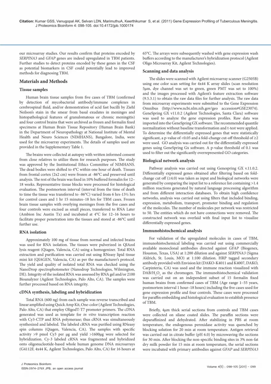
Results and DiscussionGene expression profiling using DNA microarrays was carried
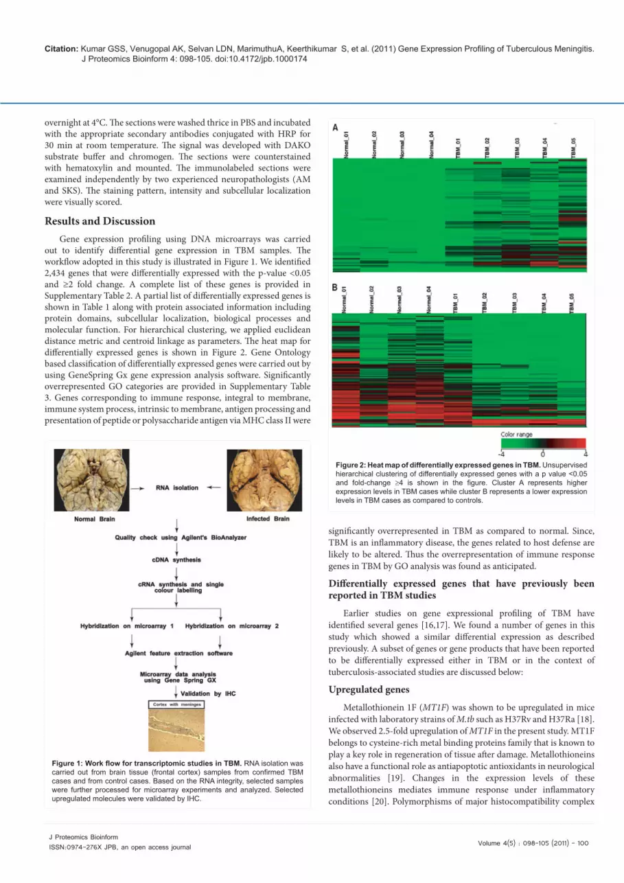
out to identify differential gene expression in TBM samples. The workflow adopted in this study is illustrated in Figure 1. We identified 2,434 genes that were differentially expressed with the p-value <0.05 and ≥2 fold change. A complete list of these genes is provided in Supplementary Table 2. A partial list of differentially expressed genes is shown in Table 1 along with protein associated information including protein domains, subcellular localization, biological processes and molecular function. For hierarchical clustering, we applied euclidean distance metric and centroid linkage as parameters. The heat map for differentially expressed genes is shown in Figure 2. Gene Ontology based classification of differentially expressed genes were carried out by using GeneSpring Gx gene expression analysis software. Significantly overrepresented GO categories are provided in Supplementary Table 3. Genes corresponding to immune response, integral to membrane, immune system process, intrinsic to membrane, antigen processing and presentation of peptide or polysaccharide antigen via MHC class II were
significantly overrepresented in TBM as compared to normal. Since, TBM is an inflammatory disease, the genes related to host defense are likely to be altered. Thus the overrepresentation of immune response genes in TBM by GO analysis was found as anticipated.
Differentially expressed genes that have previously been reported in TBM studies
Earlier studies on gene expressional profiling of TBM have identified several genes [16,17]. We found a number of genes in this study which showed a similar differential expression as described previously. A subset of genes or gene products that have been reported to be differentially expressed either in TBM or in the context of tuberculosis-associated studies are discussed below:
Upregulated genes
Metallothionein 1F (MT1F) was shown to be upregulated in mice infected with laboratory strains of M.tb such as H37Rv and H37Ra [18]. We observed 2.5-fold upregulation of MT1F in the present study. MT1F belongs to cysteine-rich metal binding proteins family that is known to play a key role in regeneration of tissue after damage. Metallothioneins also have a functional role as antiapoptotic antioxidants in neurological abnormalities [19]. Changes in the expression levels of these metallothioneins mediates immune response under inflammatory conditions [20]. Polymorphisms of major histocompatibility complex
Figure 1: Work flow for transcriptomic studies in TBM. RNA isolation was carried out from brain tissue (frontal cortex) samples from confirmed TBM cases and from control cases. Based on the RNA integrity, selected samples were further processed for microarray experiments and analyzed. Selected upregulated molecules were validated by IHC.
Figure 2: Heat map of differentially expressed genes in TBM. Unsupervised hierarchical clustering of differentially expressed genes with a p value <0.05 and fold-change ≥4 is shown in the figure. Cluster A represents higher expression levels in TBM cases while cluster B represents a lower expression levels in TBM cases as compared to controls.
Citation: Kumar GSS, Venugopal AK, Selvan LDN, MarimuthuA, Keerthikumar S, et al. (2011) Gene Expression Profiling of Tuberculous Meningitis. J Proteomics Bioinform 4: 098-105. doi:10.4172/jpb.1000174
Volume 4(5) : 098-105 (2011) - 101 J Proteomics Bioinform ISSN:0974-276X JPB, an open access journal
S.no GeneSymbol Architecture/Domain Primary ocalization
Alternate localization Biological process Molecular function Molecule class +/- Fold change
TBM/Normal1 CHI3L2 SP Nucleus N/A Bone remodeling Molecular function
unknown Unclassified + 58
2 UBD UBQ Nucleus Cytoplasm Protein metabolism Ubiquitin-specific protease activity
Ubiquitin proteasome system protein
+ 56
3 IGL@ IGC; IGV; SP N/A N/A Immune response Antigen binding Immunoglobulin + 42.54 IGHA2 Ig_LIKE; IGV; SP N/A N/A Immune response Antigen binding Immunoglobulin + 405 CXCL9 SCY; CC; CXC; SP Extracellular N/A Immune response Chemokine activity Chemokine + 366 IGHG1 Ig_LIKE; IGV N/A N/A Immune response Antigen binding Immunoglobulin + 327 SLAMF8 TM; SP Plasma
membrane N/A Immune response Receptor activity Cell surface receptor + 24
8 GBP5 CC N/A N/A Cell communication; Signal transduction GTPase activity GTPase + 22
9 CARTPT SP Extracellular N/A Biological_process unknown
Molecular function unknown Unclassified + 20
10 CIITA LRR Nucleus N/A Immune response Transcription regulator activity
Transcription regulatory protein + 20
11 LOC100131733 N/A N/A N/A N/A N/A N/A + 19
12 APOL6 TM; CC Cytoplasm N/A Metabolism; Energy pathways Transporter activity Transport/cargo
protein + 18.5
13 CHI3L1 SP Extracellular N/A Cell growth and/or maintenance
Extracellular matrix structural constituent
Extracellular matrix protein + 16
14 IL4I1 SP N/A N/A Apoptosis; Immune response Catalytic activity Enzyme: Oxidase + 15
15 SERPINA3 SERPIN; SP Extracellular Cytoplasm; Nucleus Protein metabolism Protease inhibitor
activity Protease inhibitor + 14
16 MYBPH FN3; IGC2 Cytoplasm N/A Cell growth and/or maintenance
Structural molecule activity Structural protein + 14
17 SLAMF7 IG; TM; SP Plasma membrane N/A Immune response Receptor activity Cell surface
receptor + 14
18 IL21R FN3; TM; SP; WSXWS Plasma membrane N/A Immune response Transmembrane
receptor activity Cytokine receptor + 13
19 IDO1 N/A Cytoplasm N/A Metabolism; Energy pathways Catalytic activity Enzyme:
Oxygenase + 13
20 SLC14A1 TM Plasma membrane N/A Transport Transporter activity Transport/cargo
protein + 13
21 SCIN GEL Cytoplasm Plasma membrane
Cell growth and/or maintenance
Cytoskeletal protein binding
Cytoskeletal associated protein + 11
22 PKD2L1 EF; TM; CC Plasma membrane N/A Transport Ion channel activity Ion channel + 11
23 CTAG1A N/A N/A N/A Biological_process unknown
Molecular function unknown Unclassified + 11
24 TYMP N/A Extracellular Cytoplasm; Nucleus
Cell communication; Signal transduction Growth factor activity Growth factor + 11
25 LAX1 TM Integral to membrane N/A Cell communication;
Signal transduction
Receptor signaling complex scaffold activity
Adapter molecule + 10
26 SPOCD1 TFS2M N/A N/A Biological_process unknown
Molecular function unknown Unclassified + 10
27 CD2 TM; SP Plasma membrane N/A Immune response Receptor activity Cell surface
receptor + 10
28 TTR TRANSTHYR; SP Extracellular N/A Transport Transporter activity Transport/cargo protein - 23
29 RELN EGF; SP Extracellular N/A Protein metabolism Serine-type peptidase activity Serine protease - 19
30 LYVE1 LINK; TM; SP Plasma membrane N/A Cell communication;
Signal transduction Receptor activity Cell surface receptor - 19
31 DEFA3 SP Extracellular N/A Immune response Defense/immunity protein activity Defensin - 15
32 HIAT1 TM N/A N/A Transport Transporter activity Transport/cargo protein - 13
33 EXPH5 N/A Nucleus N/A Biological_process unknown
Molecular function unknown Unclassified - 12
34 GLIPR1L2 SCP N/A N/A Biological_process unknown
Molecular function unknown Unclassified - 12
35 TMED7-TICAM2 N/A N/A N/A NA NA NA - 1236 ABHD3 SP N/A N/A Biological_
process unknownMolecular function unknown Unclassified - 11
37 CNTN6 FN3; Ig_LIKE; IGC2; SP N/A N/A Cell communication; Signal transduction
Cell adhesion molecule activity Adhesion molecule - 11
38 C9orf5 TM N/A N/A Biological_process unknown
Molecular function unknown
Integral membrane protein - 11
39 EDNRB TM; SP Plasma membrane
Lysosome; Endoplasmic reticulum
Cell communication; Signal transduction
G-protein coupled receptor activity
G protein coupled receptor - 11
40 TMCO3 TM; CC; SP Integral to membrane N/A Biological_
process unknownMolecular function unknown
Integral membrane protein - 10
41 APPBP2 CC CytoplasmCytoplasmic vesicle; Golgi apparatus
Regulation of gene expression, epigenetic
Molecular function unknown Adapter molecule - 10
42 S100A12 N/A Cytoplasm N/A Cell communication; Signal transduction Calcium ion binding Calcium binding
protein - 10
Note: Positive sign (+) indicates upregulation, Negative sign (-) indicates downregulation and N/A: Not availableTable 1: A partial list of differentially regulated genes expressed in TBM.

Citation: Kumar GSS, Venugopal AK, Selvan LDN, MarimuthuA, Keerthikumar S, et al. (2011) Gene Expression Profiling of Tuberculous Meningitis. J Proteomics Bioinform 4: 098-105. doi:10.4172/jpb.1000174
Volume 4(5) : 098-105 (2011) - 102 J Proteomics Bioinform ISSN:0974-276X JPB, an open access journal
class II, DR beta 1 (HLA-DRB1) gene have been found to be associated with susceptibility to TB [21]. HLA-DRB1 belongs to an important class of molecules which play a crucial role in the process of antigen presentation [22]. HLA-DRB1 was shown to be upregulated 3-fold in the current study. IL12RB1 encoded protein is a type-1 transmembrane protein, which belongs to the hemopoietin receptor superfamily [23]. Upregulation of IL12RB1 has been reported earlier in M.tb infected mice [18]. Further, nucleotide polymorphisms in IL12RB1 have also been reported to be associated with susceptibility to TB [21]. In this study, we found 3-fold upregulation of IL12RB1 transcript in TBM. The protein encoded by solute carrier family 11, member 1 (SLC11A1) belongs to the solute carrier family and is also known as natural resistance-associated macrophage protein 1. Polymorphisms in SLC11A1 have been associated with susceptibility to TB [21]. We observed a 4-fold upregulation of SLC11A1 transcript in TBM cases. Tumor necrosis factor receptor superfamily, member 4 (TNFRSF4) is known to be involved in the pathogenesis of various immunological abnormalities including infectious, autoimmune, inflammatory related diseases. It has been shown to be upregulated in mice infected with M.tb strains H37Rv and H37Ra [18,24]. In this study, TNFRSF4 transcript was 2-fold upregulated in TBM as compared to uninfected controls. Chemokine (C-X-C motif) ligand 9 (CXCL9) is a T cell trafficking chemokine [25], which is known to be upregulated in mice infected with laboratory strains of M.tb and high concentrations of CXCL9 have been reported in the CSF of TBM patients [18,26]. We observed a 36-fold upregulation of CXCL9 transcript in our study. Met proto-oncogene (MET), also known as hepatocyte growth factor receptor, encodes a receptor tyrosine kinase, which has shown to be overexpressed in M.tb stimulated monocyte-derived macrophages (MDMs) [17]. We found a 9-fold upregulation of this transcript in our transcriptomic study. CHI3L1 (Chitinase 3-like 1) is involved in the process of inflammation and tissue remodeling. CHI3L1 was also shown to be upregulated in peripheral blood mononuclear cells (PBMCs) of recovered extra pulmonary tuberculosis patients upon incubating their PBMCs with whole lysates of M.tb [16]. CHI3L1 transcript was 16-fold upregulated in the present study.
Downregulated genes
SLC15A2 (Solute carrier family 15, member 2 ) is known to be involved in the induction of proton-dependent transport for transporting small peptides [27]. In an ex vivo experiment, M.tb-stimulated MDMs showed downregulation of SLC15A2 [17]. In this study, we found 5-fold downregulation of SLC15A2 transcript in TBM. ITGB1 (Integrin beta 1) belongs to the membrane receptor family, which is involved in cell adhesion and recognition. ITGB1 is also involved in several cellular processes including immune response [28]. ITGB1 transcript was observed to be 3-fold downregulated in this study. It has been reported that ITGB1 was downregulated in fetal lung cell line in the presence of M.tb recombinant CFP-10/ESAT-6 protein (rCFES) [29]. Cathepsin L1 (CTSL1) encoded protein is a lysosomal cysteine proteinase, which is involved in intracellular protein catabolism. CTSL1 transcript was shown to be 2-fold downregulated in this study and the encoded protein is localized to endosomes. CTSL1 activity and maturation was affected by Mycobacterium avium and M.tb infected macrophages [30].
Differentially expressed genes that were not reported earlier
A large number of differentially expressed genes identified in our study have not been previously reported in the literature to be
associated with TBM. These genes are involved in various biological functions including cell-cell communication, enzymatic activity and signal transduction.
Novel upregulated genes
Over the past several years, various studies have proven the overexpression of GFAP (Glial fibrillary acidic protein) in CNS tuberculosis, toxocariasis and pneumococcal meningitis [31-33]. It has been shown to be overexpressed in astrocytes when there is an astrogliosis during CNS inflammation. In the present study, GFAP transcript showed 5-fold upregulation. Interleukin 4 induced 1 (IL4I1) expression has been shown to be induced by IL-4 in B-cells and this secreted protein is known to be involved in the regulation of T lymphocytes [34]. IL4I1 transcript was found to be 15-fold upregulation in our study. Interleukin 21 receptor (IL21R) belongs to the type 1 cytokine receptor family and is involved in the maturation of natural killer cell and regulation of T lymphocytes [35]. IL21R binds to its ligand and activates the downstream signaling molecules like JAK 1, JAK3, STAT1 and STAT3 [36]. IL21R transcript showed 13-fold upregulation in the current study. Serpin peptidase inhibitor, clade A (alpha-1 antiproteinase, antitrypsin), member 3 (SERPINA3) is a plasma protease inhibitor and is a member of the serine protease inhibitor class. SERPINA3 transcript was found to be 14-fold upregulated in the current study. The overexpression of this protein has been reported in several neurological disorders like schizophrenia [37,38]. GFAP and SERPINA3 were chosen for further validation by immunohistochemical analysis which is explained in the later section.
Novel downregulated genes
RELN mRNA was observed to be 19-fold downregulated in the Figure 3
Expression
Binding
Regulation
Promoter binding
TransportMember
Metabolism
Protein modi�cation
(+) Catalyst
(+) Modulator(+) Regulator
(+) Target(-) Catalyst(-) Modulator(-) Regulator(-) Target
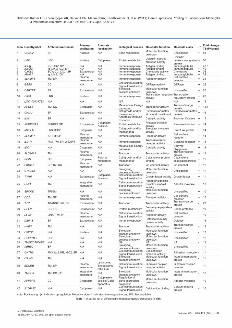
Figure 3: Biological network analysis of differentially expressed genes in TBM. Illustration of subnetwork identified by biological network analysis. CHI3L1 and GFAP which were upregulated in TBM formed a highly interconnected network through SERPINA3.

Citation: Kumar GSS, Venugopal AK, Selvan LDN, MarimuthuA, Keerthikumar S, et al. (2011) Gene Expression Profiling of Tuberculous Meningitis. J Proteomics Bioinform 4: 098-105. doi:10.4172/jpb.1000174
Volume 4(5) : 098-105 (2011) - 103 J Proteomics Bioinform ISSN:0974-276X JPB, an open access journal
present study. Reelin (RELN) encodes a secreted extracellular matrix protein. RELN is involved in the molecular mechanism of cognitive functions. Defects in RELN expression have been associated with the abnormality of neuronal position and dendritic development in mouse models [39]. The protein encoded by Vacuolar protein sorting 26 (VPS26) is involved in retrograde transport of proteins. VPS26 was found to be 2-fold downregulated in TBM cases. Sorting nexin 3 (SNX3), sorting nexin 12 (SNX12) and sorting nexin 18 (SNX18) belong to the sorting nexin family and are involved in intracellular trafficking. Inhibited expression of the SNX3 has been shown to affect membrane trafficking from early endosomes to recycling endosomes [40]. In this study, SNX3 transcript was found to be 3-fold downregulated and protein encoded by this gene is localized to the endosomes. SNX12 and SNX18 were found to be downregulated 3-fold and 4-fold, respectively. Defensin alpha 3, neutrophil-specific (DEFA3) protein belongs to the family of microbicidal peptides which is involved in antimicrobial activity. DEFA3 mRNA and protein expression levels have been reported to be decreased in transmigrated monocytes [41]. It showed 15-fold downregulation in our study.
Biological network data analysis
Several genes are involved in carrying out a specific biological process and they often interact with each other. We carried out biological network analysis using GeneSpring Gx software to identify such
networks in relation to TBM. Natural language processing is used by GeneSpring to generate a database of interacting molecules. Genes that were differentially expressed in TBM were used as input which resulted in the generation of a complex network depending on the connectivity between the genes. It was comprised of several nodes forming distinct subnetworks. Expression values were overlaid onto the network. We identified a subnetwork with SERPINA3 being the key molecule which is shown in Figure 3. SERPINA3 is known to be associated with several neurological disorders including Parkinson’s disease [42], Alzheimer’s disease [43], schizophrenia and cerebrovascular disease [44]. Notably, we observed GFAP and CHI3L1 which were upregulated in TBM to be part of this subnetwork. Earlier reports have shown that CHI3L1 and GFAP were co localized in the brain infarction and in other neurological diseases [45]. Other molecules that were part of the subnetwork include P704P, FKSG30, ACTBL2, ACTG2, ACTO7 and FHL5. Although it is intriguing that we see the interaction of these molecules in the represented network, the exact functional role and significance with respect to TBM has to be investigated further.
Figure 4: Immunohistochemical labeling of GFAP. A: Whole mount preparation of frontal cortex from a control shows thin subpial band of gliosis (arrow) and very light labeling of protoplasmic astrocytes. The white matter (w) shows diffuse, low intensity staining. (GFAP, Obj x5). B: Whole mount preparation of frontal cortex from a case of TBM showing thick subpial carpet (arrow) extending into the cortical grey matter. Note the reduced gradient of labeling of grey matter and diffuse dark staining of white matter (w). (GFAP, Obj x 5). C: Low power view of the frontal cortex from a case of TBM shows thick gliotic pial band (arrow) and hypertrophic glial cells in the superficial cortex. Above the pial band in the subarachnoid space, inflammatory exudates are observed. (GFAP, Obj x 10). D: The white matter at higher magnification from the control shows astrocytes with small body and thin long processes, some of them impinging on the vessel wall to form a fine lacy background. (GFAP, Obj x 20). E: Higher magnification of the subpial zone from the case of TBM showing large, hypertrophic reactive astrocytes with darkly labeled body and thick cell processes. The neuropil between the cells also has coarse fibres unlike the control. (GFAP, Obj x 40).
Figure 5: Immunohistochemical labeling of SERPINA3. A: Whole mount preparation of frontal cortex from a control showing low diffuse labeling of the cortical ribbon and low intensity staining of white matter (w). (SERPINA3, Obj x 5). B: Whole mount preparation of frontal cortex from a case of TBM showing diffuse intense staining of the cortical ribbon and exudates in the subarachnoid space (w - white matter). (SERPINA3, Obj x 5). C: The pyramidal neurons of the superficial cortex in controls have cytoplasmic labeling. (SERPINA3, Obj x 20). D: The astrocytes in the cortex of controls are stained well, though the density of labeled astrocytes is low. (SERPINA3, Obj x 20). E: The white matter shows a higher density of the SERPINA3 labeled astrocytes in TBM. (SERPINA3, Obj x 10). F: Low power view of the frontal cortex showing dense labeling of subpial glial membrane, reactive astrocytes underneath and the granulomatous exudates in the subarachnoid space. (TBM case) (v: vessel) (SERPINA3, Obj x 20). G: Higher magnification of the subarachnoid exudates showing labeling of the round histocytes. (TBM case) (SERPINA3, Obj x 20).
Citation: Kumar GSS, Venugopal AK, Selvan LDN, MarimuthuA, Keerthikumar S, et al. (2011) Gene Expression Profiling of Tuberculous Meningitis. J Proteomics Bioinform 4: 098-105. doi:10.4172/jpb.1000174
Volume 4(5) : 098-105 (2011) - 104 J Proteomics Bioinform ISSN:0974-276X JPB, an open access journal
Validation of candidate upregulated biomarkers by immunohistochemical labeling
In the present study, we chose to carry out IHC-based validation for two of the novel upregulated molecules identified in this study based on their biological significance and their fold expression. This was performed in 15 TBM cases, including the cases used for gene expression profiling.
GFAP: The staining pattern of GFAP in TBM cases as compared to control cases is shown in histological microphotographs in Figure 4. As shown in the figure, we observed thick subpial carpet staining of GFAP in the whole mount preparation of the frontal cortex of the TBM cases (Figure 4B) as compared to the control groups (Figure 4A). In the frontal cortex, astrocytes in the superficial cortex along with thick processes (Figure 4C), the white matter in the gyri and subcortical band were densely stained with GFAP in TBM cases. In controls (Figure 4D), GFAP labeling is conspicuous around the blood vessels with the foot processes (arrow) impinging on the vessel walls and forming a fine lacy background. In TBM cases, we found strong labeling of GFAP in the hypertrophic astrocytes (Figure 4E) with dark cell body and thick processes.
SERPINA3: Whole mount preparations of immuno stained sections from the frontal cortex in TBM (Figure 5B) showed marginally enhanced staining of the cortical ribbon in contrast to control (Figure 5A). On closer examination in controls, the neuronal cytoplasm in the superficial layers (Figure 5C) and astrocytes in the grey and white matter was labeled strongly with thick processes, but the density of astrocytes was conspicuously lower (Figure 5D). In the case of TBM, a thick subpial carpet was found in addition to numerous subpial astrocytes beneath the dense chronic granulomatous exudates (Figure 5F) and the cortex and white matter (Figure 5E). In the subarachnoid space the histiocytes in the exudates and around the vessels were densely labeled (Figure 5F and Figure 5G). The cortical neuronal labeling is essentially similar to controls, but marginally higher intensity of cell labeling involving all the layers of cortical ribbon unlike control. All these cellular elements contribute to the upregulated gene expression protein profile.
ConclusionsThough many clinical, morphological and biochemical studies
have been carried out earlier on tuberculous meningitis, only a limited number of gene expression profiling studies are have been undertaken. In tropical developing countries, TBM is one of the commonest forms of chronic meningitis. A rise in HIV cases in these geographic regions further worsens the situation. Biomarkers that could facilitate early diagnosis of TBM provide a better opportunity for clinical management of this disease. By carrying out gene expression profiling of infected brain from TBM patients and control subjects, we have identified several differentially expressed genes. Systematic validation of some of the candidate molecules may provide biomarkers with potential clinical utility. Some of the differentially expressed genes encode proteins that are detectable in CSF and targeted studies to develop assay systems to monitor these molecules in CSF may prove useful.
Acknowledgments
The study was supported by a research grant “DBT Programme Support on Neuroproteomics of Neurological Disorders” to the Institute of Bioinformatics and National Institute of Mental Health and Neurosciences by the Department of Biotechnology (DBT), Government of India. This study was supported in part by an NIH roadmap grant for Technology Centers of Networks and Pathways (U54RR020839). T. S. Keshava Prasad is supported by a research grant on “Establishment of a National Database on Tuberculosis” and is a recipient of a Young Investigator Award from DBT. Harsha Gowda is a Wellcome Trust-DBT India Alliance Early Career Fellow. Human brain tissues for the study were obtained from
the Human Brain Bank in the Department of Neuropathology, National Institute of Mental Health and Neuro Sciences, Bangalore, India. Secretarial assistance of Mrs. Manjula Madan is acknowledged.
Conflict of Interest
Authors have declared no conflict of interest.
References
1. Fanning A (1999) Tuberculosis: 6. Extrapulmonary disease. Cmaj 160: 1597-1603.
2. Garg RK (1999) Tuberculosis of the central nervous system. Postgrad Med J 7: 133-140.
3. Hosoglu S, Geyik MF, Balik I, Aygen B, Erol S, et al. (2002) Predictors of outcome in patients with tuberculous meningitis. Int J Tuberc Lung Dis 6: 64-70.
4. Thwaites GE, Nguyen DB, Nguyen HD, Hoang TQ, Do TT, et al. (2004) Dexamethasone for the treatment of tuberculous meningitis in adolescents and adults. N Engl J Med 351: 1741-1751.
5. Berenguer J, Moreno S, Laguna F, Vicente T, Adrados M, et al. (1992) Tuberculous meningitis in patients infected with the human immunodeficiency virus. N Engl J Med 326: 668-672.
6. Horn DL, Hewlett D Jr, Peterson S, Sabido D, Opal SM (1993) RISE-resistant tuberculous meningitis in AIDS patient. Lancet 341: 177-178.
7. Eintracht S, Silber E, Sonnenberg P, Koornhof HJ, Saffer D (2000) Analysis of adenosine deaminase isoenzyme-2 (ADA(2)) in cerebrospinal fluid in the diagnosis of tuberculosis meningitis. J Neurol Neurosurg Psychiatry 69: 137-138.
8. Garg SK, Tiwari RP, Tiwari D, Singh R, Malhotra D, et al. (2003) Diagnosis of tuberculosis: available technologies, limitations, and possibilities. J Clin Lab Anal 17: 155-163.
9. Juan RS, Sanchez-Suarez C, Rebollo MJ, Folgueira D, Palenque E, et al. (2006) Interferon gamma quantification in cerebrospinal fluid compared with PCR for the diagnosis of tuberculous meningitis. J Neurol 253: 1323-1330.
10. Patil SA, Gourie-Devi M, Anand AR, Vijaya AN, Pratima N, et al. (1996) Significance of mycobacterial immune complexes (IgG) in the diagnosis of tuberculous meningitis. Tuber Lung Dis 77: 164-167.
11. Donald PR, Malan C (1985) Cerebrospinal fluid lactate and lactate dehydrogenase levels as diagnostic aids in tuberculous meningitis. S Afr Med J 67: 19-20.
12. Tang LM (1988) Serial lactate determinations in tuberculous meningitis. Scand J Infect Dis 20: 81-83.
13. Gautam N, Aryal M, Bhatta N, Bhattacharya SK, Baral N, et al. (2007) Comparative study of cerebrospinal fluid adenosine deaminase activity in patients with meningitis. Nepal Med Coll J 9: 104-106.
14. Lorber J (1954) The results of treatment of 549 cases of tuberculous meningitis. Am Rev Tuberc 69: 13-25.
15. Verdon R, Chevret S, Laissy JP, Wolff M (1996) Tuberculous meningitis in adults: review of 48 cases. Clin Infect Dis 22: 982-988.
16. Kim DK, Park GM, Hwang YI, Kim HJ, Han SK, et al. (2006) Microarray analysis of gene expression associated with extrapulmonary dissemination of tuberculosis. Respirology 11: 557-565.
17. Thuong NT, Dunstan SJ, Chau TT, Thorsson V, Simmons CP, et al. (2008) Identification of tuberculosis susceptibility genes with human macrophage gene expression profiles. PLoS Pathog 4: e1000229.
18. Beisiegel M, Mollenkopf HJ, Hahnke K, Koch M, Dietrich I, et al. (2009) Combination of host susceptibility and Mycobacterium tuberculosis virulence define gene expression profile in the host. Eur J Immunol 39: 3369-3384.
19. Asmussen JW, Von Sperling ML, Penkowa M (2009) Intraneuronal signaling pathways of metallothionein. J Neurosci Res 87: 2926-2936.
20. Yin X, Knecht DA, Lynes MA (2005) Metallothionein mediates leukocyte chemotaxis. BMC Immunol 6: 21.
21. Cheepsattayakorn A, Cheepsattayakorn R (2009) Human genetic influence on

Citation: Kumar GSS, Venugopal AK, Selvan LDN, MarimuthuA, Keerthikumar S, et al. (2011) Gene Expression Profiling of Tuberculous Meningitis. J Proteomics Bioinform 4: 098-105. doi:10.4172/jpb.1000174
Volume 4(5) : 098-105 (2011) - 105 J Proteomics Bioinform ISSN:0974-276X JPB, an open access journal
susceptibility of tuberculosis: from infection to disease. J Med Assoc Thai 92: 136-141.
22. Cooke GS, Hill AV (2001) Genetics of susceptibility to human infectious disease. Nat Rev Genet 2: 967-977.
23. Chua AO, Chizzonite R, Desai BB, Truitt TP, Nunes P, et al. (1994) Expression cloning of a human IL-12 receptor component. A new member of the cytokine receptor superfamily with strong homology to gp130. J Immunol 153: 128-136.
24. Hori T (2006) Roles of OX40 in the pathogenesis and the control of diseases. Int J Hematol 83: 17-22.
25. Muller M, Carter S, Hofer MJ, Campbell IL (2010) Review: The chemokine receptor CXCR3 and its ligands CXCL9, CXCL10 and CXCL11 in neuroimmunity--a tale of conflict and conundrum. Neuropathol Appl Neurobiol 36: 368-387.
26. Simmons CP, Thwaites GE, Quyen NT, Chau TT, Mai PP, et al. (2005) The clinical benefit of adjunctive dexamethasone in tuberculous meningitis is not associated with measurable attenuation of peripheral or local immune responses. J Immunol 175: 579-590.
27. Liu W, Liang R, Ramamoorthy S, Fei YJ, Ganapathy ME, et al. (1995) Molecular cloning of PEPT 2, a new member of the H+/peptide cotransporter family, from human kidney. Biochim Biophys Acta 1235: 461-466.
28. Barreiro LB, Quintana-Murci L (2009) From evolutionary genetics to human immunology: how selection shapes host defence genes. Nat Rev Genet 11: 17-30.
29. Tsai KN, Chan EC, Tsai TY, Chen KT, Chen CY, et al. (2009) Cytotoxic effect of recombinant Mycobacterium tuberculosis CFP-10/ESAT-6 protein on the crucial pathways of WI-38 cells. J Biomed Biotechnol 2009: 917084.
30. Nepal RM, Mampe S, Shaffer B, Erickson AH, Bryant P (2006) Cathepsin L maturation and activity is impaired in macrophages harboring M. avium and M. tuberculosis. Int Immunol 18: 931-939.
31. Harris JE, Nuttall RK, Elkington PT, Green JA, Horncastle DE, et al. (2007) Monocyte-astrocyte networks regulate matrix metalloproteinase gene expression and secretion in central nervous system tuberculosis in vitro and in vivo. J Immunol 178: 1199-1207.
32. Kadurugamuwa JL, Modi K, Coquoz O, Rice B, Smith S, et al. (2005) Reduction of astrogliosis by early treatment of pneumococcal meningitis measured by simultaneous imaging, in vivo, of the pathogen and host response. Infect Immun 73: 7836-7843.
33. Liao CW, Cho WL, Kao TC, Su KE, Lin YH, et al. (2008) Blood-brain barrier
impairment with enhanced SP, NK-1R, GFAP and claudin-5 expressions in experimental cerebral toxocariasis. Parasite Immunol 30: 525-534.
34. Boulland ML, Marquet J, Molinier-Frenkel V, Moller P, Guiter C, et al. (2007) Human IL4I1 is a secreted L-phenylalanine oxidase expressed by mature dendritic cells that inhibits T-lymphocyte proliferation. Blood 110: 220-227.
35. Parrish-Novak J, Dillon SR, Nelson A, Hammond A, Sprecher C, et al. (2000) Interleukin 21 and its receptor are involved in NK cell expansion and regulation of lymphocyte function. Nature 408: 57-63.
36. Asao H, Okuyama C, Kumaki S, Ishii N, Tsuchiya S, et al. (2001) Cutting edge: the common gamma-chain is an indispensable subunit of the IL-21 receptor complex. J Immunol 167: 1-5.
37. Arion D, Unger T, Lewis DA, Levitt P, Mirnics K (2007) Molecular evidence for increased expression of genes related to immune and chaperone function in the prefrontal cortex in schizophrenia. Biol Psychiatry 62: 711-721.
38. Saetre P, Emilsson L, Axelsson E, Kreuger J, Lindholm E, et al. (2007) Inflammation-related genes up-regulated in schizophrenia brains. BMC Psychiatry 7: 46.
39. D’Arcangelo G (2006) Reelin mouse mutants as models of cortical development disorders. Epilepsy Behav 8: 81-90.
40. Worby CA, Dixon JE (2002) Sorting out the cellular functions of sorting nexins. Nat Rev Mol Cell Biol 3: 919-931.
41. Williams MR, Sakurai Y, Zughaier SM, Eskin SG, McIntire LV (2009) Transmigration across activated endothelium induces transcriptional changes, inhibits apoptosis, and decreases antimicrobial protein expression in human monocytes. J Leukoc Biol 86: 1331-1343.
42. Wang YC, Liu HC, Liu TY, Hong CJ, Tsai SJ (2001) Genetic association analysis of alpha-1-antichymotrypsin polymorphism in Parkinson’s disease. Eur Neurol 45: 254-256.
43. Yu G, Jia J (2010) Is there an association of regulatory region polymorphism in the alpha-1-antichymotrypsin gene with sporadic Alzheimer’s disease in the northern Han-Chinese population? J Clin Neurosci 17: 766-769.
44. Liu W, Zhu Y, Ge M, Pang Q, Yu Y (2010) Polymorphism rs4934 of SERPINA3 and sporadic intracranial aneurysms in the Chinese population. Cerebrovasc Dis 29: 68-72.
45. Bonneh-Barkay D, Wang G, Starkey A, Hamilton RL,Wiley CA In vivo CHI3L1 (YKL-40) expression in astrocytes in acute and chronic neurological diseases. J Neuroinflammation 7: 34.
Submit your next manuscript and get advantages of OMICS Group submissionsUnique features:
• Userfriendly/feasiblewebsite-translationofyourpaperto50world’sleadinglanguages• AudioVersionofpublishedpaper• Digitalarticlestoshareandexplore
Special features:
• 100OpenAccessJournals• 10,000editorialteam• 21daysrapidreviewprocess• Qualityandquickeditorial,reviewandpublicationprocessing• IndexingatPubMed(partial),Scopus,DOAJ,EBSCO,IndexCopernicusandGoogleScholaretc• SharingOption:SocialNetworkingEnabled• Authors,ReviewersandEditorsrewardedwithonlineScientificCredits• Betterdiscountforyoursubsequentarticles
Submityourmanuscriptat:http://www.editorialmanager.com/proteomics