epidemiology of meningitis in an hiv-infected ugandan cohort
TRANSCRIPT
Epidemiology of Meningitis in an HIV-infected Ugandan Cohort
Radha Rajasingham 1,2
Joshua Rhein 1,2
Kate Klammer 3
Abdu Musubire 2
Henry Nabeta 2
Andrew Akampurira 2
Eric C Mossel 4
Darlisha A Williams 1,2
Dave J Boxrud 3
Mary B Crabtree 4
Barry R Miller 4
Melissa A Rolfes 1
Supatida Tengsupakul 1
Alfred O Andama 5
David B Meya 1,2,5
David R Boulware 1
1. University of Minnesota, Minneapolis, Minnesota2. Infectious Disease Institute, Makerere University, Kampala, Uganda3. Minnesota Department of Health, St. Paul, Minnesota4. Centers for Disease Control and Prevention, Fort Collins, Colorado5. Department of Medicine, Makerere University College of Health Sciences,
Kampala, Uganda
Corresponding Author:Radha Rajasingham420 Delaware Street, MMC250Minneapolis, MN [email protected]
MeSH Keywords: Meningitis; Diagnostic Techniques and Procedures; Cryptococcal Meningitis; Tuberculosis Meningitis; bacterial meningitis; opportunistic infection; HIV; Africa
Word CountAbstract: 150 wordsManuscript: 3105 words

Abstract
There is limited understanding of the epidemiology of meningitis amongst HIV-infected
populations in sub-Saharan Africa. We conducted a prospective cohort study of HIV-infected
adults with suspected meningitis in Uganda, to comprehensively evaluate the etiologies of
meningitis. Intensive CSF testing was performed to evaluate for bacterial, viral, fungal, and
mycobacterial etiologies, including neurosyphilis,16s ribosomal DNA (rDNA) PCR for bacteria,
Plex-ID broad viral assay, quantitative-PCR for HSV-1/2, CMV, EBV, and Toxoplasma gondii;
RT-PCR for Enteroviruses and arboviruses, and Xpert MTB/RIF assay. Cryptococcal meningitis
accounted for 60% (188/314) of all causes of meningitis. Of 117 samples sent for viral PCR,
36% were EBV positive. Among cryptococcal antigen negative patients, the yield of Xpert
MTB/RIF assay was 22% (8/36). After exclusion of cryptococcosis and bacterial meningitis,
61% (43/71) with an abnormal CSF profile had no definitive diagnosis. Exploration of new TB
diagnostics and diagnostic algorithms for evaluation of meningitis in resource-limited settings
remains critical.
2

Introduction
Meningitis is a common cause of death in Africa. Historically in the African meningitis belt,
bacterial pathogens such as Neisseria meningitidis and Streptococcus pneumoniae have been the
most common etiologies, resulting in an estimated 800,000 cases between 1996 and 2010.1
During non-epidemic situations in the African meningitis belt, surveillance conducted in Burkina
Faso, Ghana, Niger, and Mali estimate the most common etiological agents of meningitis are:
Neisseria meningitidis (37–52%), Streptococcus pneumoniae (27–43%), and Haemophilus
influenzae (5–31%).2 Because of these findings, the World Health Organization (WHO)
emphasizes empiric ceftriaxone as the first-line therapy,2 despite the geographic diversity of the
continent.
The higher prevalence of HIV infection in Eastern and Southern Africa has a dramatic impact
on the etiologies of meningitis. In Malawi, South Africa, Uganda, Zambia, and Zimbabwe,
cryptococcal meningitis (CM) is the predominant etiology of meningitis followed by TB
meningitis (TBM) in adults.3-8 In both South Africa and Malawi, Cryptococcus is more common
than all causes of bacterial meningitis combined. In Ethiopia, the most common neurologic
manifestation has been thought to be cerebral toxoplasmosis, followed by TBM and
Cryptococcus,11 although recent studies suggest a very large burden of cryptococcosis among
those with advanced HIV.
Unfortunately in many African countries, outside of a research setting, microbiological
diagnoses of meningitis are difficult to ascertain, and patients are treated empirically – often
initially with antibacterial agents per WHO guidance.2 Even in the setting of well-resourced
3
research studies, microbiological meningitis diagnoses are made in a minority of cases. TBM is
notoriously difficult to diagnose with widely differing data on the utility of microscopy for acid
fast bacilli (AFB) smear on CSF.16-19 Mycobacterial culture takes up to 6 weeks to grow, making
culture obsolete for guiding clinical management. CSF interferon-gamma (IFN-γ) levels have
been suggested as a TBM diagnostic biomarker, yet thresholds vary. The debut of the Xpert
MTB/RIF assay, an automated PCR molecular assay, is encouraging. The assay is relatively
rapid (results in ~2.5 hours) and has demonstrated excellent performance with sputum for
pulmonary TB (i.e. sensitivity 86%, specificity >95%).22 Currently, performance data of the
Xpert MTB/RIF in CSF suggest a more limited sensitivity (~60%).
The present study aimed to comprehensively evaluate the microbiologic etiologies of
meningitis in an HIV-infected inpatient adult population with suspected meningitis in Kampala,
Uganda.
Methods
Setting and Patients
We conducted a prospective cohort study of HIV-infected adult inpatients consecutively
presenting with suspected meningitis between November 2010 and October 2012 at Mulago
National Hospital –a tertiary referral hospital in Kampala, Uganda, as part of screening for the
Cryptococcal Optimal ART Timing (COAT) trial.25 We included HIV-infected patients, not
receiving anti-retroviral therapy (ART) presenting with suspected meningitis, and gave written
informed consent. Persons known to be receiving ART were excluded as immune reconstitution
inflammatory syndrome (IRIS) creates different diagnostic considerations beyond the scope of
our aims.26 Clinical suspicion for meningitis was based on 1 or more symptoms of headache,
4
fever, neck stiffness, or altered mental status. CSF was obtained for routine examination and
microbiological testing, as described below. If HIV-serostatus was unknown, rapid HIV testing
was performed. If clinically indicated chest radiograph and sputum examination were obtained.
Neuroradiology was obtained if patients had focal neurologic deficits.
Routine CSF Examination
Prospectively, the Makerere Microbiology Laboratory performed CSF testing of: white cell
count, protein, and India ink. CSF glucose testing was unavailable. Cryptococcal antigen by
latex agglutination was performed by the Makerere University Johns Hopkins University lab.
After centrifugation (3000g x10 minutes) of >5mL initial CSF volume, Gram stain, India ink,
and acid fast bacilli (AFB) stain were performed on the concentrated pellet and the CSF
supernatant was frozen at -80ºC for later testing. Additionally, fungal and bacterial CSF cultures
were performed; however, these later culture results did not influence TBM testing.
Mycobacterial culture was unavailable. Cryptococcal meningitis was diagnosed by CSF culture
or positive CSF cryptococcal antigen by latex agglutination or lateral flow assay (Immy Inc.,
Norman, Oklahoma). Lateral flow assay was prospectively used beginning in 2011.27 Definitive
bacterial meningitis was diagnosed by CSF culture, Gram’s stain, or 16s rDNA sequencing.
Further Diagnostic Studies
Additional CSF testing was performed on 63 samples negative for cryptococcosis to
determine the etiology of meningitis, and a subset of 54 samples with Cryptococcus to identify
potential concurrent infections. Samples were selected based on available remaining CSF for
further testing. 16s rDNA amplification was performed at the University of Minnesota to detect
potential culture-negative bacterial meningitis (Supplemental Methods). At the Minnesota
5

Department of Health (MDH), quantitative polymerase chain reaction (PCR) was performed for
herpes simplex (HSV-1/2), cytomegalovirus (CMV), Epstein-Barr virus (EBV), Toxoplasma
gondii and RT-PCR for enterovirus. Neurosyphilis testing was performed using venereal disease
research laboratory (VDRL).
Additionally, the Ibis PLEX-ID (Abbott, Illinois, USA) Broad Viral 1 Assay was performed,
per manufacturer’s instructions, to detect herpesviruses, adenoviruses, parvovirus B19,
enteroviruses, and polyomaviruses through a broad range mass spectrometry platform. MDH
tested specimens in parallel to evaluate the PLEX-ID system in comparison with established
PCR/RT-PCR methods.
Arboviral meningoencephalitis evaluation was performed on 111 samples negative for
cryptococcosis and TBM, in collaboration with Centers for Disease Control and Prevention
(CDC), by culture and genus-specific PCR for alphavirus, orthobunyavirus, and flavivirus.
Sixty-two serum samples with sufficient remaining sample were evaluated for immunoglobulin
M (IgM) for West Nile, yellow fever, dengue, chikungunya, and Zika viruses.
Xpert MTB/RIF Assay
In 2011-2012, Xpert MTB/RIF assay (Cepheid, Sunnyvale, CA) was performed in Uganda,
according to manufacturer’s instructions, on 63 CSF samples, at physician discretion, for patients
with a lymphocytic pleocytosis to evaluate for TBM. The 1 mL CSF was mixed with 1mL of
Xpert MTB/RIF sample reagent to a total volume of 2.0mL. Specimens were incubated at room
temperature for 15 minutes with a manual vigorous shaking 10-20 times after 5-10 minutes. A
minimum of 2mL of specimen was transferred by sterile technique into the Xpert MTB/RIF
cartridge and evaluated immediately.28
6
Cytokine evaluation
We additionally measured CSF cytokine levels, with specific interest in IFN-γ. Frozen CSF
supernatant was shipped on dry ice (-20ºC) to the University of Minnesota. We measured CSF
cytokines using a Luminex instrument and reagents, according to manufacturer’s instructions
(Bio-Rad, Hercules, CA). IFN-γ measurements in pg/mL were converted to IU/mL (40 pg/mL =
1 IU/mL).
Ethics Statement
All participants (or a surrogate) provided written informed consent for lumbar puncture, CSF
testing, CSF storage, and data collection. Institutional review board approval occurred at all
institutions.
Data Analysis and Statistics
Statistical analysis was primarily descriptive with distributions characterized by median and
interquartile range (IQR). A Fisher’s exact test compared the frequency of EBV detection with
and without a co-infection. Statistical analysis was conducted using SPSS version 22 (IBM,
Armonk, NY).
Results
Three hundred and fourteen consecutive HIV-infected patients presenting to Mulago Hospital
in Kampala, Uganda with suspected meningitis were enrolled. Baseline demographic
characteristics, along with characteristics of presentation are summarized in Table 1. One
hundred and eighty eight (60%) had confirmed cryptococcal meningitis by CSF culture or
7

cryptococcal antigen (Figure 1). Of the remaining 126 patients, 8 were missing either CSF WBC
or protein information, and were excluded from the analysis given insufficient information.
Forty-two (13.4%) had a normal CSF WBC count (<5 cells/L), normal CSF protein (<45
mg/dL), and cryptococcal negative. These 42 were classified as not having meningitis. Bacterial
meningitis was present in five patients (1.6%) with 4 positive Gram stains (S. pneumoniae n=3,
Neisseria meningitidis) and one additional CSF sample with S. pneumoniae detected by 16s
PCR; bacterial cultures were negative in these samples.
The remaining 71 CSF samples (22.6%) had either an elevated WBC count or elevated
protein with a clinical diagnosis of meningitis of unknown etiology. The median age for this
subgroup was 33 years (range 22 to 81) with 51% male. The median CD4 count was 29 cells/μL
(IQR, 6 to 48), with median Glasgow coma score of 14 (IQR, 12 to 15) and Karnofsky score of
40 (IQR, 40 to 50). Among these 71 persons of interest, the median CSF WBC count was 45
(IQR, <5 to135, max 880) cells/L with a median lymphocyte percentage of 100% (IQR 81-
100%, min 52%). Median CSF protein was 121 (IQR, 72-240, max 730) mg/dL, and CSF
opening pressure was 18 (IQR, 13-29) cmH2O (normal <20 cmH2O). The median CSF removed
for diagnostics was 16 (IQR, 10-25) mL. Ziehl-Neelsen AFB stain was negative on all CSF
samples.
Of the 71 patients with abnormal CSF of unknown etiology, 39% (n=29) survived to hospital
discharge, 31% died (n=22), 4% (n=3) formally left the hospital against medical advice, and 24%
(n=17) had unknown outcome.
Results from further diagnostics
8
One hundred and seventeen CSF samples (63 non-Cryptococcus, 54 Cryptococcus) were sent
for further analysis through Toxoplasma gondii PCR, viral q-PCR/RT-PCR, and PLEX-ID
(Table 2). Notably, 50% (58/117) of samples were negative for all tested viruses. The most
prevalent virus isolated was EBV (36%), with a similar prevalence in those with and without
cryptococcal infection (39% vs. 33% respectively, P=0.8). Neurosyphilis was detected in 2.7%
(3/111) of persons tested. Though testing for enterovirus, HSV-1, and HSV-2 was performed,
there were no positive results by either PLEX-ID or RT-PCR.
Arboviral Testing
Of 111 samples with sufficient CSF volume for arboviral testing, genus-specific RT-PCR for
alphavirus, orthobunyavirus, and flavivirus were negative. In 62 serum specimens sent, IgM
serologies were negative for West Nile, yellow fever, and chikungunya. The only non-negative
serologies were for dengue (n=1 positive, n=2 equivocal) and Zikavirus (n=1 equivocal). Virus
isolation by culture was negative on serum IgM positive and equivocal samples.
Xpert MTB/RIF Assay Results
Sixty-three CSF samples had sufficient CSF volume for further TB investigation among
those with suspected TB meningitis. Of 36 patients with available demographic data, 64% were
male with a median age of 35 years. Fifty of the CSF samples were run prospectively and 13
retrospectively on the on the GeneXpert platform. Eight CSF samples were found to be Xpert
MTB/RIF positive (12.7% of those tested), none of which were rifampin resistant. One sample
was uninterpretable by Xpert. Twenty-seven of the 63 CSF samples tested by GeneXpert were
cryptococcal antigen positive. One sample was both cryptococcal antigen positive and Xpert
9
MTB/RIF positive. Among cryptococcal-negative samples, 8 out of 36 were positive for Xpert
MTB/RIF for a prevalence of 22.2% (95%CI: 10-39%).
Of eight patients Xpert MTB/RIF assay positive, seven died. Six deaths occurred in the
hospital, and one died approximately one month after initiating anti-TB treatment. One TBM
patient was already receiving treatment for disseminated TB when diagnosed with TBM and
concurrent Cryptococcus (also EBV and JC positive), was readmitted for paradoxical CNS IRIS
(TB and/or Cryptococcal-related), and ultimately survived one year. Five of the eight Xpert-
positive patients also had concurrent EBV or JC present.
Among specimens run for Xpert MTB/RIF, CSF IFN-γ levels were measured on 47 CSF
samples retrospectively (14 with cryptococcal meningitis, 33 without). The mean IFN-γ level
from 4 Xpert MTB/RIF positive samples was 4.05 IU/mL (95%CI: 1.4 to 6.7). Of the remaining
43 samples, the mean interferon- level was 1.45 IU/mL (95%CI: 0.96 to 1.94). Of the samples
with cryptococcal meningitis, mean CSF IFN- γ was 3.45 IU/mL (95%CI: 0.78 to 6.12).
Finally, after performing CSF Cryptococcal antigen testing, CSF culture, AFB smear, Xpert
MTB/RIF, along with extensive viral testing, there were 16.3% (43/264) with an abnormal CSF
profile and clinical signs of meningitis without a microbiological diagnosis. Amongst these 43,
the median CSF WBC was 45 cells/L (IQR, 4 to 153), with 100% lymphocytes (IQR, 81% to
100%), and median CSF protein of 111 mg/dL (IQR, 71 to 227). Of these 22 (51%) were alive
at discharge, 7 (16%) were dead, and 14 (33%) had unknown outcomes.
Discussion
10

In this prospective cohort, after the roll out of antiretroviral therapy, we report that
cryptococcal meningitis still causes the majority of meningitis among hospitalized adult patients
in 2010-2012. Similar to three other recent publications, cryptococcal meningitis was more
common than all other identified etiologies of meningitis combined.8-10 Thereafter, a confirmed
diagnosis was difficult to ascertain. The prevalence of aseptic meningitis was 23% (71/314).
Using intensive diagnostics for TB meningitis, we detected only 8 cases (2.5%) of TB meningitis
diagnosed by Xpert MTB/RIF assay. AFB smear was negative in all of our samples. We also
identified 1.6% cases of bacterial meningitis, 1% toxoplasmosis, and a number of patients with
EBV, CMV, JC, or BK virus present in CSF. It is also possible that some of the unknown cases
may have been due to HIV infection itself. There appears to be little need for expansion of HSV
PCR testing or intravenous acyclovir in our population. Although if further patients were tested,
HSV likely would have been detected, the prevalence appears quite low and testing would not be
cost-effective compared to other interventions.29 Overall, meningitis remains a highly prevalent
condition amongst hospitalized HIV-infected adults in our setting, where HIV prevalence is
estimated at 64% in hospitalized patients.30 Other settings that are similar with endemic TB,
delayed HIV diagnosis, and challenges with retention-in-care likely also struggle with diagnosis
and management of meningitis.
The recommended empiric use of ceftriaxone for meningitis should be re-considered for
patients with advanced HIV.2 Clearly bacterial meningitis is infrequent in our patient population,
as well as in Zambia among those with HIV.8 In the general population in Malawi, Cryptococcus
is nearly 2.3-times more common than all causes of bacterial meningitis combined in adults,9
with similar national epidemiology in South Africa.10 With these results, in both the general
population and the population of inpatients with advanced HIV disease, and knowing that the
11
cost of ceftriaxone 2g (US$2) is the same as the cryptococcal antigen lateral flow assay (CrAg
LFA, $2 Immy, Inc., Norman, Oklahoma), one might consider an algorithm for those HIV-
infected patients initially presenting with meningitis, involving an initial cryptococcal antigen
point-of-care test in the blood. If the LFA were positive, the physician would then measure CSF
opening pressure,31 or perform a large volume lumbar puncture to reduce intracranial pressure. If
cryptococcal antigen negative, one could do the lumbar puncture and perform gram stain,
culture, and consider empiric ceftriaxone. Intensive TB diagnostics may be valuable in those
who are cryptococcal antigen negative, including a chest radiograph, induced sputum, and/or
further evaluation of lymphadenopathy. From a cost-effectiveness perspective, rapid expansion
of CrAg LFA is a prudent public health investment.
Few studies have investigated the validity of Xpert MTB/RIF in CSF specifically.32-34 The
largest investigation thus far has been a South African cohort of 204 patients (87% HIV-infected)
with suspected meningitis. Xpert MTB/RIF was found to be 67% sensitive and 94% specific
when compared to PCR or culture confirmed TB meningitis, and was better than smear
microscopy.23 Similar findings were described in a predominantly HIV-negative cohort in
Vietnam with suspected TB meningitis, where Xpert MTB/RIF had a sensitivity of 59%
compared to clinical diagnosis.24 Centrifuging the CSF should increase the sensitivity of the
Xpert assay.
Among 148 HIV-infected patients with suspected TBM in an Indian cohort, 2.1% of all CSF
samples tested detected AFB by light-emitting diode (LED) auramine fluorescent microscopy, as
compared to 24.6% of CSF samples that were Xpert MTB/RIF assay positive.36 In a
retrospective Italian study, Xpert MTB/RIF detected 11 out of 13 CSF samples that were culture
positive, and an additional case that was culture negative but clinically presumed to be TB.37
12

Another Indian study reported excellent sensitivity and specificity of Xpert MTB/RIF in
extrapulmonary samples, except for CSF, where sensitivity was only 29%.38
The role of complementary immunologic tests, such as CSF IFN-γ levels is yet to be
determined. In our samples, IFN-γ levels were substantially higher in CSF that was positive by
Xpert MTB/RIF – suggesting a possible role in the diagnosis of TB meningitis. Patel, et al.,
reported significantly higher IFN-γ levels in the definite-TBM group than in the non-TBM
group.21 The authors reported 92% sensitivity and 100% specificity in measuring IFN-γ > 0.24
IU/mL in persons with negative Gram stain and cryptococcal antigen CSF against 48 non-TBM
controls.21 Further prospective evaluation is warranted to determine the utility of IFN-γ in those
with suspected TB meningitis, after exclusion of Cryptococcus, also a type-1 helper T cell (Th1)
pathogen.
Finally, we identified EBV in 42% of patients tested; many with EBV alone and others co-
infected with Cryptococcus, TB, or other viral pathogens. Similar findings have been confirmed
in a Zambian cohort with advanced HIV, where 28% of CSF samples had EBV detected by
PCR,8 and in two additional studies from Malawi Of note, one of the studies from Malawi
described a prevalence of 61% among those with HIV and bacterial meningitis and found EBV
to be associated with HIV infection, and EBV viral load to be associated with increased
mortality.39 Kelly et al speculated that EBV might be a non-pathogenic marker of
immunosuppression, being shed from activated B-cells trafficking into the CSF during
meningitis. Conversely, an alternative view would be that EBV infection is the primary insult,
which predisposes severely immunocompromised patients to other forms of meningitis. CNS
lymphoma may also be associated with EBV in the CSF, though further diagnostics for
evaluation are lacking in our setting.
13
Another study out of Malawi evaluated viral etiologies of aseptic meningitis, and found a
very small proportion of patients with aseptic meningitis (8%).40 Of these, 25% were EBV
positive in the CSF, all of whom were HIV-infected. Further investigation into the significance
of EBV in the CSF is needed. Quantitative values from the CSF and serum may be helpful to
distinguish those with CNS lymphoma from those with non-pathogenic presence of EBV.
Several weaknesses of the current cohort and setting are worth mentioning. First, as the main
focus of the study was to evaluate the etiology of meningitis, valuable clinical results such as
chest radiograph results, response to empiric therapies, and clinical outcomes were incomplete.
The local laboratory was unable to provide Mycobacteria culture or CSF glucose. Ideally TB
culture, though not clinically timely, would provide a gold standard by which to compare the
Xpert MTB/RIF assay. In 2013, we began processing CSF glucose using a handheld glucometer.
Additionally, CSF VDRL is an insensitive diagnostic test for syphilis. Thus, a negative value
does not rule out a diagnosis of syphilis. Serum RPR may have been more informative.
In summary, we identified a microbiologic diagnosis for clinical meningitis in a cohort of
HIV-infected Ugandan inpatients. The most common etiology was cryptococcal meningitis.
Among persons negative for Cryptococcus, the Xpert MTB/RIF assay was of moderate yield
(22% prevalence) in the CSF (2.5% overall prevalence). Forty-two percent of CSF samples
tested were EBV-positive, though the significance of this requires additional quantitative
investigation. Exploration of new TB diagnostics along with diagnostic algorithms for
evaluation of meningitis in resource-limited settings remains critical.
Acknowledgements:
14

Research support for aspects of this project was received from the National Institutes of
Health and Fogarty International Center (R21NS065713, K23AI073192, U01AI089244,
T32AI055433, R25TW009345). Partially funded by an Interagency Agreement between CDC
and the Defense Threat Reduction Agency. The findings and conclusions in this document are
those of the authors and do not necessarily represent the views of the Centers for Diseases
Control and Prevention or Minnesota Department of Health.
15

References:
16
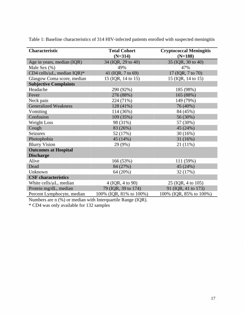
Table 1: Baseline characteristics of 314 HIV-infected patients enrolled with suspected meningitis
Characteristic Total Cohort(N=314)
Cryptococcal Meningitis (N=188)
Age in years, median (IQR) 34 (IQR, 29 to 40) 35 (IQR, 30 to 40)Male Sex (%) 49% 47%CD4 cells/L, median IQR)* 41 (IQR, 7 to 69) 17 (IQR, 7 to 70)Glasgow Coma score, median 15 (IQR, 14 to 15) 15 (IQR, 14 to 15)Subjective Complaints Headache 290 (92%) 185 (98%)Fever 276 (88%) 165 (88%)Neck pain 224 (71%) 149 (79%)Generalized Weakness 128 (41%) 76 (40%)Vomiting 114 (36%) 84 (45%)Confusion 109 (35%) 56 (30%)Weight Loss 98 (31%) 57 (30%)Cough 83 (26%) 45 (24%)Seizures 52 (17%) 30 (16%)Photophobia 45 (14%) 31 (16%)Blurry Vision 29 (9%) 21 (11%)Outcomes at Hospital DischargeAlive 166 (53%) 111 (59%)Dead 84 (27%) 45 (24%)Unknown 64 (20%) 32 (17%)CSF characteristicsWhite cells/L, median 4 (IQR, 4 to 90) 25 (IQR, 4 to 105)Protein mg/dL, median 79 (IQR, 39 to 174) 91 (IQR, 41 to 173)Percent Lymphocyte, median 100% (IQR, 81% to 100%) 100% (IQR, 85% to 100%)Numbers are n (%) or median with Interquartile Range (IQR).* CD4 was only available for 132 samples
17
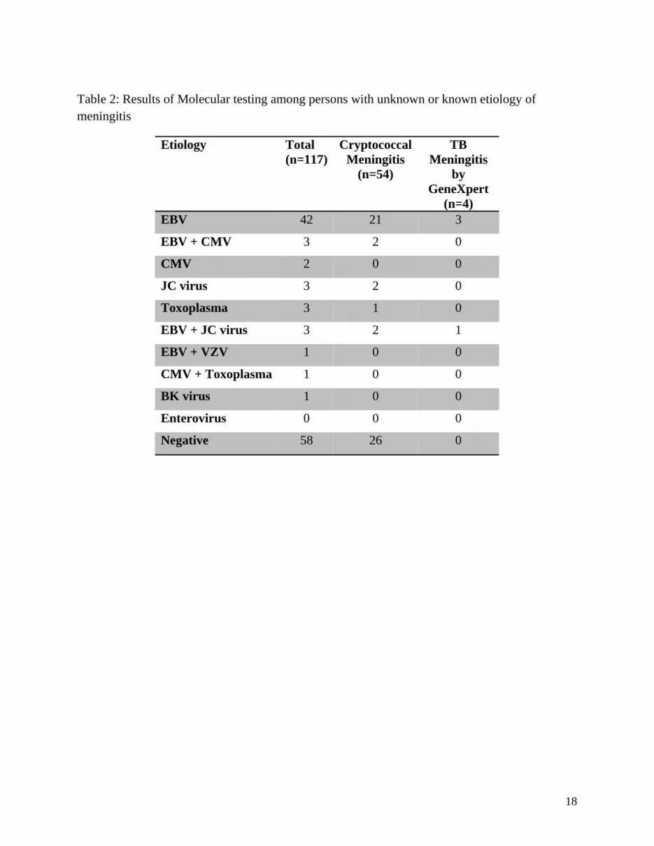
Table 2: Results of Molecular testing among persons with unknown or known etiology of meningitis
Etiology Total(n=117)
Cryptococcal Meningitis
(n=54)
TB Meningitis
by GeneXpert
(n=4)EBV 42 21 3
EBV + CMV 3 2 0
CMV 2 0 0
JC virus 3 2 0
Toxoplasma 3 1 0
EBV + JC virus 3 2 1
EBV + VZV 1 0 0
CMV + Toxoplasma 1 0 0
BK virus 1 0 0
Enterovirus 0 0 0
Negative 58 26 0
18
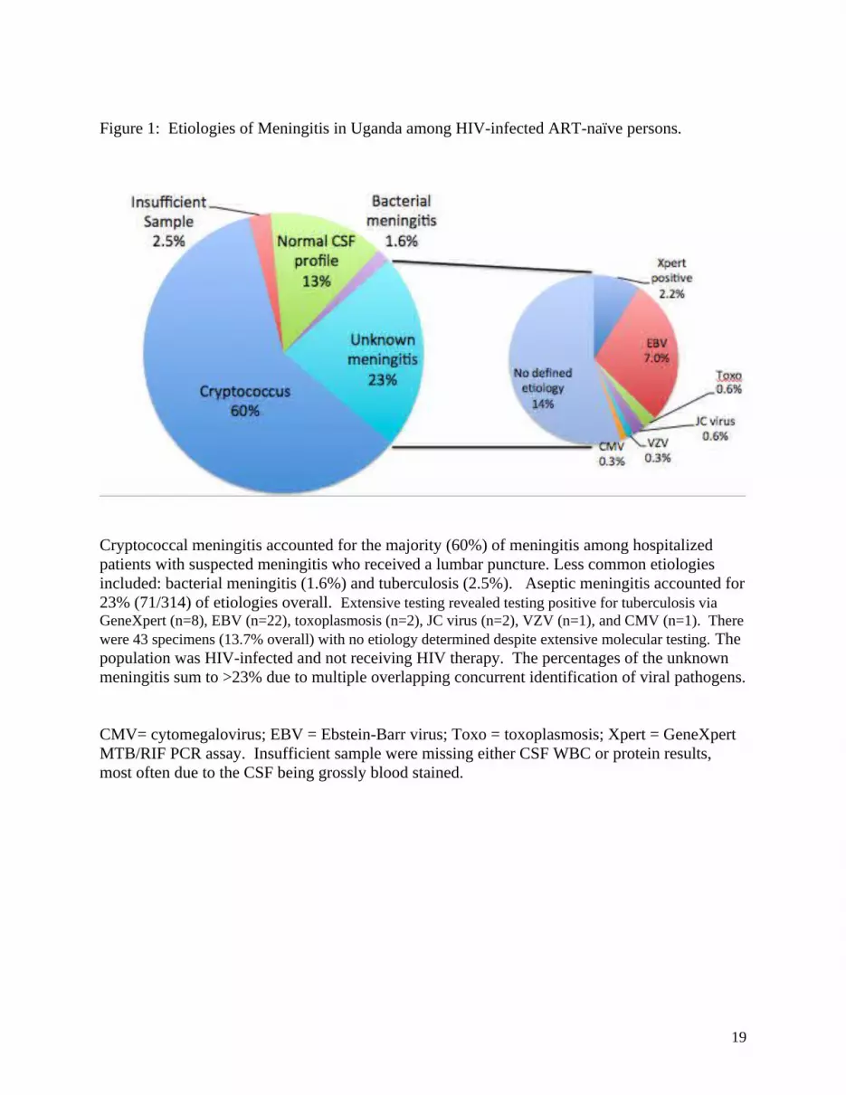
Figure 1: Etiologies of Meningitis in Uganda among HIV-infected ART-naïve persons.
Cryptococcal meningitis accounted for the majority (60%) of meningitis among hospitalized patients with suspected meningitis who received a lumbar puncture. Less common etiologies included: bacterial meningitis (1.6%) and tuberculosis (2.5%). Aseptic meningitis accounted for 23% (71/314) of etiologies overall. Extensive testing revealed testing positive for tuberculosis via GeneXpert (n=8), EBV (n=22), toxoplasmosis (n=2), JC virus (n=2), VZV (n=1), and CMV (n=1). There were 43 specimens (13.7% overall) with no etiology determined despite extensive molecular testing. The population was HIV-infected and not receiving HIV therapy. The percentages of the unknown meningitis sum to >23% due to multiple overlapping concurrent identification of viral pathogens.
CMV= cytomegalovirus; EBV = Ebstein-Barr virus; Toxo = toxoplasmosis; Xpert = GeneXpert MTB/RIF PCR assay. Insufficient sample were missing either CSF WBC or protein results, most often due to the CSF being grossly blood stained.
19

Supplemental Methods16s Ribosomal DNA Analysis
DNA extraction
For each CSF sample, DNA of two aliquots (200 ul each) was extracted in separate experiments using Ultraclean DNA blood spin kit (MO Bio Lab Inc., Carlsbad, California) according to manufacturer’s instructions. For each batch of extraction, a negative control (PCR grade water) was processed.
PCR and sequencing primers
The primer pair used for amplification and sequencing consisted of fD1 (5-AGAGTTTGATCCTGGCTCAG-3) and rP2 (5-ACGGCTACCTTGTTACGACTT-3), which produce an approximately 1,500-bp fragment and is considered to be universal for most eubacteria.
16 rDNA PCR Amplification
PCR was performed in duplicate in a blinded manner.
The final PCR mixture (50 µl) contained 1.5 µM of MgCl2, 1X of PCR buffer, 0.5 µg/µl of bovine serum albumin, 0.2 mM of dioxynucleoside triphosphate (dNTPs), 0.5 µM of each primer and 1 U of native Taq polymerase. The thermal cycle profile used was as follows: 5 min at 95 ◦C, followed by 40 cycles of 45 sec at 95 ◦C, 45 sec at 57 ◦C, 1.5 min at 72 ◦C, and finally followed by 5 min at 72◦C. Amplicon detection was carried out by agarose gel electrophoresis of 10 µL of amplification product through 1.5% agarose gel containing ethidium bromide (EtBr) 0.5 µg/ml of agarose in 1X of Tris-Acetate EDTA (TAE) buffer. Amplicons were visualized under UV irradiation.
The presence of amplifiable DNA was assessed by (i) using qualitative PCR to detect NRAS gene (human housekeeping gene) in each DNA eluate2 and (ii) spiking two of culture negative CSF samples with Escherichia coli before DNA extraction. DNA eluates from CSF sample known to have negative culture and negative 16S rDNA PCR were spiked with 1380 pg of purifed E. coli DNA to assess for PCR inhibitor. The detection level of the assay was determined by plate counting of 10-fold serial dilution of E. coli laboratory strain HB-101. Detection level of this PCR assay was 600 cfu/ml.
Sequencing
Crude PCR product was sent to Functional Biosciences, Inc. (Madison, Wisconsin) for sequencing. The sequence was compared to the sequences stored in GenBank by using Basic Local Alignment Search Tool (BLAST) analysis (http://blast.ncbi.nlm.nih.gov/Blast.cgi).
20