galectin-1 suppresses experimental colitis in mice
TRANSCRIPT
Galectin-1 Suppresses Experimental Colitis in Mice
LUCA SANTUCCI,* STEFANO FIORUCCI,* NATALIA RUBINSTEIN,‡ ANDREA MENCARELLI,*BARBARA PALAZZETTI,* BARBARA FEDERICI,* GABRIEL A. RABINOVICH,‡ and ANTONIO MORELLI**Clinica di Gastroenterologia ed Epatologia, Dipartimento di Scienze Chirurgiche, Gastroenterologiche ed Epatologiche, Universita degli Studidi Perugia, Italy; and ‡Laboratorio de Inmunogenetica, Hospital de Clinicas Jose de San Martin, Facultad de Medicina, Universidad deBuenos Aires, Buenos Aires, Argentina
Background & Aims: Uncontrolled T-cell activation playsa critical role in the pathogenesis of inflammatory boweldiseases. Therefore, pharmacologic strategies directedto restore the normal responsiveness of the immunesystem by deleting inappropriately activated T cellscould be efficacious in the treatment of these pathologicconditions. Galectin-1 is an endogenous lectin ex-pressed in lymphoid organs that plays a role in themaintenance of central and peripheral tolerance. Theaim of the present study was to evaluate the therapeuticeffects of galectin-1 on T-helper cell type 1-mediatedexperimental colitis induced by intrarectal administra-tion of 2,4,6-trinitrobenzene sulfonic acid (TNBS) inmice. Methods: Cells and tissues from mice with TNBScolitis receiving treatment with several doses of humanrecombinant galectin-1 (hrGAL-1) were analyzed formorphology, cytokine production, and apoptosis.Results: Prophylactic and therapeutic administration ofrhGAL-1 resulted in a striking improvement in the clini-cal and histopathologic aspects of the disease. hrGAL-1reduced the number of hapten-activated spleen T cells,decreased inflammatory cytokine production, and pro-foundly reduced the ability of lamina propria T cells toproduce IFN� in vitro. Moreover, hrGAL-1 led to theappearance of apoptotic mononuclear cells in colontissue when administered in vivo and induced selectiveapoptosis of TNBS-activated lamina propria T cells invitro. Conclusion: Collectively, these data show that hr-GAL-1 exerts protective and immunomodulatory activityin TNBS-induced colitis and it might be effective in thetreatment of inflammatory bowel diseases.
Inflammatory bowel diseases (IBD), encompassingCrohn’s disease (CD) and ulcerative colitis, are idio-
pathic chronic inflammatory conditions occurring withincreasing frequency in western populations.1Althoughthe etiology of IBD remains unknown, there is circum-stantial evidence to link this condition to a failure of themucosal immune system to attenuate the immune re-sponse to endogenous antigens, such as the normal en-teric bacterial flora.2 Support for this view has come fromanimal models of colitis, including the hapten model of
colonic inflammation induced by intrarectal delivery of2,4,6-trinitrobenzene sulfonic acid (TNBS), which con-sistently exhibit an imbalance of regulatory cytokines,most notably an excessive production of T helper cell(Th)-1–derived cytokines.3,4
Because activation-induced cell death of antigen-acti-vated lamina propria (LP) T cells represents the crucialmechanism involved in the attenuation of the mucosalimmune response, it has been postulated that a defectiveactivation-induced cell death may be the key factor forthe inappropriate mucosal T-cell accumulation observedin IBD and experimental colitis.5 This scenario is sup-ported by the recent demonstration that LP T cellsisolated from CD patients are resistant to multiple apo-ptotic signals6–8 and that the beneficial effects of anti-TNF� antibodies in CD are mediated by promotingmucosal T-cell death.9 In addition, treatment with anti–IL-12 and anti–IL-6 receptor monoclonal antibodies(mAbs) suppresses experimental colitis in mice by induc-ing LP T-cell death.3,8 Therefore, deletion of inappropri-ately activated LP T cells might be a therapeutic targetto counteract locally overshooting immune system inIBD.
Galectin-1, a 14.5-kilodalton homodimer, is a mem-ber of a family of �-galactoside binding proteins thatshare growth regulatory and immunomodulatory activi-ties.10,11 Galectin-1 is constitutively expressed by smoothcardiac and skeletal muscles, thymus, kidney, and pla-centa; a number of galectin-1 receptors have been iden-tified, including laminin, fibronectin, and the hemato-poietic cell surface membrane proteins CD43 and
Abbreviations used in this paper: ELISA, enzyme-linked immunosor-bent assay; FITC, fluorescence isothiocyanate; hrGAL, human recom-binant galectin; IBD, irritable bowel disease; IFN�, interferon gamma;IL, interleukin; IL2R, interleukin 2 receptor; LP, lamina propria; mAb,monoclonal antibody; MPO, myeloperoxidase; PCR, polymerase chainreaction; Th, T-helper; TNBS, 2,4,6-trinitrobenzene sulfonic acid; TNF�tumor necrosis factor alpha.
© 2003 by the American Gastroenterological Association0016-5085/03/$30.00
doi:10.1016/S0016-5085(03)00267-1
GASTROENTEROLOGY 2003;124:1381–1394
CD45.12,13 Galectin-1 has been shown to induce apopto-sis of a subset of negatively selected CD4loCD8lo imma-ture thymocytes and activated mature T lymphocytes,suggesting that this endogenous lectin is involved ingenerating and maintaining central and peripheral tol-erance.12–18 Recently, we have provided evidence thatgalectin-1 exerts immunomodulatory activity in vivo,suppressing disease in 2 experimental models of T-cellmediated diseases: collagen-induced arthritis in mice andconcanavalin A-induced hepatitis in mice.19,20 In thesemodels, galectin-1 administration resulted in a selectiveelimination of antigen-activated T cells and in a Th1 toTh2 shift that induced a remission state in the evolutionof the ongoing inflammatory disease.19,20 We have nowextended this finding by demonstrating that galectin-1exerts therapeutic activity in TNBS-induced colitis inmice by eliminating the uncontrolled Th1 response tothe hapten. Because this experimental model has a num-ber of clinical, histologic, and immunologic analogieswith CD, these data suggest the potential use of galec-tin-1 for the treatment of this disease.
Materials and Methods
Animals
Balb/c mice were obtained from Charles River (Monza,Italy). They were group-housed under controlled temperature(22°C) and photoperiod (12:12-hour light-dark cycle). Themice were allowed unrestricted access to standard mouse chowand tap water. They were allowed to acclimate to these con-ditions for at least 5 days before inclusion in an experiment.Protocols were approved by the Animal Study Committees ofthe University of Perugia according to governmental guide-lines for animal care.
Induction of Colitis and Study Design
Colitis was induced in Balb/c mice as previously de-scribed.3,21 Briefly, one-day fasted mice were anesthetized withhalothane and O2 and a 3.5F catheter was carefully insertedinto the colon via the anus until approximately the splenicflexure (4 cm from the anus). To induce colitis, 2 mg of thehapten reagent TNBS (Sigma Chemical Co, Milan, Italy) in50% ethanol (to break the intestinal epithelial barrier) wasslowly administered into the lumen via the catheter fitted onto
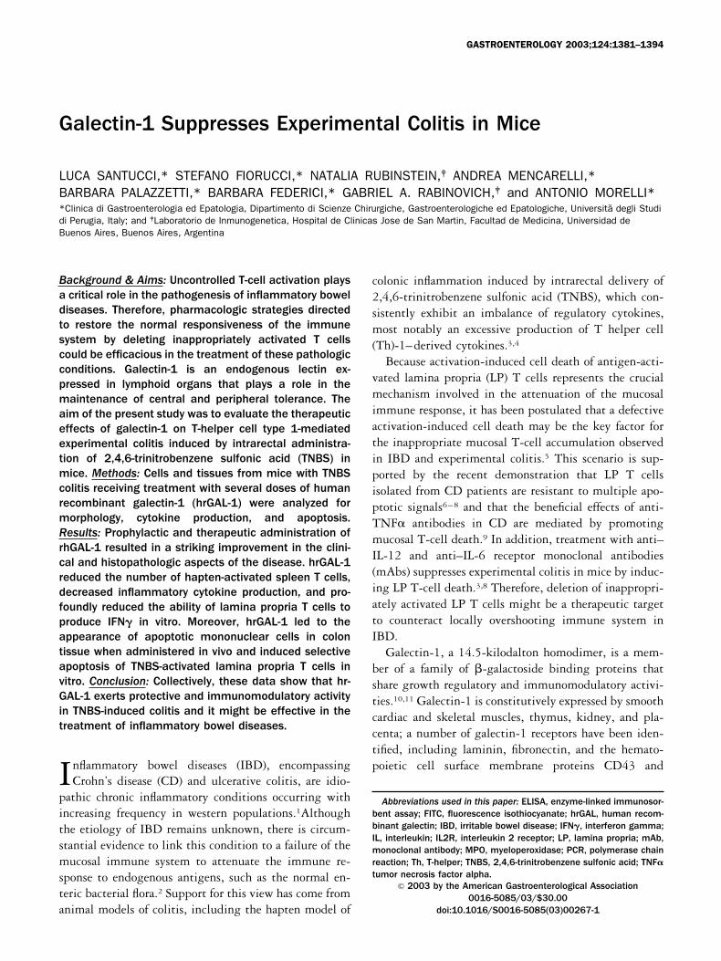
Figure 1. Early administration of rhGAL-1 protects against development of TNBS-induced colitis. Balb/c mice were given 2 mg TNBS intraco-lonically and rh-GAL1 was administered IV daily for 7 days, starting at the same time of intrarectal instillation of TNBS. (A) Wasting disease inmice with TNBS colitis is improved by rhGAL-1 in a dose-dependent manner. Each point represents the mean � SE of 8–12 mice. *P � 0.05versus TNBS alone and mice treated with 0.04 mg/kg rhGAL-1. (B, C, and D) Severity of TNBS-induced inflammation (microscopic score and MPOactivity) is reduced by rhGAL-1 administration in a dose-dependent manner. Bars represent the mean � SE of 8–12 mice. *P � 0.05 versuscontrol (ethanol-treated) mice; **P � 0.05 versus mice treated with TNBS alone and mice treated with 0.04 mg/kg rhGAL-1.
1382 GASTROENTEROLOGY Vol. 124, No. 5
a 1-mL syringe (injection volume, 100 �L). In control exper-iments, mice received 50% ethanol alone using the sametechnique. The effect of recombinant human galectin-1 (rh-GAL-1) on TNBS-induced colitis was investigated in 2 differ-ent protocols. To assess whether early administration of rh-GAL-1 protects against development of colitis (prophylacticprotocol), mice receiving 2 mg TNBS were randomized to
receive saline or rhGAL-1. rhGAL-1 was given IV followingTNBS injection and daily thereafter for 7 days at the doses of0.04, 0.4, and 1 mg/kg. Mice were killed at day 7 and theircolon analyzed. To address whether rhGAL-1 treatment wasbeneficial in treating established colitis (therapeutic protocol),administration of rhGAL-1 was started 2 weeks after colitisinduction. Mice were treated IV daily for 1 week with 1 mg/kg
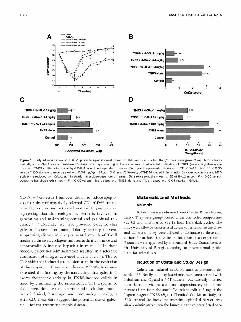
Figure 2. Early administration of rhGAL-1 protectsagainst the development of TNBS-induced colitis.Histologic analysis (H&E staining) of the colonfrom mice killed 7 days after intrarectal instillationof TNBS, with or without treatment with rhGAL-1.(A) H&E-stained paraffin section of control (etha-nol-treated) mouse (original magnification, 100�).(B) (Original magnification, 100�.) (C) (Originalmagnification, 400�.) H&E-stained paraffin sec-tion from a mouse killed 7 days after TNBS admin-istration alone, showing thickening of the colonwall and massive inflammatory infiltrate in the lam-ina propria. (D) (Original magnification, 100�.) Ef-fect of daily administration of 1 mg/kg rhGAL-1 onhistologic colitis induced by TNBS. Subepithelialedema with no inflammatory infiltrate in the mu-cosa and submucosa is shown.
Figure 4. Immunodetection of en-dogenous galectin-1 by immunogoldstaining. (A) No labeling was de-tected in colon sections using anormal rabbit serum (original magni-fication, 100�). (B) (Original magni-fication, 100�.) and (C) (Originalmagnification, 400�.) A strong stain-ing was detected, mainly at the levelof the epithelial cells, using a rabbitanti–galectin-1 polyclonal antibody.(D) Galectin-1 staining was less pro-nounced in colon sections from amouse killed 7 days after intrarectalinstillation of TNBS.
May 2003 GALECTIN-1 AND TNBS-INDUCED COLITIS 1383
rhGAL-1 or saline. At day 21 mice were sacrificed and theircolon analyzed.
Real-Time Reverse Transcription-Polymerase Chain Reaction (PCR)
Mice were sacrificed and the colons were removed,immediately snap-frozen on liquid nitrogen, and stored at�80°C until used. Total RNA was isolated by using theTRIzol reagent (Life Technologies, Milan, Italy) as previouslydescribed.21 PCR was performed using specific primers, de-signed using software PRIMER3 (Whitehead Institute, Cam-bridge, MA) with published sequence data from the NationalCenter Biotechnology Information database. Primers were syn-thesized by Sigma Genosys (The Woodlands, TX). For mouseRNA 18s, the sense primer was 5�-ACA CGG ACA GGATTG ACA GAT T -3� and antisense 5�- CGT TCG TTA TCGGAA TTA ACC A-3�. For mouse galectin-1, the sense primerwas: 5� -TGA ACC TGG GAA AAG ACA GC- 3� and theantisense was 5� -TCA GCC TGG TCA AAG GTG AT- 3�. Incontrol experiments with 3 replicates, no false positives weredetected. Amplification reactions contained 5 �L cDNA, 12.5�L of the 2 � Quanti tect SYBR Green reverse transcription-PCR Master Mix (Qiagen, Milan, Italy), and 0.75 �L of eachof the specific primers. Primer concentrations in a final volumeof 25 �L were 300 nmol/L for galectin-1 and 100 nmol/L for�-actin (housekeeping gene). All reactions were performed intriplicate in an iCycler iQ system (Biorad, Hercules, CA) andthe thermal cycling conditions were: 15 minutes at 95°C,followed by 50 cycles of 95°C for 10 seconds and 60°C for 30seconds. We used the expression of RNA 18s to normalize theexpression data of galectin-1 cDNA. RNA 18s was used tocorrect for the differences in the amount of total RNA addedto a reaction and to compensate for different levels of inhibi-tion during reverse transcription of RNA and during PCR.
Immunohistochemistry for EndogenousGalectin-1 Expression
For immunohistochemical staining, paraffin sectionswere mounted on glass slide coated with 1% polylisine, depar-
affinized with xilene, rehydrated and incubated with a rabbitanti–galectin-1 polyclonal antibody (diluted 1:1000 in 1%bovine serum albumin-PBS) for 24 hours at 4°C. The poly-clonal anti–galectin-1 antibody, obtained as previously de-scribed,22 was specific because it did not recognize othergalectins, such as galectin-3.23 Samples without primary an-tibody served as negative control. After 3 washes with PBS,slides were incubated with a 1:6 dilution of the anti-rabbitIgG-gold complex for 1 hour at room temperature. Colloidalgold particles (average diameter, 16 nm) were prepared aspreviously described, using sodium citrate as a reducingagent.24 A silver enhancement kit (Sigma Chemical Co, St.Louis, MO) was used to visualize the gold particles at thelight-microscopic level. After washing with tridistilled water,the sections were mounted on glass slides and studied in aAxioskop Zeiss (Germany). Photographs were taken with aHitachi CDD Color Camera (Japan).
Histological Grading of Colitis
For histologic examination, tissues were removed, fixedin 10% buffered formalin phosphate, embedded in paraffin,sectioned, and stained with hematoxylin and eosin (H&E). Thedegree of inflammation on microscopic cross sections of thecolon was graded semiquantitatively from 0 to 4 (0, no signsof inflammation; 1, very low level; 2, low level of leukocyteinfiltration; 3, high level of leukocyte infiltration, high vascu-lar density, thickening of the colon wall; and 4, transmuralinfiltration, loss of goblet cells, high vascular density, thick-ening of the colon wall). Grading was performed in a blindedfashion by 2 experienced pathologists. Thickness of the colonwall was determined on cross sections by measuring the dis-tance from the serosal surface to the luminal surface at 2-mmintervals along the entire length of each section by using anOlympus BX60 microscope (Olympus Co, Tokyo, Japan).Images were captured by a digital camera (SPOT-2, DiagnosticInstruments Inc, Burroughs, MI) and analyzed by a specificsoftware (Delta Sistemi, Rome, Italy).
Cytokine and Myeloperoxidase (MPO)Assays
Immunoreactive murine TNF�, IFN, IL-12, andIL-1� were quantified in plasma and colon homogenates byusing commercially available enzyme-linked immunosorbentassay (ELISA) kits according to the manufacturer’s instructions(R&D Systems, Abingdon, UK). Neutrophil infiltration in thecolon was monitored by measuring tissue MPO activity usinga spectrophotometric assay with tetramethylbenzidine as sub-strate, according to a previously published method.25
Flow-Cytometric Analysis of Spleen T Cells
Mice were killed 7 days after TNBS or ethanol injec-tion, and the spleen removed. After monocyte elimination byadhesion and lysis of red cells with 0.15 mol/L NH4Cl and1mmol/L KHCO3, lymphocytes were resuspended in RPMI/
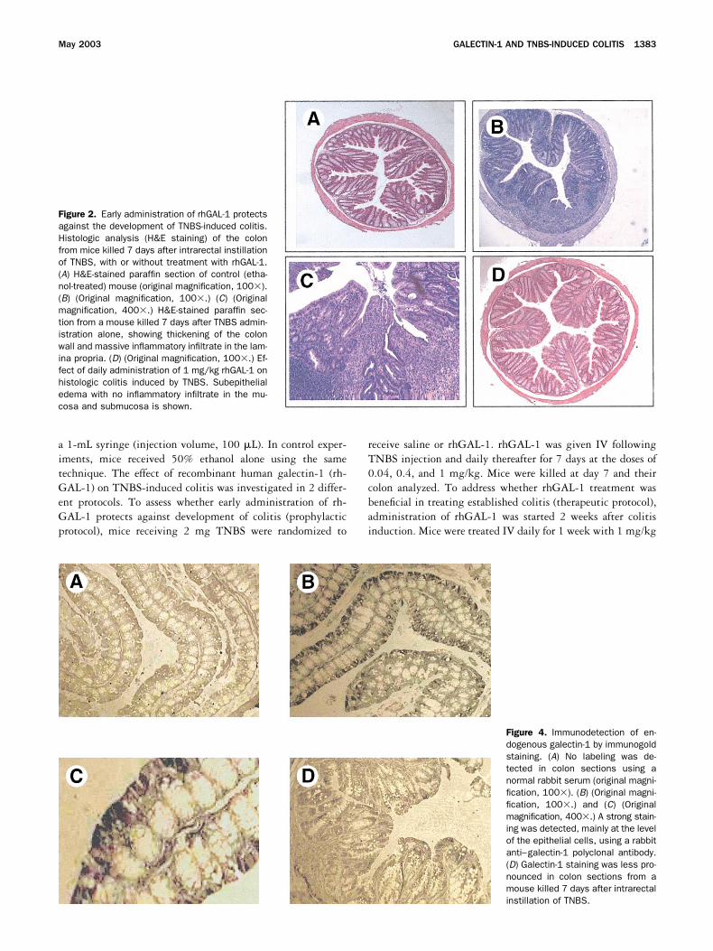
Figure 3. Real-time quantitative PCR of galectin-1 expression in co-lonic tissues. Intrarectal TNBS instillation resulted in a 4-fold de-crease in galectin-1 colonic mRNA content. The means of triplicatedeterminations are shown. Data was normalized against 18S ribo-somal RNA. *P � 0.05 versus control (ethanol-treated) mice.
1384 GASTROENTEROLOGY Vol. 124, No. 5
fetal calf serum. T cells were then separated from B cells usingnylon wool columns, as previously described.26 After thisprocedure, about 90% of lymphocytes were CD3-positive Tcells. Lymphocytes were then incubated with fluorescence iso-thiocyanate (FITC)-conjugated anti-Fas, anti-FasL, and anti-IL2R mAbs for 30 minutes at 4°C. Cells were then washedtwice and resuspended in PBS/formaldehyde (0.5%). Stainedcells were then analyzed on a FACScan cytofluorimeter (BectonDickinson, San Jose, CA), and cells were gated using forwardversus side scatter to exclude dead cells and debris.
Isolation of LP T Cells
Colonic LP T cells were prepared as previously de-scribed.27 In brief, after excision of all visible lymphoid folli-cles, colonic tissue was treated with 1 mmol/L EDTA in PBSfor 20 minutes to remove the epithelium. The tissue was thendigested with collagenase (type IV; Sigma) for 20 minutes ina shacking incubator at 37°C; this step was repeated twice.The released cells were then layered on a 40–100% Percollgradient (Pharmacia, Uppsala, Sweden) and spun at 1.800 rpm
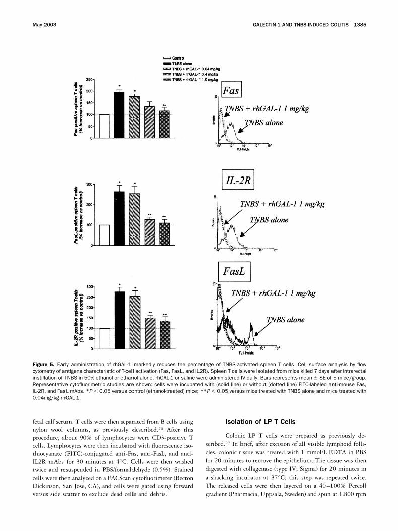
Figure 5. Early administration of rhGAL-1 markedly reduces the percentage of TNBS-activated spleen T cells. Cell surface analysis by flowcytometry of antigens characteristic of T-cell activation (Fas, FasL, and IL2R). Spleen T cells were isolated from mice killed 7 days after intrarectalinstillation of TNBS in 50% ethanol or ethanol alone. rhGAL-1 or saline were administered IV daily. Bars represents mean � SE of 5 mice/group.Representative cytofluorimetric studies are shown: cells were incubated with (solid line) or without (dotted line) FITC-labeled anti-mouse Fas,IL-2R, and FasL mAbs. *P � 0.05 versus control (ethanol-treated) mice; **P � 0.05 versus mice treated with TNBS alone and mice treated with0.04mg/kg rhGAL-1.
May 2003 GALECTIN-1 AND TNBS-INDUCED COLITIS 1385
to obtain the lymphocyte-enriched populations at the 40–100% interface. An enriched CD4 T-cell population wasobtained by positive selection using FACSorter (Becton Dick-inson). Flow cytometric analysis showed that the resultant cellpopulation contained about 90% CD4 T lymphocytes.
Culture of LP CD4� T Cells for Assay ofCytokine Production
LP CD4 T cells from different treatment regimenswere suspended in complete medium (RPMI 1640, 10%heat-inactivated fetal calf serum, 3 mmol/L L-glutamine, 10mmol/L Hepes buffer, 10 �g/mL penicillin, 100 U/mLstreptomycin, and 0.05 mmol/L 2-ME) and cultured at aconcentration of 105 cells/mL. To measure cytokine produc-tion, LP CD4 T cells were placed for 48 hours ontouncoated culture wells (to measure production by unstimu-lated cells) or onto wells containing immobilized murineanti-CD3� mAb (Pharmingen, San Diego, CA) and 1�g/mL soluble anti-CD28 antibody (Pharmingen) (to mea-sure production by stimulated cells). At the end of theincubation period, culture supernatants were harvested andassayed for cytokine concentration by using specific ELISAkits (R&D systems).
Evaluation of T-cell Apoptosis In Vitro
To investigate the effect of rhGAL-1 on T-cell ap-optosis, freshly isolated spleen and LP T cells were incu-bated for 8 hours in complete medium with 3 mmol/Ldithiothereitol, with increasing doses of rhGAL-1. At theend of the incubation period, apoptotic cells were identified
at FACScan cytofluorimeter (Becton Dickinson) using an-nexin V and propidium iodide (PI) according to the man-ufacturer’s instructions (R&D Systems). The percent of celldeath was calculated by determining the percent of viablecells:
[% of viable � (% of annexin V� and PI�,
rhGAL-1 treated)/
(% of annexin V�
and PI�, control treated)
� 100 and % of death � 100 – % of viable].
To assess the mechanism underlying T-cell apoptosis in-duced by rhGAL-1, caspase 8 and caspase 9 activities weremeasured 2 and 8 hours after the incubation with rhGAL-1 byusing specific caspase fluorometric protease assay according tomanufacturer’s instructions (ApoAlert, Clontech, Palo Alto,CA).
In Situ TUNEL Staining
For detection of apoptotic cells in tissue, terminaldeoxynucleotidyl transferase-mediated deoxyuridine trisphos-phate nick-end labeling (TUNEL) staining was performed inparaffin sections of colon from control (ethanol-treated) mice,mice with fully established TNBS-induced colitis, and micewith TNBS-induced colitis treated with 1mg/kg rhGAL-1.Colon sections were deparaffinized and the proteins present inthe sections digested with 20 �g/mL proteinase K for 15minutes at room temperature. Following 4 washes in distilledwater, endogenous peroxidase was quenched with 2.0% H2O2
at room temperature and sections were washed 2 times withPBS. The labeling of degraded DNA specific to apoptotic cellswas performed with a peroxidase-conjugated in situ apoptosisdetection kit (ApopTag, Intergen, NY), according to the man-ufacturer’s instructions. Detection of labeled ends was per-formed with anti-digoxigenin peroxidase antibody and sub-strate.
Drugs and Reagents
Routine buffer reagents were obtained from SigmaChemical Co (Milan, Italy). All other chemicals were obtainedfrom the sources indicated or Sigma Chemical.
Statistical Analysis
All values are expressed as mean � SEM of n mice pergroup. Comparisons of more than 2 groups were made with a1-way analysis of variance with post-hoc Tukey’s test. Com-parison of 2 groups was made using Student’s t test forunpaired data when appropriate. Differences were consideredstatistically significant if P was � 0.05.
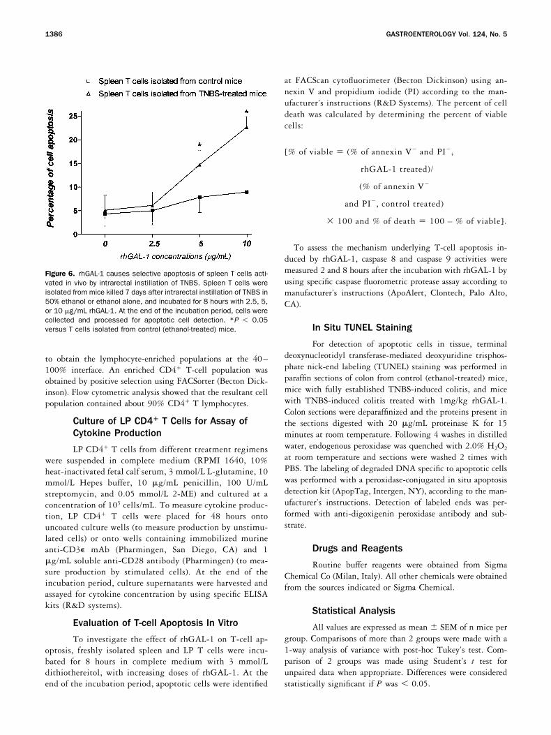
Figure 6. rhGAL-1 causes selective apoptosis of spleen T cells acti-vated in vivo by intrarectal instillation of TNBS. Spleen T cells wereisolated from mice killed 7 days after intrarectal instillation of TNBS in50% ethanol or ethanol alone, and incubated for 8 hours with 2.5, 5,or 10 �g/mL rhGAL-1. At the end of the incubation period, cells werecollected and processed for apoptotic cell detection. *P � 0.05versus T cells isolated from control (ethanol-treated) mice.
1386 GASTROENTEROLOGY Vol. 124, No. 5
Results
rhGAL-1 Protects Against Development ofTNBS-Induced Colitis (Prophylactic Protocol)
Effect of early administration of rhGAL-1 on clin-ical and histologic signs of colitis. Intracolonic admin-istration of TNBS in Balb/c mice induced a severe illnesscharacterized by loss of body weight that remained ap-proximately 15% lower than controls during all post-
treatment periods (Figure 1A ). Early administration ofrhGAL-1 resulted in a dose-dependent prevention ofwasting syndrome induced by TNBS administration(Figure 1A ). The 2 higher doses of rhGAL-1 preventedthe TNBS-induced wasting syndrome at similar extent(P 0.05 between 0.4 and 1mg/kg). Distal colons ofmice sacrificed 7 days after TNBS administration ap-peared hyperemic and thickened, with adhesion to othersegments of intestine. On microscopic examination the
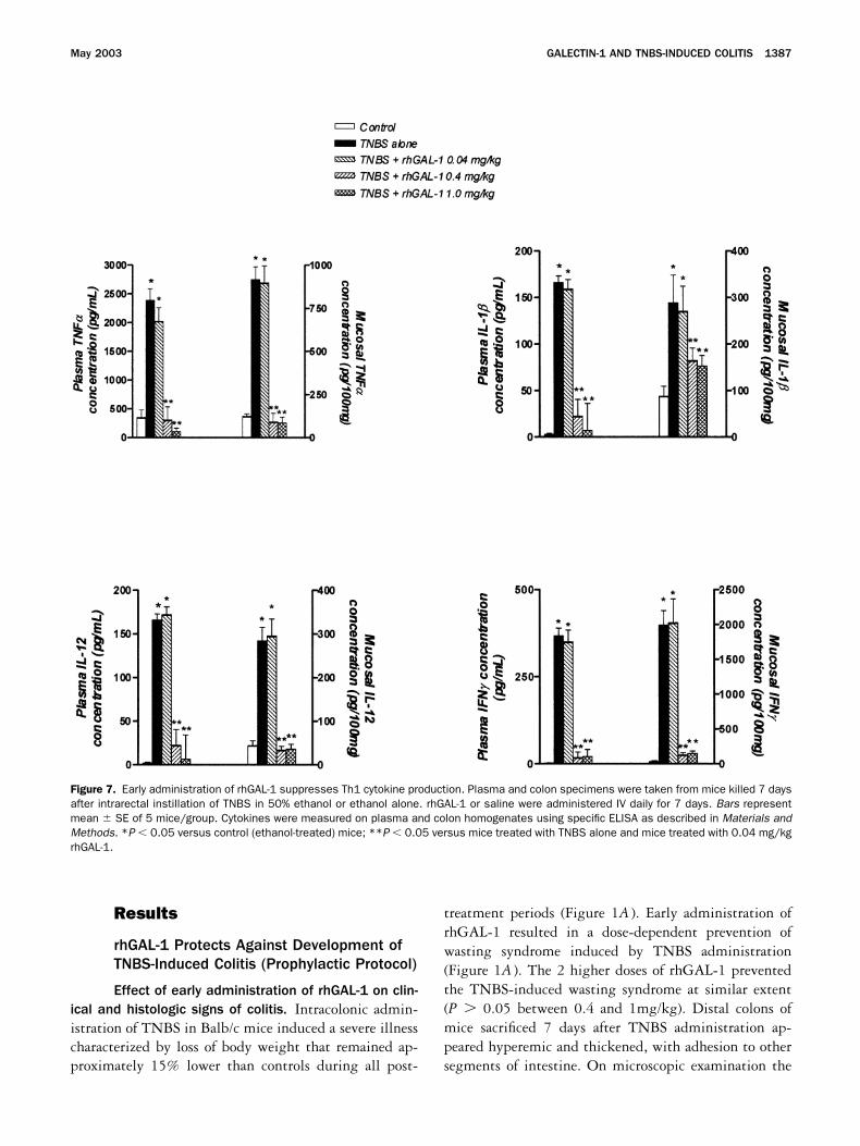
Figure 7. Early administration of rhGAL-1 suppresses Th1 cytokine production. Plasma and colon specimens were taken from mice killed 7 daysafter intrarectal instillation of TNBS in 50% ethanol or ethanol alone. rhGAL-1 or saline were administered IV daily for 7 days. Bars representmean � SE of 5 mice/group. Cytokines were measured on plasma and colon homogenates using specific ELISA as described in Materials andMethods. *P � 0.05 versus control (ethanol-treated) mice; **P � 0.05 versus mice treated with TNBS alone and mice treated with 0.04 mg/kgrhGAL-1.
May 2003 GALECTIN-1 AND TNBS-INDUCED COLITIS 1387
colons of TNBS-treated mice demonstrated a markedmononuclear cell infiltration associated with loss of gob-let cells (Figure 2B and C ). Some parts of the mucosallayer lost crypts and these areas were replaced by lym-phocytes and macrophages (Figure 2C ). The histologicgrading of colon sections increased from 0.5 � 0.1 incontrol mice to 2.7 � 0.3 in TNBS-treated mice (P �0.01) (Figure 1B). In line with these histologic alter-ations, intrarectal instillation of TNBS resulted in asignificant increase in colon mucosal wall thickness andMPO activity (Figure 1C and D). Early administration ofrhGAL-1 reduced the extent of colonic damage inducedby TNBS in a dose-dependent manner, as measured bythe reduction in histologic injury score and colonic MPOcontent (Figure 1B, C, and D; Figure 2D and E ).
Colonic galectin-1 expression. High levels of ga-lectin-1 mRNA was detected in the colon of controlmice. Real-time quantitative PCR demonstrated thatrectal TNBS instillation caused a 4-fold decrease incolonic galectin-1 mRNA expression (Figure 3). As as-sessed by immunohistochemistry, normal colon consti-tutively expresses high levels of galectin-1, which wasmainly localized at the level of the superficial epithelialcells (Figure 4B). In line with the results of quantitativereal-time PCR, galectin-1 expression was markedly re-duced in colon sections from mice with TNBS-inducedcolitis (Figure 4D ).
Effect of early administration of rhGAL-1 onspleen T cells. As assessed by the expression on cellmembranes of the activation-induced molecules Fas,FasL, and IL2R, colonic instillation of TNBS caused asignificant increase in the number of activated spleen Tcells (Figure 5). Early administration of rhGAL-1 re-duced the percentage of activated spleen T cells in adose-dependent manner (Figure 5). To investigate if thereduction in the percentage of activated spleen T cellsobserved in rhGAL-1–treated mice was related to thedeletion of TNBS-activated T cells, spleen T cells wereisolated from TNBS or ethanol-treated mice and culturedwith increasing doses of rhGAL-1. As shown in Figure 6,rhGAL-1 increased the percentage of apoptosis only inspleen T cells isolated from TNBS-treated mice in adose-dependent manner, while it had no effect on T cellsisolated from control mice. rhGAL-1–induced cell deathwas reverted by the addition of lactose, indicating thatT-cell apoptosis was mediated by carbohydrate-specificinteractions (data not shown). These data suggest thatintrarectal administration of TNBS results in activationof a subset of TNBS-specific T cells that are highlysusceptible to the proapoptotic activity of rhGAL-1.
Effect of early administration of rhGAL-1 onTNBS-induced pro-inflammatory cytokine release. Asshown in Figure 7, colonic instillation of TNBS resultedin a marked increase in mucosal and plasma concentra-tions of several Th1 cytokines, such as TNF�, IL-1�,IL-12, and IFN. In agreement with the reduction ofcolonic inflammation, early administration of rhGAL-1dose-dependently reduced plasma levels of TNF�, IL-1�,IL-12, and IFN and the concentration of these proin-flammatory cytokines in the colonic mucosa (Figure 7).
Effect of early administration of rhGAL-1 on IFN�
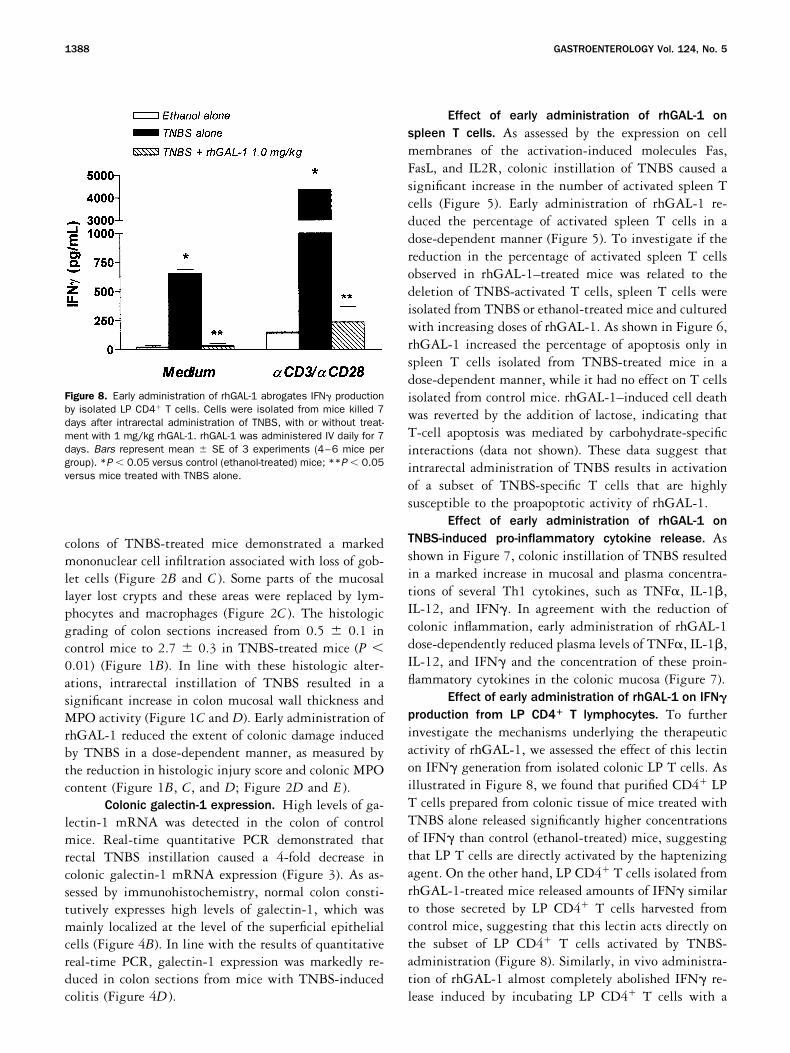
production from LP CD4� T lymphocytes. To furtherinvestigate the mechanisms underlying the therapeuticactivity of rhGAL-1, we assessed the effect of this lectinon IFN generation from isolated colonic LP T cells. Asillustrated in Figure 8, we found that purified CD4 LPT cells prepared from colonic tissue of mice treated withTNBS alone released significantly higher concentrationsof IFN than control (ethanol-treated) mice, suggestingthat LP T cells are directly activated by the haptenizingagent. On the other hand, LP CD4 T cells isolated fromrhGAL-1-treated mice released amounts of IFN similarto those secreted by LP CD4 T cells harvested fromcontrol mice, suggesting that this lectin acts directly onthe subset of LP CD4 T cells activated by TNBS-administration (Figure 8). Similarly, in vivo administra-tion of rhGAL-1 almost completely abolished IFN re-lease induced by incubating LP CD4 T cells with a
Figure 8. Early administration of rhGAL-1 abrogates IFN productionby isolated LP CD4 T cells. Cells were isolated from mice killed 7days after intrarectal administration of TNBS, with or without treat-ment with 1 mg/kg rhGAL-1. rhGAL-1 was administered IV daily for 7days. Bars represent mean � SE of 3 experiments (4–6 mice pergroup). *P � 0.05 versus control (ethanol-treated) mice; **P � 0.05versus mice treated with TNBS alone.
1388 GASTROENTEROLOGY Vol. 124, No. 5
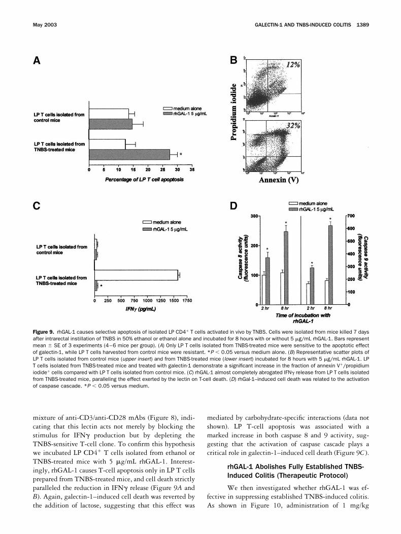
mixture of anti-CD3/anti-CD28 mAbs (Figure 8), indi-cating that this lectin acts not merely by blocking thestimulus for IFN production but by depleting theTNBS-sensitive T-cell clone. To confirm this hypothesiswe incubated LP CD4 T cells isolated from ethanol orTNBS-treated mice with 5 �g/mL rhGAL-1. Interest-ingly, rhGAL-1 causes T-cell apoptosis only in LP T cellsprepared from TNBS-treated mice, and cell death strictlyparalleled the reduction in IFN release (Figure 9A andB). Again, galectin-1–induced cell death was reverted bythe addition of lactose, suggesting that this effect was
mediated by carbohydrate-specific interactions (data notshown). LP T-cell apoptosis was associated with amarked increase in both caspase 8 and 9 activity, sug-gesting that the activation of caspase cascade plays acritical role in galectin-1–induced cell death (Figure 9C ).
rhGAL-1 Abolishes Fully Established TNBS-Induced Colitis (Therapeutic Protocol)
We then investigated whether rhGAL-1 was ef-fective in suppressing established TNBS-induced colitis.As shown in Figure 10, administration of 1 mg/kg
Figure 9. rhGAL-1 causes selective apoptosis of isolated LP CD4 T cells activated in vivo by TNBS. Cells were isolated from mice killed 7 daysafter intrarectal instillation of TNBS in 50% ethanol or ethanol alone and incubated for 8 hours with or without 5 �g/mL rhGAL-1. Bars representmean � SE of 3 experiments (4–6 mice per group). (A) Only LP T cells isolated from TNBS-treated mice were sensitive to the apoptotic effectof galectin-1, while LP T cells harvested from control mice were resistant. *P � 0.05 versus medium alone. (B) Representative scatter plots ofLP T cells isolated from control mice (upper insert) and from TNBS-treated mice (lower insert) incubated for 8 hours with 5 �g/mL rhGAL-1. LPT cells isolated from TNBS-treated mice and treated with galectin-1 demonstrate a significant increase in the fraction of annexin V/propidiumiodide cells compared with LP T cells isolated from control mice. (C) rhGAL-1 almost completely abrogated IFN release from LP T cells isolatedfrom TNBS-treated mice, paralleling the effect exerted by the lectin on T-cell death. (D) rhGal-1–induced cell death was related to the activationof caspase cascade. *P � 0.05 versus medium.
May 2003 GALECTIN-1 AND TNBS-INDUCED COLITIS 1389
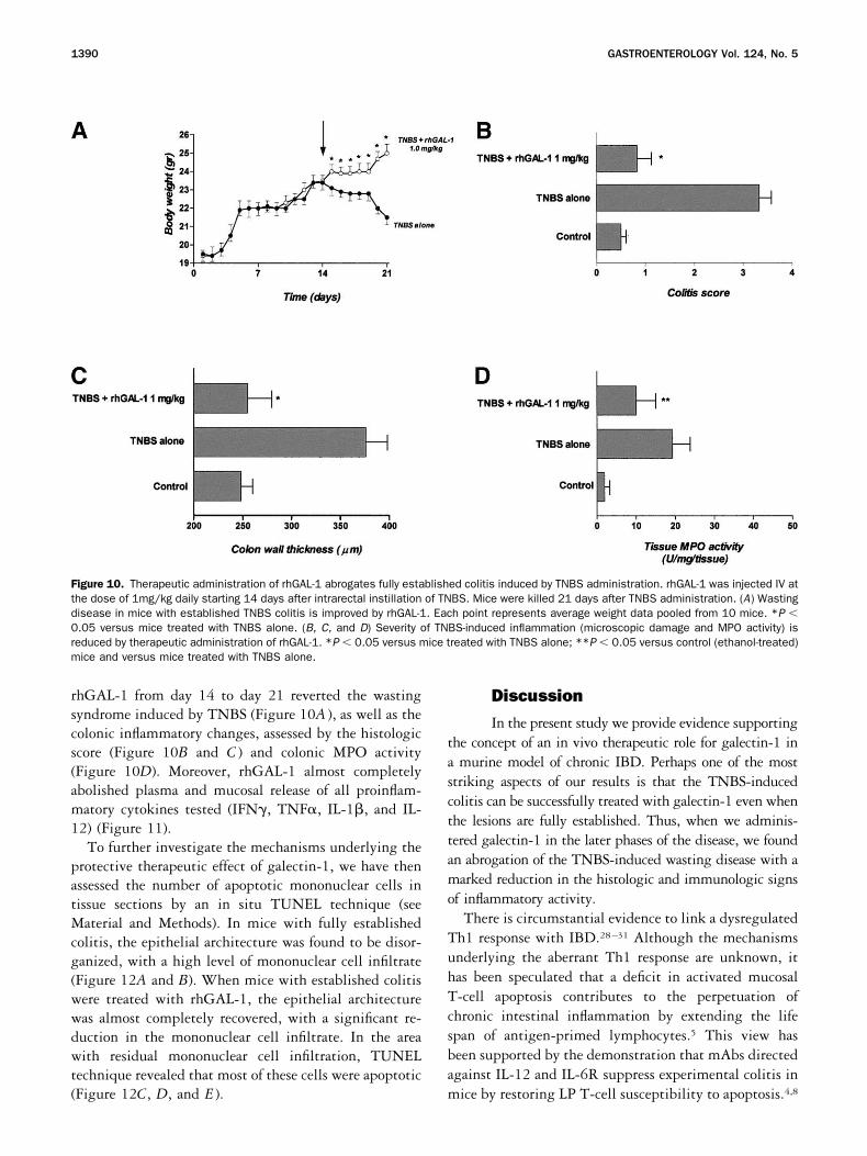
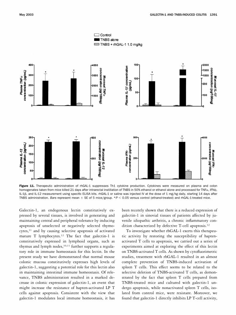
rhGAL-1 from day 14 to day 21 reverted the wastingsyndrome induced by TNBS (Figure 10A ), as well as thecolonic inflammatory changes, assessed by the histologicscore (Figure 10B and C ) and colonic MPO activity(Figure 10D). Moreover, rhGAL-1 almost completelyabolished plasma and mucosal release of all proinflam-matory cytokines tested (IFN, TNF�, IL-1�, and IL-12) (Figure 11).
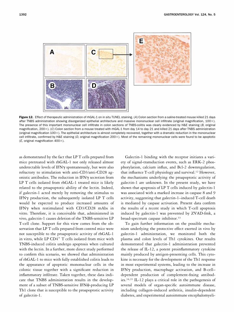
To further investigate the mechanisms underlying theprotective therapeutic effect of galectin-1, we have thenassessed the number of apoptotic mononuclear cells intissue sections by an in situ TUNEL technique (seeMaterial and Methods). In mice with fully establishedcolitis, the epithelial architecture was found to be disor-ganized, with a high level of mononuclear cell infiltrate(Figure 12A and B). When mice with established colitiswere treated with rhGAL-1, the epithelial architecturewas almost completely recovered, with a significant re-duction in the mononuclear cell infiltrate. In the areawith residual mononuclear cell infiltration, TUNELtechnique revealed that most of these cells were apoptotic(Figure 12C, D, and E ).
Discussion
In the present study we provide evidence supportingthe concept of an in vivo therapeutic role for galectin-1 ina murine model of chronic IBD. Perhaps one of the moststriking aspects of our results is that the TNBS-inducedcolitis can be successfully treated with galectin-1 even whenthe lesions are fully established. Thus, when we adminis-tered galectin-1 in the later phases of the disease, we foundan abrogation of the TNBS-induced wasting disease with amarked reduction in the histologic and immunologic signsof inflammatory activity.
There is circumstantial evidence to link a dysregulatedTh1 response with IBD.28–31 Although the mechanismsunderlying the aberrant Th1 response are unknown, ithas been speculated that a deficit in activated mucosalT-cell apoptosis contributes to the perpetuation ofchronic intestinal inflammation by extending the lifespan of antigen-primed lymphocytes.5 This view hasbeen supported by the demonstration that mAbs directedagainst IL-12 and IL-6R suppress experimental colitis inmice by restoring LP T-cell susceptibility to apoptosis.4,8
Figure 10. Therapeutic administration of rhGAL-1 abrogates fully established colitis induced by TNBS administration. rhGAL-1 was injected IV atthe dose of 1mg/kg daily starting 14 days after intrarectal instillation of TNBS. Mice were killed 21 days after TNBS administration. (A) Wastingdisease in mice with established TNBS colitis is improved by rhGAL-1. Each point represents average weight data pooled from 10 mice. *P �0.05 versus mice treated with TNBS alone. (B, C, and D) Severity of TNBS-induced inflammation (microscopic damage and MPO activity) isreduced by therapeutic administration of rhGAL-1. *P � 0.05 versus mice treated with TNBS alone; **P � 0.05 versus control (ethanol-treated)mice and versus mice treated with TNBS alone.
1390 GASTROENTEROLOGY Vol. 124, No. 5
Galectin-1, an endogenous lectin constitutively ex-pressed by several tissues, is involved in generating andmaintaining central and peripheral tolerance by inducingapoptosis of unselected or negatively selected thymo-cytes,11 and by causing selective apoptosis of activatedmature T lymphocytes.12 The fact that galectin-1 isconstitutively expressed in lymphoid organs, such asthymus and lymph nodes,10,11 further supports a regula-tory role in immune homeostasis for this lectin. In thepresent study we have demonstrated that normal mousecolonic mucosa constitutively expresses high levels ofgalectin-1, suggesting a potential role for this lectin alsoin maintaining intestinal immune homeostasis. Of rele-vance, TNBS administration resulted in a marked de-crease in colonic expression of galectin-1, an event thatmight increase the resistance of hapten-activated LP Tcells against apoptosis. Consistent with the view thatgalectin-1 modulates local immune homeostasis, it has
been recently shown that there is a reduced expression ofgalectin-1 in sinovial tissues of patients affected by ju-venile idiopathic arthritis, a chronic inflammatory con-dition characterized by defective T-cell apoptosis.32
To investigate whether rhGAL-1 exerts this therapeu-tic activity by restoring the susceptibility of hapten-activated T cells to apoptosis, we carried out a series ofexperiments aimed at exploring the effect of this lectinon TNBS-activated T cells. As shown by cytofluorimetricstudies, treatment with rhGAL-1 resulted in an almostcomplete prevention of TNBS-induced activation ofspleen T cells. This effect seems to be related to theselective deletion of TNBS-activated T cells, as demon-strated by the fact that spleen T cells prepared fromTNBS-treated mice and cultured with galectin-1 un-dergo apoptosis, while nonactivated spleen T cells, iso-lated from control mice, were resistant. Moreover, wefound that galectin-1 directly inhibits LP T-cell activity,
Figure 11. Therapeutic administration of rhGAL-1 suppresses Th1 cytokine production. Cytokines were measured on plasma and colonhomogenates taken from mice killed 21 days after intrarectal instillation of TNBS in 50% ethanol or ethanol alone and processed for TNF�, IFN�,IL-1�, and IL-12 measurement using specific ELISA kits. rhGAL-1 or saline was injected IV at the dose of 1 mg/kg daily, starting 14 days afterTNBS administration. Bars represent mean � SE of 5 mice/group. *P � 0.05 versus control (ethanol-treated) and rhGAL-1-treated mice.
May 2003 GALECTIN-1 AND TNBS-INDUCED COLITIS 1391
as demonstrated by the fact that LP T cells prepared frommice pretreated with rhGAL-1 not only released almostundetectable levels of IFN spontaneously, but were alsorefractory to stimulation with anti-CD3/anti-CD28 ag-onistic antibodies. The reduction in IFN secretion fromLP T cells isolated from rhGAL-1 treated mice is likelyrelated to the proapoptotic ability of the lectin. Indeed,if galectin-1 acted merely by removing the stimulus toIFN production, the subsequently isolated LP T cellswould be expected to produce increased amounts ofIFN when restimulated with CD3/CD28 mAbs invitro. Therefore, it is conceivable that, administered invivo, galectin-1 causes deletion of the TNBS-sensitive LPT-cell clone. Support for this view comes from the ob-servation that LP T cells prepared from control mice werenot susceptible to the proapoptotic activity of rhGAL-1in vitro, while LP CD4 T cells isolated from mice withTNBS-induced colitis undergo apoptosis when culturedwith the lectin. In a further, more direct study performedto confirm this scenario, we showed that administrationof rhGAL-1 to mice with fully established colitis leads tothe appearance of apoptotic mononuclear cells in thecolonic tissue together with a significant reduction ininflammatory infiltrate. Taken together, these data indi-cate that TNBS administration results in the develop-ment of a subset of TNBS-sensitive IFN�-producing LPTh1 clone that is susceptible to the proapoptotic activityof galectin-1.
Galectin-1 binding with the receptor initiates a vari-ety of signal-transduction events, such as ERK-2 phos-phorylation, calcium influx, and Bcl-2 downregulation,that influence T-cell physiology and survival.12 However,the mechanisms underlying the proapoptotic activity ofgalectin-1 are unknown. In the present study, we haveshown that apoptosis of LP T cells induced by galectin-1was associated with a marked increase in caspase 8 and 9activity, suggesting that galectin-1–induced T-cell deathis mediated by caspase activation. Present data confirmthe results of a recent study in which T-cell apoptosisinduced by galectin-1 was prevented by ZVAD-fmk, abroad-spectrum caspase inhibitor.33
To gain further information on the possible mecha-nism underlying the protective effect exerted in vivo bygalectin-1 administration, we monitored both theplasma and colon levels of Th1 cytokines. Our resultsdemonstrated that galectin-1 administration preventedthe release of IL-12, a potent proinflammatory cytokinemainly produced by antigen-presenting cells. This cyto-kine is necessary for the development of the Th1 responsein most experimental systems, leading to the increase inIFN production, macrophage activation, and B-cell–dependent production of complement-fixing antibod-ies.34,35 IL-12 plays a critical role in the pathogenesis ofseveral models of organ-specific autoimmune disease,including collagen-induced arthritis, insulin-dependentdiabetes, and experimental autoimmune encephalomyeli-
Figure 12. Effect of therapeutic administration of rhGAL-1 on in situ TUNEL staining. (A) Colon section from a saline-treated mouse killed 21 daysafter TNBS administration showing disorganized epithelial architecture and massive mononuclear cell infiltrate (original magnification, 100�).The presence of this important mononuclear cell infiltrate in colon sections of TNBS-colitis was clearly evidenced by H&E staining (B, originalmagnification, 200�). (C) Colon section from a mouse treated with rhGAL-1 from day 14 to day 21 and killed 21 days after TNBS administration(original magnification 100�). The epithelial architecture is almost completely recovered, together with a dramatic reduction in the mononuclearcell infiltrate, confirmed by H&E staining (D, original magnification 200�). Most of the remaining mononuclear cells were found to be apoptotic(E, original magnification 400�).
1392 GASTROENTEROLOGY Vol. 124, No. 5
tis36,37; it is also involved in the pathogenesis of IBD38,39
and TNBS-induced colitis.3,4 Our data, showing thatgalectin-1 administration almost completely abolishedIL-12 release induced by TNBS suggests that the immu-nomodulatory activity of this lectin is mediated not onlyby the deletion of the TNBS-sensitive T-cell clone, butalso by blocking the synthesis and/or the release of thispotent proinflammatory cytokine. This hypothesis issupported by previous demonstrations that galectin-1prevents proinflammatory cytokine release from spleenmacrophages stimulated by LPS and from T cells stim-ulated by IL-2, without affecting cell viability.20,40
Therefore, inhibition of proinflammatory cytokine syn-thesis and/or release might represent another mechanismunderlying the therapeutic activity of galectin-1 onTNBS-induced colitis in mice.
In summary, we have shown that galectin-1 exerts amultilevel regulation of mucosal immune system in arodent model of colitis. Because galectin-1 is an endog-enous lectin that is neither immunogenic nor cytotoxic,it could be potentially useful in treating human diseasesinvolving dysregulated T-cell activation. In this context,galectin-1 would have distinct advantages over the un-selective immunosuppressive agents currently used in thetreatment of human IBDs.
References1. Podolsky DK. Inflammatory bowel disease. N Engl J Med 1991;
325:928–937.2. Schreiber S, MacDermott RP, Raedler A, Pinnau R, Bertovich MJ,
Nash GS. Increased activation of isolated intestinal lamina pro-pria mononuclear cells in inflammatory bowel disease. Gastroen-terology 1991;101:1020–1030.
3. Neurath MF, Fuss I, Kelsall BL, Stuber E, Strober W. Antibodies tointerleukin 12 abrogate established experimental colitis. J ExpMed 1995;182:1281–1290.
4. Fuss IJ, Marth T, Neurath MF, Pearlstein GR, Jain A, Strober W.Anti-interleukin 12 treatment regulates apoptosis of Th1 T cells inexperimental colitis in mice. Gastroenterology 1999;117:1078–1088.
5. Neurath MF, Finotto S, Fuss I, Boirivant M, Galle PR, Strober W.Regulation of T-cell apoptosis in inflammatory bowel disease: todie or not to die, that is the mucosal question. Trends Immunol2001;22:21–26.
6. Ina K, Itoh J, Fukushima K, Kusugami K, Yamaguchi T, Kyokane K,Imada A, Binion DG, Musso A, West GA, Dobrea GM, McCormickTS, Lapetina EG, Levine AD, Ottaway CA, Fiocchi C. Resistance ofCrohn’s disease T cells to multiple apoptotic signals is associ-ated with a Bcl-2/Bax mucosal imbalance. J Immunol 1999;163:1081–1090.
7. Boirivant M, Marini M, Di Felice G, Pronio AM, Montesani C,Tersigni R, Strober W. Lamina propria T cells in Crohn’s diseaseand other gastrointestinal inflammation show defective CD2 path-way-induced apoptosis. Gastroenterology 1999;116:557–565.
8. Atreya A, Mudter J, Finotto S, Mullberg J, Jostock T, Wirtz S,Schutz M, Bartsh B, Holtmann M, Becker C, Strand D, Czaja J,Schlaak JF, Lehr HA, Autschbach F, Schurmann G, Nishimoto N,Yoshizaki K, Ito H, Kishimoto T, Galle PR, Rose-John S, NeurathMF. Blockade of interleukin 6 trans signaling suppresses T-cell
resistance against apoptosis in chronic intestinal inflammation:evidence in Crohn disease and experimental colitis in vivo. NatMed 2000;6:583–588.
9. ten Hove T, van Montfrans C, Peppelenbosch MP, van DeventerSJ. Infliximab treatment induces apoptosis of lamina propria Tlymphocytes in Crohn’s disease. Gut 2002;50:206–211.
10. Barondes SH, Castronovo V, Cooper DNW, Cummings RD, Dricka-mer K, Feizi T, Gitt MA, Hirabayashi J, Hughes C, Kasai K.Galectins: a family of animal �-galactoside–binding lectins. Cell1994;76:597–598.
11. Barondes SH, Cooper DNW, Gitt MA, Leffler H. Galectins: struc-ture and function of a large family of animal lectins. J Biol Chem1994;269:20807–20810.
12. Rabinovich GA, Baum LG, Tinari N, Paganelli R, Natoli C, Liu FT,Iacobelli S. Galectins and their ligands: amplifiers, silencers ortuners of the inflammatory response. Trends Immunol 2002;23:313–320.
13. Baum LG, Pang M, Perillo NL, Wu T, Delegeane A, UittenbogaartCH, Fukuda M, Seilhamer JJ. Human thymic epithelial cells ex-press an endogenous lectin, galectin-1, which binds to core 2O-glycans on thymocytes and T lymphoblastoid cells. J Exp Med1995;181:877–887.
14. Perillo NL, Uittenbogaart CH, Nguyen JT, Baum LG. Galectin-1, anendogenous lectin produced by thymic epithelial cells, inducesapoptosis of human thymocytes. J Exp Med 1997;185:1851–1858.
15. Perillo NL, Pace KE, Seilhamer JJ, Baum LG. Apoptosis of T cellsmediated by galectin-1. Nature 1995;378:736–739.
16. Blaser C, Kaufmann M, Muller C, Zimmermann C, Wells V, Mal-lucci L, Pircher H. Beta-galactoside-binding protein secreted byactivated T cells inhibits antigen-induced proliferation of T cells.Eur J Immunol 1998;28:2311–2319.
17. Pace KE, Lee C, Stewart PL, Baum LG. Restricted receptor seg-regation into membrane microdomains on human T cells duringapoptosis induced by galectin-1. J Immmunol 1999;163:3801–3811.
18. Pace KE, Hahn HP, Pang M, Nguyen JT, Baum LG. CD7 delivers aproapoptotic signal during galectin-1–induced T cell death. J Im-munol 2000;165:2331–2334.
19. Rabinovich GA, Daly G, Dreja H, Tailor H, Riera CM, HirabayashiJ, Chernajovsky Y. Recombinant galectin-1 and its genetic deliv-ery suppress collagen-induced arthritis via T cell apoptosis. J ExpMed 1999;190:385–397.
20. Santucci L, Fiorucci S, Cammilleri F, Servillo G, Federici B, MorelliA. Galectin-1 exerts immunomodulatory and protective effects inconcanavalin A-induced hepatitis in mice. Hepatology 2000;31:399–406.
21. Fiorucci S, Mencarelli A, Palazzetti B, Sprague AG, Distrutti E,Morelli A, Novobrantseva TI, Cirino G, Koteliansky VE, de Foug-erolles AR. Importance of innate immunity and collagen bindingintegrin �1�1 in TNBS-induced colitis. Immunity 2002;17:769–780.
22. Hirabayashi J, Ayaki H, Soma G, Kasai K. Production and purifi-cation of a recombinant human 14-kDa beta-galactoside-bindinglectin. FEBS Lett 1989;250:161–165.
23. Zuniga E, Rabinovich GA, Iglesias MM, Gruppi A. Regulated ex-pression of galectin-1 during B cell activation: potential implica-tions on T cell apoptosis. J Leukoc 2001;70:70–73.
24. Frens G. Controlled nucleation of the regulation of the particlesize in monodisperse gold solution. Nature Phys Sci 1973;241:20–22.
25. Santucci L, Fiorucci S, Di Matteo FM, Morelli A. Role of tumornecrosis factor-� release and leukocyte margination in indometh-acin-induced gastric injury in rats. Gastroenterology 1995;108:393–401.
26. Julius MH, Simpson E, Herenberg LA. A rapid method for the
May 2003 GALECTIN-1 AND TNBS-INDUCED COLITIS 1393
isolation of functional thymus-derived murine lymphocytes. EurJ Immunol 1973;3:645–649.
27. Dohi T, Fujihashi K, Rennert PD, Iwatani K, Kiyono H, McGhee J.Hapten-induced colitis is associated with colonic patch hypertro-phy and T helper cell 2-type responses. J Exp Med 1999;189:1169–1179.
28. Sartor RB. Cytokines in intestinal inflammation: pathophysiolog-ical and clinical consideration. Gastroenterology 1994;106:533–539.
29. Strater J, Wellisch I, Riedl S, Henning W, Koretz K, Tandara A,Krammer PH, Moller P. CD95 (APO1/Fas)-mediated apoptosis incolon epithelial cells: a possible role in ulcerative colitis. Gastro-enterology 1997;113:160–167.
30. MacDonald TT, Bajiaj-Elliott M, Pender SLF. T cells orchestrateintestinal mucosal shape and integrity. Immunol Today 1999;20:505–510.
31. Kanai T, Watanabe M, Okazawa A, Sato T, Yamazaki M, OkamotoS, Ishii H, Totsuka T, Iiyama R, Okamoto R, Ikeda M, Kurimoto M,Takeda K, Akira S, Hibi T. Macrophage-derived IL-18-mediatedintestinal inflammation in the murine model of Crohn’s disease.Gastroenterology 2001;121:875–888.
32. Harjacek M, Diaz-Cano S, De Miguel M, Wolfe H, Maldonado CA,Rabinovich GA. Expression of galectins-1 and –3 correlates withdefective mononuclear cell apoptosis in patients with juvenileidiopathic arthritis. J Rheumatol 2001;28:1914–1922.
33. Rabinovich GA, Ramhorst RE, Rubinstein N, Corigliano A, DaroquiMC, Kier-Joffe EB, Fainboim L. Induction of allogenic T-cell hypo-responsiveness by galectin-1–mediated apoptotic and non-apop-totic mechanisms. Cell Death Differ 2002;9:661–670.
34. Trinchieri G. Interleukin-12: a cytokine produced by antigen-pre-senting cells with immunoregulatory functions in the generationof T-helper cells type 1 and cytotoxic lymphocytes. Blood 1994;84:4006–4027.
35. Seder RA, Gazzinelli R, Sher A, Paul WE. Interleukin 12 actsdirectly on CD4 T cells to enhance priming for interferon �production and diminishes interleukin 4 inhibition of such prim-ing. Proc Natl Acad Sci U S A 1993;90:10188–10192.
36. Trembleau S, Penna G, Bosi E, Mortara A, Gately MK, Adorini L.Interleukin 12 administration induces T helper type 1 cells andaccelerates autoimmune diabetes in NOD mice. J Exp Med 1995;181:817–821.
37. Leonard JP, Walburger KE, Goldman SJ. Prevention of experimen-tal autoimmune encephalomyelitis by antibodies against interleu-kin 12. J Exp Med 1995;181:381–386.
38. Monteleone G, Biancone L, Marasco R, Morrone G, Marasco O,Luzza F, Pallone F. Interleukin 12 is expressed and activelyreleased by Crohn’s disease intestinal lamina propria mononu-clear cells. Gastroenterology 1997;112:1169–1178.
39. Monteleone G, MacDonald TT, Wathen NC, Pallone F, Pender SL.Enhancing lamina propria Th1 cell responses with interleukin 12produces severe tissue injury. Gastroenterology 1999;117:1069–1077.
40. Rabinovich GA, Ariel A, Hershkoviz R, Hirabayashi J, Kasai K,Lider O. Specific inhibition of T-cell adhesion to extracellularmatrix and proinflammatory cytokine secretion by human recom-binant galectin-1. Immunology 1999;97:100–106.
Received April 24, 2002. Accepted February 6, 2003.Address reprint requests to Luca Santucci, M.D., Ph.D., Clinica di
Gastroenterologia ed Epatologia, Dipartimento di Scienze Chirurgiche,Gastroenterologiche ed Epatologiche, Universita di Perugia, PoliclinicoMonteluce, 06122, Perugia, Italy. e-mail: [email protected] authors thank Dr. Linda Baum (University of California–Los
Angeles) and Dr. Ken Scott (School of Biological Sciences, University ofAuckland, New Zealand) for the generous gift of rhGAL-1.
1394 GASTROENTEROLOGY Vol. 124, No. 5