gain control from beyond the classical receptive field in primate primary visual cortex
TRANSCRIPT
Gain control from beyond the classical receptive fieldin primate primary visual cortex
BEN S. WEBB, CHRIS J. TINSLEY, NICK E. BARRACLOUGH, AMANDA PARKER,and ANDREW M. DERRINGTONSchool of Psychology, University of Nottingham, University Park, Nottingham, NG7 2RD, UK
(Received September 6, 2002;Accepted April 8, 2003)
Abstract
Gain control is a salient feature of information processing throughout the visual system. Heeger (1991, 1992)described a mechanism that could underpin gain control in primary visual cortex (V1). According to this model, aneuron’s response is normalized by dividing its output by the sum of a population of neurons, which are selectivefor orientations covering a broad range. Gain control in this scheme is manifested as a change in the semisaturationconstant (contrast gain) of a V1 neuron. Here we examine how flanking and annular gratings of the same ororthogonal orientation to that preferred by a neuron presented beyond the receptive field modulate gain in V1neurons in anesthetized marmosets (Callithrix jacchus). To characterize how gain was modulated by surroundstimuli, the Michaelis–Menten equation was fitted to responseversuscontrast functions obtained under eachstimulus condition. The modulation of gain by surround stimuli was modelled best as a divisive reduction inresponse gain. Response gain varied with the orientation of surround stimuli, but was reduced most when theorientation of a large annular grating beyond the classical receptive field matched the preferred orientation ofneurons. The strength of surround suppression did not vary significantly with retinal eccentricity or laminardistribution. In the marmoset, as in macaques (Angelucci et al., 2002a,b), gain control over the sort of distancesreported here (up to 10 deg) may be mediated by feedback from extrastriate areas.
Keywords: Marmoset, Primary visual cortex, Receptive field, Gain, Context
Introduction
The classical receptive field has long been regarded as the funda-mental functional unit in primary visual cortex (V1) (Hubel &Wiesel, 1962, 1968). Numerous physiological studies have shown,however, that descriptions of the classical receptive field may alsoneed to take account of input from beyond the classical receptivefield (for reviews see, Allman et al., 1985; Fitzpatrick, 2000;Lamme & Spekreijse, 2000; Wörgötter & Eysel, 2000; Albright &Stoner, 2002). A stimulus beyond the classical receptive field is, bydefinition, not able to drive a V1 neuron, but typically it canmodulate its response (Blakemore & Tobin, 1972; Maffei & Fioren-tini, 1976; Nelson & Frost, 1978; Gilbert & Wiesel, 1990; DeAn-gelis et al., 1994; Li & Li, 1994; Sillito et al., 1995; Levitt & Lund,1997; Polat et al., 1998; Sugita, 1999; Walker et al., 1999; Palmer& Nafziger, 2002).
V1 neurons are thus able to integrate information over largeareas, and may be able to perform much more complex operationsthan previously thought. Some higher order visual processing—traditionally postulated as an emergent property of extrastriate
neurons with large receptive fields—may be carried out as early asV1. Several studies with higher order stimuli have shown thatinteractions between stimuli inside and outside the receptive fieldof V1 neurons may form the substrate for contour integration(Kapadia et al., 1995), perceptual grouping (Mizobe et al., 2001),perceptual “pop-out” (Kastner et al., 1997; Nothdurft et al., 1999),and figure-ground segregation (Lamme, 1995; Zipser et al., 1996,but see Rossi et al., 2001). These perceptual phenomena tend to berepresented in V1 as a relative increase in neural activity.
Research on the properties of surround interactions has shownthat the response of a V1 neuron can be increased or decreased bychanging the relative contrast and0or orientation (Toth et al., 1996;Levitt & Lund, 1997; Polat et al., 1998; Chen et al., 2001; Mizobeet al., 2001) or direction of motion (Palmer & Nafziger, 2002)inside and outside the receptive field. However, there is skepticismwhether increases in the activity of V1 neurons due to the presenceof surround stimuli genuinely originate from beyond the classicalreceptive field (Walker et al., 2000). Because of the dynamicnature of the receptive field (Kapadia et al., 1999; Sceniak et al.,1999) and underestimation of its size in some studies (Walkeret al., 2000), such increases in activity may be explained bysurround stimuli encroaching onto and activating the receptivefield (Walker et al., 2000; Cavanaugh et al., 2002a,b). Much morerobust is the finding that surround interactions in V1 are predom-
Address correspondence and reprint requests to: Ben Webb, School ofPsychology, University of Nottingham, University Park, Nottingham, NG72RD, UK. E-mail: [email protected]
Visual Neuroscience(2003),20, 221–230. Printed in the USA.Copyright © 2003 Cambridge University Press 0952-5238003 $16.00DOI: 10.10170S0952523803203011
221
inantly suppressive, and maximal when surround stimuli matchstimuli on the receptive field (DeAngelis et al., 1994; Walker et al.,2000; Cavanaugh et al., 2002b).
Little attention has been given to the function(s) served byinput from beyond the classical receptive field. Gain control,which is fundamental to information processing in V1, is a strongcandidate (Albrecht & Hamilton, 1982; Ohzawa et al., 1985; Sclaret al., 1989; Heeger, 1991, 1992; Geisler & Albrecht, 1992;Carandini et al., 1997; Truchard et al., 2000). Heeger (1991,1992)proposed a mechanism for contrast gain control in V1. The modelposits that the range of contrasts over which a V1 neuron respondsis reset by dividing its output by the total output of a nearbyensemble of neurons. Contrast gain control scales the semisatura-tion constant by a factork; put another way, effective stimuluscontrast is changed by 10k. Moreover, according to this model,gain controls signals are pooled over a broad range of orientationsand spatial frequencies (Heeger, 1991, 1992).
Sengpiel et al. (1998) tested whether a range of inhibitoryphenomena could be explained in terms of a single divisivegain-control mechanism. Contrary to the predictions of Heeger’smodel, they found that surround suppression, cross-orientationsuppression, and interocular suppression, in cat V1, were bestexplained by different inhibitory mechanisms. A response gainmodel provided the best description of surround suppression, acontrast gain model described cross-orientation suppression best,and a combined response gain and contrast gain model providedthe best description of interocular suppression (Sengpiel et al.,1998). However, surround modulation of gain was measured onlywith annular gratings of the same orientation as that preferred byneurons. It is not clear from this study whether gain-control signalsfrom beyond the classical receptive field are pooled over a rangeof orientations or activated only by a restricted range of orientations.
In the current experiment, we examine how flanking andannular gratings of the same and orthogonal orientation to thatpreferred by neurons modulate the gain of V1 neurons in anesthe-tized marmosets. In the lateral geniculate nucleus (LGN) of themarmoset, we found that large surround stimuli of the same ororthogonal orientation to that on the receptive field reduced re-sponse gain in a divisive fashion (Webb et al., 2002). In the LGN,gain control is probably mediated by the feedback projection fromV1 (Przybyszewski et al., 2000; Webb et al., 2002). Here we findthat gain-control signals from beyond the classical receptive fieldof V1 neurons are also modelled best as a reduction in responsegain. We also found that response gain was not the same for allorientations: gain was reduced most when the orientation of sur-round stimuli matched the preferred orientation of the neuron.
Materials and methods
Animal preparation
Extracellular recordings were made in four adult marmosets (Cal-lithrix jacchus) that weighed between 362–414 g. One of theanimals was male and the others were female. All surgical andpreparatory procedures were in accordance with the guidelines ofthe UK Animals (Scientific Procedures) Act of 1986.
Animals were initially anesthetized with Saffan (Alphadalone0Alphaxalone acetate; 1.5 ml0kg i.m.). The lateral tail veins and thetrachea were cannulated. In the male the urethra was catheterized.Animals were artificially respired with a N2O (70%), O2 (30%)mixture. Surgical anesthesia was maintained with a venous infu-sion of fentanyl citrate (20mg{kg21 h21) in a saline-glucose
solution, at a rate of 1.5 ml0kg0h. Skeletal muscles were paralyzedwith vecuronium bromide (0.1 mg{kg21 h21). End-expired CO2was maintained at between 4 and 5% in the expired air byadjusting the stroke volume of the animal. Body temperature wasmaintained close to 37.58C by an electric blanket controlled by arectal thermistor. An appropriate depth of anesthesia was main-tained by continuous monitoring of electrocardiogram and electro-encephalogram activity. Supplementary anesthesia (fluothane) wasgiven if necessary.
Physiological recording
A craniotomy was made over V1 and a dural flap was reflected toexpose the surface of the cortex. Extracellular responses of singleneurons were recorded with either glass-insulated tungsten elec-trodes (Merrill & Ainsworth, 1972) or epoxy-coated tungstenelectrodes (FHC Inc., Maine). The signal from the electrode wasamplified, band-pass filtered, sampled, and time-stamped with aresolution of 100ms. Single units were isolated by matchingincoming spikes to a template constructed from the shape of actionpotential traces. To prevent respiratory movement of the brainduring recording sessions, the craniotomy was filled with agar (3%in saline) and sealed with candle wax.
Optics
The pupils were dilated with atropine sulphate and the eyes wereprotected by gas-permeable contact lenses of zero-added power.The refractive error of each eye was corrected with miniaturespectacle lenses that optimized the response of an isolated neuronto a high spatial frequency sine-wave grating. The optic disk andfovea of each eye were plotted on a tangent screen 57 cm in frontof the animal, with a reversing ophthalmoscope.
Visual stimuli
Stimuli were generated by a Macintosh computer using a Radius10-bit graphics card and presented initially on a tangent projectionscreen, subtending approximately 87 deg3 67 deg at a viewingdistance of 57 cm. On the tangent projection screen, we searchedfor cells with a large patch of drifting sinusoidal grating that variedits orientation. Receptive fields were mapped with a driftinggrating at the preferred orientation, and then positioned on thecenter of a CRT display monitor with a front-surfaced mirror. TheCRT display (Sony Model No. GDM 200PST) subtended 15.5deg 3 11.5 deg at a viewing distance of 114 cm, had a meanluminance of approximately 50 cd0m2, and a frame rate of 120 Hz.The display nonlinearity was corrected using a lookup table.Contrast of visual stimuli was specified by Michelson contrast~Lmax 2 Lmin)0~Lmax 1 Lmin).
On the CRT display, we used drifting sinusoidal gratings tomap the receptive field and obtain tuning curves for each neuron.We mapped receptive fields by measuring the response of theneuron as the length and width and position of a patch of gratingwere independently changed. The position of a 0.5–2 deg patch ofgrating that evoked the maximum obtainable response was desig-nated as the center of the receptive field. All stimuli used here werecentered on this location. We regularly checked the centering of thereceptive field. If it shifted from its original position, it wasrecentered using the same method.
Spatial-frequency, temporal-frequency, orientation, and size tun-ing curves were obtained in separate tests by varying a circular
222 B.S. Webb et al.
patch of drifting grating along each of these dimensions. The peakvalues for each of these measures were used in subsequent tests.We then compared the response of the neuron to an annular patchof drifting grating of different inner diameters and a blank screenof the same mean luminance. The inner diameter of the annulargrating that evoked no response from the neuron was designated asthe diameter of the summation field.
In the experiment reported here a drifting sinusoidal grating, atthe preferred orientation, spatial frequency, and temporal fre-quency of the neuron, was presented within the classical receptivefield for 1000 ms. The grating could have several different con-trasts, covering the range 0–1, and was presented alone or sur-rounded by a drifting grating of the same or orthogonal orientationcontained within either (1) a larger annular field that was 10 deg310 deg in size, (2) flanks at the preferred orientation that were thelength of the receptive field and 10 deg wide, or (3) flanks at theorthogonal orientation that were the width of the receptive fieldand 10 deg in length. The spatial phase of surround stimuli was thesame as the stimulus on the classical receptive field.
The different conditions were presented 60 times (10 repeti-tions of 6 contrast levels of the grating presented to the classicalreceptive field), in an interleaved fashion with interstimulus inter-vals of 500 ms.
Histology and track tracing
Microlesions (5–10mA for 5–10 s, electrode negative) were madeat different depths on each penetration. These were used to recon-struct each electrode track and assign cells to layers. At the end ofeach experiment an overdose of pentobarbitone (Sagatal; 60 mg0kg) was given. Once electrocardiogram, electroencephalogram,and CO2 traces were flat, animals were perfused through the leftventricle, initially with phosphate buffer, and then with 4% form-aldahyde. The brain was removed and stored in a 30% sucrosesolution until it sank. Sagittal sections were taken every 60mmwith a freezing microtome; sections were mounted and stainedwith cresyl violet.
Data analysis
To quantify the differential modulation of the responseversuscontrast function by stimuli beyond the classical receptive field,the response of a neuron to each stimulus configuration wascompared, by fitting the Michaelis–Menten equation to each cell’sresponseversuscontrast function by minimizing the squared error.The equation is
R 5 Rmaxcn0~cn 1 c50
n ! 1 M. (1)
Rmax is the maximum attainable response,c50 is the contrast atwhich the response reached half its maximum value,n indicatesthe steepness of the curve, andM is the spontaneous firing rate. Weused the Nelder–Mead simplex search method to generate thecurve fits. The fraction of variance explained by the curve fits wasused as a goodness-of-fit criterion.
Cells were designated as simple or complex by calculating theratio of the first harmonic~F1) and mean of the response~F0) to anoptimal drifting grating (Skottun et al., 1991).F1 andF0 amplitudewere used as the measure of a simple and complex cell’s response,respectively. We calculated an index of direction selectivity withthe following equation:
~RBD 2 ROD!0~RBD 1 ROD!, (2)
where RBD is the response to the preferred direction of motionminus the spontaneous activity, andROD is the response to theopposite direction of motion minus the spontaneous activity. Cellswere designated as direction selective if the index of directionselectivity was.0.3̂.
Results
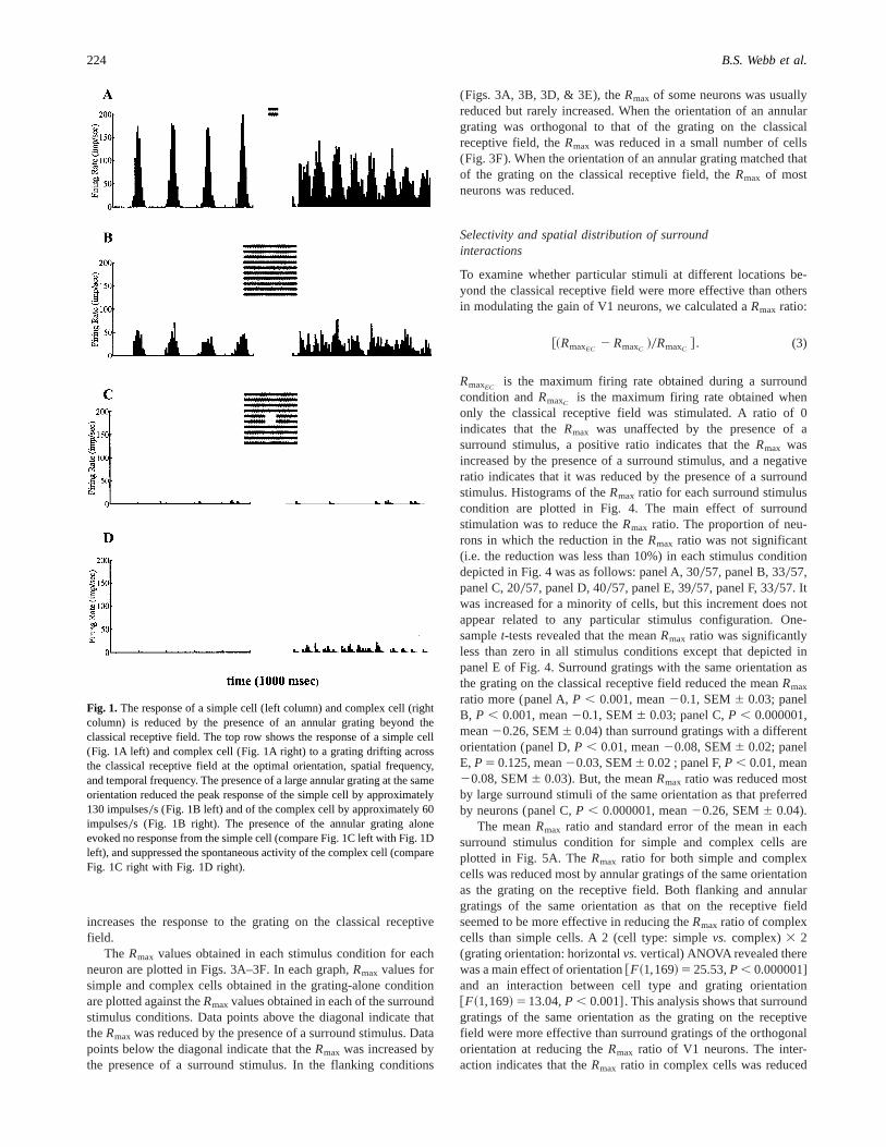
The data described here were obtained from 34 simple and 33complex cells in primary visual cortex (V1) of the marmoset.Neurons were recorded at eccentricities between 1.02 and 9.9 deg.Fig. 1 shows typical responses of a simple cell (left column) andcomplex cell (right column). The top row of Fig. 1 illustrates thata grating drifting across the receptive field at the preferred orien-tation, spatial frequency, and temporal frequency evoked a re-sponse that peaks at 180 impulses0s from the simple cell (Fig. 1Aleft) and one that peaks at 120 impulses0s from the complex cell(Fig. 1A right). The presence of an annular grating beyond theclassical receptive field of the same orientation as that preferred bythe neuron reduced the peak response of the simple cell byapproximately 130 impulses0s (Fig. 1B left), and of the complexcell by about 60 impulse0s. When an annular grating was pre-sented alone, it evoked no response from the simple cell (compareFig. 1C left with Fig. 1D left) and suppressed the spontaneousactivity of the complex cell (compare Fig. 1C right with Fig. 1Dright).
Surround modulation of gain
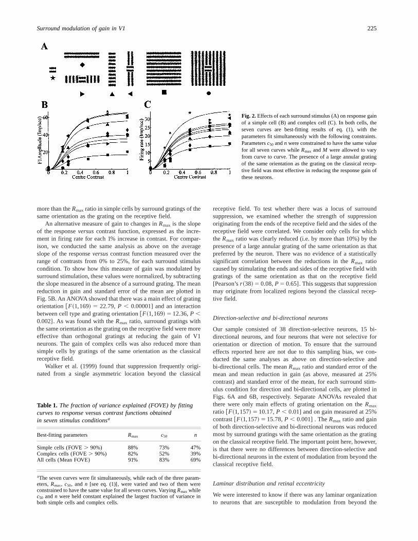
The primary aim of the study was to examine how flanking andannular stimuli of the same and orthogonal orientation to thereceptive field presented beyond the receptive field modulated thegain of responses to an optimal stimulus on the receptive field. Tocharacterize how different surround stimuli modulated gain, theMichaelis–Menten equation was fitted to responseversuscontrastfunctions obtained under seven stimulus conditions (Fig. 2A). Weestimated values of each of the parametersRmax, c50, andn thatwere required to produce curves that fit the neuron’s responseversuscontrast function under the seven stimulus conditions. Foreach neuron, the seven curves were fit simultaneously and twoparameters were constrained to have the same value for all sevencurves, while the third parameter andM were allowed to vary fromcurve to curve. We compared the fits produced by varying each ofthe three curve parameters,Rmax, c50, andn. Allowing c50 to varyexplained more variance than allowingn to vary, but allowingRmax to vary explained the most variance (see Table 1). The meanfraction of variance explained by allowingRmax to vary was 91%.More than 90% of the variance was explained in 30 (88%) simplecells and 27 (82%) complex cells. The following analysis isconfined toRmax values obtained from these neurons.
Figs. 2B and 2C are examples of constrained fits to dataobtained under seven stimulus conditions from a simple and acomplex cell, respectively. In both examples, surround stimulireducedRmax to the grating on the classical receptive field bydifferent amounts.Rmax was reduced most when the surroundstimulus was a large annular grating of the same orientation as thegrating on the classical receptive field. The response was reducedby up to 75% of theRmax in the simple cell, and by up to 60% inthe complex cell. There is no evidence that surround stimulation
Surround modulation of gain in V1 223
increases the response to the grating on the classical receptivefield.
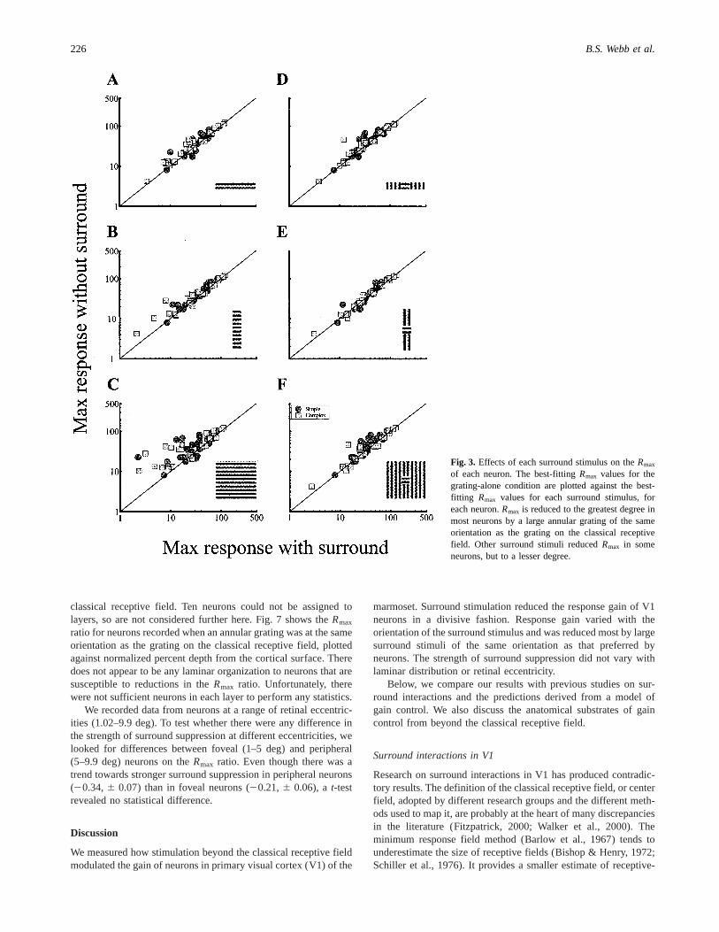
The Rmax values obtained in each stimulus condition for eachneuron are plotted in Figs. 3A–3F. In each graph,Rmax values forsimple and complex cells obtained in the grating-alone conditionare plotted against theRmaxvalues obtained in each of the surroundstimulus conditions. Data points above the diagonal indicate thattheRmax was reduced by the presence of a surround stimulus. Datapoints below the diagonal indicate that theRmax was increased bythe presence of a surround stimulus. In the flanking conditions
(Figs. 3A, 3B, 3D, & 3E), theRmax of some neurons was usuallyreduced but rarely increased. When the orientation of an annulargrating was orthogonal to that of the grating on the classicalreceptive field, theRmax was reduced in a small number of cells(Fig. 3F). When the orientation of an annular grating matched thatof the grating on the classical receptive field, theRmax of mostneurons was reduced.
Selectivity and spatial distribution of surroundinteractions
To examine whether particular stimuli at different locations be-yond the classical receptive field were more effective than othersin modulating the gain of V1 neurons, we calculated aRmax ratio:
@~RmaxEC2 RmaxC !0RmaxC # . (3)
RmaxECis the maximum firing rate obtained during a surround
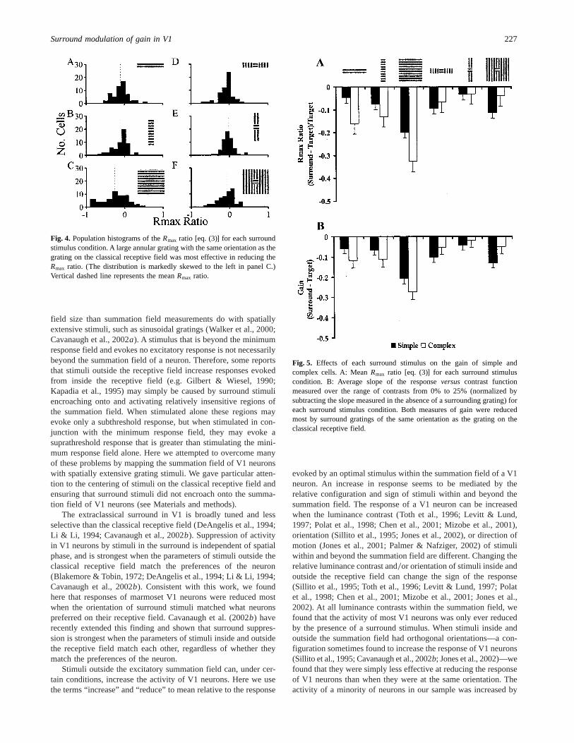
condition andRmaxC is the maximum firing rate obtained whenonly the classical receptive field was stimulated. A ratio of 0indicates that theRmax was unaffected by the presence of asurround stimulus, a positive ratio indicates that theRmax wasincreased by the presence of a surround stimulus, and a negativeratio indicates that it was reduced by the presence of a surroundstimulus. Histograms of theRmax ratio for each surround stimuluscondition are plotted in Fig. 4. The main effect of surroundstimulation was to reduce theRmax ratio. The proportion of neu-rons in which the reduction in theRmax ratio was not significant(i.e. the reduction was less than 10%) in each stimulus conditiondepicted in Fig. 4 was as follows: panel A, 30057, panel B, 33057,panel C, 20057, panel D, 40057, panel E, 39057, panel F, 33057. Itwas increased for a minority of cells, but this increment does notappear related to any particular stimulus configuration. One-samplet-tests revealed that the meanRmax ratio was significantlyless than zero in all stimulus conditions except that depicted inpanel E of Fig. 4. Surround gratings with the same orientation asthe grating on the classical receptive field reduced the meanRmax
ratio more (panel A,P , 0.001, mean20.1, SEM6 0.03; panelB, P , 0.001, mean20.1, SEM6 0.03; panel C,P , 0.000001,mean20.26, SEM6 0.04) than surround gratings with a differentorientation (panel D,P , 0.01, mean20.08, SEM6 0.02; panelE, P5 0.125, mean20.03, SEM6 0.02 ; panel F,P , 0.01, mean20.08, SEM6 0.03). But, the meanRmax ratio was reduced mostby large surround stimuli of the same orientation as that preferredby neurons (panel C,P , 0.000001, mean20.26, SEM6 0.04).
The meanRmax ratio and standard error of the mean in eachsurround stimulus condition for simple and complex cells areplotted in Fig. 5A. TheRmax ratio for both simple and complexcells was reduced most by annular gratings of the same orientationas the grating on the receptive field. Both flanking and annulargratings of the same orientation as that on the receptive fieldseemed to be more effective in reducing theRmax ratio of complexcells than simple cells. A 2 (cell type: simplevs. complex)3 2(grating orientation: horizontalvs.vertical) ANOVA revealed therewas a main effect of orientation@F~1,169! 5 25.53,P , 0.000001]and an interaction between cell type and grating orientation@F~1,169! 513.04,P , 0.001]. This analysis shows that surroundgratings of the same orientation as the grating on the receptivefield were more effective than surround gratings of the orthogonalorientation at reducing theRmax ratio of V1 neurons. The inter-action indicates that theRmax ratio in complex cells was reduced
Fig. 1. The response of a simple cell (left column) and complex cell (rightcolumn) is reduced by the presence of an annular grating beyond theclassical receptive field. The top row shows the response of a simple cell(Fig. 1A left) and complex cell (Fig. 1A right) to a grating drifting acrossthe classical receptive field at the optimal orientation, spatial frequency,and temporal frequency. The presence of a large annular grating at the sameorientation reduced the peak response of the simple cell by approximately130 impulses0s (Fig. 1B left) and of the complex cell by approximately 60impulses0s (Fig. 1B right). The presence of the annular grating aloneevoked no response from the simple cell (compare Fig. 1C left with Fig. 1Dleft), and suppressed the spontaneous activity of the complex cell (compareFig. 1C right with Fig. 1D right).
224 B.S. Webb et al.
more than theRmax ratio in simple cells by surround gratings of thesame orientation as the grating on the receptive field.
An alternative measure of gain to changes inRmax is the slopeof the responseversuscontrast function, expressed as the incre-ment in firing rate for each 1% increase in contrast. For compar-ison, we conducted the same analysis as above on the averageslope of the responseversuscontrast function measured over therange of contrasts from 0% to 25%, for each surround stimuluscondition. To show how this measure of gain was modulated bysurround stimulation, these values were normalized, by subtractingthe slope measured in the absence of a surround grating. The meanreduction in gain and standard error of the mean are plotted inFig. 5B. An ANOVA showed that there was a main effect of gratingorientation@F~1,169! 5 22.79,P , 0.00001] and an interactionbetween cell type and grating orientation@F~1,169! 5 12.36,P ,0.002]. As was found with theRmax ratio, surround gratings withthe same orientation as the grating on the receptive field were moreeffective than orthogonal gratings at reducing the gain of V1neurons. The gain of complex cells was also reduced more thansimple cells by gratings of the same orientation as the classicalreceptive field.
Walker et al. (1999) found that suppression frequently origi-nated from a single asymmetric location beyond the classical
receptive field. To test whether there was a locus of surroundsuppression, we examined whether the strength of suppressionoriginating from the ends of the receptive field and the sides of thereceptive field were correlated. We consider only cells for whichtheRmax ratio was clearly reduced (i.e. by more than 10%) by thepresence of a large annular grating of the same orientation as thatpreferred by the neuron. There was no evidence of a statisticallysignificant correlation between the reductions in theRmax ratiocaused by stimulating the ends and sides of the receptive field withgratings of the same orientation as that on the receptive field[Pearson’sr ~38! 5 0.08,P5 0.65]. This suggests that suppressionmay originate from localized regions beyond the classical recep-tive field.
Direction-selective and bi-directional neurons
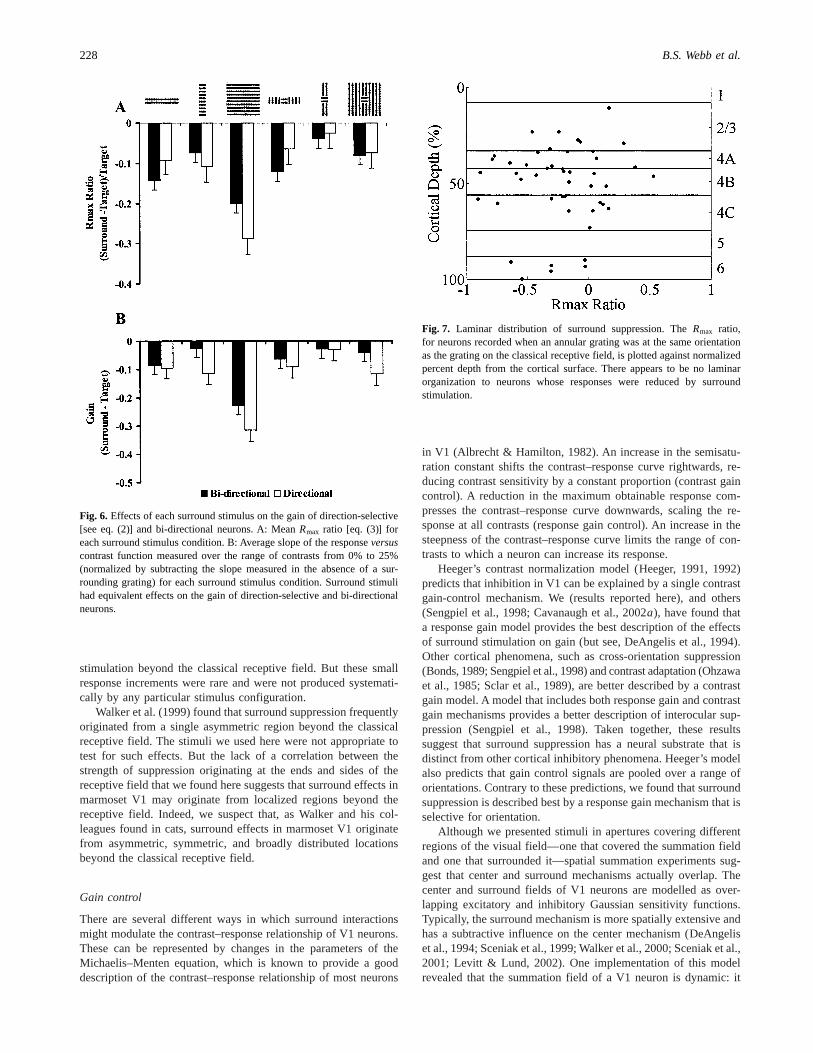
Our sample consisted of 38 direction-selective neurons, 15 bi-directional neurons, and four neurons that were not selective fororientation or direction of motion. To ensure that the surroundeffects reported here are not due to this sampling bias, we con-ducted the same analyses as above on direction-selective andbi-directional cells. The meanRmax ratio and standard error of themean and mean reduction in gain (as above, measured at 25%contrast) and standard error of the mean, for each surround stim-ulus condition for direction and bi-directional cells, are plotted inFigs. 6A and 6B, respectively. Separate ANOVAs revealed thatthere were only main effects of grating orientation on theRmax
ratio @F~1,157! 5 10.17,P , 0.01] and on gain measured at 25%contrast@F~1,157! 5 15.78,P , 0.001] . TheRmax ratio and gainof both direction-selective and bi-directional neurons was reducedmost by surround gratings with the same orientation as the gratingon the classical receptive field. The important point here, however,is that there were no differences between direction-selective andbi-directional neurons in the extent of modulation from beyond theclassical receptive field.
Laminar distribution and retinal eccentricity
We were interested to know if there was any laminar organizationto neurons that are susceptible to modulation from beyond the
Fig. 2.Effects of each surround stimulus (A) on response gainof a simple cell (B) and complex cell (C). In both cells, theseven curves are best-fitting results of eq. (1), with theparameters fit simultaneously with the following constraints.Parametersc50 andn were constrained to have the same valuefor all seven curves whileRmax andM were allowed to varyfrom curve to curve. The presence of a large annular gratingof the same orientation as the grating on the classical recep-tive field was most effective in reducing the response gain ofthese neurons.
Table 1. The fraction of variance explained (FOVE) by fittingcurves to responseversuscontrast functions obtainedin seven stimulus conditionsa
Best-fitting parameters Rmax c50 n
Simple cells (FOVE. 90%) 88% 73% 47%Complex cells (FOVE. 90%) 82% 52% 39%All cells (Mean FOVE) 91% 83% 69%
aThe seven curves were fit simultaneously, while each of the three param-eters,Rmax, c50, and n [see eq. (1)], were varied and two of them wereconstrained to have the same value for all seven curves. VaryingRmaxwhilec50 andn were held constant explained the largest fraction of variance inboth simple cells and complex cells.
Surround modulation of gain in V1 225
classical receptive field. Ten neurons could not be assigned tolayers, so are not considered further here. Fig. 7 shows theRmax
ratio for neurons recorded when an annular grating was at the sameorientation as the grating on the classical receptive field, plottedagainst normalized percent depth from the cortical surface. Theredoes not appear to be any laminar organization to neurons that aresusceptible to reductions in theRmax ratio. Unfortunately, therewere not sufficient neurons in each layer to perform any statistics.
We recorded data from neurons at a range of retinal eccentric-ities (1.02–9.9 deg). To test whether there were any difference inthe strength of surround suppression at different eccentricities, welooked for differences between foveal (1–5 deg) and peripheral(5–9.9 deg) neurons on theRmax ratio. Even though there was atrend towards stronger surround suppression in peripheral neurons(20.34,6 0.07) than in foveal neurons (20.21,6 0.06), at-testrevealed no statistical difference.
Discussion
We measured how stimulation beyond the classical receptive fieldmodulated the gain of neurons in primary visual cortex (V1) of the
marmoset. Surround stimulation reduced the response gain of V1neurons in a divisive fashion. Response gain varied with theorientation of the surround stimulus and was reduced most by largesurround stimuli of the same orientation as that preferred byneurons. The strength of surround suppression did not vary withlaminar distribution or retinal eccentricity.
Below, we compare our results with previous studies on sur-round interactions and the predictions derived from a model ofgain control. We also discuss the anatomical substrates of gaincontrol from beyond the classical receptive field.
Surround interactions in V1
Research on surround interactions in V1 has produced contradic-tory results. The definition of the classical receptive field, or centerfield, adopted by different research groups and the different meth-ods used to map it, are probably at the heart of many discrepanciesin the literature (Fitzpatrick, 2000; Walker et al., 2000). Theminimum response field method (Barlow et al., 1967) tends tounderestimate the size of receptive fields (Bishop & Henry, 1972;Schiller et al., 1976). It provides a smaller estimate of receptive-
Fig. 3. Effects of each surround stimulus on theRmax
of each neuron. The best-fittingRmax values for thegrating-alone condition are plotted against the best-fitting Rmax values for each surround stimulus, foreach neuron.Rmax is reduced to the greatest degree inmost neurons by a large annular grating of the sameorientation as the grating on the classical receptivefield. Other surround stimuli reducedRmax in someneurons, but to a lesser degree.
226 B.S. Webb et al.
field size than summation field measurements do with spatiallyextensive stimuli, such as sinusoidal gratings (Walker et al., 2000;Cavanaugh et al., 2002a). A stimulus that is beyond the minimumresponse field and evokes no excitatory response is not necessarilybeyond the summation field of a neuron. Therefore, some reportsthat stimuli outside the receptive field increase responses evokedfrom inside the receptive field (e.g. Gilbert & Wiesel, 1990;Kapadia et al., 1995) may simply be caused by surround stimuliencroaching onto and activating relatively insensitive regions ofthe summation field. When stimulated alone these regions mayevoke only a subthreshold response, but when stimulated in con-junction with the minimum response field, they may evoke asuprathreshold response that is greater than stimulating the mini-mum response field alone. Here we attempted to overcome manyof these problems by mapping the summation field of V1 neuronswith spatially extensive grating stimuli. We gave particular atten-tion to the centering of stimuli on the classical receptive field andensuring that surround stimuli did not encroach onto the summa-tion field of V1 neurons (see Materials and methods).
The extraclassical surround in V1 is broadly tuned and lessselective than the classical receptive field (DeAngelis et al., 1994;Li & Li, 1994; Cavanaugh et al., 2002b). Suppression of activityin V1 neurons by stimuli in the surround is independent of spatialphase, and is strongest when the parameters of stimuli outside theclassical receptive field match the preferences of the neuron(Blakemore & Tobin, 1972; DeAngelis et al., 1994; Li & Li, 1994;Cavanaugh et al., 2002b). Consistent with this work, we foundhere that responses of marmoset V1 neurons were reduced mostwhen the orientation of surround stimuli matched what neuronspreferred on their receptive field. Cavanaugh et al. (2002b) haverecently extended this finding and shown that surround suppres-sion is strongest when the parameters of stimuli inside and outsidethe receptive field match each other, regardless of whether theymatch the preferences of the neuron.
Stimuli outside the excitatory summation field can, under cer-tain conditions, increase the activity of V1 neurons. Here we usethe terms “increase” and “reduce” to mean relative to the response
evoked by an optimal stimulus within the summation field of a V1neuron. An increase in response seems to be mediated by therelative configuration and sign of stimuli within and beyond thesummation field. The response of a V1 neuron can be increasedwhen the luminance contrast (Toth et al., 1996; Levitt & Lund,1997; Polat et al., 1998; Chen et al., 2001; Mizobe et al., 2001),orientation (Sillito et al., 1995; Jones et al., 2002), or direction ofmotion (Jones et al., 2001; Palmer & Nafziger, 2002) of stimuliwithin and beyond the summation field are different. Changing therelative luminance contrast and0or orientation of stimuli inside andoutside the receptive field can change the sign of the response(Sillito et al., 1995; Toth et al., 1996; Levitt & Lund, 1997; Polatet al., 1998; Chen et al., 2001; Mizobe et al., 2001; Jones et al.,2002). At all luminance contrasts within the summation field, wefound that the activity of most V1 neurons was only ever reducedby the presence of a surround stimulus. When stimuli inside andoutside the summation field had orthogonal orientations—a con-figuration sometimes found to increase the response of V1 neurons(Sillito et al., 1995; Cavanaugh et al., 2002b; Jones et al., 2002)—wefound that they were simply less effective at reducing the responseof V1 neurons than when they were at the same orientation. Theactivity of a minority of neurons in our sample was increased by
Fig. 4. Population histograms of theRmax ratio [eq. (3)] for each surroundstimulus condition. A large annular grating with the same orientation as thegrating on the classical receptive field was most effective in reducing theRmax ratio. (The distribution is markedly skewed to the left in panel C.)Vertical dashed line represents the meanRmax ratio.
Fig. 5. Effects of each surround stimulus on the gain of simple andcomplex cells. A: MeanRmax ratio [eq. (3)] for each surround stimuluscondition. B: Average slope of the responseversus contrast functionmeasured over the range of contrasts from 0% to 25% (normalized bysubtracting the slope measured in the absence of a surrounding grating) foreach surround stimulus condition. Both measures of gain were reducedmost by surround gratings of the same orientation as the grating on theclassical receptive field.
Surround modulation of gain in V1 227
stimulation beyond the classical receptive field. But these smallresponse increments were rare and were not produced systemati-cally by any particular stimulus configuration.
Walker et al. (1999) found that surround suppression frequentlyoriginated from a single asymmetric region beyond the classicalreceptive field. The stimuli we used here were not appropriate totest for such effects. But the lack of a correlation between thestrength of suppression originating at the ends and sides of thereceptive field that we found here suggests that surround effects inmarmoset V1 may originate from localized regions beyond thereceptive field. Indeed, we suspect that, as Walker and his col-leagues found in cats, surround effects in marmoset V1 originatefrom asymmetric, symmetric, and broadly distributed locationsbeyond the classical receptive field.
Gain control
There are several different ways in which surround interactionsmight modulate the contrast–response relationship of V1 neurons.These can be represented by changes in the parameters of theMichaelis–Menten equation, which is known to provide a gooddescription of the contrast–response relationship of most neurons
in V1 (Albrecht & Hamilton, 1982). An increase in the semisatu-ration constant shifts the contrast–response curve rightwards, re-ducing contrast sensitivity by a constant proportion (contrast gaincontrol). A reduction in the maximum obtainable response com-presses the contrast–response curve downwards, scaling the re-sponse at all contrasts (response gain control). An increase in thesteepness of the contrast–response curve limits the range of con-trasts to which a neuron can increase its response.
Heeger’s contrast normalization model (Heeger, 1991, 1992)predicts that inhibition in V1 can be explained by a single contrastgain-control mechanism. We (results reported here), and others(Sengpiel et al., 1998; Cavanaugh et al., 2002a), have found thata response gain model provides the best description of the effectsof surround stimulation on gain (but see, DeAngelis et al., 1994).Other cortical phenomena, such as cross-orientation suppression(Bonds, 1989; Sengpiel et al., 1998) and contrast adaptation (Ohzawaet al., 1985; Sclar et al., 1989), are better described by a contrastgain model. A model that includes both response gain and contrastgain mechanisms provides a better description of interocular sup-pression (Sengpiel et al., 1998). Taken together, these resultssuggest that surround suppression has a neural substrate that isdistinct from other cortical inhibitory phenomena. Heeger’s modelalso predicts that gain control signals are pooled over a range oforientations. Contrary to these predictions, we found that surroundsuppression is described best by a response gain mechanism that isselective for orientation.
Although we presented stimuli in apertures covering differentregions of the visual field—one that covered the summation fieldand one that surrounded it—spatial summation experiments sug-gest that center and surround mechanisms actually overlap. Thecenter and surround fields of V1 neurons are modelled as over-lapping excitatory and inhibitory Gaussian sensitivity functions.Typically, the surround mechanism is more spatially extensive andhas a subtractive influence on the center mechanism (DeAngeliset al., 1994; Sceniak et al., 1999; Walker et al., 2000; Sceniak et al.,2001; Levitt & Lund, 2002). One implementation of this modelrevealed that the summation field of a V1 neuron is dynamic: it
Fig. 6. Effects of each surround stimulus on the gain of direction-selective[see eq. (2)] and bi-directional neurons. A: MeanRmax ratio [eq. (3)] foreach surround stimulus condition. B: Average slope of the responseversuscontrast function measured over the range of contrasts from 0% to 25%(normalized by subtracting the slope measured in the absence of a sur-rounding grating) for each surround stimulus condition. Surround stimulihad equivalent effects on the gain of direction-selective and bi-directionalneurons.
Fig. 7. Laminar distribution of surround suppression. TheRmax ratio,for neurons recorded when an annular grating was at the same orientationas the grating on the classical receptive field, is plotted against normalizedpercent depth from the cortical surface. There appears to be no laminarorganization to neurons whose responses were reduced by surroundstimulation.
228 B.S. Webb et al.
samples more of the visual field at low contrast than at highcontrast (Sceniak et al., 1999). Our data, however, are clearly moreconsistent with a model in which the surround has a divisiveinfluence on the center mechanism (Cavanaugh et al., 2002a;Sceniak et al., 2001). Cavanaugh et al. (2002a) have devised amodel that implements a general form of divisive suppression.Center and surround mechanisms are modelled as overlappingGaussians, but each has its own gain control. Independent gain-control mechanisms in the center and surround enable the divisiveinfluence of the surround to scale responses up and down, depend-ing on the relative strength of each mechanism. The apparentincrease in size with contrast occurs because of this scaling ofsensitivity (Cavanaugh et al., 2002a).
Anatomical substrates for surround interactions
Surround interactions were not localized to any particular corticallayer(s). In each layer from which we obtained data, the strengthof signals from the surround field of neurons ranged from negli-gible to weak to very strong. As others (Sceniak et al., 2001;Cavanaugh et al., 2002a; Levitt & Lund, 2002; Yao & Li, 2002)have found, it seems that surround interactions are a generalproperty of V1, with different neurons being affected to differentextents.
What is the circuitry that mediates surround interactions in V1?Surround interactions may be mediated by intrinsic lateral and0orfeedback connections in V1 (Rockland & Lund, 1982, 1983;Livingstone & Hubel, 1984; Kennedy & Bullier, 1985; Gilbert &Wiesel, 1989; Malach et al., 1993; Budd, 1998; Lamme & Roelf-sema, 2000; Bullier, 2001). Combined anatomical and physiolog-ical studies conducted by Angelucci and her colleagues (Angelucciet al., 2002a,b) suggest that feedback connections from extrastri-ate cortex are the most likely substrate. They compared the spatialextent of feedforward input from the lateral geniculate nucleus(LGN), intrinsic lateral connections, and feedback connectionsfrom extrastriate cortex with that of the minimum response field,the summation field, and extraclassical surround field of V1 neu-rons. They concluded that in V1 LGN input mediates activity insummation fields measured with high contrast stimuli, horizontalconnections mediate activity in summation fields measured withlow contrast stimuli, and feedback connections mediate inter-actions between stimuli within and beyond summation fields (An-gelucci et al., 2002a,b).
Gain control appears to be a fundamental property of earlyvisual processing. We have found that, in both the LGN (Webbet al., 2002) and V1 of marmosets (results reported here), responsegain is reduced in a divisive fashion by input from beyond theclassical receptive field. In the LGN, gain control may be mediatedby the feedback projection from V1 (Przybyszewski et al., 2000;Webb et al., 2002). In V1, modulation of gain-control signals frombeyond the summation field are probably mediated by feedbackfrom extrastriate cortex (Angelucci et al., 2002a,b).
Cortical feedback projections may control information flowingthrough the visual hierarchy by modulating the gain of neurons atthe preceding stage. In the LGN, this mechanism is mediated by anexcitatory feedback projection from V1 that influences responsesevoked from inside the classical receptive field (Przybyszewskiet al., 2000). Inhibition from outside the classical receptive fieldmodulates the excitatory influence of the cortex in the LGN (Webbet al., 2002). In V1, feedback from extrastriate cortex may have amore direct effect on inhibitory influences from outside the sum-mation field, possiblyvia inhibitory interneurons.
Conclusion
Input from beyond the classical receptive field reduced responsegain in a divisive fashion. Reductions in response gain were mostprominent when the orientation of large stimuli beyond the clas-sical receptive field matched that preferred by neurons. Responsegain control in the early visual system may be mediated by theinteraction between cortical feedback and inhibitory influencesfrom beyond the classical receptive field.
Acknowledgments
This work was supported by grants from the Wellcome Trust, the BBSRC,and a Wellcome Prize studentship to Ben S. Webb. We thank Carl Espin fortechnical support, Peter Lennie for allowing us to use his software, and thetwo anonymous reviewers for their comments.
References
Albrecht, D.G. & Hamilton, D.B. (1982). Striate cortex of monkey andcat: Contrast response function.Journal of Neurophysiology48, 217–237.
Albright, T.D. & Stoner, G.R. (2002). Contextual influences on visualprocessing.Annual Review of Neuroscience25, 339–379.
Allman, J., Miezin, F. & McGuinness, E. (1985). Stimulus specificresponses from beyond the classical receptive field: Neurophysiologi-cal mechanisms for local-global comparisons in visual neurons.AnnualReview of Neuroscience8, 407–430.
Angelucci, A., Levitt, J.B. & Lund, J.S. (2002a). Anatomical origins ofthe classical receptive field and modulatory surround field of singleneurons in macaque visual cortical area V1.Progress in Brain Research136, 373–388.
Angelucci, A., Levitt, J.B., Walton, E.J.S., Hupé, J.M., Bullier, J. &Lund, J.S. (2002b). Circuits for local and global signal integration inprimary visual cortex.Journal of Neuroscience22, 8633–8646.
Barlow, H.B., Blakemore, C. & Pettigrew, J.D. (1967). The neuralmechanism of binocular depth discrimination.Journal of Physiology193, 327–342.
Bishop, P.O. & Henry, G.H. (1972). Striate neurons: Receptive fieldconcepts.Investigative Ophthalmology11, 346–354.
Blakemore, C. & Tobin, E.A. (1972). Lateral inhibition between orien-tation detectors in the cat’s visual cortex.Experimental Brain Research15, 439–440.
Bonds, A.B. (1989). Role of inhibition in the specification of orientationselectivity of cells in the cat striate cortex.Visual Neuroscience2,41–55.
Budd, J.M.L. (1998). Extrastriate feedback to primary visual cortex inprimates: A quantitative analysis of connectivity.Proceedings of theRoyal Society (B)(London)265, 1037–1044.
Bullier, J. (2001). Integrated model of visual processing.Brain ResearchReviews36, 96–107.
Carandini, M., Heeger, D.J. & Movshon, J.A. (1997). Linearity andnormalization in simple cells of the macaque primary visual cortex.Journal of Neuroscience17, 8621–8644.
Cavanaugh, J.R., Bair, W. & Movshon, J.A. (2002a). Nature andinteraction of signals from the receptive field center and surround inmacaque V1 neurons.Journal of Neurophysiology88, 2530–2546.
Cavanaugh, J.R., Bair, W. & Movshon, J.A. (2002b). Selectivity andspatial distribution of signals from the receptive field surround inmacaque V1 neurons.Journal of Neurophysiology88, 2547–2556.
Chen, C.C., Kasamatsu, T., Polat, U. & Norcia, A.M. (2001). Contrastresponse characteristics of long-range lateral interactions in cat striatecortex.Neuroreport12, 655–661.
Deangelis, G.C., Freeman, R.D. & Ohzawa, I. (1994). Length andwidth tuning of neurons in the cat’s primary visual cortex.Journal ofNeurophysiology71, 347–374.
Fitzpatrick, D. (2000). Seeing beyond the receptive field in primaryvisual cortex.Current Opinion in Neurobiology10, 438–443.
Geisler, W.S. & Albrecht, D.G. (1992). Cortical neurons: Isolation ofcontrast gain control.Vision Research32, 1409–1410.
Gilbert, C.D. & Wiesel, T.N. (1989). Columnar specificity of instrinsichorizontal and corticocortical connections in cat visual cortex.Journalof Neuroscience9, 2432–2442.
Surround modulation of gain in V1 229
Gilbert, C.D. & Wiesel, T.N. (1990). The influence of contextual stimulion the orientation selectivity of cells in primary visual cortex of the cat.Vision Research30, 1689–1701.
Heeger, D.J. (1991). Nonlinear model of neural responses in cat visualcortex. InComputational Models of Visual Processing, ed.Landy, M.& Movshon, J.A., pp. 119–133. Cambridge, Massachusetts: MITPress.
Heeger, D.J. (1992). Normalization of cell responses in cat striate cortex.Visual Neuroscience9, 181–197.
Hubel, D.H. & Wiesel, T.N. (1962). Receptive fields, binocular inter-action and functional architecture in cat’s visual cortex.Journal ofPhysiology(London)160, 106–154.
Hubel, D.H. & Wiesel, T.N. (1968). Receptive fields and functionalarchitecture of monkey striate cortex.Journal of Physiology(London)195, 215–243.
Jones, H.E., Grieve, K.L., Wang, W. & Sillito, A.M. (2001). Surroundsuppression in primate V1.Journal of Neurophysiology86, 2011–2028.
Jones, H.E., Wang, W. & Sillito, A.M. (2002). Spatial organization andmagnitude of orientation contrast interactions in primate V1.Journal ofNeurophysiology88, 2796–2808.
Kapadia, M.K., Ito, M. & Gilbert, C.D. (1995). Improvement in visualsensitivity by changes in local context: Parallel studies in humanobservers and V1 of alert monkeys.Neuron15, 843–856.
Kapadia, M.K., Westheimer, G. & Gilbert, C.D. (1999). Dynamicsof spatial summation in primary visual cortex of alert monkeys.Proceedings of the National Academy of Sciences of the U.S.A.96,12073–12078.
Kastner, S., Nothdurft, H.C. & Pigarev, I.N. (1997). Neuronal corre-lates of pop-out in cat striate cortex.Vision Research37, 371–376.
Kennedy, H. & Bullier, J. (1985). A double-labeling investigation of theafferent connectivity to cortical areas V1 and V2 of the macaquemonkey.Journal of Neuroscience5, 2815–2830.
Lamme, V.A.F. (1995). The neurophysiology of figure-ground segregationin primary visual cortex.Journal of Neuroscience15, 1605–1615.
Lamme, V.A.F. & Spekreijse, H. (2000). Contextual modulation in pri-mary visual cortex and scene perception. InThe New Cognitive Neuro-sciences, ed.Gazzaniga, M., pp. 279–290. Cambridge, Massachusetts:MIT Press. .
Lamme, V.A.F. & Roelfsema, P.R. (2000). The distinct modes of visionoffered by feedforward and recurrent processing.Trends in Neurosci-ences23, 571–579.
Levitt, J.B. & Lund, J.S. (1997). Contrast dependence of contextualeffects in primate visual cortex.Nature387, 73–76.
Levitt, J.B. & Lund, J.S. (2002). The spatial extent over which neurons inmacaque striate cortex pool visual signals.Visual Neuroscience19,439–452.
Li, C.Y. & Li, W. (1994). Extensive integration field beyond the classicalreceptive field of cat’s striate cortical neurons—classification andtuning properties.Vision Research34, 2337–2355.
Livingstone, M.S. & Hubel, D.H. (1984). Specificity of intrinsic con-nections in primate primary visual cortex.Journal of Neuroscience4,2830–2835.
Maffei, L. & Fiorentini, A. (1976). The unresponsive regions of visualcortical receptive fields.Vision Research16, 1131–1139.
Malach, R., Amir, Y., Harel, M. & Grinvald, A. (1993). Relationshipbetween intrinsic connections and functional architecture revealed byoptical imaging andin vivo targeted biocytin injections in primatestriate cortex.Proceedings of the National Academy of Sciences of theU.S.A.90, 10469–10473.
Merrill, E.G. & Ainsworth, A. (1972). Glass-coated platinum-platedtungsten microelectrodes.Medical and Biological Engineering10,662–672.
Mizobe, K., Polat, U., Pettet, M.W. & Kasamatsu, T. (2001). Facili-tation and suppression of single striate-cell activity by spatially discretepattern stimuli presented beyond the receptive field.Visual Neurosci-ence18, 377–391.
Nelson, J.I. & Frost, B. (1978). Orientation selective inhibition frombeyond the classical receptive field.Brain Research139, 359–365.
Nothdurft, H.C., Gallant, J.L. & Van Essen, D.C. (1999). Responsemodulation by texture surround in primate area V1: Correlates of“popout” under anesthesia.Visual Neuroscience16, 15–34.
Ohzawa, I., Sclar, G. & Freeman, R.D. (1985). Contrast gain control inthe cat’s visual system.Journal of Neurophysiology3, 651–667.
Palmer, L.A. & Nafziger, J.S. (2002). Effects of surround motion onreceptive-field gain and structure in area 17 of the cat.Visual Neuro-science19, 335–353.
Polat, U., Mizobe, K., Pettet, M.W., Kasamatsu, T. & Norcia, A.M.(1998). Collinear stimuli regulate visual responses depending on cell’scontrast threshold.Nature391, 580–584.
Przybyszewski, A.W., Gaska, J.P., Foote, W. & Pollen, D.A. (2000).Striate cortex increases contrast gain of macaque LGN neurons.VisualNeuroscience17, 485–494.
Rockland, K.S. & Lund, J. (1982). Widespread periodic intrinsic con-nections in the tree shew visual cortex.Science215, 1532–1534.
Rockland, K.S. & Lund, J. (1983). Intrinsic laminar lattice connectionsin primate visual cortex.Journal of Comparative Neurology216,303–318.
Rossi, A.F., Desimone, R. & Ungerleider, L.G. (2001). Contextualmodulation in primary visual cortex of macaques.Journal of Neuro-science21, 1698–1709.
Sceniak, M.P., Ringach, D.L., Hawken, M.J. & Shapley, R. (1999).Contrast’s effect on spatial summation by macaque V1 neurons.NatureNeuroscience2, 733–739.
Sceniak, M.P., Hawken, M.J. & Shapley, R. (2001). Visual spatialcharacterization of macaque V1 neurons.Journal of Neurophysiology85, 1873–1887.
Schiller, P.H., Finlay, B.L. & Volman, S.F. (1976). Quantative studiesof single-cell properties in monkey striate cortex. I. Spatiotemporalorganization of receptive field.Journal of Neurophysiology39, 1288–1319.
Sclar, G., Lennie, P. & Depriest, D.D. (1989). Contrast adaptation instriate cortex of macaque.Vision Research7, 747–755.
Sengpiel, F., Baddeley, R., Freeman, T.C.B., Harrad, R. & Blakemore,C. (1998). Different mechanisms underlie three inhibitory phenomenain cat area 17.Vision Research38, 2067–2080.
Sillito, A.M., Grieve, K.L., Jones, H.E., Cudeiro, J. & Davis, J. (1995).Visual cortical mechanisms detecting focal orientation discontinuities.Nature378, 492–496.
Skottun, B.C., De Valois, R.L., Grosof, D.H. & Movshon, J.A. (1991).Classifying simple and complex cells on the basis of response modu-lation. Vision Research31, 1079–1986.
Sugita, Y. (1999). Grouping of image fragments in primary visual cortex.Nature401, 269–272.
Toth, L.J., Rao, S.C., Kim, D., Somers, D. & Sur, M. (1996). Sub-threshold facilitation and suppression in primary visual cortex revealedby intrinsic signal imaging.Proceedings of the National Academy ofSciences of the U.S.A. 93, 9869–9874.
Truchard, A.M., Ohzawa, I. & Freeman, R.D. (2000). Contrast gaincontrol in the visual cortex: Monocular versus binocular mechanisms.Journal of Neuroscience20, 3017–3032.
Walker, G.A., Ohzawa, I. & Freeman, R.D. (1999). Asymmetric sup-pression outside the classical receptive field of the visual cortex.Journal of Neuroscience19, 10536–10553.
Walker, G.A., Ohzawa, I. & Freeman, R.D. (2000). Suppression outsidethe classical cortical receptive field.Visual Neuroscience17, 369–379.
Webb, B.S., Tinsley, C.J., Barraclough, N.E., Easton, A., Parker, A.& Derrington, A.M. (2002). Feedback from V1 and inhibition frombeyond the classical receptive field modulates the responses of neuronsin the primate lateral geniculate nucleus.Visual Neuroscience19,583–592.
Wörgötter, F. & Eysel, U.T. (2000). Context, state and the receptivefields of striatal cortex cells.Trends in Neurosciences23, 497–503.
Yao, H. & Li, C.Y. (2002). Clustered organization of neurons with similarextra-receptive field properties in the primary visual cortex.Neuron35,547–553.
Zipser, K., Lamme, V.A.F. & Schiller, P.H. (1996). Contextual modula-tion in primary visual cortex.Journal of Neuroscience16, 7376–7389.
230 B.S. Webb et al.