functional comparison of mouse cire/mouse dc-sign and human dc-sign
TRANSCRIPT
Functional comparison of mouse CIRE/mouseDC-SIGN and human DC-SIGN
Irina Caminschi1,2, Alexandra J. Corbett3, Corina Zahra1, Mireille Lahoud1, Karen M. Lucas1,2,Mariam Sofi4, David Vremec1, Thomas Gramberg5, Stefan Pohlmann5, Joan Curtis1, EmanuelaHandman1, Serani L. H. van Dommelen3,6, Peter Fleming3,6, Mariapia A. Degli-Esposti3,6, KenShortman1,2 and Mark D. Wright4
1Walter and Eliza Hall Institute of Medical Research and 2Cooperative Research Centre for Vaccine Technology, Melbourne,Victoria 3050, Australia3Centre for Experimental Immunology, Lions Eye Institute, Western Australia4Mac Farlane Burnet Institute for Medical Research and Public Health, Kronheimer Building, A&RMC, Studley RoadHeidelberg, Victoria 3084, Australia5Institute for Clinical and Molecular Virology and Nikolaus-Fiebiger Center, University of Erlangen-Nurnberg,91054 Erlangen, Germany6Immunology and Virology Program, Centre for Ophthalmology and Visual Science, University of Western Australia,Western Australia
Keywords: cell-surface molecules, dendritic cells, rodents
Abstract
CIRE/mDC-SIGN is a C-type lectin we originally identified as a molecule differentially expressedby mouse dendritic cell (DC) populations. Immunostaining with a CIRE/mDC-SIGN-specific mAbrevealed that CIRE/mDC-SIGN is indeed on the surface of some CD41, CD4�8� DCs and plasmacytoidpre-DCs, but not on CD81 DCs. It has been proposed that CIRE/mDC-SIGN is the functionalorthologue of human DC-SIGN (hDC-SIGN), a molecule that both enhances T cell responses andfacilitates antigen uptake. We assessed if CIRE/mDC-SIGN and hDC-SIGN exhibit functionalsimilarities. CIRE/mDC-SIGN is down-regulated upon activation, but unlike hDC-SIGN, incubation withIL-4 and IL-13 did not enhance CIRE/mDC-SIGN expression, indicating differences in gene regulation.Like hDC-SIGN, CIRE/mDC-SIGN bound mannosylated residues. However, we could detect no role forCIRE/mDC-SIGN in T cell–DC interactions and the protein did not bind to pathogens known to interactwith hDC-SIGN, including Leishmania mexicana, cytomegalovirus, HIV and lentiviral particles bearingthe Ebolavirus glycoprotein. The binding of CIRE/mDC-SIGN to hDC-SIGN ligands was not rescuedwhen CIRE/mDC-SIGN was engineered to express the stalk region of hDC-SIGN. We conclude thatthere are significant differences in the fine specificity of the C-type lectin domains of hDC-SIGN andCIRE/mDC-SIGN and that these two molecules may not be functional orthologues.
Introduction
Antigen-presenting dendritic cells (DCs) are essential forinitiating immune responses because of their unique abilityto stimulate naive T lymphocytes (1). However, DCs are het-erogeneous in both their ontogeny and function (2). In mousespleen, there exist three major populations of conventionaldendritic cells (cDCs): CD4+CD8� (CD4+ DC), CD4�CD8�
(double negative [DN] DC) and CD4�CD8+ DC (CD8+ DC)(3). While there is debate over the haematological originsof these populations (4–6), they are functionally distinct. Forexample, CD8+ DCs are relatively poor stimulators of T cells(7, 8), and were originally proposed to have a regulatoryfunction (9). However, if activated, CD8+ and CD8� DCs can
both induce immune responses (10, 11), though they pre-ferentially induce Th1 and Th2 type responses, respectively(12, 13). This is in part due to CD8+ DCs being more potentproducers of IL-12 (14, 15). CD8+ DCs have a unique ability totake-up dying cells (16), and are especially adept at cross-presenting exogenous antigen on class I MHC (17, 18). Lymphnode CD8+ DCs are the most efficient at presenting viralantigen and initiating T cell responses (19). Finally, DC pop-ulations occupy different micro-environments in lymphoidorgans (15, 20), CD8+ DCs tend to concentrate in T cell areas,whereas CD8� DCs are found in marginal zones but migrateto T cell areas upon stimulation (21).
Correspondence to: I. Caminschi; E-mail: [email protected] Received 5 June 2005, accepted 16 February 2006
Transmitting editor: C. Goodnow Advance Access publication 28 March 2006
International Immunology, Vol. 18, No. 5, pp. 741–753doi:10.1093/intimm/dxl011
ª The Japanese Society for Immunology. 2006. All rights reserved.For permissions, please e-mail: [email protected]
by guest on January 1, 2014http://intim
m.oxfordjournals.org/
Dow
nloaded from
Molecules whose expression differs among these DC pop-ulations are of particular interest as they may underpin theirfunctional differences. Using both genetic (22, 23) andimmunophenotyping approaches (3, 24, 25), several suchmolecules have been identified and recently we cloned thecell-surface protein CIRE, which is expressed by DN DCs andCD4+ DCs, but not by CD8+ DCs (26).
CIRE is a type II membrane protein with a C-type lectindomain (CTLD) at the cell surface. The CIRE protein sequenceshows a 57% identity with the human molecule DC-specificintercellular adhesion molecule (ICAM)-grabbing non-integrin(hDC-SIGN) (26, 27). Human DC-SIGN facilitates interactionswith T cells by binding ICAM-3, and with endothelial cells bybinding ICAM-2 (28, 29). It also plays a role in the capture,internalisation and presentation of foreign antigen (30). Notably,several pathogens interact with hDC-SIGN, including virusessuch as HIV, human cytomegalovirus (HCMV) and Ebolavirus(EBOV), bacteria such as Mycobacterium and parasites suchas Leishmania and the eggs of Schistosoma mansoni (31).
The sequence similarity, together with the cellular expressionand genomic localisation data, suggest that CIRE is mouse DC-SIGN (26, 27) and therefore will be referred to as CIRE/mDC-SIGN. However, whether CIRE/mDC-SIGN and hDC-SIGN areindeed functional orthologues is open to question. The issue isclouded by gene duplication events in both the mouse andhuman genomes. In humans, there is a DC-SIGN homologue(DC-SIGNR or L-SIGN), which is just as similar to CIRE/mDC-SIGN as is hDC-SIGN (32). In mice, there are four CIRE/mDC-SIGN homologues whose CTLDs display a level of identity tohDC-SIGN as close as CIRE/mDC-SIGN itself (26, 27).
We have now generated a mAb to CIRE/mDC-SIGN,enabling us to characterise this molecule at the protein level.We confirm the differential expression of CIRE/mDC-SIGN inDC subsets and demonstrate that CIRE/mDC-SIGN is indeeda lectin able to bind mannosylated ligands in a calcium-dependent manner. However, CIRE/mDC-SIGN does not bindto any of the hDC-SIGN ligands tested, indicating thatsignificant differences exist in the ligand-binding specificityof the two molecules. The function of CIRE/mDC-SIGN mighttherefore differ from that of hDC-SIGN.
Methods
Mice
C57BL/6J wehi, CBA/CaH mice and Wistar rats were bredunder specific pathogen-free (SPF) conditions at the Walterand Eliza Hall Institute (WEHI). Germ-free C57BL/6J mice werebred at the WEHI facility and sacrificed within 12 h of arrivalinto our SPF holding facility.
Generating an anti-CIRE/mDC-SIGN mAb
A synthetic peptide was synthesised (H-MSKESTWYWVDG-SPLTLSFMKYWSKC-NH2) and conjugated to keyhole limpethaemocyanin (KLH) (Mimotopes, Victoria, Australia). To gen-erate mAbs, Wistar rats were immunised intra-peritoneally(i.p.) with 75 lg of KLH-conjugated peptide in CFA, boosted5 weeks later with 50 lg of KLH-conjugated peptide in incom-plete Freunds adjuvant and again 4 days prior to fusion in-travenously and i.p. with 10 lg of KLH-conjugated peptide in
aqueous solution. Hybridomas secreting specific mAbs wereidentified by flow cytometric analysis of supernatants usingFLAG-CIRE/mDC-SIGN-CHO and Neo-CHO (26). Four clones,from several thousand screened, produced anti-CIRE/mDC-SIGN mAb, but only one clone, 5H10, remained stable in cul-ture and continued to produce mAb.
Antibodies
The following fluorochrome-conjugated mAbs were used: anti-CD11c (N418)–allophycocyanin, –Cy5 or –FITC; anti-CIRE/mDC-SIGN (5H10)–biotin; anti-CD4 (GK1.5)–Alexa 594, anti-CD8 (53-6.7 or YTS 169.4)–FITC; anti-CD45RA (14.8)–FITC;isotype control IgG2a–biotin (PharMingen, San Diego, CA,USA); goat anti-rat–FITC antibody (Caltag, Burlingame, CA,USA); streptavidin–PE (PharMingen). To better visualise CIRE/mDC-SIGN on the DC surface, the amplification system Flow-Amp (Flow-Amp Systems, Cleveland, OH, USA) was usedaccording to the manufacturer’s guidelines. CIRE/mDC-SIGNstaining was always after pre-incubation with rat Ig and 2.4G2(10 min 4�C), and anti-CIRE/mDC-SIGN or isotype controlmAb was then added into the pre-blocking mix. The anti-hDC-SIGN (AZN-1) supernatant was kindly donated by D. Hart(Mater Institute, Queensland, Australia).
Isolation of DCs, macrophages and peripheral bloodmonocytes
The isolation of DC sub-populations has been described(3, 33). Briefly, tissues were chopped, digested with collage-nase and DNAase at room temperature and treated withEDTA. Low-density cells were enriched by density centrifuga-tion. Non-DC-lineage cells were coated with mAb (KT3-1.1,T24/31.7, TER119, RB6-8C5, ID3) and then removed usingimmunomagnetic beads. Coating with RB6-8C5 mAb (anti-Gr-1) did not result in the depletion of plasmacytoid pre-dendritic cells (pDCs) (33). The remaining cells were stainedwith various combinations of fluorochrome-conjugated mAband populations enriched for CD11c+CIRE/mDC-SIGN+ andCD11c+CIRE/mDC-SIGN� cells, or purified as CD11c+CD8+
CD4�, CD11c+CD8�CD4+ and CD11c+CD8� CD4� or asCD11cint CD45RA+ and CD11chi and CD45RA�, all by sortingon a MoFlo Instrument (Cytomation Inc.). Due to the lowlevel and frequency of CIRE/mDC-SIGN expression on DCs,the CD11c+CIRE/mDC-SIGN� DC purity was above 98%,but the CD11c+CIRE/mDC-SIGN+ DC purity was only 50–75%,the main contaminants being CD11c+CIRE/mDC-SIGN� DCs.To obtain blood mononuclear cells, mice were bled by cardiacpuncture into tubes containing heparinized buffered salinesolution. Mononuclear cells were isolated by density centrifu-gation using Histopaque 1.083 (Sigma, Castle Hill, Australia)and cells bearing CD3, Thy-1, Gr-1 and the erythrocyte markerTER119 removed by immunomagnetic bead depletion. Cellsfrom the peritoneal cavity or the bone marrow were obtainedby flushing with medium, and then removing erythrocyteswith lysis buffer (0.099 mM EDTA disodium, 0.145 M NH4Cl,0.012 M NaHCO3).
DC activation
Isolated DCs (2 3 106 cells ml�1) were cultured in 24-wellplates for 18–20 h in modified RPMI-1640 medium containing
742 CIRE/mDC-SIGN, DC-SIGN-like mouse DC protein
by guest on January 1, 2014http://intim
m.oxfordjournals.org/
Dow
nloaded from
10% FCS, antibiotics, 10�4 M 2-mercaptoethanol, granulocytemacrophage colony-stimulating factor (GM-CSF) (50 U ml�1)and CpG-1668 (0.5 lM) (GeneWorks, Adelaide, Australia) orin 96-well flat-bottom plates with murine recombinant IL-4(Immunex) and IL-13 (R&D Systems, Minneapolis, MN, USA).
Mixed leucocyte reaction
CD4 or CD8 T cells were isolated from lymph node cellsuspensions by coating irrelevant cells with mAb (anti-erythrocytes, TER119; anti-B220, RA3-6B2; anti-Gr-1, RB6-6CS; Mac-1, M1/70 and anti-CD8, 53.6-7 or anti-CD4, GK1.5),and removing coated cells using IgG-coupled magneticbeads (Dynabeads, Dynal) at a 1:10 cell-to-bead ratio; puritywas >95%. DCs were isolated and sorted as CD11c+CIRE/mDC-SIGN+ and CD11c+CIRE/mDC-SIGN� cells. Varyingnumbers of DCs were incubated for 3–4 days with 20 000CD4 or CD8 T cells in V-bottom 96-well plates in modifiedRPMI-1640 medium containing 10% FCS, antibiotics and 10�4
M 2-mercaptoethanol. Cultures were pulsed at the end of theincubation period with 1 lCi per well of [3H]thymidine for 6 h,harvested onto glass-fibre filters and thymidine incorporationwas counted by liquid scintillation. Five replicates of allcultures were done.
Generating constructs
CIRE/mDC-SIGN and hDC-SIGN cDNAs were cloned into theEcoRV restriction site of the pIRES-Neo plasmid (Clonetech,Heidelberg, Germany). Briefly, CIRE/mDC-SIGN cDNA wasamplified from previously isolated clones (26) using the PCRand Pwo polymerase (Roche, Mannheim, Germany) understandard amplification conditions: 25 cycles: 94�, 30 s; 55�,30 s; 72�, 1 min. The primers used in this reaction were: CIRE/mDC-SIGN-For primer 59-TAG TAG ATA TCG GCG CGC CTCACT TGC TAG GGC AGG A and -Rev primer 59-TAG TAG ATATCG GCG CGC CTG AAA CAT GAG TGA TTC TAA G. Simi-larly, hDC-SIGN was amplified from cDNA derived fromhuman DCs, using primers designed from published sequen-ces (34): hDC-SIGN-For primer 59-TAG TAG ATA TCT GGGGTG ACATGA GTG AC and DC-SIGN-Rev primer 59-TAG TAGATA TCT ACG CAG GAG GGG GGT TT. The CIRE/mDC-SIGN/hDC-SIGN hybrid molecules were generated using an over-lapping PCR strategy. To generate CIRE/mDC-SIGN/h-CTLD,cDNA encoding the cytoplasmic domains, transmembranedomain and hinge region of CIRE/mDC-SIGN was amplified byPCR using the CIRE/mDC-SIGN-For primer, plus the CIRE/mDC-SIGN/hCTLD-Rev primer 59-TGT CCA TTC CCA GGGACA GGA GCG GCA CAG TCG AT. The first 17 nt of thisprimer correspond to sequence encoding the C-terminus ofthe CIRE/mDC-SIGN hinge region, whereas the last 17 ntencode the N-terminus of the hDC-SIGN CTLD. Similarly,cDNA corresponding to the hDC-SIGN CTLD was amplifiedwith the hDC-SIGN-Rev primer and the CIRE/mDC-SIGN/hCTLD-For primer ATC GAC TGT GCC GCT CCT GTC CCTGGG AAT GGA CA, whose sequence is the reverse andcomplement of the CIRE/mDC-SIGN/hCTLD-Rev primer. ThecDNAs encoding the CIRE/mDC-SIGN cytoplasmic, trans-membrane and hinge regions and the DC-SIGN CTLD werepurified by conventional methods, and joined by PCR usingthe CIRE/mDC-SIGN-For and hDC-SIGN-Rev primers. The
resulting cDNA was then cloned into the EcoRV site of pIRES-Neo. A parallel strategy was used to generate the hDC-SIGN/mCTLD hybrid. Here cDNA encoding the hDC-SIGN cyto-plasmic, transmembrane and hinge regions was amplifiedusing the hDC-SIGN-For primer and the DC-SIGN/mCTLD-Rev primer 59-CGT CCA GTC CCA GGG GCA GGG GTG GCACAG GCG TT. Similarly, cDNA encoding the mouse CIRE/mDC-SIGN CTLD was amplified using the CIRE/mDC-SIGN-Rev primer and primer whose sequence was the reverseand complement of DC-SIGN/mCTLD-Rev, namely DC-SIGN/mCTLD-For primer 59-AAC GCC TGT GCC ACC CCT GCCCCT GGG ACT GGA CG. Again, the two resulting cDNA frag-ments were joined in an overlapping PCR using the hDC-SIGN-For and CIRE/mDC-SIGN-Rev primers. Mouse SIGNR1was sub-cloned into the expression plasmid pcDNA3.1Zeo(Invitrogen, CA, USA) and has the AU1 tag fused to thecarboxy terminal (35).
Transfection
CHO-KI (CHO) cells were transfected with the pIRES-Neoplasmid containing the neomycin phosphotransferase geneplus inserted gene or with the control pCI-neo plasmid(Promega, Annandale, NSW, Australia) only, using the Fu-GENE 6 Transfection Reagent (Roche, Indianapolis, IN, USA)according to the manufacturer’s guidelines. Transfectantswere allowed to recover for 24 h before selection with 750lg ml�1 G418 (Geneticin, GIBCO). Mouse SIGNR1 trans-fectants were selected with Zeocin (250 lg ml�1) (Invitrogen).All tranfectants were stained with specific antibodies [anti-human DC-SIGN (120507; R&D Systems); anti-CIRE (5H10)and biotinylated AU1 mAb (AU1; Covance, CA, USA],visualised with the appropriate secondary reagent and sortedon a MoFlo Instrument (Cytomation Inc.)
Binding affinity of CIRE/mDC-SIGN
Adherent CHO-K1 cells expressing FLAG-CIRE/mDC-SIGN,CIRE/mDC-SIGN, mouse SIGNR1 or the neomycin resistancegene only were made into single-cell suspension by a briefincubation with 0.01 M EDTA/PBS and then washed twicewith 5% FCS–RPMI-1640. Cells (105) were re-suspended invarious dilutions of mannan (Sigma, Castle Hill, NSW, Australia)in an ice-cold buffered balanced salt solution containing 2%FCS, Ca2+ and Mg2+, or alternatively, in a similar solution where5 mM EDTA substituted for Ca2+ and Mg2+, then incubated withmannosylated FITC-conjugated BSA (10 lg ml�1) (Sigma) for20 min at 37�C. Binding was visualised by flow cytometry.
Parasites and parasite-binding assays
Leishmania promastigotes (World Health Organization’s Ref-erence Centre for Leishmaniases, Jerusalem, Israel) weremaintained by passaging in athymic nude mice. Promasti-gotes were grown in M199 medium (Invitrogen) containing10% FCS and 2 mM glutamine. Parasites were cultured for nolonger than 6 weeks to assure virulence. The Leishmaniaparasites used were a cloned line of Leishmania majorV121 (MHOM/IL/67/JERICHO II) and Leishmania mexicana(MNYC/BZ/62/M379). Transfected cells were plated out onglass cover slips in 24-well trays at a density of 5 3 104 cellsper well and incubated at 37�C for 24 h before infection. Cells
CIRE/mDC-SIGN, DC-SIGN-like mouse DC protein 743
by guest on January 1, 2014http://intim
m.oxfordjournals.org/
Dow
nloaded from
744 CIRE/mDC-SIGN, DC-SIGN-like mouse DC protein
by guest on January 1, 2014http://intim
m.oxfordjournals.org/
Dow
nloaded from
in duplicate wells were infected with promastigotes at amultiplicity of infection of 5:1. Infection was allowed to proceedfor 24 h at 33�C for L. mexicana and 37�C for L. major, beforefree parasites were removed by washing, and cells fixed inmethanol and stained with Giemsa. The percent infected cellsor cells with attached parasites and the number of parasitesattached or internalised in each cell were determined aftercounting at least 400 cells in each of duplicate samples. Insome experiments, the cells were infected with L. majoramastigotes obtained from infected CBA/N athymic nude miceas described (36).
HCMV-binding assay
HCMV strain AD169 and MCMV strain K181-Perth wereincubated, at multiplicities of infection ranging from 50 to 1,with 53 104 CHO transfectants for 1 h at 4�C in PBS/0.1% BSA/1mM CaCl2/2 mM MgCl2, as described previously (37). Thepercentage of cells to which AD169 and K181 attached wasdetermined by flow cytometric analysis using an anti-HCMVgB mAb (clone 1-M-12, Biodesign) and an anti-MCMV gHmAb (clone 8D1.22A, kindly provided by L. Loh, University ofSaskatchewan, Saskatchewan, Canada), respectively. A FITC-conjugated anti-mouse antibody was used to detect binding.CHO cells transfected with the neomycin resistance gene alonewere included in all the assays as a negative control.
Enhancement of HIV-1 transmission and Ebolavirusglycoprotein-mediated infection
Ebolavirus glycoprotein (EBOV-GP)-bearing lentiviral pseudo-types and replication competent HIV-1 NL4-3 harbouring theluciferase gene in place of nef (NL4-3luc) were generated asdescribed (38, 39). In brief, 293T cells were either transientlyco-transfected with pNL4-3 E�R� Luc (40) and an expressionplasmid for EBOV-GP of the Zaire subspecies (for generationof EBOV-GP-bearing pseudotypes), or transfected with NL4-3luc (for generation of replication competent HIV-1 NL4-3reporter virus), using the calcium phosphate method. The cul-ture medium was replenished after 16 h and then harvested48 h post-transfection. The supernatants were passed through0.2-lm filters and stored at �80�C. Lectin-mediated enhance-ment of viral infection was assessed employing lectin ex-pressing CHO transfectants. To analyse the impact of lectinexpression on EBOV-GP-driven infection, the CHO cell lines[CHO cells are permissive to EBOV-GP-dependent entry (41)]were seeded in 96-well plates and spin infected with EBOV-GP-bearing pseudotypes at 2000 r.p.m. for 2.5 h as described(42). After overnight incubation, the infection medium wasreplaced by fresh culture medium and cells cultivated for
3 days before cells were lysed and luciferase activities incell lysate determined using a commercially available kit(Promega, Madison, WI, USA). To assess lectin-mediatedenhancement of HIV-1 transmission, the CHO cell lines (whichare not permissive to HIV-1 infection) were seeded in 96-wellplates, incubated with HIV-1 NL4-3luc for 2 h at 37�C, washedwith fresh culture medium and co-cultivated with receptor-positive CEMx174 5.25 M7 cells (43). Luciferase activitiesin cell lysates were determined 3 days after the start of theco-culture.
Western blot analysis of chimeric molecules expressedin CHO cells
Cell lysates from parental CHO cells or CHO cells expressinghDC-SIGN, or the chimeric molecules (CIRE/mDC-SIGN lec-tin fused to hDC-SIGN stalk and hDC-SIGN lectin fused toCIRE/mDC-SIGN stalk) and CIRE/mDC-SIGN, were separatedon SDS-PAGE under reducing and non-reducing conditions.For reducing conditions, samples were re-suspended inbuffer (62.5 mM Tris–HCl pH 6.8, 10% glycerol, 4% SDS andbromophenol blue) containing 5% b-mercaptoethanol andboiled (5 min) and vortexed several times, whereas the non-reducedsampleswere lysed in buffer withoutb-mercaptoethanol.Samples were transferred onto mobilon-P polyvinylidene di-fluoride membrane (Millipore) according to the manufacturer’sinstruction. Membranes were blocked with 2% skim milk, thenprobed with mouse anti-human DC-SIGN mAb (120507; R&DSystems) and anti-CIRE/mDC-SIGN (LWC06; eBiosciences,San Diego, CA, USA) and revealed using donkey anti-mouse–HRP antibody (Chemicon International, Boronia, Australia) andanti-rat–HRP (Amersham Life Science), respectively. Themembranes were developed with Super Signal West PicoChemiluminescent Substrate (Pierce, Rockford), according tomanufacturer’s guidelines.
Results
Surface expression of CIRE/mDC-SIGN on differentcell types
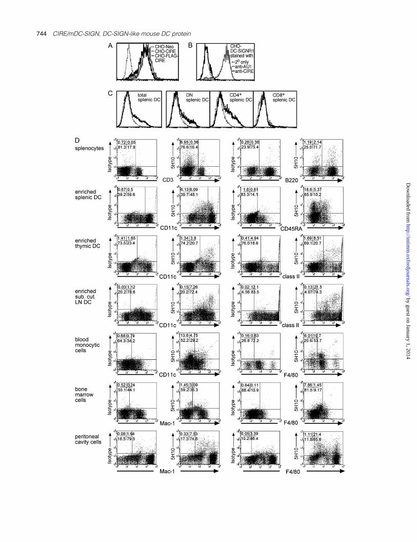
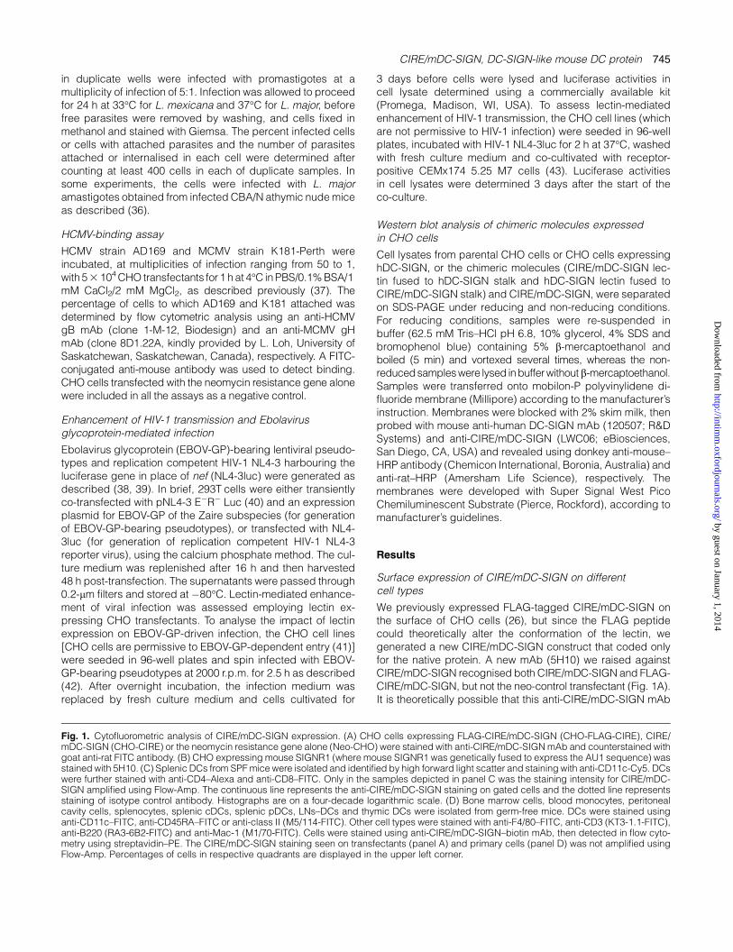
We previously expressed FLAG-tagged CIRE/mDC-SIGN onthe surface of CHO cells (26), but since the FLAG peptidecould theoretically alter the conformation of the lectin, wegenerated a new CIRE/mDC-SIGN construct that coded onlyfor the native protein. A new mAb (5H10) we raised againstCIRE/mDC-SIGN recognised both CIRE/mDC-SIGN and FLAG-CIRE/mDC-SIGN, but not the neo-control transfectant (Fig. 1A).It is theoretically possible that this anti-CIRE/mDC-SIGN mAb
Fig. 1. Cytofluorometric analysis of CIRE/mDC-SIGN expression. (A) CHO cells expressing FLAG-CIRE/mDC-SIGN (CHO-FLAG-CIRE), CIRE/mDC-SIGN (CHO-CIRE) or the neomycin resistance gene alone (Neo-CHO) were stained with anti-CIRE/mDC-SIGN mAb and counterstained withgoat anti-rat FITC antibody. (B) CHO expressing mouse SIGNR1 (where mouse SIGNR1 was genetically fused to express the AU1 sequence) wasstained with 5H10. (C) Splenic DCs from SPF mice were isolated and identified by high forward light scatter and staining with anti-CD11c-Cy5. DCswere further stained with anti-CD4–Alexa and anti-CD8–FITC. Only in the samples depicted in panel C was the staining intensity for CIRE/mDC-SIGN amplified using Flow-Amp. The continuous line represents the anti-CIRE/mDC-SIGN staining on gated cells and the dotted line representsstaining of isotype control antibody. Histographs are on a four-decade logarithmic scale. (D) Bone marrow cells, blood monocytes, peritonealcavity cells, splenocytes, splenic cDCs, splenic pDCs, LNs–DCs and thymic DCs were isolated from germ-free mice. DCs were stained usinganti-CD11c–FITC, anti-CD45RA–FITC or anti-class II (M5/114-FITC). Other cell types were stained with anti-F4/80–FITC, anti-CD3 (KT3-1.1-FITC),anti-B220 (RA3-6B2-FITC) and anti-Mac-1 (M1/70-FITC). Cells were stained using anti-CIRE/mDC-SIGN–biotin mAb, then detected in flow cyto-metry using streptavidin–PE. The CIRE/mDC-SIGN staining seen on transfectants (panel A) and primary cells (panel D) was not amplified usingFlow-Amp. Percentages of cells in respective quadrants are displayed in the upper left corner.
CIRE/mDC-SIGN, DC-SIGN-like mouse DC protein 745
by guest on January 1, 2014http://intim
m.oxfordjournals.org/
Dow
nloaded from

cross-reacted with the mouse DC-SIGN siblings (SIGNR1-4),despite the fact that the immunogenic peptide selected waschosen from a region of genetic variability. Since mAb 5H10reacted with splenocytes (see below) and only mouse SIGNR1is expressed in the spleen and on macrophages (27, 44–47),we determined whether this mAb cross-reacted with mouseSIGNR1. SIGNR1 expression was detected with an antibodyagainst the antigenic AU1 tag, but not by mAb 5H10, indicatingthat 5H10 does not recognise mouse SIGNR1 (Fig. 1B).
Splenic DC subsets were then investigated for surfaceexpression of CIRE/mDC-SIGN. Only a small proportion oftotal cDCs expressed CIRE/mDC-SIGN. This protein wasonly found on a fraction of CD4+ and DN DCs, but not on CD8+
DCs (Fig. 1C). Thus, the protein expression of CIRE/mDC-SIGN agreed with the expression profile predicted by thereverse transcription (RT)–PCR analysis (26). Interestingly,CIRE/mDC-SIGN was also expressed on a large proportionof pDCs (33; Fig. 2A).
Next we determined whether CIRE/mDC-SIGN was ex-pressed on other cell types. Bone marrow cells, peritonealcells, splenocytes, blood monocytes, Tand B cells and variousorgan-derived DCs were assessed for their expression ofCIRE/mDC-SIGN. Splenic DCs consistently contained a CIRE/mDC-SIGN+ sub-population. However, unseparated cellsderived from bone marrow, blood or lymphoid organs of ourstandard laboratory mice produced conflicting results, withstaining for CIRE/mDC-SIGN varying from undetectable levelsup to 25% of the cells analysed (data not shown).
Since we had already shown by RT–PCR that CIRE/mDC-SIGN is down-regulated upon activation (26), we reasonedthat the mice might be exposed to undefined pathogens inour holding rooms, and this may have activated their immunesystem. Thus, germ-free mice were obtained and sacrificedwithin 12 h of entering the animal holding facilities. Indeed,germ-free mice consistently displayed a significant pro-portion of CIRE/mDC-SIGN+ cells in the bone marrow, peri-toneal cavity, spleen and blood (Fig. 1D). The majority ofpositive cells were myeloid (monocytes, macrophages) sincelymphocytes were almost all negative. While T cells did notexpress CIRE/mDC-SIGN, a minority of splenocytes (2%),which were CD19+ and presumed to be B cells, expressedthis protein (data not shown). Interestingly, the proportion ofcDCs and pDCs expressing CIRE/mDC-SIGN differed be-tween lymphoid compartments (Fig. 1D); 10–20% splenicDCs were positive for CIRE/mDC-SIGN, while only 7–9%of lymph node DCs and 2% of thymic DCs were positivefor this marker. The reason for this variation remains to beelucidated.
To determine whether CIRE/mDC-SIGN protein was down-regulated upon activation, as predicted by RT–PCR, cDCs andpDCs were isolated and stained before and after overnightincubation with GM-CSF or GM-CSF and CpG. While only asmall fraction of freshly isolated cDCs (5–10%) from normal B6mice were CIRE/mDC-SIGN+, up to 50% of pDCs expressedthis marker. Upon activation, the expression of CIRE/mDC-SIGN on both cDCs and pDCs was completely lost (Fig. 2A).
Fig. 2. CIRE/mDC-SIGN is down-regulated upon activation and is not up-regulated by IL-4 and IL-13. (A) Isolated splenic cDCs (98% <CD11hiCD45RA�) and pDCs (98% < CD11cint CD45RA+) from SPF mice were purified by cell sorting and cultured in the presence of GM-CSF orGM-CSF and CpG-1668. The continuous line represents the anti-CIRE/mDC-SIGN staining and the dotted line is the background staining of anisotype control antibody. Histographs are on a logarithmic scale. The data are representative of minimum five independent experiments.(B) Freshly isolated DCs (>90% CD11c+) from SPF mice were cultured in the presence of GM-CSF plus various concentrations of IL-4 or IL-13 for18 h. The level of CIRE expression was determined by flow cytometry. Open bars represent the control staining with an isotype-matched mAb.Data presented are representative of a minimum of four independent experiments.
746 CIRE/mDC-SIGN, DC-SIGN-like mouse DC protein
by guest on January 1, 2014http://intim
m.oxfordjournals.org/
Dow
nloaded from

Since purified CIRE/mDC-SIGN+ and CIRE/mDC-SIGN� cellshad comparable survival rates in cell culture (data notshown), the disappearance of CIRE/mDC-SIGN+ cells re-flected down-regulation rather than preferential cell death. Ithas been reported that incubation with IL-4 (48) and IL-13 (49)increases the expression levels of hDC-SIGN. However, whenfreshly isolated DCs from SPF mice were incubated withvarious concentrations of IL-4 or IL-13, there was no up-regulation of CIRE/mDC-SIGN (Fig. 2B). This discrepancycould reflect the different cell types used; we used freshlyisolated splenic DCs not monocyte-derived DCs or macro-phages or could be due to genuine differences in regulationof gene expression.
Binding of mannose by CIRE/mDC-SIGN
Sequence analysis of CIRE/mDC-SIGN predicts a binding ofmannosylated residues similar to hDC-SIGN. To establishwhether CIRE/mDC-SIGN had a mannose-binding specificity,CHO cells expressing FLAG-CIRE/mDC-SIGN, CIRE/mDC-SIGN, mouse SIGNR1 or the neomycin resistance gene alonewere incubated with mannosylated FITC-conjugated BSA.FLAG-CIRE/mDC-SIGN and CIRE/mDC-SIGN CHO cellsclearly bound mannosylated FITC-conjugated BSA (Fig. 3A).Furthermore, this binding was inhibited by mannan (Fig. 3A) ina dose-dependent manner (Fig. 3B), and by EDTA (Fig. 3Aand B), showing that CIRE/mDC-SIGN, like hDC-SIGN, boundligands in a mannose-specific and Ca2+-dependent fashion.However, binding of the mannosylated FITC-conjugatedBSA was not inhibited by the mAb 5H10, indicating this mAbdoes not neutralise the CIRE/mDC-SIGN lectin-binding site(data not shown). Interestingly, while CIRE/mDC-SIGN boundsa-D-mannosylated FITC-conjugated BSA, FITC–ovalbuminpeptide containing mainly (Man)nGlcNAcGlcNAc-Asn failedto bind CIRE/mDC-SIGN (50). The reason for this discrepancyis not clear but may reflect difference in the substrate (typeor level of mannosylation). Importantly, both human DC-SIGNand mouse SIGNR1 also bind mannosylated FITC-conjugatedBSA in a Ca2+-dependent manner (Fig. 3C), indicating thatother lectins are capable of binding this substrate.
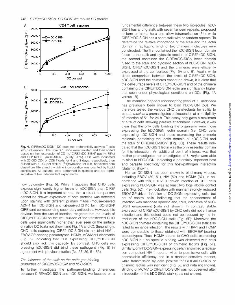
DCs expressing CIRE/mDC-SIGN are not endowed withenhanced stimulatory capacity
Human DC-SIGN has been proposed to facilitate the inter-action between T cells and DCs thereby supporting primaryimmune responses (28). To test whether mouse DCs express-ing CIRE/mDC-SIGN could induce a more potent proliferativeresponse than their CIRE/mDC-SIGN� counterparts, DC sub-sets were purified based on their expression of CIRE/mDC-SIGN and incubated with allogeneic T cells. Since theanti-CIRE/mDC-SIGN mAb does not block binding of man-nosylated BSA (data not shown), we assumed that the CIRE/mDC-SIGN molecule is still free to interact with its naturalligand partner. However, the expression of CIRE/mDC-SIGNdid not correlate with any enhanced CD8 T cell or CD4 T cellproliferative response (Fig. 4).
Lack of pathogen binding by CIRE/mDC-SIGN
Human DC-SIGN has been shown to bind many pathogens(31). To determine whether the same pathogens can bind
CIRE/mDC-SIGN, CHO cells were transfected with vectorsthat expressed CIRE/mDC-SIGN, hDC-SIGN or the neomycinresistance cassette. The levels of CIRE/mDC-SIGN and hDC-SIGN on the surface of CHO transfectants were measured by
Fig. 3. CHO cells expressing CIRE/mDC-SIGN bind mannosylatedprotein in a calcium-dependent, mannan-inhibitable manner. CHOcells (105) expressing FLAG-CIRE/mDC-SIGN (FLAG-CIRE-CHO),CIRE/mDC-SIGN (CIRE-CHO) or neomycin resistance gene only(Neo-CHO) were re-suspended in various dilutions of mannan in ice-cold medium containing 2% FCS and Ca2+ and Mg2+ or in mediumwhere the Ca2+ and Mg2+ were replaced by 5 mM EDTA. (A) Mannanand EDTA inhibit the binding of mannosylated FITC-conjugated BSAto CIRE/mDC-SIGN. (B) Mannan inhibits the binding of mannosylatedFITC-conjugated BSA by CIRE/mDC-SIGN in a dose-dependentmanner. Data are representative of a minimum of five independentexperiments. (C) Human DC-SIGN and mouse SIGNR1 also bindmannosylated FITC-conjugated BSA in a Ca2+-dependent manner.Cells were then incubated with mannosylated FITC-conjugated BSAand fluorescence levels quantitated by flow cytometry. Data arerepresentative of two independent experiments.
CIRE/mDC-SIGN, DC-SIGN-like mouse DC protein 747
by guest on January 1, 2014http://intim
m.oxfordjournals.org/
Dow
nloaded from
flow cytometry (Fig. 5). While it appears that CHO cellsexpress significantly higher levels of hDC-SIGN than CIRE/mDC-SIGN, it is important to note that a direct comparisoncannot be drawn: expression of both proteins was detectedupon staining with different primary mAbs (mouse-derivedAZN-1 for hDC-SIGN and rat-derived 5H10 for mDC-SIGN/CIRE) and corresponding secondary antibodies. However, it isobvious from the use of identical reagents that the levels ofCIRE/mDC-SIGN on the cell surface of the transfected CHOcells were significantly higher than ever seen on the surfaceof native DC (data not shown and Fig. 1A and C). Surprisingly,CHO cells expressing CIRE/mDC-SIGN did not bind HIV-1,EBOV-GP-bearing pseudotypes, HCMV, MCMV or Leishmania(Fig. 5), indicating that DCs expressing CIRE/mDC-SIGNshould also lack this capacity. By contrast, CHO cells ex-pressing hDC-SIGN did bind these pathogens (Fig. 5) inagreement with previous studies (34, 37, 39, 51–53).
The influence of the stalk on the pathogen-bindingproperties of CIRE/mDC-SIGN and hDC-SIGN
To further investigate the pathogen-binding differencesbetween CIRE/mDC-SIGN and hDC-SIGN, we focused on a
fundamental difference between these two molecules. hDC-SIGN has a long stalk with seven tandem repeats, proposedto form an alpha helix and allow tetramerisation (54), whileCIRE/mDC-SIGN has a short stalk with no tandem repeats. Todetermine the relative importance of the stalk and the lectindomain in facilitating binding, two chimeric molecules wereconstructed. The first contained the hDC-SIGN lectin domainfused to the stalk and cytosolic section of CIRE/mDC-SIGN,the second contained the CIRE/mDC-SIGN lectin domainfused to the stalk and cytosolic section of hDC-SIGN. hDC-SIGN, CIRE/mDC-SIGN and the chimeras were efficientlyexpressed at the cell surface (Fig. 5A and B). Again, whiledirect comparison between the levels of CIRE/mDC-SIGN,hDC-SIGN and the chimeras cannot be drawn, it is clear thatthe cell-surface levels of CIRE/mDC-SIGN and of the chimeracontaining the CIRE/mDC-SIGN lectin are significantly higherthat seen under physiological conditions on DCs (Fig. 1Aversus 1B).
The mannose-capped lipophosphoglycan of L. mexicanahas previously been shown to bind hDC-SIGN (53). Wetherefore tested the various CHO transfectants for ability tobind L. mexicana promastigotes on incubation at a multiplicityof infection of 5:1 for 24 h. This assay only gave a maximumof 15% of cells showing parasite attachment. However, it wasclear that the only cells binding the organisms were thoseexpressing the hDC-SIGN lectin domain (i.e. CHO cellsexpressing hDC-SIGN and those expressing the chimericmolecule containing the lectin domain of hDC-SIGN andthe stalk of CIRE/mDC-SIGN) (Fig. 5C). These results indi-cated that the hDC-SIGN lectin was the only essential domainfor this interaction. An additional point of interest was thatneither promastigotes nor amastigotes of L. major were ableto bind to hDC-SIGN, indicating a potentially important hostand parasite specificity for this host–pathogen interaction(data not shown).
Human DC-SIGN has been shown to bind many viruses,including EBOV (39, 51), HIV (52) and HCMV (37). In ac-cordance with this, EBOV-GP-driven infection of CHO cellsexpressing hDC-SIGN was at least two logs above controlcells (Fig. 5D). Pre-incubation with mannan strongly reducedEBOV-GP-driven infection of DC-SIGN-expressing cells butnot of control cells, indicating that the enhancement ofinfection was mannose specific and, thus, indicative of hDC-SIGN engagement (data not shown). In contrast, stableexpression of CIRE/mDC-SIGN by CHO cells did not enhanceinfection and this defect could not be rescued by the in-troduction of the hDC-SIGN stalk (Fig. 5F). Moreover, thehDC-SIGN chimera containing the CIRE/mDC-SIGN stalk alsofailed to enhance infection. The results with HIV-1 and HCMVwere comparable to those obtained with EBOV-GP-bearingpseudotypes. Thus, HCMV bound to CHO cells expressinghDC-SIGN but no specific binding was observed with cellsexpressing CIRE/mDC-SIGN or chimeric lectins (Fig. 5F).Similarly, only hDC-SIGN-expressing cells transmitted a replica-tion competent HIV-1 reporter virus to permissive cells withappreciable efficiency and in a mannan-sensitive manner,while transmission by cells positive for CIRE/mDC-SIGN orchimeric lectins was inefficient (Fig. 5E and data not shown).Binding of MCMV to CIRE/mDC-SIGN was not observed afterintroduction of the hDC-SIGN stalk (data not shown).
Fig. 4. CIRE/mDC-SIGN+ DC does not preferentially activate T cellsinto proliferation. DCs from SPF mice were isolated and then sortedbased on their expression of CD11c+CIRE/mDC-SIGN+ (purity: 75%)and CD11c+CIRE/mDC-SIGN� (purity: 98%). DCs were incubatedwith 20 000 CD4 or CD8 T cells for 4 and 3 days, respectively, thenpulsed with 1 lCi per well of [3H]thymidine for 6 h, harvested ontoglass-fibre filters and thymidine incorporation was counted by liquidscintillation. All cultures were performed in quintets and are repre-sentative of two independent experiments.
748 CIRE/mDC-SIGN, DC-SIGN-like mouse DC protein
by guest on January 1, 2014http://intim
m.oxfordjournals.org/
Dow
nloaded from
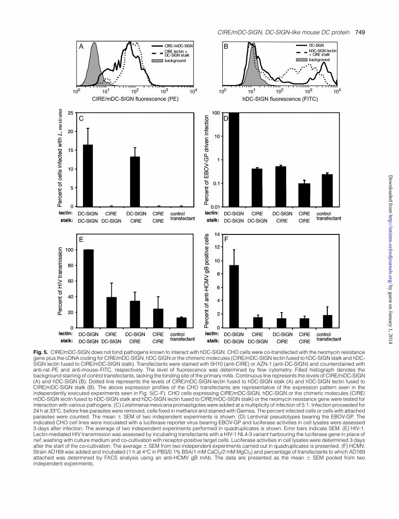
Fig. 5. CIRE/mDC-SIGN does not bind pathogens known to interact with hDC-SIGN. CHO cells were co-transfected with the neomycin resistancegene plus the cDNA coding for CIRE/mDC-SIGN, hDC-SIGN or the chimeric molecules (CIRE/mDC-SIGN lectin fused to hDC-SIGN stalk and hDC-SIGN lectin fused to CIRE/mDC-SIGN stalk). Transfectants were stained with 5H10 (anti-CIRE) or AZN-1 (anti-DC-SIGN) and counterstained withanti-rat–PE and anti-mouse–FITC, respectively. The level of fluorescence was determined by flow cytometry. Filled histograph denotes thebackground staining of control transfectants, lacking the binding site of the primary mAb. Continuous line represents the levels of CIRE/mDC-SIGN(A) and hDC-SIGN (B). Dotted line represents the levels of CIRE/mDC-SIGN-lectin fused to hDC-SIGN stalk (A) and hDC-SIGN lectin fused toCIRE/mDC-SIGN stalk (B). The above expression profiles of the CHO transfectants are representative of the expression pattern seen in theindependently executed experiments seen in Fig. 5(C–F). CHO cells expressing CIRE/mDC-SIGN, hDC-SIGN or the chimeric molecules (CIRE/mDC-SIGN lectin fused to hDC-SIGN stalk and hDC-SIGN lectin fused to CIRE/mDC-SIGN stalk) or the neomycin resistance gene were tested forinteraction with various pathogens. (C) Leishmania mexicana promastigotes were added at a multiplicity of infection of 5:1. Infection proceeded for24 h at 33�C, before free parasites were removed, cells fixed in methanol and stained with Giemsa. The percent infected cells or cells with attachedparasites were counted. The mean 6 SEM of two independent experiments is shown. (D) Lentiviral pseudotypes bearing the EBOV-GP. Theindicated CHO cell lines were inoculated with a luciferase reporter virus bearing EBOV-GP and luciferase activities in cell lysates were assessed3 days after infection. The average of two independent experiments performed in quadruplicates is shown. Error bars indicate SEM. (E) HIV-1.Lectin-mediated HIV transmission was assessed by incubating transfectants with a HIV-1 NL4-3 variant harbouring the luciferase gene in place ofnef, washing with culture medium and co-cultivation with receptor-positive target cells. Luciferase activities in cell lysates were determined 3 daysafter the start of the co-cultivation. The average 6 SEM from two independent experiments carried out in quadruplicates is presented. (F) HCMV.Strain AD169 was added and incubated (1 h at 4�C in PBS/0.1% BSA/1 mM CaCl2/2 mM MgCl2) and percentage of transfectants to which AD169attached was determined by FACS analysis using an anti-HCMV gB mAb. The data are presented as the mean 6 SEM pooled from twoindependent experiments.
CIRE/mDC-SIGN, DC-SIGN-like mouse DC protein 749
by guest on January 1, 2014http://intim
m.oxfordjournals.org/
Dow
nloaded from

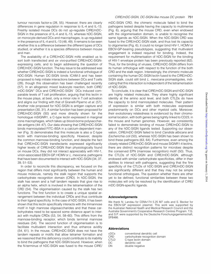
To determine whether the stalk of hDC-SIGN did indeedfacilitate multimerisation when fused to CIRE/mouse DC-SIGN,or conversely whether the stalk of CIRE/mouse DC-SIGNabolished tetramerisation, we conducted western blot analysisto estimate the sizes of the chimeric proteins expressed bythe various transfectants. Proteins in the cell lysate of parentalCHO-KI cells that do not express mouse or human DC-SIGNare not detected with the anti-CIRE/mDC-SIGN and anti-human DC-SIGN mAb (Fig. 6; lane 1). DC-SIGN is a 46-kDaprotein containing one N-linked glycosylation site, which isutilised (38). In accordance with this, under reducing con-ditions, the predominant product of DC-SIGN (lane 2) resolvedat 46 kDa with a minor product detected at ~44 kDa (pre-sumed to be non-glycosylated protein). In contrast, under non-reducing conditions, an additional DC-SIGN (lane 2) productwas detected at ~180 kDa, corresponding to the expectedtetramer (184 kDa). The chimeric protein of CIRE/mDC-SIGNlectin fused to hDC-SIGN stalk is expected to be 44 kDa(lane 3) and, accordingly, under reducing conditions a band of44 kDa was detected. Interestingly, under non-reducing con-ditions, the predominant product runs at ~180 kDa, the ex-pected size (176 kDa) of the tertramerised protein, and only aminor monomeric product is detected at 44 kDa. The secondchimera (hDC-SIGN lectin fused to CIRE/mDC-SIGN stalk)was expected to be 29 kDa and under reducing conditionsa band is indeed detected at 29 kDa (lane 4). However,regardless of whether the cell lysates were run under reducingor non-reducing conditions, no multimer was detected, con-sistent with the notion that the CIRE/mDC-SIGN stalk fails toallow multimerisation. In accordance with this, CIRE/mDC-SIGN (27 kDa) failed to multimerise, since an oligomerisationproduct of CIRE/mDC-SIGN was not detected under non-reducing or reducing conditions.
Thus, the stalk of hDC-SIGN does not endow CIRE/mDC-SIGN with the capacity to bind any of the tested viruses,
arguing that the mouse lectin has a different binding specificitycompared with hDC-SIGN. Furthermore, replacing the stalk ofhDC-SIGN with that of CIRE/mDC-SIGN abolished binding tothree of the four pathogens analysed, suggesting that hDC-SIGN does indeed require the oligomerisation and spatialorientation facilitated by its long stalk in order to functionallyinteract with many ligands.
Discussion
The development of a mAb against CIRE/mDC-SIGN hasenabled us to study CIRE/mDC-SIGN expression at the pro-tein level. Our initial attempt to determine CIRE/mDC-SIGNexpression on various leukocytes harvested from mice pro-duced variable results. However, cells from germ-free micewith restricted exposure to conventional mouse holding roomconditions showed higher and more reproducible CIRE/mDC-SIGN expression. This suggests that CIRE/mDC-SIGNexpression is exquisitely sensitive to activation or mild in-flammation induced by microbial exposure. These results arein keeping with our experimental data showing that activationof cDCs or pDCs results in complete down-regulation of CIRE/mDC-SIGN expression (26).
Using the CIRE/mDC-SIGN mAb, we confirmed at the pro-tein level that among cDCs, CIRE/mDC-SIGN is expressedby CD4+, DN DCs but not CD8+ DCs (26). CIRE/mDC-SIGN isalso strongly expressed by a proportion of splenic pDCs (33),macrophages and monocytes. There is little expression ofCIRE/mDC-SIGN on lymphocytes, except perhaps on a veryminor subset of B cells.
It has been proposed that mouse CIRE/mDC-SIGN is theorthologue of hDC-SIGN. Complicating this proposition isthe fact that in the mouse there are at least five closely relatedC-type lectins (CIRE/mDC-SIGN, mSIGNR1—4), all sharingcomparable amino acid identity with the two human C-typelectins, hDC-SIGN and hDC-SIGNR (26, 27). However, sincelike human DC-SIGN, CIRE/mDC-SIGN is the only mouseDC-SIGN gene that is closely juxtaposed to the CD23 gene,and is the only mouse lectin expressed in DCs, it wasconcluded to be the hDC-SIGN orthologue. In this report,using a mAb against mouse CIRE/mDC-SIGN, we show thatits expression pattern has some similarity to that of hDC-SIGN. Mouse CIRE/mDC-SIGN is expressed on freshly iso-lated splenic, thymic and lymph node DCs; unfortunately,these tissue sources have not been used for isolation ofhuman DCs. However, CIRE/mDC-SIGN can be detected onmonocyte-derived DCs (data not shown) and macrophagesand hDC-SIGN is expressed on monocyte-derived DCs,monocyte-derived Langerhans cells, CD1alo dermal DCsand macrophage subsets (28, 49, 55–57). In the mouse,CIRE/mDC-SIGN is also on the cell surface of spleen pDCs;notably, there are reports of its expression on human pDCs,but the data are conflicting (49, 55). Interestingly, mouse bloodmonocytes express CIRE/mDC-SIGN, whereas human mono-cytes do not express DC-SIGN (28, 55, 56). There maybesome parallels between the down-regulation of CIRE/mDC-SIGN on freshly isolated mouse DCs when activated withCpG (Fig. 2) LPS, IFN-c and poly-IC (data not shown) andthe reduction observed when human monocyte-derived DCsare ‘matured’ with factors such as LPS, prostaglandin E2 and
Fig. 6. The chimeric molecule containing the CIRE/mDC-SIGN lectindomain fused to human DC-SIGN stalk appears to form multimerssimilarly to hDC-SIGN. Cell lysates from parental CHO cells (lane 1)or CHO cells expressing hDC-SIGN (lane 2), or the chimeric mol-ecules [CIRE/mDC-SIGN lectin fused to hDC-SIGN stalk (lane 3) andhDC-SIGN lectin fused to CIRE/mDC-SIGN stalk (lane 4)] and CIRE/mDC-SIGN (lane 5) were separated on SDS-PAGE (10%) under re-ducing and non-reducing conditions, probed with mouse anti-humanDC-SIGN mAb and anti-CIRE/mDC-SIGN and then revealed usinganti-rat–HRP and anti-mouse–HRP antibody.
750 CIRE/mDC-SIGN, DC-SIGN-like mouse DC protein
by guest on January 1, 2014http://intim
m.oxfordjournals.org/
Dow
nloaded from
tumour necrosis factor-a (28, 55). However, there are clearlydifferences in gene regulation in response to IL-4 and IL-13.Freshly isolated mouse DCs do not up-regulate CIRE/mDC-SIGN in the presence of IL-4 and IL-13, whereas hDC-SIGN,on monocyte-derived DCs and macrophages, is up-regulatedin the presence of IL-4 and IL-13 (48, 49). It remains to be seenwhether this is a difference between the different types of DCsstudied, or whether it is a species difference between mouseand man.
The availability of a CIRE/mDC-SIGN mAb enabled us tosort both transfected and ex vivo-purified CIRE/mDC-SIGN-expressing cells, and to begin addressing the question ofCIRE/mDC-SIGN function. The key issue to be addressed waswhether CIRE/mDC-SIGN is indeed a functional orthologue ofhDC-SIGN. Human DC-SIGN binds ICAM-3 and has beenproposed to help initiate interactions between DCs and T cells(28), though this observation has been challenged recently(57). In an allogeneic mixed leukocyte reaction, both CIRE/mDC-SIGN+ DCs and CIRE/mDC-SIGN� DCs induced com-parable levels of T cell proliferation, arguing that this lectin inthe mouse plays, at best, only a minor role in T cell activationand aligns our finding with that of Granelli-Piperno et al. (57).Another role proposed for hDC-SIGN is antigen capture andpresentation (30, 31). A similar function has been proposed forCIRE/mDC-SIGN (50) and more directly shown for thehomologue mSIGNR1, a C-type lectin expressed in marginalzone macrophages, which takes up blood-borne polysacchar-ide antigens (44–47). Our data showing that CIRE/mDC-SIGNbinds mannosylated FITC–BSA in a calcium-dependent man-ner (Fig. 3) demonstrates that this molecule is also a C-typelectin with mannose-binding specificity. However, despitethe common mannose-binding function, and despite the factthat CIRE/mDC-SIGN transfectants expressed significantlyhigher levels of CIRE/mDC-SIGN than physiologically foundon mouse DCs, they did not interact appreciably with HIV-1,virion-associated EBOV-GP, HCMV or L. mexicana, pathogensthat have been documented to interact with hDC-SIGN (34, 37,39, 51–53).
In order to reconcile this discrepancy, we focused on theregion that differs most significantly between the human andmouse molecule, namely the stalk region that supports thecarbohydrate recognition domain (CRD). In hDC-SIGN, thestalk has seven and a half tandem repeats that give rise toan alpha helix, which is involved in the tetramerisation of theCRD (54). The oligomerisation caused by the stalk has twofunctions. The first function is to create a unique spatial ar-rangement between the individual CRDs and thus contributeto their ligand specificity. In the case of hDC-SIGN, it has beenshown that this lectin specifically interacts with the trimannosemotif in high mannose oligosaccharides and that these car-bohydrates need to be spaced some distance apart to inter-act with multiple CRDs (53, 54, 58–60). This differs from themannose-binding receptor, which binds terminal mannoseresidues (54). The second function of oligomerisation is tofacilitate multivalent interaction and thus enhance avidity(54, 61). In the mouse, CIRE/mDC-SIGN does not have thetandem repeats or motifs that allow tetramer formation andwe reasoned that this deficiency could contribute to its inabilityto bind the pathogens that hDC-SIGN bound. However, whenthe N-terminus of hDC-SIGN was fused to the mouse CIRE/
mDC-SIGN CRD, the chimeric molecule failed to bind thepathogens tested despite having the capacity to multimerise(Fig. 6), arguing that the mouse CRD, even when endowedwith the oligomerisation domain, is unable to recognise thesame ligands as hDC-SIGN. When the hDC-SIGN CRD wasfused to the CIRE/mDC-SIGN stalk, and thus lost its capacityto oligomerise (Fig. 6), it could no longer bind HIV-1, HCMV orEBOV-GP-bearing pseudotypes, suggesting that multivalentengagement is indeed required for binding. Indeed, therequirement for multimerisation of hDC-SIGN for the bindingof HIV-1 envelope protein has been previously reported (62).Thus, for the binding of viruses, CIRE/mDC-SIGN differs fromits human orthologue with respect to the function of the bothCRD and the stalk region. Interestingly, the chimeric moleculecontaining the human DC-SIGN lectin fused to the CIRE/mDC-SIGN stalk, could still bind L. mexicana promastigotes, indi-cating that this interaction is independent of the tetramerisationof hDC-SIGN.
To conclude, it is clear that CIRE/mDC-SIGN and hDC-SIGNare highly related molecules. They share highly significantidentity at the amino acid level, and both CTLDs do havethe capacity to bind mannosylated molecules. Their patternof expression is similar with both molecules expressedpredominantly on DCs and other myeloid cells. Moreover,their evolutionary relatedness is evidenced by their chromo-somal location, with both genes being tightly linked to CD23, inthe mouse and human genomes. However, we consistentlyfailed to demonstrate binding of mouse CIRE/mDC-SIGN toany of the hDC-SIGN ligands tested. Supporting our obser-vation, CIRE/mDC-SIGN failed to bind Candida albicans andEscherichia coli (50), whereas hDC-SIGN has been shown tobind these pathogens (63, 64). Furthermore, even among theclosely related CIRE/mDC-SIGN and mouse SIGNR1-4 lectins,there are distinct recognition patterns for microbes despitethe conserved EPN (mannose recognition) motif (50). Thus,the CTLDs of hDC-SIGN and CIRE/mDC-SIGN, althoughendowed with similar carbohydrate specificities, differ in theirabilities to interact with pathogens, suggesting that the finespecificity of the CTLDs of hDC-SIGN and CIRE/mDC-SIGNare significantly different and that they may not be simplefunctional orthologues. The question whether there are otheryet to be defined, functional similarities between these twomolecules will only be resolved by the identification of CIRE/mDC-SIGN-specific ligands.
Acknowledgements
We thank N. Landau for CEMx174 5.25 M7 cells and S. Becker forthe EBOV-GP expression plasmid. This work was supported bythe Australian National Health and Medical Research Council and theAustralia Government’s Cooperative Research Centres Program. T.G.and S.P. were supported by the Deutsche Forschungsgemeinschaft,SFB466.
Abbreviations
cDCs conventional dendritic cellCRD carbohydrate recognition domainCTLD C-type lectin domainDC dendritic cellDN double negative
CIRE/mDC-SIGN, DC-SIGN-like mouse DC protein 751
by guest on January 1, 2014http://intim
m.oxfordjournals.org/
Dow
nloaded from
EBOV-GP Ebolavirus glycoproteinGM-CSF granulocyte macrophage colony-stimulating factorHCMV human cytomegalovirusICAM intercellular adhesion moleculei.p. intra-peritoneallyKLH keyhole limpet haemocyaninMCMV mouse cytomegaloviruspDC plasmacytoid pre-dendritic cellRT reverse transcriptionSPF specific pathogen freeWEHI Walter and Eliza Hall Institute
References
1 Steinman, R. M. 1991. The dendritic cell system and its role inimmunogenicity. Annu. Rev. Immunol. 9:271.
2 Shortman, K. and Liu, Y. J. 2002. Mouse and human dendritic cellsubtypes. Nat. Rev. Immunol. 2:151.
3 Vremec, D., Pooley, J., Hochrein, H., Wu, L., and Shortman, K.2000. CD4 and CD8 expression by dendritic cell subtypes inmouse thymus and spleen. J. Immunol. 164:2978.
4 Shortman, K. and Wu, L. 2001. Parentage and heritage of dendriticcells. Blood 97:3325.
5 Traver, D., Akashi, K., Manz, M. et al. 2000. Development ofCD8alpha-positive dendritic cells from a common myeloid pro-genitor. Science 290:2152.
6 Martin, P., del Hoyo, G. M., Anjuere, F. et al. 2000. Concept oflymphoid versus myeloid dendritic cell lineages revisited: bothCD8alpha(�) and CD8alpha(+) dendritic cells are generated fromCD4(low) lymphoid-committed precursors. Blood 96:2511.
7 Kronin, V., Winkel, K., Suss, G. et al. 1996. A subclass of dendriticcells regulates the response of naive CD8 T cells by limiting theirIL-2 production. J. Immunol. 157:3819.
8 Suss, G. and Shortman, K. 1996. A subclass of dendritic cells killsCD4 T cells via Fas/Fas-ligand-induced apoptosis. J. Exp. Med.183:1789.
9 Fazekas de St Groth, B. 1998. The evolution of self-tolerance:a new cell arises to meet the challenge of self-reactivity. Immunol.Today 19:448.
10 Ruedl, C. and Bachmann, M. F. 1999. CTL priming by CD8(+) andCD8(�) dendritic cells in vivo. Eur. J. Immunol. 29:3762.
11 Schlecht, G., Leclerc, C. and Dadaglio, G. 2001. Induction of CTLand nonpolarized Th cell responses by CD8alpha(+) andCD8alpha(�) dendritic cells. J. Immunol. 167:4215.
12 Maldonada-Lopez, R., De Smedt, T., Pajak, B. et al. 1999. Role ofCD8alpha+ and CD8alpha� dendritic cells in the induction ofprimary immune responses in vivo. J. Leukoc. Biol. 66:242.
13 Pulendran, B., Smith, J. L., Caspary, G. et al. 1999. Distinctdendritic cell subsets differentially regulate the class of immuneresponse in vivo. Proc. Natl Acad. Sci. 96:1036.
14 Hochrein, H., Shortman, K., Vremec, D., Scott, B., Hertzog, P. andO’Keeffe, M. 2001. Differential production of IL-12, IFN-alpha, andIFN-gamma by mouse dendritic cell subsets. J. Immunol. 166:5448.
15 Pulendran, B., Lingappa, J., Kennedy, M. K. et al. 1997. De-velopmental pathways of dendritic cells in vivo: distinct function,phenotype, and localization of dendritic cell subsets in FLT3ligand-treated mice. J. Immunol. 159:2222.
16 Iyoda, T., Shimoyama, S., Liu, K. et al. 2002. The CD8+ dendriticcell subset selectively endocytoses dying cells in culture andin vivo. J. Exp. Med. 195:1289.
17 Pooley, J., Heath, W. R. and Shortman, K. 2001. Intravenoussoluble antigen is presented to CD4 Tcells by CD8� dendritic cellsbut cross-presented to CD8+ T cells by CD8+ dendritic cells.J. Immunol. 166:5327.
18 den Haan, J. M., Lehar, S. M. and Bevan, M. J. 2000. CD8(+) butnot CD8(�) dendritic cells cross-prime cytotoxic T cells in vivo.J. Exp. Med. 192:1685.
19 Allan, R. S., Smith, C. M., Belz, G. T. et al. 2003. Epidermal viralimmunity induced by CD8alpha+ dendritic cells but not byLangerhans cells. Science 301:1925.
20 Steinman, R. M., Pack, M. and Inaba, K. 1997. Dendritic cells in theT-cell areas of lymphoid organs. Immunol. Rev. 156:25.
21 Reis e Sousa, C., Hieny, S., Scharton-Kersten, T. et al. 1997. In vivomicrobial stimulation induces rapid CD40 ligand-independentproduction of interleukin 12 by dendritic cells and their re-distribution to T cell areas. J. Exp. Med. 186:1819.
22 Caminschi, I., Lucas, K. M., O’Keeffe, M. A. et al. 2001. Molecularcloning of F4/80-like-receptor, a seven-span membrane protein ex-pressed differentially by dendritic cell and monocyte-macrophagesubpopulations. J. Immunol. 167:3570.
23 Edwards, A. D., Chaussabel, D., Tomlinson, S., Schulz, O., Sher, A.and Reis, E. S. C. 2003. Relationships among murine CD11c(high)dendritic cell subsets as revealed by baseline gene expressionpatterns. J. Immunol. 171:47.
24 Vremec, D. and Shortman, K. 1997. Dendritic cell subtypes inmouse lymphoid organs: cross-correlation of surface markers,changes with incubation, and differences among thymus, spleen,and lymph nodes. J. Immunol. 159:565.
25 McLellan, A. D., Kapp, M., Eggert, A. et al. 2002. Anatomic locationand T-cell stimulatory functions of mouse dendritic cell subsetsdefined by CD4 and CD8 expression. Blood 99:2084.
26 Caminschi, I., Lucas, K. M., O’Keeffe, M. A. et al. 2001. Molecularcloning of a C-type lectin superfamily protein differentially ex-pressed by CD8alpha(�) splenic dendritic cells. Mol. Immunol.38:365.
27 Park, C. G., Takahara, K., Umemoto, E. et al. 2001. Five mousehomologues of the human dendritic cell C-type lectin, DC-SIGN.Int. Immunol. 13:1283.
28 Geijtenbeek, T. B., Torensma, R., van Vliet, S. J. et al. 2000.Identification of DC-SIGN, a novel dendritic cell-specific ICAM-3receptor that supports primary immune responses. Cell 100:575.
29 Geijtenbeek, T. B., Krooshoop, D. J., Bleijs, D. A. et al. 2000. DC-SIGN-ICAM-2 interaction mediates dendritic cell trafficking. Nat.Immunol. 1:353.
30 Engering, A., Geijtenbeek, T. B., van Vliet, S. J. et al. 2002. Thedendritic cell-specific adhesion receptor DC-SIGN internalizesantigen for presentation to T cells. J. Immunol. 168:2118.
31 van Kooyk, Y. and Geijtenbeek, T. B. 2003. DC-SIGN: escapemechanism for pathogens. Nat. Rev. Immunol. 3:697.
32 Soilleux, E. J., Barten, R. and Trowsdale, J. 2000. DC-SIGN;a related gene, DC-SIGNR; and CD23 form a cluster on 19p13.J. Immunol. 165:2937.
33 O’Keeffe, M., Hochrein, H., Vremec, D. et al. 2002. Mouseplasmacytoid cells: long-lived cells, heterogeneous in surfacephenotype and function, that differentiate into CD8(+) dendriticcells only after microbial stimulus. J. Exp. Med. 196:1307.
34 Curtis, B. M., Scharnowske, S. and Watson, A. J. 1992. Sequenceand expression of a membrane-associated C-type lectin thatexhibits CD4-independent binding of human immunodeficiencyvirus envelope glycoprotein gp120. Proc. Natl Acad. Sci. USA89:8356.
35 Marzi, A., Gramberg, T., Simmons, G. et al. 2004. DC-SIGN andDC-SIGNR interact with the glycoprotein of Marburg virus and theS protein of severe acute respiratory syndrome coronavirus.J. Virol. 78:12090.
36 Glaser, T. A., Wells, S. J., Spithill, T. W., Pettit, J. M., Humphris, D. C.and Mukada, A. J. 1990. Leishmania major and L. donovani: amethod for rapid purification of amastigotes. Exp. Parasitol. 71:343.
37 Halary, F., Amara, A., Lortat-Jacob, H. et al. 2002. Humancytomegalovirus binding to DC-SIGN is required for dendritic cellinfection and target cell trans-infection. Immunity 17:653.
38 Pohlmann, S., Baribaud, F., Lee, B. et al. 2001. DC-SIGNinteractions with human immunodeficiency virus type 1 and 2and simian immunodeficiency virus. J. Virol. 75:4664.
39 Simmons, G., Reeves, J. D., Grogan, C. C. et al. 2003. DC-SIGNand DC-SIGNR bind ebola glycoproteins and enhance infection ofmacrophages and endothelial cells. Virology 305:115.
40 Connor, R. I., Chen, B. K., Choe, S. and Landau, N. R. 1995. Vpr isrequired for efficient replication of human immunodeficiency virustype-1 in mononuclear phagocytes. Virology 206:935.
41 Wool-Lewis, R. J. and Bates, P. 1998. Characterization of Ebolavirus entry by using pseudotyped viruses: identification ofreceptor-deficient cell lines. J. Virol. 72:3155.
752 CIRE/mDC-SIGN, DC-SIGN-like mouse DC protein
by guest on January 1, 2014http://intim
m.oxfordjournals.org/
Dow
nloaded from

42 O’Doherty, U., Swiggard, W. J. and Malim, M. H. 2000. Humanimmunodeficiency virus type 1 spinoculation enhances infectionthrough virus binding. J. Virol. 74:10074.
43 Hsu, M., Harouse, J. M., Gettie, A., Buckner, C., Blanchard, J. andCheng-Mayer, C. 2003. Increased mucosal transmission but notenhanced pathogenicity of the CCR5-tropic, simian AIDS-inducingsimian/human immunodeficiency virus SHIV(SF162P3) maps toenvelope gp120. J. Virol. 77:989.
44 Baribaud, F., Pohlmann, S., Sparwasser, T. et al. 2001. Functionaland antigenic characterization of human, rhesus macaque,pigtailed macaque, and murine DC-SIGN. J. Virol. 75:10281.
45 Geijtenbeek, T. B., Groot, P. C., Nolte, M. A. et al. 2002. Marginalzone macrophages express a murine homologue of DC-SIGN thatcaptures blood-borne antigens in vivo. Blood 100:2908.
46 Kang, Y. S., Kim, J. Y., Bruening, S. A. et al. 2004. The C-type lectinSIGN-R1 mediates uptake of the capsular polysaccharide ofStreptococcus pneumoniae in the marginal zone of mouse spleen.Proc. Natl Acad. Sci. USA 101:215.
47 Kang, Y. S., Yamazaki, S., Iyoda, T. et al. 2003. SIGN-R1, a novelC-type lectin expressed by marginal zone macrophages in spleen,mediates uptake of the polysaccharide dextran. Int. Immunol.15:177.
48 Relloso, M., Puig-Kroger, A., Pello, O. M. et al. 2002. DC-SIGN(CD209) expression is IL-4 dependent and is negatively regulatedby IFN, TGF-beta, and anti-inflammatory agents. J. Immunol. 168:2634.
49 Soilleux, E. J., Morris, L. S., Leslie, G. et al. 2002. Constitutive andinduced expression of DC-SIGN on dendritic cell and macrophagesubpopulations in situ and in vitro. J. Leukoc. Biol. 71:445.
50 Takahara, K., Yashima, Y., Omatsu, Y. et al. 2004. Functionalcomparison of the mouse DC-SIGN, SIGNR1, SIGNR3 andLangerin, C-type lectins. Int. Immunol. 16:819.
51 Alvarez, C. P., Lasala, F., Carrillo, J., Muniz, O., Corbi, A. L. andDelgado, R. 2002. C-type lectins DC-SIGN and L-SIGN mediatecellular entry by Ebola virus in cis and in trans. J. Virol. 76:6841.
52 Geijtenbeek, T. B., Kwon, D. S., Torensma, R. et al. 2000. DC-SIGN,a dendritic cell-specific HIV-1-binding protein that enhances trans-infection of T cells. Cell 100:587.
53 Appelmelk, B. J., van Die, I., van Vliet, S. J. et al. 2003. Cuttingedge: carbohydrate profiling identifies new pathogens that interact
with dendritic cell-specific ICAM-3-grabbing nonintegrin on den-dritic cells. J. Immunol. 170:1635.
54 Mitchell, D. A., Fadden, A. J. and Drickamer, K. 2001. A novelmechanism of carbohydrate recognition by the C-type lectinsDC-SIGN and DC-SIGNR. Subunit organization and bindingto multivalent ligands. J. Biol. Chem. 276:28939.
55 Turville, S. G., Cameron, P. U., Handley, A. et al. 2002. Diversity ofreceptors binding HIV on dendritic cell subsets. Nat. Immunol.3:975.
56 Turville, S. G., Arthos, J., Donald, K. M. et al. 2001. HIV gp120receptors on human dendritic cells. Blood 98:2482.
57 Granelli-Piperno, A., Pritsker, A., Pack, M. et al. 2005. Dendriticcell-specific intercellular adhesion molecule 3-grabbing non-integrin/CD209 is abundant on macrophages in the normalhuman lymph node and is not required for dendritic cellstimulation of the mixed leukocyte reaction. J. Immunol. 175:4265.
58 Lin, G., Simmons, G., Pohlmann, S. et al. 2003. Differential N-linkedglycosylation of human immunodeficiency virus and Ebola virusenvelope glycoproteins modulates interactions with DC-SIGN andDC-SIGNR. J. Virol. 77:1337.
59 Feinberg, H., Mitchell, D. A., Drickamer, K. and Weis, W. I. 2001.Structural basis for selective recognition of oligosaccharides byDC-SIGN and DC-SIGNR. Science 294:2163.
60 Guo, Y., Feinberg, H., Conroy, E. et al. 2004. Structural basis fordistinct ligand-binding and targeting properties of the receptorsDC-SIGN and DC-SIGNR. Nat. Struct. Mol. Biol. 11:591.
61 Weis, W. I., Taylor, M. E. and Drickamer, K. 1998. The C-type lectinsuperfamily in the immune system. Immunol. Rev. 163:19.
62 Bernhard, O. K., Lai, J., Wilkinson, J., Sheil, M. M. andCunningham, A. L. 2004. Proteomic analysis of DC-SIGN ondendritic cells detects tetramers required for ligand binding butno association with CD4. J. Biol. Chem. 279:51828.
63 Cambi, A., Gijzen, K., de Vries, J. M. et al. 2003. The C-type lectinDC-SIGN (CD209) is an antigen-uptake receptor for Candidaalbicans on dendritic cells. Eur. J. Immunol. 33:532.
64 Klena, J., Zhang, P., Schwartz, O., Hull, S. and Chen, T. 2005. Thecore lipopolysaccharide of Escherichia coli is a ligand for thedendritic-cell-specific intercellular adhesion molecule nonintegrinCD209 receptor. J. Bacteriol. 187:1710.
CIRE/mDC-SIGN, DC-SIGN-like mouse DC protein 753
by guest on January 1, 2014http://intim
m.oxfordjournals.org/
Dow
nloaded from