frequency, severity, and prediction of tuberculous meningitis immune reconstitution inflammatory...
TRANSCRIPT
H I V / A I D S M A J O R A R T I C L E
Frequency, Severity, and Prediction of TuberculousMeningitis Immune Reconstitution InflammatorySyndrome
Suzaan Marais,1,2,3 Graeme Meintjes,1,2,3,4 Dominique J. Pepper,1,2,5 Lori E. Dodd,6 Charlotte Schutz,1,2,3 Zahiera Ismail,2
Katalin A. Wilkinson,1,7 and Robert J. Wilkinson1,2,3,4,7
1Clinical Infectious Diseases Research Initiative, Institute of Infectious Diseases and Molecular Medicine, University of Cape Town, 2InfectiousDiseases Unit, GF Jooste Hospital, Cape Town, 3Department of Medicine, University of Cape Town, South Africa; 4Department of Medicine, ImperialCollege London, United Kingdom; 5Department of Medicine, University of Mississippi Medical Center, Jackson, 6Biostatistics Research Branch,National Institute of Allergy and Infectious Diseases, National Institutes of Health, Bethesda, Maryland; and 7Division of Mycobacterial Research,MRC National Institute for Medical Research, London, United Kingdom
Background. Tuberculosis immune reconstitution inflammatory syndrome (IRIS) is a common cause of dete-rioration in human immunodeficiency virus (HIV)–infected patients receiving tuberculosis treatment after startingantiretroviral therapy (ART). Potentially life-threatening neurological involvement occurs frequently and has beensuggested as a reason to defer ART.
Methods. We conducted a prospective study of HIV-infected, ART-naive patients with tuberculous meningitis(TBM). At presentation, patients started tuberculosis treatment and prednisone; ART was initiated 2 weeks later.Clinical and laboratory findings were compared between patients who developed TBM-IRIS (TBM-IRIS patients)and those who did not (non-TBM-IRIS patients). A logistic regression model was developed to predict TBM-IRIS.
Results. Forty-seven percent (16/34) of TBM patients developed TBM-IRIS, which manifested with severefeatures of inflammation. At TBM diagnosis, TBM-IRIS patients had higher cerebrospinal fluid (CSF) neutrophilcounts compared with non-TBM-IRIS patients (median, 50 vs 3 cells ×106/L, P = .02). Mycobacterium tuberculosiswas cultured from CSF of 15 TBM-IRIS patients (94%) compared with 6 non-TBM-IRIS patients (33%) at time ofTBM diagnosis; relative risk of developing TBM-IRIS if CSF was Mycobacterium tuberculosis culture positive = 9.3(95% confidence interval [CI], 1.4–62.2). The combination of high CSF tumor necrosis factor (TNF)-α and lowinterferon (IFN)-γ at TBM diagnosis predicted TBM-IRIS (area under the curve = 0.91 [95% CI, .53–.99]).
Conclusions. TBM-IRIS is a frequent, severe complication of ART in HIV-associated TBM and is character-ized by high CSF neutrophil counts and Mycobacterium tuberculosis culture positivity at TBM presentation. Thecombination of CSF IFN-γ and TNF-α concentrations may predict TBM-IRIS and thereby be a means to individ-ualize patients to early or deferred ART.
Keywords. meningitis; tuberculosis; neutrophils; pathogenesis.
Paradoxical tuberculosis immune reconstitution in-flammatory syndrome (IRIS) occurs in 8%–43% of
human immunodeficiency virus (HIV)–infected pa-tients receiving tuberculosis treatment after startingantiretroviral therapy (ART) [1–4]. Tuberculosis IRISresults from rapid restoration of Mycobacteriumtuberculosis–specific immune responses, but its patho-genesis remains poorly understood [1, 5, 6].
Neurological tuberculosis IRIS occurs in a substan-tial proportion (12%) of tuberculosis IRIS cases and isthe commonest cause of central nervous system (CNS)deterioration during the first year of ART in settingsof high tuberculosis/HIV prevalence [7, 8]. Mortality ishigh (up to 30%) in those affected [8]. Manifestationsof neurological tuberculosis IRIS include meningitis
Received 28 May 2012; accepted 11 September 2012; electronically published24 October 2012.
Correspondence: Suzaan Marais, MBChB, Room 3.03, Clinical Infectious Dis-eases Research Initiative, Institute of Infectious Diseases and Molecular Medi-cine, Wolfson Pavilion, Faculty of Health Sciences, University of Cape Town,Observatory 7925, Cape Town, South Africa ([email protected]).
Clinical Infectious Diseases 2013;56(3):450–60© The Author 2012. Published by Oxford University Press on behalf of the InfectiousDiseases Society of America. This is an Open Access article distributed under theterms of the Creative Commons Attribution License (http://creativecommons.org/licenses/by/3.0/), which permits unrestricted reuse, distribution, and reproduction inany medium, provided the original work is properly cited.DOI: 10.1093/cid/cis899
450 • CID 2013:56 (1 February) • HIV/AIDS
[7–11], intracranial tuberculomata [7, 8, 12–14], brain abscess-es [12, 15], radiculomyelitis [7, 8, 11], and spinal epidural ab-scesses [7]. There are no prospective studies describingtuberculous meningitis (TBM) IRIS; only isolated cases [9–15]and 1 case series of neurological tuberculosis IRIS [8, 16] havebeen published. Although consensus now exists that ARTshould be started early (around 2 weeks) in HIV/tuberculosis-coinfected patients with severe immunosuppression, a poten-tial exception is TBM because of the perceived risk of TBM-IRIS [17, 18]. However, the frequency and severity of thiscomplication are not well documented and no means exist topredict the syndrome.
We therefore investigated clinical and laboratory findings inART-naive HIV-infected patients who presented with TBM.We undertook serial cerebrospinal fluid (CSF) sampling in pa-tients who did and did not develop TBM-IRIS.
MATERIALS AND METHODS
SettingThis prospective, observational study was performed at GFJooste Hospital, a public sector referral hospital in CapeTown.Thehospital servesa low-income,high-densitypopulationin which the tuberculosis notification rate exceeds 1.5% peryear with 70% of tuberculosis cases coinfected with HIV [19].
ParticipantsART-naive HIV-infected patients aged ≥18 years presentingwith meningitis from March 2009 through October 2010 werescreened for study inclusion. HIV infection was diagnosed using2 rapid HIV antibody tests and confirmed by HIV load. DefiniteTBM was diagnosed when acid-fast bacilli were seen, or whenM. tuberculosis was cultured from CSF. Probable TBM was diag-nosed when a patient showed clinical, laboratory, and radiologi-cal features of TBM in the absence of other infective causes forpresentation [20]. Paradoxical TBM-IRIS was diagnosed accord-ing to a published definition for tuberculosis IRIS modified formeningitis [7, 8]. The definition had 3 components: (1) TBMdiagnosis before starting ART and improvement on tuberculosistreatment prior to ART initiation; (2) onset of TBM-IRIS mani-festations (ie, new, recurrent, or worsening clinical features ofTBM) within 3 months of ART initiation; and (3) exclusion ofalternative causes for clinical deterioration.
Patients were ineligible if they had a contraindication tolumbar puncture, including unequal pressures between individ-ual brain compartments on brain imaging, or severe TBM (ie,modified British Medical Research Council [BMRC] grade IIIdisease severity) [21]. The University of Cape Town ResearchEthics Committee approved the study and written informedconsent was obtained from all patients or their relatives.
ProceduresDemographic data and history of tuberculosis disease, HIVinfection, and other systemic illnesses and medications wererecorded. Patients underwent general physical andneurological examination. Chest radiography, phlebotomy, andlumbar puncture were performed. In patients with suspectedraised intracranial pressure or focal neurological deficits, braincomputed tomography scanning was performed prior tolumbar puncture. CSF analysis included biochemistry, cytolo-gy, microbiology (including microscopy and culture for fungiand pyogenic bacteria), syphilis serology, HIV load, and Cryp-tococcus latex agglutination titer. Ziehl-Neelsen staining ofsediment and M. tuberculosis culture was performed. If myco-bacteria were cultured from CSF, tuberculosis polymerasechain reaction (PCR; Genotype MTBDRplus, Hain Lifesciences)was performed to determine susceptibility to rifampicin andisoniazid. CSF varicella zoster virus PCR was performed if theetiology was suspected. CSF was also stored at −80°C andanalyzed for a range of inflammatory markers on the Bio-Plexplatform (Bio-Rad Laboratories, Hercules, CA) using custom-ized Milliplex kits (Millipore, St Charles, MO) according to themanufacturer’s instructions.
At TBM diagnosis, patients started tuberculosis treatmentaccording to national guidelines [22] and prednisone (1.5 mg/kg/day). After 2 weeks of treatment and prior to initiation ofART, patients were assessed for improvement on tuberculosistreatment. The initial ART regimen was stavudine, lamivu-dine, and efavirenz. Later during the study, tenofovir replacedstavudine according to revised national guidelines. CSF inves-tigations performed at TBM diagnosis were repeated at thetime of ART initiation, 2 weeks later, and at time of TBM-IRIS presentation and 2 weeks thereafter. Prednisone wasreduced to 0.75 mg/kg/day 4 weeks after starting ART and dis-continued 2 weeks thereafter, unless the patient developedTBM-IRIS. At TBM-IRIS presentation, investigations wereperformed to exclude alternative causes of deterioration. Pred-nisone was either recommenced or the dose increased. Pa-tients were followed for the duration of tuberculosis treatment(9 months); routine visits were at 2 weeks, 4 weeks, 6 weeks,12 weeks, 6 months, and 9 months after TBM diagnosis. Pa-tients were seen more frequently during deterioration.
Statistical AnalysisStatistical analysis was performed using the GraphPad Prismversion 5, R version 2.14.1, and StatXact version 9 softwarepackages. Categorical variables were compared using χ2 orFisher exact test. Continuous variables were compared betweengroups and time points within groups, using the Wilcoxonrank sum and Wilcoxon matched pairs tests, respectively.Adjusted relative risks (RRs) were evaluated using Cochran-Mantel-Haenszel tests and tests of homogeneity when
HIV/AIDS • CID 2013:56 (1 February) • 451
considering categorical risk factors. Log-binomial models werefitted to continuous risk factors. Significance testing was doneusing 2-sided P values with P < .05 taken as significant.
The predictive accuracy of CSF neutrophil counts at TBMdiagnosis for TBM-IRIS was assessed using nonparametric areaunder the receiver operating characteristic curve (AUC). Addi-tionally, a model to predict TBM-IRIS risk was developed from5 prespecified cytokines measured in CSF at time of TBM diag-nosis. Interleukin 6 (IL-6), interleukin 10, interleukin 12p40,interferon gamma (IFN-γ), and tumor necrosis factor alpha(TNF-α) were selected as candidate markers of TBM-IRISbased on previous studies [23, 24]. We prespecified the analysisfor evaluating the multivariate cytokine model as follows. Sig-nificant cytokines comparing TBM-IRIS and non-TBM-IRISusing Wilcoxon rank sum tests were selected for a logisticregression model. Nonsignificant cytokines were dropped,
resulting in a final model. The entire model building processwas evaluated using leave-one-out cross-validation, the boot-strap method [25], and the permutation test to provide a cross-validated (nonparametric) estimate of the AUC, P values, and95% confidence intervals (CIs). As a secondary analysis, we ex-amined whether the addition of CSF neutrophils and/or lym-phocyte counts would improve the model’s predictive ability.
RESULTS
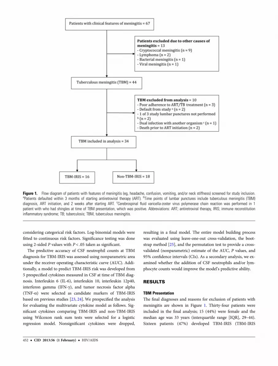
TBM PresentationThe final diagnoses and reasons for exclusion of patients withmeningitis are shown in Figure 1. Thirty-four patients wereincluded in the final analysis; 15 (44%) were female and themedian age was 33 years (interquartile range [IQR], 29–44).Sixteen patients (47%) developed TBM-IRIS (TBM-IRIS
Figure 1. Flow diagram of patients with features of meningitis (eg, headache, confusion, vomiting, and/or neck stiffness) screened for study inclusion.aPatients defaulted within 3 months of starting antiretroviral therapy (ART). bTime points of lumbar punctures include tuberculous meningitis (TBM)diagnosis, ART initiation, and 2 weeks after starting ART. cCerebrospinal fluid varicella-zoster virus polymerase chain reaction was performed in 1patient with who had shingles at time of TBM presentation, which was positive. Abbreviations: ART; antiretroviral therapy, IRIS; immune reconstitutioninflammatory syndrome; TB; tuberculosis; TBM, tuberculous meningitis.
452 • CID 2013:56 (1 February) • HIV/AIDS
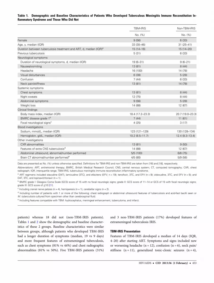
patients) whereas 18 did not (non-TBM-IRIS patients).Tables 1 and 2 show the demographic and baseline character-istics of these 2 groups. Baseline characteristics were similarbetween groups, although patients who developed TBM-IRIShad a longer duration of symptoms (median, 19 vs 9 days)and more frequent features of extrameningeal tuberculosis,such as chest symptoms (81% vs 44%) and chest radiographicabnormalities (81% vs 50%). Five TBM-IRIS patients (31%)
and 3 non-TBM-IRIS patients (17%) developed features ofextrameningeal tuberculosis IRIS.
TBM-IRIS PresentationFeatures of TBM-IRIS developed a median of 14 days (IQR,4–20) after starting ART. Symptoms and signs included newor worsening headache (n = 12), confusion (n = 6), neck pain/stiffness (n = 11), generalized tonic-clonic seizures (n = 4),
Table 1. Demographic and Baseline Characteristics of Patients Who Developed Tuberculous Meningitis Immune Reconstitution In-flammatory Syndrome and Those Who Did Not
TBM-IRIS Non-TBM-IRIS
No. (%) No. (%)
Female 9 (56) 6 (33)Age, y, median (IQR) 33 (30–46) 31 (25–41)
Duration between tuberculosis treatment and ART, d, median (IQR)a 15 (14–16) 15 (14–20)
Previous tuberculosis 5 (31) 6 (33)Neurological symptoms
Duration of neurological symptoms, d, median (IQR) 19 (6–31) 9 (6–21)
Nausea/vomiting 13 (81) 8 (44)Headache 16 (100) 14 (78)
Visual disturbances 6 (38) 5 (28)
Confusion 7 (44) 6 (33)Neck pain/stiffness 13 (81) 14 (78)
Systemic symptoms
Chest symptoms 13 (81) 8 (44)Night sweats 12 (75) 8 (44)
Abdominal symptoms 9 (56) 5 (28)
Weight loss 14 (88) 12 (67)Clinical findings
Body mass index, median (IQR) 18.4 (17.2–23.3) 20.7 (19.0–23.3)
BMRC disease grade 1b 7 (44) 11 (61)Focal neurological signsc 4 (25) 3 (17)
Blood investigations
Sodium, mmol/L, median (IQR) 123 (121–129) 130 (128–134)Hemoglobin, g/dL, median (IQR) 10.2 (8.5–11.7) 12.4 (9.3–13.4)
Other investigations
CXR abnormalities 13 (81) 9 (50)Features of extra CNS tuberculosisd 14 (88) 12 (67)
Abdominal ultrasound, abnormal/number performed 5/5 (100) 6/8 (75)
Brain CT abnormal/number performede 4/5 (80) 5/9 (56)
Data are presented as No. (%) unless otherwise specified. Definitions for TBM-IRIS and non-TBM-IRIS are taken from [16] and [18], respectively.
Abbreviations: ART, antiretroviral therapy; BMRC, British Medical Research Council; CNS, central nervous system; CT, computed tomography; CXR, chestradiograph; IQR, interquartile range; TBM-IRIS, tuberculous meningitis immune reconstitution inflammatory syndrome.a ART regimens included stavudine (D4T), lamivudine (3TC), and efavirenz (EFV; n = 19); tenofovir, 3TC, and EFV (n = 9); zidovudine, 3TC, and EFV (n = 5); andD4T, 3TC, and lopinavir/ritonavir (n = 1).b BMRC grade I: Glasgow Coma Scale (GCS) score of 15 with no focal neurologic signs; grade II: GCS score of 11–14 or GCS of 15 with focal neurologic signs;grade III: GCS score of ≤10 [21].c Including cranial nerve palsies (n = 4), hemiparesis (n = 1), cerebellar signs (n = 2).d Including number of patients with 1 or more of the following: chest radiograph or abdominal ultrasound features of tuberculosis and acid-fast bacilli seen orM. tuberculosis cultured from specimen other than cerebrospinal fluid.e Including features compatible with TBM: hydrocephalus, meningeal enhancement, tuberculoma, and infarct.
HIV/AIDS • CID 2013:56 (1 February) • 453
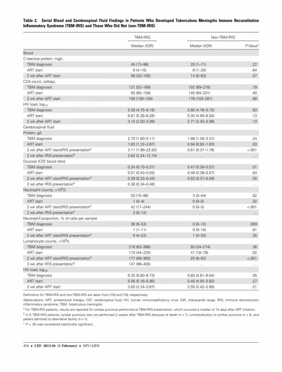
Table 2. Serial Blood and Cerebrospinal Fluid Findings in Patients Who Developed Tuberculous Meningitis Immune ReconstitutionInflammatory Syndrome (TBM-IRIS) and Those Who Did Not (non-TBM-IRIS)
TBM-IRIS Non-TBM-IRIS
Median (IQR) Median (IQR) P Valuec
BloodC-reactive protein, mg/L
TBM diagnosis 45 (13–98) 25 (1–71) .22
ART start 6 (4–15) 8 (1–20) .642 wk after ART start 56 (20–105) 14 (6–63) .07
CD4 count, cells/μL
TBM diagnosis 131 (52–169) 102 (69–278) .79ART start 93 (65–158) 145 (64–231) .40
2 wk after ART start 158 (139–193) 178 (103–261) .68
HIV load, log10TBM diagnosis 5.39 (4.75–6.16) 5.60 (4.76–5.72) .83
ART start 5.61 (5.26–6.26) 5.30 (4.90–6.04) .13
2 wk after ART start 3.15 (2.50–3.36) 2.71 (2.43–2.98) .13Cerebrospinal fluid
Protein, g/L
TBM diagnosis 2.70 (1.80–5.11) 1.88 (1.28–3.31) .24ART start 1.63 (1.22–2.67) 0.94 (0.82–1.63) .03
2 wk after ART start/IRIS presentationa 3.11 (1.99–22.83) 0.61 (0.37–1.76) <.001
2 wk after IRIS presentationb 2.62 (2.24–12.74) … …
Glucose (CSF blood ratio)
TBM diagnosis 0.24 (0.15–0.31) 0.47 (0.28–0.57) .01
ART start 0.51 (0.43–0.55) 0.48 (0.38–0.57) .632 wk after ART start/IRIS presentationa 0.39 (0.33–0.43) 0.53 (0.37–0.59) .05
2 wk after IRIS presentationb 0.38 (0.34–0.48) … …
Neutrophil counts, ×106/LTBM diagnosis 50 (15–86) 3 (0–44) .02
ART start 1 (0–4) 0 (0–5) .32
2 wk after ART start/IRIS presentationa 42 (17–244) 0 (0–3) <.0012 wk after IRIS presentationb 3 (0–12) … …
Neutrophil proportion, % of cells per sample
TBM diagnosis 36 (6–53) 0 (0–12) .009ART start 1 (1–11) 0 (0–18) .61
2 wk after ART start/IRIS presentationa 9 (4–22) 1 (0–33) .35
Lymphocyte counts, ×106/LTBM diagnosis 218 (63–366) 93 (24–274) .36
ART start 110 (44–225) 41 (18–79) .02
2 wk after ART start/IRIS presentationa 177 (69–363) 25 (6–52) <.0012 wk after IRIS presentationb 147 (98–405) … …
HIV load, log10TBM diagnosis 6.35 (5.80–6.73) 5.60 (4.81–6.54) .05ART start 5.56 (5.16–5.95) 5.40 (4.93–5.82) .27
2 wk after ART start 3.00 (2.24–3.67) 2.55 (2.42–2.80) .21
Definitions for TBM-IRIS and non-TBM-IRIS are taken from [16] and [18], respectively.
Abbreviations: ART, antiretroviral therapy; CSF, cerebrospinal fluid; HIV, human immunodeficiency virus; IQR, interquartile range; IRIS, immune reconstitutioninflammatory syndrome; TBM, tuberculous meningitis.a For TBM-IRIS patients, results are reported for lumbar puncture performed at TBM-IRIS presentation, which occurred a median of 14 days after ART initiation.b In 5 TBM-IRIS patients, lumbar puncture was not performed 2 weeks after TBM-IRIS because of death (n = 1), contraindication to lumbar puncture (n = 3), andpatient admitted to alternative facility (n = 1).c P < .05 was considered statistically significant.
454 • CID 2013:56 (1 February) • HIV/AIDS
vomiting (n = 5), paraparesis (n = 3), myoclonic jerks (n = 1),dysconjugate eye movements (n = 1), and aphasia (n = 1). Attime of TBM-IRIS presentation, 15 patients underwent brainimaging, including computed tomography (n = 14) or magnet-ic resonance imaging (n = 1). Imaging showed features ofTBM in 14 of these patients. Magnetic resonance imaging ofthe spine was performed in 2 patients with paraparesis; bothhad features of radiculomyelitis (Supplementary Figure 1).
Serial Blood and CSF FindingsBaseline blood investigations were similar between TBM-IRISand non-TBM-IRIS patients with the exception of serumsodium concentrations, which were lower in TBM-IRIS pa-tients (median, 123 vs 130 mmol/L, P = .01, Table 1). Table 2and Figure 2 show serial blood and CSF findings. There was asignificant rise in CD4 counts between starting ART and 2weeks later in both TBM-IRIS (median, 93–158 cells/μL,P = .009) and non-TBM-IRIS (median, 145–178 cells/μL,P = .04) patients. Between these time points, blood and CSFHIV loads decreased significantly (P < .001) in both groups.
At TBM diagnosis, TBM-IRIS patients had higher CSF cellcounts, in particular neutrophils (median, 50 vs 3 cells ×106/L,P = .02, Table 2). Similarly, neutrophil percentages from indi-vidual samples were higher in TBM-IRIS patients comparedwith non-TBM-IRIS patients (median, 36% vs 0%, P = .009).CSF to blood glucose ratios were lower in TBM-IRIS patients(median, 0.24 vs 0.47, P = .005). In both groups, CSF parame-ters initially improved on tuberculosis treatment (Table 2 andFigure 2). However, at TBM-IRIS presentation, TBM-IRIS pa-tients showed findings of recurrent inflammation. In thisgroup, lymphocyte and neutrophil counts at TBM-IRIS pre-sentation were similar, and protein concentrations higher,compared with the same parameters at TBM diagnosis(median protein, 3.11 g/L at TBM-IRIS vs 2.70 g/L at TBMdiagnosis, P = .007).
Mycobacterium tuberculosis was cultured from the CSF of15 TBM-IRIS patients (94%) and 6 non-TBM-IRIS patients(33%) at TBM diagnosis; the risk of developing TBM-IRIS ifCSF M. tuberculosis culture was positive at this time point was71.4% (15/21), compared with a risk of 7.7% (1/13), corre-sponding to an RR of 9.3 (95% CI, 1.4–62.2, P = .004). Addi-tional analyses considered the RRs of culture positivityadjusting for the following known or potential risk factors fortuberculosis IRIS: baseline viral load (median, ≥330 000copies/mL, equivalent to log10 = 5.52, vs <330 000), CD4count (median, ≤137 cells/μL vs >137), evidence of dissemi-nated disease in the form of an abnormal chest radiographsuggesting miliary disease, and duration of illness (median du-ration, ≥2 weeks vs <2 weeks). Mycobacterium tuberculosisculture positivity remained a significant risk factor after ad-justing for these factors (Supplementary Table 1). There was
no evidence of differences in the RRs according to thesefactors after considering culture status. Some TBM-IRIS pa-tients remained culture positive after 2 weeks (n = 7) and 4weeks (n = 2) of tuberculosis treatment. No non-TBM-IRISpatients were culture positive after starting tuberculosis treat-ment. All cultures were fully drug sensitive with the exceptionof one, which was monoresistant to isoniazid.
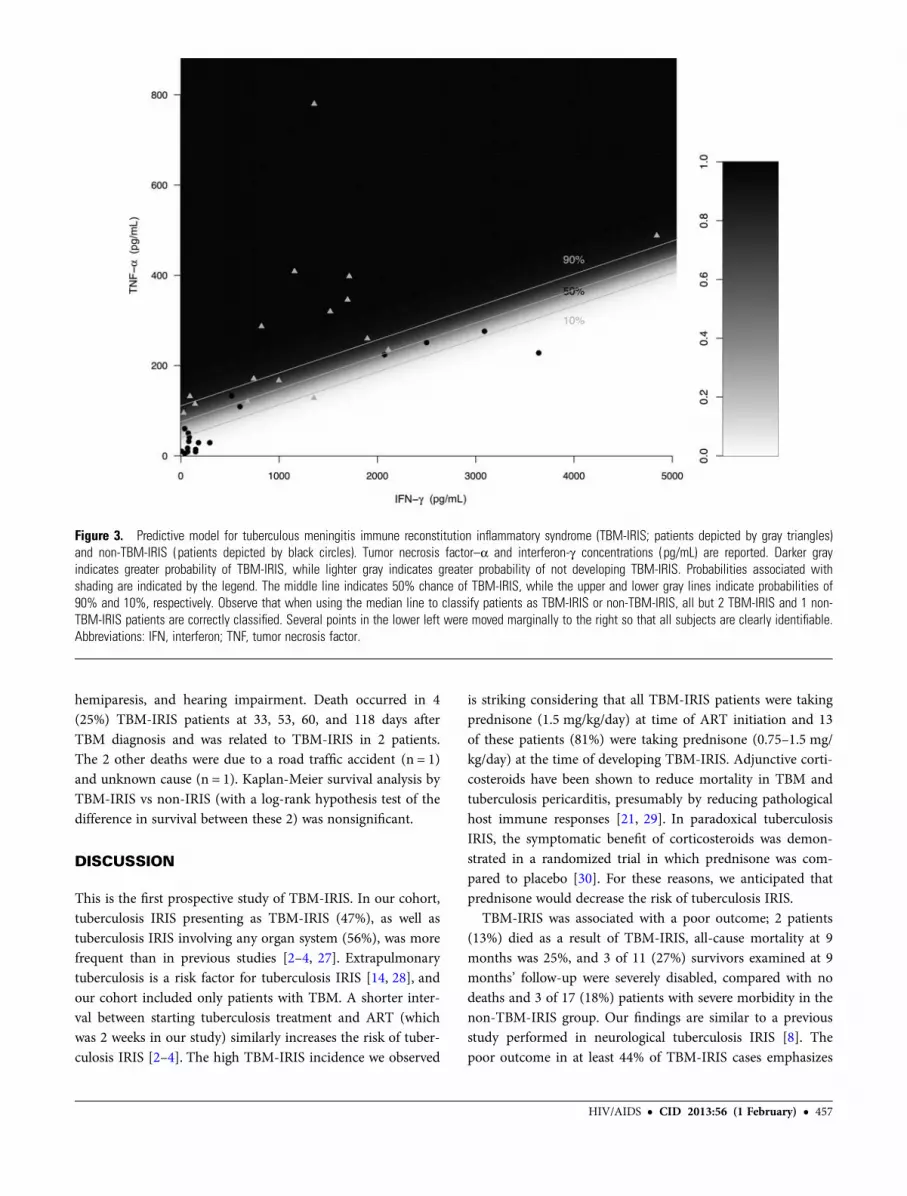
Analysis of Baseline CSF Neutrophil Count and CytokineConcentrations to Predict TBM-IRISConcentrations of the prespecified cytokines from which themodel to predict TBM-IRIS was developed are shown in Sup-plementary Table 2 and Supplementary Figure 2. The finalmultivariate logistic regression model included IFN-γ andTNF-α and produced a cross-validated AUC of 0.91 (95% CI,.53–.99, P = .02), indicating high diagnostic accuracy whenjointly considering these 2 cytokines to differentiate TBM-IRIS from non-TBM-IRIS at time of TBM diagnosis. Theodds ratio (OR) for TNF-α was 1.85 (per 10 pg/mL, P = .006),indicating an 85% increase in the odds of IRIS for every 10pg/mL increase in TNF-α (after adjusting for IFN-γ). The ORfor IFN-γ was 0.64 (per 100 pg/mL, P = .01), indicating de-creased odds of IRIS with increasing IFN-γ (after adjusting forTNF-α). Figure 3 provides a heatmap representation of thepredicted probabilities of the resulting model with the ob-served values overlaid. Neutrophil counts produced an AUCof 0.72 (95% CI, .54–.90, P = .03), indicating modest discrimi-natory accuracy for TBM-IRIS. However, including CSF neu-trophil and lymphocyte counts in the model did not improveits ability to predict TBM-IRIS.
Management and OutcomeIn 13 patients prescribed prednisone (0.75–1.5 mg/kg/day),the dose was increased at TBM-IRIS diagnosis. Prednisonewas restarted at a dose of 1.5 mg/kg/day in the other 3 TBM-IRIS patients. The median total duration of corticosteroidtreatment in TBM-IRIS patients was 109 days (IQR, 69–141)compared with 35 days (IQR, 20–43) in non-TBM-IRIS pa-tients. ART was interrupted during TBM-IRIS in 1 patientbecause of brainstem involvement. This patient made a fullrecovery and had no recurrent symptoms after recommence-ment of ART under prednisone cover. At 9 months’ follow-up, all non-TBM-IRIS patients were alive (including 1 whohad defaulted study follow-up but continued tuberculosistreatment from a primary care tuberculosis clinic), 2 hadmarked cognitive impairment (international HIV dementiascale <10) [26], and 1 patient had marked cognitive impair-ment with residual hemiparesis. Twelve IRIS patients (75%)were alive at 9 months’ follow-up, 2 patients showed markedcognitive impairment, 1 patient defaulted study follow-up butwas alive, and 1 had marked cognitive impairment, residual
HIV/AIDS • CID 2013:56 (1 February) • 455
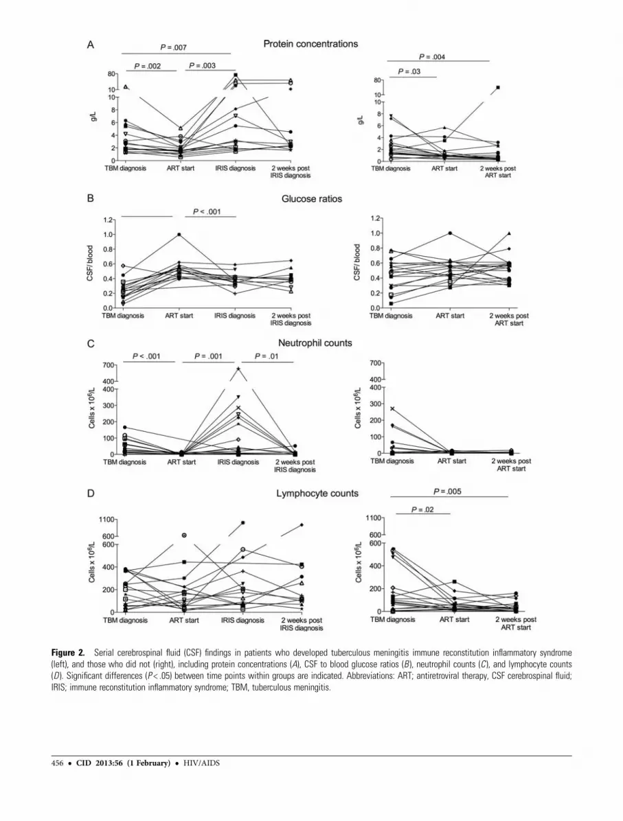
Figure 2. Serial cerebrospinal fluid (CSF) findings in patients who developed tuberculous meningitis immune reconstitution inflammatory syndrome(left), and those who did not (right), including protein concentrations (A), CSF to blood glucose ratios (B ), neutrophil counts (C ), and lymphocyte counts(D ). Significant differences (P < .05) between time points within groups are indicated. Abbreviations: ART; antiretroviral therapy, CSF cerebrospinal fluid;IRIS; immune reconstitution inflammatory syndrome; TBM, tuberculous meningitis.
456 • CID 2013:56 (1 February) • HIV/AIDS
hemiparesis, and hearing impairment. Death occurred in 4(25%) TBM-IRIS patients at 33, 53, 60, and 118 days afterTBM diagnosis and was related to TBM-IRIS in 2 patients.The 2 other deaths were due to a road traffic accident (n = 1)and unknown cause (n = 1). Kaplan-Meier survival analysis byTBM-IRIS vs non-IRIS (with a log-rank hypothesis test of thedifference in survival between these 2) was nonsignificant.
DISCUSSION
This is the first prospective study of TBM-IRIS. In our cohort,tuberculosis IRIS presenting as TBM-IRIS (47%), as well astuberculosis IRIS involving any organ system (56%), was morefrequent than in previous studies [2–4, 27]. Extrapulmonarytuberculosis is a risk factor for tuberculosis IRIS [14, 28], andour cohort included only patients with TBM. A shorter inter-val between starting tuberculosis treatment and ART (whichwas 2 weeks in our study) similarly increases the risk of tuber-culosis IRIS [2–4]. The high TBM-IRIS incidence we observed
is striking considering that all TBM-IRIS patients were takingprednisone (1.5 mg/kg/day) at time of ART initiation and 13of these patients (81%) were taking prednisone (0.75–1.5 mg/kg/day) at the time of developing TBM-IRIS. Adjunctive corti-costeroids have been shown to reduce mortality in TBM andtuberculosis pericarditis, presumably by reducing pathologicalhost immune responses [21, 29]. In paradoxical tuberculosisIRIS, the symptomatic benefit of corticosteroids was demon-strated in a randomized trial in which prednisone was com-pared to placebo [30]. For these reasons, we anticipated thatprednisone would decrease the risk of tuberculosis IRIS.
TBM-IRIS was associated with a poor outcome; 2 patients(13%) died as a result of TBM-IRIS, all-cause mortality at 9months was 25%, and 3 of 11 (27%) survivors examined at 9months’ follow-up were severely disabled, compared with nodeaths and 3 of 17 (18%) patients with severe morbidity in thenon-TBM-IRIS group. Our findings are similar to a previousstudy performed in neurological tuberculosis IRIS [8]. Thepoor outcome in at least 44% of TBM-IRIS cases emphasizes
Figure 3. Predictive model for tuberculous meningitis immune reconstitution inflammatory syndrome (TBM-IRIS; patients depicted by gray triangles)and non-TBM-IRIS (patients depicted by black circles). Tumor necrosis factor–α and interferon-γ concentrations (pg/mL) are reported. Darker grayindicates greater probability of TBM-IRIS, while lighter gray indicates greater probability of not developing TBM-IRIS. Probabilities associated withshading are indicated by the legend. The middle line indicates 50% chance of TBM-IRIS, while the upper and lower gray lines indicate probabilities of90% and 10%, respectively. Observe that when using the median line to classify patients as TBM-IRIS or non-TBM-IRIS, all but 2 TBM-IRIS and 1 non-TBM-IRIS patients are correctly classified. Several points in the lower left were moved marginally to the right so that all subjects are clearly identifiable.Abbreviations: IFN, interferon; TNF, tumor necrosis factor.
HIV/AIDS • CID 2013:56 (1 February) • 457
the need to predict and prevent, and improve the treatment of,TBM-IRIS.
Low serum sodium concentration is associated with deathin HIV-associated TBM [31]. In our study, serum sodiumconcentration was lower in TBM-IRIS patients compared withnon-TBM-IRIS patients at the time of TBM diagnosis. Thismay reflect the higher degree of tuberculosis disseminationobserved in TBM-IRIS patients, which could have contributedto their risk of developing TBM-IRIS.
Our finding of an association between higher CSF neutro-phils at TBM presentation and subsequent development ofTBM-IRIS provides important and novel insight into the path-ogenesis of tuberculosis IRIS. Not only were neutrophil countshigher in TBM-IRIS patients compared with non-TBM-IRISpatients, but neutrophil percentages for individual patientswere similarly raised in TBM-IRIS. The neutrophil countsshowed dynamic fluctuations over time in TBM-IRIS patientswith a marked decrease on tuberculosis treatment, and a strik-ing increase at TBM-IRIS onset. Similar changes in lympho-cyte counts were not observed. Studies of tuberculosis IRIShave hitherto focused on the contribution of helper T-celltype 1 lymphocyte responses [32, 33]. However, a role formyeloid cells in tuberculosis IRIS is suggested by a case reportof a patient who died from unmasking pulmonary tuberculosisIRIS; postmortem histological examination of diseased lungshowed a marked macrophage infiltrate [34]. We have foundcytokines of predominantly myeloid origin (IL-6 and TNF-α)to be consistently elevated in patients with tuberculosis IRIS,compared with those who did not develop IRIS [23]. Oliveret al [6] also reported an association between plasma cytokines(interleukin 18) and chemokines (CXCL-10) of the innateimmune system and tuberculosis IRIS. In an animal model,immune reconstitution following transfer of mycobacteria-specific CD4 T cells to T-cell–deficient mice infected withMycobacterium avium was associated with marked increasesof both blood and lung CD11b cells (likely representing in-flammatory monocytes and neutrophils) [35]. Our resultssuggest that neutrophils contribute to tuberculosis IRIS patho-genesis. The combination of high CSF TNF-α and low IFN-γconcentrations at the time of TBM diagnosis predicted TBM-IRIS in this cohort. Simmons et al [36] reported a negativecorrelation between CSF IFN-γ and mortality in HIV-infectedpatients with TBM. Conversely, a positive correlation wasfound between CSF IFN-γ and TNF-α concentrations andTBM disease severity by others [37].
Several studies have shown an association between dissemi-nated and extrapulmonary tuberculosis and subsequent tuber-culosis IRIS [14, 28, 38, 39]. At TBM diagnosis, CSF M.tuberculosis culture positivity, which reflects mycobacterialantigen load, was a major risk factor for developing TBM-IRIS(RR = 9.3). Furthermore, 7 TBM-IRIS patients (44%) were
persistently CSF M. tuberculosis culture positive after 2 weeksof tuberculosis treatment and 2 patients (13%) remainedculture positive after 4 weeks of tuberculosis treatment. Thisstrongly supports the inference that a high M. tuberculosis ba-cillary load at time of starting ART is a risk factor for tubercu-losis IRIS [40]. The findings suggest it important to optimizetuberculosis treatment prior to starting ART in patients athigh risk of developing TBM-IRIS.
We acknowledge several limitations. Because of the relative-ly small sample size, the study may not have been powered todetect further differences between IRIS and non-TBM-IRISpatients. Only patients with less severe disease (BMRC TBMgrade 1 and 2) and those without contraindications to lumbarpuncture were enrolled, resulting in the exclusion of a signifi-cant proportion of TBM patients presenting in our setting[41]; our results may therefore not be generalizable to ART-naive patients presenting with severe HIV-associated TBM.The model to predict TBM-IRIS needs further validation andexploration with independent data.
In conclusion TBM-IRIS complicated the course of treat-ment of HIV-associated TBM in nearly half our patients,despite the use of adjunctive corticosteroid therapy. The mani-festations were severe, fatal in 2 cases. The occurrence ofTBM-IRIS associated with CSF M. tuberculosis culture positiv-ity and a high neutrophil count at both baseline and at thetime of TBM-IRIS. The baseline relationship between CSFTNF-α and IFN-γ predicted TBM-IRIS. These observationsprovide novel insight into the pathogenesis of this conditionand provide rationale to individualize ART beyond 2 weeks inthis devastating, partly iatrogenic, condition.
Supplementary Data
Supplementary materials are available at Clinical Infectious Diseases online(http://www.oxfordjournals.org/our_journals/cid/). Supplementary materi-als consist of data provided by the author that are published to benefit thereader. The posted materials are not copyedited. The contents of all sup-plementary data are the sole responsibility of the authors. Questions ormessages regarding errors should be addressed to the author.
Notes
Acknowledgments. We thank the patients who participated in thestudy and Monica Magwayi for care provided to patients during the study.We also thank the Radiology Department at GF Jooste Hospital, in particu-lar Dr Ashmitha Rajkumar and Dr Marisa Mezzabotta, who reported thebrain imaging.Disclaimer. The funders had no role in study design, data collection
and analysis, decision to publish, or preparation of the manuscript.Financial support. This work was supported by the Carnegie Corpo-
ration Training Award and Discovery Foundation Academic FellowshipAward (S. M.); Perinatal HIV Research Unit, the US Agency for Interna-tional Development, and the President’s Emergency Plan for AIDS Relief(S. M., D. J. P., and C. S.); Wellcome Trust (S. M., R. J. W., and G. M.,WT 097254, 081667, 084323, and 088316); Fogarty International CenterSouth Africa TB/AIDS Training Award (G. M., D. J. P., and C. S., NIH/
458 • CID 2013:56 (1 February) • HIV/AIDS
FIC U2R TW007373-01A1 and U2R TW007370-01A1); European UnionGrant (R. J. W., SANTE/2005/105-061-102); Medical Research CouncilGrant (R. J. W., U.1175.02.002.00014.01).Potential conflicts of interest. All authors: No reported conflicts.All authors have submitted the ICMJE Form for Disclosure of Potential
Conflicts of Interest. Conflicts that the editors consider relevant to thecontent of the manuscript have been disclosed.
References
1. Meintjes G, Lawn SD, Scano F, et al. Tuberculosis-associated immunereconstitution inflammatory syndrome: case definitions for use in re-source-limited settings. Lancet Infect Dis 2008; 8:516–23.
2. Blanc FX, Sok T, Laureillard D, et al. Earlier versus later start of anti-retroviral therapy in HIV-infected adults with tuberculosis. N Engl JMed 2011; 365:1471–81.
3. Havlir DV, Kendall MA, Ive P, et al. Timing of antiretroviral therapyfor HIV-1 infection and tuberculosis. N Engl J Med 2011; 365:1482–91.
4. Abdool Karim SS, Naidoo K, Grobler A, et al. Integration of antiretro-viral therapy with tuberculosis treatment. N Engl J Med 2011;365:1492–501.
5. Meintjes G, Rabie H, Wilkinson RJ, Cotton MF. Tuberculosis-associat-ed immune reconstitution inflammatory syndrome and unmasking oftuberculosis by antiretroviral therapy. Clin Chest Med 2009;30:797–810.
6. Oliver BG, Elliott JH, Price P, et al. Mediators of innate and adaptiveimmune responses differentially affect immune restoration disease as-sociated with Mycobacterium tuberculosis in HIV patients beginningantiretroviral therapy. J Infect Dis 2010; 202:1728–37.
7. Asselman V, Thienemann F, Pepper DJ, et al. Central nervous systemdisorders after starting antiretroviral therapy in South Africa. AIDS2010; 24:2871–6.
8. Pepper DJ, Marais S, Maartens G, et al. Neurologic manifestations ofparadoxical tuberculosis-associated immune reconstitution inflamma-tory syndrome: a case series. Clin Infect Dis 2009; 48:e96–107.
9. Torok ME, Kambugu A, Wright E. Immune reconstitution disease ofthe central nervous system. Curr Opin HIVAIDS 2008; 3:438–45.
10. Dautremer J, Pacanowski J, Girard PM, Lalande V, Sivignon F,Meynard JL. A new presentation of immune reconstitution inflamma-tory syndrome followed by a severe paradoxical reaction in an HIV-1-infected patient with tuberculous meningitis. AIDS 2007; 21:381–2.
11. Tuon FF, Mulatti GC, Pinto WP, de Siqueira Franca FO, GryschekRC. Immune reconstitution inflammatory syndrome associated withdisseminated mycobacterial infection in patients with AIDS. AIDSPatient Care STDS 2007; 21:527–32.
12. Lee CH, Lui CC, Liu JW. Immune reconstitution syndrome in apatient with AIDS with paradoxically deteriorating brain tuberculoma.AIDS Patient Care STDS 2007; 21:234–9.
13. Crump JA, Tyrer MJ, Lloyd-Owen SJ, Han LY, Lipman MC, JohnsonMA. Miliary tuberculosis with paradoxical expansion of intracranialtuberculomas complicating human immunodeficiency virus infectionin a patient receiving highly active antiretroviral therapy. Clin InfectDis 1998; 26:1008–9.
14. Manosuthi W, Kiertiburanakul S, Phoorisri T, Sungkanuparph S.Immune reconstitution inflammatory syndrome of tuberculosisamong HIV-infected patients receiving antituberculous and antiretro-viral therapy. J Infect 2006; 53:357–63.
15. Vidal JE, Cimerman S, Schiavon Nogueira R, et al. Paradoxical reac-tion during treatment of tuberculous brain abscess in a patient withAIDS. Rev Inst Med Trop Sao Paulo 2003; 45:177–8.
16. Marais S, Scholtz P, Pepper DJ, Meintjes G, Wilkinson RJ, Candy S.Neuroradiological features of the tuberculosis-associated immune re-constitution inflammatory syndrome. Int J Tuberc Lung Dis 2010;14:188–96.
17. Torok ME, Yen NT, Chau TT, et al. Timing of initiation of antiretro-viral therapy in human immunodeficiency virus (HIV)-associatedtuberculous meningitis. Clin Infect Dis 2011; 52:1374–83.
18. Torok ME, Farrar JJ. When to start antiretroviral therapy in HIV-associated tuberculosis. N Engl J Med 2011; 365:1538–40.
19. Médecins Sans Frontières, Western Cape Province Department ofHealth, City of Cape Town Department of Health, and University ofCape Town Infectious Disease Epidemiology Unit. ComprehensiveTB/HIV services at primary health care level, Khayelitsha annual ac-tivity report: 2007–2008. Available at: http://www.msf.or.jp/info/pressreport/pdf/2009_hiv01.pdf Accessed 28 October 2012.
20. Bhigjee AI, Padayachee R, Paruk H, Hallwirth-Pillay KD, Marais S,Connoly C. Diagnosis of tuberculous meningitis: clinical and laborato-ry parameters. Int J Infect Dis 2007; 11:348–54.
21. Thwaites GE, Nguyen DB, Nguyen HD, et al. Dexamethasone for thetreatment of tuberculous meningitis in adolescents and adults. N EnglJ Med 2004; 351:1741–51.
22. Antimycobacterials. In: Gibbon CJ, Blockman M, eds. South Africanmedicines formulary. 8th ed. Cape Town, South Africa: Division ofClinical Pharmacology, Faculty of Health Sciences, University of CapeTown, 2008:301–2.
23. Tadokera R, Meintjes G, Skolimowska KH, et al. Hypercytokinaemiaaccompanies HIV-tuberculosis immune reconstitution inflammatorysyndrome. Eur Respir J 2011; 37:1248–59.
24. Meintjes G, Skolimowska KH, Wilkinson KA, et al. Corticosteroid-modulated immune activation in the tuberculosis immune reconstitu-tion inflammatory syndrome. Am J Respir Crit Care Med 2012;186:369–77.
25. Simon RM, Korn EL, McShane LM, Radmacher MD, Wright GW,Zhao Y. Class prediction. In: Dietz K, Samet J, Gail M, et al., eds.Design and analysis of DNA microarray investigations. 1st ed.New York: Springer, 2004:108–14.
26. Sacktor NC, Wong M, Nakasujja N, et al. The International HIV De-mentia Scale: a new rapid screening test for HIV dementia. AIDS2005; 19:1367–74.
27. Muller M, Wandel S, Colebunders R, Attia S, Furrer H, Egger M.Immune reconstitution inflammatory syndrome in patients startingantiretroviral therapy for HIV infection: a systematic review and meta-analysis. Lancet Infect Dis 2010; 10:251–61.
28. Burman W, Weis S, Vernon A, et al. Frequency, severity and durationof immune reconstitution events in HIV-related tuberculosis. Int JTuberc Lung Dis 2007; 11:1282–9.
29. McGee S, Hirschmann J. Use of corticosteroids in treating infectiousdiseases. Arch Intern Med 2008; 168:1034–46.
30. Meintjes G, Wilkinson RJ, Morroni C, et al. Randomized placebo-controlled trial of prednisone for paradoxical tuberculosis-associatedimmune reconstitution inflammatory syndrome. AIDS 2010; 24:2381–90.
31. Torok ME, Chau TT, Mai PP, et al. Clinical and microbiological fea-tures of HIV-associated tuberculous meningitis in Vietnamese adults.PLoS One 2008; 3:e1772.
32. Bourgarit A, Carcelain G, Martinez V, et al. Explosion of tuberculin-specific Th1-responses induces immune restoration syndrome in tu-berculosis and HIV co-infected patients. AIDS 2006; 20:F1–7.
33. Meintjes G, Wilkinson KA, Rangaka MX, et al. Type 1 helper T cellsand FoxP3-positive T cells in HIV-tuberculosis-associated immune re-constitution inflammatory syndrome. Am J Respir Crit Care Med2008; 178:1083–9.
34. Lawn SD, Wainwright H, Orrell C. Fatal unmasking tuberculosisimmune reconstitution disease with bronchiolitis obliterans organizingpneumonia: the role of macrophages. AIDS 2009; 23:143–5.
35. Barber DL, Mayer-Barber KD, Antonelli LR, et al. Th1-drivenimmune reconstitution disease in Mycobacterium avium-infectedmice. Blood 2010; 116:3485–93.
36. Simmons CP, Thwaites GE, Quyen NT, et al. Pretreatment intracere-bral and peripheral blood immune responses in Vietnamese adults
HIV/AIDS • CID 2013:56 (1 February) • 459
with tuberculous meningitis: diagnostic value and relationship todisease severity and outcome. J Immunol 2006; 176:2007–14.
37. Patel VB, Bhigjee AI, Bill PL, Connolly CA. Cytokine profiles in HIVseropositive patients with tuberculous meningitis. J Neurol NeurosurgPsychiatry 2002; 73:598–9.
38. Breton G, Duval X, Estellat C, et al. Determinants of immune recon-stitution inflammatory syndrome in HIV type 1-infected patients withtuberculosis after initiation of antiretroviral therapy. Clin Infect Dis2004; 39:1709–12.
39. Michailidis C, Pozniak AL, Mandalia S, Basnayake S, Nelson MR,Gazzard BG. Clinical characteristics of IRIS syndrome in patients withHIV and tuberculosis. Antivir Ther 2005; 10:417–22.
40. Lawn SD, Meintjes G. Pathogenesis and prevention of immune recon-stitution disease during antiretroviral therapy. Expert Rev Anti InfectTher 2011; 9:415–30.
41. Marais S, Pepper DJ, Schutz C, Wilkinson RJ, Meintjes G. Presenta-tion and outcome of tuberculous meningitis in a high HIV prevalencesetting. PLoS One 2011; 6:e20077.
460 • CID 2013:56 (1 February) • HIV/AIDS