fouling and wetting in membrane distillation: mechanisms and
TRANSCRIPT

FOULING AND WETTING IN MEMBRANE DISTILLATION: MECHANISMS AND
MITIGATION STRATEGIES
By
Zhangxin Wang
Dissertation
Submitted to the Faculty of the
Graduate School of Vanderbilt University
in partial fulfillment of the requirements
for the degree of
DOCTOR OF PHILOSOPHY
in
Environmental Engineering
September 30, 2018
Nashville, Tennessee
Approved:
Shihong Lin, Ph.D.
Florence Sanchez, Ph.D.
Alan Bowers, Ph.D.
Paul E. Laibinis, Ph.D.
G. Kane Jennings, Ph.D.

ii
DEDICATION
To my parents,
For their love, encouragement, and support

iii
ACKNOWLEDGEMENTS
I would like to express my great thankfulness to Dr. Shihong Lin, who has been an excellent
advisor and mentor throughout my Ph.D. study. Without his guidance and support, I would not be
able to have such fulfillments in the past 4 years. I also want to thank Dr. Florence Sanchez, Dr.
Alan Bowers, Dr. Paul Laibinis, and Dr. Kane Jennings for serving on my dissertation committee
and giving me valuable suggestions.
I would like to give thanks to my colleagues in Dr. Lin’s group, including Deyin Hou, Yuxi
Huang, Yuanmiaoliang Chen, Thomas Horseman, Kofi Christie, Li Wang, Yuanzhe Liang and so
on. These people are not only my teammates who have lent me hands in my academic journey, but
also my friends who have decorated my life at Vanderbilt University more colorful. Best luck to
all of you and I hope we can have our paths crossing again in the future.
I want to thank Rossane Delapp and Dalesha Blackwell, the CEE lab managers who have
offered me a lot of help in my experiments. I also want to thank the ViNSE faculty members for
training and helping me to use the lab instruments.
Last but not the least, I want to express my deepest gratitude to my parents for their love and
encouragement. They always stand with me as my strongest backup upon every decision I made.
Without their support, I would not become the person I am today.

iv
TABLE OF CONTENTS
Page
Dedication ....................................................................................................................................... ii
Acknowledgements ........................................................................................................................ iii
List of Figures .............................................................................................................................. viii
List of Tables ................................................................................................................................ xii
Chapter 1 Introduction .................................................................................................................... 1
1.1. The Emerging Need for Hypersaline Industrial Wastewater Desalination ...................... 1
1.2. Membrane Distillation as a Promising Solution ............................................................... 2
1.3. Challenges with Conventional Hydrophobic MD Membranes ........................................ 3
1.3.1. Membrane Fouling ................................................................................................. 4
1.3.2. Membrane Wetting ................................................................................................. 5
1.4. The Applications of Membranes with Special Wettability ............................................... 6
1.5. Objectives and Hypothesis ............................................................................................... 9
1.6. Structure of the Dissertation ........................................................................................... 10
Chapter 2 Impact of Surface Wettability on Fouling Resistance of MD Membranes .................. 11
2.1. Introduction ..................................................................................................................... 11
2.2. Materials and Methods ................................................................................................... 13
2.2.1. Materials and Chemicals ...................................................................................... 13
2.2.2. Composite Membrane Fabrication ....................................................................... 14
2.2.3. Membrane Characterizations ................................................................................ 14
2.2.4. Preparation and Characterizations of Oil Feed Solution ...................................... 15
2.2.5. Membrane Fouling Test ....................................................................................... 15
2.2.6. Force Measurement of Underwater Oil-Membrane Interaction ........................... 16
2.3. Results and Discussions .................................................................................................. 17
2.3.1. Morphology and Wetting Properties of the Pristine and Modified Membranes .. 17
2.3.2. Properties of Oily Feed Solution .......................................................................... 20
2.3.3. Membrane Performance in Direct Contact Membrane Distillation ..................... 21
2.3.4. Underwater Oil-Membrane Interaction ................................................................ 24
2.4. Implications .................................................................................................................... 26
Chapter 3 Influence of Surface Charge on Fouling Resistance of MD Membranes .................... 28

v
3.1. Introduction ..................................................................................................................... 28
3.2. Materials and Methods ................................................................................................... 31
3.2.1. Materials and Chemicals ...................................................................................... 31
3.2.2. Fabrication of the Underwater Oleophobic Membranes ...................................... 31
3.2.2.1. Composite membrane coated with polydopamine (PDA) and SiNPs ........ 31
3.2.2.2. Composite membrane coated with PDDA and SiNPs ............................... 32
3.2.3. Characterizations of Morphology and Wetting Property ..................................... 32
3.2.4. Preparation and Characterization of Oily Feed Solution ...................................... 33
3.2.5. Streaming Potential Measurement of the Membranes .......................................... 33
3.2.6. Membrane Fouling Tests ...................................................................................... 34
3.2.7. Underwater Oil-Adhesion Force Measurements .................................................. 34
3.3. Results and discussions................................................................................................... 35
3.3.1. Membrane Surface Properties .............................................................................. 35
3.3.1.1. Membrane Surface Morphology ................................................................ 35
3.3.1.2. Membrane Surface Potential ...................................................................... 37
3.3.1.3. Membrane Surface Wetting Property ......................................................... 38
3.3.2. Properties of Saline Crude Oil Emulsion ............................................................. 40
3.3.3. Membrane Performances in MD Experiments ..................................................... 41
3.3.4. Force Spectroscopy with Oil Droplet Probe ......................................................... 43
3.3.5. Implications of Electrostatic Interaction .............................................................. 45
3.3.6. The Relative Irrelevance of the DLVO Approach ............................................... 47
3.4. Conclusion ...................................................................................................................... 48
Chapter 4 Mechanism of Membrane Pore Wetting in Membrane Distillation ............................. 50
4.1. Introduction ..................................................................................................................... 50
4.2. Theory and Model Development .................................................................................... 53
4.2.1. General Criterion of Pore Wetting ....................................................................... 53
4.2.2. Membrane Wetting by Alcohol Solution ............................................................. 55
4.2.3. Membrane Wetting by Surfactants ....................................................................... 56
4.3. Materials and Methods ................................................................................................... 59
4.3.1. Materials ............................................................................................................... 59
4.3.2. Surface Tension Measuremnts ............................................................................. 59
4.3.3. Principle and Experimental Setup of impedance-based Wetting Monitoring ...... 59
4.3.4. MD Wetting Experiments ..................................................................................... 61
4.4. Results and Discussion ................................................................................................... 62

vi
4.4.1. Triton X-100 vs. Ethanol as a Wetting Agent: Drastically Different Wetting
Behaviors .............................................................................................................. 62
4.4.2. Ethanol-Induced Wetting: a Simple Application of the General Wetting Criterion
.............................................................................................................................. 65
4.4.3. Surfactant-Induced Wetting: a Dynamic Process Limited by Surfactant Transport
.............................................................................................................................. 66
4.5. Conclusion ...................................................................................................................... 71
Chapter 5 Kinetic Model for Surfactant-Induced Pore Wetting in Membrane Distillation .......... 72
5.1. Introduction ..................................................................................................................... 72
5.2. Theory and Model Development .................................................................................... 74
5.3. Materials and Methods ................................................................................................... 80
5.3.1. Materials and Chemicals ...................................................................................... 80
5.3.2. Surface Tension Measurements ............................................................................ 81
5.3.3. Intrinsic Contact Angle Measurements ................................................................ 81
5.3.4. Determination of LEP ........................................................................................... 82
5.3.5. Surface Excess Concentration .............................................................................. 82
5.3.6. DCMD Wetting Experiments ............................................................................... 83
5.4. Results and Discussion ................................................................................................... 84
5.4.1. The Impact of SDS Concentration ....................................................................... 84
5.4.2. The Impact of Vapor Flux .................................................................................... 85
5.4.3. The Impact of Transmembrane Hydraulic Pressure ............................................. 86
5.4.4. Membrane Wetting Kinetics for Different Surfactant Species ............................. 90
5.5. Conclusion ...................................................................................................................... 93
Chapter 6 Stategies for Fouling and Wetting Mitigation in Membrane Distillation Opertation
Using Membranes with Different Wettibility .............................................................. 96
6.1. Introduction ..................................................................................................................... 96
6.2. Materials and Methods ................................................................................................... 99
6.2.1. Materials and Chemicals ...................................................................................... 99
6.2.2. Fabrication of Composite Membrane with Superhydrophilic Skin Layer ........... 99
6.2.3. Membrane Characterization ............................................................................... 100
6.2.4. Crude Oil-in-water Emulsions ............................................................................ 100
6.2.5. Membrane Fouling Test ..................................................................................... 100
6.2.6. Oil-probe Force Spectroscopy ............................................................................ 101
6.3. Results and Discussion ................................................................................................. 101
6.3.1. Membrane Morphology ...................................................................................... 101

vii
6.3.2. Membrane Wetting Properties ............................................................................ 102
6.3.3. Characterizations of Crude Oil-in-Water Emulsions ......................................... 105
6.3.4. Membrane Performance with Oil-in-Water Emulsion without Synthetic
Surfactants .......................................................................................................... 106
6.3.5. Membrane Performance with Oil-in-Water Emulsion Stabilized by Synthetic
Surfactants .......................................................................................................... 109
6.3.6. Force Spectroscopy to Elucidate Oil-Membrane Interaction ............................. 111
6.4. Conclusion .................................................................................................................... 114
Chapter 7 Conclusions and Future Work .................................................................................... 116
Appendix A ................................................................................................................................. 120
Supplementary Materials for Chapter 2 ...................................................................................... 120
Appendix B ................................................................................................................................. 122
Supplementary Materials for Chapter 3 ...................................................................................... 122
Appendix C ................................................................................................................................. 124
Supplementary Materials for Chapter 4 ...................................................................................... 124
Appendix D ................................................................................................................................. 135
Supplementary Materials for Chapters 5 .................................................................................... 135
References ................................................................................................................................... 143

viii
LIST OF FIGURES
Figure Page
Figure 1.1 The mechanism of direct contact membrane distillation (DCMD) ............................... 3
Figure 1.2 Oil fouling of a hydrophobic membrane ....................................................................... 4
Figure 1.3 Membrane pore wetting by surfactants ......................................................................... 6
Figure 1.4 Definition of contact angle. ........................................................................................... 7
Figure 1.5 The relations between different types of most investigated surfaces with special
wettability.. ..................................................................................................................................... 8
Figure 2.1 Characterizations of the composite membrane and PVDF membrane. ....................... 18
Figure 2.2 Contact angle measurements of the composite membrane and PVDF membrane. ..... 19
Figure 2.3 Characteriziation of the crude oil-in-water emulsion. ................................................. 21
Figure 2.4 Normalized water flux for the composite membrane and PVDF membrane in DCMD
fouling tests. .................................................................................................................................. 22
Figure 2.5 Force spectroscopy of the composite membrane and pristine PVDF membrane with an
oil droplet. ..................................................................................................................................... 24
Figure 3.1 SEM images of the composite membranes and PVDF membrane. ............................. 36
Figure 3.2 Zeta potentials of the composite membranes and PVDF membrane. ......................... 37
Figure 3.3 Contact angle measurements of the composite membranes and PVDF membrane. ... 39
Figure 3.4 Characteriziation of the crude oil-in-water emulsion. ................................................. 40
Figure 3.5 Normalized water flux and for the composite membranes and PVDF membrane in
DCMD fouling tests. ..................................................................................................................... 41
Figure 3.6 Force spectroscopy of the composite memrbanes and pristine PVDF membrane with
an oil droplet. ................................................................................................................................ 44
Figure 4.1 Illustration of wetting of a cylindrical pore by saline feed solutions containing ethanol
and Triton X-100........................................................................................................................... 54
Figure 4.2 Illustration of the possible membrane pore wetting mechanism and a schematic of a
DCMD system equipped in-situ impedance measurements capability. ........................................ 60
Figure 4.3 Impedance across the PVDF membrane and salt rejection in DCMD experiments in
the presence of Triton X-100 and ethanol solution. ...................................................................... 64

ix
Figure 4.4 Experimentally measured transmembrane impedance with feed solution containing
ethanol.. ......................................................................................................................................... 66
Figure 4.5 Experimentally measured impedances across the PVDF membrane in DCMD
experiments with different Triton X-100 concentrations and vapor fluxes. ................................. 68
Figure 4.6 Experimentally measured impedances across the PVDF membrane in DCMD
experiments with different transmembrane hydraulic pressures. ................................................. 69
Figure 5.1 Schematic illustration of a cylindrical pore wetting by SDS at different
concentrations. .............................................................................................................................. 76
Figure 5.2 Experimental results and model prediction of MD membrane wetting time with
different SDS concentrations in feed solution. ............................................................................. 84
Figure 5.3 Experimental results and model prediction of MD membrane wetting at different
vapor fluxes. .................................................................................................................................. 86
Figure 5.4 Experimental results and model predictions of DCMD membrane wetting experiments
with different critical concentrations ............................................................................................ 88
Figure 5.5 Surface tension as a function of surfactant concentration for different types of
surfactants. .................................................................................................................................... 90
Figure 5.6 Wetting breakthrough time a function of Γ. ................................................................ 91
Figure 5.7 Theoretical predictions and experimental observations of the wetting breakthrough
time. .............................................................................................................................................. 93
Figure 6.1 SEM images of the local surface morphology for the PVDF membrane, composite
membrane, and omniphobic membrane. ..................................................................................... 102
Figure 6.2 Contact angle measurements of the local surface morphology for the PVDF
membrane, composite membrane, and omniphobic membrane. ................................................. 104
Figure 6.3 Characterizations of emulsion with and without any synthetic surfactant. ............... 105
Figure 6.4 Normalized water fluxes of the PVDF membrane, composite membrane, and
omniphobic membrane over time and specific volume of permeate water normalized by the
membrane area in DCMD fouling experiments. ......................................................................... 107
Figure 6.5 Normalized water fluxes and corresponding salt rejection rates for the PVDF
membrane, composite membrane, and omniphobic membrane over time in DCMD fouling and
wetting experiments. ................................................................................................................... 110

x
Figure 6.6 Force spectroscopy of the PVDF membrane, composite membrane, and omniphobic
membrane with a mineral oil droplet. ......................................................................................... 112
Figure A.1 Polarization Modulation-Infrared Reflection-Adsorption Spectroscopy (PM-IRRAS)
for the PVDF and the modified surface of the composite membrane......................................... 121
Figure B.1 Pore size distributions of the composite membranes and PVDF membrane. ........... 122
Figure B.2 Infrared absorbance spectra obtained from PMIRRAS for the composite membranes
and PVDF membrane .................................................................................................................. 122
Figure C.1 Impedance and phase angle spectra of a PVDF membrane in DCMD operation..... 124
Figure C.2 Schematic of membrane pore wetting in DCMD.. ................................................... 125
Figure C.3 The concentration profile of Triton X-100 in the solution for 𝑙 = 30 𝜇𝑚.. ............. 128
Figure C.4 The surface tensions of NaCl aqueous solutions with different concentrations of
ethanol and Triton X-100. ........................................................................................................... 130
Figure C.5 Salt rejection rate and normalized water flux in DCMD experiments corresponding to
Figure 4.3 of the main text.. ........................................................................................................ 131
Figure C.6 Salt rejection rate and normalized water flux in DCMD experiments corresponding
to Figure 4.4 of the main text. ..................................................................................................... 131
Figure C.7 Salt rejection rate and normalized water flux in DCMD experiments corresponding
to Figure 4.5a of the main text. ................................................................................................... 132
Figure C.8 Salt rejection rate and normalized water flux in DCMD experiments corresponding
to Figure 4.5b of the main text. ................................................................................................... 133
Figure C.9 Salt rejection rate and normalized water flux in DCMD experiments corresponding
to Figure 4.6 of the main text. ..................................................................................................... 134
Figure D.1 Normalized water fluxes and salt rejection data for DCMD membrane wetting
experiments (experiments 1-9 in Table D1).. ............................................................................. 137
Figure D.2 Normalized water fluxes and salt rejection rates data for DCMD membrane wetting
experiments (experiments 6 and 10-14 in Table D1). ................................................................. 138
Figure D.3 Normalized water fluxes and salt rejection rates data for DCMD membrane wetting
experiments (experiments 6, 15 and 16 in Table D1). ................................................................ 139
Figure D.4 AFM images of the PVDF film reconstructed from porous PVDF membrane.. ...... 139
Figure D.5 Surface tensions of NaCl solutions with different concentrations of SDS. .............. 140

xi
Figure D.6 Intrinsic contact angles of NaCl solutions with different concentrations of SDS on
PVDF surface. ............................................................................................................................. 140
Figure D.7 Estimated LEP of the PVDF membrane for NaCl solutions with different SDS
concentrations ............................................................................................................................. 141
Figure D.8 Salt rejection rate as a function of time in DCMD wetting experiments. ................ 141

xii
LIST OF TABLES
Table Page
Table D1. Summary of experimental conditions of DCMD wetting experiments ..................... 136
Table D2. The parameters for model prediction of pore wetting by different surfactant species
..................................................................................................................................................... 142

1
CHAPTER 1
CHAPTER 1 INTRODUCTION CHAPTER 1
1.1. The Emerging Need for Hypersaline Industrial Wastewater Desalination
Intensified freshwater scarcity and growing public environmental awareness have
progressively lead to stricter regulations on industrial wastewater disposal. As a consequence,
disposal of industrial wastewater has become increasingly costly and in some cases even
prohibited. Global interest in zero liquid discharge (ZLD) has thus been increasing in both
developed and developing countries[1]. Since many types of industrial wastewater contain
significant amount of salts, achieving ZLD will involve desalinating the saline wastewater,
concentrating the brine to the greatest extent possible, and eventually crystallizing the brine to
obtain solid waste. Regardless what initial salinity the industrial wastewater has,
desalination/concentration of the hypersaline brine is an inevitable step toward ZLD.
Another area that would foresee an appreciable growth in wastewater desalination is the
industry of shale oil/gas production[2]. The development of horizontal drilling and hydraulic
fracturing (fracking) has enabled economical exploitation of oil and gas from shale formations—
a tremendous source that was once hardly accessible with the conventional extraction
technologies[3]. As a result, the production of shale oil/gas has drastically altered the energy
portfolio of U.S. in recent years[4] and has appreciably impacted the global energy production
landscape[5]. However, shale oil/gas production faces multiple significant environmental and
public health challenges[3,6–8]. Environmentally responsible management of the tremendous
amount of wastewater is key to the sustainability of the fracking industry.
Fracking wastewater is composed of both the flow-back and the produced water[9]. The flow-
back water is the aqueous solution that flows back to the surface upon the completion of fracking.
The produced water is the water that originates from the shale formation and flows to the surface
during the oil/gas production. Collectively, fracking wastewater presents significant
environmental challenges as it contains a wide variety of contaminants that are detrimental to the
surface and ground water quality[2]. These waters are usually rich in mineral ions, colloids,
hydrocarbon, a variety of chemical additives, and in many cases, radionuclides. Existing practices

2
for managing fracking wastewater include underground injection[10], disposal to surface water or
POTW, onsite treatment, and reuse within and outside the shale oil/gas industry[11].
While reuse within the shale oil/gas industry is currently the most practical management
approach, there are multiple reasons for the growing need of onsite treatment of these wastewaters
for beneficial reuse outside the oil/gas industry or for safe discharge. These reasons include, but
are not limited to, tightening environmental regulations on direct disposal, growing fresh water
scarcity in certain areas (e.g. California)[12], and the expectable diminishing opportunities for
local reuse as fracturing fluid[13]. Therefore, appropriate treatment of fracking wastewater is
required to meet the water quality standards for surface discharge or beneficial reuses outside the
oil/gas industry. Depending on the specific shale formation, some of the fracking wastewater is
highly saline with a total dissolved solid (TDS) several times as high as that of seawater in certain
cases[4]. Desalination is thus required to reduce the TDS to an acceptable level for reuse or
discharge of these wastewaters.
For these reasons, viable technologies are in need for the desalination of industrial wastewater
and fracking wastewater, not only to comply with growingly stringent regulations but also to
recover valuable resources from waste streams.
1.2. Membrane Distillation as a Promising Solution
Reverse osmosis (RO) is a pressure-driven desalination technology that currently dominates
the market of desalination[14,15]. Although from the energy efficiency perspective, RO is almost
unbeatable, it still has some limitations in application. For hypersaline wastewaters that have
extremely high osmotic pressures, such as produced wastewater, RO cannot be utilized as the
allowable operating pressure is far exceeded. For the desalination of these wastewaters, thermal
desalination is by far the most practical choice[13].
Membrane distillation (MD) is an emerging thermal desalination technology using a
microporous hydrophobic membrane[16–19]. As shown in Figure 1.1, the microporous
hydrophobic membrane was used to isolate a hot salty stream (feed) and a cold fresh stream
(distillate). Driven by the partial vapor pressure difference generated from temperature difference,
fresh water vapor would permeate through the membrane pores from the feed side to the distillate

3
side and condense as liquid water. In the meantime, the hydrophobicity of the membrane keeps
the salty liquid water from the fresh water and thereby realizes the salty water desalination.
Figure 1.1 The mechanism of direct contact membrane distillation (DCMD): a hydrophobic
membrane, separating the hot salty feed stream and the cold pure distillate stream, only allows
water vapor to pass through its pores and condense on the distillate-membrane interface. The
transport of vapor through the pores is driven by the partial vapor pressure difference across the
membrane.
Compared with RO, the state-of-the-art desalination technology, MD has several advantages.
First, MD can be used to desalinate hypersaline wastewater, such as RO brine and shale gas/oil
produced water, whose salinity is too high for RO to deal with[20]. Second, as a low-temperature
desalination technology, MD is able to harness low-quality energy such as waste heat, solar energy,
and geothermal energy[21]. Third, the capital cost of MD is relatively low, which renders MD an
appealing process for sustainable and off-grid desalination[22].
1.3. Challenges with Conventional Hydrophobic MD Membranes
The MD membrane is the critical component in an MD system. Conventional MD membranes
are hydrophobic and made of common polymeric materials such as polyvinylidene fluoride
(PVDF), polypropylene (PP), and polytetrafluroethylene (PTFE). The hydrophobicity is required
to prevent direct liquid permeation through the micropores. However, when hydrophobic
membranes are used in MD to treat hypersaline wastewater of a complex composition, two
potential problems may lead to MD operation failures[23].

4
1.3.1. Membrane fouling
The first potential problem is membrane fouling. Membrane fouling is a universal problem in
all membrane processes[23–26]. It refers to the accumulation of unwanted materials on the
membrane surface or inside the membrane pores during operation, which leads to detrimental
effect of the overall membrane performance[23]. In MD operations, membrane fouling can be
induced by a variety of foulants including salt scalants (e.g. CaSO4 and CaCO3), particulates and
colloids (e.g. silica and silts), and biofoulants (e.g. biofilms and natural organic matters)[23,27,28].
In particular, membrane fouling is a critical concern when a hydrophobic membrane is used
in MD to treat feed waters with an abundance of hydrophobic contaminants (e.g. oil, hydrophobic
organics) due to the strong hydrophobic-hydrophobic interaction[27,29–31]. As shown in Figure
1.2, with the presence of oil droplets in the feed solution of an MD operation, the membrane pore
would be readily blocked leading to a significant vapor flux decline[32]. Recent studies using
hydrophobic MD membranes for desalinating brine water rich in oil or organics have shown the
rapid and severe flux decline as a result of membrane fouling[33].
Figure 1.2 (A) Oil fouling of a hydrophobic membrane: the hydrophobic oil droplets accumulate
at the hydrophobic membrane surface, blocking the pores for vapor transfer. (B) Experimental
results showing rapid flux decline as evidence of the membrane oil-fouling.
To mitigate the membrane fouling problem in MD, numerous strategies have been proposed
and tested. For instance, pretreating the feed solution using microfiltration (MF), ultrafiltration
(UF), or nanofiltration (NF) can effectively remove the potential foulants such as particulates,

5
colloids, and microorganisms[23,34]; implementing regular membrane flushing or gas bubbling
in MD operation can prevent salt scalant-induced fouling[35,36]; modifying the membrane surface
to be more hydrophobic can significantly lessen biofouling[37]; adding chemicals such as
antiscalants, cleaning agents, and biocides can also successfully improve alleviate the
corresponding membrane fouling problems[38,39]. Unfortunately, in MD with membrane fouling
by hydrophobic contaminants, the strategies mentioned above would either drastically increase the
treatment cost or are not efficient at all. Thus, for desalinating shale/gas wastewater or industrial
brines, which are enriched in hydrophobic contaminants, novel membrane fouling mitigation
strategies are needed.
1.3.2. Membrane wetting
The second major problem that may lead to MD operation failure is membrane wetting[16,40–
42]. It refers to the intrusion of liquid water into the membrane pore, which establishes a pathway
for liquid water transfer across the membrane. Different from membrane fouling, membrane
wetting is a unique problem in MD operations, as for other membrane processes such as MF, UF,
NF, and reverse osmosis (RO), the membranes are wetted by default.
Previous studies have shown that membrane wetting can be induced by various contaminants
such as low-surface-tension and water miscible liquids (e.g. alcohols), amphiphilic molecules (e.g.
surfactants), or other natural surface active agents[40,43–45]. Figure 1.3 shows the schematic of
membrane wetting process induced by surfactants. With the presence of surfactants in the feed
solution, the surface tension of the feed solution would be reduced, leading to a reduced liquid
entry pressure, and thereby driving the feed solution to intrude the membrane pore. Meanwhile, in
the membrane wetting process, the hydrophobic tails of the surfactant molecules will attach onto
the hydrophobic membrane pore surface, leaving the hydrophilic head exposed and eventually
rendering the membrane pores hydrophilic. The consequence of membrane wetting in MD is the
direct permeation of feed water into the distillate stream and significantly undermined salt rejection
rate[44,46–49].

6
Figure 1.3 (A) Membrane pore wetting by surfactants: the attachment of surfactants onto the pore
surface rendered the pore hydrophilic, providing channels for direct liquid passage (B)
Experimental results showing significant reduction in rejection as evidence of membrane pore
wetting.
So far, much effort has been made to mitigate membrane pore wetting in MD, such as the
development of omniphobic membranes[44,48,50,51], composite membranes with hydrophilic
surface layers[52,53], and superhydrophobic membranes with air-recharging during
operation[43,54]. However, in spite of the achievements in membrane pore wetting mitigation, the
mechanism of membrane pore wetting remains not fully understood, especially for surfactant-
induced pore wetting phenomena.
1.4. The Applications of Membranes with Special Wettability
The wettability of a solid surface is a macroscopic representation of the interaction between
the liquid and the substrate solid material. The most common way to quantify surface wettability
is contact angle (CA) measurement. Either sessile drop method in air (Figure 1.4A) or captive
bubble method in water (Figure 1.4B) has been utilized in contact angle measurements. A surface
is typically considered hydrophobic if the water CA is higher than 90° or hydrophilic if the water
CA is lower than 90°. Similarly, a surface can be determined as oleophobic if the oil CA is higher
than 90° or oleophilic if the oil CA is lower than 90°. CA alone, however, does not fully capture
the wetting behavior of a surface. The maximum and minimum CA of a liquid droplet with a given
solid−liquid boundary are defined as the advancing CA, θA (Figure 1.4C), and the receding CA,

7
θR (Figure 1.4D), respectively[55–57]. CA hysteresis (CAH) is usually defined as the difference
between θA and θR (i.e., θA − θR)[58,59]. By tradition, if the CA is higher than 150°, and the CAH
is smaller than 5° or 10°, the surface would be considered as
superhydrophobic/superoleophobic[60,61]. In constrast, the definition of superhydrophilic surface
is controversial. While some suggest a maximum water CA of 5° as the upper limit[62,63], others
confine the definition of superhydrophilicity to only a rough or porous surface[64].
Figure 1.4 (A) Definition of contact angle (CA), θ, based on a sessile liquid drop on a solid surface.
(B) Definition of CA based on a captive bubble. (C) Advancing CA, θA(>θ): the maximum CA of
a sessile drop before its boundary expands upon increase of drop volume. (D) Receding CA,
θR(<θ): the minimum CA of sessile drop before its boundary shrinks upon reduction of drop
volume.
Special wettability typically refers to surface wetting properties that are not commonly
encountered in daily life. Some of the most investigated surfaces of special wettability include
those that are extremely repellent to water (superhydrophobic)[61], to oil (superoleophobic)[65–
67], or simultaneously repellent to both water and oil (omniphobic)[68,69]. There are also surfaces
that are in-air hydrophilic but oleophobic (i.e., wetted by water but not oil in air)[66]; such surfaces
are more challenging to develop because liquids of a lower surface tension, such as oil, usually
wet a surface more readily than water which has a very high surface tension. In addition to surfaces
of homogeneous wettability, there exist surfaces of patterned wettability that are partially
hydrophobic and partially hydrophilic[70–73]. Smart surfaces that can switch wettability in
response to a variety of environmental stimuli have also been developed[74,75]. A schematic

8
diagram illustrating the concepts and relations between different types of surface wettability is
given in Figure 1.5.
Figure 1.5 The relationships between different types of most investigated surfaces with special
wettability. A surface is considered specially wettable if it has wetting properties that are not
usually observed in daily life materials, which include not only those that are super wetting or
nonwetting but also those with heterogeneous and stimuli-responsive switchable wetting
properties. We note that oleophilicity is not explicitly listed here, as most materials are oleophilic
due to the low surface tension of oil.
Recently, a variety of membranes with special wettability have been developed by the material
science community. These membranes are applied in various membrane processes. For instance,
omniphobic membranes have been proposed to be utilized in MD processes for resisting membrane
wetting by surfactants[44,48,49]; superhydrophilic membranes are employed in
ultrafiltration[76,77], nanofiltration[78,79], reverse osmosis[80,81], and forward osmosis for
mitigating organic or biological fouling[82–84]; some recent studies also suggest that novel
ultrafiltration membranes with in-air hydrophilicity and oleophobicity offer antifouling
performance that is unmatched by membranes that are in-air oleophilic[85,86].
However, to date, few studies have been reported to employ membrane with special
wettability to solve the membrane wetting problem. In addition, although the omniphobic
membranes show excellent anti-wetting property, the exact mechanism of membrane pore wetting
still remains unclear, which in turn imposes confusion about the anti-wetting mechanism of the
omniphobic membranes.

9
1.5. Objectives and Hypothesis
The overall objective of this dissertation is to elucidate the failure mechanisms and mitigation
strategies in MD operations in regards to membrane fouling and wetting problems. There are five
specific objectives in details as below:
The first objective is to investigate the impact of surface wettability on fouling resistance of
MD membranes. We fabricated a composite membrane with asymmetric wettability for MD
operation with oily feed solution. The composite membrane was composed of an in-air
superhydrophilic/underwater oleophobic surface layer and a hydrophobic substrate. Our
hypothesis is that compared to the conventional hydrophobic MD membrane, this composite
membrane is able to sustain stable MD operation in the presence of oily contaminants in feed as
the in-air superhydrophilic/underwater oleophobic surface layer can eliminate the hydrophobic-
hydrophobic interaction between the oil droplet and hydrophobic MD membrane surface
underwater.
The second objective is to explore the influence of surface charge on the fouling resistance of
MD membranes besides surface wettability. Since both the oil droplet and MD membrane are
surface charged underwater, our hypothesis is that although the membrane surface wettability
dominates the fouling resistance of the membrane, the membrane surface charge would also impact
the membrane performance.
The third objective is to study the mechanism of MD membrane pore wetting. As is well
known, the membrane pore wetting can be induced by different wetting agents, such as low-
surface-tension and water miscible liquids (e.g. alcohols), and amphiphilic molecules
(e.g. surfactants). Our hypothesis is that for membrane pore wetting resulting from ethanol and
surfactants, albeit the wetting criterions are the same, the wetting dynamics are different due to
surfactant adsorption onto the membrane pore surface.
The fourth objective is to develop a mathematical model that can comprehensively elucidate
the kinetics of MD membrane pore wetting induced by surfactant. Our hypothesis is that the
kinetics of membrane pore wetting induced by surfactant is controlled by the surfactant adsorption,
and it can be predicted with the operation conditions and surfactant properties.
The fifth objective is to provide the mitigation strategies for fouling and wetting in MD
operations employing membranes with special wettability. Our hypothesis is that although the

10
composite membranes can resist oil fouling, they will still be wetted by surfactants. Likewise, we
hypothesize that albeit the omniphobic membranes exhibit good resistance to membrane pore
wetting by surfactants, they will still be fouled by oils.
1.6. Structure of the Dissertation
This dissertation is organized into seven chapters. Chapter 2 discusses the implementation of
composite membrane with asymmetric wettability in MD operation with oily contaminants as well
as the corresponding anti-fouling mechanism. Chapter 3 presents the synergistic effects of surface
wettability and surface charge on fouling resistance of MD membranes. Chapter 4 shows the
comparison of mechanisms of membrane pore wetting induced by different wetting agents
including ethanol and surfactant in MD operations with a novel impedance-based membrane pore
wetting monitoring technique. Chapter 5 presents a theoretical model that is able to
comprehensively describe the kinetics of MD membrane pore wetting induced by surfactants with
regards to operation conditions and surfactant properties. Chapter 6 systematically investigates the
performances of composite membrane and omniphobic membrane on MD operations with
different types of feed and provides the strategy of MD membrane selection for robust MD
operation based on feed type. Finally, Chapter 7 summarizes the results of this research and
presents recommendations for future work.

11
CHAPTER 2
CHAPTER 2 IMPACT OF SURFACE WETTABILITY
ON FOULING RESISTANCE OF MD MEMBRANES CH
This chapter has been published in the journal Environmental Science & Technology as the
following peer-reviewed manuscript: Wang Z., Hou D., and Lin S., Composite membrane with
Underwater-oleophobic surface for anti-oil-fouling membrane distillation. Environmental Science
& Technology 50 (2016). p. 3866-3874. doi: 10.1021/acs.est.5b05976. 2
2.1. Introduction
Membrane distillation (MD) is a thermal desalination process using a micro-porous
hydrophobic membrane[16,18,19,87]. In an MD process, a hydrophobic membrane is employed
to create a vapor gap that separates the cold distilled water (the distillate) from the hot salty water
(the feed solution). The partial vapor pressure difference induced by trans-membrane temperature
difference drives the water vapor to transport across the hydrophobic membrane from the feed
stream to the distillate stream, rejecting most of the non-volatile species and producing high quality
distillate water. As a low-temperature, low-pressure, and low capital cost membrane-based process
that can desalinate highly saline feed water with low-grade thermal energy, MD is a promising
desalination technology for small-scale and off-grid applications, especially when low-grade
thermal energy such as waste heat, solar thermal, or geothermal energy is available[88–92].
Recently, MD has also been suggested as one of the few viable technological candidates for onsite
desalination of shale oil and gas wastewater[13,44,93].
The production of shale oil and gas with hydraulic fracturing generates a tremendous volume
of wastewater[2,3,8,94,95]. Managing this wastewater from shale oil and gas production is very
challenging, with the optimal approach highly dependent on the geologic condition of the
production sites and the chemical composition of the wastewater. Hypersaline shale oil and gas
wastewater might eventually need to be desalinated for environmentally responsible discharge or
beneficial reuse outside the oil and gas industry, as opportunities of reuse for hydraulic fracturing
will eventually diminish. Reverse osmosis (RO), the state-of-the-art technology for seawater
desalination[14], is not applicable to treat hypersaline wastewater in many cases as the osmotic
pressure of the wastewater can far exceed the maximum working pressure of existing RO

12
systems[2,96]. In this scenario, MD, a thermal desalination process the performance of which is
relatively independent of salinity, becomes one of the few viable options[13].
Commercially available MD membranes are typically made of hydrophobic materials[97],
such as polyvinylidene fluoride (PVDF), polypropylene (PP), and polytetrafluoroethylene (PTFE).
However, hydrophobic membranes used for MD are inherently prone to fouling by oil and
hydrophobic organics[27,98–100] due to the long-range hydrophobic-hydrophobic interaction[29–
31]. Depending on the production sites, the shale oil/gas wastewater can be rich in organic
constituents. In certain cases, the total organic carbon (TOC) can be as high as 5,000 to 6,000
ppm[12,101]. The necessity of using hydrophobic membranes in MD and the abundance of
hydrophobic contaminants in shale oil/gas wastewaters, pose a challenging dilemma that prevents
MD—an otherwise highly promising technology—from desalinating shale oil and gas wastewater.
To develop underwater anti-oil-adhesion surfaces, or surfaces with underwater oelophobicity
(oleophobic means “oil repelling”), the materials science community has been resorting to
biological systems for hints[102]. Biological surfaces such as fish scales, clamshell, and sharkskin
can resist marine fouling by organic matter, microorganisms, or even by oil in seawater polluted
by oil spill[103,104]. Investigating these natural underwater oleophobic surfaces reveals that this
remarkable anti-oil-adhesion surface property is attributable to the combination of both rough
surface morphology and surpehydrophilic surface chemistry. Following this principle, a wide
variety of the underwater superoleophobic surfaces have been prepared using different approaches
on different substrate materials[105–111]. In particular, underwater superoleophobic meshes and
membranes have been developed for effective oil-water separation[112].
While these proven techniques can definitely offer insights to the fabrication of anti-oil-
fouling MD membranes, techniques that modify the wetting property of the entire membrane (e.g.
dip coating, in-situ modification) are not directly applicable, because a hydrophobic substrate
needs to be preserved as a barrier for liquid transfer in MD operations. The key requirement for
developing an anti-oil-fouling MD membrane is the construction of a composite membrane
structure with a hydrophilic-underwater oleophobic surface coating to impart fouling resistance,
and a hydrophobic substrate to serve as the liquid barrier and vapor gap for the MD process. In
other words, a composite membrane with asymmetric wetting properties is required.

13
A plethora of composite MD membranes has been developed in previous studies. Several of
those studies focused on achieving surface superhydrophobicity by decorating the membrane
surface with nanoparticles[46,47,113,114]. Composite membranes with asymmetric wettability
have also been fabricated for enhancing water vapor flux, with the rationale that a thin hydrophobic
layer with hydrophilic support has a lower overall resistance for vapor transfer[115–118]. It has
been found that certain composite MD membranes, including the superhydrophobic[46,114],
omniphobic[44], and hydrophilic/hydrophobic membranes[52], can enhance the membrane anti-
wetting property to different degrees. A recent study reported a PVDF membrane with both
surfaces modified either by polyethylene glycol (PEG) along or by both PEG coated titanium
dioxide nanoparticles and showed that the modified membrane exhibited good resistance against
both wetting and fouling by an emulsified feed solution with 100 ppm mineral oil[100].
In this study, we fabricate a composite membrane with asymmetric wetting properties for anti-
oil-fouling MD operation. The composite membrane has a PVDF hydrophobic substrate and a
nanoparticle-polymer composite surface coating that is in-air superhydrophilic and underwater
oleophobic. We compare the morphology and wetting properties of the composite membrane and
the pristine PVDF membrane. We then conducted comparative direct contact MD (DCMD)
experiments with saline feed solution containing a high concentration of crude oil (1,000 ppm) to
show the different fouling propensities between the two membranes. Finally, we conducted force
spectroscopy for the underwater interaction of both the composite membrane and the pristine
PVDF membrane with an oil droplet to unravel the mechanism for the different fouling behaviors
observed in DCMD experiments.
2.2. Materials and Methods
2.2.1. Materials and Chemicals
The substrate membrane was a hydrophobic PVDF microfiltration membrane with symmetric
surface wetting property and a nominal pore size of 0.45 μm (GE Healthcare Life Sciences,
Pittsburg, PA). Chitosan (CTS, 90% Deacetylated) was obtained from Chemsavers (Bluefield,
WV). Perfluorooctanoic acid, sodium hydroxide (NaOH), sodium chloride (NaCl), acetic acid
(>99%), and silica nanoparticles (Ludox, HS-40, 40 wt.% aqueous suspension) were purchased

14
from Sigma Aldrich (St. Louis, MO). The crude oil, as the fouling agent, was acquired from Texas
Raw Crude Oil (Midland, TX).
2.2.2. Composite Membrane Fabrication
The in-air superhydrophilic and underwater oleophobic coating was synthesized using a 4-
step procedure[119,120]. First, 0.2 g chitosan (CTS) was dissolved in 100 mL of 1 % acetic acid
solution with a mixer. Second, 0.3 g silica nanoparticles (SiNPs) with an average diameter of 20
nm were dispersed in the CTS suspension under sonication for 15 min to form a dispersion of
CTS-SiNPs. After that, 20 mL aqueous solution of sodium perfluorooctanoate (PFO, 0.1M),
resulting from the reaction of perfluorooctanonic acid with NaOH, was added to the CTS-SiNPs
solution dropwise under stirring. The PFO anions coordinate to the amino groups of the CTS,
forming a white suspension of SiNPs coated with a mixture of CTS and PFO (CTS-PFO/SiNPs).
Finally, the resulting product was filtered and rinsed, first with deionized water, and then ethanol
before drying at room temperature.
To prepare the coating suspension, 0.3 g of the synthesized nanocomposite particles (CTS-
PFO/SiNPs) were dispersed in 20 mL of ethanol using bath sonication. The nanocomposite
suspension was spray-coated onto one side of the hydrophobic PVDF membrane substrate with an
area of 20 cm × 20 cm using a spray gun with an operating pressure of 0.2 MPa air. Special caution
was taken to ensure that the hydrophobic PVDF membrane was not fully wetted by the ethanol
based suspension, so that only the wetting property of the top surface but not that of the entire
porous structure was modified. The coated membrane was heat treated at 80 °C for 1 hour to
thoroughly remove any remaining solvent.
2.2.3. Membrane Characterizations
The surface morphology of the pristine PVDF membrane and fabricated composite membrane
was observed using scanning electron microscopy (SEM, Merlin, Zeiss, Thornwood, NY). The
porosity of the pristine and modified membrane was measured by gravimetric method with a
wetting agent (Porefil, IB-GT GmbH, Germany) of a low surface tension of 16 mN/m.
Furthermore, we measured the pore size distributions for both membranes with a capillary flow
porometer (Poroflux 1000, IB-FT GmbH, Germany) following a procedure described in
literature[121]. In addition to characterizing the membrane morphology and structure, we also

15
conducted polarization modulation-infrared reflection-adsorption spectroscopy (PM-IRRAS,
PMA 50, Bruker, Billerica, MA) to verify changes in surface chemical composition.
To compare the surface wetting properties of the two membranes, we measured their static
contact angle (CA) using an optical tensiometer (TL100, Attension, Finland). Both the in-air
sessile drop CA with water and underwater CA with crude oil were measured. Before measuring
the in-air water CA, we immersed the membranes in water for 2 minutes, removed the membranes
from water, and then carefully removed any water puddle remaining on the membrane surfaces
using blotting paper. Sessile drop CA measurements using this approach yield more relevant
information compared to those conducted without conditioning the membrane surfaces with water,
because in actual MD operations the membrane surfaces are in constant contact with the feed and
distillate solutions. The measurement of underwater CA is similar to that of measuring the captive
bubble CA but with the bubble being replaced by a crude oil droplet. We note that the density of
the crude oil is lower than that of water. Finally, the liquid entry pressure (LEP), which is a
parameter highly relevant to MD operation, was also measured using a well-established
method[122].
2.2.4. Preparation and Characterizations of Oil Feed Solution
The oily feed solution was prepared by vigorously mixing 2 g of crude oil and 2 L of NaCl
aqueous solution (600 mM) using a laboratory homogenizer (Fisher Scientific, Pittsburgh, PA) at
16,000 rpm for 15 minutes. The crude oil concentration of the prepared feed solution was 1,000
ppm (by weight). The morphology of oil droplets in the suspension was observed using an optical
microscopy (Nikon LV100, Japan). The size distribution of the oil droplets in the emulsion was
determined by dynamic light scattering using a Zetasizer (Malvern Nano ZS, UK).
2.2.5. Membrane Fouling Test
DCMD experiments with oily and saline feed solution were conducted to compare the fouling
behavior of the pristine PVDF membrane and the fabricated composite membrane. In these
experiments, small membrane coupons (2.5 cm × 8 cm) were used in a cross-flow test cell in co-
current flow configuration. The influent temperatures of the oily feed stream and the deionized
distillate stream were 60 °C and 20 °C, respectively. In the experiments with the composite
membrane, the modified surface of the membrane faced the oily feed solution and the unmodified

16
surface was in contact with the distillate. The mass and conductivity of the solution in the distillate
reservoir were constantly measured and recorded. The salinity of the solution was calculated from
the measured conductivity based on a pre-established calibration curve. The water vapor flux and
the salt rejection were calculated from the rates of change of the distillate mass and salinity.
The flowrates of feed and distillate streams were controlled to be 0.45 L/min (9.6 cm/s in our
test cell) and 0.2 L/min (4.3 cm/s), respectively, so that the hydraulic pressure of the feed stream
was slightly higher than that of the distillate stream in our specific system. It was visually
observable that the MD membranes were pushed from feed channel to the distillate channel. Such
an experimental setup facilitated the detection of the membrane wetting: if certain membrane pores
were wetted and thus served as channels for liquid water transport as in microfiltration, water
would transport from the feed stream to the distillate stream due to the trans-membrane pressure
difference, resulting in a clearly observable increase of the distillate salinity caused by reduced salt
rejection.
2.2.6. Force Measurement of Underwater Oil-Membrane Interaction
To mechanistically elucidate the different fouling propensities of the pristine PVDF
membrane and the composite membrane, we measured the force curves for the interaction between
an oil droplet and membrane surfaces underwater using a force tensiometer (Sigma701, Attension,
Finland)[104,123–125]. In these measurements, the membrane was fixed to the bottom of a
transparent cell using double-sided tape before the cell was filled with water. A small droplet of
mineral oil droplet (~6 μL, Sigma Aldrich, MO) was carefully hung onto an adhesion force probe
(T114, Attension, Finland) underwater. The adhesion force probe, with an oil droplet attached,
approached the membrane surface at a constant speed of 0.3 mm/min until the oil droplet contacted
the membrane surface, and then retracted from the membrane surface at the same speed. The
results from the force spectroscopy are presented as force being a function of displacement of the
adhesion force probe.
Although our ultimate intention was to probe the interaction between the oil droplet and the
membrane surface, what the force tensiometer factually measured was the force sensed by the
microelectro-mechanical system pulled by the adhesion force probe (similar to a small version of
a Du Noüy ring) hanging the oil droplet. When acquiring the force curves, the initiation point of
the retraction, which demarcated the advancing and receding stages, was set to be the moment

17
right after the advancing oil droplet contacted the membrane surface. The measurement was
conducted in this way to avoid actively pressing the oil droplet by the force probe after the oil was
in contact with the membrane surface, as the ring-shaped force probe could be forced into the oil
droplet and generate irrelevant information that complicates the data interpretation.
2.3. Results and Discussions
2.3.1. Morphology and Wetting Properties of the Pristine and Modified Membranes
The modified surface of the composite membrane (Figure 2.1B) exhibits a different surface
morphology compared to that of the pristine PVDF membrane (Figure 2.1A) due to the presence
of the CTS-PFO/SiNPs nanocomposite coating. The comparison of the PM-IRRAS spectra for the
pristine and the modified membranes also provides compositional evidence of surface
modification (Supporting Information). Figure 2.1B suggests that the nanocomposite coating
thoroughly covered the surface of the PVDF substrate and formed agglomerates that blocked some
of the membrane pores. These nanocomposite agglomerates, together with the inherent roughness
of the pristine membrane, created a hierarchical structure with a high degree of surface roughness.
According to the Wenzel theory, roughness is of critical importance in enhancing the intrinsic
wetting property of a surface[126,127].
The measured porosity of the modified membrane was 72.5 ± 0.2%, which was only slightly
lower than that of the pristine membrane (75.1 ± 0.1%). Similarly, the pore size distribution was
not significantly affected by the modification (Figure 2.1C), with the average pore size of pristine
PVDF membrane being 392 ± 23 nm and that of the modified membrane being 388 ± 28 nm. The
relatively small change in the structural property of the porous membrane is expected, as the
modification only affected the top surface of the membrane by adding about 4% to the overall
thickness of pristine the membrane (172.6 ± 7.6 μm for pristine membrane vs. 180.1± 7.1 μm for
the modified membrane).

18
Figure 0.1 SEM images featuring the morphology of (A) a commercial PVDF membrane and (B)
a composite membrane modified by coating the commercial PVDF membrane with a CTS-
PFO/SiNPs nanocomposite layer. The inset images in (A) and (B) show the local morphology of
the PVDF membrane and composite membrane, respectively. (C). Pore size distributions of the
pristine and modified PVDF membrane.
The pristine PVDF membrane and the fabricated composite membrane showed drastically
different surface wetting properties. After briefly soaking in water, the modified surface of the
composite membrane was fully wetted (Figure 2.2A), with a clearly observable film of bounded
water; while the unmodified surface of the composite membrane and both surfaces of the pristine
PVDF membrane remained non-wetted in air with water droplets beading up on the surfaces
(Figure 2.2B and 2.2C). More quantitatively, the in-air sessile drop CA with water were 106.6±0.2°
for the pristine PVDF membrane (Figure 2.2F) and 104.1±0.5° for the unmodified surface of the
composite membrane (Figure 2.2E), which indicate that both surfaces were reasonably
hydrophobic in air. In contrast, the sessile drop CA was zero for the modified surface of the
composite membrane (Figure 2.2D), which suggests that the modified surface of the composite
membrane was in-air superhydrophilic.
1 μm1 μm
100 nm 100 nm
A B C

19
Figure 0.2 Photographic images of water wetting on (A) the modified surface of the composite
membrane, (B) the unmodified surface of the composite membrane, and (C) the pristine PVDF
membrane. These images were taken right after the membranes were removed from water after
being soaked for 2 minutes. In-air water CA of (D) the modified surface of the composite
membrane, (E) the unmodified surface of the composite membrane, and (F) the pristine PVDF
membrane. These CA were measured with membranes pre-conditioned by soaking in water.
Underwater oil CA of (G) the modified surface of the composite membrane, (H) the unmodified
surface of the composite membrane, and (I) the pristine PVDF membrane. Crude oil, which served
as the foulant in subsequent MD experiments, was the oil phase for these underwater oil CA
measurements.
To acquire information of membrane wetting properties that are directly relevant to oil
fouling, we also measured the CA with crude oil underwater. The unmodified surface of the
composite membrane and both surfaces of the pristine PVDF membrane were wetted by crude oil,
resulting in very small CA (Figure 2.2H and 2.2I). In comparison, the modified surface of the
composite membrane showed underwater oleophobicity with a very high oil CA (149.5±0.5°,
Figure 2.2G). Such underwater oleophobicity resulted from the favorable interactions between
water and the highly hydrophilic surface coating[128,129]. As a hydrogel, chitosan has a wide
variety of surface functional groups that can form hydrogen bonds with water molecules. These
hydrogen forming functional groups, including alcohol, amine, amide and ether, strongly interact
with the surrounding water, resulting in an interfacial hydration layer[130–132]. Wetting such a
D E F
G H I
Composite (Top) Composite (Bottom) Pristine
In-A
ir W
ate
r C
A
Un
de
rwate
r O
il C
A
Aft
er
Wate
r
Imm
ers
ion
A B C

20
highly hydrophilic layer underwater by an oil droplet requires the destruction of the hydration layer
at the membrane surface, which is energetically highly unfavorable due to the relatively large
energy associated with hydrogen bonding.
Despite the drastic change in surface wetting property, the LEP of the membrane was barely
influenced. The LEP for the pristine and the modified membranes were both 3.26±0.2 bar.
Considering the significantly different CA between the modified and the pristine membranes, the
conventional equation for quantifying LEP based on CA and pore size apparently cannot directly
apply to explain their almost identical LEP[133]. This can be readily explained by the fact that the
LEP is not determined by the properties (e.g. pore size, wetting property) of the membrane top
surface but rather by those of the hydrophobic layer. The hydrophilic coating layer is immediately
wetted by water and thus does not contribute to any resistance against pressurized water from
breaking through. Because the spray coating technique employed allowed modification of only the
top surface, the presence of the coating did not affect the LEP.
2.3.2. Properties of Oily Feed Solution
The crude oil emulsion with 1,000 ppm oil prepared using physical emulsification was highly
turbid and stable for at least 24 hours without observable phase separation (Figure 2.3A). The
kinetic stability of the crude oil emulsion is attributable to the presence of naturally occurring
emulsifiers such as asphaltenes, naphthenic acid, carboxylic acid, and other oil-soluble acids and
bases[134,135]. Optical microscopic images suggest that the size (diameter) of the oil droplets was
in the range of a few micrometers (Figure 2.3B). More quantitative size distribution of oil droplets
in the aqueous suspension was measured using dynamic light scattering, which yielded a
distribution of hydrodynamic diameter with 5.4±0.6 μm (Figure 2.3C). The overall size of the oil
droplet in the emulsion was significantly larger than the nominal pore size of the pristine PVDF
membrane (0.45 μm) and even more so than the coated surface of the composite membrane.
Therefore, if the oil droplets have a strong affinity toward the membrane surface, they will readily
block the membrane pores and lead to significant membrane fouling.

21
Figure 0.3 (A) A photographic image of the crude oil (in water) emulsion prepared using vigorous
physical emulsification. The emulsion has a high kinetic stability with no observable phase
separation for at least 24 hours. (B) A microscopic image of crude oil droplets in the emulsion
shown in Figure 2.3A, taken by an optical microscope. (C) The distribution of hydrodynamic
diameters of crude oil droplets measured using dynamic light scattering with a ZetaSizer.
2.3.3. Membrane Performance in Direct Contact Membrane Distillation
DCMD experiments with feed solution containing crude oil, using the fabricated composite
membrane and pristine PVDF membrane, demonstrated drastically different fouling behaviors
between the two membranes (Figure 2.4). Significant fouling was observed in DCMD experiments
with the pristine PVDF membrane, as indicated by an appreciable decline of water vapor flux in a
short period of time (red circles in Figure 2.4). Since the pristine PVDF membrane was underwater
hydrophobic and oleophilic (Figure 2.2I), the oil droplets would tend to attach onto the surface of
the pristine PVDF membrane via the long-range hydrophobic-hydrophobic interaction.
The attachment and accumulation of the oil droplets on the membrane surface resulted in
blockage of membrane pores that served as the pathways for water vapor, and consequently led to
the decline of water vapor flux. In addition, because the oil droplet sizes were significantly larger
than the membrane pore size, the fouling occurred almost immediately when the MD operation
started.
2000 4000 6000 80000
10
20
30
40
Pe
rce
nta
ge
Oil Droplet Size (d.nm)
A B
C
100 µm

22
Figure 0.4 Normalized water (vapor) flux for the fabricated composite membrane (blue squares)
and pristine PVDF membrane (red circles) with feed solution containing 1,000 ppm crude oil in
the DCMD experiments. The time-dependent water fluxes were normalized by the initial water
fluxes. The cross flow velocities of the streams in the membrane cell of all DCMD experiments
were 9.6 cm/s and 4.3 cm/s in the feed stream and distillate stream, respectively. The feed and
distillate temperatures were 60°C and 20°C, respectively. The initial water fluxes for PVDF
membrane and composite membrane were 30.96 L m-2 h-1, and 26.15 L m-2 h-1, respectively.
In comparison, the DCMD performance of the composite membrane with asymmetric
wettability was significantly more stable. When challenged by the oily feed solution that markedly
fouled the pristine PVDF membrane, the fabricated composite membrane maintained a stable
water vapor flux without significant flux decline in the 36-hour operation (blue squares). In
addition, the salt rejection rate of the composite membrane always remained ~100%.
The anti-oil-fouling property of the composite membrane is attributable to the in-air
hydrophilic and underwater oelophobic CTS-PFO/SiNPs nanocomposite coating on the membrane
surface. The hydrophilic moieties on the chitosan strongly interact with water and keep the
membrane surface hydrated, providing a hydration layer that deters the oil droplet from
attaching[128,129]. Such anti-fouling mechanism for the composite membrane is similar to the
mechanism for its underwater oleophobicity suggested by the underwater oil CA measurements.
0 1 2 3 4 32 33 34 35 360.0
0.5
1.0
Composite
PVDFN
orm
aliz
ed W
ate
r F
Lu
x (
J/J
0)
Time (hr)
0 9 18 27 360.0
0.5
1.0
1.5
J/J
0
Time (hr)

23
The low surface tension fluoro- functional groups on PFO, on the other hand, may further
contribute to fouling mitigation due to its weak interaction with the oil foulants. The comparative
advantage of the membranes with both hydrophilic and oleophobic functional groups, over those
with only hydrophilic functional groups, in the effectiveness of fouling control, has been reported
in literature for microfiltration membranes[86].
The fact that the composite membrane maintained a perfect salt rejection throughout the
experiment suggests that the PVDF substrate of the composite membrane remained hydrophobic
and functioned as a barrier for liquid transport as expected. The prevention of undesired wetting
of the hydrophobic substrate was achieved by carefully coating only the membrane surface with
the CTS-PFO/SiNPs nanocomposite by maintaining a low spraying rate, a small spraying angle,
and a high degree of nebulization. Other common surface modification approaches, such as dip-
coating or spin-coating[106,108,110,111], may result in membranes that are partially or entirely
wetted when immersed in water. Furthermore, even in the presence of naturally occurring
surfactants that wetted the pristine PVDF membrane, the highly hydrophilic coating on the
composite membrane helped alleviate surfactant-induced wetting and maintained an
uncompromised salt rejection. A similar phenomenon has been observed in other studies in which
composite membranes were challenged with alternative wetting agents[52]. However, the exact
mechanism behind this anti-wetting property of the highly hydrophilic coating remains unclear.
It is worth noting that the initial water vapor flux with the fabricated composite membrane
(26.15 L m-2 h-1) was lower than that with the pristine PVDF membrane (30.96 L m-2 h-1). The
reduction of the initial water flux might be attributable to the blocking of the membrane pores by
the CTS-PFO/SiNPs nanocomposite as suggested by the SEM image presented in Figure 1B.
However, the detrimental impact of the nanocomposite coating on the vapor permeability of the
membrane might not be as significant as the portion of the pore blockage suggests, because CTS-
PFO/SiNPs coating was a porous and superhydrophilic hydrogel network that liquid water might
freely penetrate.
On the other hand, the hydrodynamic condition in such a hydrogel network was stagnant
compared to that in the cross-flow feed stream as a result of both the absence of hydrodynamic
mixing and strong interaction between water and hydrophilic functional groups in such a network.
The relatively ineffective mass and heat transfer of liquid water in the coating layer enhanced
temperature polarization[16,136,137], curtailing the driving force for vapor transfer and

24
consequently reducing the water vapor flux. This effect of enhanced temperature polarization is a
very important, if not the dominant, contribution to the reduction of initial vapor flux.
2.3.4. Underwater Oil-Membrane Interaction
To further understand the anti-oil-fouling behavior of the composite membrane in DCMD
experiments, underwater dynamic oil adhesion measurements with the modified surface of the
composite membrane and the pristine PVDF membrane were conducted. A mineral oil droplet was
employed as the force probe interacting with membrane surfaces. The force curves, which quantify
the position dependent interaction force between the oil droplet and the membrane surfaces, are
presented in Figure 2.5A for both membranes tested. The “adhesion force” reported in Figure 2.5A
was in essence the force experienced by the micro-electro-mechanical sensor (MEMS) due to the
pulling of the adhesion force probe.
Figure 0.5 (a) Force-displacement curves (or force curves) recorded in experiments where a
mineral oil droplet interacted with the composite membrane (black curve) and the pristine PVDF
membrane (red curve) underwater. The advancing parts of the force curves represent the
approaching of the oil droplet toward the membrane surface before any interaction occurred and
are therefore mostly flat (event 1). For the interaction with the pristine PVDF membrane, the oil
droplet experienced a strong attraction to the membrane immediately upon the initial physical
contact, as reflected by a sudden increase in adhesion force (event 2). The adhesion force continued
to increase as the oil droplet moved away from the PVDF membrane surface (event 3) until the oil
droplet split into two smaller parts (event 4), one remaining on the force probe and the other
retained by the hydrophobic membrane surface. The residual “adhesion force” measured after
droplet breakage in the receding stage (event 5) was an artifact that stemmed from the change of

25
oil droplet volume that affected the flotation force of the oil droplet and thus the pulling force on
the MEMS. For the interaction with the underwater oleophobic surface of the composite
membrane, the overall pattern of the advancing and receding curves resembled that measured with
the hydrophobic PVDF membrane, except for the absence of event 2 and significantly lower
magnitude of interactions in all events.
To facilitate understanding the force curves on Figure 2.5A, Figure 2.5B illustrates the five
distinct events (denoted in Figure 2.5A) that occurred in different stages of the interaction between
the oil droplet and the pristine PVDF membrane (red curve in Figure 2.5A). In event 1, the oil
droplet approached the membrane at a constant speed (0.3 mm/min). Because the force initially
sensed by the MEMS was set as the baseline and the movement of oil droplet reached a steady
state, the adhesion force for event 1 was zero by default. In event 2, the bottom tip of the oil droplet
was in its first contact with PVDF membrane. Due to the strong hydrophobic-hydrophobic
interaction, the oil droplet experienced a sudden attraction from the membrane, resulting in a
significant and abrupt increase of adhesion force as shown in Figure 2.5A.
The retraction started upon the oil-membrane contact and the adhesion force gradually
increased as the oil droplet continued to be stretched (event 3). Further retracting the force probe
from the membrane eventually split the remaining oil droplet from the oil attached on the
membrane, which was indicated by an abrupt decrease in the adhesion force (event 4). Finally, the
force probe assumed a steady-state upward movement that again yielded a flat force curve similar
to the baseline (event 5). Even though there was no interaction between oil and membrane in event
5, an artifact of “positive adhesion force” was measured by the MEMS. Such an artifact can be
explained by the fact that the floating force on a smaller remaining oil droplet was lower than that
on the larger original oil droplet, and that the so called “adhesion force” was factually the pulling
force experienced by the MEMS. Consequently, the artificial “adhesion force” in event 5 can also
be applied for quasi-quantification of the amount of oil retained by the membrane.
In strong contrast, the interaction between the oil droplet and the fabricated composite
membrane was very weak, which was supported by several features of the corresponding force
curve (black curve on Figure 5A). Most prominently, the abrupt increase of adhesion (event 2)
observed with pristine PVDF membrane did not occur for the composite membrane. Instead, zero
or even slightly negative adhesion force (which suggests possible repulsion) was observed upon
the initial contact between the oil droplet and the composite membrane. Oil-membrane adhesion
did occur after extended and forced contact, as evidenced by the presence of the gradual growth,

26
and, more clearly, a precipitous drop of the adhesion force (event 3 and 4, respectively). However,
the strength of the adhesion was significantly lower than that for the pristine PVDF membrane.
Last but not least, the artificial “adhesion force” measured in event 5 was appreciably smaller than
that for the pristine PVDF membrane, which implies that considerably less oil was retained by the
composite membrane than by the pristine PVDF membrane. These evidences collectively
demonstrate that the oil droplet has significantly lower affinity toward the underwater oleophobic
surface of the composite membrane than the pristine hydrophobic PVDF membrane, which
convincingly explained the markedly enhanced anti-oil-fouling performance of the composite
membrane observed in DCMD experiments.
2.4. Implications
As a membrane-based technology that can harness low-grade heat to desalinate hypersaline
waters, MD has the potential to become a highly promising technology for desalinating
challenging saline wastewaters such as shale oil/gas wastewater and industrial wastewater.
However, while MD excels for desalinating relatively clean water such as seawater, the intrinsic
incompatibility between the hydrophobic membranes required for MD operation and saline
feedwater with an abundance of hydrophobic contaminants results in significant fouling that
hinders MD from achieving its full potential in other more challenging applications. The composite
MD membrane with asymmetric surface wetting properties developed in this study unlocks the
potential of MD for treating highly challenging wastewaters that are rich in hydrophobic foulants.
Beyond its anti-oil-fouling capability, the composite membrane also seemed to resist wetting by
naturally occurring surfactants in crude oil.
Even for feed waters with lower concentrations of foulants, membrane fouling is still a long-
term concern for practical MD operation[17,28,32]. Although this study only demonstrates the
excellent fouling resistance of the composite membrane using relatively short experiments with a
high concentration of a very potent foulant (i.e. oil), it is likely that the observed improvement in
anti-fouling performance of the composite membrane over the conventional hydrophobic
membrane will also apply to longer-term MD operations with foulants of other types, such as
organic, colloidal and biological fouling, and at lower concentrations. However, whether a
composite oleophobic membrane can offer appreciable enhancement in fouling control over a
broader range of foulant types and concentrations remains to be experimentally confirmed. Lastly,

27
the specific modification technique employed in this study, and the general surface modification
strategy to yield underwater oleophobicity, may have the potential to mitigate organic and
biological fouling in other pressurized membrane processes.
Supporting Information
See Appendix A. Details on the methodology of measuring membrane porosity, pore size
distribution, and liquid entry pressure; infrared adsorption spectra for both the pristine membrane
and the modified surface of the composite membrane acquired using polarization modulation-
infrared reflection-adsorption spectroscopy (Figure A1).
Acknowledgement
We acknowledge the financial support from Bureau of Reclamation, Department of Interior, via
DWPR Agreement R15AC00088. We also acknowledge the support received by Dr. Deyin Hou
from the Chinese Scholarship Council.

28
CHAPTER 3
CHAPTER 3 INFLUENCE OF SURFACE CHARGE
ON FOULING RESISTANCE OF MD MEMBRANESCHAPTER
This chapter has been published in the journal Journal of Membrane Science as the following peer-
reviewed manuscript: Wang Z., Jin J., and Lin S., Tailoring Surface Charge and Wetting Property
for Robust Oil-fouling Mitigation in Membrane Distillation. Journal of Membrane Science. 516
(2016). p. 113-122. doi:10.1016/j.memsci.2016.06.011.
3.1. Introduction
Membrane distillation (MD) is an emerging thermal desalination process capable of utilizing
low-grade heat. In an MD process, a microporous hydrophobic membrane is employed as the
barrier for liquid water transport and the medium for water vapor transport. The temperature
difference between hot salty water (the feed solution) and cold deionized water (the distillate)
results in a partial vapor pressure difference which drives the vapor to transport from the feed
stream to the distillate stream, thereby desalinating the feed water [16,87,138]. Research interests
in MD have recently grown significantly due to the increasing demand for desalination and the
potential of MD to become a competitive technology in certain challenging desalination
applications. For example, a distinct advantage of MD is the ability to harness low-grade thermal
energy such as waste heat, geothermal energy, and solar thermal energy for desalination
[21,139,140]. Such an advantage, together with the small system footprint and low capital cost,
render MD an appealing process for sustainable and off-grid desalination[88–92]. On the other
hand, MD has also been identified as one of the only few promising technological candidates for
desalinating shale gas produced water, which, in certain cases, is too saline for reverse osmosis to
treat[13,44,93].
Most existing MD membranes are only applicable to desalinate relatively ‘clean’ water (e.g.
seawater) with a very low concentration of hydrophobic contaminants. This is because the
commercially available hydrophobic membranes are inherently susceptible to fouling by
hydrophobic contaminants due to the strong and long-range hydrophobic-hydrophobic
interactions[29–31]. The direct consequence of MD membrane fouling is flux decline attributed to

29
the blocking of membrane pores by the accumulated foulants. This problem of membrane fouling
by hydrophobic contaminants is particularly conspicuous when MD is employed to desalinate
saline industrial wastewaters enriched in hydrophobes. One prominent example is shale gas
produced water, which is not only hyper-saline but in many cases abundant with hydrocarbons.
Previous MD studies using a hydrophobic polyvinylidene fluoride (PVDF) membrane to treat a
feed solution with oil reported rapid flux decline that led to process failure in a relatively short
time [100,141]. Another example is the application of MD to treat anaerobic digestate which,
though not as potent as oil emulsion as a foulant, still led to considerable flux decline in a long
term operation[142].
An effective approach to mitigate oil fouling in MD is to develop membranes with an
underwater oleophobic surface that resists oil adhesion when submerged in water. The underwater
oleophobicity is to be distinguished from the in-air oleophobicity which stands for resistance to oil
adhesion in air. Inspired by biological surfaces such as sharkskin and clamshells that are resistant
to biological or organic adhesion underwater, the materials science community has developed a
plethora of interfacial materials with underwater superoleophobicity for different applications such
as marine biofouling control [143,144] and oil-water separations [102–109,111,112,145]. In
general, there are two requirements for constructing an underwater superoleophobic surface: rough
(textured) surface morphology and hydrophilic surface chemistry. Following this principle,
composite MD membranes with a hydrophobic substrate and an underwater oleophobic top surface
have been developed using hydrogel-nanoparticle composites for oil fouling mitigation[100,141].
The presence of highly hydrophilic surface coating renders the feed-facing surface of the MD
membrane in-air superhydrophilic and underwater superoleophobic, enabling MD to desalinate
oily feed water with robust anti-fouling performance.
It is well known in surface science that a hydrogen-bond forming, superhydrophilic surface
can resist the adhesion of hydrophobic substances underwater due to the formation of a surface
hydration layer that serves as an energetic barrier for hydrophobic foulants to attach onto substrate
surface[130–132,146,147]. This mechanism, namely the hydration force, has been a pivotal
guiding principle for developing fouling resistant membranes and anti-oil-adhesion
surfaces[148,149]. Although the effect of hydration force is indeed prominent, there is another
important factor that plays a critical role in oil-membrane interaction but is nonetheless often

30
neglected in developing underwater oleophobic surfaces[107,150–152]. This factor is surface
charge, which plays a significant role in controlling both electrical double layer (EDL) interaction
and interfacial tension[153,154].
In a recent study where a PVDF MD membrane was coated with titanium dioxide (TiO2)
nanoparticles and polyethylene glycol (PEG) to acquire resistance to mineral oil fouling, it has
been observed that varying the pH around the isoelectric point of the modified PVDF slightly
affected the fouling kinetics[100]. However, such an effect was not appreciable, which may be
explained by the fact that both the modified PVDF membrane and the mineral oil emulsion had
similar isoelectric points and their interaction was thus always repulsive regardless of the solution
pH. On the other hand, many studies conducted to develop underwater superoleophobic meshes or
membranes for oil-water separation only focused on engineering the surface wetting properties but
largely overlooked the impact of the surface charge. Because both the electrostatic interaction and
the hydration force may be important as suggested by the extended Derjaguin Landau Verwey
Overbeek (x-DVLO) theory[155], it is our objective in this study to systematically assess the
impacts of these two interactions on the oil-membrane interaction and consequently on the
propensity of an MD membrane to be fouled by an oil-in-water emulsion.
In this study, we compare the performance of three membranes with different surface wetting
and charge properties in direct contact membrane distillation (DCMD) experiments with feed
water containing crude oil. A hydrophobic and negatively charged PVDF membrane is used as the
reference. The other two membranes are composite membranes prepared by modifying the
hydrophobic PVDF membranes with nanocomposite coating comprising silica nanoparticles and
hydrophilic polymers with different surface charges. We characterize these membranes to acquire
information regarding their morphological and wetting properties and conduct DCMD
experiments with these membranes to compare their fouling kinetics. We also conduct the force
spectroscopy using a force tensiometer to probe the underwater interactions between a crude oil
droplet and different membranes, and apply these force curves to elucidate the difference of these
membranes in their fouling propensity.

31
3.2. Materials and Methods
3.2.1. Materials and Chemicals
The hydrophobic microporous membranes, used as the reference in fouling experiments and
as the substrates for surface modification, were made of PVDF with a nominal pore size of 0.45
μm (GE Healthcare Life Sciences, Pittsburg, PA). Dopamine hydrochloride, Trizma hydrochloride
buffer, poly(diallyldimethylammonium chloride) solution (PDDA, 20% in H2O), acetone, sodium
chloride (NaCl), potassium chloride (KCl), sodium hydroxide (NaOH), and hydroochloric acid
(HCl) were all procured from Sigma Aldrich (St. Louis, MO). Silica nanoparticles (SiNPs) with
diameters ranging between 20 and 60 nm were purchased from SkySpring Nanomaterials
(Houston, TX). The crude oil as the fouling agent was acquired from Texas Raw Crude Oil
(Midland, TX).
3.2.2. Fabrication of the Underwater Oleophobic Membranes
We prepared two different composite membranes by modifying the PVDF substrate
membrane. Both membranes were underwater oleophobic due to the presence of the highly
hydrophilic coating. However, due to the different hydrophilic surface modifiers used, the two
membranes were of opposite charges at environmentally relevant pH. This subsection describes
the experimental details of fabricating the two composite membranes.
3.2.2.1. Composite membrane coated with polydopamine (PDA) and SiNPs
The first composite membrane was prepared by coating a PVDF membrane with a layer of
negatively charged nanocomposite of PDA and SiNPs (PDA/SiNPs). We prepared such a
composite membrane by first spray-coating the substrate membrane with SiNPs and then dip-
coating the SiNPs coated membrane with PDA[156,157]. The SiNPs, which are inexpensive and
chemically inert, were used to increase the surface roughness and create hierarchical morphology
that are essential for in-air superhydrophilicity and underwater superoleophobicity. For spray
coating, 0.5 g SiNPs were dispersed in 100 mL acetone under sonication for 5 min to obtain a 5
g/L SiNPs suspension. 20 mL of SiNPs suspension were spray-coated onto a PVDF membrane
substrate with an area of 20 cm × 10 cm using a spray gun with 0.2 MPa air.

32
After briefly dried in air, the SiNPs coated membrane was submerged in a dopamine solution
prepared by dissolving 0.1 g dopamine powder in 100 mL of a 15 mM Trizma hydrochloride buffer
solution with the pH adjusted to 8.5. A layer of PDA formed as a result of self-polymerization of
dopamine, covering the SiNPs and the top surface of the PVDF membrane. During the dip-coating
process, the PVDF membrane was taped around on a glass substrate to ensure that only the SiNPs
coated surface of the membrane was exposed to the dopamine suspension. Because the hydrophilic
PDA solution could not wet the hydrophobic pores of the PVDF membrane, the hydrophobicity of
the inner pore structure of the PVDF substrate was not compromised, i.e., only the wetting property
of the membrane surface, not that of the entire membrane, was altered by this membrane
modification approach. After 24-hour self-polymerization of dopamine, the PDA/SiNPs coated
PVDF (PDA/SiNPs-PVDF) membrane was washed with DI water and dried at room temperature.
3.2.2.2. Composite membrane coated with PDDA and SiNPs
The second composite membrane with PDDA and SiNPs nanocomposite (PDDA/SiNPs) was
fabricated via layer-by-layer spray coating[158]. All the spray coating steps were conducted using
a spray gun with 0.2 MPa pressurized air. First, a PVDF membrane substrate with an area of 20
cm × 10 cm was spray coated with a layer of PDDA with 5 mL aqueous PDDA solution (52 g/L)
and then dried at room temperature. Second, 10 mL SiNPs suspension (5 g/L in acetone) was
sprayed onto the PDDA coated membrane to form a SiNPs layer. The function of first PDDA layer
was to impart positive charge to the PVDF substrate so that the SiNPs can attach onto the PDDA
coated membrane via electrostatic interaction. Lastly, the air-dried SiNPs decorated membrane
was spray coated with another layer of PDDA solution (1 ml, 52 g/L). The resulting membrane
was heated at 100 °C for 1 hour. The coating applied using this approach has been tested to be
mechanically robust[158].
3.2.3. Characterizations of Morphology and Wetting Property
The surface morphology of the pristine PVDF membrane and the two fabricated composite
membranes was observed using scanning electron microscopy, or SEM (Merlin, Zeiss,
Thornwood, NY) and atomic force spectroscopy, or AFM (Dimension Icon, Bruker, Billerica,
MA). The pore size distribution and liquid entry pressure, LEP, of each membrane were
determined with a capillary flow porometer (Poroflux 1000, IB-GT GmbH, Germany)[121,122].

33
In addition, the chemical composition of each membrane surface was confirmed via polarization
modulation-infrared reflection-adsorption spectroscopy, or PM-IRRAS (PMA 50, Bruker,
Billerica, MA).
To compare the wetting properties of the three membranes, the static contact angle (CA) was
measured using an optical tensiometer (TL100, Attension, Finland). For each membrane, both the
in-air sessile drop CA with water and underwater CA with crude oil were measured. The
measurements of underwater CA with crude oil were similar to measuring captive bubble CA but
with the bubble replaced by a floating crude oil droplet. Each contact angle was measured at five
different locations on the membrane sample[159,160].
3.2.4. Preparation and Characterization of Oily Feed Solution
The saline crude oil emulsion (1000 ppm wt%) was prepared by mixing 2 g of crude oil with
2 L of NaCl aqueous solution (0.6 M) at 16,000 rpm for 15 min using a homogenizer (Fisher
Scientific, Waltham, MA). The morphology of the oil droplets in the prepared solution was
observed using an optical microscope (Nikon LV100, Japan). The size distribution and zeta
potential of the oil droplets in the emulsion were determined by dynamic light scattering (DLS)
and electrophoresis, respectively, both with a Zetasizer (Malvern Nano ZS, UK). The pH of the
prepared feed solution was also measured.
3.2.5. Streaming Potential Measurement of the Membranes
We measured the zeta potential of the pristine PVDF membranes and the composite
membranes using a streaming potential analyzer (SurPASS, Anton Paar, Ashland, VA). In these
measurements, the membrane coupons were taped onto two parallel and opposing planar surfaces
across a slit with adjustable width. The electrolyte flowed, in alternating directions, along the slit
composed of the two parallel surfaces covered by the membrane samples. This flow of electrolyte
solution generated streaming potentials from which the zeta potential of the membrane surface was
calculated using the Helmholtz–Smoluchowski equation[161,162]. The electrolyte solution was 1
mM KCl and the pH was automatically adjusted by the addition of KOH and HCl.

34
3.2.6. Membrane Fouling Tests
DCMD experiments were conducted with the saline crude oil emulsion to compare the fouling
behaviors of the pristine PVDF membrane and the fabricated composite membranes. In these
experiments, membrane coupons with an area of 2.5 cm × 8 cm were used in a cross-flow test cell
using a co-current flow configuration. In the experiments with composite membranes, the
modified, underwater oleophobic surfaces of the membrane were in contact with the feed solution,
whereas the unmodified sides faced the distillate. The influent temperatures of the oily feed
solution and deionized distillate were 60 °C and 20 °C, respectively.
To facilitate the detection of membrane wetting, the flowrates of feed and distillate streams
were controlled to be 0.45 L/min and 0.2 L/min (9.6 cm/s and 4.3 cm/s in our test cell), respectively
[44,141]. These operation conditions resulted in a slightly higher hydraulic pressure in the feed
stream than in the distillate stream in this specific test cell, so that the liquid flow through any
potentially wetted pores would always be driven from the feed stream to the distillate stream and
thereby increase the salinity of the distillate stream. The mass and conductivity of the solution in
the distillate reservoir were constantly measured and recorded. The water vapor flux and salt
rejection were calculated from the temporal change of mass and conductivity of the distillate.
3.2.7. Underwater Oil-Adhesion Force Measurements
To mechanistically understand the fouling behaviors of the pristine PVDF membrane and the
fabricated composite membranes, force spectroscopy was conducted with a force tensiometer
(Sigma701, Attention, Finland) to probe the underwater interaction between a crude oil droplet
and the membrane surfaces[104,123–125]. In these force spectroscopic experiments, the
membrane samples were fixed at the bottom of a transparent test cell filled with electrolyte
solution. A small droplet (~6 μL) of crude oil was carefully hung on to a submerged force probe
(T114, Attention, Finland) using a syringe. For collecting the force curves, the force probe with
the attached oil droplet approached the membrane sample surface at a constant speed of 0.3
mm/min until the oil droplet contacted with the membrane surface. Once physical contact between
the oil droplet and the membrane sample occurred, the force probe retracted from the membrane
surface at the same speed as approaching. Compressing the oil droplet against the sample surface
was deliberately avoided, because the force probe was an open ring that can be readily forced into
the crude oil phase and generate data that is not directly relevant to the oil-membrane interaction.

35
The results from the force spectroscopy were force curves that quantify force, measured by the
micro-electro-mechanical sensor, as a function of the force probe position.
3.3. Results and discussions
3.3.1. Membrane Surface Properties
3.3.1.1. Membrane Surface Morphology
The SEM and AFM images of the pristine PVDF membrane (Figure 3.1 A, D and G), the
PDA/SiNPs-PVDF membrane (Figure 1B, E and H), and the PDDA/SiNPs-PVDF (Figure 3.1 C,
F and I), show that surface modifications drastically affected the morphology of the PVDF
membranes. Specifically, both the top view (Figure 3.1 B and C) and the cross-section view (Figure
3.1E and F) of the SEM images suggest that the nanocomposite coatings thoroughly cover the
surface of the PVDF substrate and block most of the membrane pores on the surface. However,
the modifications did not significantly affect the pore size distribution (Figure B.1) of the
membrane because the thickness of coating layer is very small compared to the thickness of the
PVDF membrane which was 176.4±7.3 μm.

36
Figure 0.1 SEM images featuring the local morphology of (A) the commercial PVDF membrane,
(B) the PDA/SiNPs-PVDF composite membrane, and (C) the PDDA/SiNPs-PVDF composite
membrane. The inset images show the membranes surface at a larger scale. The PDA/SiNPs and
PDDA/SiNPs coatings covered most of the membrane pores. Cross-section SEM images of (D)
the commercial PVDF membrane, (E) the PDA/SiNPs-PVDF composite membrane, and (F) the
PDDA/SiNPs-PVDF composite membrane. Surface AFM images of (G) the commercial PVDF
membrane, (H) the PDA/SiNPs-PVDF composite membrane, and (I) the PDDA/SiNPs-PVDF
composite membrane.
The surface modifications did not lead to any appreciable change in LEP either. Compared to
the PVDF membrane with an LEP of 2.93 bar, the LEP of the PDDA/SiNPs and PDA/SiNPs
modified PVDF membranes were 2.98 and 3.01 bar, respectively. Although the surface
modification significantly reduced the size of the membrane surface pores, the strong
hydrophilicity of the coating layer facilitated the permeation of water with little extra resistance.
Consequently, the LEP should still be primarily determined by the wetting property and pore size
of the hydrophobic substrate which in all three cases were the same PVDF membrane. For the
similar reason, the coated membranes still maintained significant water vapor permeability, which

37
will be further discussed in Section 3.3.3. The hydrophilic nanocomposite PDA/SiNPs and
PDDA/SiNPs coatings exhibited hierarchical roughness which, according to Wenzel theory,
enhances the hydrophilicity of the surface[126,127]. The results from PM-IRRAS spectra
confirmed the chemical compositions of the surface coatings on the modified composite
membranes (Figure B.2).
3.3.1.2. Membrane Surface Potential
Surface potential is an important membrane property affecting the electrostatic interaction
between membranes and oil droplets. In this study, we measured the zeta potentials of each
membrane at different pH (Figure 3.2). The zeta potential varied with pH due to protonation or
deprotonation of surface functional groups[163,164]. Throughout the entire pH range tested, the
pristine PVDF membrane was negatively charged (green triangles in Figure 3.2) and the
PDDA/SiNPs-PVDF composite membrane was positively charged (red circles in Figure 3.2). The
point of zero charge (PZC) for the PDA/SiNPs-PVDF membrane was slightly below 4,
significantly lower than the pH of the saline crude oil emulsion, which was between 5-6.
Figure 0.2 Zeta potentials of the unmodified PVDF membrane (green triangles) and the
PDDA/SiNPs-PVDF composite membrane (red circles) and PDA/SiNPs composite membrane
(blue squares) as a function of pH. The background electrolyte concentration was 1 mM KCl.

38
3.3.1.3. Membrane Surface Wetting Property
The wetting properties of the composite membranes were drastically different from that of the
pristine PVDF membrane. The in-air water CA (blue bars in Figure 3.3) of the pristine PVDF, the
PDA/SiNPs-PVDF composite membrane, and the PDDA/SiNPs-PVDF composite membrane
were 106.7±3.7°, 35.1±5° and 17.9±2°, respectively. Both nanocomposite coatings imparted
strong hydrophilicity to the PVDF membrane that was originally hydrophobic, which can be
explained by the strong attractive interaction between water and the hydrophilic polymer on the
modified membrane surfaces.
To obtain the membrane wettability information directly relevant to oil fouling in MD, we
also measured the underwater CA with crude oil for the three membranes (orange bars in Figure
3.3). The unmodified PVDF membrane was instantly wetted by the crude oil with a negligibly
small oil CA, indicating that the membrane was underwater superoleophilic. In comparison, the
PDA/SiNPs-PVDF composite membrane exhibited underwater superoleophobicity with a very
high oil CA (162.8±2.4°). Such underwater superoleophobicity is attributable to the strong
interaction between the water and the hydrophilic surface coating (i.e., the hydration force). As a
highly hydrophilic polymer, PDA has a variety of amide and alcohol functional groups that can
form hydrogen bonds with water and create an interfacial hydration layer on the membrane
surface[128–132]. Wetting the hydrogel coated membrane by the crude oil requires surface
dehydration, which is energetically highly unfavorable.

39
Figure 0.3 Comparison of in-air water CA (blue bars) and underwater oil CA (orange bars) between
the pristine PVDF membrane and the fabricated composite membranes. The images for the sessile
water drop and captive oil drop are juxtaposed with the corresponding CA. The underwater oil CA
for the PDDA/SiNPs-PVDF composite membrane decreased with time (shown in the bar chart)
and eventually became zero as the crude oil wicked the membrane.
With a similar principle, underwater superoleophobicity was also expected for the
PDDA/SiNPs-PVDF composite membrane, because PDDA is also a highly hydrophilic polymer
that forms abundant hydrogen bonds with water. Indeed, the initial underwater oil CA for
PDDA/SiNPs-PVDF membrane was also very high (157.7±1.5°), indicating the composite
membrane was superoleophobic upon immediate contact with water. However, the oil CA
decreased over time to 57.1±2° in about 30 min, which implies that the PDDA/SiNPs-PVDF
composite membrane became oleophilic if sufficient time was given for the oil-membrane
interaction to reach equilibrium. In comparison, the underwater oil CA for the PDA/SiNPs-PVDF
membrane was stable with no observable change over 1 h.
Such a difference between the two composite membranes in their dynamic wetting behavior
is most likely attributable to the difference in their surface charges and hence the electrostatic
interaction with the crude oil. In fact, the x-DLVO theory accounts for the electrical double layer,
van der Waals, and the Lewis acid-based (AB) interactions, two of which depend on surface charge
[153,155]. The x-DLVO theory can in part explain the observed difference in the wetting behaviors
between the two composite membranes, as the electrostatic interaction between the PDA/SiNPs-
PVDF membrane and the oil droplet was repulsive and that for between the PDDA/SiNPs-PVDF

40
membrane and the oil droplet was attractive. More detailed discussion will be given in section
3.4.1 and 3.4.2 regarding the applicability of the x-DLVO theory in explaining the oil-membrane
interaction.
3.3.2. Properties of Saline Crude Oil Emulsion
The crude oil emulsion (1000 ppm wt%), prepared as oil feed solution, was highly turbid
(Figure 3.4A inset). The emulsion was stable without observable phase separation for at least 24
hours. Such kinetic stability of the emulsion can be attributed to the presence of naturally occurring
emulsifier such as asphaltenes, naphthenic acid, carboxylic acid, and other oil-soluble acids and
bases in crude oil[134,135].
Observation using optical microscopy suggested that the diameters of the oil droplets in the
emulsion were in the range of few microns (Figure 3.4A), which was confirmed by DLS
measurements that yielded a distribution of hydrodynamic diameter of 4.9±0.8 μm (Figure 3.4B).
Since the nominal pore size of the PVDF membrane substrate was only 0.45 μm, the oil droplets
are expected to effectively block the membrane pores once they attach onto the membrane
surfaces. The zeta potential of the oil droplets in the emulsion was -16.4±0.8 mV derived from the
measured particle electrophoretic mobility. The pH of the prepared feed solution was measured to
be between 5 and 6, probably due to the various amphiphilic acids present at the water-oil interface.
Figure 0.4 (A) A microscopic image of crude oil droplets in the crude oil (in water) emulsion
prepared via vigorous physical emulsification. The inset showed a photographic image of the

41
emulsion. The emulsion was kinetically stable with no observable phase separation for at least 24
hours. (B) The hydrodynamic size (diameter) distribution of the crude oil droplets measured using
dynamic light scattering.
3.3.3. Membrane Performances in MD Experiments
DCMD experiments with the saline crude oil emulsion using the pristine PVDF membrane
and the two composite membranes showed drastically different fouling behaviors (Figure 3.5). In
each experiment, the monitored conductivity of the distillate was observed to be decreasing with
the more feed water recovered, yielding a salt rejection rate of over 99.9% for every membrane
tested. The fouling of the pristine PVDF membrane was very fast as indicated by drastic decline
of water vapor flux in a short period of time (green triangles in Figure 3.5). In fact, the water flux
dropped to almost zero in only about two hours. Since the PVDF membrane was in-air hydrophobic
and underwater oleophilic as suggested by the CA measurements, the very strong long-range
hydrophobic-hydrophobic interaction between the crude oil droplets and the membrane surface
promoted the retention of oil droplets on the membrane surface and resulted in blockage of
membrane pores that served as the pathways for water vapor flux.
Figure 0.5 Normalized water (vapor) flux for PDA/SiNPs-PVDF composite membranes (blue
squares), PDDA/SiNPs-PVDF composite membrane (red circles), and pristine PVDF membrane
(green triangles) with a saline feed solution containing 1000 ppm (% wt) crude oil in the DCMD
experiments. The time-dependent water fluxes were normalized by the initial water fluxes of the
respective membranes. The cross flow velocities in the feed stream and distillate stream were 9.6
cm/s and 4.3 cm/s, respectively. The feed and distillate temperatures were 60 °C and 20 °C,

42
respectively. The initial water fluxes for PVDF membrane and composite membranes with
PDA/SiNPs and PDDA/SiNPs were 30.96 L m-2 h-1, and 24.29 L m-2 h-1 and 23.6 L m-2 h-1,
respectively.
In comparison, the DCMD performance of the PDA/SiNPs-PVDF composite membrane was
appreciably more stable. With the same crude oil emulsion that severely fouled the pristine PVDF
membrane, the PDA/SiNPs-PVDF composite membrane was able to maintain a nearly constant
water vapor flux along the 12 h MD operation (blue squares in Figure 3.5). The more robust MD
performance of the PDA/SiNPs-PVDF composite membrane can be explained by the underwater
superoleophobicity of the composite membrane as suggested by its underwater oil CA. The strong
hydration force due to the presence of the highly hydrophilic PDA polymer, and the electrostatic
repulsion between the nanocomposite and the oil droplets, both contributed to mitigating the
adhesion of the oil droplets to the membrane surface.
The PDDA/SiNPs nanocomposite coating slowed down the membrane fouling as reflected by
the appreciably slower flux decline of the water vapor flux (red circles in Figure 3.5) as compared
to that with the pristine PVDF membrane. However, the fouling control was significantly less
effective with the PDDA/SiNPs nanocomposite than with the PDA/SiNPs nanocomposite. The
difference in anti-fouling performance between these two nanocomposite membranes is in good
accordance with the results observed in underwater oil CA measurements. The initial underwater
superoleophobicity of the PDDA/SiNPs-PVDF membrane, which was a result of the repulsive
hydration force, contributed to the slower flux decline as compared to the pristine PVDF
membrane. However, the PDDA/SiNPs-PVDF membrane gradually lost its oleophobicity possibly
due to electrostatic attraction, which eventually led to significant membrane fouling within a
relatively short time. In comparison, the PDA/SiNPs-PVDF was able to sustain a much more
robust MD performance due to the presence of both hydration force and electrostatic repulsion.
The initial fluxes of the PDDA/SiNPs-PVDF composite membrane (23.6 LMH) and
PDA/SiNPs-PVDF composite membrane (24.3 LMH) were lower than that of the pristine PVDF
membrane (31.0 LMH). The flux reduction was obviously consequent of the surface modification
that blocked a significant portion of the membrane. On the other hand, the percentages of flux
reduction were clearly not in proportion to the fraction of pore blockage shown in the SEM images.
Because the nanocomposite layers were porous hydrophilic networks, the liquid feed water should
be able to permeate through layer with limited resistance. We therefore speculate that the reduction

43
of vapor flux was mostly attributable to the enhanced temperature polarization in the coating layer.
The hydrated nanocomposite coating is a hydraulically stagnant layer insusceptible to the cross-
flow induced mixing in the feed channel[16,136,137]. Similar to the internal concentration
polarization in forward osmosis which has a detrimental impact on mass transfer[165], the
temperature polarization across the hydrated nanocomposite layer was also “internal” in the sense
that it occurred within the membrane structure. This additional temperature polarization served to
reduce the driving force for vapor transfer across the MD membranes.
3.3.4. Force Spectroscopy with Oil Droplet Probe
To acquire more fundamental understanding about the interaction between the oil droplet and
the membrane surface and to explain the different fouling behaviors with different membranes, we
conducted force spectroscopy with an oil droplet as the force probe and water as the medium. The
position dependent interaction forces between an oil droplet and different membrane surfaces are
presented in Figure 3.6 for comparison. A complete interaction curve includes two stages, the
advancing and receding stages, and five distinct events, including “approach”, “contact”, “retract”,
“split” and “detach”. The details for general interpretation of the force curves were given in our
previous publication[141].
Before the oil droplet force probe contacted the surface, the “adhesion force” was set to zero
by default, as there was no physical interaction between the oil droplet and the membrane. Upon
contact between the oil droplet force probe and the pristine PVDF membrane, a strong and sudden
attraction was detected. This strong attraction can be attributed to the hydrophobic interaction
between the oil droplet and the PVDF membrane[29,30,166]. As the force probe retracted, it
experienced an attractive force until the split of the oil droplet. The oil droplet split was reflected
on the force curve as a precipitous drop in the “adhesion force”. A finite “adhesion force” was
measured after the split of the oil droplet, which was an artifact resulting from a smaller oil droplet
remaining on the force probe. The new “baseline” was non-zero because the difference between
buoyancy and gravity for the remaining oil droplet was smaller than that of the original oil
droplet[141]. However, there was no physical oil-membrane interaction or “adhesion”
corresponding to the finite “adhesion force”, because the oil droplet had already detached from the
surface. Although such an “adhesion force” measured by the tensiometer was an artifact in essence,
its magnitude positively correlates with the size of the oil droplet remaining on the force probe,

44
which still yields a useful piece of information regarding the affinity between oil droplet and the
membrane surface.
Figure 0.6 Force curves for the interactions of a crude oil droplet with the pristine PVDF membrane
(green curve), the PDA/SiNPs-PVDF membrane (blue curve) and the PDDA/SiNPs-PVDF
membrane (red curve). The advancing stage was represented by the flat baselines with close-to-
zero adhesion force measured when the oil droplet probe approached the membrane surface (the
bottom parts on the force curves, proceeding from left to right). There was no difference between
the three tested membranes in the force curves during the advancing stage since no interaction with
the membrane had occurred. The remaining parts of the force curves were obtained from the
receding stage in which the oil droplet probe was pulled away from the surface (proceeding from
right to left on the figure). This part of the force curve was distinctive for each membrane as it was
strongly dependent on the oil-membrane interaction. A precipitous drop in adhesion force was
present in every single force curve, representing the split of the oil droplet probe into two parts
with one remaining on the probe and the other staying on the membrane surface. A constant
“adhesion force” was measured, without exception, in the end of the each force curve because the
oil droplet probe no long interacted with the membrane after the split. The non-zero and constant
“adhesion forces” at the end of all force curves were nothing but an artifacts (i.e. they do not
represent actual adhesion), resulting from the change of oil droplet volume and thus the forces
measured by the tensiometer.
For the PDA/SiNPs-PVDF composite membrane, no strong and sudden attraction between
the oil droplet and the membrane was observed. On the contrary, a slight repulsive interaction was
detected as suggested by a negative “adhesion force” measured upon the contact between the oil
droplet and the composite membrane. We did not attempt to quantify such a repulsive force by
further pressing the oil probe against the membrane, as the ring-shaped force probe could be forced

45
into the compressed oil droplet and generates irrelevant interaction information. Therefore, the
force probe was retracted almost instantly after the oil droplet contacted the membrane.
Nonetheless, the repulsion between the oil droplet and the PDA/SiNPs-PVDF composite
membrane upon their first contact was conspicuous from the recorded force curve. The retraction
of the force probe resulted in attractive interaction between the oil droplet and the membrane as
evidenced by the increasing adhesion force during the receding stage, before the oil droplet split.
However, such an adhesion force was very small for the PDA/SiNPs-PVDF composite membrane,
as revealed by the force curve (blue curve in Figure 3.6). Furthermore, the remaining part of the
force curve after oil droplet split only shifted slightly from the original baseline, indicating that
only a small proportion of the oil droplet remained on the membrane surface.
The shape of the force curve for the interaction of an oil droplet with the PDDA/SiNPs-PVDF
composite membrane resembled that for the interaction with the PDA/SiNPs-PVDF membrane.
For instance, repulsion was also observed upon the initial contact between the oil droplet and the
composite membrane. However, the attractive interaction between the oil droplet and the
PDDA/SiNPs-PVDF was significantly stronger than that between the oil droplet and the
PDA/SiNPs-PVDF membrane during the force probe retraction. Since the primary difference
between the two composite membranes was the surface zeta potential, it is likely that electrostatic
interaction played an important role that differentiated two composite membranes in their
interactions with the oil droplet probe.
3.3.5. Implications of Electrostatic Interaction
The results from the force spectroscopy can be employed to explain the different fouling
behaviors observed with different MD membranes. The pristine PVDF membrane was most
susceptible to fouling as evidenced by the fastest flux decline among three membranes. This can
be explained by the instant and very strong attraction between the oil droplets and the PVDF
membrane via the strong hydrophobic-hydrophobic interaction.
On the other hand, the other two underwater oleophobic membranes both mitigated membrane
fouling due to the presence of the repulsive hydration force. However, the extents to which
membrane fouling was alleviated by the two composite membranes were very different due to the
different surface charges and consequently different electrostatic interactions. The attractive
electrostatic interaction between the positively charged PDDA/SiNPs-PVDF membrane and the

46
negatively charged crude oil particles offset the repulsive hydration force, leading to a membrane
fouling rate between that with the hydrophobic and underwater oleophilic PVDF membrane and
that with the underwater oleophobic and negatively charged PDA/SiNPs-PVDF membrane.
The response for the wettability-based interaction seemed to be much faster than that for the
electrostatic interaction. This phenomenon was observed in both the underwater oil CA
measurements and the oil droplet probe force spectroscopy. In the underwater oil CA measurement
using the PDDA/SiNPs-PVDF membrane, the initial underwater oil CA was very high due to the
in-air hydrophilic surface coating of the PDDA/SiNPs. The gradual transition of the membrane
surface from being underwater superoleophobic to underwater oleophilic can be attributed to
electrostatic interaction which did not take effect immediately after the initial oil-membrane
contact.
Different responses based on the interaction mechanisms were also observed in force
spectroscopy. For example, the precipitous attraction upon the first contact between the membrane
and the oil droplet was manifest for PVDF membrane but absent for the PDDA/SiNPs-PVDF
membrane, which can be explained by their different surface wetting properties. The relatively
significant electrostatic interaction between the positively charged PDDA/SiNPs-PVDF
membrane and the negatively charged oil droplet did not become effective until the oil droplet
force probe was being retracted from the membrane surface.
The experimental evidences from CA measurements, force spectroscopy, and fouling
experiments consistently lead to a cogent conclusion that surface potentials (charges) played a
critical role, in addition to wettability, in determining the oil-membrane interaction in water.
However, electrostatic interaction is a rather broad concept with many manifestations. For
example, ionic bonding, electrical double layer interaction, or even the Keesom type of vdW
interaction (e.g. hydrogen bond) can all be considered as electrostatic interaction[166]. The exact
mechanism of electrostatic interaction relevant to our experimental observations in this study still
needs to be explicated. In the following subsections, we will discuss the impact of the surface
potentials on the oil-membrane interaction from both the DLVO and the interfacial tension
perspectives.

47
3.3.6. The relative Irrelevance of the DLVO Approach
When particle-membrane interaction (colloidal deposition) is involved, the DLVO theory has
been often cited for theoretical explanation[167–169]. The DLVO theory states that the affinity
between a particle and a surface is primarily determined by the electrical double layer (EDL)
interaction and the van der Waals (vdW) interaction. The attachment efficiency, defined as the
probability for successful attachment per collision, has been employed to quantify such an affinity.
However, the applicability of the DLVO theory, for explaining the experimental results of
membrane fouling in our study, is questionable. One major problem is that the EDL interaction,
which comprises both osmotic and Coulombic contributions[166,170], should theoretically have
negligible impact on the overall interaction due to the very short Debye length (calculated to be
~0.4 nm for the feed solution used). Within such a short Debye length, the vdW force should
dominate the overall interaction and eclipse any difference in the EDL interaction. Therefore, the
EDL interaction was unlikely the manifestation of the differential electrostatic interactions
between the PDA/SiNPs-PVDF and PDDA/SiNPs-PVDF membranes that led to drastically
different fouling behaviors.
To account for the impact of surface wetting properties, the extended DLVO (x-DLVO) theory
has often been cited to quantify the affinity between a particle and a membrane surface[171]. The
x-DLVO theory incorporate the Lewis acid-base (AB) interaction that accounts for the wetting
property based short-range interactions such as hydration force. The short range vdW and AB
interactions contribute to the interfacial tension and the free energy of adhesion that not only
determine how easy a particle can attach onto the membrane surface (i.e. the attachment efficiency)
but also how readily a particle can detach from the membrane surface upon local perturbation.
According to van Oss, the AB component of the interfacial tension involves interaction
between electron acceptors and electron donors and only takes effect when the two interacting
surfaces are both polar[82,171,172]. The surface charges of the membranes and the oil droplets
have significant impact on the electron donor and electron acceptor parameters, which can explain
the drastically different wetting, adhesion, and fouling behaviors between the positively charged
PDDA/SiNPs-PVDF membrane and the negatively charged PDA/SiNPs-PVDF membrane. In
other words, the electrostatic interaction most likely exerted its impact via the polar interaction
component in surface adhesion, rather than the EDL interaction.

48
However, as free energy of adhesion is a thermodynamic property, it still cannot explain the
dynamic behaviors of the PDA/SiNPs-PVDF membrane in the CA measurement and force
spectroscopy. We speculate that such dynamic wetting behavior involves the redistribution of
natural anionic surfactants at the interface between crude oil and water. These anionic surfactants
have lateral mobility along the interface and thus can migrate under the influence of electric field.
When the oil droplet was in contact with a positively charged surface, these anionic surfactants
gradually concentrated toward the contact area and thereby increased the local electronegativity,
which resulted in a higher electron donor parameter of the oil surface and thus a stronger attractive
AB interaction between the oil droplet and the membrane.
3.4. Conclusion
In this study, we tested a hydrophobic PVDF membrane, a PDA/SiNPs-PVDF composite
membrane with a hydrophilic and negatively charged surface, and a PDDA/SiNPs-PVDF
composite membrane with a hydrophilic and positively charged surface in
membrane distillation experiments with a saline crude oil emulsion. While the fouling resistance
of the PVDF membrane was expectably poor, both composite membranes were effective in fouling
control, but to different extents. The negatively charged PDA/SiNPs-PVDF membrane was highly
effective and robust in mitigating the fouling of the negatively charged crude oil emulsion; whereas
the positively charged PDDA/SiNPs-PVDF membrane only offered limited improvement over the
hydrophobic PVDF membrane. The difference in the fouling behaviors between the three
membranes was, to a high degree, corroborated by results obtained from underwater oil CA
measurement and from oil droplet probe force spectroscopy.
Dynamic behaviors of the wetting and adhesion were observed when the negatively charged
crude oil droplet interacted with the hydrophilic but positively charge PDDA/SiNPs-PVDF
membrane. The fundamental mechanism underlying this dynamic behavior remains uncertain. We
also demonstrated the relative irrelevance of the colloidal deposition approach and the strong
relevance of the adhesion energy approach for elucidating the impact of electrostatic interaction on
oil-membrane interaction. One possible explanation for the dynamic wetting and adhesion
behaviors was the redistribution of charge baring surfactants at the oil water interface under the
electrostatic influence of the oppositely charged membrane, which led to increasing attractive
interaction between the crude oil droplet and the PDDA/SiNPs-PVDF membrane.

49
Supporting Information
See Appendix B. Details about pore size distribution and FTIR spectra of each membrane can be
found in Figure B.1 and B.2, respectively.
Acknowledgement
The author acknowledge the financial support from Bureau of Reclamation, Department of
Interior, via DWPR Agreement R15AC00088.

50
CHAPTER 4
CHAPTER 4 MECHANISM OF MEMBRANE PORE WETTING
IN MEMBRANE DISTILLATION CHAPTER 4
This chapter has been published as two peer-reviewed manuscripts: the first one is on
Environmental Science & Technology Letters as “Chen Y.1, Wang Z.1, Jennings G. K., and Lin
S., Probing Pore Wetting in Membrane Distillation Using Impedance: Early Detection and
Mechanism of Surfactant-Induced Wetting, Environmental Science & Technology Letters 4
(2017), p. 505-510. (1equal contributions) doi:10.1021/acs.estlett.7b00372.”, and the other one is
on Journal of Membrane Science as “Wang Z.1, Chen Y.1, Sun X., Duddu R., and Lin S.,
Mechanism of Pore Wetting in Membrane Distillation with Achohol vs. Surfactant, Journal of
Membrane Science 559 (2018). p. 183-195. (1equal contributions) doi:
10.1016/j.memsci.2018.04.045.”.
4.1. Introduction
Membrane distillation (MD) is a thermal desalination technology capable of using low-
temperature heat to desalinate hypersaline water[16,19,138,173–175]. In an MD process, a
microporous hydrophobic membrane is applied to separate a hot salty stream, known as feed
solution, and a cold distillate stream. Air pockets are trapped within such a membrane because the
membrane hydrophobicity prevents the feed solution and distillate from intruding into the
membrane pores. Driven by the partial water vapor pressure difference resulting from the
temperature difference of the two streams, water evaporates from the feed solution, transports
across the air pockets, and condenses to become part of the distillate. The hydrophobic membrane
thereby serves as a barrier to prevent the salty feed stream from passing through the membrane in
its liquid form and mixing with the distillate. MD has several major technological advantages,
including the capability of utilizing low-grade energy, the ability to treat hypersaline wastewaters,
small system foot print, and low capital cost due to the absence of high pressure and high
temperature components[1,13,20,88,90,91,176–178].
In spite of all its advantages, MD has not been widely applied in industry, likely due to several
major technical challenges. One of the major technical challenges that limit the application of MD
is membrane pore wetting[42,179,180]. Membrane pore wetting refers to the penetration of feed

51
water through the membrane pores and the consequent failure of MD operation due to the
compromised salt rejection rate. Previous studies have shown that the presence of low-surface-
tension (LST) or amphiphilic agents in the feed solution induces pore wetting in MD with
hydrophobic membranes[40,43,44,94]. Recent studies have explored both material and operational
solutions to this technical challenge. Specifically, the development of omniphobic membranes has
been shown to be a promising material solution[44,48,50,51]. Alternatively, composite
membranes with a hydrophilic coating have been shown to enhance wetting resistance[52,53]. On
the other hand, a combined material and operational approach using a superhydrophobic
membrane with air-layer recharging has also proven to be a viable solution[43,54].
Despite all these efforts in addressing the challenge of pore wetting in MD, the exact
mechanism of pore wetting has yet to be thoroughly elucidated. It is generally believed that wetting
occurs if the transmembrane hydraulic pressure difference exceeds the liquid entry pressure (LEP)
that depends on membrane material, pore structure, and the surface tension of the feed solution.
This theoretical framework can explain pore wetting induced by lowering the surface tension of
the feed solution via addition of LST and water miscible liquid, such as alcohol, into the aqueous
feed solution. However, when surfactants are used to promote pore wetting, it is unclear if wetting
occurs simply because surfactants reduce the surface tension of the feed solution, or because the
pore surface becomes hydrophilic due to surfactant adsorption. While the first mechanism is
certainly important, the second mechanism also sounds probable and has been suggested by several
studies to be a possible contribution[40,181]. However, if the first mechanism is the sole
contribution to pore wetting, a testable hypothesis is that feed solutions containing LST liquid and
surfactants should behave similarly in inducing pore wetting, as long as their surface tension is
adjusted to be the same. Differentiating these mechanisms requires more advanced techniques for
probing wetting dynamics which is inaccessible by simply monitoring salt rejection.
The conventional approach of wetting detection is by monitoring the electrical conductivity
of the distillate[182,183]. Despite its simplicity and prevalent use, this conductivity-based
approach can detect wetting only when certain pores have been penetrated and the MD membrane
has already failed. Recently, an alternative wetting detection technique based on measuring the
direct current across a conductive MD membrane was proposed[184]. While this approach appears
to be more sensitive than measuring distillate conductivity for monitoring the onset of membrane
wetting, it still cannot detect imminent wetting of membrane pores that have not yet been wicked

52
through. A novel monitoring approach that can provide effective early detection of imminent
membrane wetting is highly desirable, as it may potentially enable the implementation of timely
measures to prevent process failure due to wetting.
Electrochemical impedance spectroscopy (EIS) is a versatile characterization technique that
probes the magnitude and phase of the electrochemical impedance of a system. It works by
measuring the electrical response of a system upon application of alternating potentials with a wide
spectrum of frequencies[185,186]. Recently, EIS has been utilized to determine the wetting state
of textured surfaces, taking advantage of the fact that the impedance varies depending on whether
the grooves (or pores) of a rough (or porous) surface are filled with an electrolyte solution or
air[187–189]. Similarly, EIS has also recently been utilized to construct a novel porometry for
characterizing pore size distribution of microporous membranes[190]. In addition, several studies
about the successful implementations of EIS to monitor fouling in membrane systems such as RO,
microfiltration, and forward osmosis (FO) haven been reported[185,191–197]. Through the use of
EIS, early detection of RO membrane fouling has been achieved[185,198]. The mechanisms of
various types of membrane fouling have also been investigated using EIS[185,192,193,199,200].
MD differs from all these membrane processes in that pores in a functional MD membrane should
always be filled with air. These air-filled pores, sandwiched by the feed solution and distillate,
comprise an equivalent capacitor for which impedance-based characterization is strongly relevant.
Surprisingly, however, no impedance-based study of MD pore wetting has been reported.
In this study, we explore the use of an impedance-based technique to monitor pore wetting in
MD. Here, we construct a direct contact MD (DCMD) system with the capability of in-situ
impedance measurement and use such a system to investigate the dynamics of surfactant-induced
pore wetting. Specifically, we perform DCMD experiments with a commercial hydrophobic
polyvinylidene fluoride (PVDF) membrane and induce pore wetting by adding surfactants into the
feed solution. Two real-time monitoring techniques, one based on cross-membrane impedance and
the other based on distillate conductivity, are employed and compared in probing membrane pore
wetting. Exploiting this impedance-based technique, we comparatively investigate the behaviors
of pore wetting in direct contact membrane distillation (DCMD) with feed solutions containing
two different types of wetting agents. The first wetting agent, ethanol, is a LST and non-adsorbing
liquid miscible with water. The second wetting agent, Triton X-100, is a charge-neutral surfactant.
Using the wetting monitoring technique based on single-frequency impedance measurement, we

53
study the dynamic wetting of PVDF membranes in DCMD experiments to elucidate the
fundamental difference between wetting induced by ethanol and by surfactant. We also develop a
novel theoretical model to describe the dynamics of surfactant-induced pore wetting, and use
DCMD experiments to verify several key predictions from such a model regarding the impacts of
surfactant concentration, feed temperature, and transmembrane hydraulic pressure on wetting
kinetics.
4.2. Theory and Model Development
4.2.1. General criterion of pore wetting
The minimum transmembrane pressure difference required for liquid to intrude into a
membrane pore is the liquid entry pressure (LEP) as given by equation 4.1:
𝐿𝐸𝑃 = −2𝐵𝛾𝐿𝑐𝑜𝑠𝜃
𝑟 (4.1)
where 𝛾𝐿 is the liquid surface tension, 𝜃 is the intrinsic liquid contact angle between the liquid and
the solid membrane material, 𝑟 is the equivalent pore radius, and 𝐵 is the geometric factor
accounting for the non-cylindrical pore geometry and is unity for perfect cylindrical pores. The
addition of wetting agents, regardless of chemical compositions, reduces 𝛾𝐿 of the feed solution,
thereby decreasing LEP and facilitating pore wetting[19,40,50,201,202]. We note that the change
of 𝛾𝐿 also affects 𝜃, as the intrinsic contact angle depends on both 𝛾𝐿 and the surface energy of
the solid material. In this sense, 𝛾𝐿 and 𝑐𝑜𝑠𝜃 can be grouped into one single parameter, 𝛾𝐿𝑐𝑜𝑠𝜃,
because they cannot vary independently as 𝛾𝐿 changes.
Both alcohol and surfactants can reduce 𝛾𝐿, 𝑐𝑜𝑠𝜃, and consequently, 𝛾𝐿𝑐𝑜𝑠𝜃. However, the
fundamental difference between these two wetting agents is that the surface energy of the solid
pore surface can be modified by surfactants via surface adsorption (Figure 4.1b) but not by simple
alcohols which is hardly surface active (Figure 4.1a). Is it possible, then, that surface adsorption is
one of the reasons for pore wetting induced by surfactants, because the membrane pores are
rendered hydrophilic by the adsorbed surfactants? This mechanism of pore wetting induced by
surfactant adsorption has indeed been suggested in literature[40,181,203]. However, we contend
here with two arguments that surface adsorption of surfactants unlikely has positive contribution
to pore wetting.

54
Figure 4.1 Illustration of wetting of a cylindrical pore by saline feed solutions containing (A)
ethanol and (B) Triton X-100. For pore wetting induced by ethanol, the solution enters the
membrane pore when the transmembrane pressure exceeds the LEP. Adsorption of ethanol
molecules onto the pore surface is negligible. For pore wetting by Triton X-100 solution, the
liquid-air interface (i.e. the wetting frontier) propagates forward toward the distillate when the
transmembrane pressure exceeds the LEP corresponding to the Triton X-100 concentration at the
liquid-air interface. Triton X-100 in the feed solution inside the pores are constantly removed by
the pore surface via adsorption, which tends to reduce the surfactant concentration and increase
the LEP at the liquid-air interface. The kinetics of interface propagation is therefore controlled by
the kinetics of surfactant adsorption.
First, surface adsorption of surfactants is possible only when the surfactant-containing feed
solution is already in contact with the solid surface, in which case the solid surface has already
been wetted by definition. In other words, it is improbable that surfactants can adsorb onto the
non-wetted area of the solid surface and render it hydrophilic. Altering the surface energy of the
already-wetted region of a solid surface has little relevance to whether the non-wetted portion of a
surface can be wetted by the surfactant solution or not, i.e. the relevant surface energy that should
be used in equation 4.1 is the intrinsic surface energy of the membrane material, not that of the
surfactant-modified surface. The second argument regards the ability of omniphobic membrane to
resist wetting by surfactant solution[44,48,50,51]. According to well-established theory,
omniphobicity is attainable only if the surface has both low surface energy and reentrant
structure[204–206]. If surface hydrophilization were indeed a mechanism for surfactant-induced
pore wetting, omniphobic membrane should not have wetting resistance to surfactant solutions,
which is inconsistent with experimental observations[44,49,50]. Therefore, surfactant adsorption

55
should not contribute positively to pore wetting. On the contrary, surfactant adsorption deters pore
wetting by surfactant-containing solutions in MD, as we will explain below.
Now we assume that surface hydrophilization by surfactant adsorption is not a positive
contribution to membrane pore wetting, the general criterion of pore wetting is simply that ∆𝑃 is
higher than LEP according to the general criterion of wetting[19,40]:
∆𝑃 > 𝐿𝐸𝑃 (4.2)
The reason of using ∆𝑃 instead of just the feed hydraulic pressure when comparing with LEP
has been elucidated in literature [207] and summarized in Appendix C.
This general criterion should apply to wetting induced by both alcohols and surfactants.
However, we note that the LEP should be calculated based on the solution properties of the wetting
frontier but not those of the bulk solution. This is of particular importance for surfactant-induced
wetting, as adsorption of surfactants reduces the surfactant concentration at the wetting frontier
and leads to a frontier LEP that may be much higher than that of the bulk solution.
4.2.2. Membrane Wetting by Alcohol Solution
For wetting of an ideal cylindrical pore by feed solution containing ethanol, which is non
surface active, the kinetics of propagation of the wetting frontier toward the distillate can be
modeled by treating the solution within a pore as an expanding Poiseuille flow[208,209]. In this
case, the wetting kinetics is described by equation 4.3 (detailed derivation shown in Appendix C):
𝑙(𝑡) = √𝑟2
4𝜇(∆𝑃 − 𝐿𝐸𝑃)𝑡 (4.3)
where 𝑙 is the intrusion depth defined as the distance between the wetting frontier and the pore
entrance, 𝑟 is the pore radius, 𝜇 is the dynamic viscosity of the feed solution, and 𝑡 is the time
since feed solution enters the pore. The function 𝑙(𝑡) describes the time-dependent position of the
wetting frontier, which can be semi-quantitatively probed by measuring the transmembrane
impedance[181].

56
4.2.3. Membrane wetting by surfactants
Albeit pore wetting by surfactant solution should follow the same general criterion given by
equation 4.2, its wetting dynamics differs drastically from that of alcohol-induced wetting due to
the strong adsorption of surfactant onto the pore surface (Figure 4.2b). The relevant LEP for the
propagation of the wetting frontier (i.e. the liquid-air interface) depends on the interfacial
concentration of surfactants, which is in turn controlled by the transport of surfactants to the
wetting frontier. On the one hand, adsorption of surfactants onto the pore surface surrounding the
wetting frontier tends to reduce the surfactant concentration, which in turn tends to increase 𝛾𝐿 at
the wetting frontier and retards further wetting. On the other hand, diffusion of surfactants down
concentration gradient and convection induced by transmembrane vapor flux, both in the axial
direction of the pore, replenish the surfactants at the wetting frontier, which in turn tend to decrease
𝛾𝐿 and further promote wetting.
Theoretically, we can solve a partial differential equation (PDE) regarding the diffusion-
convection-adsorption of surfactants to obtain the spatiotemporal distribution of surfactant
concentration in the water column within a partially wetted pore (Appendix C). One key boundary
condition is that the surfactant concentration at the wetting frontier is such that the corresponding
LEP equals ∆𝑃. In addition, the spatial boundary for the PDE, which is the position of the wetting
frontier, constantly changes as interface continuously propagates toward the distillate. The goal of
solving the PDE is to quantify the position of the shifting boundary as a function of time, i.e., 𝑙(𝑡).
The numerical solution of such a PDE of a time-dependent boundary condition is challenging to
obtain, and more importantly, provides limited intuitive insight to understanding the dynamics of
surfactant-induced pore wetting. Here, we present a simplified quasi-equilibrium-adsorption
model with which we can derive a simple analytical approximation of 𝑙(𝑡) that captures the most
important features of surfactant-induced dynamic wetting.
Two primary simplifying assumptions are applied in our derivation. First, it can be shown that
the longitudinal diffusive contribution to surfactant transport is negligible compared to the
convective contribution (Appendix C). Second, we assume that adsorption is very fast compared
to longitudinal surfactant transport so that local adsorption equilibrium is reached at any moment,
i.e., the local surface concentration of surfactants, 𝜏(𝑥), can be determined by the local aqueous
surfactant concentration, 𝑐(𝑥) , following an adsorption isotherm. This assumption of quasi-
equilibrium-adsorption is a reasonable approximation because (1) the mean radial diffusion

57
distance, i.e., the mean distance for which surfactant molecules travel radially before they adsorb
onto the pore surface, is roughly the membrane pore size and thus very small compared to 𝑙(𝑡)
except at the beginning of wetting; and (2) physical adsorption of surfactants via hydrophobic
interaction involves no specific chemical interaction that requires activation energy, and is
therefore not rate-limiting.
To describe the adsorption equilibrium, we first consider the Langmuir isotherm which is
widely applied for monolayer adsorption as in the case of surfactant adsorption. The Langmuir
isotherm is given by
𝜏(𝑥) = 𝜏𝑚𝑎𝑥
𝐾𝑐(𝑥)
1 + 𝐾𝑐(𝑥) (4.4)
where 𝜏𝑚𝑎𝑥 is the maximum area density of Triton X-100 (surfactant to be used in this study) on
pore surface and 𝐾 is an equilibrium constant that governs the partition of Triton X-100 between
the membrane surface and the solution phase. Here, we consider an MD membrane made of
polyvinyldine fluoride (PVDF) for obtaining the parameters in equation 4.4. With the Triton X-
100 concentration used in this study and the relevant 𝐾 value reported in literature for PVDF[210],
it can be shown that 𝐾𝑐(𝑥) ≫ 1, which reduces the Langmuir isotherm to a stepwise adsorption
isotherm employed in the previous literature[209]:
𝜏(𝑥) = {𝜏𝑚𝑎𝑥, 𝑐(𝑥) > 0
0, 𝑐(𝑥) = 0 (4.5)
Equation 4.5 implies that, as long as the PVDF surface is in equilibrium with a Triton X-100
solution of a non-trivial concentration, the surface will always be fully saturated by the Triton X-
100 molecules. Consequently, if the PVDF surface has not yet been fully saturated, which is the
case for the differential portion of the pore surface surrounding the wetting frontier, the aqueous
concentration of Triton X-100 in equilibrium with such an unsaturated part of the surface should
approach zero. In other words, the wetting frontier is depleted of surfactants due to the continuous
and fast removal of surfactants from the solution via adsorption onto the pore surface near the
frontier. As a result, the 𝛾𝐿 of the wetting frontier is close to the 𝛾𝐿 of surfactant-free water due to
the very low interfacial surfactant concentration. Therefore, wetting frontier does not proceed
toward the distillate until its surrounding pore surface is fully saturated with surfactant molecules
that are constantly replenished to the wetting frontier by axial convection. In other words, the

58
wetting kinetics is mostly controlled by the kinetics of convective transport of Triton X-100
molecules to the wetting frontier.
The mass balance for surfactants in a partially wetted pore, including both the feed solution
and the wetted PVDF surface, is expressed by equation 4.6.
𝑣𝑐0𝑡𝜋𝑅2 = ∫ [𝜏𝑚𝑎𝑥2𝜋𝑅(1 − 휀) + 𝑐(𝑥)𝜋𝑅2]𝑑𝑥𝑙(𝑡)
0
(4.6)
where 𝑐0 is the concentration of Triton X-100 in the bulk solution outside the membrane pore, and
𝑣 is the convective flow velocity, which is approximately equal to vapor flux, 𝐽 , divided by
porosity, 휀 (i.e. 𝑣 = 𝐽/휀). Specifically, the term on the left-hand-side represents the total amount
of Triton X-100 entering the pore from time zero to 𝑡, considering only convection. The first part
of the integral on the right-hand-side quantifies the amount of Triton X-100 adsorbed onto the pore
surface that has been already wetted, with the factor 1 − 휀 accounting for the fact that a typical
pore wall (except for those in a track-etched membrane) is fragmented in the axial direction and
membrane porosity is assumed to be isotropic. The second part of the integral represents the
amount of Triton X-100 in the aqueous solution in the partially wetted pore. According to previous
literature[211,212], 𝜏𝑚𝑎𝑥 is estimated to be 3×10⁻⁶ mole m-2 for Triton X-100 adsorption on
PVDF, rendering the second term in the integral negligible compared to the first term. Therefore,
equation 4.6 can be simplified as
𝑙(𝑡) ≈𝑟
2휀(1 − 휀)𝜏𝑚𝑎𝑥𝐽𝑐0𝑡 (4.7)
Equation 4.7 suggests that for given membrane and surfactant, in which case 𝑟, 휀, and 𝜏𝑚𝑎𝑥
are constant, the kinetic rate of wetting frontier propagation is proportional to the feed surfactant
concentration, 𝑐0, and to vapor flux, 𝐽. Remarkably, equation 4.7 also implies that the kinetics of
wetting does not depend on ∆𝑃, except when ∆𝑃 is too high that it exceeds the LEP of surfactant-
free feed solution. These unique dependences of wetting kinetics on 𝑐0, 𝐽, and ∆𝑃 can be readily
and reliably tested using MD experiments.

59
4.3. Materials and Methods
4.3.1. Materials
The hydrophobic PVDF membrane with a nominal pores size of 0.45 µm was purchased from
GE Healthcare (Pittsburg, PA). Sodium chloride (NaCl), Triton X-100, and ethanol (200 proof)
were all procured from Sigma Aldrich (St. Louis, MO) and used without further purification.
4.3.2. Surface tension Measurements
The surface tensions of aqueous NaCl solution (0.6 M) with different concentrations of
ethanol and Triton X-100 were measured at 60 °C by analyzing the shape of a submerged air
bubble in a given solution using an optical tensiometer (TL100, Attension, Finland) with drop
shape analysis software[213–215]. Additionally, the surface tensions of 50 ppm Triton X-100 in
NaCl aqueous solutions at temperatures of 50, 60, and 70 °C were also measured.
4.3.3. Principle and experimental setup of impedance-based wetting monitoring
In an MD process, the pores in the microporous membrane have three possible wetting states:
the non-wetted state, the transition state, and the wetted state (Figure 4.2a). In the non-wetted state,
the membrane pores are fully filled with air[23]. According to literature[187,188], the air-filled
microporous membrane can be modeled as a parallel circuit of a resistor and a capacitor when
considering its impedance. Specifically, the polymeric membrane material and the air-filled pores
between the two liquid –air interfaces constitute the resistor and the capacitor, respectively.

60
Figure 4.2 (a) Illustration of the possible membrane pore wetting mechanism and corresponding
working principle for impedance-based monitoring. The equivalent circuit only captures the region
of the MD membrane with air pockets and does not consider contributions to impedance at and
outside the membrane-solution interfaces. (b) A schematic of a DCMD system equipped in-situ
impedance measurements capability.
In the presence of surfactant molecules in the feed solution, the feed solution can partially
penetrate the air-filled pores (i.e. the transition state). This partial penetration of the feed solution
decreases the average distance between the two liquid-air interfaces, or the thickness of the air-
gap within the pores, which consequently increases the capacitance of the air pocket and reduces
the resistance of the polymeric membrane, both leading to a reduction of the overall system
impedance (at a given frequency). If such a picture of partial wetting without pore breakthrough is
true, one should expect the observation of reduced impedance but no change in distillate salinity,
as the pores have not yet been penetrated to become available channels for direct liquid permeation
of the feed solution.
As surfactant-induced wetting proceeds further, some membrane pores are eventually fully
penetrated (i.e. the wetted state)[23], in which case the salt in the feed solution can freely move
through the wetted pores and thereby significantly undermine the salt rejection of the MD process.
In this wetted state, the equivalent capacitor is short-circuited by the wicked-through pores, leading
to a drastic decrease in the overall impedance. The final system impedance in the wetted state is

61
expected to be negligible compared to the initial impedance as the resistance due to the wetted
pores is negligible compared to the capacitive contribution to the overall impedance.
Following this principle, a DCMD system with the capability of measuring impedance in the
wetting experiments is shown in Figure 4.2b. A commercial hydrophobic PVDF membrane
coupon of 8 cm 2.5 cm (GE Health Life Sciences, Pittsburg, PA) was used in this study. The
nominal pore size and average thickness of the membrane are 0.45 and 170 m, respectively. In
all DCMD experiments, a 0.6 M NaCl solution was used as the feed solution and deionized water
was used as the distillate. The influent temperatures of feed and distillate streams were maintained
at 60℃ and 20℃, respectively. The hydraulic pressure of the feed stream was kept higher than that
of the distillate stream to facilitate unequivocal detection of membrane pore wetting that will be
reflected by the increased salinity of the distillate[44,49,50,216].
A potentiostat (SP-150, BioLogic Science) was coupled with the DCMD system to conduct
in-situ measurement of the impedance across the membrane during MD. Two titanium electrodes
(1cm × 0.5 cm) were placed in direct contact with the two sides of the MD membrane. To identify
a suitable operational frequency for time-resolved impedance measurement, a frequency scan was
first conducted in an operating DCMD system. On the basis of the results of the frequency scan,
we chose 100 kHz as the operational frequency for measuring time-resolved impedance.
(justifications given in Appendix C).
4.3.4. MD Wetting Experiments
For each membrane wetting experiment, the time-resolved impedance and the salt rejection
rate of the membrane were both monitored. Cross-membrane impedance at a single-frequency of
100 kHz was measured over time to probe the dynamics of membrane wetting. Real-time salt
rejection of the membrane was obtained from real-time water flux and distillate conductivity. Prior
to the addition of wetting agent (ethanol or Triton X-100), the system was operated with feed
solution (0.6 M NaCl aqueous solution) free of wetting agent for half an hour to establish baselines
of flux and impedance. Five different sets of DCMD wetting experiments were conducted. The
first set of experiments compared wetting induced by ethanol solution and by Triton X-100
solution with the same surface tension. The second set of experiments was designed to study the
impact of transmembrane hydraulic pressure in wetting induced by ethanol. The third and fourth

62
sets of experiments were performed to investigate the influences of surfactant concentration and
vapor flux in membrane wetting induced by Triton X-100, respectively. The last set of experiments
was conducted to show the effect of transmembrane hydraulic pressure on surfactant-induced
wetting.
When interpreting the measured impedance, the MD membrane can be considered as
equivalent to a parallel circuit of a capacitor and a resistor[181,187,188]. At high frequencies, the
capacitive contribution dominates the transmembrane admittance (i.e. reciprocal of impedance).
Therefore, the impedance is roughly proportional to the thickness of the remaining air gap. The
time-resolved transmembrane impedance normalized by the initial transmembrane impedance can
thus provide a semi-quantification of the wetting frontier position following equation 4.8
(Appendix C):
𝑍(𝑡)
𝑍0=
𝑑0 − 𝑙(𝑡)
𝑑0 (4.8)
Where, 𝑍0 is the initial transmembrane impedance, 𝑍(𝑡) is the transmembrane impedance at time
𝑡, 𝑑0 is the thickness of the membrane which was 180 µm for the membrane used in this study.
4.4. Results and Discussion
4.4.1. Triton X-100 vs. ethanol as a wetting agent: drastically different wetting behaviors
Representative data of transmembrane impedance (red squares) and salt rejection (blue
circles) in DCMD wetting experiments with Triton X-100 or ethanol as wetting agent are shown
in Figure 4.3.
Before the addition of Triton X-100, both constant impedance and perfect salt rejection were
observed (Figure 4.3a), indicating that the PVDF membrane was in a nonwetted state
(Figure 4.2a). Soon after the addition of surfactants, the impedance began to decrease, whereas the
salt rejection remained almost perfect until the impedance became negligibly small. This stage
marked by decreasing impedance suggests the system to be in a transition state of membrane
wetting (Figure 4.2a). In this state, the impedance was constantly reduced as the feed solution–air
interface gradually propagated toward the distillate. However, because the membrane pores were

63
not yet wicked through by the feed solution in this transition state, the partially air-filled membrane
pores still served as a barrier for direct liquid permeation and maintained a near-perfect salt
rejection. In addition, the normalized water flux remained at unity during this transition stage
(Figure C.5), again supporting the argument that none of the pores were fully penetrated.
Eventually, the feed solution penetrated through some membrane pores, resulting in a noticeable
drop in the salt rejection rate. Such a drop in the salt rejection rate, however, was not observed
until the impedance became negligibly small. The time difference between the onsets of impedance
reduction and salt rejection rate reduction represents the duration of the transition state in a
membrane pore wetting process. Even with these surfactant-containing feed solutions of a
relatively strong wetting propensity, such a delay was still quite significant (Figure 4.3a).
Therefore, this impedance-based technique, capable of capturing the transition state, can be
utilized for monitoring the membrane pore wetting kinetics and the membrane pore wetting
induced by Triton X-100 was progressive after the addition of Triton X-100 (Figure 4.3a).
For membrane wetting caused by ethanol, the transmembrane impedance and salt rejection
rate responded almost simultaneously and the wetting was instantaneous (Figure 4.3b). The
wetting occurred so fast that it required almost no time, after the addition of ethanol, for the feed
solution to fully wick through the pores.

64
Figure 4.3 Impedance (red squares, left Y-axis) across the PVDF membrane and salt rejection
(blue circles, right Y-axis) in DCMD experiments in the presence of (a) 50 ppm Triton X-100 and
(b) 20 wt% ethanol solution. The impedances are normalized by the initial impedance (4299 Ohms
in panel a, and 4439 Ohms in panel b). In both DCMD experiments, 0.6 M NaCl solution and DI
water were used as the feed and distillate streams, respectively. The temperatures of the feed and
distillate streams were kept at 60 and 20 °C, respectively. The transmembrane hydraulic pressure
was set at 5 kPa. The black dashed line indicates the addition of wetting agent (Triton X-100 in
panel a, and ethanol in panel b) to the feed solution, which resulted in a feed solution with surface
tension of around 35 mN/m according to the surface tension measurements data (Figure C4 in
Appendix C). The water flux data corresponding to the experiments are shown in Figure C5 in
Appendix C.

65
For the two experiments shown in Figure 4.3, the surface tension of the feed solutions after
adding Triton X-100 or ethanol were controlled to be approximately the same (~35 mN/m,
Appendix C), despite the drastically different wetting behaviors observed. Whereas wetting
induced by ethanol was instantaneous, wetting induced by Triton X-100 was gradual and dynamic.
The dynamic behavior of surfactant-induced wetting is attributable to the adsorption of Triton X-
100 to the membrane pore surface which constantly removes the amphiphilic wetting agent from
the wetting frontier and thereby slows down the wetting process. In contrast, because ethanol does
not adsorb onto the membrane pore surface (even if it does, adsorption would barely change bulk
ethanol concentration which is very large), as long as 𝛥𝑃 exceeds LEP, pore wetting occurs at a
kinetic rate following the Poiseuille flow. Using equation 4.3 with the experimental conditions,
the time for complete wetting is estimated to be less than 1 s (Appendix C), which is in accordance
with the experimentally observed instantaneity of ethanol-induced wetting.
4.4.2. Ethanol-induced wetting: a simple application of the general wetting criterion
When ethanol was employed as the wetting agent, we can simply apply the general wetting
criterion given by equation 4.2 using the LEP of the bulk solution: if ∆𝑃 is lower than LEP,
membrane wetting does not occur; if ∆𝑃 is higher than LEP, wetting occurs instantly. The
experiment shown in Figure 4.3b is an example of inducing wetting via lowering the LEP by
reducing the surface tension of the feed solution via adding ethanol. When the surface tension of
the feed solution was low enough, LEP became lower than ∆𝑃, which led to instant and complete
pore wetting. Alternatively, one can also induce wetting by increasing ∆𝑃 to an extent that it
surpasses the LEP of all pores. For example, we conducted an MD wetting experiment with a
saline feed solution containing 15 wt% ethanol using different transmembrane hydraulic pressures
(Figure 4.4). The results suggest that an MD membrane that was not wetted with ∆𝑃 =1 kPa was
immediately fully wetted as soon as ∆𝑃 was raised to 11 kPa. The results on Figure 4.3b and Figure
4.4 both demonstrate that, for ethanol-induced wetting, one can simply determine if wetting can
occur using equation 4.2 with the LEP of bulk solution.

66
Figure 4.4 Experimentally measured transmembrane impedance with feed solution containing
ethanol. The transmembrane hydraulic pressure was changed from 1 kPa (before the dash line) to
11 kPa (at and after the dash line). The impedances are normalized by the initial impedance which
was 4854 Ohms. The ethanol concentration of the feed solution was 15 wt%. The temperatures of
feed and distillate streams were set at 60 and 20 °C, respectively. The water flux and salt rejection
are shown in Figure C.6 in Appendix C.
4.4.3. Surfactant-induced wetting: a dynamic process limited by surfactant transport
When surfactants are employed to induce wetting in MD, more complex dynamic wetting
occur due to the adsorption of surfactants onto the pore surface. By treating surfactant transport
and adsorption as a pseudo-steady-state processes, we have developed a simplified model that
describes the kinetics of wetting frontier propagation (equation 4.7) which predicts several key
features of surfactant-induced wetting. Here, using Triton X-100 as the model surfactant, we
experimentally assess these key features and verify their consistence with the equation 4.7.
Specifically, we want to evaluate whether the model summarized in equation 4.7 can, at least
qualitatively, predict the impacts of several key factors on the kinetics of surfactant-induced
wetting. The kinetics of wetting frontier propagation can be semi-quantitatively approximated by
the transmembrane impedance using the following obtained simply by combining equations 4.7
and 4.8:
𝑍(𝑡)
𝑍0= 1 −
𝑅
2휀(1 − 휀)𝜏𝑚𝑎𝑥𝑑0𝐽𝑐0𝑡 (4.9)
Norm
aliz
edI
mp
ed
an
ce
0
0.2
0.4
0.6
0.8
1
Time(s)0 500 1,000
NormalizedImpedance
1 kPa 11 kPa

67
Equation 4.9 suggests that the rate of impedance decline is proportional to vapor flux, 𝐽, and
the feed concentration of surfactants, 𝑐0. It also implies, rather surprisingly, that ∆𝑃, does not have
a direct impact on wetting kinetics, which we will elaborate later. While equation 4.9 suggests a
linear decline of normalized transmembrane impedance, we note that such linearity results from a
highly simplified model with the ideal assumptions that the membrane pores are perfectly
cylindrical and of a uniform size and that diffusive contribution to overall surfactant transport is
negligible. Because these ideal assumptions, which are required to derive equation 4.9, are most
certainly not satiated with any realistic MD membrane, we do not expect to apply equation 4.9 for
accurate quantitative prediction of experimental results. Rather, we want to experimentally assess
the semi-quatitative dependence of impedance decline rate on critical operating parameters such
as 𝑐0, 𝐽 and ∆𝑃.
For wetting experiments with different Triton X-100 concentrations in feed solutions (Figure
4.5a), it was observed that the impedance decline was significantly faster with a feed solution of a
higher Triton X-100 concentration (100 ppm, blue squares) than with a feed solution of a lower
surfactant concentration (50 ppm, red circles), which is semi-quantitatively consistent with
equation 4.9. Based on the developed model, the wetting frontier does not proceed unless its
surrounding pore surface has been fully saturated with the adsorbed surfactants. For multiple
factors that have been discussed in the model development section, the kinetics of Triton X-100
adsorption around the wetting frontier is limited by how fast the surfactants are replenished via
convection from the bulk solution to the wetting frontier. Since the rate of convective transport is
primarily determined by 𝑐0 and 𝐽, a higher bulk concentration of surfactants leads to a faster
wetting process.

68
Figure 4.5 (a) Experimentally measured impedances across the PVDF membrane in DCMD
experiments in the presence of 50 ppm Triton X-100 (red circles) and 100 ppm Triton X-100 (blue
rectangles) in the feed solutions. The impedances are normalized by the initial impedances which
were 4299 and 4199 Ohms for experiments with feed solutions containing 50 ppm ethanol and 100
ppm Triton X-100, respectively. The temperatures of feed and distillate streams were kept 60 and
20 °C, respectively. (b) Experimentally measured impedances across the PVDF membrane in
DCMD experiments with different fluxes resulting from different feed temperatures. With a
constant distillate temperature of 20 °C, the feed temperatures were set at 40°C (black triangles),
50 °C (blue rectangles), 60 °C (red circles) and 70 °C (green diamonds) to yield vapor fluxes of
14.97, 29.95, and 47.13 L m2- h-1, respectively. The measured impedances are normalized by the
initial impedances which were 5007, 4480, and 3329 Ohms for experiments with feed temperatures
of 40°C, 50 °C, 60 °C and 70 °C, respectively. In all panels, the black dash lines indicate the
additions of Triton X-100 to the feed solutions. The resulting Triton X-100 concentration was 50
ppm. The transmembrane hydraulic pressure was kept at 5 kPa in all experiments. The water fluxes
and salt rejections corresponding to the experiments are shown in Figure C.7 and C.8 in Appendix
C.
For the similar reason, the wetting kinetics also positively correlates with vapor flux, 𝐽 (Fig.
4.5b). To show this, a set of MD wetting experiments were conducted to achieve different vapor
fluxes by adjusting feed temperature. Specifically, the measured vapor fluxes with a constant
distillate temperature of 20 ºC but different feed temperatures at 40, 50, 60, and 70 °C were 8.60,
14.97, 29.97, and 47.13 L m-2 h-1, respectively. The experimental results (Fig. 4.5b) shows a
positive correlation between vapor flux and wetting kinetics. It is worth noting that changing the
temperature from 50 to 70 °C does not significantly affect the surface tension of the feed solution
with 50 ppm Triton X-100: the measured surface tensions of the solution are 32.5 ± 0.5, 32.4 ±
0.4, 32.9 ± 0.8, and 32.2 ± 0.9 mN/m at 40 °C, 50 °C, 60 °C, and 70 °C, respectively. Therefore,
No
rma
lize
dIm
pe
da
nce
0
0.2
0.4
0.6
0.8
1
Time(s)0 2,000 4,000 6,000 8,000
40oC50oC60oC70oC
Norm
aliz
ed
Im
pe
dan
ce
0
0.2
0.4
0.6
0.8
1
Time(s)0 1,000 2,000 3,000 4,000
50ppm100ppm
Addition of Triton X-100
(a)
Addition of Triton X-100
(final concentration of 50 ppm)
(b)

69
the primary impact of feed temperature on the wetting kinetics was realized mostly via the
enhanced vapor flux.
One counterintuitive prediction from equation 4.9 is that the rate of surfactant-induced pore
wetting is independent of the ∆𝑃. This theoretical prediction is particularly surprising especially
considering that the general wetting criterion solely compares the ∆𝑃 with LEP regardless of the
chemical nature of the wetting agent. Nonetheless, controlled wetting experiments with all other
parameters maintained the same except for different ∆𝑃 yielded results that support the theoretical
prediction (Figure 4.6). Pore wetting in experiments with two very different transmembrane
pressures (5 and 11 kPa) was both progressive instead of instantaneous, which is characteristic of
surfactant-induced wetting, and has remarkably similar kinetics of impedance decline.
Figure 4.6 Experimentally measured impedances across the PVDF membrane in DCMD
experiments with transmembrane hydraulic pressures of 5 kPa (blue circles) and 11 kPa (red
rectangles). The impedances are normalized by the initial impedances which were 4270 and 4377
Ohms for experiments with transmembrane pressures of 5 kPa and 11 kPa, respectively. The black
dash line indicates the addition of Triton X-100 to the feed solution which yielded a Triton X-100
concentration of 100 ppm. The vapor flux was controlled to be around 30 LMH by adjusting the
feed temperatures to account for the different hydrodynamic impacts resulting from having
different feed pressures. The vapor flux and salt rejection in these experiments are shown in Figure
C.9 in Appendix C.
The insensitivity of wetting kinetics to ∆𝑃 can be interpreted in the following way. Let us
apply the general wetting criterion stipulated by equation 4.2 and assume a critical condition that
wetting frontier moves forward as long as ∆𝑃 = 𝐿𝐸𝑃. Here, LEP is defined not based on the
property of the bulk feed solution, but rather on the property of the solution at the wetting frontier
No
rma
lize
dIm
pe
da
nce
0
0.2
0.4
0.6
0.8
1
Time(s)0 1,000 2,000 3,000
5kPa11kPa
Addition of Triton X-100
(final concentration: 100 ppm)

70
which has a surfactant concentration significantly lower than the bulk concentration. Due to the
dependence of 𝛾𝐿 on surfactant concentration and the dependence of LEP on 𝛾𝐿, there exists a
specific surfactant concentration at the wetting frontier that corresponds to a specific LEP. Let us
define the critical surfactant concentrations at the wetting frontier with which wetting occurs with
an ∆𝑃 of 5 kPa and 11 kPa as 𝑐𝐴′ and 𝑐𝐵
′ , respectively, with the superscript emphasizing the
concentrations are the defined for the wetting frontier but not the bulk solution. From equations
4.1 and 4.2, together with the dependence of 𝛾𝐿 on Triton X-100 concentration (Figure C.4b), it
can be readily shown that 𝑐𝐴′ is significantly lower than 𝑐𝐵
′ . However, the adsorption isotherm
given by equation 4.4 suggests that, regardless of the aqueous Triton X-100 concentration (except
when it is very close to zero), the equilibrium surface concentration of the adsorbed Triton X-100
should always be roughly equal to the maximum surface concentration, 𝜏𝑚𝑎𝑥, which can also be
mathematically written as
𝜏(𝑐𝐴′ ) ≈ 𝜏(𝑐𝐵
′ ) ≈ 𝜏𝑚𝑎𝑥 (4.10)
In other words, a lower critical concentration of surfactants at the wetting frontier does not
lead to a lower equilibrium surface concentration of the adsorbed surfactants on the pore surface
near the wetting frontier. In any case, the aqueous surfactant concentration at the wetting frontier
cannot increase to the critical concentration required for forward propagation of the frontier until
the surrounding pore surface is almost fully saturated with the adsorbed surfactants. Therefore, the
wetting kinetics is eventually controlled by the rate at which pore surface is saturated by the
adsorbed surfactants, which is in turn controlled by the rate at which surfactants are transported to
wetting frontier via convection. Transmembrane hydraulic pressure has relatively little
significance in determining wetting kinetics because it has no impact on neither the convective
transport nor the adsorption of surfactants.
The only exceptional scenario in which ∆𝑃 will play a role is when it becomes higher than
the LEP of a surfactant-free feed solution (significantly higher than 100 kPa). In this case, ∆𝑃 is
always higher than the LEP of the feed solution, regardless of the surfactant concentration at the
wetting frontier. Wetting will become instantaneous, similar to ethanol-induced wetting.
However, this is a rather trivial scenario in the current context because wetting of such kind is
not really induced by surfactants.

71
4.5. Conclusion
Pore wetting is a failure mechanism unique to MD, and at the same time, a technical challenge
that limits the applications of MD. Unlike membrane fouling, which is a common failure
mechanism across different membrane processes, pore wetting in MD has been understood to a
significantly lesser extent. In this study, we develop a novel impedance-based monitoring
technique that is able to capture the dynamic membrane pore wetting processes. Furthermore, by
implementing this impedance-based technique in DCMD wetting experiments with different
wetting agents, we acquire new fundamental insights to understanding MD pore wetting and
elucidates the mechanism of surfactant-induced wetting. Specifically, we demonstrate the
fundamental difference between wetting induced by surfactants and by alcohols, and show that the
two drastically different phenomena share the same simple underlying wetting principle.
Ethanol wetting is instant as long as ∆𝑃 exceeds the LEP. In comparison, surfactant-induced
wetting is dynamic, with its kinetic rate strongly dependent on several factors that affect the
kinetics of surfactant adsorption to pore surface. Both theoretical model and experimental results
suggest that bulk concentration of surfactants and water vapor flux in MD have strong impacts on
the wetting kinetics. Notably, a surprising yet justifiable feature of surfactant-induced wetting is
that its kinetics are mostly independent of transmembrane hydraulic pressure.
While the current study provides new and mechanistic insights to understanding MD pore
wetting, the conclusions from this study may not be universally applicable to all pore wetting
phenomena in MD. In particular, compared to pore wetting induced by highly effective wetting
agents, pore wetting in many practical MD processes involving mineral scaling or fouling by
natural organic matter occurs in much lower rates and may involve different mechanisms. Future
work is needed to further elucidate wetting in those more complicated and longer-term scenarios
before we can arrive in a comprehensive understanding of pore wetting in MD.
Supporting Information
See Appendix C.
Acknowledgement
The authors are grateful to the National Science Foundation for its support via Grants CBET
1705048 and CBET 1739884.

72
CHAPTER 5
CHAPTER 5 KINETIC MODEL FOR SURFACTANT-INDUCED PORE
WETTING IN MEMBRANE DISTILLATION
This chapter has been divided into two peer-reviewed manuscripts: the first one has been published
on Journal of Membrane Science as “Wang Z., Chen Y., and Lin S., Kinetic Model for Surfactant-
Induced Pore Wetting in Membrane Distillation, Journal of Membrane Science 564 (2018), p. 275-
288. doi:10. 1016/j.memsci.2018.07.010.”, and the other one has been submitted to Desalination
as “Wang Z.1, Chen Y.1, and Lin S., Surface Excess Concentration of Surfactants Controls the
Wetting Kinetics in Membrane Distillation”.
5.1. Introduction
Membrane distillation (MD) is a membrane-based thermal desalination technology
[16,19,87]. In a typical MD process, a microporous hydrophobic membrane is employed to
separate a hot salty stream (the feed solution) and a cold salt-free distillate stream. Driven by the
partial vapor pressure difference resulting from the temperature difference between the feed and
distillate streams, water vapor transports through the membrane pores from the feed stream to the
distillate stream. Any non-volatile solute (e.g. salt) is rejected by the hydrophobic membrane.
Compared to reverse osmosis (RO), the state-of-the-art desalination technology, MD has several
advantages including the capability of treating hypersaline brines, the ability to utilize low grade
thermal energy, relatively low capital cost due to the absence of high pressure and high temperature
components, and small system footprint that is important to mobile desalination
applications[1,13,20,88–92,177,178].
However, large scale practical applications of MD are still limited due to a variety of technical
challenges, several of which are related to the use of conventional hydrophobic membranes. In
particular, membrane pore wetting is an important technical challenge that is unique to MD (i.e. it
does not exist in other membrane processes)[42,180,217,218]. Membrane pore wetting refers to
the penetration of feed solution through membrane pores, which in most cases leads to
unacceptable salt rejection. Membrane pore wetting can be induced by low-surface-tension liquids
(e.g. alcohol), amphiphilic molecules (e.g. surfactants), or other natural surface active
agents[40,43–45]. Much effort has been made on developing novel membrane materials and

73
operating strategies to prevent or mitigate pore wetting in MD, which includes the development of
omniphobic membranes[44,48,50,51], composite membranes with a hydrophilic surface
layer[52,53], and superhydrophobic membrane coupled with air-layer recharging during
operation[43,54].
Interestingly, despite the technological successes in developing wetting prevention measures,
the exact mechanism of pore wetting in MD has not been fully understood. Most past studies
applied the very simple wetting criterion based on comparing transmembrane pressure and liquid
entry pressure[19,133,201]. Some recent studies suggested amphiphilic contaminants can render
the membrane surface hydrophilic and thereby induce wetting[181,203]. In a recent study, we
showed the fundamental difference between pore wetting induced by alcohol, with which the
impact of surface adsorption is negligible, and by surfactants, with which the impact of surface
adsorption is significant[219]. It was shown that alcohol-induced wetting was instantaneous
whereas surfactant-induced wetting was progressive[208,209]. Our study also suggests that, while
the presence of surfactants in feed solution promotes wetting, the adsorption of surfactants onto
pore surface does not promote wetting by rendering the pores hydrophilic, but instead deters pore
wetting by reducing the surface tension of the feed solution at the liquid-air interface[219]. To
accurately describe the kinetics of surfactant-induced wetting, we need to develop a model that
accounts for all major mechanisms for mass transport of surfactants into the membrane pores,
which has never been reported in literature.
In addition, it has been reported in literature that different types of surfactants have very
different wetting behaviors in MD[203,220]. Specifically, different surfactants at the same molar
concentration can result in very different wetting kinetics. Such difference in wetting behavior was
attributed to the different hydrophilic-lipophilic balance (HLB) which is one of the most important
parameters for a surfactant. HLB quantifies to what degree a surfactant is hydrophilic or lipophilic
and thus dictates its partition between water and oil. However, the adsorption of amphiphilic
molecules onto a hydrophobic surface is energetically highly favorable and should thus be
controlled by the rate of surfactant transport. In other words, even though strength of the interaction
between surfactants and hydrophobic pore surface may vary depending on HLB, it should have
negligible impact on the adsorption kinetics, because the adsorption is limited by transport in the
absence of energy barrier. An analogous scenario can be found in colloidal aggregation or
deposition, whereas the strength of particle-particle or particle-surface interaction is relevant only

74
if an energy barrier exists[221]. When the energy barrier is eliminated, the aggregation or
deposition kinetics become diffusion limited. Based on this argument, the kinetics of wetting
should have little direct correlation with HLB.
In this study, we investigate the kinetics of pore wetting in MD with feed solutions containing
surfactants. We first develop a theoretical model to predict the kinetic rate of wetting frontier
propagation in a progressively wetted membrane pore. This model is based on three major
assumptions including force equilibrium at the liquid-air interface, pseudo-steady state transport
of surfactants, and pseudo-equilibrium surfactant adsorption. This kinetic model accounts for the
major transport mechanisms of convection, diffusion, and adsorption of surfactants in a slowly
expanding volume of feed solution in a partially wetted pore. We use sodium dodecyl sulfate (SDS)
as a model surfactant to validate several key characteristics predicted by the model, and then study
the membrane pore wetting induced by different surfactant species with the model.
5.2. Theory and Model Development
The widely accepted wetting criterion in an MD process is that the transmembrane hydraulic
pressure (∆P) exceeds the liquid entry pressure, LEP, of the membrane pore (Eq. 5.1)[22,207,222].
∆𝑃 ≥ 𝐿𝐸𝑃 (5.1)
By definition, LEP is the minimum pressure for a liquid to enter a pore and can be calculated
using with Eq. 5.2.
𝐿𝐸𝑃 = −2𝐵𝛾𝐿cos𝜃
𝑅 (5.2)
where 𝛾𝐿 is the surface tension of the feed solution, 𝜃 is the intrinsic contact angle between the
feed solution and the membrane surface, 𝑅 is the equivalent radius of the membrane pore, B is the
geometric factor accounting for the non-cylindrical pore geometry (𝐵 = 1 for cylindrical pore).
The presence of surfactants (e.g. SDS) in an aqueous solution reduces both 𝛾𝐿 and cos𝜃 in Eq. 5.2
and thereby results in a decreased LEP[40,50,202,223]. The LEP decreases as the surfactant
concentration increases. We note that transmembrane hydraulic pressure, instead of just the
hydraulic pressure of the feed stream, should be used in Eq. 5.1. This is rather counter-intuitive
but has been thoroughly explained by Zmievskii[207].

75
If the SDS concentration in the feed solution is sufficiently high (Fig. 5.1(a)), the
corresponding LEP would be lower than ∆P, and the feed solution would enter the membrane pore
according to the wetting criterion. As the feed solution intrudes into the membrane pore, the SDS
in the feed solution would adsorb onto the hydrophobic surface in the membrane pore. The
adsorption causes a decrease of SDS concentration at the wetting frontier and consequently leads
to an increased LEP (calculated based on the reduced frontier SDS concentration), which impedes
the further progression of wetting frontier in the membrane pore. With the supplement of SDS to
the wetting frontier by convection induced by vapor flux and diffusion induced by concentration
gradient, the SDS concentration in the wetting frontier could be maintained at a critical
concentration (c∗) if force balance at the liquid-air interface (i.e. LEP=∆P at the wetting frontier)
is assumed. This force balance assumption is reasonable because a hypothetic Hagan-Poiseuille
flow of a force unbalanced water column would take only a fraction of a second to penetrate the
pores, which is significantly faster than what have been observed. The force equilibrium at the
wetting frontier can be maintained until the SDS concentration at the wetting frontier has to
increase beyond c∗ when SDS can no longer adsorb onto the pore surface near the wetting frontier
due to surface saturation. Because the surface tension of the feed solution at the wetting frontier,
which is dependent on c∗, and the surface tension of the unwetted pore surface, which is relevant
to LEP calculation, are both constant, the static contact angle at the triple phase boundary at the
wetting frontier can also be approximated to be constant.

76
Figure 5.1 Schematics of a cylindrical pore wetting by SDS at (a) a high concentration and (b) low
concentration, respectively. In both scenarios, for the SDS solution to enter the membrane pore,
the SDS concentration in the solution frontier must reach the critical concentration c∗
(corresponding to LEP=∆P). Once the SDS solution enters the membrane pore, within a control
volume (orange dash rectangle) consisting of the solution inside the membrane pore and the
boundary layer outside the membrane pore, the mass balance of SDS can be performed considering
SDS transports via convection, diffusion, accumulation (in the solution) and adsorption. The
distinction between (a) and (b) is the concentration profile of SDS in the control volume (green
lines). SDS diffuses into and out of the control volume in (a) and (b), respectively.
For the feed solution with SDS at a low concentration, the LEP corresponding to the bulk SDS
concentration may be higher than ∆P, which may suggest that pore intrusion should not occur.
However, the SDS concentration at the pore entrance (i.e. the feed/membrane interface) can be
significantly higher than the bulk concentration due to concentration polarization (CP)[136,224].
If the elevated SDS concentration reaches the critical concentration (c∗), the feed solution is able
to enter the membrane pore. Once in the pore, the SDS can adsorb onto the pore surface. The sole
effect of adsorption is to reduce the SDS concentration and thus the liquid surface tension at the
wetting frontier, not to change the pore surface tension used in LEP calculation. On the other hand,
convective transport of SDS tends to increase the SDS concentration at the wetting frontier. These
two effects of convective transport and surface adsorption are exactly the same as in the case with
high bulk concentration of SDS described in the last paragraph. However, with low bulk

77
concentration of SDS, the direction of diffusion along the pore is opposite to that when the bulk
concentration of SDS is high. In another word, diffusion tends to reduce the SDS concentration at
the wetting frontier when the bulk SDS concentration is low. The concentration profile of SDS in
the case of low bulk SDS concentration is illustrated in Fig. 5.1b.
Regardless of the SDS concentration in the feed solution, the transport of SDS within an ideal
cylindrical pore can be described by Eq. 5.3 that is derived based on the mass balance of SDS in a
control volume including the solution in the boundary layer and the partially wetted membrane
pore (Fig. 5.1):
(Jwc0 − D∂c(x, t)
∂x x=0) dt
= ∫ [c(x, t + dt) − c(x, t)]dxδ𝑏
0
+ ε {∫ [2
𝑅τ(x, t + dt)(1 − ε) + c(x, t + dt)] dx
δ𝑏+l(t+dt)
δ𝑏
− ∫ [2
𝑅τ(x, t)(1 − ε) + c(x, t)] dx
δ𝑏+l(t)
δ𝑏
}
(5.3)
where Jw is the vapor flux in MD, ε is the porosity of the membrane (ε = 0.6 in our simulation),
c0 is the SDS concentration in bulk solution, t is time, R is the radius of the ideal cylindrical pore,
D is the diffusion coefficient of the SDS (D = 7.310−10 m2/s according to literatures[225]), x is
the position of a certain point from the starting point of the boundary layer (x=0, see Fig.1) in the
direction pointing from the feed solution to the distillate, c(x, t) is the SDS concentration at
position “x” and time “t”, δ𝑏 is the thickness of the boundary layer (estimated to be 15 μm based
on Sherwood correlation and the flow conditions [136,224,226]), l(t) is distance between the pore
entrance and the wetting frontier, τ(x, t) is the surface density of SDS adsorbed on the pore surface
at position x, and time t. The left-hand-side of Eq. 5.3 represents the amount of SDS entering the
control volume in the period from time 𝑡 to 𝑡 + 𝑑𝑡, with the first and second terms representing
the convective and diffusive components to the flux at the entrance of the membrane pores,
respectively. The right-hand-side of Eq. 5.3 represents increase of the total amount of SDS in the

78
control volume within that time period. More detailed explanation of the development of Eq. 5.3
is given in Appendix D. We note that the control volume as defined is expanding over time as the
wetting frontier moves deeper into the pore.
Our ultimate goal of solving Eq. 5.3 is to identify l(t), which quantifies the kinetics of wetting
frontier propagation. Solving Eq. 5.3 requires the relationship between τ(x, t) and c(x, t) which is
described by the adsorption isotherm. The simplest adsorption isotherm is the stepwise adsorption
isotherm:
τ(x, t) = {τmax, c(x, t) > 0
0, c(x, t) = 0 (5.4)
where τmax is the maximum surface adsorption density of SDS on polyvinylidene fluoride (PVDF)
surface (~10 × 10−6 mole m-2 according to literature[227]). The stepwise adsorption isotherm is
a valid approximation of Langmuir adsorption isotherm for SDS adsorption onto PVDF membrane
surface as justified in Appendix D. With the assumption of stepwise adsorption isotherm, the
surface of the wetted pores has τmax everywhere except for the negligibly small differential area
at the wetting frontier.
Finding l(t) from Eq. 5.3 also requires the knowledge of c(x, t), which is described by the
convection-diffusion equation in the boundary layer (Eq. 5.5) and by the convection-diffusion-
adsorption equation within the pore (Eq. 5.6):
∂c(x, t)
∂t= −Jw
∂c(x, t)
∂x+ D
∂2c(x, t)
∂x2 (for 0 ≤ x ≤ δ𝑏) (5.5)
∂c(x, t)
∂t= −
Jw
ε
∂c(x, t)
∂x+ D
∂2c(x, t)
∂x2−
2
R
∂τ
∂t (for δ ≤ x ≤ δ𝑏 + l(t)) (5.6)
The initial condition of these two equations is c(x, 0) = 0 except for c(0,0) = c0. The two
boundary conditions are c(0, t) = c0, and c(δ𝑏 + l(t), t) = c∗, respectively.

79
The challenge for solving Eqs. 5.5 and 5.6 is the moving boundary of the feed solution with
in the pore (i.e. l(t) is a function of t). To overcome this problem, we assume a pseudo-steady state
for SDS transport at any moment (i.e. ∂c/ ∂t ≈ 0). This is a reasonable approximation because the
time scale for wetting frontier propagation is significantly larger than the relevant time scale for
surfactant transport. With the pseudo-steady assumption, Eqs. 5.5 and 5.6 can now be rewritten as:
0 = −Jw
dc
dx+ D
d2c
dx2 (for 0 ≤ x ≤ δ𝑏) (5.7)
0 = −Jw
ε
dc
dx+ D
d2c
dx2 (for δ𝑏 ≤ x ≤ δ𝑏 + l(t)) (5.8)
with the boundary conditions being c(0) = c0, and c(δ𝑏 + l(t)) = c∗, respectively.
We note that Eqs. 5.7 and 5.8 are similar to the equations describing the concentration
polarization (CP) phenomenon in forward osmosis, FO[228–230]. Specifically, Eq. 5.7 accounts
for CP in the boundary layer, which is the analogous to external concentration polarization (ECP)
in FO; whereas Eq. 5.8 accounts for CP within the membrane pore, which is equivalent to internal
concentration polarization (ICP) in FO. Analytical solutions of Eqs. 5.7 and 5.8 are available and
presented as Eqs. 5.9 and 5.10 for the boundary layer and the wetted region within the pore,
respectively:
c(x) =(1 − ε) exp (
Jwδ𝑏
D ) c0 + εexp [Jw
D (δ𝑏 +l(t)
ε )] c0 − c∗
(1 − ε) exp (Jwδ𝑏
D ) + εexp [Jw
D (δ𝑏 +l(t)
ε )] − 1
+c∗ − c0
(1 − ε) exp (Jwδ𝑏
D ) + εexp [Jw
D (δ𝑏 +l(t)
ε )] − 1exp (
Jwx
D)
(for 0 ≤ x ≤ δ𝑏)
(5.9)

80
c(x)
=c∗ [(1 − ε) exp (
Jwδ𝑏
D ) − 1] + εc0exp [Jw
D (δ𝑏 +l(t)
ε )]
(1 − ε) exp (Jwδ𝑏
D ) + εexp [Jw
D (δ𝑏 +l(t)
ε )] − 1
+ε exp (
Jwδ𝑏
D ) (c∗ − c0)
(1 − ε) exp (Jwδ𝑏
D ) + εexp [Jw
D (δ𝑏 +l(t)
ε )] − 1exp (Jw
x − δ𝑏
Dε)
(for δ𝑏 ≤ x ≤ δ𝑏 + l(t))
(5.10)
Given the position of wetting frontier as quantified by l(t), the SDS concentration profile can
be obtained using Eqs. 5.9 and 5.10. Inserting the spatial distributions of solution concentration of
SDS, c(x), at time t and 𝑡 + d𝑡, into Eq. 5.3 allows us to perform a numerical evaluation of the
differential time, dt, that is required for the wetting frontier to move forward by a differential
distance, d𝑙. Numerical integration of d𝑙 from 0 to 𝛿𝑚, with 𝛿𝑚 being the membrane thickness
(180 μm for the membrane tested), yields the time required for the feed solution to fully penetrate
a cylindrical pore after the introduction of surfactants into the feed solution. We define this as the
wetting breakthrough time, 𝑡wetting.
5.3. Materials and Methods
Direct contact membrane distillation (DCMD) experiments were performed to validate the
results simulated using the model presented in Section 5.2. Here, we provide relevant information
regarding these experiments.
5.3.1. Materials and Chemicals
The hydrophobic PVDF membrane with a nominal pore size of 0.45 μm and a thickness of
180 μm was procured from GE Healthcare (Pittsburgh, PA). Sodium chloride (NaCl), SDS, and
Triton X-100 were all purchased from Sigma Aldrich (St. Louis, MO) and used without further
purification. Sodium chloride (NaCl) and different types of surfactants, including SDS, sodium
dodecylbenzenesulfonate (SDBS), Triton X-100, Cetrimonium bromide (CTAB), Tween 20,

81
Tween 85, and Span 20 were all purchased from Sigma Aldrich and used without further
purification.
5.3.2. Surface tension measurements
The surface tensions of aqueous NaCl solutions (0.6 M) containing different concentrations
of SDS were measured at 60 °C using drop shape analysis of air bubbles in respective solutions by
an optical tensiometer (TL100, Attension Finland). The surface tensions of NaCl solution (0.6 M)
with 0.5 mM SDS at different temperatures from 35 to 70 °C were also measured using the same
method. For each solution, the reported surface tension is the average of five different
measurements.
The surface tension of NaCl aqueous solutions with different types of surfactants at different
concentrations were evaluated by analyzing the shape of reverse pendant drops using an optical
tensiometer (TL100, Attention, Finland). Specifically, an air bubble was extruded into the solution
using a submerged micro-syringe and stayed attached to the micro-syringe tip. The shape of the
air bubble was analyzed using the built-in software of the instrument to obtain the surface tension
of the solutions. All measurements were conducted at 60 °C which was the feed temperature for
the MD experiments. In almost all cases, the NaCl concentration was maintained at 0.6 M which
was the feed concentration in the MD experiments. An additional concentration of 0.3 M was also
used for measuring the surface tensions of SDBS for investigating the impact of ionic strength
which also has a strong impact on surface tension. Five measurements were performed for each
set of conditions and the mean values were reported with standard deviations.
5.3.3. Intrinsic contact angle measurements
The direct measurements of contact angle (CA) using PVDF membranes cannot yield relevant
information that can be used for calculating LEP based on Eq. 5.2. This is because the CA in Eq.
5.2 is the intrinsic CA which can only be measured using a smooth and non-porous surface, as
otherwise the system would be in a Cassie-Baxter state and yield a CA that is significantly higher
than the intrinsic CA. Therefore, a smooth and non-porous PVDF film was fabricated by first
melting the PVDF membrane at 200 °C and then cooling it at room temperature. The roughness of
the reconstructed PVDF film was measured by atomic force microscopy (AFM, Dimension ICON,

82
Bruker, Billerica, MA). With the reconstructed PVDF film, the intrinsic CA between the PVDF
membrane and the given solutions were measured using captive bubble method. For each SDS
concentration, CA measurements were conducted at 5 different positions on the reconstructed
PVDF film and the average and standard deviation of the measurements are reported.
5.3.4. Determination of LEP
With the surface tension of the feed solution and intrinsic CA of the feed solution on the
reconstructed PVDF film, we use Eq. 5.2 to calculate the LEP for each feed solution assuming
perfect cylindrical pores (B=1) with a pore radius of R = 0.225 μm. We note that due to fast
adsorption of surfactants in membrane pores, LEP cannot be accurately determined using
conventional experimental protocol[97,122] because the properties of the wetting frontier differ
significantly from those of the bulk solution. We also note that the calculated LEP based on
measured parameters and Eq. 5.2 is merely an estimation due to the simplifying assumption of
ideal cylindrical pore.
5.3.5. Surface excess concentration
The accurate experimental determination of 𝜏𝑚𝑎𝑥 for a given surfactant on PVDF surface, or
on any other solid surface, is practically rather challenging. However, there is another readily
assessible parameters that can be used as a proxy of 𝜏𝑚𝑎𝑥 , which is the surface excess
concentration, 𝛤 [231,232]. By definition, 𝛤 is the areal concentration of a surfactant at the air-
water interface. Surface excess concentration is numerically similar to 𝜏𝑚𝑎𝑥 for PVDF given that
both air and PVDF are sufficiently hydrophobic so that adsorption of surfactants onto the liquid-
air interface and the liquid-solid interface is energetically highly favorable in both cases. The
accurate estimation of 𝛤 can be readily performed by measuring the surface tension of the
solutions with different concentrations of surfactant and applying the Gibbs adsorption equation
[233,234]:
𝛤 = −1
𝑅𝑇
𝜕𝛾
𝜕𝑙𝑛𝐶
(5.11)
where 𝛾 is the surface tension of the liquid, 𝐶 is the molar concentration of the surfactant, 𝑅 is the
ideal gas constant, 𝑇 is the temperature (333K in our experiments).

83
5.3.6. DCMD wetting experiments
The membrane wetting experiments were conducted with a standard DCMD experimental
setup that was described in detail in our previous studies[235,236]. The feed solution was 0.6 M
NaCl aqueous solution with SDS, whereas deionized water was utilized as permeate stream. Both
the vapor flux and the conductivity of the permeate stream were continually monitored and
recorded with a time interval of 1 min. The real-time salt rejection of the membrane was calculated
using the monitored vapor flux and conductivity data. Without wetting, the salt rejection of the
PVDF membrane is always above 99.9%. In this study, we quantify the kinetic rate of pore wetting
by measuring the breakthrough time, 𝑡wetting. The breakthrough moment is defined as the point at
which a small portion of membrane pores have already been fully penetrated by the feed solution.
To be consistent between experiments, we arbitrarily set the time when salt rejection dropped to
99% as the breakthrough moment.
In each experiment, prior to the addition of SDS to induce membrane wetting, the system was
operated for half an hour to establish a stable baseline of vapor flux. Three different sets of wetting
experiments were performed for the purposes of studying the impacts of three parameters on
membrane wetting process. These three parameters include bulk concentration of SDS in feed
solution, vapor flux, and transmembrane hydraulic pressure, respectively. In each set of
experiments, only the target parameter under investigation was varied, while the other two
parameters were maintained constant. The detailed experimental conditions are given in the
following discussion section and also summarized in Table D1 in Appendix D. The experimental
results shown in the figures from section 5.4.1 to 5.4.3 are marked with an experimental number
that can be referred to Table D1.
Comparative DCMD wetting experiments with different surfactant species were carried out
using commercial PVDF membrane with a dimension of 8 cm × 2.5 cm. For each surfactant
species, 0.6 M NaCl solution and deionized water were used as feed solution and distillate,
respectively. For SDBS, an additional set of experiments were performed with a NaCl
concentration of 0.3 M. The temperatures of the feed solution and distillate were maintained at 60
and 20 °C, respectively, which resulted in vapor fluxes in a range of 30.5±1.2 L m-2 hr-1. Before
the addition of surfactants, the system was operated for 10 minutes to develop a stable baseline for
vapor flux. The dosing of surfactants resulted in a surfactant concentration of 0.3 mM in the feed

84
solution. During the entire experiment, the mass and conductivity of permeate solution were
constantly monitored to determine the water (vapor) flux and salt rejection. To quantify the kinetics
of pore wetting, we measured the breakthrough time defined as the time from the addition of
surfactant to the point when the salt rejection dropped to 99%. Three replicate experiments were
performed for each set of experimental conditions and the experimental results were reported as
average values with error bars.
5.4. Results and Discussion
5.4.1. The impact of SDS concentration
The model predictions for DCMD membrane wetting with different bulk concentrations of
SDS, c0, are remarkably consistent with the experimental data (Fig. 5.2). In general, a higher c0
results in a shorter 𝑡wetting. Based on the mass transfer model elaborated in section 5.2, the kinetics
of forward propagation of wetting frontier largely depends on how fast the pore surface is saturated
by SDS adsorption, which in turn depends on how fast the SDS molecules are transported from
the bulk solution to the wetting frontier. When water flux, 𝐽w, is constant, a higher 𝑐0 enhances
both the convective and diffusive transport of SDS to the wetting frontier (when 𝑐0 < 𝑐∗, a higher
𝑐0 deters the back diffusion), which leads to faster pore wetting as indicated by Fig. 5.2.
Figure 5.2 Experimental results (blue circles) and model prediction (red line) of MD membrane
wetting time with different SDS concentrations in feed solution. For the DCMD wetting

85
experiments, different SDS concentrations c0 ranging from 0.17 to 0.85 mM were evaluated. All
other experimental conditions are maintained constant except for c0. Specifically, the feed and
permeate solution temperatures were maintained at 60 and 20 °C, respectively, which resulted in
a constant vapor water flux Jw at 32.3 ± 0.9 L m-2 h-1 prior to the addition of SDS. The
transmembrane hydraulic pressure ∆P was maintained at 6 kPa. The time series of water flux and
salt rejection for DCMD wetting experiments are given in Fig. A1 in Appendix D. For the
simulation, the c0 and Jw are based the experimental conditions, and the critical SDS concentration
c∗ in the wetting frontier is assumed to be 0.42 mM which corresponds to an LEP of 6 kPa.
Fig. 5.2 also suggests that when c0 was lower than a critical bulk concentration c0′ (~0.35
mM), the membrane was not wetted by the feed solution, which can also be explained by the
wetting model. For the feed solution to enter the membrane pore, the SDS concentration at the
pore entrance must at least equal the critical SDS concentration c∗ (0.42 mM for ∆P = 6 kPa) so
that ∆P is higher than LEP calculated using the concentration at the pore entrance. If the bulk
concentration, c0 , is too low, the concentration at the pore entrance cannot exceed c∗ even
considering the effect of external concentration polarization (ECP), in which case the feed solution
cannot even enter the pores. This critical bulk concentration c0′ below which feed solution cannot
enter membrane pore can be determined based on the equation that describes ECP:
c0′ = c∗exp (−
Jwδ𝑏
D) (5.12)
5.4.2. The impact of vapor flux
When flux is increased, convective transport of SDS to the wetting frontier is faster, which in
turn accelerates the saturation of the pore surface by SDS and thus the forward propagation of the
wetting frontier. Such an effect of vapor flux on wetting kinetics is consistently observed from
both experimental data and model simulation (Fig. 5.3). Specifically, Fig. 5.3 suggests that
1/twetting is a linear function of vapor flux, i.e., the wetting time is roughly inversely proportional
to vapor flux. We note that the surface tension of the feed solution did not change much with
temperature varying from 35 to 70 °C as indicated by experimental measured surface tensions (in
this temperature window) that all fall into the range of 32.4±3.5 mN/m.

86
Figure 5.3 Experimental results (blue circles) and model prediction (red line) of MD membrane
wetting at different vapor fluxes. In the DCMD wetting experiments, the permeate solution
temperature was maintained at 20 °C, whereas the feed solution temperatures varied from 35 to 70
°C, which resulted in varied vapor water flux Jw from 6.0 to 45.8 L m-2 h-1. Except for the feed
solution temperature, all experimental conditions are maintained the same. Specifically, the SDS
concentration c0 of the feed solution was 0.43 mM, and the transmembrane hydraulic pressure ∆P
was 6 kPa. The detailed water flux and salt rejection rate data for DCMD wetting experiments
were shown in Figure D.2 in Appendix D. For model simulation, c0 and Jw are based on the
experimental conditions, and the critical SDS concentration c∗ in the wetting frontier is assumed
to be 0.42 mM which corresponds to an LEP of 6 kPa.
The simulated the “1/twetting vs. flux” line does not pass the origin, which suggests that
wetting can still occur with zero vapor flux, although it takes a very long breakthrough time.
Wetting can occur in the absence of vapor flux due to transport of SDS to the wetting frontier via
diffusion, if the bulk surfactant concentration is sufficiently high. However, in the case described
by Fig. 5.3, we can conclude that vapor flux induced convective transport has a dominant effect
on the overall SDS transport as long as the flux is not too low.
5.4.3. The impact of transmembrane hydraulic pressure
In addition to the influences of surfactant concentration and vapor flux, the impact of
transmembrane hydraulic pressure, ∆P, was also investigated. As described in section 5.2, the
system is considered to be always in a force balance described by the continuous equality between

87
∆P and 𝐿𝐸𝑃. Because 𝐿𝐸𝑃 is a function of 𝛾𝐿cos𝜃 (we do not separate 𝛾𝐿 and cos𝜃, because 𝜃 is
dependent on 𝛾𝐿 , anyway) which is in turn a function of surfactant concentration, each ∆P
corresponds to a critical surfactant concentration, c∗, at the wetting frontier. With the force balance
condition (𝐿𝐸𝑃 = ∆P), the impact of ∆P is eventually exerted via the corresponding c∗ that serves
as the boundary condition for solving the pseudo-steady state surfactant transport equation (Eq.
5.8).
We note that for a given ∆P, c∗ is dependent on the surfactant species, salt composition, and
surface tension of the pore surface, which all have significant influence on 𝛾𝐿cos𝜃 (as a function
of surfactant concentration), and on pore geometry, which also has a strong impact on LEP.
However, the dependence of c∗ on temperature is weak due to the weak dependence of 𝛾𝐿 on
temperature. Due to the complex relation between c∗ and ∆P the establishment of which requires
the knowledge of other parameters and relation (e.g. 𝛾𝐿cos𝜃 as a function of surfactant
concentration), in the following analysis, we will use c∗ as a proxy variable of ∆P when we
systematically study the impact of ∆P on the wetting kinetics. For a given system with known
surfactant species, salt composition, material and morphological properties of the membrane, there
exists a one-to-one relation between ∆P and its proxy variable c∗ . Although not particularly
straightforward, this treatment using c∗ as a proxy variable allows us to focus on the wetting
mechanism itself without having to arbitrate several unknown parameters and relations. In the
following analysis, all simulations will be carried out using c∗ as the variable to generate the
wetting time. In the few experimental measurements, however, we will estimate the c∗ based on
∆P and other estimated properties and relations.
Fig. 5.4 shows the simulation results of 1/twetting as a function of c∗ with different c0 at the
same vapor flux. The relative positions of the different 1/twetting vs. c∗ curves with different c0
suggest that increasing the bulk concentration of SDS in the feed solution accelerates the wetting
process, which is consistent with the results in Fig. 5.2. Within each curve, increasing c∗, which is
equivalent to decreasing ∆P, slows down pore wetting. While ∆P itself does not have direct impact
on SDS transport, a lower ∆P leads to a higher c∗ based on the force equilibrium assumption.
Regardless of c0, a higher c∗ always contributes negatively to the transport of SDS to the wetting
frontier. In particular, when c∗ is sufficiently high, the forward convection is offset by the back

88
diffusion, resulting in no net transport of SDS to the wetting frontier. This maximum c∗, c𝑚𝑎𝑥∗ ,
beyond which wetting does not occur is given by
cmax∗ = c0exp (
Jwδ𝑏
D) (5.13)
This cmax∗ correspond to a critical ∆P below which wetting would not occur. Such ∆P is
essentially the LEP calculated using Eq. 5.2 with the 𝛾𝐿cos𝜃 the surfactant concentration of the
cmax∗ . We note that LEP can even become negative when the surfactant concentration is sufficiently
high, in which the force balance condition requires that distillate hydraulic pressure be higher than
the feed hydraulic pressure.
Figure 5.4 Experimental results (blue circles) and model predictions (red line for c0 = 0.43 mM,
black line for c0 = 0.51 mM, and green line for c0 = 0.68 mM) of DCMD membrane wetting
experiments with different critical concentrations c∗ that depends on the transmembrane hydraulic
pressure ∆P and is thus employed to serve as a proxy variable to ∆P without the detailed
knowledge of solution and membrane properties. For DCMD wetting experiments with c0 = 0.43
mM, three different ∆P were employed (0, 6, and 12 kPa), and the critical concentrations c∗ were
estimated by LEP calculation results in Appendix D. For DCMD wetting experiments with c0 =
0.51 and 0.68 mM, ∆P was controlled to be 6 kPa. In all wetting experiments, the feed and
permeate solution temperatures were maintained at 60 and 20 °C, respectively, which resulted in
a constant vapor water flux Jw at 32.1 ± 0.8 L m-2 h-1 prior to the addition of SDS. The detailed
water flux and salt rejection rate data for DCMD wetting experiments were shown in Fig. D.3 in
Appendix D. For the model simulations, three different c0 (0.43, 0.51, 0.68 mM) were investigated
with the same Jw at 32 L m-2 h-1, respectively. For the experimental results, with c0 = 0.43 mM,

89
twetting is 25, 24, and 24 min for ∆P = 0, 6, and 13 kPa, respectively; with c0 = 0.51 mM,
twetting = 17 min for ∆P = 6 kPa; with c0 = 0.68 mM, twetting = 9 min for ∆P = 6 kPa.
To compare the model prediction with the experimental data from MD wetting experiments
using different ∆P, the quantitative relationship between ∆P and c∗ is required. However, accurate
measurement of LEP using conventional approach is not applicable for solution with surfactants
due to the rapid adsorption of surfactants onto the pore surface which leads to a significant
difference of surfactant concentration between the bulk solution and the wetting frontier.
Therefore, we have estimated the LEP using Eq. 5.2 with measured surface tension and the intrinsic
CA of the solution on a smooth, reconstructed PVDF surface (detailed results are shown in Fig.
D.4, D.5 and D.6). The intrinsic CA cannot be measured using the PVDF membrane due to the
presence of the pores, as a sessile drop on a porous PVDF membrane surface is in a Cassie-Baxter
state that yields a CA significantly higher than the intrinsic CA[112,237].
As shown in Fig. 5.4, LEP decreases with the increasing c∗. It can be observed from Fig. D.3
and D.7 (Appendix D) that the range of ∆P investigated in our experiments only corresponds to
very small change in c∗. Using linear interpolation, we estimate the c∗ corresponding to the tested
∆P of 0, 6, and 12 kPa to be 0.43, 0.42, and 0.41 mM, respectively. Within such a small range of
c∗, the kinetic model predicts that breakthrough time should be very similar, which is indeed
observed from experiments. While the range of c∗ of the three investigated cases seems small, the
corresponding range of ∆P is actually significant in the context of MD, because (1) MD is not a
pressurized membrane process and only requires relatively small hydraulic pressures to circulate
the feed and distillate streams, and more importantly, (2) ∆P represents the transmembrane
hydraulic pressure which is typically zero when the feed and distillate stream have the same
hydraulic pressures. Therefore, the experimental data was not collected to validate a full simulated
curve on Fig. 5.4 because ∆P for the lower range of c∗ is too high in the context of MD and is thus
practically irrelevant.
The “clustering” of the three data points for ∆P=0, 6, and 12 kPa (when c0 = 0.43 mM)
suggests that wetting kinetics in MD is virtually independent of ∆P, which is quite counter-
intuitive because the very criterion of wetting is ∆P > LEP. The key to resolve this counter-
intuition is to understand the pseudo steady-state and force balance assumption in the wetting
model, that, ∆P does not exert its impact directly by inducing force unbalance, but rather by

90
affecting the SDS concentration at the wetting frontier required to maintain the force balance,
which in turn impacts the mass transfer rate of SDS to the wetting frontier.
5.4.4. Membrane wetting kinetics for different surfactant species
The surface tension of each surfactant solution decreases as the surfactant concentration
increases (Fig. 5.5a). Following the Gibbs adsorption equation (Eq. 5.11), 𝛤 can be estimated using
the slope of “ 𝛾 vs. ln (𝐶) ” before the surfactant concentration reaches the critical micelle
concentration, CMC. A more negative slope represents a larger 𝛤 which suggests that the
surfactants occupy less surface area and that it requires more such surfactants to saturate a given
surface area. Likewise, a less negative slope indicates that the surfactants are “larger” and fewer
of such surfactants are needed to saturate a given surface area (Fig. 5.5b).
Figure 5.5 (a) surface tension, 𝛾, as a function of surfactant concentration for different types of
surfactants. The solution temperature was 60 ºC and the default NaCl concentration was 0.6 M.
For SDBS, an additional NaCl concentration of 0.3 M was also used. We were interested in the
slope of the linear portion of “𝛾 vs. ln (𝐶)” from which the surface excess concentration, 𝛤, can be
determined using equation 1. (b) illustration of the concept of surface packing density: compared
to “smaller” surfactants (high 𝛤, bottom), “larger” surfactants (low 𝛤, top) have a lower surface
packing density and require less surfactants to saturate a surface of given area.
The breakthrough time, 𝑡𝑤𝑒𝑡𝑡𝑖𝑛𝑔, determined from the data of the wetting experiments (see
Fig. D.8 in Appendix D), is linearly correlated with 𝛤 in all occasions with the exception of SDS
(Fig. 5.6a). Because 𝛤 is strongly dependent on ionic strength, solutions dosed with the same
concentration of SDBS (0.3 mM) but different concentrations of NaCl (0.3, and 0.6 M) have very
different 𝛤 which also linearly scales with 𝑡𝑤𝑒𝑡𝑡𝑖𝑛𝑔. In comparison, there is virtually no correlation
between 𝑡𝑤𝑒𝑡𝑡𝑖𝑛𝑔 and HLB (Fig. 5.6b), which is consistent with our hypothesis that HLB should

91
play no role in the kinetics of wetting. Neither is any correlation observed between the
breakthrough time and the charge of surfactants (negative for SDS and SDBS, positive for CTAB,
neutral for all others).
Figure 5.6 (a) Wetting breakthrough time, 𝑡𝑤𝑒𝑡𝑡𝑖𝑛𝑔, as a function of 𝛤 estimated from the data in
Fig 5.2(a). (b) 𝑡𝑤𝑒𝑡𝑡𝑖𝑛𝑔 as a function of HLB. In each MD experiment, the inlet temperatures of the
feed and permeate streams were maintained at 60 and 20 °C, respectively, which resulted in a
water vapor flux of 30.5±1.2 L m-2 hr-1 in our system. In most cases, the feed solutions were 0.6
M NaCl solutions dosed with different surfactants at 0.3 mM. For SDBS, an additional NaCl
concentration (0.3 M) was also tested.
The general linear relationship between 𝑡𝑤𝑒𝑡𝑡𝑖𝑛𝑔 and 𝛤 can be explained by the kinetic model
which suggests that the kinetic rate of pore wetting is essentially determined by how fast the pore
surface is saturated by adsorbed surfactants. With this kinetic model, the rate of pore surface
saturation is primarily controlled by (1) the rate of surfactant transport from the bulk solution to
the wetting frontier, and (2) the surface packing density, which is equivalent to 𝛤. Because we
controlled the surfactant concentration in the bulk solution and the vapor flux to be same in all
experiments, the molar flux of all tested surfactants (except SDS) were similar. Therefore,
surfactants with low 𝛤, which are “large” surfactants that saturate a unit pores area with less
surfactant molecules, promote faster saturation of the pore surface and thus faster wetting.
Similarly, “smaller” surfactants with high 𝛤 saturate the pore surface more slowly because more
surfactant molecules are required to saturate a unit area of surface. Consequently, surfactants with
higher 𝛤 resulted in slower wetting with longer 𝑡𝑤𝑒𝑡𝑡𝑖𝑛𝑔.
The argument of similar molar flux (for all surfactants except SDS) in the above discussion
implicitly assumes that evaporation-induced convection dominates over the concentration
gradient-induced diffusion for the axial transport of surfactants from the bulk solution to the

92
wetting frontier. In fact, the violation of this assumption can be employed to explain the significant
deviation of SDS-induced wetting from the linear relationship between 𝑡𝑤𝑒𝑡𝑡𝑖𝑛𝑔 and 𝛤 observed in
experiments with other surfactants. As shown in Table D2 in Appendix D, the diffusion coefficient
of SDS is at least an order of magnitude higher than that of other surfactants. In addition, SDS is
also significantly less effective in reducing the surface tension of a solution, which is evidenced
by the fact that it has a significantly higher CMC than most other surfactants except for SDBS.
In the context of pore wetting, SDS has a higher critical concentration, 𝐶′, than most other
surfactants (Table D2). This critical concentration is defined as the surfactant concentration that
leads to an 𝐿𝐸𝑃′ (at the wetting frontier) equal to 𝛥𝑃. The pseudo force equilibrium assumption
demands that the actual surfactant concentration at the wetting frontier be maintained as 𝐶′. With
𝐶′at the wetting frontier and the diffusion coefficient both being significantly higher than other
surfactants, diffusion plays much more important role in the axial transport for SDS than for other
surfactants, which explains the significant deviation of SDS-induced wetting from the linear
correlation between 𝑡𝑤𝑒𝑡𝑡𝑖𝑛𝑔 and 𝛤 for pore wetting induced by other surfactants.
If we employ the full kinetic model that accounts for both the convective and diffusive
contributions to surfactant transport, we can simulate 𝑡𝑤𝑒𝑡𝑡𝑖𝑛𝑔 for different types of surfactants
considering both the impacts of the surface packing density and diffusion coefficient. The
theoretical predictions based on the full kinetic model match reasonably well with the experimental
observations (Fig. 5.7), including the data point measured with SDS. The very good agreement
between experimental observations and theoretical predictions does not only apply to different
surfactants but also to the same surfactant (SDBS in this case) with different background
electrolyte concentrations. The ability of the kinetic model to quantitatively predict the
experimental results is quite satisfactory especially considering the many simplifying assumptions
(e.g. cylindrical pore geometry) made in the model.

93
Figure 5.7 Theoretical predictions and experimental observations of the wetting breakthrough
time, 𝑡𝑤𝑒𝑡𝑡𝑖𝑛𝑔. The theoretical predictions are simulated using the full kinetic model of surfactant-
induced pore wetting as reported in our previous publication [238] and briefly summarized in
Appendix D. The dash line represents perfect match between the theoretical predictions and
experimental observations.
According to the kinetic model, the critical concentration (𝐶′) at the wetting frontier influences
the diffusive transport of surfactants because it affects the concentration gradient. While 𝐶′ is
difficult to determine experimentally, simulation results using arbitrary 𝐶′ from 0.01 mM to the
CMC of each surfactant suggest that 𝑡𝑤𝑒𝑡𝑡𝑖𝑛𝑔 is virtually independent of 𝐶′ except for SDS (Table
D2). This further affirms our previous argument that convective contribution dominates the
transport of all surfactants but SDS to the wetting frontier. For SDS, simulating 𝑡𝑤𝑒𝑡𝑡𝑖𝑛𝑔 requires
more accurate estimation of 𝐶′ by measuring both the surface tension and contact angle of the
solution on a smooth PVDF surface, which has been performed experimentally (Appendix D).
Randomly selecting a 𝐶′ for SDS would lead to a huge range of predicted 𝑡𝑤𝑒𝑡𝑡𝑖𝑛𝑔 from 530s to
infinity (i.e. the membrane would never be wetted), which highlights the importance of diffusion
in the axial transport of SDS in the pore wetting process.
5.5. Conclusion
In this study, we developed and numerically solved a kinetic model for dynamic pore wetting
induced by surfactants in MD. This kinetic transport model is developed by solving the coupled
convection-diffusion-adsorption mass transport equations using assumptions of pseudo-

94
equilibrium adsorption and force balance at the wetting frontier, as well as pseudo-steady state for
surfactant transport in the boundary layer and within the partially wetted pore. The kinetic model
suggests several characteristic dependences of wetting kinetics on bulk surfactant concentration,
vapor flux, and transmembrane hydraulic pressure, which have been validated by MD experiments
with pore wetting induced by addition of SDS surfactant. We believe the developed model is
universally applicable for predicting kinetics of pore wetting induced by other surfactants or any
highly surface-active agents that can (1) significantly change the surface tension of an aqueous
solution even at low concentration, and (2) strongly adsorb onto the pore surface.
The proposed and validated model reveals several important insights regarding surfactant-
induced pore wetting, which are summarized below:
(1) Surfactants promote wetting by reducing the surface tension of the feed solution,
not by making the pore surface hydrophilic.
(2) For the above reason, adsorption of surfactants onto the pore surface does not
promote wetting. On the contrary, it deters pore wetting by continuously removing the
surfactant at the wetting frontier and thereby increasing its LEP.
(3) The kinetics of surfactant-induced pore wetting is determined dominantly by the
transport of surfactants to the wetting frontier, which is governed by convection, diffusion,
and surface adsorption.
(4) For different surfactant species, surface excess concentration has a strong influence
on the kinetics of surfactant-induced pore wetting. For highly effective surfactants that can
significantly reduce surface tension with a very low surfactant concentration, surface excess
concentration is arguably the single most important property of a surfactant that affects the
wetting kinetics. For less effective surfactants, such as SDS, the diffusion coefficient of the
surfactants also has a noticeable impact on the wetting kinetics.
These insights will help us better understand pore wetting in MD and develop effective
strategies to mitigate pore wetting in MD operations.
Supporting Information
See Appendix D.

95
Acknowledgement
This work is supported by National Science Foundation via research grant CBET-1739884. Z.
Wang also acknowledge the support from American Chemical Society Petroleum Research
Foundation via award ACS-PRF 57353 DNI.

96
CHAPTER 6
CHAPTER 6 STATEGIES FOR FOULING AND WETTING MITIGATION
IN MEMBRANE DISTILLATION OPERTATION
USING MEMBRANES WITH DIFFERENT WETTIBILITY
This chapter has been published in the journal Water Research as the following peer-reviewed
manuscript: Wang Z., and Lin S., Membrane fouling and wetting in membrane distillation and
their mitigation by novel membranes with special wettability. Water Research 112 (2017). p. 38-
47. doi: 10.1016/j.watres.2017.01.022.
6.1. Introduction
Membrane distillation (MD) is a thermal desalination process capable of utilizing low-grade
energy to desalinate highly saline water, such as RO brine and shale gas/oil wastewater
[16,20,44,87,138]. In a typical MD process, a hydrophobic microporous membrane is used to
separate a hot saline water stream (feed) and a cold fresh water stream (distillate). Driven by the
trans-membrane temperature difference between the hot feed and cold distillate streams, water
evaporates from the feed/membrane interface, transports down the partial vapor pressure gradient,
and condenses on the distillate/membrane interface. Although MD has been studied for decades,
it has recently gained momentum in academic research and industrial development because it has
been proposed as one of the few promising solutions to the desalination of highly saline brine such
as shale gas/oil produced water or briny wastewater in zero liquid discharge[1,13,44,93]. Due to
the very high salinity, the osmotic pressures of these wastewaters can be significantly higher than
the allowable working pressure in reverse osmosis—the state-of-the-art technology for seawater
desalination.
The MD membrane is a critical component in an MD system, serving as a barrier for direct
liquid water transfer and a medium for water vapor transfer. Conventional MD membranes are
hydrophobic and made of common polymeric materials such as polyvinylidene fluoride (PVDF),
polypropylene (PP), and polytetrafluoroethylene (PTFE). The hydrophobicity is required to
prevent direct liquid permeation through the micropores. However, when hydrophobic membranes
are used in MD to treat hypersaline wastewater of a complex composition, two potential problems
may lead to MD operation failures [23].

97
The first potential problem is membrane fouling in which fouling agents (or foulants) attach
onto the hydrophobic membrane surface, block the membrane pores, and consequently cause
significantly reduced water vapor flux[32,239]. Membrane fouling is a particular concern when
hydrophobic membrane is used in MD to treat feed waters with an abundance of hydrophobic
contaminants (e.g. oil, hydrophobic organics) due to the strong hydrophobic-hydrophobic
interaction[27,29–31]. Recent studies using hydrophobic MD membranes for desalinating brine
water rich in oil or organics have shown the rapid and severe flux decline as a result of membrane
fouling[33,239,240].
The second major problem that may lead to MD operation failure is membrane wetting. When
a hydrophobic membrane is employed in an MD process to desalinate brine water containing
amphiphilic molecules, such as surfactants and other amphiphilic organics, the hydrophobic tails
of the amphiphilic molecules will attach onto the hydrophobic membrane pore surface, leaving the
hydrophilic head exposed and eventually rendering the membrane pores hydrophilic. The
consequence of membrane pore wetting is the direct permeation of feed water into the distillate
stream and significantly undermined salt rejection rate[44,46–49].
The potential problems of fouling and wetting constraint the applications of conventional
hydrophobic MD membrane to treating only relatively “clean” feed water without hydrophobic
and amphiphilic contaminants. For desalinating shale/gas wastewater or industrial brines, which
are enriched in hydrophobic or/and amphiphilic contaminants, conventional hydrophobic
membranes could readily fail due to wetting or/and fouling. In this case, either extensive
pretreatments are required to remove the hydrophobic and amphiphilic contaminants before the
MD process, which will significantly increase the treatment cost; or advanced membranes that can
resist wetting and fouling need to be developed.
Significant advances have been made recently in the materials science community to elucidate
the mechanisms of special wettability observed in biological systems and to apply those principles
to fabricate artificial interfacial materials with similar special wettability[112]. Two types of
special wetting properties have been recently leveraged to enhance MD membrane performance.
First, composite membranes with a hydrophobic substrate and a hydrophilic or superhydrophilic
surface layer have been developed to impart robust resistance to oil fouling[33,239,240]. Such an
approach of integrating a superhydrophilic skin layer has also been employed for fouling control
in other membrane processes[105–109,148]. The underlying mechanism for fouling resistance is

98
the formation of hydration shell on the superhydrophilic coating which renders the membrane
surface superoleophobic underwater[82,130,132,146,149,241].
On the other hand, omniphobic membranes have been also developed to mitigate membrane
wetting induced by surfactants[44,48,49]. These membranes resist wetting by water and low-
surface-tension liquids (e.g. oil) in air, and prevent feed solutions containing surfactants from
penetrating into the membrane pores. The key to impart omniphobicity, especially oleophobicity,
is to create a rough surface of reentrant structure and very low surface energy[69,242]. Detailed
theory regarding the criteria of developing surfaces with robust resistance to wetting by low-
surface-tension liquids has been established. Different methods have been also developed to create
interfacial materials with omniphobicity using a variety of substrates[204,243–248].
The development of omniphobic membranes and composite membranes with
superhydrophilic skin layer has significantly expanded the applications of MD and enabled MD to
desalinate more challenging feed water[48,239]. However, a systematic understanding is still
lacking regarding how these membranes perform with feed water of different characteristics. For
example, while omniphobic membranes robustly resist oil wetting in air, its underwater antifouling
performance against oil has not been studied. On the other hand, a previous study has reported that
a composite membrane with both hydrophilic and hydrophobic layers was effective in preventing
the wetting by feed water containing ethanol, which seems to suggest that a composite membrane
may also resist wetting by feed water with surfactants. It is also intriguing to investigate whether
each of these two membranes, or both of them, can deliver stable MD performance in complex
feed water, such as an oil-in-water emulsion stabilized by surfactants.
In this study, we employ direct contact membrane distillation (DCMD) to systematically
investigate the impact of membrane wetting properties on their anti-fouling and anti-wetting
performance when used to treat oil-in-water emulsion with and without added synthetic
surfactants. Three types of membranes with distinct wetting properties, including a hydrophobic
(but oleophilic) membrane, an omniphobic membrane, and a composite membrane with a
hydrophobic substrate and a superhydrophilic skin layer, are compared for their wetting properties
and MD performance. We also conduct force spectroscopy to assess the underwater interaction
between an oil droplet and the three different membranes to elucidate the different observed
fouling behaviors.

99
6.2. Materials and Methods
6.2.1. Materials and Chemicals
The hydrophobic PVDF membrane and omniphobic membrane, both with a nominal pore size
of 0.45 μm, were purchased from GE Healthcare Life Sciences (Pittsburg, PA), and Pall
Corporation (Exton, PA), respectively. This commercial omniphobic membrane belongs to the
Versapor® series and is made of acrylic copolymer. Although this Versapore® membrane is
developed for venting and moisture control applications and has never been used for MD or any
type of water treatment process, we have tested it to be applicable for MD, yielding a flux
comparable to the PVDF membrane.
Chitosan (CTS, medium molecular weight, deacetylated chitin), silica nanoparticles, or
SiNPs, (LUDOX® HS-40), perfluorooctanoic acid, TritonTM X-100, acetic acid, sodium hydroxide
(NaOH) and sodium chloride (NaCl) were all procured from Sigma Aldrich (St. Louis, MO). The
crude oil was purchased from Texas Raw Crude Oil (Midland, TX).
6.2.2. Fabrication of Composite Membrane with Superhydrophilic Skin Layer
The composite membrane was fabricated following the procedures reported in
literature[120,239]. Briefly, a PFO/CTS nanoparticle-polymer composite was prepared via adding
20 mL of perfluorooctanoate (PFO) aqueous solution (0.1M, acquired from reacting
perfluorooctanoic acid with NaOH) to a 100 mL dispersion of CTS and SiNPs (0.2 g CTS
dissolved in 1% acetic solution in the presence of 0.3 g SiNPs) dropwise under vigorous stirring.
After rinsed with DI water and dried under room temperature, 0.3 g of the PFO/CTS composite
was dispersed in 20 mL ethanol and then spray coated onto a PVDF membrane coupon (20 cm ×
10 cm) using a spray gun under an air pressure of 0.2 MPa. After heat treatment at 80 °C for 1 h,
the resulting composite membrane, namely CTS/PFO-PVDF membrane, was ready to be used in
MD experiments. Here, the chitosan was mainly responsible for imparting the hydrophilicity. The
SiNPs were added to enhance the surface roughness required to achieve superhydrophilicity. The
PFO, a low-surface-energy molecule, was blended to the composite coating to further enhance the
anti-fouling performance of the coating. Previous studies have suggested a hydrophilic surface
impregnated with low-surface-energy functional groups can outperform a purely hydrophilic

100
surface in resisting fouling[86,249–251]. It has been suggested that the lower-surface-energy
groups serve to promote the release of foulants upon perturbation[252].
6.2.3. Membrane Characterization
The surface morphology of each membrane was characterized using scanning electron
microscopy (SEM, Merlin, Zeiss, Thornwood, NY). The wetting properties of the membrane
surfaces were quantified by static contact angle (CA) measurements. For each membrane, the in-
air CA with water or mineral oil, and the underwater CA with mineral oil were determined by an
optical tensiometer (TL100, Attension Finland). The in-air CAs were measured with the sessile
drop method, and the underwater CAs were obtained using an approach similar to captive bubble
method except that the air phase was replaced by the oil phase. For each membrane, at least five
CAs at different locations of the membrane surface were measured.
6.2.4. Crude Oil-in-water Emulsions
Crude oil-in-water emulsions were prepared using two different approaches. In both cases,
the resulting emulsions have a salinity of 600 mM (NaCl) and an oil concentration of 500 ppm.
With the first approach, the crude oil/water mixture was rigorously stirred at 16,000 rpm for 15
min using a laboratory homogenizer (Fisher Scientific, Pittsburgh, PA) without adding any
synthetic surfactant. The second approach differs from the first approach only in that an anionic
surfactant (Triton X-100) at a concentration of 100 ppm was added to the emulsion before stirring.
We note that 100 ppm is below the critical micelle concentration for Triton-X 100. To compare
the two prepared emulsions, microscopic images of the oil droplets in the emulsions were obtained
using an optical microscopy (Nikon LV100, Japan) and size distributions of the oil droplets were
also determined by dynamic light scattering (DLS) with a Zetasizer (Malvern Nano ZS, UK).
6.2.5. Membrane fouling test
The three different membranes, each of an area of 8 cm × 2.5 cm, were tested in a DCMD
system with the two different oil-in-water emulsions as feed solution and deionized water as
distillate.
In each test, the influent temperatures of the feed and distillate streams were controlled at 65
and 20 °C, respectively. The flow rates of the feed and the distillate streams were maintained at

101
500 mL/min (10.7 cm/s in our test cell) and 250 mL/min (5.4 cm/s), respectively. As a result, the
hydraulic pressure of the feed stream was slightly higher than that of the distillate stream, which
facilitated the detection of membrane wetting. If any pore was wetted, the saline feed water would
permeate through the wetted pore to the distillate stream under hydraulic pressure gradient and
thereby increase the distillate salinity, providing an unequivocal evidence of pore
wetting[44,48,49]. In all MD experiments, the mass and conductivity of the distillate were
measured continuously, from which the real time flux and salt rejection rate were calculated.
6.2.6. Oil-probe Force Spectroscopy
Force spectroscopy regarding the underwater oil-membrane interaction was conducted for
each membrane using a force tensiometer (Sigma 701, Attension, Finland) to elucidate the
different membrane fouling behaviors[104,123,125]. In all measurements, a membrane coupon (2
cm × 2 cm) was first attached onto the bottom of a transparent test cell before the cell was filled
with water. A small droplet of mineral oil (~6 μL) was carefully hung onto a ring-shaped force
probe underwater in the cell using a syringe. In each measurement, the force probe with the
attached oil droplet first approached the membrane surface at a constant speed of 0.3 mm/min, and
then retracted from the membrane surface at the same speed immediately after oil-membrane
contact. The force applied on the probe at different position was measured by the micro-electro-
mechanical senor built in the force tensiometer. For the omniphobic membrane, an additional force
spectroscopy was performed in the presence of 100 ppm Triton X-100.
6.3. Results and Discussion
6.3.1. Membrane Morphology
The SEM images (Fig. 6.1) show that the pristine PVDF membrane (Fig. 6.1A and 6.1D), the
fabricated composite membrane (Fig. 6.1B and 6.1E), and the omniphobic membrane (Fig. 6.1C
and 6.1F) have drastically different surface morphology. Compared with the PVDF membrane, the
composite membrane has a PFO/CTS composite surface coating with nanoscale roughness
imparted by the SiNPs. It can be observed from the cross-section SEM image (Fig. 6.1E) that the
PFO/CTS coating was only applied onto the top surface but not the PVDF substrate. On the other
hand, the omniphobic membrane has a more stratified morphology that provides the reentrant

102
structure, which is required for a system to maintain a thermodynamically metastable Cassie-
Baxter (C-B) state even with low-surface-tension liquid. In a C-B state, the liquid is supported by
a composite surface composed of the porous solid surface and the air trapped within the pores.
Because of the non-wetting requirement in MD application, the feed solution-membrane contact
in MD should always be in a C-B state. The reentrant structure is critical for maintaining a C-B
with low-surface-tension liquid because only with such a structure the entrance of low-surface-
tension liquid into the pores can possibly be energetically unfavorable. For more details regarding
reentrant structure and the mechanism via which it imparts oleophobicity, interested readers are
referred to the abundant literature in materials science on omniphobic surface
development.[44,69,112,204,205,242,253,254]. The presence of nanoparticle clusters within the
porous structure also create additional multiscale reentrant structure to impart more robust
resistance against pore wicking by oil[255].
Figure 0.1 Top-view SEM images of the local surface morphology for (A) PVDF membrane, (B)
PFO-PVDF composite membrane, and (C) omniphobic membrane. The insets in (A), (B) and (C)
show the membrane surface morphology at a larger scale. Cross-section SEM images of (D) PVDF
membrane, (E) PFO/CTS-composite membrane, and (F) commercial omniphobic membrane.
6.3.2. Membrane wetting properties
The CAs that quantify the membrane wetting properties are shown in Fig. 6.2. Both the PVDF
membrane and the omniphobic membrane were in-air hydrophobic with in-air water CAs of

103
105.3±2.1° (Fig. 6.2A) and 132.5±0.9° (Fig. 6.2C), respectively. The coated surface of the
composite membrane was in-air hydrophilic with an in-air water CA of 15±1.3° (Fig. 6.2B) due to
the presence of the hydrophilic coating. The PVDF membrane was in-air superoleophilic, with no
observable in-air oil CA (Fig. 6.2D). Interestingly, the composite membrane underwent an
oleophilic-to-superoleophilic transition, with the in-air oil CA changing from its initial value of
55.4±0.9° to 0° in 10 minutes (Fig. 6.2E). The dynamic wetting of a surface with heterogeneous
surface energy has been reported[256], but its detailed mechanism remains unclear. The
reconfiguration of surface functional groups has been suggested to be a possible mechanism. For
dynamic wetting of the composite surface by an oil droplet, it was possible that the system was
originally in a Cassie-Baxter state as the oil was in contact with the low-surface-energy
perfluoroalkyl chains in the PFO. The prolonged contact between the oil droplet and the PFO/CTS
surface coating eventually became dominated by the interaction between oil and the underlying
CTS which promoted membrane wetting. However, this cannot explain why similar dynamic
wetting behavior was not observed with a water droplet on the composite membrane surface. It is
worth noting that a previous study has reported a superhydrophilic-superoleophobic surface on
which the dynamic wetting of water was observed yet the oil CA was ultra-high and stable.
Moreover, dynamic wetting of membrane has also been observed with commercial PTFE
membranes contacting different low-surface-tension liquids[44], in which the time scale of
transitioning to a fully wetted surface varied significantly depending on the contacting liquids. In
general, membrane dynamic wetting is a widely observed yet relatively poorly understood
phenomenon that warrants further investigation. The hydrophobic PVDF membrane and the
composite membrane were both wicked by mineral oil in air, as suggested by non-observable in-
air CAs (Figure 6.2D and 6.2E). In comparison, the omniphobic membrane was not subject to oil
wicking (Figure 6.2F) due to its oleophobicity made possible by the combination of reentrant
morphology and the low-surface-energy of the materials.

104
Figure 0.2 In-air water CA of (A) the PVDF membrane, (B) the composite membrane, and (C) the
ominphobic membrane; in-air oil CA of (D) the PVDF membrane, (E) the composite membrane,
and (F) the omniphobic membrane; underwater oil CA of the (G) the PVDF membrane, (H) the
composite membrane, and (I) the omniphobic membrane.
We also measured the underwater oil CA due to its strong relevance in membrane oil fouling.
The PVDF membrane was underwater oleophilic with an underwater oil CA of 23.8±1.8° (Fig.
6.2G), which can be attributed to the strong attractive hydrophobic-hydrophobic interaction
between the membrane and the oil droplet. In comparison, the composite membrane was
underwater highly oleophobic with an underwater oil CA of 139.3±1.6°. This underwater
oleophobicity can be explained by the strong repulsive hydration force experienced by the oil
droplet when it attempted to wet the superhydrophilic CTS/PFO coated surface[239]. The adhesion
between the mineral oil and the CTS/PFO underwater, which is required for the mineral oil to wick
into the porous CTS/PFO layer, necessitates the dehydration of the CTS/PFO which is
energetically highly unfavorable[128,130–132].
The wetting properties of the omniphobic membrane were significantly different from those
of the PVDF and the CTS/PFO membrane. Non-zero CAs were observed for both water and oil in
air as well as for oil underwater. Another key requirement to achieve omniphobicity, in addition
to the reentrant structure, is that the solid materials of the porous structure is of low surface energy.
A B C
D E F
G IH
10 min
PVDF
Membrane
Composite
Membrane
Omniphobic
Membrane
In-Air
Water CA
In-Air
Oil CA
Underwater
Oil CA

105
Therefore, it should be expected that an omniphobic membrane is underwater oleophilic due to the
hydrophobic-hydrophobic interaction between oil and the low-surface-energy membrane
materials. However, the underwater oil CA of the omniphobic membrane (73.8±0.7°), though
lower than 90º, was significantly higher than that of the PVDF membrane. This is because the
complete wetting of a membrane requires the transition from a C-B state to a Wenzel state via pore
wicking, which did not occur to the omniphobic membrane due to the reentrant structure.
Nonetheless, the underwater oil CA of the omniphobic membrane was appreciably lower than that
of the CTS/PFO membrane, which suggests possibly favorable attachment of oil droplets onto the
omniphobic membrane and consequently the blocking of membrane pores by the attached oil
droplets in MD operations.
6.3.3. Characterizations of crude oil-in-water emulsions
The prepared emulsions were highly turbid (Fig. 6.3A and 6.3B insets) and stable with no
observed phase separation for at least 24 h. The stability of the emulsion with Triton X-100 was
attributed to the presence of the synthetic surfactant, Triton X-100. On the other hand, the stability
of the emulsion without synthetic surfactant can be ascribed to the natural amphiphilic molecules
existing in the crude oil that acted as surfactants. These naturally occurring surfactants include,
but are not limited to, asphaltenes, naphthenic acid, carboxylic acid and other oil-soluble acids and
bases[134,135].
Figure 0.3 Microscopic images of (A) emulsion with Triton X-100, and (B) emulsion without any
synthetic surfactant. The insets show the photographic images of the emulsions. (C) The
distributions of hydrodynamic diameters of crude oil droplets in the emulsions measured by
dynamic light scattering. The emulsions were kinetically stable for at least 24 h with no phase
separation observed.
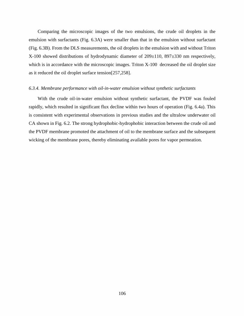
106
Comparing the microscopic images of the two emulsions, the crude oil droplets in the
emulsion with surfactants (Fig. 6.3A) were smaller than that in the emulsion without surfactant
(Fig. 6.3B). From the DLS measurements, the oil droplets in the emulsion with and without Triton
X-100 showed distributions of hydrodynamic diameter of 209±110, 897±330 nm respectively,
which is in accordance with the microscopic images. Triton X-100 decreased the oil droplet size
as it reduced the oil droplet surface tension[257,258].
6.3.4. Membrane performance with oil-in-water emulsion without synthetic surfactants
With the crude oil-in-water emulsion without synthetic surfactant, the PVDF was fouled
rapidly, which resulted in significant flux decline within two hours of operation (Fig. 6.4a). This
is consistent with experimental observations in previous studies and the ultralow underwater oil
CA shown in Fig. 6.2. The strong hydrophobic-hydrophobic interaction between the crude oil and
the PVDF membrane promoted the attachment of oil to the membrane surface and the subsequent
wicking of the membrane pores, thereby eliminating available pores for vapor permeation.

107
Figure 0.4 Normalized water fluxes of the PVDF membrane (black squares), the composite
membrane (red circles), and the omniphobic membrane (blue diamonds) over (a) time, and (b)
specific volume of permeate water normalized by the membrane area in DCMD experiments using
prepared crude oil-in-water emulsion without synthetic surfactant as the feed solution. The
normalized fluxes were the measured absolute fluxes normalized by the initial fluxes. The influent
temperatures of the feed and distillate streams were 65 and 20 ºC, respectively. The cross-flow
velocities on the membrane for the feed and distillate streams were 10.7 and 5.4 cm/s, respectively.
The initial fluxes for the PVDF membrane, the composite membrane and the omniphobic
membrane were 35.67, 30.15 and 25.19 LMH, respectively. The salt rejection rates for all
membranes are over 99.9% in all experiments. The inset in (a) is the DCMD result for composite
membrane operating with the same feed water over 20 hours.

108
Similar fouling behavior was also observed for the omniphobic membrane (blue diamonds on
Fig. 6.4). As discussed in section 6.3.2, although the underwater CAs with oil were drastically
different between the omniphobic membrane and the hydrophobic PVDF membrane due to the
presence of reentrant structure in the omniphobic membrane, the low-surface-energy material
comprising the omniphobic membrane was still attractive to oil via the hydrophobic-hydrophobic
interaction underwater. Such an attraction led to the spreading of the oil droplets on the membrane
surface, which eliminated the available pores of the omniphobic membrane for vapor transfer and
thereby led to significant flux decline. The major difference in fouling between the PVDF and the
omniphobic membranes, however, was the permeation of oil into the pores of the PVDF membrane
but not the omniphobic membrane. Evidences in support of such difference include the underwater
oil CA for both membranes reported in section 6.3.2, as well as the force curves measured for the
interaction between an oil droplet and both membranes, which will be discussed in detail in section
6.3.6.
In a stark contrast, the composite membrane was able to deliver a highly stable performance
with negligible flux decline in 20 h (red circles on Fig. 6.4a). The excellent fouling resistance of
the composite membrane was attributable to the presence of the superhydrophilic PFO/CTS
coating which created a hydration layer on the membrane surface. The strong and repulsive
hydration force prevented the oil from attaching onto the surface, thereby keeping the pores open
and available for vapor transfer. On the other hand, the low-surface-energy perfluoro functional
groups in the PFO/CTS coating served to promote the release of any attached oil foulant upon
local hydrodynamic perturbation[252]. Our recent study has shown that the PFO/CTS coating was
more effective in mitigating oil fouling in MD than a pure crosslinked CTS coating which was
superhydrophilic but without low-surface-energy functional groups[249]. Similar improvements
on anti-fouling performance by the integration of low-surface-energy functional groups into
superhydrophilic matrixes have also been observed in other pressurized membrane
processes[86,252,259–261].
For all three membranes, the calculated salt rejection rates were all over 99.9%. We therefore
conclude no membrane wetting occurred with this feed solution containing no synthetic surfactant.
The natural surfactants that stabilized the oil-in-water emulsions did not induce membrane pore
wetting even for PVDF membrane which has been demonstrated to be very susceptible to wetting
by surfactants[44]. It was likely that the naturally occurring surfactants were mostly bound to the

109
oil-water interface and did not exist substantially as unassociated surfactants in the feed water due
to their relatively low concentrations.
6.3.5. Membrane performance with oil-in-water emulsion stabilized by synthetic surfactants
When the crude oil-in-water emulsion stabilized by synthetic surfactants (Triton X-100) was
used as the feed solution in MD, it completely changed the membrane performance. For the PVDF
membrane, very rapid fouling was observed, leading to drastic flux decline within 10 min (Fig.
6.5a). The fouling of the PVDF membrane by feed solution stabilized by Triton X-100 seemed to
be faster than that with feed solution without synthetic surfactants (Fig.6.4). During this fouling
stage, the salt rejection remained near perfect (Fig. 6.5b), implying that the membrane pores were
not yet wicked through by the feed solution. Most interestingly, the fouling stage only lasted for
about 10 min before pore wetting became dominant. Both the increase of permeate flux, which
eventually surpassed the initial flux, as well as the dramatically reduced salt rejection, which
eventually became zero, suggest rapid and severe pore wetting. Previous studies have shown
failure of MD due to membrane pore wetting by unbound anionic surfactants[44,48,49]. Here, the
wetting could be caused by either the surfactants that bound directly to the membrane pores or by
surfactants that bound to the absorbed oil on the surface of the membrane pores, although the exact
contributions are difficult to determine.
The composite membrane, which robustly resisted fouling against crude oil-in-water emulsion
without synthetic surfactant, was highly susceptible to pore wetting when the feed solution
contained Triton X-100 (Fig. 6.5). The absence of flux decline suggests that no fouling occurred.
Therefore, the wetting was attributable to the free surfactants (i.e. unbound to oil droplets) but not
oil-bound surfactants. The unbound surfactants were able to transport across the superhydrophilic
skin layer and absorb onto the underlying hydrophobic PVDF substrates, which rendered the
membrane pores hydrophilic, appreciably reduced the liquid entry pressure, and allowed direct
permeation of the saline feed water through the pores. The failure of the composite membrane in
MD performance was similar to that of the PVDF membrane in both the degree and the kinetics
of wetting: the salt rejection rate decreased to almost zero in just about half an hour (Fig. 6.5b).
The major difference was the absence of the fouling stage for the composite membrane with a
superhydrophilic surface.

110
Figure 0.5 (a) Normalized water fluxes for the PVDF membrane (black squares), the composite
membrane (blue diamonds), and the omniphobic membrane (red circles) over time in DCMD
experiments using high-surfactant-content crude oil-in-water emulsion as feed. The experimental
conditions were the same as those described in Fig. 6.4. The initial fluxes for the PVDF membrane,
composite membrane and omniphobic membrane were 36.15, 30.76 and 24.46 L/(m2hr1),
respectively. (b) Corresponding salt rejection rates for the PVDF membrane (black squares), the
composite membrane (blue diamonds), and the omniphobic membrane (red circles) over time in
the DCMD experiments. The insets in both (a) and (b) show the DCMD results for the omniphobic
membrane operating with the same feed water over a long time period.
In comparison, the omniphobic membrane exhibited excellent anti-fouling and anti-wetting
performance in DCMD experiments using crude oil-in-water emulsion stabilized by synthetic
surfactants as the feed solution: no flux decline or decrease in salt rejection rate was observed over
20 h of operation (Fig. 6.5), which suggested the absence of fouling or wetting. Previous studies

111
have shown outstanding anti-wetting performance of omniphobic membranes when feed solution
was dosed with free surfactants[44,48,49]. It has been suggested that the combination of re-entrant
structure and low surface energy sustains a metastable Cassie-Baxter state for the contact between
an omniphobic membrane with low surface tension liquids. With the exact same mechanism, the
unbound surfactants in the feed solution did not induce pore wetting of the omniphobic membrane
in our experiments. A recent study also showed that an omniphobic membrane delivered
significantly more stable performance than a PVDF membrane when being used to desalinate real
shale gas wastewater[50].
The omniphobic membrane performed very differently with different feed solutions: we have
already shown, in section 6.3.4, that crude oil-in-water emulsion without synthetic surfactants
readily fouled the omniphobic membrane; however, when the emulsion was stabilized by synthetic
surfactants, Triton X-100, no fouling was observed over 20 h of MD operation (Fig. 6.5). In an
abundance of surfactants in the emulsion, the crude oil droplets were “wrapped” by the surface-
adsorbed surfactants and thereby became ‘hydrophilic’. The hydrophilic oil droplets were not able
to attach onto the membrane surface and block the membrane pores, because not only the
hydrophobic-hydrophobic attraction was eliminated but the oil droplets were also stabilized by a
hydration shell due to the strong affinity between the hydrophilic heads of the surfactants and the
surrounding water.
6.3.6. Force spectroscopy to elucidate oil-membrane interaction
To acquire fundamental understandings of the interaction between oil and membranes with
different special wetting properties, underwater force spectroscopy was conducted for each
membrane using an oil droplet as the force probe. The force curves from the force spectroscopy
are shown in Fig. 6.6, showing a unique behavior for the interaction between each membrane with
the oil droplet probe. In each measurement, the oil droplet probe first approached the membrane
and retracted immediately after contacting the membrane, both at the same speed. Due to oil-
membrane adhesion, the retraction stretched the oil droplet and eventually split the droplet into
two portions with one remaining on the probe and the other retained by the membrane surface.
Physically, there should be no baseline adhesion force before the oil droplet contacted the
membrane and after the split of the oil droplet, as there was no oil-membrane interaction in both
cases. However, the measured “pseudo-adhesion-forces”, which were essentially the force exerted

112
on the force sensor of the tensiometer, could be different for the two baselines in the receding stage
due to the reduction of the oil droplet volume as a result of the split. The decrease of the oil droplet
volume caused the simultaneous decrease of the flotation force, gravitational force, and viscous
force (for constant-speed movement), which collectively led to a net positive pseudo-adhesion-
force compared to the baseline before splitting. Such a baseline difference represents the amount
of oil droplet retained by the membrane and thus complements the actual adhesion force in
assessing the oil-membrane interaction.
Figure 0.6 Force curves for the interactions between the PVDF membrane (black curve), the
composite membrane (red curve), and the omniphobic membrane (blue and green curves) and a
mineral oil droplet. The force curves display the forces measured by the tensiometer sensor as a
function of probe displacement. All force curves were collected for the oil-membrane interaction
under DI water, with the exception for the one that was collected in a Triton-X solution (the green
curve). The initial position of the oil droplet and the initial force applied on the oil droplet were
both set as zero.
For the PVDF membrane, upon the physical contact between the oil droplet and the membrane
surface, there was a drastic increase in the adhesion force. This strong attraction between the PVDF
membrane surface and the approaching oil droplet is attributable to the strong and attractive
hydrophobic-hydrophobic interaction[241]. The underwater adhesion force between the oil droplet
and the PVDF membranes (~200 μN) was significantly stronger than that of the composite
membrane and the omniphobic membrane. In addition, a large fraction of oil was retained by the
PVDF membrane, which reduced the volume of the oil droplet probe that translated to a net force
of ~130 μN for the new baseline in the receding stage.

113
The oil droplet interacted with the composite membrane in a drastically different manner
compared to that with the PVDF membrane. First of all, no strong adhesion force was detected
upon the contact between the oil droplet and the superhydrophilic surface of the composite
membrane. On the contrary, a small repulsion (reflected as negative adhesion force) was observed,
most likely due to the repulsive hydration force. Stronger repulsion was likely to be observed if
we further pressed the oil droplet probe against the membrane surface. However, we intentionally
chose to retract immediately right after the oil-membrane contact because the ring-shaped
tensiometer probe (to which the oil droplet was hung) could be forced into oil droplet and generate
information irrelevant to the actual oil-membrane interaction. Furthermore, the pseudo-adhesion
force for the baseline in the receding stage was very low compared to that with the PVDF
membrane, suggesting little retention of oil on the membrane surface due to the very weak
adhesion between the oil and the superhydrophilic surface of membrane.
The interactions between the oil droplet and the omniphobic membrane were the most
interesting. For the interaction in DI water, similar to the interaction with PVDF membrane, a
sudden increase in adhesion force was observed at the end of the advancing stage due to the
hydrophobic-hydrophobic interaction. This is consistent with the results of our fouling
experiments which showed similar fouling behaviors for both the hydrophobic PVDF membrane
and the omniphobic membrane. However, the fraction of oil retained by the omniphobic membrane
was significantly lower than that by the PVDF membrane, as indicated by the very low pseudo-
adhesion force measured in the baseline of the receding stage. In fact, the fraction of oil droplet
retained by omniphobic membrane was even similar to that retained by the composite membrane
with a superhydrophilic surface, even though the two surfaces have drastically different upon-
contact interactions with the oil droplet. In this regard, the omniphobic membrane can be
considered “attractive but not adhesive” to oil, which is consistent with the observation in the CA
measurements that the oil droplet spread reasonably well on the omniphobic membrane surface
but did not penetrate into the membrane pores because of the reentrant structure.
The presence of the excess synthetic surfactants, Triton X-100, in the solution where the force
curve was measured, drastically changed the oil-membrane interaction. The force curve for the
interaction of the oil droplet with the omniphobic membrane in the presence of the surfactants was
similar to that with the composite membrane in DI water. The observation of the oil-membrane
interaction in the presence of surfactants was very interesting but not particularly surprising. As

114
discussed in section 6.3.5, the surfactants ‘wrapped’ the oil droplets adsorbing onto the oil-water
interface and rendered the oil droplets hydrophilic. Consequently, the attractive oil-membrane
interaction was significantly reduced due to hydration force resulting from strong hydration of the
hydrophilic heads of the adsorbed surfactants. Similar behavior of fouling mitigation has also been
observed in a previous study in which Pickering emulsion stabilized by nanoparticles showed
appreciably lower membrane fouling propensity in ultrafiltration compared to an emulsion
stabilized simply by naturally occurring surfactants[148].
6.4. Conclusion
To successfully utilize MD to desalinate challenging saline wastewater with a complex
composition, such as shale oil/gas produced wastewater or industrial brine, the selection of proper
MD membrane is of vital importance. With the recent innovations in membrane materials, three
types of membranes are currently available for MD operations, including conventional
hydrophobic membranes, composite membranes with a superhydrophilic surface and a
hydrophobic substrate, and omniphobic membranes. This study employs CA measurements,
DCMD fouling and wetting experiments, as well as oil-probe force spectroscopy to systematically
investigate the performance of different MD membranes for desalinating saline oil-water
emulsions.
When the oil emulsion was stabilized only by a limited amount of naturally occurring
surfactants, severe fouling was observed for both the hydrophobic and the omniphobic membranes
due to the very low surface energy of these membranes that resulted in the attractive hydrophobic-
hydrophobic interaction. However, the omniphobic membrane differed from the hydrophobic
membrane in that the oil droplets could not penetrate into the pores of the omniphobic membrane
due to the reentrant structure. The appropriate membrane to be employed for such a feed solution
is the composite membrane with a superhydrophilic surface that has been shown to be capable of
mitigating oil fouling and sustaining robust MD operation.
For feed solution containing crude oil droplets stabilized by excess synthetic surfactants, the
membrane performance became dramatically different. Both the composite membrane and the
hydrophobic PVDF membrane were eventually completely wetted by the excess surfactants,
leading to near-zero salt rejection. While no fouling of the composite membrane was observed, the
PVDF membrane was rapidly fouled first before wetting occurred. The omniphobic membrane,

115
on the other hand, was able to resist both fouling and wetting with this feed solution because the
omniphobic membrane was intrinsically anti-wetting (as shown in previous studies) and the
emulsified oil droplets became hydrophilic due to adsorbed surfactants on the oil-water interface.
Acknowledgement
The authors acknowledge the financial support from Bureau of Reclamation, Department of
Interior, via DWPR Agreement R15AC00088.

116
CHAPTER 7
CHAPTER 7 CONCLUSIONS AND FUTURE WORK CHAPTER 8
Membrane fouling and wetting are two prominent challenges limiting the wide application
of membrane distillation (MD) in industry. In this dissertation, the mechanisms and mitigation
strategies of membrane fouling and wetting in MD have been investigated. For the membrane
fouling problem, it has been long known that the conventional hydrophobic membrane is prone to
fouling by hydrophobic contaminants (e.g. oils) due to the strong hydrophobic-hydrophobic
interaction underwater. In this dissertation, we utilized a composite membrane with an in-air
superhydrophilic/underwater oleophobic surface layer and a hydrophobic substrate in MD
operation with oily feed and found that the composite membrane can largely mitigate oil fouling
problem because of the elimination of hydrophobic-hydrophobic interaction. In addition, we
revealed that besides surface wetting property, the surface charge is also able to influence the
fouling resistance of the MD membrane. For membrane wetting problem, despite of the efforts
spent on mitigation strategy, the fundamental mechanism still remains unclear. To unveil the
mechanism of membrane pore wetting, we firstly developed a novel impedance-based technique
that is able to monitor the membrane wetting pore process in MD operations and then exploited
this technique to study the membrane pore wettings induced by two different wetting agents,
ethanol (a water-miscible and low-surface-tension liquid) and Triton X-100 (amphiphilic
molecules). The results indicate that membrane pore wettings resulted from these two different
wetting agents share the same wetting criterion but exhibit distinctive kinetics, and the difference
in wetting dynamics stems from the adsorption of Triton X-100 onto the membrane pore surface.
Furthermore, we developed a mathematical model for surfactant-induced membrane pore wetting
process that can quantitatively predict the pore wetting breakthrough time regarding the MD
operation conditions and surfactant properties. After all the studies on the mechanisms of
membrane failures and mitigation strategies, we compared the performances of MD membranes
with different wettability in MD operations with different types of feed, and developed a general
membrane selection strategy for sustaining robust MD operations depending on feed type. A
detailed summary of the conclusions from the main chapters of this dissertation is provided in the
following:

117
Chapter 2. Inspired by the development of surface materials with special wettability in material
science community, a composite membrane with an in-air superhydrophilic/underwater
oleophobic surface layer and a hydrophobic substrate has been fabricated, characterized, and
employed in direct contact membrane distillation (DCMD) operation with crude oil-in-water
emulsion as feed. Compared with the conventional hydrophobic MD membrane, the composite
MD membrane exhibited excellent fouling resistance in the DCMD operation. The oil probe force
spectroscopy acquired with a force tensiometer indicates that the excellent fouling resistance of
the composite membrane can be attributed to the elimination of the strong hydrophobic-
hydrophobic interaction between the hydrophobic membrane surface and the oil droplet
underwater. The results from this chapter suggest that composite membranes with asymmetric
wettability can enable MD to desalinate hypersaline wastewater with high concentrations of
hydrophobic contaminants.
Chapters 3. The efforts of developing anti-fouling membranes have so far been focusing on
tailoring membrane surface wetting properties, whereas the impacts of surface charge are often
eclipsed. In this chapter, we investigated the synergistic impact of surface wetting property and
surface charge on anti-fouling property of the MD membrane. Two composite membranes with
different surface charges were fabricated. The fabricated composite membranes and the pristine
hydrophobic membrane were firstly characterized using contact angle measurements, and then
tested in DCMD operations with crude oil-in-water emulsion as feed, respectively. It was found
that the negatively charged composite membrane performed the best in mitigating membrane
fouling by the negatively charged oil emulsion, followed by the positively charged composite
membrane, with the pristine hydrophobic membrane being the most susceptible to oil fouling in
MD operations. Oil probe force spectroscopy has been conducted for each membrane, and the
results corroborated with the contact angle measurement results to unveil the anti-fouling
mechanism associated with surface charge.
Chapter 4. Pore wetting is an important failure mechanism unique to MD. While it is well known
that both low-surface-tension and water miscible liquids, such as alcohols, and amphiphilic
molecules, such as surfactants, are effective wetting agents, the detailed mechanisms for these
agents to induce pore wetting remain unclear. In particular, the role of surface adsorption in
surfactant-induced wetting remains to be elucidated. To obtain the fundamental insights to

118
understanding the mechanism of pore wetting, we developed a novel and simple method, based on
measurement of cross-membrane impedance, for monitoring the dynamics of membrane pore
wetting and enabling early detection of imminent wetting-based membrane failure. Using this
wetting monitoring method, we experimentally probed the kinetics of wetting frontier propagation
induced by different wetting agents including ethanol and Triton X-100 surfactant. We
demonstrated that ethanol-induced wetting is instantaneous whereas surfactant-induced wetting is
dynamic with its kinetic rate dependent on several critical factors. We also developed a theoretical
model to qualitatively explain the important features experimentally observed in surfactant-
induced membrane pore wetting. Specifically, it was found that the kinetics of surfactant-induced
wetting strongly depends on the vapor flux and the bulk concentration of surfactants in the feed
solution, but surprisingly not on the transmembrane hydraulic pressure difference. The results from
this chapter also suggest that while the presence of surfactants promotes wetting, adsorption of
surfactants onto pore surface actually deters pore wetting instead of promoting it by rendering the
surface hydrophilic.
Chapter 5. To enrich the understanding of the mechanism of surfactant-induced pore wetting in
MD, in this chapter, we developed a mathematical model, based on surfactant transport in a
partially wetted membrane pore under the pseudo-steady state assumption, to quantify the kinetics
of pore wetting. The developed model predicted several key dependences of wetting kinetics on
operating parameters and solution properties, which were highly consistent with results from MD
experiments using feed solution containing sodium dodecyl sulfate. It was found that kinetics of
pore wetting is strongly dependent on vapor flux, surfactant concentration, but relatively
independent of the transmembrane hydraulic pressure. The critical surfactant concentration below
which pore wetting does not occur was also predicted by the wetting model. In addition, the wetting
propensity of different surfactant species was investigated, and the membrane pore wetting
predictions from the developed model matched the experimental results very well, which further
validated the developed model. The results indicate that for membrane wettings induced by
different surfactants, the surface excess concentration is the most important surfactant property in
affecting the pore wetting kinetics. Other parameters of a surfactant, such as molecular charge and
hydrophilic-lipophilic balance, have no observable direct impact on the wetting kinetics.

119
Chapter 6. In this chapter, we systematically investigated the impact of the surface wetting
properties on the membrane wetting and/or fouling behaviors in MD. Specifically, we compared
the wetting and fouling resistance of three types of membranes of different wetting properties,
including hydrophobic and omniphobic membranes as well as composite membranes with a
hydrophobic substrate and a superhydrophilic top surface. We challenged the MD membranes with
hypersaline feed that contained a relatively high concentration of crude oil with and without added
synthetic surfactant, Triton X-100. We found that the composite membranes with superhydrophilic
top surface were robustly resistant to oil fouling in the absence of Triton X-100, but were subject
to pore wetting in the presence of Triton X-100. On the other hand, the omniphobic membranes
were easily fouled by oil-in-water emulsion without Triton X-100, but successfully sustained
stable MD performance with Triton X-100 stabilized oil-in-water emulsion as the feed solution. In
contrast, the conventional hydrophobic membranes failed readily regardless whether Triton X-100
was present, although via different mechanisms. These findings are corroborated by contact angle
measures as well as oil-probe force spectroscopy. This chapter provides a holistic picture regarding
how a hydrophobic membrane fails in MD and how we can leverage membranes with special
wettability to prevent membrane failure in MD operations.
Future Work. This dissertation can be further extended from the following perspectives:
The composite MD membrane should be further tested with real hypersaline wastewater
rich of hydrophobic contaminants (e.g. shale oil/gas produced water) as feed to confirm its
practical viability in field work.
Besides surface wettability and surface charge, other surface properties of MD membrane
can be further investigated to construct MD membranes with more robust fouling
resistance.
The mechanism of membrane pore wetting induced by inorganic fouling (e.g. salt scaling)
still remains unknown and needs to be explored.
A novel MD membrane that has simultaneous fouling and wetting resistances is needed.

120
APPENDIX A
SUPPLEMENTARY MATERIALS FOR CHAPTER 2
Membrane Porosity Quantification
The porosity of the membrane was defined as the volume of the pores divided by the total volume
of the membrane. The flat-sheet membrane porosity ε was determined by gravimetric method and
calculated by the following equation[262]:
1 2
L
m m
A l
( A 1 )
where m1 is the weight of the wet membrane, m2 is the weight of the dry membrane, L is the
liquid density, A is the effective area of the membrane and l is the membrane thickness. The liquid
used for porosity measurement named Porefil was supported by IB-FT GmbH (Germany) and its
surface tension and density were 16 dyn/cm and 1.87 g/ml, respectively.
Pore Size Distribution
The pore size distribution of the membrane was investigated by using a Capillary Flow
Porometer, CFP (Porolux 1000, IB-FT GmbH, Germany). The membrane sample was fully wetted
with the Porefil, and then the measurement was carried out following the procedure described in
literature[121]. The CFP was fully automated to generate the pore size distribution following the
cited method.
Liquid Entry Pressure
The liquid entry pressure (LEP) of water was obtained using the method described by Smolders
and Franken[122]. The membrane sample with effective area of 2.69 cm2 was assembled into a
test membrane cell filled with distilled water. Hydraulic pressure was gradually increased by
regulating the outlet pressure of the nitrogen cylinder. The minimum applied pressure at which a
continuous liquid flow was observed was recorded as the LEP.

121
Chemical Evidence for Surface Modification
Figure A.1 Polarization Modulation-Infrared Reflection-Adsorption Spectroscopy (PM-IRRAS)
for the PVDF and the modified surface of the composite membrane. The major difference between
the two spectra is the presence of additional bonds such as Si-O, C-N, and O-H that are
characteristic of the nanocomposite (silica nanoparticles/chitosan/PFO) employed for surface
modification.[263–266]

122
APPENDIX B
SUPPLEMENTARY MATERIAL FOR CHAPTER 3
Figure B.1 Pore size distributions of PVDF membrane (black squares), PDA/SiNPs-PVDF
composite membrane (red circles) and PDDA/SiNPs-PVDF composite membrane (blue triangles).
The distributions are bimodal in all cases.
Figure B.0.2 Infrared absorbance spectra obtained from PMIRRAS for the PVDF membrane
(black curve), PDA/SiNPs-PVDF composite membrane (red curve) and PDDA/SiNPs-PVDF
composite membrane (blue curve). In the spectrum of PDA/SiNPs-PVDF composite membrane,
the peak at 1617 cm-1 resulted from C=C resonance vibration in the aromatic ring and N-H
bending; the peak at 1529 cm-1 was attributed to the N-H shearing vibration and N-O stretching;
the peak at 1419 cm-1 was attributable to phenolic O-H deformation and phenolic C-O stretching

123
[267,268]. In the spectrum of PDDA/SiNPs-PVDF composite membrane, the peaks at 1614 cm-1
was owing to the deformation vibration of N-H; the peaks at 1446 cm-1 and 2918 cm-1 were resulted
from C-H bending; the peak at 3327 cm-1 was attributed to a deformation vibration of N-H and a
stretching vibration of –NR3+

124
APPENDIX C
SUPPLEMENTARY MATERIAL FOR CHAPTER 4
Selection of Frequency for Impedance Measurement
The selection of a specific frequency for measuring the time-resolved impedance across the MD
membrane follows two basic principles. First, the phase should not be close to zero so that
capacitive contribution becomes insignificant. Second, the initial overall impedance should be
reasonably high before any wetting occurs so that the measurement of the impedance change can
be more accurate. Following these rules, we conducted an EIS measurement in the frequency range
of 103 to 106 Hz and summarized the dependence impedance and phase on frequency in a Bode
plot (Figure C.1). The measurement was conducted in an operating DCMD system with surfactant-
free saline feed solution. Based on these spectra, we chose a working frequency of 105 Hz. Other
medium frequencies should also work, as long as they are not too high in which case the impedance
is too low, or not low in which case the phase is too high (close to 0).
Figure C.1 Impedance (red squares, left Y-axis) and phase angle (blue circles, right Y-axis) spectra
of a PVDF membrane in DCMD operation with 0.6 M NaCl solution on feed side and DI water on
permeate side.

125
Justification of using transmembrane hydraulic pressure instead of feed hydraulic pressure
for wetting analysis
In this analysis, we present the proof that the transmembrane hydraulic pressure, ∆𝑃, instead of
the hydraulic pressure of the feed stream, should be used to compared LEP as a criterion for pore
wetting. Such a proof was first given by Zmievskii[207] and is simply summarized here. We start
with conducting a force balance analysis on the water and air trapped in an ideal cylindrical pore
between the feed solution and the distillate (Figure C.2).
Figure C.2 Schematic of membrane pore wetting in DCMD. 𝑃𝑓 and 𝑃𝑝 are the applied hydraulic
pressures on the feed and permeate sides, respectively. 𝑃𝑎𝑖𝑟 is the pressure of the trapped air in the
membrane pore. 𝛾𝑓 and 𝛾𝑝 are the surface tensions of the feed and permeate solutions,
respectively. 𝜃𝑓 and 𝜃𝑝 are the contact angles of the feed and permeate solutions on the PVDF
membrane surface, respectively.
Let us assume that the hydraulic pressure on the feed side, 𝑃𝑓 , is higher than that on the
distillate side, 𝑃𝑝 , so that ∆𝑃 is always positive. The force balance for the feed solution and
distillate within the pore can be described by the two following equations:
𝑃𝑓𝜋𝑅2 = 𝑃𝑎𝑖𝑟𝜋𝑅2 + (− cos 𝜃𝑓)𝛾𝑓2𝜋𝑅 (C1)
𝑃𝑝𝜋𝑅2 = 𝑃𝑎𝑖𝑟𝜋𝑅2 + (− 𝑐𝑜𝑠 𝜃𝑝)𝛾𝑝2𝜋𝑅 (C2)
where 𝑅 is the radius of the membrane pore; 𝑃𝑎𝑖𝑟 is the pressure of air trapped in the
membrane pore; 𝜃𝑓 and 𝜃𝑝 are the intrinsic contact angles of the feed solution and the distillate on
the membrane material, respectively; 𝛾𝑓 and 𝛾𝑝 are the surface tensions of the feed solution and
distillate, respectively.

126
When feed solution has not yet intruded into the membrane pore, increasing 𝑃𝑓 first increases
𝜃𝑓. But 𝑃𝑎𝑖𝑟 remains constant because the increasing curvature of feed/air interface barely changes
the volume of the air trapped in the pore. Based on equation C1, the increased hydraulic pressure
on the feed solution is balanced by the increased interfacial force caused by an increased 𝜃𝑓. As
𝑃𝑓 further increases, 𝜃𝑓 will reach a maximum, 𝜃𝑓,𝑚𝑎𝑥, that is determined by the surface tensions
of the feed solution and membrane material. Further increasing 𝑃𝑓 will lead to intrusion of the feed
solution into the pore, which will in turn compress the air trapped in the pore and increase its
pressure.
Once the air pressure starts to increases, force balance on the distillate (equation C2) dictates
that 𝜃𝑝 decreases because 𝑃𝑎𝑖𝑟 increases yet 𝑃𝑝 remains constant. As 𝑃𝑎𝑖𝑟 continues to increase,
𝜃𝑝 will become less than zero and an air bubble will emerge in the distillate until 𝜃𝑝 reaches −90°.
Further increase of 𝑃𝑎𝑖𝑟 will cause detachment of air bubble, which will result in destabilization of
the system and complete pore wetting. At this critical point of pore wetting, equations C1 and C2
can be combined to obtain equation C3, with 𝜃𝑓 = 𝜃𝑓,𝑚𝑎𝑥 and 𝜃𝑝 = −90°:
(𝑃𝑓 − 𝑃𝑝)𝜋𝑅2 = (− cos 𝜃𝑓,𝑚𝑎𝑥)𝛾𝑓 ∙ 2𝜋𝑅 (C3)
Rearranging equation C3 yields equation C4:
𝛥𝑃 = 𝑃𝑓 − 𝑃𝑝 =−2𝛾𝑓
𝑅cos 𝜃𝑓,𝑚𝑎𝑥 = 𝐿𝐸𝑃 (C4)
Equation C4 suggests that at the critical point of pore wetting, LEP should be equal to the
transmembrane hydraulic pressure difference, 𝛥𝑃, not the feed hydraulic pressure, 𝑃𝑓. Therefore,
the criterion for pore wetting is that 𝛥𝑃 > 𝐿𝐸𝑃.
Derivation of Equation 4.3
According to Poiseuille equation[269,270],
∆𝑃 − 𝐿𝐸𝑃 =8𝜇𝑙𝑄
𝜋𝑟4 (C5)
where ∆𝑃 is the applied transmembrane pressure, 𝐿𝐸𝑃 is the liquid entry pressure of the
membrane pore, 𝑙 is the distance between the wetting frontier and the pore entrance, 𝜇 is the
dynamic viscosity, 𝑄 is the volumetric flow rate (i.e. 𝑄 = π𝑟2 𝑑𝑙
𝑑𝑡), 𝑟 is the pore radius.
Rearranging equation C5 gives

127
𝑙𝑑𝑙 =8(∆𝑃 − 𝐿𝐸𝑃)𝜇
𝑟2𝑑𝑡 (C6)
Integrating equation C6 from 𝑙 = 0 to 𝑙, and 𝑡 = 0 to 𝑡, we can obtain,
𝑙2
2=
(∆𝑃 − 𝐿𝐸𝑃)𝑟2
8𝜇𝑡 (C7)
We can get equation C8, which is equation 4.3 in the main text, from rearranging equation C7
𝑙(𝑡) = √𝑅2
4𝜇(∆𝑃 − 𝐿𝐸𝑃)𝑡 (C8)
In our experiments, 𝑟 = 0.225 𝜇𝑚, 𝜇 = 0.47 𝑚𝑃𝑎 ∙ 𝑠, for ∆𝑃 − 𝐿𝐸𝑃 = 0.25 𝑘𝑃𝑎, which is
a very small pressure, the time 𝑡 it takes for the liquid to penetrate through the entire membrane
(𝑙(𝑡) = 𝑑0, where the membrane thickness 𝑑0 = 180 𝜇𝑚) is calculated to be 1.22 𝑠.
Partial Differential Equation for Surfactant Transport
The partial differential equation regarding the diffusion-convection-adsorption of surfactant within
a partially wetted pore[209],
𝜕𝑐
𝜕𝑡+
2
𝑟
𝜕𝜏
𝜕𝑡= 𝐷
𝜕2𝑐
𝜕𝑥2+ 𝐷𝑠𝑙
2
𝑟
𝜕2𝑐
𝜕𝑥2− (
𝐽
휀+ 𝑣𝑠)
𝜕𝑐
𝜕𝑥 (C9)
Where 𝑐 (abbreviation for 𝑐(𝑥, 𝑡)) is the concentration of surfactants at position 𝑥, and time
𝑡, 𝜏 is the surface concentration of the surfactant on the pore surface, 𝑅 is the pore radius, 𝐷 is the
diffusion coefficient of surfactants in the solution, 𝐷𝑠𝑙 is the surface diffusion coefficient over the
filled portion of the pore, 𝐽 is the vapor flux, 휀 is the porosity of the membrane, 𝑣𝑠 is the moving
speed of the solution in the pore (𝑣𝑠 ≪𝐽
𝜀 in our experiments). The initial conditions for the equation
is 𝑐 = 0 except for 𝑐(0,0) = 𝑐0, where 𝑐0 is the surfactant concentration in the bulk solution. The
left-hand-side boundary condition is 𝑐(0, 𝑡) = 𝑐0, and the right-hand-side boundary condition is
𝑐(𝑙, 𝑡) = 𝑐′, where 𝑐′ is the critical concentration of surfactant that satisfies 𝐿𝐸𝑃 = ∆𝑃.
Relative Insignificance of Axial Diffusion
According to literatures[212,271,272], 𝐷 = 4 × 10⁻⁷𝑐𝑚2𝑠−1, and the adsorption occurs so fast
that the pore surface would be saturated immediately as the solution is in contact with the pore

128
surface (𝜕𝜏
𝜕𝑡= 0 for 𝑐 ≠0). We can solve a specific case of the PDE at steady state with 𝐽 = 14 L
m-2 h-1 (relatively low), 𝑐0 = 0.08 𝑚𝑀, 𝑐∗ = 0 (the extreme case), and 𝑙 = 30 𝜇𝑚 (less than 20%
of the membrane thickness). The result is plotted in Figure C.3. In this specific case, the diffusion
term at the pore entrance, 𝐷𝑑𝑐
𝑑𝑥|
𝑥=0, is only 3.22% of the convection term, 𝐽𝑐0. This suggests that
except for the beginning of the wetting when 𝑙 is very short, axial diffusion is relatively
insignificant compared to convection for surfactant transport, and can thus be numerically ignored.
When a higher 𝐽 or/and 𝑐∗ , the axial diffusion at the pore entrance will become even more
negligible compared to convection.
Figure C.3 The concentration profile of Triton X-100 in the solution for 𝑙 = 30 𝜇𝑚. In this specific
case, the bulk concentration 𝑐0 = 0.08 𝑚𝑀 , the critical concentration 𝑐∗ = 0 , the diffusion
coefficient 𝐷 = 4 × 10⁻⁷𝑐𝑚2𝑠−1, and the vapor flux with 𝐽 = 14 L m-2 h-1.
Derivation of Equation 4.8
As the membrane is equivalent to a parallel circuit of a resistor and a capacitor and at a high
frequency (100 kHz), the transmembrane impedance is approximately equal to the impedance of
the capacitor. The capacitance of the capacitor 𝐶 can be calculated as
𝐶 =휀𝑝𝐴
𝑑 (C10)

129
Where, 휀𝑝 is the permittivity of the air, 𝐴 is the area of the air, 𝑑 is the distance between the
two air-liquid interfaces across the membrane. Following the equation of capacitor impedance
calculation, the transmembrane impedance 𝑍 can be obtained with the frequency 𝜔 and imaginary
unit 𝑖
𝑍 =1
𝑖𝜔𝐶 (C11)
The distance 𝑑 can be expressed using the membrane thickness 𝑑0, and the position of the
wetting frontier at time 𝑡, 𝑙(𝑡):
𝑑 = 𝑑0 − 𝑙(𝑡) (C12)
Combining equations C10 to C12 yields
𝑍 =𝑑0 − 𝑙(𝑡)
𝑖𝜔휀𝑝𝐴 (C13)
Normalizing 𝑍 with 𝑍0 yields
𝑍
𝑍0=
𝑑0 − 𝑙(𝑡)
𝑑0 (C14)
which is equation 4.8 in the main text.
Surface Tension Measurements
The results of surface tension measurement using pedant drop method are shown in Figure
C.4. Figure C.4 suggests that both the 20 wt% ethanol salty solution (red circles) and 50 ppm
Triton X-100 salty solution have a surface tension of ~35 mN/m. Therefore, in our wetting
experiments to compare the wetting behaviors of ethanol and Triton X-100, 20 wt% ethanol salty
solution and 50 ppm Triton X-100 salty solution were chosen as the feed solutions, respectively.

130
Figure C.4 The surface tensions of 0.6 M NaCl aqueous solutions with different concentrations of
(a) ethanol (red circles), and (b) Triton X-100 (blue squares) at 60 °C.
Water Flux and Salt Rejection Rate
The normalized water fluxes and salt rejection rate as functions of time in DCMD experiments are
shown in Figure C.5, C.6, C.7, C.8, and C.9. In all DCMD experiments, the baseline normalized
water flux was 1, and the initial salt rejection rate was near 100%. As the membrane became
wetted, some membrane pores were fully wicked through by the feed solution, resulting in a
drastically reduced salt rejection rate and significantly increased normalized water flux. The
increase of normalized water flux could be attributed to the fact that the fully wicked-through
membrane pores became available for convective liquid water flow. As the hydarulic pressure on
the feed side was controlled to be higher than that on the distillate side, the convective water flow
through the wicked-through pores was always from the feed stream to the distillate stream.
Because such a hydraulic pressure driven convective water flux was significantly faster than the
vapor flux driven by partial vapor pressure difference, the total water flux, as a sum of the liquid
flux and vapor flux, increased as more pores became fully wicked through.
Su
rfa
ce
Te
nsio
n(
mN
/m)
30
40
50
60
70
Concentration(ppm)0 20 40 60 80 100
TritonX-100SaltySolution
Surf
aceT
ensio
n(
mN
/m)
30
40
50
60
70
Concentration(wt%)0 5 10 15 20 25
EthanolSaltySolution
(a) (b)

131
Figure C.5 Salt rejection rate (red rectangle, left Y-axis) and normalized water (liquid and vapor)
flux (blue circle, right Y-axis) in DCMD experiments the impedance and salt rejection rate of
which are reported in Figure 4.3 of the main text. The water fluxes are normalized by the initial
water fluxes of the respective measurements. The initial water (only vapor, no liquid) fluxes in (a)
and (b) were 29.48 and 29.95 LMH, respectively.
Figure C.6 Salt rejection rate (red rectangle, left Y-axis) and normalized water (liquid and vapor)
flux (blue circle, right Y-axis) in DCMD experiments the impedance and salt rejection rate of
which are reported in Figure 4.4 of the main text. The water flux is normalized by the initial water
flux. The initial water (only vapor, no liquid) flux was 29 LMH.
Sa
ltR
eje
ctio
nR
ate
(%
)
0
50
100
No
rmaliz
ed
Wa
terF
lux
0
0.5
1
1.5
Time(s)0 500 1,000
11 kPa1 kPa
Salt Rejection Rate
Normalized Water Flux

132
Figure C.7 Salt rejection rate (red rectangle, left Y-axis) and normalized water (liquid and vapor)
flux (blue circle, right Y-axis) in DCMD experiments the impedance and salt rejection rate of
which are reported in Figure 4.5a of the main text. The water fluxes are normalized by the initial
water fluxes of the respective measurements. The initial water (only vapor, no liquid) fluxes in (a)
and (b) were 29.48 and 29 LMH, respectively.

133
Figure C.8 Salt rejection rate (red rectangle, left Y-axis) and normalized water (liquid and vapor)
flux (blue circle, right Y-axis) in DCMD experiments the impedance and salt rejection rate of
which are reported in Figure 4.5b of the main text. The water fluxes are normalized by the initial
water fluxes of the respective measurements. The initial water (only vapor, no liquid) fluxes in (a)
(b), (c), and (d) were 8.60, 14.97, 29.95, and 47.13 LMH, respectively.

134
Figure C.9 Salt rejection rate (red rectangle, left Y-axis) and normalized water (liquid and vapor)
flux (blue circle, right Y-axis) in DCMD experiments the impedance and salt rejection rate of
which are reported in Figure 4.6 of the main text. The water fluxes are normalized by the initial
water fluxes of the respective measurements. The initial water (only vapor, no liquid) fluxes in (a)
and (b) were 28.85, and 29.48 LMH, respectively.

135
APPENDIX D
SUPPLEMENTARY MATERIAL FOR CHAPTERS 5
Derivation of mass balance equation for surfactants (Eq. 5.3).
Eq. 5.3 with a slight modification is shown below and denoted as Eq. 5.3’. Eq. 5.3 in the main text
can be obtained by dividing both sides of Eq. 5.3’ by πR2. Eq. 5.3 or Eq. 5.3’ describes the mass
balance of surfactants in the control volume (CV, including boundary layer and pores). The
definition of each term can be found in the main text and the nomenclature. Here, we provide a
more detailed description of the meaning of each term.
πR2
휀(Jwc0 − D
∂c(x, t)
∂x x=0) dt
=πR2
휀∫ [c(x, t + dt) − c(x, t)]dx
δ𝑏
0
+ {∫ [τ(x, t + dt)2πR(1 − ε) + c(x, t + dt)πR2]dxδ𝑏+l(t+dt)
δ𝑏
− ∫ [τ(x, t)2πR(1 − ε) + c(x, t)πR2]dxδ𝑏+l(t)
δ𝑏
}
(5.3’)
Here we consider a cylindrical pore of radius R and its corresponding portion in the boundary
layer. Because the boundary layer has no solid material and is 100% porous, the area of the
boundary layer corresponding to a pore of an area πR2 is πR2/휀. The first term on the left-hand-
side (LHS), Jwc0πR2/휀, is the convective flux of the surfactants into the CV corresponding to a
pore (including the boundary layer outside the pore). The second term on the LHS is the diffusive
flux of surfactants into the same CV. The first integral on the right-hand-side (RHS) represents the
accumulation of surfactants in the boundary layer from t to t + dt.
The first part of the second integral on the RHS, ∫ τ(x, t + dt)2πR(1 − ε)dxδ𝑏+l(t+dt)
δ𝑏,
quantifies the amount of surfactant on the pore surface at time t + dt. The term (1 − ε) accounts
for the fact that a pore does not have continuous, non-porous wall across the thickness of the
membrane. Rather, we assume the isotropy for the membrane porosity so that the available area of
solid surface in the pore is (1 − ε) of the area calculated using a cylindrical tube (i.e.

136
πR2l(t + dt)). The second part of the second integral on the RHS is the amount of surfactant in
the solution within the pore. The third integral on the RHS is almost exactly the same as the second
integral on the RHS except for a different time point, t + dt (instead of t). Together, the difference
between the second and the third integrals on the RHS represents the accumulation of surfactants
within a pore (not including the boundary layer). The RHS quantifies the accumulation of
surfactants within a CV including both the pore and the boundary layer.
Surfactant adsorption isotherm.
Assuming Langmuir adsorption isotherm for SDS adsorption onto PVDF surface[227,273], the
area density of the adsorbed SDS on PVDF surface, τ, can be expressed as
τ = τmax
Kc
1 + Kc (D1)
where τmax is the maximum area density of SDS on PVDF surface and K is an equilibrium
constant that governs the partition of SDS between the pore surface and the solution phase.
According to literature[227], K can be approximated as 180 mM-1. In our study, the minimum c0
we utilized was over 0.1 mM. According Eq. D1, with K = 180 mM-1and c = 0.1 mM , τ =
0.95τmax. With all other concentrations tested, the pore surface can be considered to be practically
saturated with SDS as long as it is in contact with the solution, in which case we can simplify the
Langmuir adsorption isotherm to a stepwise adsorption isotherm:
τ = {τmax, c > 0
0, c = 0 (D2)
Summary of experimental conditions of DCMD wetting experiments
Table D1. . Summary of experimental conditions of DCMD wetting experiments
Experiment
No.
SDS
concentration
(mM)
Feed
temperature
(°C)
Permeate
temperature
(°C)
Feed velocity
(m/s)
Permeate
velocity (m/s)
(1) 0.85 60 20 0.29 0.18
(2) 0.77 60 20 0.29 0.18
(3) 0.68 60 20 0.29 0.18
(4) 0.60 60 20 0.29 0.18
(5) 0.51 60 20 0.29 0.18
(6) 0.43 60 20 0.29 0.18
(7) 0.34 60 20 0.29 0.18
(8) 0.17 60 20 0.29 0.18

137
(9) 0.43 70 20 0.29 0.18
(10) 0.43 65 20 0.29 0.18
(11) 0.43 55 20 0.29 0.18
(12) 0.43 50 20 0.29 0.18
(13) 0.43 40 20 0.29 0.18
(14) 0.43 35 20 0.29 0.18
(15) 0.43 62 20 0.18 0.18
(16) 0.43 62 20 0.29 0.02
Water fluxes and salt rejection rates data for Fig. 5.2.
Figure D.1 Normalized water fluxes and salt rejection data for DCMD membrane wetting
experiments (experiments 1-9 in Table D1). In each panel, the water vapor fluxes were normalized
by the initial vapor flux. The initial water vapor fluxes were (a) 31.2, (b) 32.7, (c) 32.2, (d) 31.2,
(e) 31.4, (f) 32.7, (g) 31.8, and (h) 33.5 L m-2 h-1, respectively. The transmembrane hydraulic
pressure in all the experiments was 6 kPa. We note that after the membrane was wetted, the
normalized water flux increased over unity. This is because the higher hydraulic pressure of the
feed stream (than that of the distillate stream) drives convective flow of feed solution through the
wetted pores, thereby increasing the total flux.

138
Water fluxes and salt rejection rates data for Fig. 5.3.
Figure D.2 Normalized water fluxes and salt rejection rates data for DCMD membrane wetting
experiments (experiments 6 and 10-14 in Table D1) with initial vapor fluxes of (a) 6.0, (b) 10.3,
(c) 16.5, (d) 23.1, (e) 32.2, (f) 39.2, and (g) 45.8 L m-2 h-1, respectively. In each panel, the water
fluxes were normalized by the initial vapor flux. The SDS concentration of 0.43 mM and the
transmembrane hydraulic pressure of 6 kPa were used in all the experiments. The normalized water
flux increased over unity after wetting occurred, as explained in the caption Fig. D1.
Water fluxes and salt rejection rates data for Fig. 5.4.

139
Figure D.3 Normalized water fluxes and salt rejection rates data for DCMD membrane wetting
experiments (experiments 6, 15 and 16 in Table D1) with SDS concentration of 0.43 mM and
transmembrane hydraulic pressure of (a) 0 kPa, (b) 6 kPa, and (c) 13 kPa, respectively. Normalized
water fluxes and salt rejection rates data for DCMD membrane wetting experiments (experiments
5 and 3 in Appendix C) with transmembrane hydraulic pressure of 6 kPa and SDS concentration
of (d) 0.51 mM and (e) 0.68 mM, respectively. In each panel, the water fluxes were normalized by
the initial vapor flux. The initial vapor fluxes were (a) 31.2, (b) 32.2, (c) 33.5, (d) 31.2 and (e) 32.7
L m-2 h-1, respectively. The normalized water flux increased over unity after wetting occurred, as
explained in the caption Fig. D1.
Surface roughness of the reconstructed PVDF film
Figure D.4 AFM images of the PVDF film reconstructed from porous PVDF membrane. The
measured Ra and Rq were 2.5 and 3.12 nm, respectively. No pore was observed and the roughness

140
is negligibly small. Therefore, CA measured using this film can be considered as an approximation
of the intrinsic CA.
Surface tensions of SDS solutions with different concentrations
Figure D.5 Surface tensions of NaCl solutions (0.6 M) with different concentrations of SDS.
Intrinsic contact angles of NaCl solutions with different concentrations of SDS on a PVDF
surface;
Figure D.6 Intrinsic contact angles of NaCl solutions (0.6 M) with different concentrations of SDS
on PVDF surface.

141
Estimated LEP for NaCl solutions with different SDS concentrations
Figure D.7 Estimated LEP of the PVDF membrane for NaCl solutions with different SDS
concentrations. These values were calculated using Eq. 5.2 based on the surface tensions (Fig. D5)
and intrinsic contact angles (Fig. D6) of NaCl solutions with different SDS concentrations.
Results of Membrane Wetting Experiments
Figure D.8 Salt rejection rate as a function of time in DCMD wetting experiments. Each curve
represents the change of salt rejection rate in a wetting experiment, and the color of the curve
specifies the surfactant species utilized in the experiment. The purple dash line denotes to a salt
rejection rate of 99%, which is defined as the criterion for the breakthrough of the membrane pore

142
by salty feed solution. The breakthrough time is attained as the time at which a curve drops below
the purple dash line.
Model Parameters for Different Surfactant Species
In each model simulation, the water flux Jw was 30.5 L m-2 hr-1, and the bulk concentration for
surfactants was 0.3 mM. The maximum packing density of surfactant on the pore surface τmax
was approximated using the measured surface excess concentration and the coefficients of
determination for surface excess concentrations R2 were also provided. The diffusion coefficient
D of each surfactant was acquired from literatures[225,272,274–278].The critical concentrations
C′ for SDS was estimated using the following equation with an LEP of 6 kPa (i.e. the
transmembrane pressure in the experiments)
LEP = −2𝛾(𝐶′)cos (𝜃(𝐶′))
𝑅
where 𝛾(𝐶′) and 𝜃(𝐶′) are the surface tension of the solution and its contact angle on a PVDF
surface, respectively, both being a function of 𝐶′ (ref [25] in the main text for more details). For
other surfactants, we performed the simulations using 𝐶′ from 0.01 mM (extremely low) to the
CMC of the surfactants from the surface tension measurements. We note that LEP does not change
when 𝐶′ is beyond CMC because 𝛾(𝐶′) and cos (𝜃(𝐶′)) becomes constant when 𝐶′ is beyond
CMC. We find that for all other surfactants except SDS, the choice of 𝐶′ within this range does
not have any appreciable impact on the simulated 𝑡wetting because diffusion is unimportant
compared to convection in the axial transport of these surfactants. A summary of surface excess
concentration, Γ, coefficient of determination R2 (for Γ), critical concentration, 𝐶′ , diffusion
coefficient, 𝐷, and the simulated 𝑡wetting for different surfactant species are shown in Table D2.
Table D2. The parameters for model prediction of pore wetting by different surfactant species
(corresponding to Fig. 5.7 in the main text)
Surfactant
Species
𝛤
(× 10−6 𝑚𝑜𝑙𝑒 𝑚−2)
R2
(for 𝛤 )
C’
(mM) 𝐷
(× 10−10 𝑚2 𝑠−1)
𝑡wetting
(C’=0.01 mM)
𝑡wetting
(C’=CMC)
SDS 5.31 0.977 0.36 7.3 7130 (C’=0.36 mM)
Triton X100 3.50 0.990 0.01-0.2 0.4 890 890
Tween 20 2.96 0.998 0.01-0.05 0.75 700 700
Tween 85 3.62 0.975 0.01-0.05 0.7 860 860
Span 20 4.13 0.989 0.01-0.05 0.12 1060 1060
CTAB 4.31 0.999 0.01-0.05 0.39 1090 1110
SDBS
(0.6 Μ ΝaCl)
2.62 0.945 0.01-0.2 0.2 680 680
SDBS
(0.3 M NaCl)
4.19 0.987 0.01-0.2 0.8 980 990

143
REFERENCES
[1] T. Tong, M. Elimelech, The Global Rise of Zero Liquid Discharge for Wastewater
Management: Drivers, Technologies, and Future Directions, Environ. Sci. Technol. 50
(2016) 6846–6855. doi:10.1021/acs.est.6b01000.
[2] K.B. Gregory, R.D. Vidic, D. a. Dzombak, Water management challenges associated with
the production of shale gas by hydraulic fracturing, Elements. 7 (2011) 181–186.
doi:10.2113/gselements.7.3.181.
[3] S.G. Osborn, A. Vengosh, N.R. Warner, R.B. Jackson, Methane contamination of drinking
water accompanying gas-well drilling and hydraulic fracturing., Proc. Natl. Acad. Sci. U.
S. A. 108 (2011) 8172–8176. doi:10.1073/pnas.1109270108.
[4] EIA, Annual Energy Outlook 2017, Eia. (2017).
[5] IEA, World Energy Outlook 2011 Special Report - Are We Entering a Golden Age of
Gas?, 2011. doi:10.1515/reveh-2014-0002.
[6] R.W. Howarth, A. Ingraffea, T. Engelder, Natural gas: Should fracking stop?, Nature. 477
(2011) 271–275. doi:10.1038/477271a.
[7] L.M. McKenzie, R.Z. Witter, L.S. Newman, J.L. Adgate, Human health risk assessment of
air emissions from development of unconventional natural gas resources, Sci. Total
Environ. 424 (2012) 79–87. doi:10.1016/j.scitotenv.2012.02.018.
[8] R.D. Vidic, S.L. Brantley, J.M. Vandenbossche, D. Yoxtheimer, J.D. Abad, Impact of
shale gas development on regional water quality., Science. 340 (2013) 1235009.
doi:10.1126/science.1235009.
[9] United States Environmental Protection Agency, The Hydraulic Fracturing Water Cycle,
EPA’s Study Hydraul. Fract. Its Potential Impact Drink. Water Resour. (2016) 1–666.
https://www.epa.gov/hfstudy/hydraulic-fracturing-water-
cycle%0Ahttps://www.epa.gov/sites/production/files/2016-
12/documents/hfdwa_executive_summary.pdf.
[10] C. Clark, J. Veil, Produced Water Volumes and Management Practices in the United
States, Argonne Natl. Lab. Rep. (2009) 64. doi:10.2172/1007397.
[11] B.G. Rahm, J.T. Bates, L.R. Bertoia, A.E. Galford, D. a. Yoxtheimer, S.J. Riha,
Wastewater management and Marcellus Shale gas development: Trends, drivers, and
planning implications, J. Environ. Manage. 120 (2013) 105–113.
doi:10.1016/j.jenvman.2013.02.029.
[12] A. Vengosh, R.B. Jackson, N. Warner, T.H. Darrah, A. Kondash, A critical review of the
risks to water resources from unconventional shale gas development and hydraulic
fracturing in the United States, Env. Sci Technol. 48 (2014) 8334–8348.
doi:10.1021/es405118y.

144
[13] D.L. Shaffer, L.H. Arias Chavez, M. Ben-Sasson, S. Romero-Vargas Castrillón, N.Y. Yip,
M. Elimelech, Desalination and reuse of high-salinity shale gas produced water: drivers,
technologies, and future directions., Environ. Sci. Technol. 47 (2013) 9569–83.
doi:10.1021/es401966e.
[14] C. Fritzmann, J. Löwenberg, T. Wintgens, T. Melin, State-of-the-art of reverse osmosis
desalination, Desalination. 216 (2007) 1–76. doi:10.1016/j.desal.2006.12.009.
[15] M. Elimelech, W. a Phillip, The future of seawater desalination: energy, technology, and
the environment., Science. 333 (2011) 712–717. doi:10.1126/science.1200488.
[16] K.W. Lawson, D.R. Lloyd, Membrane distillation, J. Memb. Sci. 124 (1997) 1–25.
doi:10.1016/S0376-7388(96)00236-0.
[17] a. M. Alklaibi, N. Lior, Membrane-distillation desalination: Status and potential,
Desalination. 171 (2005) 111–131. doi:10.1016/j.desal.2004.03.024.
[18] E. Curcio, E. Drioli, Membrane Distillation and Related Operations—A Review, Sep.
Purif. Rev. 34 (2005) 35–86. doi:10.1081/SPM-200054951.
[19] A. Alkhudhiri, N. Darwish, N. Hilal, Membrane distillation: A comprehensive review,
Desalination. 287 (2012) 2–18. doi:10.1016/j.desal.2011.08.027.
[20] C.R. Martinetti, A.E. Childress, T.Y. Cath, High recovery of concentrated RO brines
using forward osmosis and membrane distillation, J. Memb. Sci. 331 (2009) 31–39.
doi:10.1016/j.memsci.2009.01.003.
[21] S. Lin, N.Y. Yip, M. Elimelech, Direct contact membrane distillation with heat recovery:
Thermodynamic insights from module scale modeling, J. Memb. Sci. 453 (2014) 498–
515. doi:10.1016/j.memsci.2013.11.016.
[22] M.S. El-Bourawi, Z. Ding, R. Ma, M. Khayet, A framework for better understanding
membrane distillation separation process, J. Memb. Sci. 285 (2006) 4–29.
doi:10.1016/j.memsci.2006.08.002.
[23] L.D. Tijing, Y.C. Woo, J.S. Choi, S. Lee, S.H. Kim, H.K. Shon, Fouling and its control in
membrane distillation-A review, J. Memb. Sci. 475 (2015) 215–244.
doi:10.1016/j.memsci.2014.09.042.
[24] W. Gao, H. Liang, J. Ma, M. Han, Z. Chen, Z. Han, et al., Membrane fouling control in
ultra fi ltration technology for drinking water production : A review, Desalination. 272
(2011) 1–8. doi:10.1016/j.desal.2011.01.051.
[25] F. Meng, S.R. Chae, A. Drews, M. Kraume, H.S. Shin, F. Yang, Recent advances in
membrane bioreactors (MBRs): Membrane fouling and membrane material, Water Res. 43
(2009) 1489–1512. doi:10.1016/j.watres.2008.12.044.
[26] Q. She, R. Wang, A.G. Fane, C.Y. Tang, Membrane fouling in osmotically driven
membrane processes : A review, J. Memb. Sci. 499 (2016) 201–233.
doi:10.1016/j.memsci.2015.10.040.

145
[27] D.M. Warsinger, J. Swaminathan, E. Guillen-Burrieza, H. a. Arafat, J.H. Lienhard V,
Scaling and fouling in membrane distillation for desalination applications: A review,
Desalination. 356 (2014) 294–313. doi:10.1016/j.desal.2014.06.031.
[28] M. Gryta, Fouling in direct contact membrane distillation process, J. Memb. Sci. 325
(2008) 383–394. doi:10.1016/j.memsci.2008.08.001.
[29] J. Israelachvili, R. Pashley, The hydrophobic interaction is long range, decaying
exponentially with distance., Nature. 300 (1982) 341–342. doi:10.1038/300341a0.
[30] Y. Tsao, D. Evans, H. Wennerstrom, Long-range attractive force between hydrophobic
surfaces observed by atomic force microscopy, Science (80-. ). 262 (1993) 547–550.
doi:10.1126/science.8211182.
[31] E.E. Meyer, K.J. Rosenberg, J. Israelachvili, Recent progress in understanding
hydrophobic interactions., Proc. Natl. Acad. Sci. U. S. A. 103 (2006) 15739–15746.
doi:10.1073/pnas.0606422103.
[32] M. Gryta, M. Tomaszewska, J. Grzechulska, a. W. Morawski, Membrane distillation of
NaCl solution containing natural organic matter, J. Memb. Sci. 181 (2001) 279–287.
doi:10.1016/S0376-7388(00)00582-2.
[33] G. Zuo, R. Wang, Novel membrane surface modification to enhance anti-oil fouling
property for membrane distillation application, J. Memb. Sci. 447 (2013) 26–35.
doi:10.1016/j.memsci.2013.06.053.
[34] A. El-abbassi, A. Ha, M. Khayet, M.C. García-payo, Integrated direct contact membrane
distillation for olive mill wastewater treatment, 323 (2013) 31–38.
doi:10.1016/j.desal.2012.06.014.
[35] L.D. Nghiem, T. Cath, A scaling mitigation approach during direct contact membrane
distillation, Sep. Purif. Technol. 80 (2011) 315–322. doi:10.1016/j.seppur.2011.05.013.
[36] G. Chen, X. Yang, R. Wang, A.G. Fane, Performance enhancement and scaling control
with gas bubbling in direct contact membrane distillation, Desalination. 308 (2013) 47–55.
doi:10.1016/j.desal.2012.07.018.
[37] B.J. Privett, J. Youn, S.A. Hong, J. Lee, J. Han, J.H. Shin, et al., Antibacterial Fluorinated
Silica Colloid Superhydrophobic Surfaces, Langmuir. (2011) 9597–9601.
doi:10.1021/la201801e.
[38] F. He, K.K. Sirkar, J. Gilron, Effects of antiscalants to mitigate membrane scaling by
direct contact membrane distillation, J. Memb. Sci. 345 (2009) 53–58.
doi:10.1016/j.memsci.2009.08.021.
[39] S. Srisurichan, R. Jiraratananon, A.G. Fane, Humic acid fouling in the membrane
distillation process, Desalination. 174 (2005) 63–72. doi:10.1016/j.desal.2004.09.003.
[40] A.C.M. Franken, J.A.M. Nolten, M.H. V Mulder, D. Bargeman, C.A. Smolders, Wetting
criteria for the applicability of membrane distillation, J. Memb. Sci. 33 (1987) 315–328.

146
doi:10.1016/S0376-7388(00)80288-4.
[41] S. Goh, J. Zhang, Y. Liu, A.G. Fane, Fouling and wetting in membrane distillation (MD)
and MD-bioreactor (MDBR) for wastewater reclamation, Desalination. 323 (2013) 39–47.
doi:10.1016/j.desal.2012.12.001.
[42] E. Guillen-Burrieza, M.O. Mavukkandy, M.R. Bilad, H.A. Arafat, Understanding wetting
phenomena in membrane distillation and how operational parameters can affect it, J.
Memb. Sci. 515 (2016) 163–174. doi:10.1016/j.memsci.2016.05.051.
[43] M. Rezaei, D.M. Warsinger, J.H. Lienhard V, W.M. Samhaber, Wetting prevention in
membrane distillation through superhydrophobicity and recharging an air layer on the
membrane surface, J. Memb. Sci. 530 (2017) 42–52. doi:10.1016/j.memsci.2017.02.013.
[44] S. Lin, S. Nejati, C. Boo, Y. Hu, C.O. Osuji, M. Elimelech, Omniphobic Membrane for
Robust Membrane Distillation, Environ. Sci. Technol. Lett. 1 (2014) 443–447.
doi:10.1021/ez500267p.
[45] G. Naidu, S. Jeong, S.J. Kim, I.S. Kim, S. Vigneswaran, Organic fouling behavior in
direct contact membrane distillation, Desalination. 347 (2014) 230–239.
doi:10.1016/j.desal.2014.05.045.
[46] A. Razmjou, E. Arifin, G. Dong, J. Mansouri, V. Chen, Superhydrophobic modification of
TiO2 nanocomposite PVDF membranes for applications in membrane distillation, J.
Memb. Sci. 415–416 (2012) 850–863. doi:10.1016/j.memsci.2012.06.004.
[47] Y. Liao, R. Wang, A.G. Fane, Engineering superhydrophobic surface on poly(vinylidene
fluoride) nanofiber membranes for direct contact membrane distillation, J. Memb. Sci. 440
(2013) 77–87. doi:10.1016/j.memsci.2013.04.006.
[48] C. Boo, J. Lee, M. Elimelech, Engineering Surface Energy and Nanostructure of
Microporous Films for Expanded Membrane Distillation Applications, Environ. Sci.
Technol. 50 (2016) 8112–8119. doi:10.1021/acs.est.6b02316.
[49] J. Lee, C. Boo, W.H. Ryu, A.D. Taylor, M. Elimelech, Development of Omniphobic
Desalination Membranes Using a Charged Electrospun Nanofiber Scaffold, ACS Appl.
Mater. Interfaces. 8 (2016) 11154–11161. doi:10.1021/acsami.6b02419.
[50] C. Boo, J. Lee, M. Elimelech, Omniphobic Polyvinylidene Fluoride (PVDF) Membrane
for Desalination of Shale Gas Produced Water by Membrane Distillation, Environ. Sci.
Technol. 50 (2016) 12275–12282. doi:10.1021/acs.est.6b03882.
[51] Y.C. Woo, Y. Chen, L.D. Tijing, S. Phuntsho, T. He, J.S. Choi, et al., CF4plasma-
modified omniphobic electrospun nanofiber membrane for produced water brine treatment
by membrane distillation, J. Memb. Sci. 529 (2017) 234–242.
doi:10.1016/j.memsci.2017.01.063.
[52] P. Peng, A.G. Fane, X. Li, Desalination by membrane distillation adopting a hydrophilic
membrane, Desalination. 173 (2005) 45–54. doi:10.1016/j.desal.2004.06.208.

147
[53] X. Yang, R. Wang, L. Shi, A.G. Fane, M. Debowski, Performance improvement of PVDF
hollow fiber-based membrane distillation process, J. Memb. Sci. 369 (2011) 437–447.
doi:10.1016/j.memsci.2010.12.020.
[54] D.M. Warsinger, A. Servi, S. Van Belleghem, J. Gonzalez, J. Swaminathan, J. Kharraz, et
al., Combining air recharging and membrane superhydrophobicity for fouling prevention
in membrane distillation, J. Memb. Sci. 505 (2016) 241–252.
doi:10.1016/j.memsci.2016.01.018.
[55] A.B.D. Cassie, S. Baxter, Wettability of porous surfaces, Trans. Faraday Soc. 40 (1944)
546. doi:10.1039/tf9444000546.
[56] J.F. Joanny, P.G. de Gennes, P.G. De Gennes, P.G. de Gennes, P.G. De Gennes, A model
for contact angle hysteresis, J. Chem. Phys. 81 (1984) 552. doi:10.1063/1.447337.
[57] C.N.C. Lam, R. Wu, D. Li, M.L. Hair, A.W. Neumann, Study of the advancing and
receding contact angles: Liquid sorption as a cause of contact angle hysteresis, Adv.
Colloid Interface Sci. 96 (2002) 169–191. doi:10.1016/S0001-8686(01)00080-X.
[58] L. Gao, T.J. McCarthy, Contact angle hysteresis explained, Langmuir. 22 (2006) 6234–
6237. doi:10.1021/la060254j.
[59] G. McHale, N.J. Shirtcliffe, M.I. Newton, Contact-angle hysteresis on super-hydrophobic
surfaces, Langmuir. 20 (2004) 10146–10149. doi:10.1021/la0486584.
[60] P. Roach, N.J. Shirtcliffe, M.I. Newton, Progess in superhydrophobic surface
development, Soft Matter. 4 (2008) 224. doi:10.1039/b712575p.
[61] M. Ma, R.M. Hill, Superhydrophobic surfaces, Curr. Opin. Colloid Interface Sci. 11
(2006) 193–202. doi:10.1016/j.cocis.2006.06.002.
[62] P. Patel, C.K. Choi, D.D. Meng, Superhydrophilic Surfaces for Antifogging and
Antifouling Microfluidic Devices, JALA - J. Assoc. Lab. Autom. 15 (2010) 114–119.
doi:10.1016/j.jala.2009.10.012.
[63] K. Tadanaga, J. Morinaga, A. Matsuda, T. Minami, Superhydrophobic - Superhydrophilic
micropatterning on flowerlike alumina coating film by the sol - Gel method, Chem. Mater.
12 (2000) 590–592. doi:10.1021/cm990643h.
[64] J. Drelich, E. Chibowski, Superhydrophilic and superwetting surfaces: Definition and
mechanisms of control, Langmuir. 26 (2010) 18621–18623. doi:10.1021/la1039893.
[65] A.K. Kota, J.M. Mabry, A. Tuteja, Superoleophobic surfaces: design criteria and recent
studies., Surf. Innov. 1 (2013) 71–83. doi:10.1680/si.12.00017.
[66] K. Liu, Y. Tian, L. Jiang, Bio-inspired superoleophobic and smart materials: Design,
fabrication, and application, Prog. Mater. Sci. 58 (2012) 503–564.
doi:10.1016/j.pmatsci.2012.11.001.
[67] A.K. Kota, Y. Li, J.M. Mabry, A. Tuteja, Hierarchically structured superoleophobic

148
surfaces with ultralow contact angle hysteresis, Adv. Mater. 24 (2012) 5838–5843.
doi:10.1002/adma.201202554.
[68] T.-S. Wong, S.H. Kang, S.K.Y. Tang, E.J. Smythe, B.D. Hatton, A. Grinthal, et al.,
Bioinspired self-repairing slippery surfaces with pressure-stable omniphobicity, Nature.
477 (2011) 443–447. doi:10.1038/nature10447.
[69] A. Tuteja, W. Choi, J.M. Mabry, G.H. McKinley, R.E. Cohen, Robust omniphobic
surfaces., Proc. Natl. Acad. Sci. U. S. A. 105 (2008) 18200–18205.
doi:10.1073/pnas.0804872105.
[70] L. Zhai, M.C. Berg, F.Ç. Cebeci, Y. Kim, J.M. Milwid, M.F. Rubner, et al., Patterned
superhydrophobic surfaces: Toward a synthetic mimic of the namib desert beetle, Nano
Lett. 6 (2006) 1213–1217. doi:10.1021/nl060644q.
[71] R.P. Garrod, L.G. Harris, W.C.E. Schofield, J. McGettrick, L.J. Ward, D.O.H. Teare, et
al., Mimicking a Stenocara beetle’s back for microcondensation using plasmachemical
patterned superhydrophobic-superhydrophilic surfaces, Langmuir. 23 (2007) 689–693.
doi:10.1021/la0610856.
[72] Y.C. Jung, B. Bhushan, Wetting behaviour during evaporation and condensation of water
microdroplets on superhydrophobic patterned surfaces, J. Microsc. 229 (2008) 127–140.
doi:10.1111/j.1365-2818.2007.01875.x.
[73] A.M. Kietzig, S.G. Hatzikiriakos, P. Englezos, Patterned superhydrophobic metallic
surfaces, Langmuir. 25 (2009) 4821–4827. doi:10.1021/la8037582.
[74] E. Wischerhoff, K. Uhlig, A. Lankenau, H.G. Börner, A. Laschewsky, C. Duschl, et al.,
Controlled cell adhesion on PEG-based switchable surfaces, Angew. Chemie - Int. Ed. 47
(2008) 5666–5668. doi:10.1002/anie.200801202.
[75] L. Zhang, Z. Zhang, P. Wang, Smart surfaces with switchable superoleophilicity and
superoleophobicity in aqueous media: toward controllable oil/water separation, NPG Asia
Mater. 4 (2012) 1–8. doi:10.1038/am.2012.14.
[76] S. Liang, Y. Kang, A. Tiraferri, E.P. Giannelis, X. Huang, M. Elimelech, Highly
hydrophilic polyvinylidene fluoride (PVDF) ultrafiltration membranes via postfabrication
grafting of surface-tailored silica nanoparticles, ACS Appl. Mater. Interfaces. 5 (2013)
6694–6703. doi:10.1021/am401462e.
[77] S.S. Madaeni, N. Ghaemi, a. Alizadeh, M. Joshaghani, Influence of photo-induced
superhydrophilicity of titanium dioxide nanoparticles on the anti-fouling performance of
ultrafiltration membranes, Appl. Surf. Sci. 257 (2011) 6175–6180.
doi:10.1016/j.apsusc.2011.02.026.
[78] H. Guo, Y. Ma, P. Sun, S. Cui, Z. Qin, Y. Liang, Self-Cleaning and Antifouling
Nanofiltration Membranes-Superhydrophilic Multilayered Polyelectrolyte/CSH
Composite Films Towards Rejection of Dyes, RSC Adv. 5 (2015) 63429–63438.
doi:10.1039/C5RA11438A.

149
[79] A.R. Roudman, F. a. Digiano, Surface energy of experimental and commercial
nanofiltration membranes: Effects of wetting and natural organic matter fouling, J. Memb.
Sci. 175 (2000) 61–73. doi:10.1016/S0376-7388(00)00409-9.
[80] G. Kang, M. Liu, B. Lin, Y. Cao, Q. Yuan, A novel method of surface modification on
thin-film composite reverse osmosis membrane by grafting poly(ethylene glycol),
Polymer (Guildf). 48 (2007) 1165–1170. doi:10.1016/j.polymer.2006.12.046.
[81] L. Zou, I. Vidalis, D. Steele, A. Michelmore, S.P. Low, J.Q.J.C. Verberk, Surface
hydrophilic modification of RO membranes by plasma polymerization for low organic
fouling, J. Memb. Sci. 369 (2011) 420–428. doi:10.1016/j.memsci.2010.12.023.
[82] A. Tiraferri, Y. Kang, E.P. Giannelis, M. Elimelech, Superhydrophilic thin-film composite
forward osmosis membranes for organic fouling control: Fouling behavior and antifouling
mechanisms, Environ. Sci. Technol. 46 (2012) 11135–11144. doi:10.1021/es3028617.
[83] S. Romero-Vargas Castrillón, X. Lu, D.L. Shaffer, M. Elimelech, Amine enrichment and
poly(ethylene glycol) (PEG) surface modification of thin-film composite forward osmosis
membranes for organic fouling control, J. Memb. Sci. 450 (2014) 331–339.
doi:10.1016/j.memsci.2013.09.028.
[84] D.L. Shaffer, H. Jaramillo, S. Romero-Vargas Castrillón, X. Lu, M. Elimelech, Post-
fabrication modification of forward osmosis membranes with a poly(ethylene glycol)
block copolymer for improved organic fouling resistance, J. Memb. Sci. 490 (2015) 209–
219. doi:10.1016/j.memsci.2015.04.060.
[85] X. Zhu, W. Tu, K.H. Wee, R. Bai, Effective and low fouling oil/water separation by a
novel hollow fiber membrane with both hydrophilic and oleophobic surface properties, J.
Memb. Sci. 466 (2014) 36–44. doi:10.1016/j.memsci.2014.04.038.
[86] X. Zhu, H.E. Loo, R. Bai, A novel membrane showing both hydrophilic and oleophobic
surface properties and its non-fouling performances for potential water treatment
applications, J. Memb. Sci. 436 (2013) 47–56. doi:10.1016/j.memsci.2013.02.019.
[87] M. Khayet, T. Matsuura, Membrane Distillation, 2011. doi:10.1016/B978-0-444-53126-
1.10016-8.
[88] F. Banat, N. Jwaied, Economic evaluation of desalination by small-scale autonomous
solar-powered membrane distillation units, Desalination. 220 (2008) 566–573.
doi:10.1016/j.desal.2007.01.057.
[89] S. Al-Obaidani, E. Curcio, F. Macedonio, G. Di Profio, H. Al-Hinai, E. Drioli, Potential of
membrane distillation in seawater desalination: Thermal efficiency, sensitivity study and
cost estimation, J. Memb. Sci. 323 (2008) 85–98. doi:10.1016/j.memsci.2008.06.006.
[90] Y.D. Kim, K. Thu, N. Ghaffour, K. Choon Ng, Performance investigation of a solar-
assisted direct contact membrane distillation system, J. Memb. Sci. 427 (2013) 345–364.
doi:10.1016/j.memsci.2012.10.008.
[91] J. Bundschuh, N. Ghaffour, H. Mahmoudi, M. Goosen, S. Mushtaq, J. Hoinkis, Low-cost

150
low-enthalpy geothermal heat for freshwater production: Innovative applications using
thermal desalination processes, Renew. Sustain. Energy Rev. 43 (2015) 196–206.
doi:10.1016/j.rser.2014.10.102.
[92] N. Ghaffour, J. Bundschuh, H. Mahmoudi, M.F.A. Goosen, Renewable energy-driven
desalination technologies: A comprehensive review on challenges and potential
applications of integrated systems, Desalination. 356 (2014) 94–114.
doi:10.1016/j.desal.2014.10.024.
[93] L.M. Camacho, L. Dumée, J. Zhang, J. De Li, M. Duke, J. Gomez, et al., Advances in
membrane distillation for water desalination and purification applications, Water
(Switzerland). 5 (2013) 94–196. doi:10.3390/w5010094.
[94] D.M. Kargbo, R.G. Wilhelm, D.J. Campbell, Natural gas plays in the marcellus shale:
Challenges and potential opportunities, Environ. Sci. Technol. 44 (2010) 5679–5684.
doi:10.1021/es903811p.
[95] S.M. Olmstead, L. a Muehlenbachs, J. Shih, Z. Chu, A.J. Krupnick, Shale gas
development impacts on surface water quality in Pennsylvania, Proc. Natl Acad. Sci.
USA. 110 (2013) 4962–4967. doi:10.1073/pnas.1213871110/-
/DCSupplemental.www.pnas.org/cgi/doi/10.1073/pnas.1213871110.
[96] N.R. Warner, C. a. Christie, R.B. Jackson, A. Vengosh, Impacts of shale gas wastewater
disposal on water quality in Western Pennsylvania, Environ. Sci. Technol. 47 (2013)
11849–11857. doi:10.1021/es402165b.
[97] J. Zhang, N. Dow, M. Duke, E. Ostarcevic, J.D. Li, S. Gray, Identification of material and
physical features of membrane distillation membranes for high performance desalination,
J. Memb. Sci. 349 (2010) 295–303. doi:10.1016/j.memsci.2009.11.056.
[98] F. Liu, N.A. Hashim, Y. Liu, M.R.M. Abed, K. Li, Progress in the production and
modification of PVDF membranes, J. Memb. Sci. 375 (2011) 1–27.
doi:10.1016/j.memsci.2011.03.014.
[99] A. Hausmann, P. Sanciolo, T. Vasiljevic, M. Weeks, K. Schroën, S. Gray, et al., Fouling
of dairy components on hydrophobic polytetrafluoroethylene (PTFE) membranes for
membrane distillation, J. Memb. Sci. 442 (2013) 149–159.
doi:10.1016/j.memsci.2013.03.057.
[100] G. Zuo, R. Wang, Novel membrane surface modification to enhance anti-oil fouling
property for membrane distillation application, J. Memb. Sci. 447 (2013) 26–35.
doi:10.1016/j.memsci.2013.06.053.
[101] W. Orem, C. Tatu, M. Varonka, H. Lerch, A. Bates, M. Engle, et al., Organic substances
in produced and formation water from unconventional natural gas extraction in coal and
shale, Int. J. Coal Geol. 126 (2014) 20–31. doi:10.1016/j.coal.2014.01.003.
[102] S. Nishimoto, B. Bhushan, Bioinspired self-cleaning surfaces with superhydrophobicity,
superoleophobicity, and superhydrophilicity, RSC Adv. (2012) 671–690.

151
doi:10.1039/c2ra21260a.
[103] M. Liu, S. Wang, Z. Wei, Y. Song, L. Jiang, Bioinspired design of a superoleophobic and
low adhesive water/solid interface, Adv. Mater. 21 (2009) 665–669.
doi:10.1002/adma.200801782.
[104] X. Liu, J. Zhou, Z. Xue, J. Gao, J. Meng, S. Wang, et al., Clam’s shell inspired high-
energy inorganic coatings with underwater low adhesive superoleophobicity, Adv. Mater.
24 (2012) 3401–3405. doi:10.1002/adma.201200797.
[105] Z. Xue, S. Wang, L. Lin, L. Chen, M. Liu, L. Feng, et al., A novel superhydrophilic and
underwater superoleophobic hydrogel-coated mesh for oil/water separation, Adv. Mater.
23 (2011) 4270–4273. doi:10.1002/adma.201102616.
[106] Q. Wen, J.C. Di, L. Jiang, J.H. Yu, R.R. Xu, Zeolite-coated mesh film for efficient oil-
water separation, Chem. Sci. 4 (2013) 591–595. doi:10.1039/c2sc21772d.
[107] Y. Zhu, F. Zhang, D. Wang, X.F. Pei, W. Zhang, J. Jin, A novel zwitterionic
polyelectrolyte grafted PVDF membrane for thoroughly separating oil from water with
ultrahigh efficiency, J. Mater. Chem. A. 1 (2013) 5758–65. doi:10.1039/c3ta01598j.
[108] K. Rohrbach, Y. Li, H. Zhu, Z. Liu, J. Dai, J. Andreasen, et al., A cellulose based
hydrophilic, oleophobic hydrated filter for water/oil separation, Chem. Commun. 50
(2014) 13296–13299. doi:10.1039/C4CC04817B.
[109] V. Hejazi, A.E. Nyong, P.K. Rohatgi, M. Nosonovsky, Wetting transitions in underwater
oleophobic surface of brass, Adv. Mater. 24 (2012) 5963–5966.
doi:10.1002/adma.201202516.
[110] S.S. Chhatre, A. Tuteja, W. Choi, A. Revaux, D. Smith, J.M. Mabry, et al., Thermal
annealing treatment to achieve switchable and reversible oleophobicity on fabrics.,
Langmuir. 25 (2009) 13625–32. doi:10.1021/la901997s.
[111] P.S. Brown, O.D.L. a Atkinson, J.P.S. Badyal, Ultrafast oleophobic-hydrophilic switching
surfaces for antifogging, self-cleaning, and oil-water separation, ACS Appl. Mater.
Interfaces. 6 (2014) 7504–7511. doi:10.1021/am500882y.
[112] Z. Wang, M. Elimelech, S. Lin, Environmental Applications of Interfacial Materials with
Special Wettability, Environ. Sci. Technol. 50 (2016) 2132–3150.
[113] Y. Liao, R. Wang, A.G. Fane, Fabrication of bioinspired composite nanofiber membranes
with robust superhydrophobicity for direct contact membrane distillation, Environ. Sci.
Technol. 48 (2014) 6335–6341. doi:10.1021/es405795s.
[114] J. Zhang, Z. Song, B. Li, Q. Wang, S. Wang, Fabrication and characterization of
superhydrophobic poly (vinylidene fluoride) membrane for direct contact membrane
distillation, Desalination. 324 (2013) 1–9. doi:10.1016/j.desal.2013.05.018.
[115] M. Khayet, J.I. Mengual, T. Matsuura, Porous hydrophobic/hydrophilic composite
membranes: Application in desalination using direct contact membrane distillation, J.

152
Memb. Sci. 252 (2005) 101–113. doi:10.1016/j.memsci.2004.11.022.
[116] M. Qtaishat, D. Rana, M. Khayet, T. Matsuura, Preparation and characterization of novel
hydrophobic/hydrophilic polyetherimide composite membranes for desalination by direct
contact membrane distillation, J. Memb. Sci. 327 (2009) 264–273.
doi:10.1016/j.memsci.2008.11.040.
[117] M. Qtaishat, M. Khayet, T. Matsuura, Novel porous composite hydrophobic/hydrophilic
polysulfone membranes for desalination by direct contact membrane distillation, J. Memb.
Sci. 341 (2009) 139–148. doi:10.1016/j.memsci.2009.05.053.
[118] S. Bonyadi, T.S. Chung, Flux enhancement in membrane distillation by fabrication of dual
layer hydrophilic-hydrophobic hollow fiber membranes, J. Memb. Sci. 306 (2007) 134–
146. doi:10.1016/j.memsci.2007.08.034.
[119] Y. Liu, J. Tang, X. Chen, J.H. Xin, Decoration of carbon nanotubes with chitosan, Carbon
N. Y. 43 (2005) 3178–3180. doi:10.1016/j.carbon.2005.06.020.
[120] J. Yang, H. Song, X. Yan, H. Tang, C. Li, Superhydrophilic and superoleophobic
chitosan-based nanocomposite coatings for oil/water separation, Cellulose. 21 (2014)
1851–1857. doi:10.1007/s10570-014-0244-0.
[121] M. Khayet, T. Matsuura, Preparation and characterization of polyvinylidene fluoride
membranes for membrane distillation, Ind. Eng. Chem. …. 40 (2001) 5710–5718.
doi:10.1021/ie010553y.
[122] K. Smolder, A. Franken, Terminology for Membrane Distillation, Desalination. 72 (1989)
249–262.
[123] K. Liu, J. Du, J. Wu, L. Jiang, Superhydrophobic gecko feet with high adhesive forces
towards water and their bio-inspired materials, Nanoscale. 4 (2012) 768.
doi:10.1039/c1nr11369k.
[124] X. Yao, J. Gao, Y. Song, L. Jiang, Superoleophobic Surfaces with Controllable Oil
Adhesion and Their Application in Oil Transportation, Adv. Funct. Mater. 21 (2011)
4270–4276. doi:10.1002/adfm.201100775.
[125] L. Heng, J. Su, J. Zhai, Q. Yang, L. Jiang, Dual high adhesion surface for water in air and
for oil underwater, Langmuir. 27 (2011) 12466–12471. doi:10.1021/la202692c.
[126] R.N. Wenzel, Resistance of solid surfaces to wetting by water., J. Ind. Eng. Chem.
(Washington, D. C.). 28 (1936) 988–994. doi:10.1021/ie50320a024.
[127] R.N. Wenzel, Surface Roughness and Contact Angle, J. Phys. Colloid Chem. 53 (1949)
1466–1467. doi:10.1021/j150474a015.
[128] J. a Howarter, J.P. Youngblood, Amphiphile grafted membranes for the separation of oil-
in-water dispersions., J. Colloid Interface Sci. 329 (2009) 127–32.
doi:10.1016/j.jcis.2008.09.068.

153
[129] J. a. Howarter, K.L. Genson, J.P. Youngblood, Wetting behavior of oleophobic polymer
coatings synthesized from fluorosurfactant-macromers, ACS Appl. Mater. Interfaces. 3
(2011) 2022–2030. doi:10.1021/am200255v.
[130] M. Rinaudo, Chitin and chitosan: Properties and applications, Prog. Polym. Sci. 31 (2006)
603–632. doi:10.1016/j.progpolymsci.2006.06.001.
[131] D. Anjali Devi, B. Smitha, S. Sridhar, T.M. Aminabhavi, Pervaporation separation of
isopropanol/water mixtures through crosslinked chitosan membranes, J. Memb. Sci. 262
(2005) 91–99. doi:10.1016/j.memsci.2005.03.051.
[132] S. Chen, L. Li, C. Zhao, J. Zheng, Surface hydration: Principles and applications toward
low-fouling/nonfouling biomaterials, Polymer (Guildf). 51 (2010) 5283–5293.
doi:10.1016/j.polymer.2010.08.022.
[133] A.C.M. Franken, J.A.M. Nolten, M.H.V. Mulder, D. Bargeman, C.A. Smolders, Wetting
criteria for the applicability of membrane distillation, J. Memb. Sci. 33 (1987) 315–328.
doi:10.1016/S0376-7388(00)80288-4.
[134] D. Langevin, S. Poteau, I. Hénaut, J.F. Argillier, Crude Oil Emulsion Properties and Their
Application to Heavy Oil Transportation, Oil Gas Sci. Technol. 59 (2004) 511–521.
doi:10.2516/ogst:2004036.
[135] A.P. Sullivan, P.K. Kilpatrick, The Effects of Inorganic Solid Particles on Water and
Crude Oil Emulsion Stability, Ind. Eng. Chem. Res. 41 (2002) 3389–3404.
doi:10.1021/ie010927n.
[136] L. Martı́nez-Dı́ez, M.. Vázquez-González, Temperature and concentration polarization in
membrane distillation of aqueous salt solutions, J. Memb. Sci. 156 (1999) 265–273.
doi:10.1016/S0376-7388(98)00349-4.
[137] M. Qtaishat, T. Matsuura, B. Kruczek, M. Khayet, Heat and mass transfer analysis in
direct contact membrane distillation, Desalination. 219 (2008) 272–292.
doi:10.1016/j.desal.2007.05.019.
[138] M.K. Souhaimi, T. Matsuura, Membrane Distillation: Principles and Applications,
Elsevier, Amsterdam, 2011.
[139] E. Shaulsky, C. Boo, S. Lin, M. Elimelech, Membrane-based osmotic heat engine with
organic solvent for enhanced power generation from low-grade heat, Environ. Sci.
Technol. 49 (2015) 5820–5827. doi:10.1021/es506347j.
[140] G. Zuo, R. Wang, R. Field, A.G. Fane, Energy efficiency evaluation and economic
analyses of direct contact membrane distillation system using Aspen Plus, Desalination.
283 (2011) 237–244. doi:10.1016/j.desal.2011.04.048.
[141] Z. Wang, D. Hou, S. Lin, Composite Membrane with Underwater-Oleophobic Surface for
Anti-Oil-Fouling Membrane Distillation, Environ. Sci. Technol. (2016) acs.est.5b05976.
doi:10.1021/acs.est.5b05976.

154
[142] S. Kim, D.W. Lee, J. Cho, Application of direct contact membrane distillation process to
treat anaerobic digestate, J. Memb. Sci. 511 (2016) 20–28.
doi:10.1016/j.memsci.2016.03.038.
[143] J. Genzer, K. Efimenko, Recent developments in superhydrophobic surfaces and their
relevance to marine fouling: a review., Biofouling. 22 (2006) 339–360.
doi:10.1080/08927010600980223.
[144] S. Pechook, K. Sudakov, I. Polishchuk, I. Ostrova, V. Zakin, B. Pokroy, et al., Bioinspired
Passive Anti-biofouling Surfaces Preventing Biofilm Formation, J. Mater. Chem. B. 4
(2014) 1166–1169. doi:10.1039/b000000x.
[145] S.S. Chhatre, A. Tuteja, W. Choi, A. Revaux, D. Smith, J.M. Mabry, et al., Thermal
annealing treatment to achieve switchable and reversible oleophobicity on fabrics,
Langmuir. 25 (2009) 13625–13632. doi:10.1021/la901997s.
[146] R.M. Pashley, Hydration forces between mica surfaces in aqueous electrolyte solutions, J.
Colloid Interface Sci. 80 (1981) 153–162. doi:10.1016/0021-9797(81)90171-5.
[147] R.M. Pashley, DLVO and hydration forces between mica surfaces in Li+, Na+, K+, and
Cs+ electrolyte solutions: A correlation of double-layer and hydration forces with surface
cation exchange properties, J. Colloid Interface Sci. 83 (1981) 531–546.
doi:10.1016/0021-9797(81)90348-9.
[148] A. V. Dudchenko, J. Rolf, L. Shi, L. Olivas, W. Duan, D. Jassby, Coupling Underwater
Superoleophobic Membranes with Magnetic Pickering Emulsions for Fouling-Free
Separation of Crude Oil/Water Mixtures: An Experimental and Theoretical Study, ACS
Nano. 9 (2015) 9930–9941. doi:10.1021/acsnano.5b04880.
[149] H. Fe, D. Lu, W. Cheng, T. Zhang, X. Lu, Q. Liu, et al., Hydrophilic Fe2O3 dynamic
membrane mitigating fouling of support ceramic membrane in ultrafiltration of oil/water
emulsion, Sep. Purif. Technol. 165 (2016) 1–9. doi:10.1016/j.seppur.2016.03.034.
[150] P.-C. Chen, Z.-K. Xu, Mineral-coated polymer membranes with superhydrophilicity and
underwater superoleophobicity for effective oil/water separation., Sci. Rep. 3 (2013) 2776.
doi:10.1038/srep02776.
[151] H. Shi, Y. He, Y. Pan, H. Di, G. Zeng, L. Zhang, et al., A modified mussel-inspired
method to fabricate TiO2 decorated superhydrophilic PVDF membrane for oil/water
separation, J. Memb. Sci. 506 (2016) 60–70.
doi:http://dx.doi.org/10.1016/j.memsci.2016.01.053.
[152] J. Zeng, Z. Guo, Superhydrophilic and underwater superoleophobic MFI zeolite-coated
film for oil/water separation, Colloids Surfaces A Physicochem. Eng. Asp. 444 (2014)
283–288. doi:10.1016/j.colsurfa.2013.12.071.
[153] C.J. Van Oss, M.K. Chaudhury, R.J. Good, Interfacial Lifshitz-van der Waals and polar
interactions in macroscopic systems, Chem. Rev. 88 (1988) 927–941.
doi:10.1021/cr00088a006.

155
[154] C.J. van Oss, R.F. Giese, P.M. Costanzo, DLVO and Non-DLVO Interactions in
Hectorite, Clays Clay Miner. 38 (1990) 151–159. doi:10.1346/CCMN.1990.0380206.
[155] C.J. van Oss, Acid-base interfacial interactions in aqueous media, Colloids Surfaces A
Physicochem. Eng. Asp. 78 (1993) 1–49. doi:10.1016/0927-7757(93)80308-2.
[156] H.-C. Yang, K.-J. Liao, H. Huang, Q.-Y. Wu, L.-S. Wan, Z.-K. Xu, Mussel-inspired
modification of a polymer membrane for ultra-high water permeability and oil-in-water
emulsion separation, J. Mater. Chem. A. 2 (2014) 10225. doi:10.1039/c4ta00143e.
[157] N. Liu, Mussel power, Nature. 7 (2008) 2–3.
[158] P.S. Brown, B. Bhushan, Mechanically durable, superomniphobic coatings prepared by
layer-by-layer technique for self-cleaning and anti-smudge, J. Colloid Interface Sci. 456
(2015) 210–218. doi:10.1016/j.jcis.2015.06.030.
[159] D. Hou, G. Dai, J. Wang, H. Fan, L. Zhang, Z. Luan, Preparation and characterization of
PVDF/nonwoven fabric flat-sheet composite membranes for desalination through direct
contact membrane distillation, Sep. Purif. Technol. 101 (2012) 1–10.
doi:10.1016/j.seppur.2012.08.031.
[160] D. Hou, G. Dai, J. Wang, H. Fan, Z. Luan, C. Fu, Boron removal and desalination from
seawater by PVDF flat-sheet membrane through direct contact membrane distillation,
Desalination. 326 (2013) 115–124. doi:10.1016/j.desal.2013.07.023.
[161] H. Xie, T. Saito, M.A. Hickner, Zeta potential of ion-conductive membranes by streaming
current measurements, Langmuir. 27 (2011) 4721–4727. doi:10.1021/la105120f.
[162] A. V. Delgado, F. González-Caballero, R.J. Hunter, L.K. Koopal, J. Lyklema,
Measurement and interpretation of electrokinetic phenomena, J. Colloid Interface Sci. 309
(2007) 194–224. doi:10.1016/j.jcis.2006.12.075.
[163] M. Elimelech, W.H. Chen, J.J. Waypa, Measuring the zeta (electrokinetic) potential of
reverse osmosis membranes by a streaming potential analyzer, Desalination. 95 (1994)
269–286. doi:10.1016/0011-9164(94)00064-6.
[164] K. Boussu, Y. Zhang, J. Cocquyt, P. Van der Meeren, A. Volodin, C. Van Haesendonck,
et al., Characterization of polymeric nanofiltration membranes for systematic analysis of
membrane performance, J. Memb. Sci. 278 (2006) 418–427.
doi:10.1016/j.memsci.2005.11.027.
[165] D.L. Shaffer, J.R. Werber, H. Jaramillo, S. Lin, M. Elimelech, Forward osmosis: Where
are we now?, Desalination. 356 (2014) 271–284. doi:10.1016/j.desal.2014.10.031.
[166] J. Israelachvili, Intermolecular and Surface Forces, 3rd Edition, 2010. doi:10.1016/B978-
0-12-375182-9.10025-9.
[167] J.A. Brant, A.E. Childress, Colloidal adhesion to hydrophilic membrane surfaces, J.
Memb. Sci. 241 (2004) 235–248. doi:10.1016/j.memsci.2004.04.036.

156
[168] E.M. V Hoek, S. Bhattacharjee, M. Elimelech, Effect of membrane surface roughness on
colloid-membrane DLVO interactions, Langmuir. 19 (2003) 4836–4847.
doi:10.1021/la027083c.
[169] A. Subramani, E.M. V Hoek, Direct observation of initial microbial deposition onto
reverse osmosis and nanofiltration membranes, J. Memb. Sci. 319 (2008) 111–125.
doi:10.1016/j.memsci.2008.03.025.
[170] S. Lin, M.R. Wiesner, Paradox of stability of nanoparticles at very low ionic strength,
Langmuir. 28 (2012) 11032–11041. doi:10.1021/la3016589.
[171] J.A. Brant, A.E. Childress, Assessing short-range membrane-colloid interactions using
surface energetics, J. Memb. Sci. 203 (2002) 257–273. doi:10.1016/S0376-
7388(02)00014-5.
[172] C. Van Oss, R. Good, M. Chaudhury, C. Oss, Additive and nonadditive surface tension
components and the interpretation of contact angles, Langmuir. 4 (1988) 884–891.
doi:10.1021/la00082a018.
[173] E. Drioli, A. Ali, F. Macedonio, Membrane distillation: Recent developments and
perspectives, Desalination. 356 (2015) 56–84. doi:10.1016/j.desal.2014.10.028.
[174] M. Khayet, T. Matsuura, J.I. Mengual, M. Qtaishat, Design of novel direct contact
membrane distillation membranes, Desalination. 192 (2006) 105–111.
doi:10.1016/j.desal.2005.06.047.
[175] A. Deshmukh, C. Boo, V. Karanikola, S. Lin, A.P. Straub, T. Tong, et al., Membrane
Distillation at the Water-Energy Nexus: Limits, Opportunities, and Challenges, Energy
Environ. Sci. (2018). doi:10.1039/C8EE00291F.
[176] Z. Wang, S. Lin, Membrane fouling and wetting in membrane distillation and their
mitigation by novel membranes with special wettability, Water Res. 112 (2017) 38–47.
doi:http://dx.doi.org/10.1016/j.watres.2017.01.022.
[177] P.D. Dongare, A. Alabastri, S. Pedersen, K.R. Zodrow, N.J. Hogan, O. Neumann, et al.,
Nanophotonics-enabled solar membrane distillation for off-grid water purification, Proc.
Natl. Acad. Sci. 114 (2017) 6936–6941. doi:10.1073/pnas.1701835114.
[178] J. Wu, K.R. Zodrow, P.B. Szemraj, Q. Li, Photothermal Nanocomposite Membranes for
Direct Solar Membrane Distillation, J. Mater. Chem. A. (2017).
doi:10.1039/C7TA04555G.
[179] M.R. Qtaishat, T. Matsuura, 13 - Modelling of pore wetting in membrane distillation
compared with pervaporation, 2015.
doi:http://dx.doi.org.proxy2.library.illinois.edu/10.1016/B978-1-78242-246-4.00013-1.
[180] Y. Liao, R. Wang, A.G. Fane, Engineering superhydrophobic surface on poly(vinylidene
fluoride) nanofiber membranes for direct contact membrane distillation, J. Memb. Sci. 440
(2013) 77–87. doi:10.1016/j.memsci.2013.04.006.

157
[181] Y. Chen, Z. Wang, K. Jennings, S. Lin, Probing Pore Wetting in Membrane Distillation
using Impedance: Early Detection and Mechanism of Surfactant-induced Wetting,
Environ. Sci. Technol. Lett. 4 (2017) 505–510. doi:10.1021/acs.estlett.7b00372.
[182] M. Gryta, Long-term performance of membrane distillation process, J. Memb. Sci. 265
(2005) 153–159. doi:10.1016/j.memsci.2005.04.049.
[183] M. Gryta, M. Barancewicz, Influence of morphology of PVDF capillary membranes on
the performance of direct contact membrane distillation, J. Memb. Sci. 358 (2010) 158–
167. doi:10.1016/j.memsci.2010.04.044.
[184] F.E. Ahmed, B.S. Lalia, R. Hashaikeh, Membrane-based detection of wetting
phenomenon in direct contact membrane distillation, J. Memb. Sci. 535 (2017) 89–93.
doi:10.1016/j.memsci.2017.04.035.
[185] J.M. Kavanagh, S. Hussain, T.C. Chilcott, H.G.L. Coster, Fouling of reverse osmosis
membranes using electrical impedance spectroscopy: Measurements and simulations,
Desalination. 236 (2009) 187–193. doi:10.1016/j.desal.2007.10.066.
[186] E. Barsoukov, J.R. Macdonald, Impedance Spectroscopy, 2005. doi:10.1002/0471716243.
[187] J.C. Tuberquia, W.S. Song, G.K. Jennings, Investigating the superhydrophobic behavior
for underwater surfaces using impedance-based methods, Anal. Chem. 83 (2011) 6184–
6190. doi:10.1021/ac200650z.
[188] J.C. Tuberquia, N. Nizamidin, G.K. Jennings, Effect of superhydrophobicity on the barrier
properties of polymethylene films, Langmuir. 26 (2010) 14039–14046.
doi:10.1021/la102312w.
[189] C.A. Escobar, A.R. Zulkifli, C.J. Faulkner, A. Trzeciak, G.K. Jennings, Composite
fluorocarbon membranes by surface-initiated polymerization from nanoporous gold-
coated alumina, ACS Appl. Mater. Interfaces. 4 (2012) 906–915. doi:10.1021/am201565b.
[190] S. Bannwarth, H. Breisig, V. Houben, C. Oberschelp, M. Wessling, Membrane impedance
porometry, J. Memb. Sci. 542 (2017) 352–366. doi:10.1016/j.memsci.2017.08.019.
[191] Y. Gao, W. Li, W.C.L. Lay, H.G.L. Coster, A.G. Fane, C.Y. Tang, Characterization of
forward osmosis membranes by electrochemical impedance spectroscopy, Desalination.
312 (2013) 45–51. doi:10.1016/j.desal.2012.03.006.
[192] J.S. Ho, J.H. Low, L.N. Sim, R.D. Webster, S.A. Rice, A.G. Fane, et al., In-situ
monitoring of biofouling on reverse osmosis membranes: Detection and mechanistic study
using electrical impedance spectroscopy, J. Memb. Sci. 518 (2016) 229–242.
doi:10.1016/j.memsci.2016.06.043.
[193] J.S. Ho, L.N. Sim, R.D. Webster, B. Viswanath, H.G.L. Coster, A.G. Fane, Monitoring
fouling behavior of reverse osmosis membranes using electrical impedance spectroscopy:
A field trial study, Desalination. 407 (2017) 75–84. doi:10.1016/j.desal.2016.12.012.
[194] A. Antony, T. Chilcott, H. Coster, G. Leslie, In situ structural and functional

158
characterization of reverse osmosis membranes using electrical impedance spectroscopy,
J. Memb. Sci. 425–426 (2013) 89–97. doi:10.1016/j.memsci.2012.09.028.
[195] M.T. Darestani, T.C. Chilcott, H.G.L. Coster, Electrical impedance spectroscopy study of
piezoelectric PVDF membranes, J. Solid State Electrochem. 18 (2014) 595–605.
doi:10.1007/s10008-013-2286-x.
[196] H.G.L. Coster, T.C. Chilcott, A.C.F. Coster, Impedance spectroscopy of interfaces,
membranes and ultrastructures, in: Bioelectrochemistry Bioenerg., 1996: pp. 79–98.
doi:10.1016/0302-4598(96)05064-7.
[197] H.G.L. Coster, K.J. Kim, K. Dahlan, J.R. Smith, C.J.D. Fell, Characterisation of
ultrafiltration membranes by impedance spectroscopy. I. Determination of the separate
electrical parameters and porosity of the skin and sublayers, J. Memb. Sci. 66 (1992) 19–
26. doi:10.1016/0376-7388(92)80087-Z.
[198] V.S.T. Sim, J. Cen, T.H. Chong, A.P.S. Yeo, W.B. Krantz, H.G.L. Coster, et al., Novel
Monitors Enable Early Detection of RO System Fouling, IDA J. Desalin. Water Reuse. 4
(2012) 36–48. doi:10.1179/ida.2012.4.3.36.
[199] E.M. V Hoek, M. Elimelech, Cake-Enhanced Concentration Polarization: A New Fouling
Mechanism for Salt-Rejecting Membranes, Environ. Sci. Technol. 37 (2003) 5581–5588.
doi:10.1021/es0262636.
[200] J.S. Park, T.C. Chilcott, H.G.L. Coster, S.H. Moon, Characterization of BSA-fouling of
ion-exchange membrane systems using a subtraction technique for lumped data, J. Memb.
Sci. 246 (2005) 137–144. doi:10.1016/j.memsci.2004.07.022.
[201] M.M.C. Garcia-Payo, M.A.M. Izquierdo-Gil, C. Fernandez-Pineda, Wetting Study of
Hydrophobic Membranes via Liquid Entry Pressure Measurements with Aqueous Alcohol
Solutions, J. Colloid Interface Sci. 230 (2000) 420–431. doi:10.1006/jcis.2000.7106.
[202] D.E.M. Warsinger, A. Servi, G.B. Connors, M.O. Mavukkandy, H.A. Arafat, K. Gleason,
et al., Reversing membrane wetting in membrane distillation: comparing dryout to
backwashing with pressurized air, Environ. Sci. Water Res. Technol. 3 (2017) 930–939.
doi:10.1039/C7EW00085E.
[203] N.G.P. Chew, S. Zhao, C.H. Loh, N. Permogorov, R. Wang, Surfactant effects on water
recovery from produced water via direct-contact membrane distillation, J. Memb. Sci. 528
(2017) 126–134. doi:10.1016/j.memsci.2017.01.024.
[204] A. Tuteja, W. Choi, M. Ma, J.M. Mabry, S. a Mazzella, G.C. Rutledge, et al., Designing
superoleophobic surfaces., Science. 318 (2007) 1618–1622. doi:10.1126/science.1148326.
[205] A.K. Kota, W. Choi, A. Tuteja, Superomniphobic surfaces: Design and durability, MRS
Bull. 38 (2013) 383–390. doi:10.1557/mrs.2013.101.
[206] E.J. Lee, B.J. Deka, J. Guo, Y.C. Woo, H.K. Shon, A.K. An, Engineering the Re-Entrant
Hierarchy and Surface Energy of PDMS-PVDF Membrane for Membrane Distillation
Using a Facile and Benign Microsphere Coating, Environ. Sci. Technol. 51 (2017) 10117–

159
10126. doi:10.1021/acs.est.7b01108.
[207] Y.G. Zmievskii, Determination of critical pressure in membrane distillation process, Pet.
Chem. 55 (2015) 308–314. doi:10.1134/S0965544115040118.
[208] N.V. Churaev, G.A. Martynov, V.M. Starov, Z.M. Zorin, Some features of capillary
imbibition of surfactant solutions, Colloid Polym. Sci. 259 (1981) 747–752.
[209] V.M. Starov, S.A. Zhdanov, M.G. Velarde, Capillary imbibition of surfactant solutions in
porous media and thin capillaries: Partial wetting case, J. Colloid Interface Sci. 273 (2004)
589–595. doi:10.1016/j.jcis.2004.02.033.
[210] M.S. Romero-Cano, A. Martín-Rodríguez, F.J. de las Nieves, Adsorption and Desorption
of Triton X-100 in Polystyrene Particles with Different Functionality: I. Adsorption Study,
J. Colloid Interface Sci. 227 (2000) 322–328.
doi:http://dx.doi.org/10.1006/jcis.2000.6862.
[211] A. Martín-Rodríguez, M.A. Cabrerizo-Vílchez, R. Hidalgo-Álvarez, A comparative study
on the adsorption of Triton X-100 and Tween 20 onto latexes with different interfacial
properties, J. Colloid Interface Sci. 187 (1997) 139–147. doi:10.1006/jcis.1996.4681.
[212] M.S. Romero-Cano, A. Martín-Rodríguez, F.J. de las Nieves, Adsorption and Desorption
of Triton X-100 in Polystyrene Particles with Different Functionality: I. Adsorption Study,
J. Colloid Interface Sci. 227 (2000) 322–328.
doi:http://dx.doi.org/10.1006/jcis.2000.6862.
[213] J. Landers, G.Y. Gor, A. V Neimark, Colloids and Surfaces A: Physicochemical and
Engineering Aspects, Colloids Surfaces A Physicochem. Eng. Asp. 437 (2013) 3–32.
doi:10.1016/j.colsurfa.2013.01.007.
[214] J.D. Berry, M.J. Neeson, R.R. Dagastine, D.Y.C. Chan, R.F. Tabor, Measurement of
surface and interfacial tension using pendant drop tensiometry, J. Colloid Interface Sci.
454 (2015) 226–237. doi:10.1016/j.jcis.2015.05.012.
[215] M.G. Cabezas, A. Bateni, J.M. Montanero, A.W. Neumann, A new drop-shape
methodology for surface tension measurement, in: Appl. Surf. Sci., 2004: pp. 480–484.
doi:10.1016/j.apsusc.2004.05.250.
[216] Z. Wang, S. Lin, Membrane fouling and wetting in membrane distillation and their
mitigation by novel membranes with special wettability, Water Res. 112 (2017) 38–47.
doi:10.1016/j.watres.2017.01.022.
[217] M.R. Qtaishat, T. Matsuura, Modelling of pore wetting in membrane distillation compared
with pervaporation, in: Pervaporation, Vap. Permeat. Membr. Distill. Princ. Appl., 2015:
pp. 385–413. doi:10.1016/B978-1-78242-246-4.00013-1.
[218] Z. Wang, S. Lin, Membrane fouling and wetting in membrane distillation and their
mitigation by novel membranes with special wettability, Water Res. 112 (2017) 38–47.
doi:http://dx.doi.org/10.1016/j.watres.2017.01.022.

160
[219] Z. Wang, Y. Chen, X. Sun, R. Duddu, S. Lin, Mechanism of Pore Wetting in Membrane
Distillation with Alcohol vs. Surfactant, J. Memb. Sci. 559 (2018) 183–195.
doi:10.1016/j.memsci.2018.04.045.
[220] N.G.P. Chew, S. Zhao, C. Malde, R. Wang, Polyvinylidene fluoride membrane
modification via oxidant-induced dopamine polymerization for sustainable direct-contact
membrane distillation, J. Memb. Sci. 563 (2018) 31–42.
doi:10.1016/j.memsci.2018.05.035.
[221] M. Elimelech, J. Gregory, X. Jia, R.A. Williams, Modelling of particle deposition onto
ideal collectors, Part. Depos. Aggreg. - Meas. Model. Simul. (1995).
doi:http://dx.doi.org/10.1016/B978-075067024-1/50005-4.
[222] M. Khayet, Membranes and theoretical modeling of membrane distillation: A review,
Adv. Colloid Interface Sci. 164 (2011) 56–88. doi:10.1016/j.cis.2010.09.005.
[223] M.C. García-Payo, M.A. Izquierdo-Gil, C. Fernández-Pineda, Wetting study of
hydrophobic membranes via liquid entry pressure measurements with aqueous alcohol
solutions, J. Colloid Interface Sci. 230 (2000) 420–431.
doi:http://dx.doi.org/10.1006/jcis.2000.7106.
[224] R.W. Schofield, A.G. Fane, C.J.D. Fell, R. Macoun, Factors affecting flux in membrane
distillation, Desalination. 77 (1990) 279–294. doi:10.1016/0011-9164(90)85030-E.
[225] N. Mikati, S. Wall, Diffusion of Sodium Dodecyl Sulfate Studied by a Steady-State
Technique, Langmuir. 9 (1993) 113–116. doi:10.1021/la00025a026.
[226] M. Sudoh, K. Takuwa, H. Iizuka, K. Nagamatsuya, Effects of thermal and concentration
boundary layers on vapor permeation in membrane distillation of aqueous lithium bromide
solution, J. Memb. Sci. 131 (1997) 1–7. doi:10.1016/S0376-7388(97)00109-9.
[227] C.H. Chang, E.I. Franses, Adsorption dynamics of surfactants at the air/water interface: a
critical review of mathematical models, data, and mechanisms, Colloids Surfaces A
Physicochem. Eng. Asp. 100 (1995) 1–45. doi:10.1016/0927-7757(94)03061-4.
[228] J.R. McCutcheon, M. Elimelech, Influence of concentrative and dilutive internal
concentration polarization on flux behavior in forward osmosis, J. Memb. Sci. 284 (2006)
237–247. doi:10.1016/j.memsci.2006.07.049.
[229] G.T. Gray, J.R. McCutcheon, M. Elimelech, Internal concentration polarization in forward
osmosis: role of membrane orientation, Desalination. 197 (2006) 1–8.
doi:10.1016/j.desal.2006.02.003.
[230] M.C. Porter, Concentration Polarization with Membrane Ultrafiltration, Ind. Eng. Chem.
Prod. Res. Dev. 11 (1972) 234–248. doi:10.1021/i360043a002.
[231] N.R. Biswal, S. Paria, Wetting of PTFE and glass surfaces by aqueous solutions of
cationic and anionic double-chain surfactants, Ind. Eng. Chem. Res. 51 (2012) 10172–
10178. doi:10.1021/ie301198k.

161
[232] P.E. Levitz, Adsorption of non ionic surfactants at the solid/water interface, Colloids
Surfaces A Physicochem. Eng. Asp. 205 (2002) 31–38. doi:10.1016/S0927-
7757(01)01139-6.
[233] M.J. Rosen, A.W. Cohen, M. Dahanayake, X.Y. Hua, Relationship of structure to
properties in surfactants. 10. Surface and thermodynamic properties of 2-
dodecyloxypoly(ethenoxyethanol)s, C12H25(OC2H4)xOH, in aqueous solution, J. Phys.
Chem. 86 (1982) 541–545. doi:10.1021/j100393a025.
[234] B. Dong, N. Li, L. Zheng, L. Yu, T. Inoue, Surface adsorption and micelle formation of
surface active ionic liquids in aqueous solution, Langmuir. 23 (2007) 4178–4182.
doi:10.1021/la0633029.
[235] Y.X. Huang, Z. Wang, J. Jin, S. Lin, Novel Janus Membrane for Membrane Distillation
with Simultaneous Fouling and Wetting Resistance, Environ. Sci. Technol. 51 (2017)
13304–13310. doi:10.1021/acs.est.7b02848.
[236] D. Hou, Z. Wang, K. Wang, J. Wang, S. Lin, Composite membrane with electrospun
multiscale-textured surface for robust oil-fouling resistance in membrane distillation, J.
Memb. Sci. 546 (2018) 179–187. doi:10.1016/j.memsci.2017.10.017.
[237] Y. Kwon, N. Patankar, J. Choi, J. Lee, Design of surface hierarchy for extreme
hydrophobicity, Langmuir. 25 (2009) 6129–6136. doi:10.1021/la803249t.
[238] Z. Wang, Y. Chen, S. Lin, Kinetic model for surfactant-induced pore wetting in
membrane distillation, J. Memb. Sci. 564 (2018) 275–288.
doi:10.1016/j.memsci.2018.07.010.
[239] Z. Wang, D. Hou, S. Lin, Composite Membrane with Underwater-Oleophobic Surface for
Anti-Oil-Fouling Membrane Distillation, Environ. Sci. Technol. 50 (2016) 3866–3874.
doi:10.1021/acs.est.5b05976.
[240] Z. Wang, J. Jin, D. Hou, S. Lin, Tailoring surface charge and wetting property for robust
oil-fouling mitigation in membrane distillation, J. Memb. Sci. 516 (2016) 113–122.
doi:10.1016/j.memsci.2016.06.011.
[241] J. Israelachvili, Intermolecular and Surface Forces, 3rd ed., Elsevier Inc, Waltham, MA,
2011.
[242] A.K. Kota, G. Kwon, A. Tuteja, The design and applications of superomniphobic surfaces,
NPG Asia Mater. 6 (2014) e109. doi:10.1038/am.2014.34.
[243] H. Meng, S. Wang, J. Xi, Z. Tang, L. Jiang, Facile means of preparing superamphiphobic
surfaces on common engineering metals, J. Phys. Chem. C. 112 (2008) 11454–11458.
doi:10.1021/jp803027w.
[244] D. Nyström, J. Lindqvist, E. Östmark, P. Antoni, A. Carlmark, A. Hult, et al.,
Superhydrophobic and self-cleaning bio-fiber surfaces via ATRP and subsequent
postfunctionalization, ACS Appl. Mater. Interfaces. 1 (2009) 816–823.
doi:10.1021/am800235e.

162
[245] A. Ceria, P.J. Hauser, Atmospheric plasma treatment to improve durability of a water and
oil repellent finishing for acrylic fabrics, Surf. Coatings Technol. 204 (2010) 1535–1541.
doi:10.1016/j.surfcoat.2009.09.077.
[246] A. Vilčnik, I. Jerman, A.Š. Vuk, M. Koželj, B. Orel, B. Tomšič, et al., Structural
properties and antibacterial effects of hydrophobic and oleophobic sol-gel coatings for
cotton fabrics, Langmuir. 25 (2009) 5869–5880. doi:10.1021/la803742c.
[247] M. Hikita, K. Tanaka, T. Nakamura, T. Kajiyama, A. Takahara, Super-liquid-repellent
surfaces prepared by colloidal silica nanoparticles covered with fluoroalkyl groups,
Langmuir. 21 (2005) 7299–7302. doi:10.1021/la050901r.
[248] C.T. Hsieh, Y.S. Cheng, S.M. Hsu, J.Y. Lin, Water and oil repellency of flexible silica-
coated polymeric substrates, Appl. Surf. Sci. 256 (2010) 4867–4872.
doi:10.1016/j.apsusc.2010.01.081.
[249] Z. Wang, S. Lin, The Impact of Low-surface-energy Functional Groups on Oil Fouling
Resistance in Membrane Distillation, J. Memb. Sci. 527 (2016) 68–77.
doi:10.1016/j.memsci.2016.12.063.
[250] W. Chen, Y. Su, J. Peng, Y. Dong, X. Zhao, Z. Jiang, Engineering a robust, versatile
amphiphilic membrane surface through forced surface segregation for ultralow flux-
decline, Adv. Funct. Mater. 21 (2011) 191–198. doi:10.1002/adfm.201001384.
[251] W. Chen, Y. Su, J. Peng, X. Zhao, Z. Jiang, Y. Dong, et al., Efficient wastewater
treatment by membranes through constructing tunable antifouling membrane surfaces,
Environ. Sci. Technol. 45 (2011) 6545–6552. doi:10.1021/es200994n.
[252] J.R. Werber, C.O. Osuji, M. Elimelech, Materials for next-generation desalination and
water purification membranes, Nat. Rev. Mater. (2016) 16018.
doi:10.1038/natrevmats.2016.18.
[253] H.F. Hoefnagels, D. Wu, G. De With, W. Ming, Biomimetic superhydrophobic and highly
oleophobic cotton textiles, Langmuir. 23 (2007) 13158–13163. doi:10.1021/la702174x.
[254] X. Feng, L. Jiang, Design and creation of superwetting/antiwetting surfaces, Adv. Mater.
18 (2006) 3063–3078. doi:10.1002/adma.200501961.
[255] L. Ostrovskaya, H. Diamant, T.A. Witten, M. Poujade, C. Guthmann, E. Rolley, et al.,
Roughness-induced non-wetting, Europhys. Lett. 52 (2000) 165–170.
[256] J. Yang, Z. Zhang, X. Xu, X. Zhu, X. Men, X. Zhou, Superhydrophilic–superoleophobic
coatings, J. Mater. Chem. 22 (2012) 2834. doi:10.1039/c2jm15987b.
[257] L. Wang, X. Li, G. Zhang, J. Dong, J. Eastoe, Oil-in-water nanoemulsions for pesticide
formulations, J. Colloid Interface Sci. 314 (2007) 230–235.
doi:10.1016/j.jcis.2007.04.079.
[258] B. Abismaïl, J.P. Canselier, A.M. Wilhelm, H. Delmas, C. Gourdon, Emulsification by
ultrasound: Drop size distribution and stability, Ultrason. Sonochem. 6 (1999) 75–83.

163
doi:10.1016/S1350-4177(98)00027-3.
[259] Y. Li, Y. Su, X. Zhao, X. He, R. Zhang, J. Zhao, et al., Antifouling, high-flux
nanofiltration membranes enabled by dual functional polydopamine, ACS Appl. Mater.
Interfaces. 6 (2014) 5548–5557. doi:10.1021/am405990g.
[260] X. Zhao, Y. Su, W. Chen, J. Peng, Z. Jiang, Grafting perfluoroalkyl groups onto
polyacrylonitrile membrane surface for improved fouling release property, J. Memb. Sci.
415–416 (2012) 824–834. doi:10.1016/j.memsci.2012.05.075.
[261] Y. Li, Y. Su, X. Zhao, R. Zhang, J. Zhao, X. Fan, et al., Surface fluorination of polyamide
nanofiltration membrane for enhanced antifouling property, J. Memb. Sci. 455 (2014) 15–
23. doi:10.1016/j.memsci.2013.12.060.
[262] S. Rajesh, K.H. Shobana, S. Anitharaj, D.R. Mohan, Preparation, morphology,
performance, and hydrophilicity studies of poly(amide-imide) incorporated cellulose
acetate ultrafiltration membranes, Ind. Eng. Chem. Res. 50 (2011) 5550–5564.
doi:10.1021/ie1019613.
[263] G. Kang, H. Ko, C. Choi, Chemical Bond Structure of a-C:F Films with a Low Dielectric
Constant Deposited by Using CH4/CF4 ICPCVD, 42 (2003) 676–681.
[264] J. Serra, P. Gonz??lez, S. Liste, C. Serra, S. Chiussi, B. Le??n, et al., FTIR and XPS
studies of bioactive silica based glasses, J. Non. Cryst. Solids. 332 (2003) 20–27.
doi:10.1016/j.jnoncrysol.2003.09.013.
[265] B.P. Singh, D. Singh, R.B. Mathur, T.L. Dhami, Influence of surface modified MWCNTs
on the mechanical, electrical and thermal properties of polyimide nanocomposites,
Nanoscale Res. Lett. 3 (2008) 444–453. doi:10.1007/s11671-008-9179-4.
[266] S. Mallakpour, A. Zadehnazari, Nanostructure Formation in Chiral Poly(amide-imide)s
Based on Dopamine Moiety and N-Trimellitylimido-L-Amino Acids in the Main Chain, J.
Chil. Chem. Soc. 57 (2012) 1248–1252.
[267] Y. Wu, M. Yan, X. Liu, P. Lv, J. Cui, M. Meng, et al., Accelerating the design of multi-
component nanocomposite imprinted membranes by integrating a versatile metal–organic
methodology with a mussel-inspired secondary reaction platform, Green Chem. 17 (2015)
3338–3349. doi:10.1039/C5GC00453E.
[268] V.K. Thakur, M.-F. Lin, E.J. Tan, P.S. Lee, Green aqueous modification of
fluoropolymers for energy storage applications, J. Mater. Chem. 22 (2012) 5951.
doi:10.1039/c2jm15665b.
[269] B.J. Kirby, Micro- and Nanoscale Fluid Mechanics: Transport in Microfluidic Devices,
Brian. (2010) 512. doi:10.1007/s13398-014-0173-7.2.
[270] H. Bruus, Theoretical microfluidics, Physics (College. Park. Md). 18 (2008) 363.
doi:10.1111/j.1574-6968.2009.01808.x.
[271] K. Streletzky, G.D.J. Phillies, Temperature Dependence of Triton X-100 Micelle Size and

164
Hydration, Langmuir. 11 (1995) 42–47. doi:10.1021/la00001a011.
[272] H.H. Paradies, Shape and size of a nonionic surfactant micelle. Triton X-100 in aqueous
solution, J. Phys. Chem. 84 (1980) 599–607. doi:10.1021/j100443a008.
[273] C.H. Chang, E.I. Franses, Modified Langmuir-Hinselwood kinetics for dynamic
adsorption of surfactants at the air/water interface, Colloids and Surfaces. 69 (1992) 189–
201. doi:10.1016/0166-6622(92)80230-Y.
[274] A.B. Mandal, B.U. Nair, Cyclic voltammetric technique for the determination of the
critical micelle concentration of surfactants, self-diffusion coefficient of micelles, and
partition coefficient of an electrochemical probe, J. Phys. Chem. 95 (1991) 9008–9013.
doi:10.1021/j100175a106.
[275] F. Luschtinetz, C. Dosche, Determination of micelle diffusion coefficients with
fluorescence correlation spectroscopy (FCS), J. Colloid Interface Sci. 338 (2009) 312–
315. doi:10.1016/j.jcis.2009.06.064.
[276] D. Attwood, G. Ktistis, Y. Mccormick, M.J. Story, Solubilization of Indomethacin by
Polysorbate 80 in Mixed Water-Sorbitol Solvents, J. Pharm. Pharmacol. (1989) 83–86.
[277] J.R. Campanelli, X. Wang, Comments on modelling the diffusion-controlled adsorption of
surfactants, Can. J. Chem. Eng. 76 (1998) 51–57. doi:10.1002/cjce.5450760107.
[278] D.C.H. Cheng, E. Gulari, Micellization and intermicellar interactions in aqueous sodium
dodecyl benzene sulfonate solutions, J. Colloid Interface Sci. 90 (1982) 410–423.
doi:10.1016/0021-9797(82)90308-3.