femoral shaft strains during daily activities: implications for atypical femoral fractures
TRANSCRIPT
Clinical Biomechanics xxx (2014) xxx–xxx
JCLB-03824; No of Pages 8
Contents lists available at ScienceDirect
Clinical Biomechanics
j ourna l homepage: www.e lsev ie r .com/ locate /c l inb iomech
Femoral shaft strains during daily activities: Implications for atypicalfemoral fractures
Saulo Martelli a,b,⁎, Peter Pivonka b,c, Peter R. Ebeling b,c
a Medical Device Research Institute, School of Computer Science, Engineering and Mathematics, Flinders University, Adelaide, SA, Australiab North West Academic Centre, The University of Melbourne, St Albans, Australiac Australian Institute for Musculoskeletal Science, St Albans, Australia
⁎ Corresponding author at: Medical Device ResearchScience, Engineering and Mathematics, Flinders UnivAustralia.
E-mail address: [email protected] (S. Mar
http://dx.doi.org/10.1016/j.clinbiomech.2014.08.0010268-0033/© 2014 Elsevier Ltd. All rights reserved.
Please cite this article as: Martelli, S., et al., Fe(2014), http://dx.doi.org/10.1016/j.clinbiom
a b s t r a c t
a r t i c l e i n f oArticle history:
Received 18 March 2014Accepted 4 August 2014Keywords:Atypical femoral fractureFemoral strain patternsPhysical activityMusculoskeletal and finite-element modelingOsteoporosisAnti-resorptive therapy
Background: Atypical femoral fractures are low-energy fractures initiating in the lateral femoral shaft. We hy-pothesized that atypical femoral fracture onset is associated with daily femoral strain patterns. We examinedfemoral shaft strains during daily activities.Methods: We analyzed earlier calculations of femoral strain during walking, sitting and rising from a chair, stairascent, stair descent, stepping up, and squatting based on anatomically consistent musculoskeletal and finite-element models from a single donor and motion recordings from a body-matched volunteer. Femoral strainsin the femoral shaft were extracted for the different activities and compared. The dependency between femoralstrains in the lateral shaft and kinetic parameters was studied using multi-parametric linear regression analysis.Findings: Tensile strain in the lateral femoral shaft varied from 327 με (squatting) to 2004 με (walking). Walkingand stair descent imposed tensile loading on the lateral shaft,whereas the other activitiesmainly imposed tensile
loads on the anterior shaft. The multi-parametric linear regression showed a moderately strong correlationbetween tensile strains in the lateral shaft and the motion kinetic (joint moments and ground reaction force)in the proximal (R2 = 0.60) and the distal shaft (R2 = 0.46).Interpretation: Bone regions subjected to tensile strains are associatedwith atypical femoral fractures. Walking isthe daily activity that induces the highest tensile strain in the lateral femoral shaft. The kinetics of motionexplains 46%–50% of the tensile strain variation in the lateral shaft, whereas the unexplained part is likely tobe attributed to the way joint moments are decomposed into muscle forces.© 2014 Elsevier Ltd. All rights reserved.
1. Introduction
Atypical femoral fractures (AFFs) are rare low-energy stress frac-tures progressing from the lateral femoral shaft to complete predomi-nantly transverse fractures (Shane et al., 2010). Approximately 25% ofAFF patients die within two years, while most survivors experience per-manent disabilities (Ekström et al., 2009). The increasing interest of theresearch community (Shane et al., 2010, 2014) in these rare femoralfractures is to be attributed to the suspected role played in AFFs byanti-resorptive therapies, most commonly and increasingly used totreat patients with osteoporosis (Dell et al., 2010). Indeed, long-term(i.e., ~3 years and above) anti-resorptive therapymay affect bone repairand its mechanical properties increasing bone's susceptibility to stressfractures (Allen et al., 2008; Benhamou, 2007). However, the systemicbone changes caused by anti-resorptive drugs cannot explain the specificlocation where AFFs onset takes place. A complementary explanation for
Institute, School of Computerersity, South Australia 5000,
telli).
moral shaft strains during daech.2014.08.001
AFFs may reside in the daily mechanical environment of the femoralshaft.
AFFs resemble stress fractures and pseudo-fractures of the femoralshaft and occur anywhere from just below the lesser trochanter to thedistal femoral metaphysis (Shane et al., 2014). AFFs are often precededby prodromal pain with formation of a callus in the lateral cortex adja-cent to the fracture onset, are associated with absence or low trauma,are not comminuted and aremainly transverse in orientation. Investiga-tions into the effect of long-term anti-resorptive therapy on the bonemechanical properties showed different and at times contrasting ef-fects, either promoting or reducing the collagen matrix toughness(Vashishth, 2009); increasing bonemineralization; leveling themineralparticle shape and orientation, and narrowing the bone mineralizationdensity distribution (BMDD) (Bala et al., 2012; Boivin et al., 2000;Roschger et al., 2001); and suppressing excessive bone remodelingcausing a bone strength increase (Li et al., 2001) while, at the sametime, facilitating microdamage accumulation (Allen and Burr, 2007).While anti-resorptive therapy may increase bone fragility, creatingconditions conducive to development of AFFs, the drug's systemic andgeneralized effect on the femur tissue does not justify the localizedand recurrent AFFs onset in a specific femoral region, that is, the lateral
ily activities: Implications for atypical femoral fractures, Clin. Biomech.
2 S. Martelli et al. / Clinical Biomechanics xxx (2014) xxx–xxx
shaft. One possible explanation is that the femoral shaft is subjected todaily loads that, eventually combined with altered bone mechanicalproperties, create themost favorable environment for AFFs in the lateralregion. Two alternative theories have commonly been applied todescribe daily femoral shaft loads (Aamodt et al., 1997). The classicPauwels' theory states that the femur is subjected to a frontal-planebending moment generated by the gluteals and the tractus iliotibialis,causing tension on the lateral and compression on the medial femur(Kummer, 1993), whereas Fetto and Austin (1994) theorized thatmuscular forces lead to a moment-free loading, causing a uniform com-pressive state throughout the femoral shaft. Both theories, however, arebased on a simple static unilateral stance activity. The experimentalevidence supporting either theory is limited. The increased risk forAFFs in subjects with tibio-femoral misalignment (Saita et al., 2012)and pronounced femoral bowing (Sasaki et al., 2012) have been attrib-uted to an increased femoral bending, causing tension in the lateralfemoral shaft. Aamodt et al. (1997) reported tensile strains directlymeasured in a single location of the lateral femoral shaft for twopatients. Theoretical investigations reported femoral strain patternscharacterized by combined bending and torsion on a synthetic femur,mimicking walking (Duda et al., 1998). However, no studies have re-ported 3D tensile and compressive strain distributions in the femoralshaft during different daily activities. It possible that typical strainpatterns in the femoral shaft are activity type-dependent and thatsome of them create a more favorable condition for AFFs in the lateralcortex. In this study, we focus on typical strain patterns in the femoralshaft during activities of daily living.
The only viable way to estimate 3D in-vivo femoral strain distribu-tions is through computational modeling. Musculoskeletal models canbe used to calculate muscle and joint forces (Delp et al., 2007; Martelliet al., 2011) that serve as boundary conditions of finite-element (FE)models for the calculation of the bone strain distribution (Keyak et al.,1993; Schileo et al., 2007). A fewyears ago, the EU-funded project LivingHuman Digital Library (LHDL, IST-2004-026932) made publicly avail-able a complete data collection from a healthy 81 year old femaledonor (www.physiomespace.com), which included the full bodydissection, clinical computed-tomography (CT), magnetic resonanceimages (MRI), and experimental measurements of femoral strains.This data were used to generate and validate a finite-element modelof the femur and the musculoskeletal model of the donor's lower-limb, resulting in a unique consistency of models in either topologicalor geometrical terms. The femur finite-element model was shown topredict well experimental measurements of bone strains taken on thesame bone under multi-axial loads (R2 = 0.95) while the musculoskel-etal model yielded calculations of the hip contact force in agreementwith corresponding measurements taken from THR patients between51 and 76 years of age (Martelli et al., 2014). Therefore, these modelscan be used to study the typical patterns of physiological strains in thefemoral shaft of elderly women.
The aim of this study was to test the hypothesis that the predictedlocation of AFF onset is associated with the patterns of femoral strainduring daily activities. Moreover, we tested the hypothesis that bonestrains in the lateral femoral shaft are determined by the hip abductionmoment according to Pauwels' theory. To these aims, we analyzed ear-lier simulations of femoral strains obtained using the aforementionedmodels (Martelli et al., 2014) by focusing on the femoral shaft.
2. Methods
Cortical strains in the femoral shaft were studied by combiningexperimental motion data and models of femoral elasticity and forces(Fig. 1). Models and simulations were developed and run during anearlier study where further details can be found (Martelli et al., 2014).In the present study, tensile and compressive strains in the femoralshaft were collected and analyzed; motion data and models are de-scribed below with the sole intent of providing the reader with a clear
Please cite this article as: Martelli, S., et al., Femoral shaft strains during da(2014), http://dx.doi.org/10.1016/j.clinbiomech.2014.08.001
context for analysis. Models comprised a musculoskeletal model of thelower limb and a femur finite-elementmodel from a single donor to en-sure that no bias in the resultswasmade by the topological and geomet-rical differences between the two models.
2.1. Physical activities
The body-matched volunteer (female, 25 years old) was selected bymeasuring anthropometry andbodyweight tomatch that of the donor's(Table 1). Body height and weight were measured using a commercialmeter and scale. The pelvis width was assumed the distance betweenthe right and left anterior superior iliac spine (ASIS), the femoral lengthwas assumed the distance between the ASIS and the lateral femoralepicondyle (LE), and the shank length was assumed the distance be-tween LE and the lateral tibialmalleolus (LM). Individualmeasurementswere taken thrice, and then, averaged.
The participant was equipped with 51 skin-mounted reflectivemarkers (10mm. in diameter) positioned according to the protocol pro-posed by Leardini et al. (2007). Motion data comprised the markertrajectories and the ground reaction force (GRF) collected during fiverepetitions of stair ascent (32 cm. high, 54 cm. step depth), stair descent,rising from and lowering into a chair (42.5 cm. high), step up (17 cm.high), and squatting and walking at a self-selected speed (1.2 m s−1).Marker trajectories were collected using an eight-camera motion system(ViconMotion Capture, Oxford UK), using a sampling rate of 100 Hz. GRFdata were recorded using two force platforms (Kistler Instrument AG,Switzerland), using a sampling rate of 2000 Hz. Ground reaction forcepatterns displayed consistency with corresponding normality patterns(Bergmann et al., 2001; Stacoff et al., 2005) (Supplemental Fig. 1).
2.2. Musculoskeletal model
The donor's lower-limb musculoskeletal model was used to calcu-late the muscle and joint reaction forces acting on the femur duringthe investigated activities. All simulations were performed using anopen-source musculoskeletal modeling environment called OpenSim(Delp et al., 2007). The body was modeled as a 13-segment, 15degree-of-freedom (DOF) articulated system, actuated by 84 muscle-tendon units. The skeletal anatomy was extracted from the donor'sfull-body CT scan. Inertial properties of each segment were derivedfrom the CT images, assuming homogeneous density properties forboth the hard (1.42 g/cm3) and soft (1.03 g/cm3) tissues (Dumas et al.,2005). The lower-limb muscle system was defined by registering a ge-neric model (Delp et al., 1990) on the donor's anatomy using anatomytexts (Clemente, 1985) and dissection data (Valente et al., 2012) as ref-erences. The muscles' peak isometric force was calculated using thephysiological cross-sectional areas extracted from the MRI images anda specific muscle tension of 1 MPa (Glitsch and Baumann, 1997). Theposition of the virtual markers reported by Leardini et al. (2007) wasidentified in the musculoskeletal model using the donor's skin surfaceextracted from the CT images. No scaling of the musculoskeletalmodelwas necessary to adjust themodel to the body-matched anatomyof the volunteer because the volunteer and the donor were body-matched, resulting in similar intra-segmental lengths at the pelvis,thigh, and shank, and had similar body weight and height (Table 1).
The joint angle trajectories were calculated by minimizing the in-stantaneous sum of the squared distances between the volunteer'sskin-mounted markers and the virtual markers in the model. The netjoint moments were calculated using inverse dynamics from the jointangle trajectories and themeasured GRF. A static optimization problemwas solved to decompose the net joint moments among themuscles byminimizing the weighted squared sum of muscle activations (Heintzand Gutierrez-Farewik, 2007). The hip contact force was calculated bysolving for the static equilibrium at the femur.
Themodel yielded joint angles,moments, andmuscle firing patternsin agreement with normality patterns published for normal walking
ily activities: Implications for atypical femoral fractures, Clin. Biomech.
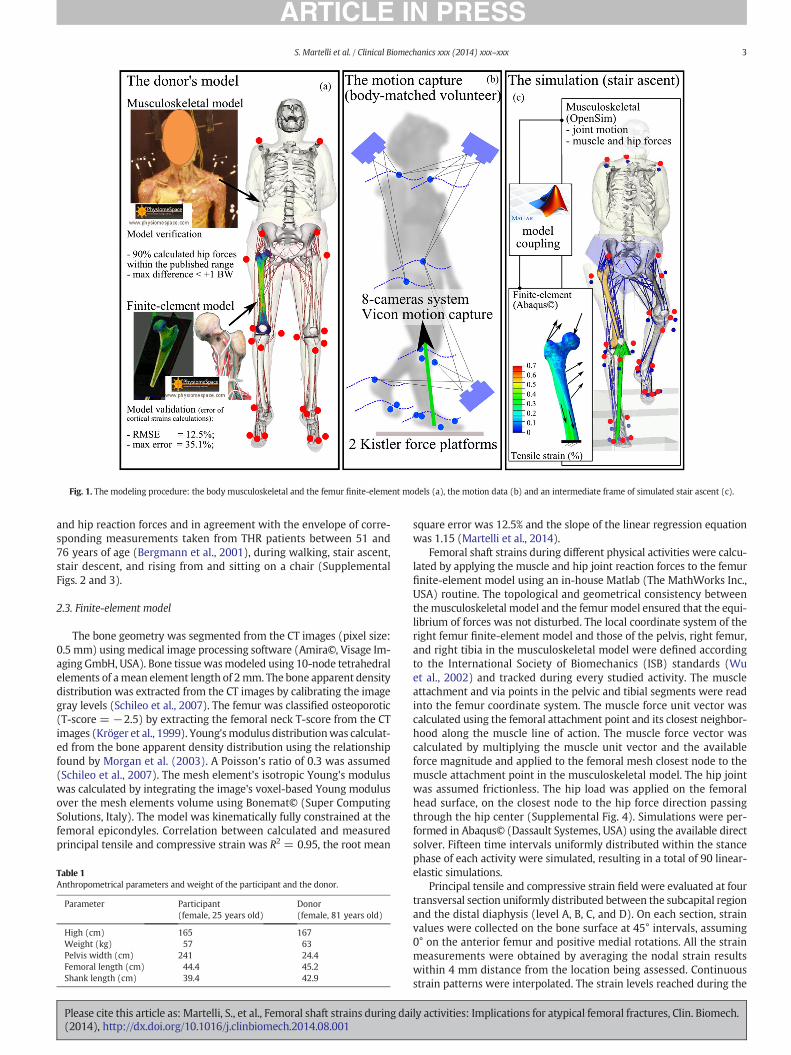
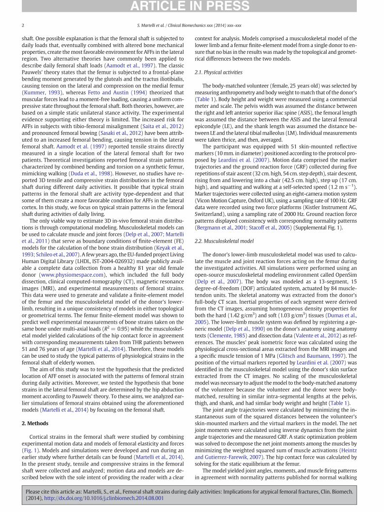
Fig. 1. The modeling procedure: the body musculoskeletal and the femur finite-element models (a), the motion data (b) and an intermediate frame of simulated stair ascent (c).
3S. Martelli et al. / Clinical Biomechanics xxx (2014) xxx–xxx
and hip reaction forces and in agreement with the envelope of corre-sponding measurements taken from THR patients between 51 and76 years of age (Bergmann et al., 2001), during walking, stair ascent,stair descent, and rising from and sitting on a chair (SupplementalFigs. 2 and 3).
2.3. Finite-element model
The bone geometry was segmented from the CT images (pixel size:0.5 mm) using medical image processing software (Amira©, Visage Im-aging GmbH, USA). Bone tissue wasmodeled using 10-node tetrahedralelements of amean element length of 2mm. The bone apparent densitydistribution was extracted from the CT images by calibrating the imagegray levels (Schileo et al., 2007). The femur was classified osteoporotic(T-score = −2.5) by extracting the femoral neck T-score from the CTimages (Kröger et al., 1999). Young'smodulus distributionwas calculat-ed from the bone apparent density distribution using the relationshipfound by Morgan et al. (2003). A Poisson's ratio of 0.3 was assumed(Schileo et al., 2007). The mesh element's isotropic Young's moduluswas calculated by integrating the image's voxel-based Young modulusover the mesh elements volume using Bonemat© (Super ComputingSolutions, Italy). The model was kinematically fully constrained at thefemoral epicondyles. Correlation between calculated and measuredprincipal tensile and compressive strain was R2 = 0.95, the root mean
Table 1Anthropometrical parameters and weight of the participant and the donor.
Parameter Participant(female, 25 years old)
Donor(female, 81 years old)
High (cm) 165 167Weight (kg) 57 63Pelvis width (cm) 241 24.4Femoral length (cm) 44.4 45.2Shank length (cm) 39.4 42.9
Please cite this article as: Martelli, S., et al., Femoral shaft strains during da(2014), http://dx.doi.org/10.1016/j.clinbiomech.2014.08.001
square error was 12.5% and the slope of the linear regression equationwas 1.15 (Martelli et al., 2014).
Femoral shaft strains during different physical activities were calcu-lated by applying the muscle and hip joint reaction forces to the femurfinite-element model using an in-house Matlab (The MathWorks Inc.,USA) routine. The topological and geometrical consistency betweenthemusculoskeletal model and the femur model ensured that the equi-librium of forces was not disturbed. The local coordinate system of theright femur finite-element model and those of the pelvis, right femur,and right tibia in the musculoskeletal model were defined accordingto the International Society of Biomechanics (ISB) standards (Wuet al., 2002) and tracked during every studied activity. The muscleattachment and via points in the pelvic and tibial segments were readinto the femur coordinate system. The muscle force unit vector wascalculated using the femoral attachment point and its closest neighbor-hood along the muscle line of action. The muscle force vector wascalculated by multiplying the muscle unit vector and the availableforce magnitude and applied to the femoral mesh closest node to themuscle attachment point in the musculoskeletal model. The hip jointwas assumed frictionless. The hip load was applied on the femoralhead surface, on the closest node to the hip force direction passingthrough the hip center (Supplemental Fig. 4). Simulations were per-formed in Abaqus© (Dassault Systemes, USA) using the available directsolver. Fifteen time intervals uniformly distributed within the stancephase of each activity were simulated, resulting in a total of 90 linear-elastic simulations.
Principal tensile and compressive strain field were evaluated at fourtransversal section uniformly distributed between the subcapital regionand the distal diaphysis (level A, B, C, and D). On each section, strainvalues were collected on the bone surface at 45° intervals, assuming0° on the anterior femur and positive medial rotations. All the strainmeasurements were obtained by averaging the nodal strain resultswithin 4 mm distance from the location being assessed. Continuousstrain patterns were interpolated. The strain levels reached during the
ily activities: Implications for atypical femoral fractures, Clin. Biomech.
4 S. Martelli et al. / Clinical Biomechanics xxx (2014) xxx–xxx
different activities were compared by calculating the principal tensileand compressive strains along the frontal, posterior, medial, andlateral femoral shaft. Ten uniformly distributed locations betweenthe subcapital region and the distal epiphysis were evaluated foreach femoral aspect. For each of the ten locations and the four femoralaspects, principal tensile and compressive strain were averaged overthe stance phase of each activity. Linear regression analysis was con-ducted to expose dependencies between cortical strain in the lateralfemoral shaft and moments acting on the femur to test Pauwels' theoryduring different activities.
3. Results
The lateral aspect of the femur was subjected to tensile strains dur-ing all the investigated activities. Fig. 2 shows the association betweentypical locations for AFFs as reported by Saleh et al. (2012) and tensilestrains in the femoral shaft while walking. Both strain intensity andorientation in the transverse planewere activity-dependent, withwalk-ing showing the highest average tensile strains in correspondence withthe location of onset of commonly observed AFFs.
The lateral femoral shaft (270°–300° in the transverse plane) wassubjected to peak tensile loads during most of the walking stancephase, whereas the peak tensile strain was slightly rotated anteriorlyduring stair descent (270°–330°). The neutral planewasmainly sagittal,and the peak compressive strain was mainly located in the medialfemoral shaft (0°–180° in the transversal plane). The anterior femoralshaft (315°–45° in the transverse plane) was subjected to peak tensileloads during most of the chair up-and-down, squatting, stair ascent,and step-up stance phases. The neutral plane was mainly coronal, andthe peak compressive strainwasmainly located in the posterior femoralshaft (0°–180° in the transversal plane). The strain field in the trans-verse plane rotated from maximal tension in the proximal–lateralshaft to maximal tension in the distal–anterior shaft during the mid-stance phase of stair ascent indicating torsion. Figs. 3 and 4 representtensile and compressive strain paths on transversal shaft sectionsduring walking and stair ascent, which showed extreme strain field
Fig. 2. Comparison of the tensile strain pattern during an intermediate frame of walking(left) with typical AFFs onset locations arrowed in an X-ray view (right) courtesy ofSaleh et al. (2012). The FE map is mirrored to facilitate comparison of the right femurmodel with the left femur represented in the X-ray.
Please cite this article as: Martelli, S., et al., Femoral shaft strains during da(2014), http://dx.doi.org/10.1016/j.clinbiomech.2014.08.001
orientations. Strain patterns for stepping up, rising from and sitting ona chair, squatting, and stair descent are reported in the SupplementaryMaterial.
The strain levels varied between activities. In the lateral femoralshaft, the average tensile strain during the stance phase of each activityvaried from 327 μεwhile squatting to 2004 μεwhile walking, and from245 με while squatting to 2337 με during stair rising in the anteriorfemoral shaft. In the medial femoral shaft, the average compressivestrain varied from −369 με during stair ascent to −1942 με whilewalking, while it varied from −305 με while squatting to −2560 μεduring stair ascent in the posterior femoral shaft (Fig. 5).
Tensile strain in theproximal–lateral femoral shaft (level A)was cor-related with the hip abduction moment; the coefficient of determina-tion was R2 = 0.94 (p-value b0.0001) during walking and R2 = 0.96(p-value b0.0001) during stair ascent (Fig. 6). In the distal–lateralfemoral shaft (level D), the coefficient of determination was R2 = 0.50while walking and R2 = 0.09 during stair ascent. No correlation wasfound between tensile strain and the hip abduction moment duringthe remaining activities. Themulti-parametric linear regression analysisusing the hip, knee, ankle moment and the ground reaction force inten-sity as predictors of tensile strains in the lateral femoral shaft during allthe studied activities showed a moderately strong correlation in theproximal (R2 = 0.60, p-value b0.0001) and in the distal femoral shaft(R2 = 0.46, p-value b0.0001).
4. Discussion
Atypical femoral fractures (AFFs) are typically transverse stressfractures arising in the lateral femoral shaft (Shane et al., 2014).While AFFs have been associated with increased bone fragility andlong-term anti-resorptive therapy (Shane et al., 2014), it is largelyunknown why these fractures propagate from the lateral femoralshaft. We tested the hypothesis that the location of AFFs onset isassociated with physiological strain distribution during daily activi-ties and that the tensile loads in the lateral femoral shaft are to be at-tributed to the hip abduction moment. We compared calculatedfemoral tensile strain distribution during walking, stair ascent, stairdescent, standing from and sitting on a chair, step up, and squattingwith common locations of AFFs onset.
The lateral femoral shaft is subjected to tensile strains during a vari-ety of physical activities and walking induces the highest tensile strainlevels. The hip abduction moment well predicts tensile strains in thelateral femoral shaft during walking and stair ascent, but not during ageneric activity. The lateral aspect of the femoral shaft was loaded intension during every studied activity. Therefore, the combination oftensile loads, to which bone is most susceptible (Bayraktar et al.,2004), and increased bone fragility that may be associated with long-term anti-resorptive therapy (Shane et al., 2014) may be an importantco-factor in creating a favorable environment for AFFs. Tensile strainmagnitudes and orientation in the transverse plane were activity-dependent, with walking inducing most of the tension in the lateralfemoral shaft, whereas chair up-and-down, squatting, stair ascent, andstep-up induced tension in the anterior femoral shaft. Walking is likelythe most critical activity in individuals susceptible to AFFs, inducinghigh tensile strain in the lateral femoral shaft, most frequently duringnormal living. A reduced walking speed may help reduce tensile strainsin the lateral femoral shaft by causing a reduction of the muscle work(Neptune et al., 2008) and of the ground reaction force (Nilsson andThorstensson, 1989).
The hip abduction moment was strongly correlated with tensilestrains in the proximo–lateral femoral shaft (levels A and B) duringwalking and stair ascent (Fig. 6), in agreement with the classicalPauwels' theory (Kummer, 1993). However, we found no correlationbetween tensile strains in the lateral femoral shaft and the hip abduc-tion moment during a generic activity (Fig. 6), whereas the multi-parametric linear regression showed that multiple kinetic variables
ily activities: Implications for atypical femoral fractures, Clin. Biomech.
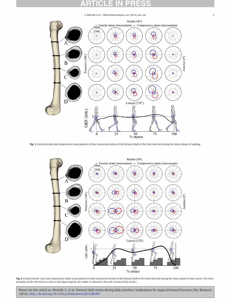
Fig. 3. Cortical tensile and compressive strain patterns in four transversal section of the femoral shaft at five time intervals during the stance phase of walking.
Fig. 4. Cortical tensile (red) and compressive (blue) strain patterns in four transversal section of the femoral shaft at five time intervals during the stance phase of stair ascent. (For inter-pretation of the references to color in this figure legend, the reader is referred to the web version of this article.)
5S. Martelli et al. / Clinical Biomechanics xxx (2014) xxx–xxx
Please cite this article as: Martelli, S., et al., Femoral shaft strains during daily activities: Implications for atypical femoral fractures, Clin. Biomech.(2014), http://dx.doi.org/10.1016/j.clinbiomech.2014.08.001
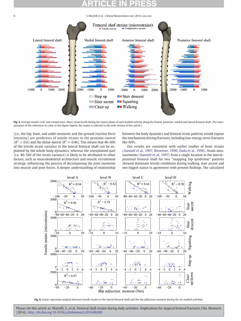
Fig. 5. Average tensile (red) and compressive (blue) strain levels during the stance phase of each studied activity along the frontal, posterior, medial and lateral femoral shaft. (For inter-pretation of the references to color in this figure legend, the reader is referred to the web version of this article.)
6 S. Martelli et al. / Clinical Biomechanics xxx (2014) xxx–xxx
(i.e., the hip, knee, and ankle moments and the ground reaction forceintensity) are predictors of tensile strains in the proximo–lateral(R2 = 0.6) and the distal–lateral (R2 = 0.46). This means that 46–60%of the tensile strain variance in the lateral femoral shaft can be ex-plained by the whole body dynamics, whereas the unexplained part(i.e. 40–54% of the strain variance) is likely to be attributed to otherfactors, such as musculoskeletal architecture and muscle recruitmentstrategy, influencing the process of decomposing the joint momentsinto muscle and joint forces. A deeper understanding of relationship
Fig. 6. Linear regression analysis between tensile strains in the lateral femor
Please cite this article as: Martelli, S., et al., Femoral shaft strains during da(2014), http://dx.doi.org/10.1016/j.clinbiomech.2014.08.001
between the body dynamics and femoral strain patterns would exposethemechanisms driving fractures, including low-energy stress fractureslike AFFs.
Our results are consistent with earlier studies of bone strains(Aamodt et al., 1997; Biewener, 1990; Duda et al., 1998). Strain mea-surements (Aamodt et al., 1997) from a single location in the lateral–proximal femoral shaft for two “snapping hip syndrome” patientsshowed dominant tensile conditions during walking, stair ascent andone legged stance in agreement with present findings. The calculated
al shaft and the hip adduction moment during the six studied activities.
ily activities: Implications for atypical femoral fractures, Clin. Biomech.
7S. Martelli et al. / Clinical Biomechanics xxx (2014) xxx–xxx
strain range (0–4667 με) is consistentwith thebone strain levels report-ed for mammalian bones during daily activities (25–50% of the yieldstrain) (Biewener, 1990). Earlier computational simulations of asynthetic femur (Duda et al., 1998) yielded up to 2000 με duringwalking, in good agreement with our results obtained for the initialtwo-third of the walking stance whereas our calculations of tensilestrains during early heel rise were higher (4667 με). This discrepancyis likely caused by the single circumstance where the ground reactionforce recorded from the volunteer (1.33BW) overestimated by ~30%corresponding normality patterns (Bergmann et al., 2001; Stacoffet al., 2005) (Supplemental Fig. 1). For the remaining walkingphases and activities, kinetic variables and hip forces were in goodagreement with correspondingmeasurements taken from THR patientsbetween 51 and 76 years of age (Bergmann et al., 2001; Stacoff et al.,2005) (Supplements Fig. 3) providing good confidence on the presentresults.
Our study has some limitations. First, muscle and joint forces from ayoung volunteer may differ from those present in older adults. However,this appears not to be true for the motion data used in the present studybecause the calculated hip-joint reaction force, which includes the contri-butions of all the hip-spanning muscles, was consistent with publishedmeasurements (Bergmann et al., 2001) in older adults. This observationis consistentwith thework of Limet al. (2012) that showedno significantdifferences in lower-limb muscle forces when younger and older adultswalk at the same speed. Second, results were generated using one ana-tomical dataset. It is possible that the inclusion of additional subjectsmay lead to different levels of bone strain in the femoral shaft. Furtherpopulation-based studies are necessary to investigate how individualanatomical parameters, bone quality, and motion patterns affect thecalculated strain patterns. Multi-scale computational modeling combin-ingmusculoskeletal and finite-elementmodels arewell suited to providesuch important information. Third, the untreated osteoporotic donor'sfemur did not account for the contrasting 9.6% BMD increase after3 years (Boivin et al., 2000) and 12% Young modulus decrease after6–10 years (Bala et al., 2012) of anti-resorptive therapy. Therefore,conclusions of the present study are directly relevant to persons whodo not use anti-resorptive therapy. In the author's opinion, however,the validity of our study lies in the fact that it provides new insightsinto our understanding of the typical patterns of femoral loads appliedto common sites of origin of AFFs and of the mechanical environmentassociated with AFF onset, i.e., tensile strain, and its changes duringdaily activities. Last, the association between tensile loads and AFFonset location shown here does not imply a cause–effect relationshipbetween tensile strain and the pathogenesis of AFFs. Present results canprovide a new base for designing new experiments investigating themechanism of AFFs.
5. Conclusion
AFFs are associated with tensile strain conditions during each dailyactivity, with walking causing the highest tensile strain in the lateralfemur. Therefore, tensile conditions are the most likely mechanicalenvironment contributing to AFF onset. The hip abduction moment isthe major determinant of tensile strain in the proximal–lateral femoralshaft during walking and stair ascent. However, this is not valid for ageneric activity forwhich amore complex interaction between dynamicparameters, muscle architecture and recruitment strategy determinesthe tensile strain in the lateral femoral shaft.
Acknowledgments
The authors are grateful to the EU-funded project LHDL (IST-2004-026932) for the data made available. This study was supported by theAustralian Research Council (DE140101530) awarded to S.M.
Please cite this article as: Martelli, S., et al., Femoral shaft strains during da(2014), http://dx.doi.org/10.1016/j.clinbiomech.2014.08.001
Appendix A. Supplementary data
Supplementary data to this article can be found online at http://dx.doi.org/10.1016/j.clinbiomech.2014.08.001.
References
Aamodt, A.,Lund-Larsen, J.,Eine, J.,Andersen, E.,Benum, P.,Husby, O.S., 1997. In vivo mea-surements show tensile axial strain in the proximal lateral aspect of the humanfemur. J. Orthop. Res. 15, 927–931. http://dx.doi.org/10.1002/jor.1100150620.
Allen, M.R.,Burr, D.B., 2007. Three years of alendronate treatment results in similar levelsof vertebral microdamage as after one year of treatment. J. Bone Miner. Res. 22,1759–1765. http://dx.doi.org/10.1359/jbmr.070720.
Allen, M.R.,Gineyts, E., Leeming, D.J.,Burr, D.B.,Delmas, P.D., 2008. Bisphosphonates altertrabecular bone collagen cross-linking and isomerization in beagle dog vertebra.Osteoporos. Int. 19, 329–337. http://dx.doi.org/10.1007/s00198-007-0533-7.
Bala, Y.,Depalle, B., Farlay, D.,Douillard, T.,Meille, S., Follet, H.,Chapurlat, R., Chevalier, J.,Boivin, G., 2012. Bone micromechanical properties are compromised during long-term alendronate therapy independently of mineralization. J. Bone Miner. Res. 27,825–834. http://dx.doi.org/10.1002/jbmr.1501.
Bayraktar, H.H.,Morgan, E.F., Niebur, G.L.,Morris, G.E.,Wong, E.K., Keaveny, T.M., 2004.Comparison of the elastic and yield properties of human femoral trabecular and cor-tical bone tissue. J. Biomech. 37, 27–35. http://dx.doi.org/10.1016/S0021-9290(03)00257-4.
Benhamou, C.-L., 2007. Effects of osteoporosis medications on bone quality. Joint BoneSpine 74, 39–47. http://dx.doi.org/10.1016/j.jbspin.2006.06.004.
Bergmann, G.,Deuretzbacher, G.,Heller,M.,Graichen, F.,Rohlmann, A.,Strauss, J.,Duda, G.N.,2001. Hip contact forces and gait patterns from routine activities. J. Biomech. 34,859–871.
Biewener, A.A., 1990. Biomechanics of mammalian terrestrial locomotion. Science 250,1097–1103.
Boivin, G.Y., Chavassieux, P.M., Santora, A.C., Yates, J.,Meunier, P.J., 2000. Alendronateincreases bone strength by increasing the mean degree of mineralization of bone tis-sue in osteoporotic women. Bone 27, 687–694.
Clemente, C., 1985. Gray’s Anatomy of the Human Body, 30th edition. Lea & Febiger.Dell, R.,Denise, G.,Ott, S.,Silverman, S.,Eisemon, E.,Funahashi, T.,Adams, A., 2010. A retro-
spective analysis of all atypical femur fractures seen in a large California HMO fromthe years 2007 to 2009. ASBMR 2010 Annual Meeting (Toronto).
Delp, S.L., Loan, J.P., Hoy, M.G., Zajac, F.E., Topp, E.L., Rosen, J.M., 1990. An interactivegraphics-based model of the lower extremity to study orthopaedic surgical proce-dures. IEEE Trans. Biomed. Eng. 37, 757–767.
Delp, S.L.,Anderson, F.C.,Arnold, A.S., Loan, P.,Habib, A., John, C.T.,Guendelman, E.,Thelen,D.G., 2007. OpenSim: open-source software to create and analyze dynamic simula-tions of movement. IEEE Trans. Biomed. Eng. 54, 1940–1950.
Duda, G.N., Heller, M., Albinger, J., Schulz, O., Schneider, E., Claes, L., 1998. Influence ofmuscle forces on femoral strain distribution. J. Biomech. 31, 841–846.
Dumas, R., Aissaoui, R., Mitton, D., Skalli, W., de Guise, J.A., 2005. Personalized bodysegment parameters from biplanar low-dose radiography. IEEE Trans. Biomed. Eng.52, 1756–1763. http://dx.doi.org/10.1109/TBME.2005.855711.
Ekström, W.,Németh, G.,Samnegård, E.,Dalen, N.,Tidermark, J., 2009. Quality of life after asubtrochanteric fracture: a prospective cohort study on 87 elderly patients. Injury 40,371–376. http://dx.doi.org/10.1016/j.injury.2008.09.010.
Fetto, J.F., Austin, K.S., 1994. A missing link in the evolution of THR: “discovery” of thelateral femur. Orthopedics 17, 347–351.
Glitsch, U.,Baumann, W., 1997. The three-dimensional determination of internal loads inthe lower extremity. J. Biomech. 30, 1123–1131.
Heintz, S.,Gutierrez-Farewik, E.M., 2007. Static optimization of muscle forces during gaitin comparison to EMG-to-force processing approach. Gait Posture 26, 279–288.
Keyak, J.H., Fourkas, M.G.,Meagher, J.M., Skinner, H.B., 1993. Validation of an automatedmethod of three-dimensional finite element modelling of bone. J. Biomech. Eng. 15,505–509.
Kröger, H., Lunt, M.,Reeve, J.,Dequeker, J.,Adams, J.E., Birkenhager, J.C., et al., 1999. Bonedensity reduction in various measurement sites inmen andwomen with osteoporot-ic fractures of spine and hip: the European quantitation of osteoporosis study. Calcif.Tissue Int. 64, 191–199.
Kummer, B., 1993. Is the Pauwels' theory of hip biomechanics still valid? A critical analy-sis, based on modern methods. Ann. Anat. 175, 203–210.
Leardini, A., Sawacha, Z., Paolini, G., Ingrosso, S.,Nativo, R., Benedetti, M.G., 2007. A newanatomically based protocol for gait analysis in children. Gait Posture 26, 560–571.
Li, J.,Mashiba, T.,Burr, D.B., 2001. Bisphosphonate treatment suppresses not only stochas-tic remodeling but also the targeted repair of microdamage. Calcif. Tissue Int. 69,281–286.
Lim, Y.P.,Lin, Y.-C.,Pandy, M.G., 2012. Muscle function during gait is invariant to age whenwalking speed is controlled. Gait Posture http://dx.doi.org/10.1016/j.gaitpost.2012.11.020.
Martelli, S.,Taddei, F.,Cappello, A.,van Sint Jan, S.,Leardini, A.,Viceconti, M., 2011. Effect ofsub-optimal neuromotor control on the hip joint load during level walking. J.Biomech. 44, 1716–1721. http://dx.doi.org/10.1016/j.jbiomech.2011.03.039.
Martelli, S.,Kersh, M.E.,Schache, A.G.,Pandy, M.G., 2014. Strain energy in the femoral neckduring exercise. J. Biomech. 47, 1784–1791. http://dx.doi.org/10.1016/j.jbiomech.2014.03.036.
Morgan, E.F., Bayraktar, H.H., Keaveny, T.M., 2003. Trabecular bone modulus–densityrelationships depend on anatomic site. J. Biomech. 36, 897–904.
ily activities: Implications for atypical femoral fractures, Clin. Biomech.
8 S. Martelli et al. / Clinical Biomechanics xxx (2014) xxx–xxx
Neptune, R.R.,Sasaki, K.,Kautz, S.A., 2008. The effect of walking speed on muscle functionand mechanical energetics. Gait Posture 28, 135–143. http://dx.doi.org/10.1016/j.gaitpost.2007.11.004.
Nilsson, J., Thorstensson, A., 1989. Ground reaction forces at different speeds of humanwalking and running. Acta Physiol. Scand. 136, 217–227.
Roschger, P., Rinnerthaler, S., Yates, J., Rodan, G.A., Fratzl, P., Klaushofer, K., 2001.Alendronate increases degree and uniformity of mineralization in cancellous boneand decreases the porosity in cortical bone of osteoporoticwomen. Bone 29, 185–191.
Saita, Y.,Ishijma, M.,Mogami, A.,Gen, H.,Kaneko, K.,Miyagawa, K.,Nemoto, M., et al., 2012.Association between the fracture site and the mechanical axis of lower extremities inpatients with atypical femoral fracture. J. Bone Miner. Res. 27.
Saleh, A.,Hegde, V.V.,Potty, A.G.,Schneider, R.,Cornell, C.N.,Lane, J.M., 2012. Managementstrategy for symptomatic bisphosphonate-associated incomplete atypical femoralfractures. HSS J. 8, 103–110. http://dx.doi.org/10.1007/s11420-012-9275-y.
Sasaki, S., Miyakoshi, N., Hongo, M., Kasukawa, Y., Shimada, Y., 2012. Low-energydiaphyseal femoral fractures associated with bisphosphonate use and severecurved femur: a case series. J. Bone Miner. Metab. 30, 561–567. http://dx.doi.org/10.1007/s00774-012-0358-0.
Schileo, E., Taddei, F.,Malandrino, A.,Cristofolini, L.,Viceconti, M., 2007. Subject-specificfinite element models can accurately predict strain levels in long bones. J. Biomech.40, 2982–2989. http://dx.doi.org/10.1016/j.jbiomech.2007.02.010.
Shane, E.,Burr, D.,Ebeling, P.R.,Abrahamsen, B.,Adler, R.A.,Brown, T.D.,Cheung, A.M., et al.,2010. Atypical subtrochanteric and diaphyseal femoral fractures: report of a task
Please cite this article as: Martelli, S., et al., Femoral shaft strains during da(2014), http://dx.doi.org/10.1016/j.clinbiomech.2014.08.001
force of the American Society for Bone and Mineral Research. J. Bone Miner. Res.25, 2267–2294. http://dx.doi.org/10.1002/jbmr.253.
Shane, E., Burr, D.,Abrahamsen, B.,Adler, R., Brown, T., Cheung, A., et al., 2014w. Atypicalsubtrochanteric and diaphyseal femoral fractures: second report of a task force ofthe american society for bone and mineral research. J. Bone Miner. Res. 29, 1–23.http://dx.doi.org/10.1002/jbmr.1998.
Stacoff, A.,Diezi, C., Luder, G., Stüssi, E.,Kramers-de Quervain, I.A., 2005. Ground reactionforces on stairs: effects of stair inclination and age. Gait Posture 21, 24–38. http://dx.doi.org/10.1016/j.gaitpost.2003.11.003.
Valente, G., Martelli, S., Taddei, F., Farinella, G., Viceconti, M., 2012. Musclediscretization affects the loading transferred to bones in lowerlimb musculoskel-etal models. Proc. Inst. Mech. Eng. H J. Eng. Med. 226, 161–169. http://dx.doi.org/10.1177/0954411911425863.
Vashishth, D., 2009. Advanced glycation end-products and bone fractures. IBMS Bonekey6, 268–278. http://dx.doi.org/10.1138/20090390.
Wu, G.,Siegler, S.,Allard, P.,Kirtley, C.,Leardini, A.,Rosenbaum, D., et al., 2002. ISB recom-mendation on definitions of joint coordinate system of various joints for thereporting of human joint motion–part I: ankle, hip, and spine. International Societyof Biomechanics. J. Biomech. 35, 543–548.
ily activities: Implications for atypical femoral fractures, Clin. Biomech.