extended extraocular phenotype of prom1 mutation in kindreds with known autosomal dominant macular...
TRANSCRIPT
1
Extended extraocular phenotype of PROM1 mutation in kindreds with known
autosomal dominant macular dystrophy
FI Arrigoni1,2*, M Matarin3*, PJ Thompson3, M Michaelides4,5, ME McClements4, E
Redmond1, L Clarke1, E Ellins1, S Mohamed3, I Pavord6, DM Hunt4, N Klein7, AT Moore4,5,
J Halcox1,8, SM Sisodiya3
1Vascular Physiology, UCL Institute of Child Health, 30 Guilford Street, London, WC1N 1EH;
2Department of Pharmacy, Kingston University, Penrhyn Road, KT1 2EE, 3Department of
Clinical and Experimental Epilepsy, UCL Institute of Neurology, Queen Square, London WC1N
3BG and National Society for Epilepsy, Bucks, SL9 0RJ; 4UCL Institute of Ophthalmology, 11-
43 Bath Street, London EC1V 9EL; 5Moorfields Eye Hospital, City Road, London, EC1V 2PD,
UK; 6Department of Respiratory Medicine, Allergy and Thoracic Surgery, Institute for Lung
Health, Glenfield Hospital, University Hospitals of Leicester NHS Trust, Leicester LE3 9QP,
UK; 7Research Department of Infection, UCL Institute of Child Health, 30 Guilford Street
London WC1N 1EH, and 8Wales Heart Research Institute, Cardiff University, Cardiff, UK
* These authors contributed equally
Author for Correspondence:
SM Sisodiya, Department of Clinical and Experimental Epilepsy, National Hospital for
Neurology and Neurosurgery, Box 29, Queen Square, London, UK WC1N 3BG
Tel 020 7391 8983
Fax 020 7391 8984
Email: [email protected]
peer
-005
7871
2, v
ersi
on 1
- 22
Mar
201
1Author manuscript, published in "European Journal of Human Genetics (2010)"
DOI : 10.1038/ejhg.2010.147
2
Running Head: Extended PROM1 phenotype
Key words: PROM1, CD 133, endothelial, tubule, sella turcica
Total Word Count: 3,019 (max 4,000)
Abstract text: 146 (max 250)
Tables and Illustrations: 3 figures and 3 tables (max 6)
Table1: Cognitive performance and plus
Table 2: Baseline characteristics
Table 3: Microparticles
Figure 1: Leucoaraiosis and empty sella turcica
Figure 2: Representative micrograph of HUVEC
Figure 3: Angiogenesis assay between HUVEC and ECFCs.
References: 42 plus 10 in supplementary material (max 50)
Supplementary material: 1,733
peer
-005
7871
2, v
ersi
on 1
- 22
Mar
201
1
3
Abstract
Mutations in prominin 1 (PROM1) have been shown to result in retinitis pigmentosa, macular
degeneration and cone-rod dystrophy. Because of the putative role of PROM1 in
hippocampal neurogenesis, we examined two kindreds with the same R373C PROM1
missense mutation using our established paradigm to study brain structure and function. As
the protein encoded by PROM1, known as CD133, is used to identify stem/progenitor cells
that can be found in peripheral blood and reflect endothelial reparatory mechanisms, other
parameters were subsequently examined that included measures of vascular function,
endothelial function and angiogenic capacity. We found that aspects of endothelial function
assayed ex vivo were abnormal in patients with the R373C PROM1 mutation, with impaired
adhesion capacity and higher levels of cellular damage. We noted also renal infections,
haematuria and recurrent miscarriages possibly reflecting consequences of abnormal tubular
modelling. Further studies are needed to confirm these findings.
peer
-005
7871
2, v
ersi
on 1
- 22
Mar
201
1
4
Introduction
The effects of a single human gene mutation may be widespread and, in their breadth,
often unsuspected. We have previously explored the cerebral structural and functional effects
of mutations in genes expressed in the eye and the brain, in individuals ascertained by their
mutant eye phenotype, in whom extraocular phenotypes were often not appreciated. Using
this paradigm, we have demonstrated roles for the genes PAX6, PITX2, SOX2, OTX2 and
RIMS1 in human brain development, and cognitive function [1- 4].
Mutations in prominin 1 (PROM1) have been shown to result in retinitis pigmentosa
[5, 6] macular degeneration [7, 8] and cone-rod dystrophy [9]. PROM1 encodes prominin-1, a
5-transmembrane glycoprotein also known as CD133 and AC133. CD133 was originally
identified as a cell surface antigen present on hematopoietic stem cells and early progenitor
cells in the bone marrow, including endothelial progenitor cells (EPC) [10, 11].
Emerging evidence suggests that EPCs are able to differentiate into mature
endothelial cells, contributing to neovascularization and re-endothelialization during both
embryonic and postnatal physiological processes [12]. Consequently, CD133 is used to
identify stem/progenitor cells that can be found in peripheral blood and thereby identify
endothelial reparatory mechanisms and also to identify tumour stem cell populations in a
variety of blood and solid cancers [13, 14]. Consistent with a role in tissue repair, increased
levels of circulating early EPCs marked by CD133 have also been found in response to brain
injury [15]. CD133 is also involved in murine and human adult hippocampal neurogenesis
[16], murine brain myelin genesis and maintenance [17]
Because of the putative role of PROM1 in hippocampal neurogenesis, we examined
two kindreds with the same known PROM1 mutation using our established paradigm to study
brain structure and function. We subsequently examined other parameters including measures
of vascular function, endothelial function and angiogenic capacity.
peer
-005
7871
2, v
ersi
on 1
- 22
Mar
201
1
5
Methods
The study included two unrelated kindreds that were already reported as having
autosomal dominant macular degeneration mapped to 4 p15.2-16.3 [5, 18]. From kindred A,
the proband (A-V:1), her mother (A-IV:2), aunt (A-IV:4) and grandmother (A-III:6) were
studied. In kindred B, the proband (B-II:1) and her mother (B-I:1) were included in the study
(Table 1). In both probands, the same R373C PROM1 mutation had been identified. Clinical
history was obtained from all participants.
Healthy control groups were recruited from staff and colleagues from the Institute of
Child Health.
The study was approved by the Joint Research Ethics Committees of the Institute of
Neurology/ National Hospital for Neurology and Neurosurgery, and Moorfields Eye Hospital
and the Institute of Child Health/Great Ormond Street Hospital for Children NHS Foundation
Trust. Subjects provided informed written consent for all parts of the study.
Brain imaging
Subjects were studied on a 3T scanner. Details of the high-resolution MRI, cerebral
and hippocampal volumetry are provided in the Supplementary material.
Cognitive tests applied to kindreds
Neuropsychological tests employed were as described previously [19]. All of the
standardised tests selected require verbal interaction only. Intellectual level, executive and
memory function were tested. Details are given in the Supplementary Material.
Clinical testing
Proband A-V:1 had renal and pulmonary assessments previously undertaken due to
relevant clinical symptoms. Olfactory testing was undertaken using the UPSIT [1] (Sensonics
Inc., Haddon Heights, NJ), with age-matched control data based upon North American
subjects, shown to be applicable to British populations [20, 21].
Blood and vascular investigations
After a 12-hour overnight fast, venous blood was taken from three subjects from
kindred A, and one from kindred B. After subjects had rested for >15 minutes, recumbent
peer
-005
7871
2, v
ersi
on 1
- 22
Mar
201
1
6
right brachial artery blood pressure was recorded using an automated sphygmomanometer
(Dinamap, Critikon, Florida, USA). Five readings were taken and mean systolic and diastolic
blood pressure calculated.
The isolation of peripheral blood mononuclear cells (PBMCs), flow cytometric
analysis for detection of EPCs and cellular microparticles, circulating endothelial cell
extraction and analyses, growth factor detection, the ability of specific cells to form colonies,
microparticle assays and in vitro colony forming and matrigel angiogenesis assays were
performed as detailed in the Supplementary Material. For kindred A, vascular structure
(carotid artery intima-media thickness, IMT) and function (flow-mediated dilatation of
brachial artery, FMD) were also assessed (Supplementary Material).
Statistics
Nonparametric tests (U-Mann-Whitney) were used with SPSS, version 14 (SPSS Inc).
Data were expressed as median and interquartile range.
peer
-005
7871
2, v
ersi
on 1
- 22
Mar
201
1
7
Results
Clinical findings
The ocular phenotypic data have been published [5, 18]. A-V:1 had steroid-resistant
asthma. She had a clinical assessment for microscopic haematuria, with a normal renal
ultrasound and intravenous pyelogram. A-IV:2 also had microscopic haematuria, with
recurrent renal infection, and renal scarring on intravenous pyelography. She had eight
pregnancies, and four children. A-III:6 had recurrent renal infections, seven pregnancies with
three miscarriages, and four children. A-IV:4 had two unprovoked seizures.
In kindred B, B-I:1 had high blood pressure and high cholesterol.
According to North-American normative data UPSIT A-IV:4 and B-II:1 had mild microsmia
and A-III:6 had moderate microsmia.
Neuroimaging
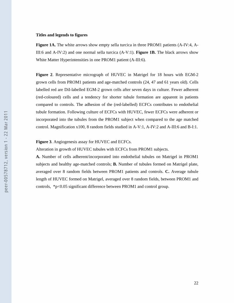
Subject A-III:6 had many small high-intensity lesions in the cerebral white matter
(Fig 1B). Subjects A-IV:4 and B-I:1 also had a few (1-2) non-specific white matter (WM)
lesions. The presence of WM hyperintensities on the FLAIR images for subject A-III:6 was
scored by a radiologist blind to the clinical data, using the Scale of Tarvonen-Schroder and
Scheltens semiquantative rating scale [22]. This scale scores the severity of hyperintensities
separately in different anatomical areas of the brain including periventricular regions, areas of
deep white matter , basal ganglia and different infratentorial regions. The total score was 19.
Lesions were focused on deep white matter (score=16/24) and pons (score=3/6). Lesions in
frontal brain area were considered as severe and mild to moderate in the other brain areas.
An empty sella turcica was noted in A-III:6, A-IV:2 and A-IV:4 (Fig 1A). Baseline
pituitary function (TSH, T4, cortisol, FSH and LH) was normal in all three. No other changes
were noted. MRI brain scans in kindred B were normal. Hippocampal and cerebral volumes
were within normal ranges for all patients from both kindreds.
peer
-005
7871
2, v
ersi
on 1
- 22
Mar
201
1
8
Cognitive function
No distinctive pattern of cognitive deficit was noted (Table 1). All subjects in kindred
A except A-IV:4 showed average to superior verbal memory scores. A-IV:4 showed poor
performance for delayed recall on the auditory verbal learning test with an average
performance on the remaining tests of memory. In kindred B, B-I:1 had impaired verbal
learning for trials ,reduced performance on tests of immediate recall and delayed verbal
learning. All subjects except A-IV:4 and B-I: 1 showed average executive functions.
Vascular and endothelial function
Vascular profiles
Subjects A-V:1, A-IV:2 and A-III:6 underwent vascular investigation. Each subject
was ranked into an age group, (1, 2 or 3), in which 12 healthy controls, with a median age
similar to that of the subject, were included (see Table 2). Patients and controls were all
female and had no known cardiovascular risk factors with the exception of high blood
pressure and high cholesterol in subject B-1:1.
Brachial artery FMD and carotid artery IMT
Detailed descriptions of procedures are in Supplementary Material. Briefly the
diameter of the brachial artery was measured at rest, during reactive hyperaemia to induce
flow-mediated endothelium-dependent dilation (FMD), again at rest, and after administration
of GTN, an endothelium-independent dilator. FMD and the ratio FMD:GTN-induced
dilatation provide the best measures of endothelial function.
Flow-mediated dilatation did not alter with age in the controls (Table 1). There was
no difference in median FMD when comparing grouped controls and patients (8.28 (5.68-
11.40) and 4.88 (4.06-12.27)). The ratio FMD:GTN did not alter with age in controls, but fell
with age in the patients. The median FMD:GTN ratio for the grouped patients did not differ
from the controls (0.95 (0.90-0.97) and 0.98 (0.96-1.02)). Intima-media thickness (IMT)
increased significantly with age in the controls (Table 2). Increasing IMT values were also
observed in the patients. However, there was no difference in the median values for IMT
between grouped controls and PROM-1 patients (0.65 (0.58-0.72) and 0.52 (0.48-0.75)).
peer
-005
7871
2, v
ersi
on 1
- 22
Mar
201
1
9
Determination of Growth Factor Concentration
Angiopoietin-2 levels and vascular endothelial growth factor (VEGF) levels were
within the normal range according to established clinical ranges.
Isolation and quantification of EPC
In controls, circulating endothelial cell (CEC) number significantly increased between
the younger groups 1 and 2, but not between either of the other age groups (Table 2). Increase
of CECs with age is less evident in PROM1 patients but falls within ranges for healthy age-
matched controls (Table 2). No change in the expression of surface markers was observed
with age in controls or PROM1 patients. Calculated as a percentage of the lymphocyte gate,
there was no significant difference between groups in the expression of any of the surface
markers alone or in combination (Table 2).
Cultivation of EPC
Colony-forming unit endothelial cells (CFU-EC) fell with age in controls. This trend
was also observed in the few PROM1 patients. Colony number tended to be low but values
fell within the normal range for their ages (controls, 20.37 (7.05- 32.57) and PROM1
patients, 11.9 (4.6-41)).
Relationship Between Cellular and Vascular Measures
Numbers of CFU-EC correlated with FMD:GTN ratio in the controls (r=0.42,
p<0.05). CFU-EC from PROM1 patients demonstrated a similar trend (Supplementary
Material ).
Microparticle number.
Total microparticle number, which consisted of endothelial microparticles (CD144,
D105 and CD62e), platelet microparticles (CD42a) and monocyte-derived microparticles
(CD14), fell within the upper range in PROM1 patients in relation to age-matched controls.
Comparisons between patients and controls independently to age, showed higher number of
total microparticles in PROM-1 patients ((2.606(1.399- 2.97); 0.3023(0.14-1.35) for PROM-1
patients and healthy age matched controls respectively, median ± interquartile range, p=
peer
-005
7871
2, v
ersi
on 1
- 22
Mar
201
1
10
0.0028). Furthermore endothelial microparticles defined by CD144 expression were also
found to be significantly elevated in the PROM-1 progeny (Table 3).
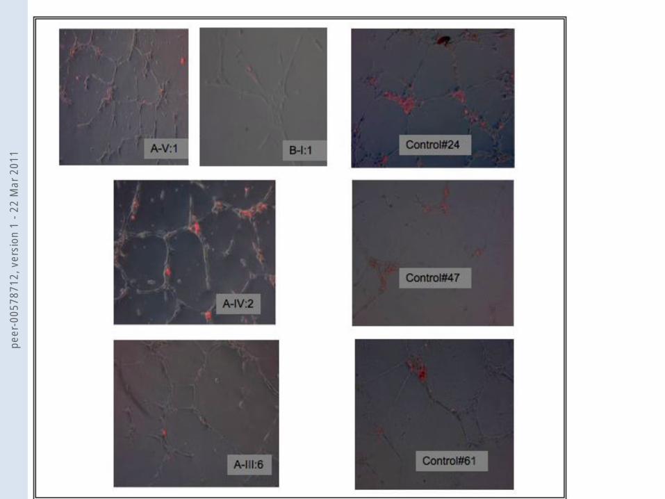
Angiogenic capability
A Matrigel assay was used to determine the effect of colony-forming unit endothelial
cells labelled with acetylated low-density lipoprotein (DiI-acLDL) on HUVEC growth.
DiI-acLDL-labelled cells from PROM1 patients did not adhere to or incorporate into
the human umbilical endothelial cells (HUVEC) as well as their age-matched controls (5.6
(0-18); 26.7 (15.5-44.3), p=0.03; Figure 3). Furthermore, a lack of DiI-acLDL-labelled cell
incorporation into the HUVEC tubules tended to produce lower tubule length in PROM1
subjects when compared to their age-matched controls (0.8 (0.74-1.2) and 1.38 (0.9-1.46),
p=0.06). No difference in tubule number was observed between the two groups (1 (1-1.14);
0.88(0.52-1.22), p=0.57; Figure 3).
peer
-005
7871
2, v
ersi
on 1
- 22
Mar
201
1
11
Discussion
We describe vascular and endothelial function in addition to cerebral structure and
function in two independent families with the same R373C mutation in the PROM1 gene.
Carriers of the mutation were already reported to present clinical features of autosomal
dominant macular dystrophy [7, 18].
Human PROM1 is expressed by various stem and progenitor cells originating from
diverse sources [23]. Due to the role of EPC in vascular and endothelial function [24], we
investigated these characteristics in five carriers of the R373C mutation. We found that the
number of circulating endothelial cells, endothelial markers such as CD133, as well as the
ability of EPC to form endothelial colonies fell within the normal range in the tested patients.
Endothelium-dependent and endothelium-independent function assessed by FMD and the
FMD:GTN ratio did not differ from controls. There was also no difference in intima-media
thickness. The basic function and EPC regenerative resources would seem unaffected by the
R373C mutation.
However the number of circulating microparticles (MPs) was raised in patients
compared to controls. MPs are membrane fragments shed by cells which have been activated
by a variety of stimuli including serine proteases, inflammatory cytokines, growth factors and
stress inducers [25]. Clinical studies have revealed elevated plasma levels of MPs in multiple
sclerosis, thrombotic thrombocytopenic purpura, coronary artery disease, hypertension,
preeclampsia, and diabetes [26, 27]. Such data have led to the concept that endothelial MP
are key factors at the crossroads between inflammation, coagulation, proteolysis and vascular
repair [28]. Elevated circulating levels of MPs in our patients could be an indicator of either
platelet, endothelial or leukocyte activation and could reflect a persistent state of endothelial
dysfunction.
We looked also at the contribution of immature ECFCs to tubule growth, of immature
ECFCs. A significantly decreased ability of these cells to incorporate or adhere to the
HUVEC was observed in carriers of the mutation. As this assay evaluates multiple cellular
peer
-005
7871
2, v
ersi
on 1
- 22
Mar
201
1
12
processes involved in blood vessel growth, EPC functional activity would appear impaired by
the mutation. In turn, vascularization might be affected in the patients, at least under certain
circumstances, perhaps those requiring neogenesis or repair.
Notably, the mutation carriers had microhaematuria and/or renal infections and a high
number of miscarriages. Although there is no study reporting endothelial dysfunction in
patients with hematuria and/or renal infections, several studies have reported that EPCs are
numerically and functionally impaired in patients with acute [29] or chronic renal failure
[30], as compared to healthy subjects and mechanisms such as inflammation or uraemic
toxins have been given as potential explanations for the observed EPC dysfunction [30].
More clear and direct is the relationship between miscarriages and possible endothelial
dysfunction, given that development, maturation and maintenance of a neovascular network
are necessary for successful haemochorial placentation as well as normal embryonic
development and growth [31]. Impairment in EPC functionality could thus affect placental
angiogenesis and contribute to history of recurrent miscarriages in our patients. This requires
further study in a larger group of patients, and might merit study in women with unexplained
recurrent miscarriages.
CD133 has been also reported to mark several cell types with a variety of putative
roles, including involvement in hippocampal neurogenesis [16, 17] and diverse
haematopoietic and extrahaematopoietic progenitive and regenerative functions [32-34].
Carriers of the PROM1 mutation showed hippocampal and cerebral volumes within normal
ranges for all patients from both kindreds. Three patients had small lesions in white matter.
Interestingly the presence of MPs has been also reported to be positively associated with
contrast-enhancing lesions on brain MRI in subjects with multiple sclerosis [35] and
furthermore endothelial dysfunction is thought to play an important role in the pathogenesis
of cerebral small-vessel disease especially in those patients with concomitant silent lacunar
infarcts and ischemic white matter lesions [36]. In line with this possible accelerated aging
effect, the older member from the kindred B and one carrier of the PROM1 mutation in
family A showed some memory disturbance and impairment in measures of executive
functions in addition to small WM lesions. The remaining carriers in both families had
average IQs and unimpaired performance on the other cognitive measures.
peer
-005
7871
2, v
ersi
on 1
- 22
Mar
201
1
13
The three (of four) members of family A showed an empty sella turcica. The familial
empty sella together with an eye phenotype could suggest a developmental defect of
mesenchymal origin caused by mutation in the same gene [37]. Familial empty sella has not,
to our knowledge, been reported previously. Three patients had impaired olfaction.
All together, the elevated plasma levels of MPs, decreased ability of EPC to
incorporate into or adhere to tubules, the presence of empty sella turcica as well as
leucoaraiosis and cognitive decline in the oldest mutation carriers suggest endothelial
dysfunction that could be more pronounced or more evident with age. The R373C mutation
results in a stable mutant protein. However, the mutant protein is not only mislocalized in
retina, but it also interferes with the action of the normal protein [7] thereby accounting for its
dominant mode of inheritance. Though we found no difference in the number of circulating
EPCs or numbers of cells expressing CD133, the mutation may impair the function. For
example the mutation could render EPCs less effective in the process of angiogenesis or
repair of damaged endothelium. Carriers of the R373C mutation showed no obvious greater
risk of vascular dysfunction, but structural and functional microcirculation alterations tend to
occur during ageing. Functional vascular examination was only undertaken in one of the two
older patients as the second declined to take part in this test. Moreover the presence of high
blood pressure and high cholesterol, as well as leucoaraiosis [38] could be a sign of vascular
alteration. Longitudinal study of more individuals with PROM1 mutation might be
informative.
To our knowledge this is the first study reporting a family with eye phenotype
together with empty sella turcica, presence of microhematuria and possible endothelial
dysfunction. In the case of the eye phenotype (macular dystrophy) the penetrance was
complete although showing differences in severity. Penetrance in the other phenotypes was
incomplete. Notable is the fact that none of the members in family B presented extra-ocular
features, nor MRI findings (other than a few WM lesions), besides the cognitive decline in
the older member, raising the possibility of organ-specific penetrance differences, perhaps
related to organ-specific splicing differences of the PROM1 transcript. PROM1 transcription
regulation is rather complicated and poorly understood: it exhibits extensive splice variation
peer
-005
7871
2, v
ersi
on 1
- 22
Mar
201
1
14
[39] with tissue-specific distribution [40], and in addition transcripts can be regulated by
epigenetic factors, as suggested by experiments with artificial in vitro methylation. However
we cannot exclude the possibility that mutation in a second gene could be influencing the
extra-ocular phenotype in family A, especially the presence of empty sella turcica.
The role of PROM1 in brain is still unclear. The gene, by itself or in combination with
other genes, seems to have a role in the development of the brain, and/or cerebral recovery
and repair [17, 41]. The heterozygous mutation studied here results in a stable protein [7] and
is perhaps insufficient to completely inhibit its function in the brain. In contrast, recessive
mutations arise from either frameshift or nonsense mutations [5, 6] that result in the
generation of a premature stop codon and truncated protein [5, 6, 42]. In these cases the
homozygous patients display a severe eye phenotype, and polydactyly was also reported in
one of the carriers, an additional symptom that confirms the existence of variable penetrance
associated with PROM1. No other documentation about vascular, endothelial or brain
structures was reported.
The nature and size of our study does not permit definitive conclusions but leads to
the suggestion that endothelial function may be affected in patients with the PROM1 R373C
mutation, despite the apparently normal levels of EPC. Further studies are needed to confirm
these interesting findings that broaden the phenotype of PROM1 mutation, and inform our
understanding of CD133 function.
Acknowledgments
We thank the patients who participated. We are grateful to the Big Lottery Fund, Wolfson
Trust and the National Society for Epilepsy for supporting the NSE MRI scanner. The work
was supported by a joint grant from the Biomedical Research Centres at UCLH/UCL, Great
Ormond Street Hospital for Children/UCL Institute of Child Health, and Moorfields Eye
Hospital/UCL Institute of Ophthamology. This work was undertaken at UCLH/UCL who
received a proportion of funding from the Department of Health’s NIHR Biomedical
peer
-005
7871
2, v
ersi
on 1
- 22
Mar
201
1

15
Research Centres funding scheme. Drs J. E. Halcox. and F. I. Arrigoni were supported by
the British Heart Foundation, Dr E. Redmond by the Coronary Artery Disease Research
Association, and Ms M. E. McClements by Fight for Sight.
Competing Interest: None declared.
peer
-005
7871
2, v
ersi
on 1
- 22
Mar
201
1
16
Bibliography
1 Sisodiya SM, Free SL, Williamson KA, et al. PAX6 haploinsufficiency causes
cerebral malformation and olfactory dysfunction in humans. Nature Genetics 2001;28(3):214-6.
2 Idrees F, Bloch-Zupan A, Free SL, et al. A novel homeobox mutation in the PITX2 gene in a family with Axenfeld-Rieger syndrome associated with brain, ocular, and dental phenotypes. American Journal of Medical Genetics Part B, Neuropsychiatric Genetics: the Official Publication of the International Society of Psychiatric Genetics 2006;141B(2):184-91.
3 Sisodiya SM, Ragge NK, Cavalleri GL, et al. Role of SOX2 mutations in human hippocampal malformations and epilepsy. Epilepsia 2006;47(3):534-42.
4 Henderson RA, Williamson K, Cumming S, et al. Inherited PAX6, NF1 and OTX2 mutations in a child with microphthalmia and aniridia. European Journal of Human Genetics 2007;15(8):898-901.
5 Zhang Q, Zulfiqar F, Xiao X, et al. Severe retinitis pigmentosa mapped to 4p15 and associated with a novel mutation in the PROM1 gene. Human Genetics 2007;122(3-4):293-9.
6 Maw MA, Corbeil D, Koch J, et al. A frameshift mutation in prominin (mouse)-like 1 causes human retinal degeneration. Human Molecular Genetics 2000;9(1):27-34.
7 Yang Z, Chen Y, Lillo C, et al. Mutant prominin 1 found in patients with macular degeneration disrupts photoreceptor disk morphogenesis in mice.[see comment]. Journal of Clinical Investigation 2008;118(8):2908-16.
8 Permanyer J, Navarro R, Friedman J, et al. A novel mutation in PROM1 that results in nonsense mediated decay causes autosomal recessive Retinitis Pigmentosa with early macular affectation.Investigative Ophthalmology & Visual Science 2010;51(5):2656-63.
9 Michaelides M, Gaillard M, Escher P, et al. The PROM1 mutation p.R373C causes an autosomal dominant bull’s eye maculopathy associated with rod, rod-cone and macular dystrophy. Investigate Ophthalmology and Visual Science 2010; [In Press].
10 Yin AH, Miraglia S, Zanjani ED, et al. AC133, a novel marker for human hematopoietic stem and progenitor cells. Blood 1997;90(12):5002-12.
11 Miraglia S, Godfrey W, Yin AH, et al. A novel five-transmembrane hematopoietic stem cell antigen: isolation, characterization, and molecular cloning. Blood 1997;90(12):5013-21.
12 Urbich C, Dimmeler S. Endothelial progenitor cells: characterization and role in vascular biology. Circulation Research 2004;95(4):343-53.
13 Singh SK, Hawkins C, Clarke ID, et al. Identification of human brain tumour initiating cells.[see comment]. Nature 2004;432(7015):396-401.
14 Zhu L, Gibson P, Currle DS, et al. Prominin 1 marks intestinal stem cells that are susceptible to neoplastic transformation.[see comment]. Nature 2009;457(7229):603-7.
15 Liu L, Liu H, Jiao J, et al. Changes in circulating human endothelial progenitor cells after brain injury.[erratum appears in J Neurotrauma. 2007 Aug;24(8):1415 Note: Jao, Junfeng [corrected to Jiao, Junfeng]. Journal of Neurotrauma 2007;24(6):936-43.
peer
-005
7871
2, v
ersi
on 1
- 22
Mar
201
1
17
16 Kempermann G, Chesler EJ, Lu L, et al. Natural variation and genetic covariance in adult hippocampal neurogenesis. Proceedings of the National Academy of Sciences of the United States of America 2006;103(3):780-5.
17 Corbeil D, Joester A, Fargeas CA, et al. Expression of distinct splice variants of the stem cell marker prominin-1 (CD133) in glial cells. GLIA 2009;57(8):860-74.
18 Michaelides M, Johnson S, Poulson A, et al. An autosomal dominant bull's-eye macular dystrophy (MCDR2) that maps to the short arm of chromosome 4. Investigative Ophthalmology & Visual Science 2003;44(4):1657-62.
19 Sisodiya SM, Thompson PJ, Need A, et al. Genetic enhancement of cognition in a kindred with cone-rod dystrophy due to RIMS1 mutation. Journal of Medical Genetics 2007;44(6):373-80.
20 Gibberd FB, Feher MD, Sidey MC, et al. Smell testing: an additional tool for identification of adult Refsum's disease. Journal of Neurology, Neurosurgery & Psychiatry 2004;75(9):1334-6.
21 Silveira-Moriyama L, Petrie A, Williams DR, et al. The use of a color coded probability scale to interpret smell tests in suspected parkinsonism. Movement Disorders 2009;24(8):1144-53.
22 Scheltens P, Barkhof F, Leys D, et al. A semiquantative rating scale for the assessment of signal hyperintensities on magnetic resonance imaging Journal of the Neurological Sciences 1993;114:7-12.
23 Wu Y, Wu P. CD133 as a marker for cancer stems cells: progresses and concerns. Stem Cells and Development 2009(May 2).
24 Werner N, Nickenig G. Influence of cardiovascular risk factors on endothelial progenitor cells: limitations for therapy? Arteriosclerosis, Thrombosis & Vascular Biology 2006;26(2):257-66.
25 Doeuvre L, Plawinski L, Toti F, et al. Cell-derived microparticles: a new challenge in neuroscience. Journal of Neurochemistry 2009;110(2):457-68.
26 Morel O, Toti F, Hugel B, et al. Procoagulant microparticles: disrupting the vascular homeostasis equation? Arteriosclerosis, Thrombosis & Vascular Biology 2006;26(12):2594-604.
27 Chironi GN, Boulanger CM, Simon A, et al. Endothelial microparticles in diseases. Cell & Tissue Research 2009;335(1):143-51.
28 Sabatier F, Camoin-Jau L, Anfosso F, et al. Circulating endothelial cells, microparticles and progenitors: key players towards the definition of vascular competence. Journal of Cellular and Molecular Medicine 2009;13(3):454-71.
29 Westerweel PE, Hoefer IE, Blankestijn PJ, et al. End-stage renal disease causes an imbalance between endothelial and smooth muscle progenitor cells. American Journal of Physiology - Renal Physiology 2007;292(4):F1132-40.
30 Herbrig K, Pistrosch F, Foerster S, et al. Endothelial progenitor cells in chronic renal insufficiency. Kidney & Blood Pressure Research 2006;29(1):24-31.
31 Demir R, Seval Y, Huppertz B. Vasculogenesis and angiogenesis in the early human placenta. Acta Histochemica 2007;109(4):257-65.
32 Urbich C, Dimmeler S. Endothelial progenitor cells functional characterization. Trends in Cardiovascular Medicine 2004;14(8):318-22.
33 Zeppernick F, Ahmadi R, Campos B, et al. Stem cell marker CD133 affects clinical outcome in glioma patients. Clinical Cancer Research 2008;14(1):123-9.
peer
-005
7871
2, v
ersi
on 1
- 22
Mar
201
1
18
34 Cantley LG. Adult stem cells in the repair of the injured renal tubule. Nature Clinical Practice Nephrology 2005;1(1):22-32.
35 Minagar A, Jy W, Jimenez JJ, et al. Elevated plasma endothelial microparticles in multiple sclerosis. Neurology 2001;56(10):1319-24.
36 Knottnerus IL, Ten Cate H, Lodder J, et al. Endothelial dysfunction in lacunar stroke: a systematic review. Cerebrovascular Diseases 2009;27(5):519-26.
37 Asahara T, Masuda H, Takahashi T, et al. Bone marrow origin of endothelial progenitor cells responsible for postnatal vasculogenesis in physiological and pathological neovascularization. Circulation Research 1999;85(3):221-8.
38 Brown WR, Moody DM, Thore CR, et al. Microvascular changes in the white mater in dementia. Journal of the Neurological Sciences 2009;283(1-2):28-31.
39 Fargeas CA, Huttner WB, Corbeil D. Nomenclature of prominin-1 (CD133) splice variants - an update. Tissue Antigens 2007;69(6):602-6.
40 Pleshkan VV, Vinogradova TV, Sverdlov ED. Methylation of the prominin 1 TATA-less main promoters and tissue specificity of their transcript content. Biochimica et Biophysica Acta 2008;1779(10):599-605.
41 Hermann A, Maisel M, Liebau S, et al. Mesodermal cell types induce neurogenesis from adult human hippocampal progenitor cells. Journal of Neurochemistry 2006;98(2):629-40.
42 Pras E, Abu A, Rotenstreich Y, et al. Cone-rod dystrophy and a frameshift mutation in the PROM1 gene. Mol Vis 2009;15:1709-16.
peer
-005
7871
2, v
ersi
on 1
- 22
Mar
201
1
19
R373C PROM1 mutation
Family A Family B
Subject A-III:6 A-IV: 4 A-IV:2 A-V:1 B-I:1 B-II:1 Relationship to
proband Maternal
GrandmotherMaternal
Aunt Mother Proband Mother Daughter
Age 65 41 44 21 65 32 VIQ 114 90 96 110 106 110
Verbal Learning Trials 46 (50th) 46 (25th) 51 (50th) 50 (25th) 22
(<1st) 56 (50th)
Verbal learning delay 7 (25th) 9 (10th) 13 (75th) 12 (50th) 6 (10th) 14 (75th) Verbal Recall Immediate
38 (75th) 31 (25th) 43 (90th) 49 (90th) 17
(10th) 48 (90th)
Verbal Recall % 105 (90th) 90 (50th) 102 (90th) 98 (90th) 88
(50th) 92 (50th)
Fluency phonemic "s" 12 19 16 18 15 24 Fluency animals 19 15 26 24 20 28
Hayling total score 14 16 13 18 11 17 Cognitive estimates 5 12 8 7 4 5 Extraocular medical
history RI, M,MC M MH,RI,MC SRA,MH,ne normal M
MRI findings WMH,EST WMH,EST EST normal WMH normal Vascular profiles EX ne EX EX ne ne
Endothelial function EX ne EX EX ne EX
Table 1: Summary of age –corrected cognitive performance (in cursive poor or impaired
scores) and extraocular findings. VIQ: Verbal Intelligence Quotient EX: Examined; ne: not
examined; RI: Renal Infections; MC: Miscarriages; M= Microsmia; MH: Microscopic
Haematuria; SRA: Steroid Resistant Asthma; WMH: White Matter Hyperintensities; EST:
Empty Sella Turcica.
peer
-005
7871
2, v
ersi
on 1
- 22
Mar
201
1
20
Age Group 1 2 3 p
ID A-V:1 B-II:1 Control A-IV:2 Control A-III:6 Control
Age 19 32 21(17-24) 46 46 (45-47) 66 55(53-61) Gender Female Female Female
% FMD 12.27 7.4 4.06 9.4 4.88 7.27 ns
(4.5-11.4) (6.1-12) (6.2-11.5)
FMD/NTG 1.02 0.95 0.98 0.9 0.96 0.95 ns
(0.9-1) (0.9-1) (0.9-1) Average IMT 0.48 0.58 0.52 0.64# 0.75 0.76 ns
(mm) (0.6-0.6) (0.6-0.7) (0.7-0.8) [*#]
Angiopoetin 1234.2
1030 1838.8 (pg/ml) VEGF 413.9 643.5 568
(pg/ml) CEC 28 16 32 56 64 48 74 ns
(ml blood) (15-38) (52-78) (27-108) [0.04]
CD 133+ 0.05 0.25 0.05 0 0.02 0.03 0.025 ns (0-0.03) (0.02-0.46) (0.02-0.6)
CD 34+ 0.01 0.17 0.13 0.2 0.2 0.2 0.25 ns (0.06-0.39) (0.12-0.37) (0.05-0.63)
CD 144+ 0.76 0.53 1.34 0.39 4.69 1.05 ns (0.44-1.71) (0.13-1.89) (0.81-2.61)
KDR/CD34+ 0.06 0.07 0.07 0.02 0.05 0.05 0.05 ns (0.06-0.2) (0.02-0.08) (0.03-0.1)
CD144/CD34+ 0.03 0.02 0.03 0.08 0.025 0.09 0.025 ns (0.01-0.21) (0.01-0.05 (0.01-0.05)
Colony 15.8 41 47.8 8 14.4 4.6 17.2 ns
number (11.1-75.1) (1.7-25.3) (10.9-22.7) [0.04]
Table 2. Blood/vasculature tests in controls and PROM1 subjects. Values represent median
(interquartile range). P values=statistics are for comparisons between patients and controls
and []= comparisons within age control groups * p=0.0008, compared to group 1, # p=0.008
compared to group 3; ns: not significant; FMD: Flow-mediated Dilatation; GTN: Glyceryl
Trinitrate (25 micrograms); CEC: Circulating Endothelial Cell; IMT : Intima Media
Thickness ; VEGF: Vascular Endothelial Growth Factor. += Surface markers
peer
-005
7871
2, v
ersi
on 1
- 22
Mar
201
1
21
Million microparticles/ml blood Age
Group 1 2 3 P values
A-V:1
B-II:1 Control
A-IV:2 Control
A-III:6 Control
Age 19 32 28 46 40 66 64 (23-31) (38-43) (61-65)
CD62e 0.015 0.556 0.009 0.086 0.005 0.08 0.14 0.0279 (0-1.08) (0-0.05) (0.01-0.02)
CD144 1.19 0.774 0.02 1.27 0.33 0.8 0.15 0.0018 (0-0.7) (0.13-0.41) (0.04-0.21)
CD105 0.7 0.085 0.04 0.16 0.06 0.39 0.035 0.0613 (0-0.71) (0.02-0.41) (0.02-0.07)
CD42 0.47 0.56 0.2 0.57 0.23 1.15 0.085 0.0115 (0-0.88) (0.18-0.38) (0.07-1.04)
CD14 2.7 0.7 0.29 1.98 0.17 2.87 0.076 0.0028 (0-1.3) (0.03-0.34) (0.06-0.94)
Total MP 2.89 1.09 0.28 2.32 0.51 3 0.19 0.0028 number (0.04-1.35) (0.18-0.55) (0.17-0.67)
Table 3. Expression of microparticle number in platelet-poor plasma taken from PROM1
subjects. Data expressed as median (range) microparticles per ml of blood or plasma. P =
statistics are for comparisons between PROM1 patients and controls. Significant p-values in
italics (after Bonferroni correction).
peer
-005
7871
2, v
ersi
on 1
- 22
Mar
201
1
22
Titles and legends to figures Figure 1A. The white arrows show empty sella turcica in three PROM1 patients (A-IV:4, A-
III:6 and A-IV:2) and one normal sella turcica (A-V:1). Figure 1B. The black arrows show
White Matter Hyperintensities in one PROM1 patient (A-III:6).
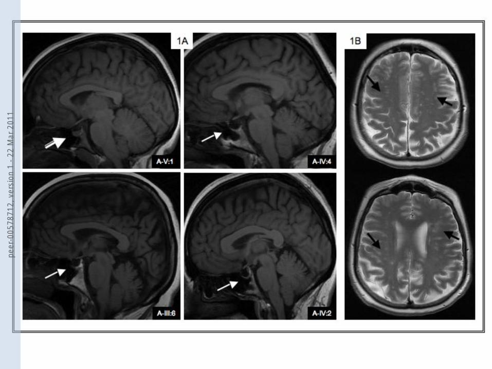
Figure 2. Representative micrograph of HUVEC in Matrigel for 18 hours with EGM-2
grown cells from PROM1 patients and age-matched controls (24, 47 and 61 years old). Cells
labelled red are DiI-labelled EGM-2 grown cells after seven days in culture. Fewer adherent
(red-coloured) cells and a tendency for shorter tubule formation are apparent in patients
compared to controls. The adhesion of the (red-labelled) ECFCs contributes to endothelial
tubule formation. Following culture of ECFCs with HUVEC, fewer ECFCs were adherent or
incorporated into the tubules from the PROM1 subject when compared to the age matched
control. Magnification x100, 8 random fields studied in A-V:1, A-IV:2 and A-III:6 and B-I:1.
Figure 3. Angiogenesis assay for HUVEC and ECFCs.
Alteration in growth of HUVEC tubules with ECFCs from PROM1 subjects.
A. Number of cells adherent/incorporated into endothelial tubules on Matrigel in PROM1
subjects and healthy age-matched controls; B. Number of tubules formed on Matrigel plate,
averaged over 8 random fields between PROM1 patients and controls. C. Average tubule
length of HUVEC formed on Matrigel, averaged over 8 random fields, between PROM1 and
controls, *p<0.05 significant difference between PROM1 and control group.
peer
-005
7871
2, v
ersi
on 1
- 22
Mar
201
1
peer
-005
7871
2, v
ersi
on 1
- 22
Mar
201
1
peer
-005
7871
2, v
ersi
on 1
- 22
Mar
201
1
1.5
form
ed
1.4
1.6
es
60*
rent
A B C
0.5
1.0
mbe
r of
tubu
les
f
1.0
1.2
leng
th o
f tub
ule
20
40
mbe
r ce
lls a
dher
prom-1 control0.0nu
m
prom-1 control
0.8
prom-1 control0
num
peer
-005
7871
2, v
ersi
on 1
- 22
Mar
201
1