explaining the encoding/retrieval flip: memory-related deactivations and activations in the...
TRANSCRIPT
Neuropsychologia 50 (2012) 3764–3774
Contents lists available at SciVerse ScienceDirect
Neuropsychologia
0028-39
http://d
n Corr
Charles
E-m
journal homepage: www.elsevier.com/locate/neuropsychologia
Reviews and perspectives
Explaining the encoding/retrieval flip: Memory-related deactivations andactivations in the posteromedial cortex
W. Huijbers a,b,n, P. Vannini a, R.A. Sperling a, C.M. Pennartzb, R. Cabeza c, S.M. Daselaar a,c,d
a Harvard Medical School, Martinos Center for Biomedical Imaging, Brigham and Women’s Hospital, Boston, MA, USAb University of Amsterdam, Swammerdam Institute for Life Sciences, Faculty of Science, Amsterdam, the Netherlandsc Duke University, Department of Psychology and Neuroscience, Duke University, Durham, NC, USAd Donders Institute for Brain, Cognition and Behaviour, Radboud University Nijmegen, Nijmegen, the Netherlands
a r t i c l e i n f o
Article history:
Received 20 March 2012
Received in revised form
21 August 2012
Accepted 27 August 2012Available online 13 September 2012
Keywords:
Episodic memory
Aging
Encoding
Retrieval
fMRI
Encoding/retrieval-flip
Alzheimers disease
Amyloid
Posteromedial cortex
Precuneus
Posterior cingulate cortex
Retrosplenial cortex
Performance
32/$ - see front matter & 2012 Elsevier Ltd. A
x.doi.org/10.1016/j.neuropsychologia.2012.08
esponding author at: Harvard Medical Schoo
town, MA 02129, United States. Tel.: þ1 617
ail address: [email protected] (
a b s t r a c t
The posteromedial cortex (PMC) is strongly linked to episodic memory and age-related memory deficits.
The PMC shows deactivations during a variety of demanding cognitive tasks as compared to passive baseline
conditions and has been associated with the default-mode of the brain. Interestingly, the PMC exhibits
opposite levels of functional MRI activity during encoding (learning) and retrieval (remembering), a pattern
dubbed the encoding/retrieval flip (E/R-flip). Yet, the exact role of the PMC in memory function has remained
unclear. This review discusses the possible neurofunctional and clinical significance of the E/R-flip pattern.
Regarding neurofunctional relevance, we will review four hypotheses on PMC function: (1) the internal
orienting account, (2) the self-referential processing account, (3) the reallocation account, and (4) the bottom-up
attention account. None of these accounts seem to provide a complete explanation for the E/R-flip pattern in
PMC. Regarding clinical relevance, we review work on aging and Alzheimer’s disease, indicating that amyloid
deposits within PMC, years before clinical memory deficits become apparent. High amyloid burden within
PMC is associated with detrimental influences on memory encoding, in particular, the attenuation of
beneficial PMC deactivations. Finally, we discuss functional subdivisions within PMC that help to provide a
more precise picture of the variety of signals observed within PMC. Collective data from anatomical, task-
related fMRI and resting-state studies all indicate that the PMC is composed of three main regions, the
precuneus, retrosplenial, and posterior cingulate cortex, each with a distinct function. We will conclude with
a summary of the findings and provide directions for future research.
& 2012 Elsevier Ltd. All rights reserved.
Contents
1. Introduction . . . . . . . . . . . . . . . . . . . . . . . . . . . . . . . . . . . . . . . . . . . . . . . . . . . . . . . . . . . . . . . . . . . . . . . . . . . . . . . . . . . . . . . . . . . . . . . . . . . . . 3765
2. The encoding/retrieval flip. . . . . . . . . . . . . . . . . . . . . . . . . . . . . . . . . . . . . . . . . . . . . . . . . . . . . . . . . . . . . . . . . . . . . . . . . . . . . . . . . . . . . . . . . . 3765
2.1. Converging evidence for the encoding/retrieval flip . . . . . . . . . . . . . . . . . . . . . . . . . . . . . . . . . . . . . . . . . . . . . . . . . . . . . . . . . . . . . . . . 3765
2.2. Competition between encoding and retrieval . . . . . . . . . . . . . . . . . . . . . . . . . . . . . . . . . . . . . . . . . . . . . . . . . . . . . . . . . . . . . . . . . . . . . 3766
2.3. Clinical relevance of the encoding/retrieval flip . . . . . . . . . . . . . . . . . . . . . . . . . . . . . . . . . . . . . . . . . . . . . . . . . . . . . . . . . . . . . . . . . . . 3767
3. Theoretical accounts . . . . . . . . . . . . . . . . . . . . . . . . . . . . . . . . . . . . . . . . . . . . . . . . . . . . . . . . . . . . . . . . . . . . . . . . . . . . . . . . . . . . . . . . . . . . . . 3768
3.1. The internal orienting account . . . . . . . . . . . . . . . . . . . . . . . . . . . . . . . . . . . . . . . . . . . . . . . . . . . . . . . . . . . . . . . . . . . . . . . . . . . . . . . . 3768
3.2. The self-referential processing account. . . . . . . . . . . . . . . . . . . . . . . . . . . . . . . . . . . . . . . . . . . . . . . . . . . . . . . . . . . . . . . . . . . . . . . . . . 3768
3.3. The reallocation account . . . . . . . . . . . . . . . . . . . . . . . . . . . . . . . . . . . . . . . . . . . . . . . . . . . . . . . . . . . . . . . . . . . . . . . . . . . . . . . . . . . . . 3769
3.4. The bottom-up attention account . . . . . . . . . . . . . . . . . . . . . . . . . . . . . . . . . . . . . . . . . . . . . . . . . . . . . . . . . . . . . . . . . . . . . . . . . . . . . . 3770
4. Functional subdivisions of PMC. . . . . . . . . . . . . . . . . . . . . . . . . . . . . . . . . . . . . . . . . . . . . . . . . . . . . . . . . . . . . . . . . . . . . . . . . . . . . . . . . . . . . . 3771
5. Conclusions . . . . . . . . . . . . . . . . . . . . . . . . . . . . . . . . . . . . . . . . . . . . . . . . . . . . . . . . . . . . . . . . . . . . . . . . . . . . . . . . . . . . . . . . . . . . . . . . . . . . . 3772
Acknowledgments . . . . . . . . . . . . . . . . . . . . . . . . . . . . . . . . . . . . . . . . . . . . . . . . . . . . . . . . . . . . . . . . . . . . . . . . . . . . . . . . . . . . . . . . . . . . . . . . . . . . 3772
References . . . . . . . . . . . . . . . . . . . . . . . . . . . . . . . . . . . . . . . . . . . . . . . . . . . . . . . . . . . . . . . . . . . . . . . . . . . . . . . . . . . . . . . . . . . . . . . . . . . . . . . . . . 3772
ll rights reserved.
.021
l, Brigham and Women’s Hospital, Department of Neurology, Athinoula A. Martinos Center for Biomedical Imaging,
726 5573.
W. Huijbers).
W. Huijbers et al. / Neuropsychologia 50 (2012) 3764–3774 3765
1. Introduction
The posteromedial cortex (PMC) is strongly associated withepisodic memory and considered a central node of the default-mode (Buckner, Andrews-Hanna, & Schacter, 2008; Raichle et al.,2001). The default-mode network (DMN) involves a set ofstrongly connected regions that in functional neuroimagingstudies tends to be activated during rest but deactivated duringdemanding cognitive tasks (Mazoyer et al., 2001; McKiernan,Kaufman, Kucera-Thompson, & Binder, 2003; Shulman et al.,1997). According to the default-mode hypothesis, these deactiva-tions arise, because PMC and other DMN regions supportcognitive processes that normally occur during rest, but must betemporarily shut down when available resources are needed foractive task performance (Raichle et al., 2001). Interestingly,successful learning of events (episodic encoding) has been asso-ciated with reduced activity in the PMC, whereas successfulretrieval of events (episodic retrieval) has been associated withincreased activity in the same region (e.g. Buckner, Raichle,Miezin, & Petersen, 1996; Daselaar, Prince, & Cabeza, 2004;Hayama, Vilberg, & Rugg, 2012; Kim, 2011; Otten & Rugg, 2001;Shrager, Kirwan, & Squire, 2008; Wagner et al., 1998; Wagner,Shannon, Kahn, & Buckner, 2005). These opposing effects, whichhave been dubbed the encoding/retrieval flip (E/R-flip), wereoriginally reported by Daselaar et al. (2009) who observed thispattern not across participants in separate encoding and retrievalstudies, but within the same study and within the same partici-pants for a variety of stimuli and memory paradigms. Since then,the E/R-flip pattern has been replicated in several other studies(Gilbert, Armbruster, & Panagiotidi, 2011; Huijbers, Pennartz,Cabeza, & Daselaar, 2009, 2011; Kim, Daselaar, & Cabeza, 2010;Vannini, O’Brien, O’Keefe, Pihlajamaki, Laviolette, & Sperling,2011). Yet, despite the robustness of the E/R-flip, the functionalsignificance of this pattern and the role of the PMC in memorystill remain unclear.
This review aims to clarify the relation between the function ofthe PMC and the E/R-flip pattern, and includes three sections.Section 2 reviews studies that found the E/R-flip pattern anddiscusses how the E/R-flip may lead to competition betweenencoding and retrieval processes. Section 2.3 discusses the rele-vance of the E/R-flip for clinical studies of aging and Alzheimer’sdisease and provides a direct link between PMC deactivationsduring encoding and memory-decline. Section 3 focuses on fourdifferent hypotheses that could potentially explain the E/R-flippattern in the PMC. Section 4 of our review discusses anatomical,functional, and connectivity findings indicating three functionallydistinct subregions within PMC; the precuneus (Pcun), posteriorcingulate cortex (PCC), and retrosplenial cortex (RsC). Distin-guishing between these subregions should help to further clarifythe role of PMC in memory function. The review ends with aconcluding section and directions for future research.
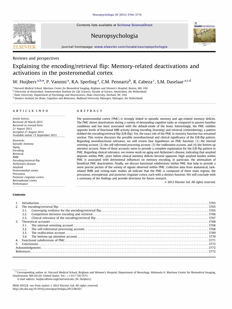
Fig. 1. Panel A: In green, overlapping activity in the medial temporal lobe and
posteromedial cortex during encoding and retrieval. The lines shows the average
time course of activity during successful encoding (ENC) in blue and successful
retrieval (RET) in orange for both regions. Adapted from Vannini et al. (2011).
Panel B shows the encoding/retrieval flip in four different experiments using
(1) faces, (2) spatial scenes, (3) word pairs, and (4) single words. Bars demonstrate
the negative encoding success effect in PMC during encoding (ENC: MISS – HIT) in
blue and the positive retrieval success effect (RET: HIT – MISS) in orange. Adapted
from Daselaar et al. (2009).
2. The encoding/retrieval flip
2.1. Converging evidence for the encoding/retrieval flip
The most powerful method for identifying brain regionsassociated with successful memory encoding processes usingfMRI is known as the subsequent memory paradigm. In thisparadigm, encoding trials are back-sorted based on whether theyare subsequently remembered (hit) or forgotten (miss). Therehave been numerous fMRI studies using this paradigm, whichhave generally found greater activity for encoding hits thanmisses, or a positive encoding success effect, in the medialtemporal lobe (MTL), a pivotal region for episodic memory
function (Kim, 2011; Paller & Wagner, 2002; Uncapher &Wagner, 2009). In contrast, several studies have also found lessactivity for hits than misses, or a negative encoding success effectin the PMC (Fig. 1; e.g. Daselaar et al., 2004; Otten & Rugg, 2001).These positive and negative encoding effects have led to the ideathat the MTL and PMC support distinct cognitive processes, whichare both important for successful memory encoding (Daselaaret al., 2009; Vannini et al., 2011). In contrast to the negative
encoding success effect in PMC during encoding, most retrievalstudies report a positive retrieval success effect in this regionduring memory retrieval, reflecting greater rather than less activityfor retrieval hits than misses (Hayama et al., 2012; Spaniol,Davidson, Kim, Han, Moscovitch, & Grady, et al., 2009). Similarto the effects of memory encoding, a positive retrieval successeffect is assumed to reflect neural mechanisms contributing to thesuccessful remembering of past events.
W. Huijbers et al. / Neuropsychologia 50 (2012) 3764–37743766
The E/R-flip pattern appears to be robust. First, it has beenshown that this pattern occurs regardless of the type of informa-tion (words, faces, spatial scenes), stimulus modality (auditory orvisual), and memory test (item or relational memory) (Fig. 1B;Daselaar et al., 2009; Huijbers et al., 2011). Initially, the E/R-flipwas defined using contrast of hits versus misses (encoding:hitsomiss\retrieval: hit4miss), but recently a similar patternhas been demonstrated using hits as compared to fixation(Vannini et al., 2012; Vannini et al., 2011). Furthermore, thepattern is not restricted to fMRI studies. Recent evidence suggeststhat the E/R-flip can also be observed when using electroence-phalography measurements during encoding and retrieval stages,from cortical sources in PMC (Jaiswal, Ray, & Slobounov, 2010).Taken together, these findings indicate that the E/R-flip patternrepresents a robust neural activity pattern that occurs indepen-dently of the specific memory task, stimulus characteristics, andneuroimaging method being used.
2.2. Competition between encoding and retrieval
Influential models of memory assume that encoding andretrieval cannot occur at the same time and that the twoprocesses compete for neural resources (Hasselmo, Bodelon, &Wyble, 2002; Norman & O’Reilly, 2003; Yassa & Stark, 2011). Inline with these models, we have recently hypothesized that theE/R-flip could also lead to a competition between encoding andretrieval states (Huijbers et al., 2009). Given that global activity ina particular brain region cannot increase and decrease at the sametime, we hypothesized that the negative encoding, and positive
retrieval, success effects in PMC cannot occur simultaneously andwill interact.
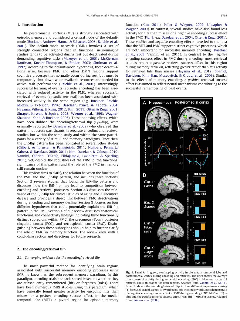
Fig. 2. Panel A shows the experimental design of the concurrent scene encoding/wo
encoding (d-prime) is lower for successful (HIT) as compared to unsuccessful (MISS) ret
concurrently. Panel D, brain activity: the green bars at the bottom represent average PM
and retrieval (RET). Panel E shows the experimental design of the attentional control exp
the behavioral results, an opposite pattern as compared concurrent encoding/retriev
compared to unsuccessful (MISS) retrieval. Adapted from Huijbers et al. (2009).
We investigated the hypothesis that the E/R-flip can lead to acompetition using an fMRI experiment in which participantsencoded and retrieved information within a brief period of time.Participants rapidly encoded words by processing their meaning(living/nonliving decisions) and then performed an old/new wordrecognition task including words presented at the word encodingphase intermixed with new words (Fig. 2A). The key differencewith a standard old/new word recognition test is that, whilerecognizing the words, participants also encoded spatial scenesthat were presented in the background. The paradigm was notsimply measuring potential interference between viewing scenesand making recognition responses, but specifically measuredinterference between successful encoding and successful retrie-val. Potential interference from perceptual or motor processeswas subtracted out, because all trials had scenes in the back-ground and all involved recognition responses. In line with amemory competition, we found that during successful wordretrieval the scenes were less likely to be successfully encoded,and vice versa (Fig. 2B). Moreover, whereas previous studiesfound the E/R-flip pattern across encoding and retrieval sessions,this study showed that the E/R-flip pattern could also be foundwithin the same session and within the same trials (Fig. 2C). Thus,PMC showed greatest activity when retrieval of words wassuccessful and encoding of spatial scenes was unsuccessful, andleast activity when encoding of spatial scenes was successful andretrieval of words unsuccessful (Fig. 2D). In order to assesswhether the apparent competition between encoding and retrievalwas the result of divided attention between word and sceneprocessing, we conducted a follow-up behavioral experiment. Forthis experiment, we replaced the encoding task with an attention-task involving the detection of a small dot that was flashed on the
rd retrieval task. Panel B shows the behavioral results, the accuracy of memory
rieval. Panel C shows brain activity within PMC when encoding and retrieval occur
C activity within the PMC for unsuccessful (MISS)/ successful (HIT) encoding (ENC)
eriment: target-detection of visual dots compared to word retrieval. Panel F shows
al. The accuracy of target detection (d-prime) is higher for successful (HIT) as
W. Huijbers et al. / Neuropsychologia 50 (2012) 3764–3774 3767
screen during memory retrieval (Fig. 2E). In this case, we actuallyfound the opposite pattern: target-detection performance wasworse, rather than better, during unsuccessful retrieval (Fig. 2F).This fits with the idea that unsuccessful retrieval tends to coincidewith a more demanding and extended search process (Rugg &Wilding, 2000), and thus less attention is available for concurrenttarget-detection. Together, these findings indicate that a mereattentional account cannot easily explain the competition betweenencoding and retrieval. Thus, similar to the findings regardingcompetition between encoding and retrieval mediated by thehippocampus (Hasselmo et al., 2002), our results suggest that theE/R-flip pattern also reflects a processing bottleneck betweenencoding and retrieval states. However, even though trail-by-trialfluctuations in theta or gamma oscillations have been associatedwith differences in fMRI signal (Scheeringa et al., 2011; Scheeringaet al., 2009) and theta power predicts encoding-related deactiva-tions of the PMC (White et al., 2012), there is currently no directevidence linking competitive neuronal processes to the E/R-flip.
2.3. Clinical relevance of the encoding/retrieval flip
Recently, clinical interest in PMC function has intensified follow-ing the development of new neuroimaging tools that allow in vivovisualization of amyloid-b deposition; one of the hallmark patholo-gies of Alzheimer’s disease (AD). The accumulation of amyloid-b infibrillar plaques in conjunction with neurofibrillary tangles are thehistopathological features required for the post-mortem confirma-tion of AD (Braak & Braak, 1992). The recent development of amolecular marker Pittsburgh Compound-B (PiB) and other tracershas made it possible to visualize fibrillar forms of amyloid-b in vivousing PET imaging (Klunk et al., 2004). Using PiB, it has been shownthat in older adults PMC is particularly vulnerable to early amyloid-b
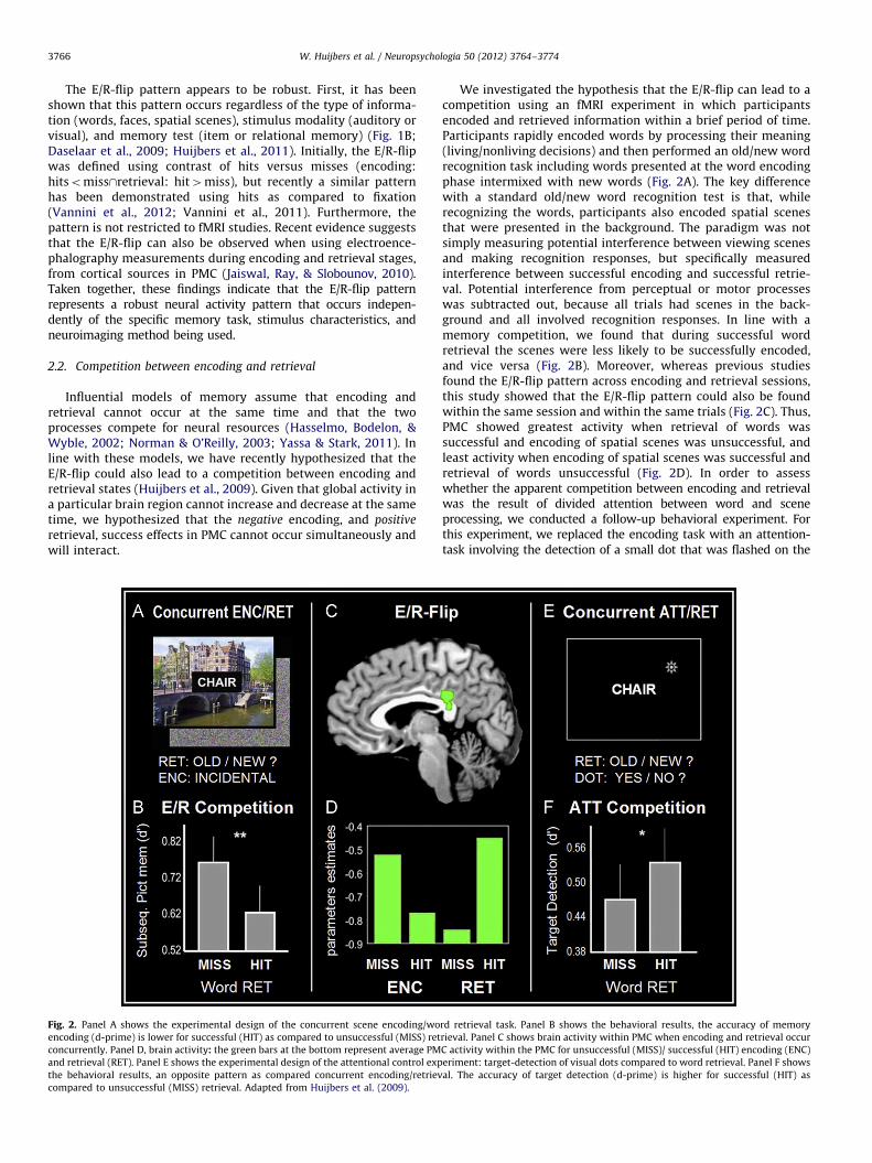
Fig. 3. Panel A: In blue, brain regions along the left midline and hemisphere that show
elderly In orange, brain regions that show activations during memory retrieval (HIT – B
(Baseline – HIT, Po0.01) and retrieval (HIT – Baseline, Po0.01). Panel B: Bars demons
separately for young (YNG), elderly (ELD) with low (PiB-) and high (PiBþ) amounts of am
and amyloid-b (R¼�0.43). The hippocampus, a control regions, shows no correlation
(2012).
deposition (Buckner et al., 2008). Although, tau-pathology andhippocampal atrophy are linked more closely to the clinical syn-drome of memory impairment, amyloid-b accumulation is one ofthe earliest pre-clinical markers of AD (Frisoni, Fox, Jack, Scheltens, &Thompson, 2010; Jack, Knopman, Jagust, Shaw, Aisen, Weiner et al.,2010), and about one third of clinically normal older adults alreadyharbor amyloid-b within PMC (Sperling et al., 2011). Therefore, it isbelieved that amyloid-b accumulation begins many years – perhapsa decade or more – prior to the emergence of the clinical syndromeof AD (Rowe et al., 2010)
Functional MRI studies have found evidence for disruptedPMC activity in older adults who diagnosed with early stages ofAD (Lustig et al., 2003). Furthermore, healthy older adults whoare at high-risk of developing AD, for example those withrelatively poor memory or who carry the APEO-4 allele, alreadyshow a reduced negative encoding success effect in the PMC,akin to AD patients (Miller et al., 2008; Pihlajamaki et al., 2010).Interestingly, several fMRI studies have shown that manynormal older adults who have high amounts of amyloid-b alsoexhibit aberrant brain activity in the PMC during memoryencoding (Kennedy et al., 2012; Mormino et al., 2012; Sperlinget al., 2009). Recently, Vannini et al. (2012) specifically investi-gated the effects of amyloid-b in relation to the E/R-flip, bydirectly contrasting the difference between the deactivationsduring successful encoding with activations during successfulretrieval (Fig. 3A). Older adults showed a reduced E/R-flip withinPMC, and this reduction was more pronounced in the olderadults with high-amounts of amyloid-b (Fig. 3B). Although, itshould be noted that this pattern was mostly driven by areduction of the negative encoding success effect. The reducedability to modulate the activation between encoding andretrieval was also related to decreased performance in thememory task. In contrast, activity in hippocampus was not
deactivations during memory encoding (Baseline – HIT, Po0.01) for young and
aseline, Po0.01). In green, E/R-flip chance: overlapping activity between encoding
trate the average activity in PMC during encoding in blue and retrieval in orange,
yloid-b deposition. Panel C: The PMC shows a correlation between the fMRI signal
between the fMRI signal and amyloid-beta (R¼0.05). Adapted from Vannini et al.
W. Huijbers et al. / Neuropsychologia 50 (2012) 3764–37743768
correlated with levels of amyloid-b (Fig. 3C) or performance.This finding provides a link between aberrant amyloid-b levels,and the E/R-flip in the PMC. Yet, its remains debated if theamyloid burden especially affects memory-related activity orleads to a more general failure to modulate activity in the PMC(Nestor, Scheltens, & Hodges, 2004; Park, Polk, Hebrank, &Jenkins, 2010).
Although amyloid accumulation has been strongly linked tosynaptic activity (Selkoe, 2001), the exact reason why amyloidstarts to aggregate within the PMC is not entirely clear. Severalstudies have shown that amyloid-b – under normal circum-stances – serves as a negative feedback signal that maintainsneuronal activity within a normal dynamic range (Cirrito,Yamada, Finn, Sloviter, Bales, May et al., 2005; Ting, Kelley,Lambert, Cook, & Sullivan, 2007). Thus, it has been suggested thatthe vulnerability of the PMC to amyloid-b might be a conse-quence of the high-levels of synaptic activity in default-moderegions (Bero et al., 2011; Zhang & Raichle, 2010). This view isconsistent with PET studies that found spatial overlap betweenhypo-metabolism, disruption of connectivity and the accumula-tion of amyloid-b (Drzezga et al., 2011). Thus, the reason why thePMC might be particularly vulnerable to amyloid-b, might be aconsequence of it metabolic demands. These metabolic demands,in turn, might reflect the PMC’s dynamic function, with both up-and down regulated activity in response to cognitive demands asreflected by the E/R-flip pattern.
3. Theoretical accounts
The clinical research reviewed in the previous section suggests alink between PMC integrity, the E/R-flip pattern, and episodicmemory. However, these findings do not explain the E/R-flip in termsof underlying cognitive processes. Four prevailing theories couldpotentially explain the E/R-flip: (1) the internal orienting account,(2) the self-referential processing account, (3) the reallocation account
and, (4) the bottom-up attention account. Below, we discuss evidencein favor of, and opposition to, each account.
3.1. The internal orienting account
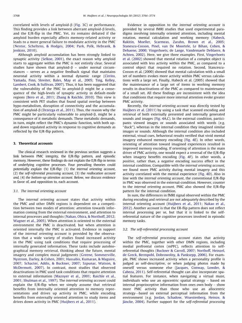
The internal orienting account states that activity withinthe PMC and other DMN regions is dependent on a competi-tion between two modes of attention: attention towards infor-mation coming from the external environment, and attention tointernal processes and thoughts (Nakao, Ohira, & Northoff, 2012;Wagner et al., 2005). When attention is oriented to the externalenvironment the PMC is deactivated, but when attention isoriented internally the PMC is activated. Evidence in supportof the internal orienting account is provided by the observa-tion that a wide variety of studies found increased activityin the PMC using task conditions that require processing ofinternally generated information. These tasks include autobio-graphical memory retrieval, thinking about the future, mentalimagery and complex moral judgments (Greene, Sommerville,Nystrom, Darley, & Cohen, 2001; Hassabis, Kumaran, & Maguire,2007; Schacter, Addis, & Buckner, 2007; Szpunar, Watson, &McDermott, 2007). In contrast, most studies that observeddeactivations in PMC used task conditions that require attentionto external information (Mazoyer et al., 2001; Raichle et al.,2001; Shulman et al., 1997). The internal orienting account couldexplain the E/R-flip when we simply assume that retrievalbenefits from internally oriented attention to memory repre-sentations and drives up activity in PMC, while encodingbenefits from externally oriented attention to study items anddrives down activity in PMC (Huijbers et al., 2011).
Evidence in opposition to the internal orienting account isprovided by several fMRI studies that used experimental para-digms involving internally oriented attention, including mentalrotation, mental calculation and working memory (Habeck,Rakitin, Moeller, Scarmeas, Zarahn, Brown et al., 2005;Stanescu-Cosson, Pinel, van De Moortele, Le Bihan, Cohen, &Dehaene, 2000; Vingerhoets, de Lange, Vandemaele Deblaere, &Achten, 2002). Here, we give three examples. First, Vingerhoetset al. (2002) showed that mental rotation of a complex object isassociated with less activity within the PMC, as compared to acontrol object that required no rotation. Second, Stancescu-Cosson et al. (2000) showed that mental calculation with a smallset of numbers evokes more activity within PMC versus calcula-tions with a large set. Finally, Habeck et al. (2005) showed thatthe maintenance of a large set of items in working memoryresults in deactivations of the PMC as compared to maintenanceof a small set. All these findings are inconsistent with the ideathat conditions that require more internal attention evoke greaterPMC activity.
Recently, the internal orienting account was directly tested byHuijbers et al. (2011) by using a task that scanned encoding andretrieval of both externally presented and internally generatedsounds and images (Fig. 4A,C). In the external condition, partici-pants viewed images or sounds associated with words (e.g.’’duck’’), whereas in the internal condition they imagined similarimages or sounds. Although the internal condition also includedexternal, visual cues, behavioral results verified that vivid mentalimagery enhanced memory encoding (Fig. 4E). In other words,orienting of attention toward imagined experiences resulted inimproved memory encoding. If orienting of attention is the maindriver of PMC activity, one would expect a reversal of the E/R-flipwhen imagery benefits encoding (Fig. 4F). In other words, apositive, rather than, a negative encoding success effect in theinternal condition. Compatible with the internal orienting account,
we found more PMC activity during mental imagery and thisactivity correlated with the mental experience (Fig. 4D). Also inline with the internal orienting account, the conventional E/R-flipin PMC was observed in the external-condition. However, counterto the internal orienting account, PMC also showed the E/R-flippattern for the internal condition.
In sum, the differences in fMRI signal observed within the PMCduring encoding and retrieval are not adequately described by theinternal orienting account (Huijbers et al., 2011; Nakao et al.,2012). Another account is that the ER-flip pattern does not reflectinternal processing per se, but that it is linked to the self-referential nature of the cognitive processes involved in episodicmemory tasks.
3.2. The self-referential processing account
The self-referential processing account states that activitywithin the PMC, together with other DMN regions, includingmedial prefrontal cortex (mPFC), reflects attention to self-referential thoughts (Buckner & Carroll, 2007; Northoff, Heinzel,de Greck, Bermpohl, Dobrowolny, & Panksepp, 2006). For exam-ple, PMC shows increased activity when a personality profile isjudged as self-descriptive, or when judging photos made byoneself versus someone else (Jacques, Conway, Lowder, &Cabeza, 2011). Self-referential thought can also incorporate spa-tial features. For instance, when navigating a virtual maze,individuals who use an egocentric spatial strategy – based oninternal proprioceptive information from ones own body – showmore PMC activity than those who use an allocentricstrategy—based on external perceptual information from theenvironment (e.g. Jordan, Schadow, Wuestenberg, Heinze, &Jancke, 2004). Further support for the self-referential processing
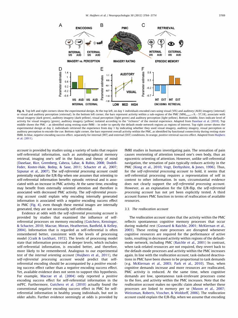
Fig. 4. Top left and right corners show the experimental design. At the top left, on day 1 individuals encoded cues using visual (VIS) and auditory (AUD) imagery (internal)
or visual and auditory perception (external). In the bottom left corner, the bars represent activity within a sub-regions of the PMC (MNI(x,y,z)¼9,�57,18), associate with
visual imagery (dark green), auditory imagery (dark yellow), visual perception (light green) and auditory perception (light yellow). Bottom middle, lines indicate level of
activity for visual imagery (green), auditory imagery (yellow) isolated according to the ‘‘richness’’ of the mental experience. Adapted from Daselaar et al. (2010). Top
middle shows the PMC – as identified using resting-state fMRI – in order to specify the default-mode network regions as regions of interest. Top right corner shows the
experimental design at day 2, individuals retrieved the experience from day 1 by indicating whether they used visual imagery, auditory imagery, visual perception or
auditory perception to encode the cue. Bottom right corner, the bars represent overall activity within the PMC, as identified by functional connectivity during resting-state
fMRI. In blue, negative encoding success effect. separately for internal (INT) and external (EXT) conditions, In orange, positive retrieval success effect. Adapted from Huijbers
et al. (2011).
W. Huijbers et al. / Neuropsychologia 50 (2012) 3764–3774 3769
account is provided by studies using a variety of tasks that requireself-referential information, such as autobiographical memoryretrieval, imaging one’s self in the future, and theory of mind(Daselaar, Rice, Greenberg, Cabeza, Labar, & Rubin, 2008; Dodell-Feder, Koster-Hale, Bedny, & Saxe, 2011; Schacter et al., 2007;Szpunar et al., 2007). The self-referential processing account couldpotentially explain the E/R-flip when one assumes that orienting toself-referential information benefits episodic retrieval and is asso-ciated with an increase in PMC activity. At the same time, encodingmay benefit from externally oriented attention and therefore isassociated with decreased PMC activity. The self-referential proces-
sing account could explain why encoding internally generatedinformation is associated with a negative encoding success effectin PMC (Fig. 4), even though these mental images are internallygenerated, they are not necessarily self-referential.
Evidence at odds with the self-referential processing account isprovided by studies that examined the influence of self-referential processes on memory encoding (Gutchess, Kensinger,& Schacter, 2010; Macrae, Moran, Heatherton, Banfield, & Kelley,2004). Information that is regarded as self-referential is oftenremembered better, consistent with the levels of processingmodel (Craik & Lockhart, 1972). The levels of processing modelstate that information processed at deeper levels, which includesself-referential information, is encoded better, and therefore,more likely to be remembered. Analogous to our experimentaltest of the internal orienting account (Huijbers et al., 2011), theself-referential processing account would predict that self-referential encoding should be accompanied by a positive encod-ing success effect rather than a negative encoding success effect.Yet, available evidence does not seem to support this hypothesis.For example, Macrae et al. (2004) only reported a positive
encoding success effect for self-referential information in themPFC. Furthermore, Gutchess et al. (2010) actually found theconventional negative encoding success effect in PMC for self-referential information in healthy young individuals, but not inolder adults. Further evidence seemingly at odds is provided by
fMRI studies in humans investigating pain. The sensation of paincauses reorienting of attention toward one’s own body, thus anegocentric orienting of attention. However, unlike self-referentialnavigation, the sensation of pain typically reduces activity in thePMC (Kong et al., 2010; Vogt, Derbyshire, & Jones, 1996). Thus,for the self-referential processing account to hold, it seems thatself-referential processing requires a representation of self incontext to other information. In sum, circumstantial evidencedoes not clearly support the self-referential processing account.
However, as an explanation for the E/R-flip, the self-referential
processing account has not yet been explicitly tested. A thirdaccount frames PMC function in terms of reallocation of availableresources.
3.3. The reallocation account
The reallocation account states that the activity within the PMCreflects spontaneous cognitive memory processes that occurduring wakeful rest (Gusnard & Raichle, 2001; McKiernan et al.,2003). These resting state processes are disrupted whenevercognitive resources are required for the performance of activetasks, resulting in decreased activity within regions of the default-mode network, including PMC (Raichle et al., 2001) In contrast,when task-related resources are not required, they revert back tothe default-mode processes and activity within the PMC increasesagain. In line with the reallocation account, task-induced deactiva-tions in PMC have been shown to be proportional to task demands(e.g. McKiernan et al., 2003; Park et al., 2010). Thus, whencognitive demands increase and more resources are reallocated,PMC activity is reduced. At the same time, when cognitivedemands are low, spontaneous task-irrelevant processes cometo the fore, and activity within the PMC increases. Note that thereallocation account makes no specific claim about whether theseprocesses are linked to memory per se (Mason et al., 2007;Weissman, Roberts, Visscher, & Woldorff, 2006). The reallocation
account could explain the E/R-flip, when we assume that encoding
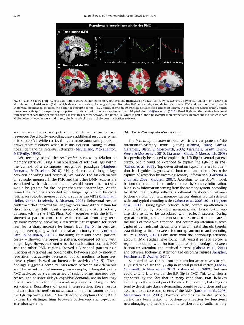
Fig. 5. Panel A shows brain regions significantly activated during memory retrieval and modulated by a task-difficulty (easy/short-delay versus difficult/long-delay). In
blue the retrosplenial cortex (RsC), which shows more activity for longer delays. Note that RsC connectivity extends into the ventral PCC and does not exactly match
anatomical boundaries. In green the posterior cingulate cortex (PCC), which shows an interaction between long and short delays. In red, the precuneus (Pcun), which
shows less activity for longer delays, a pattern consistent with the reallocation account. Adapted from Huijbers et al. (2010). Panel B shows the relative functional
connectivity of each these of regions with a distributed cortical network. In blue the RsC which is part of the hippocampal-memory network. In green the PCC which is part
of the default-mode network and in red, the Pcun which is part of the dorsal attention network.
W. Huijbers et al. / Neuropsychologia 50 (2012) 3764–37743770
and retrieval processes put different demands on corticalresources. Specifically, encoding draws additional resources whenit is successful, while retrieval – as a more automatic process –draws more resources when it is unsuccessful leading to addi-tional, demanding, retrieval attempts (McClelland, McNaughton,& O’Reilly, 1995).
We recently tested the reallocation account in relation tomemory retrieval, using a manipulation of retrieval lags withinthe context of a continuous recognition paradigm (Huijbers,Pennartz, & Daselaar, 2010). Using shorter and longer lagsbetween encoding and retrieval, we varied the task-demandson episodic memory. If the PMC and the other DMN regions areassociated with task demands, one would expect that activitywould be greater for the longer than the shorter lags. At thesame time, regions associated with longer lags should be morereliant on episodic memory regions such as the MTL (Ranganath,Heller, Cohen, Brozinsky, & Rissman, 2005). Behavioral resultsconfirmed that retrieval for long lags was more difficult than forshort lags. The fMRI results indicated three distinct activitypatterns within the PMC. First, RsC – together with the MTL –showed a pattern consistent with retrieval from long-termepisodic memory, showing a relatively flat response for shortlags, but a sharp increase for longer lags (Fig. 5). In contrast,regions overlapping with the dorsal attention system (Corbetta,Patel, & Shulman, 2008) – including Pcun and dorsal parietalcortex – showed the opposite pattern, decreased activity withlonger lags. However, counter to the reallocation account, PCCand the other DMN regions showed a V-shaped pattern as afunction of retrieval lag. Specifically, between short to mediumrepetition lags activity decreased, but for medium to long lags,these regions showed an increase in activity (Fig. 5). Thesefindings suggest a complex interaction between task demandsand the recruitment of memory. For example, at long delays thePMC activates as a consequence of task-relevant memory pro-cesses. Yet, at short delays, cognitive demands are so low theymight leave room for mind-wandering again resulting in PMCactivations. Regardless of exact interpretation, these resultsindicate that the reallocation account alone also cannot explainthe E/R-flip within PMC. A fourth account explains the E/R-flippattern by distinguishing between bottom-up and top-downattention systems.
3.4. The bottom-up attention account
The bottom-up attention account, which is a component of theAttention-to-Memory model (AtoM) (Cabeza, 2008; Cabeza,Ciaramelli, Olson, & Moscovitch, 2008; Ciaramelli, Grady, Levine,Ween, & Moscovitch, 2010; Ciaramelli, Grady, & Moscovitch, 2008)has previously been used to explain the E/R-flip in ventral parietalcortex, but it could be extended to explain the E/R-flip in PMC(Cabeza et al., 2011). Top-down attention typically refers to atten-tion that is guided by goals, while bottom-up attention refers to thecapture of attention by incoming sensory information (Corbetta &Shulman, 2002; Knudsen, 2007). According to the AtoM model,bottom-up attention is not only captured by sensory information,but also by information coming from the memory system. Accordingto AtoM, the E/R-flip reflects a different relationship betweenbottom-up attention and memory success during typical retrievaltasks and typical encoding tasks (Cabeza et al., 2008, 2011; Huijberset al., 2011). During typical retrieval tasks, bottom-up attention isoften captured by recovered memories, and hence bottom-upattention tends to be associated with retrieval success. Duringtypical encoding tasks, in contrast, to-be-encoded stimuli are atthe focus of top-down attention, and bottom-up attention is oftencaptured by irrelevant thoughts or environmental stimuli, therebyestablishing a link between bottom-up attention and encodingfailure (Cabeza, 2008). Consistent with the bottom-up attentionaccount, fMRI studies have found that ventral parietal cortex, aregion associated with bottom-up attention, overlaps betweenbottom-up attention and retrieval success (Cabeza et al., 2011)and between bottom-up attention and encoding failure (Uncapher,Hutchinson, & Wagner, 2011).
As noted above, the bottom-up attention account was origin-ally used to explain the E/R-flip in ventral parietal cortex (Cabeza,Ciaramelli, & Moscovitch, 2012; Cabeza et al., 2008), but onecould extend it to explain the E/R-flip in PMC. This extension issupported by the fact that in many conditions, PMC behavessimilarly as the ventral parietal cortex. For example, both regionstend to deactivate during demanding cognitive conditions and areassumed to be core components of the DMN (Buckner et al., 2008;McKiernan et al., 2003). However, whereas the ventral parietalcortex has been linked to bottom-up attention by functionalneuroimaging and patient data in attention and episodic memory
W. Huijbers et al. / Neuropsychologia 50 (2012) 3764–3774 3771
domains (Cabeza et al., 2008; Corbetta & Shulman, 2002), the linkin the literature between PMC and bottom-up attention is not asstrong.
In sum, none of the four accounts adequately explain the E/R-flip pattern within the PMC. These cognitive accounts are also notmutually exclusive. For example, internal orienting might engagemore self-referential processing. Likewise, bottom-up orienting tosalient information might lead to greater reallocation of resources(Raichle et al., 2001). At present, there is no overarching accountthat can fully explain PMC’s behavior in various cognitive condi-tions. The lack of a uniform theory of PMC function might alsoreflect the fact that the PMC is not a single uniform brain region,and aforementioned accounts might actually be more-or-lessapplicable to distinct regions within PMC. Thus, in order to obtaina more complete picture of the functional role of PMC, it is criticalto consider the existence of different subregions within PMC morecarefully.
Fig. 6. Simplified representation of the cortical networks involved in episodic
memory. On the left the lateral surface and right the medial surface of the left-
hemisphere. In orange, regions that tend to activate during both encoding and
retrieval, which include the hippocampal-memory network, consisting of the
hippocampal formation (HF) and the retrosplenial cortex (RsC) and the dorsal-
attention network consisting of the precuneus (Pcun), the dorsal parietal cortex
(DPC) and the lateral prefrontal cortex (lPFC). In blue regions that tend to show the
encoding/retrieval flip, including default-mode network regions: posterior cingu-
lated cortex (PCC), the ventral parietal cortex (VPC) and medial prefrontal cortex
(mPFC).
4. Functional subdivisions of PMC
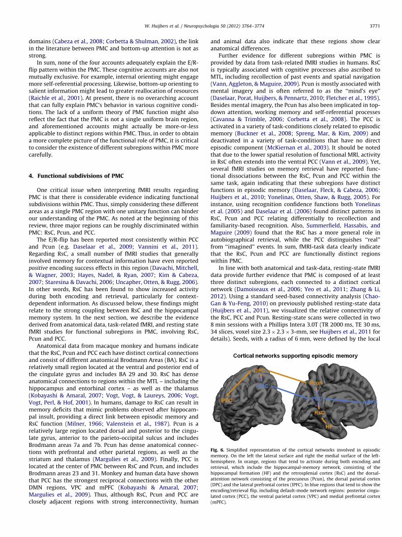
One critical issue when interpreting fMRI results regardingPMC is that there is considerable evidence indicating functionalsubdivisions within PMC. Thus, simply considering these differentareas as a single PMC region with one unitary function can hinderour understanding of the PMC. As noted at the beginning of thisreview, three major regions can be roughly discriminated withinPMC: RsC, Pcun, and PCC.
The E/R-flip has been reported most consistently within PCCand Pcun (e.g. Daselaar et al., 2009; Vannini et al., 2011).Regarding RsC, a small number of fMRI studies that generallyinvolved memory for contextual information have even reportedpositive encoding success effects in this region (Davachi, Mitchell,& Wagner, 2003; Hayes, Nadel, & Ryan, 2007; Kim & Cabeza,2007; Staresina & Davachi, 2006; Uncapher, Otten, & Rugg, 2006).In other words, RsC has been found to show increased activityduring both encoding and retrieval, particularly for context-dependent information. As discussed below, these findings mightrelate to the strong coupling between RsC and the hippocampalmemory system. In the next section, we describe the evidencederived from anatomical data, task-related fMRI, and resting statefMRI studies for functional subregions in PMC, involving RsC,Pcun and PCC.
Anatomical data from macaque monkey and humans indicatethat the RsC, Pcun and PCC each have distinct cortical connectionsand consist of different anatomical Brodmann Areas (BA). RsC is arelatively small region located at the ventral and posterior end ofthe cingulate gyrus and includes BA 29 and 30. RsC has denseanatomical connections to regions within the MTL – including thehippocampus and entorhinal cortex – as well as the thalamus(Kobayashi & Amaral, 2007; Vogt, Vogt, & Laureys, 2006; Vogt,Vogt, Perl, & Hof, 2001). In humans, damage to RsC can result inmemory deficits that mimic problems observed after hippocam-pal insult, providing a direct link between episodic memory andRsC function (Milner, 1966; Valenstein et al., 1987). Pcun is arelatively large region located dorsal and posterior to the cingu-late gyrus, anterior to the parieto-occipital sulcus and includesBrodmann areas 7a and 7b. Pcun has dense anatomical connec-tions with prefrontal and other parietal regions, as well as thestriatum and thalamus (Margulies et al., 2009). Finally, PCC islocated at the center of PMC between RsC and Pcun, and includesBrodmann areas 23 and 31. Monkey and human data have shownthat PCC has the strongest reciprocal connections with the otherDMN regions, VPC and mPFC (Kobayashi & Amaral, 2007;Margulies et al., 2009). Thus, although RsC, Pcun and PCC areclosely adjacent regions with strong interconnectivity, human
and animal data also indicate that these regions show clearanatomical differences.
Further evidence for different subregions within PMC isprovided by data from task-related fMRI studies in humans. RsCis typically associated with cognitive processes also ascribed toMTL, including recollection of past events and spatial navigation(Vann, Aggleton, & Maguire, 2009). Pcun is mostly associated withmental imagery and is often referred to as the ‘‘mind’s eye’’(Daselaar, Porat, Huijbers, & Pennartz, 2010; Fletcher et al., 1995).Besides mental imagery, the Pcun has also been implicated in top-down attention, working memory and self-referential processes(Cavanna & Trimble, 2006; Corbetta et al., 2008). The PCC isactivated in a variety of task-conditions closely related to episodicmemory (Buckner et al., 2008; Spreng, Mar, & Kim, 2009) anddeactivated in a variety of task-conditions that have no directepisodic component (McKiernan et al., 2003). It should be notedthat due to the lower spatial resolution of functional MRI, activityin RsC often extends into the ventral PCC (Vann et al., 2009). Yet,several fMRI studies on memory retrieval have reported func-tional dissociations between the RsC, Pcun and PCC within thesame task, again indicating that these subregions have distinctfunctions in episodic memory (Daselaar, Fleck, & Cabeza, 2006;Huijbers et al., 2010; Yonelinas, Otten, Shaw, & Rugg, 2005). Forinstance, using recognition confidence functions both Yonelinaset al. (2005) and Daselaar et al. (2006) found distinct patterns inRsC, Pcun and PCC relating differentially to recollection andfamiliarity-based recognition. Also, Summerfield, Hassabis, andMaguire (2009) found that the RsC has a more general role inautobiographical retrieval, while the PCC distinguishes ‘‘real’’from ‘‘imagined’’ events. In sum, fMRI-task data clearly indicatethat the RsC, Pcun and PCC are functionally distinct regionswithin PMC.
In line with both anatomical and task-data, resting-state fMRIdata provide further evidence that PMC is composed of at leastthree distinct subregions, each connected to a distinct corticalnetwork (Damoiseaux et al., 2006; Yeo et al., 2011; Zhang & Li,2012). Using a standard seed-based connectivity analysis (Chao-Gan & Yu-Feng, 2010) on previously published resting-state data(Huijbers et al., 2011), we visualized the relative connectivity ofthe RsC, PCC and Pcun. Resting-state scans were collected in two8 min sessions with a Phillips Intera 3.0T (TR 2000 ms, TE 30 ms,34 slices, voxel size 2.3�2.3�3-mm, see Huijbers et al., 2011 fordetails). Seeds, with a radius of 6 mm, were defined by the local
W. Huijbers et al. / Neuropsychologia 50 (2012) 3764–37743772
maxima from the triple dissociation between the PMC subregionstaken from the memory study that manipulated retrieval lags(Huijbers et al., 2010, MNI coordinates: RsC(x,y,z)¼[�6,�51,15],Pcun(x,y,z)¼[6,�60,51] and PCC(x,y,z)¼[0,�51,33]). Connectivitydifference maps were created by subtracting each seed-map fromthe other seed-maps (i.e., RsC–Pcun; Pcun–PCC; PCC–Pcun). Next,a random effects analysis was conducted using a one-samplet-test (Po0.001, cluster size¼10), resulting in six group-maps.Finally, using a conjunction approach, we extracted the relativeconnectivity of each PMC seed ([RsC4Pun\RsC4PCC],[Pcun4PCC\Pcun4RsC], [PCC4RsC\PCC4Pcun]). The resultingseed-maps again confirmed that the RsC, Pcun and PCC areneuroanatomically distinct regions, each preferentially connectedto a different cortical network (Fig. 5B). Specifically, the RsC isassociated more with the hippocampal-memory system (Vannet al., 2009), the Pcun is associated more with the dorsal-attentionsystem (Corbetta et al., 2008), and the PCC is associated morewith the default-mode network (Buckner et al., 2008). Note again,that due to the relatively low spatial resolution of functional MRI,connectivity of the RsC extends somewhat into the most ventralpart of PCC. In sum, the anatomical, fMRI-task, and resting-stateconnectivity data clearly indicate that the RsC, Pcun and PCC formfunctionally distinct regions within the PMC.
5. Conclusions
The PMC reliably shows opposing levels of activation duringencoding and retrieval, the E/R-flip pattern, and this can lead to acompetition between encoding and retrieval states. In terms ofclinical relevance, age-related pathology, specifically amyloiddeposition within the PMC, has detrimental effects on the E/R-flip. Thus, the E/R-flip is an interesting candidate for trackinglongitudinal changes in episodic memory during pre-clinicalstages of Alzheimer’s disease (Sperling et al., 2011). We alsoreviewed four hypotheses that may explain the E/R-flip pattern,the internal orienting account, the self-referential processing
account, the reallocation account, and the bottom-up attention
account. The internal orienting account asserts that PMC involve-ment in encoding and retrieval is dependent on the internalversus external orientation of attentional. The self-referential
processing account explains PMC activity in terms of orientingtoward self-relevant thoughts versus the external environment.The reallocation account states that the activation differences inPMC depend on task-demands and follows response times.Finally, the bottom-up attention account asserts that activitywithin default-mode regions reflects bottom-up orienting ofattention towards information retrieved from memory. Yet noneof these cognitive accounts seems to provide a full explanation forthe E/R-flip pattern in the PMC. Finally, we addressed the issuethat hinders understanding of the PMC. Specifically, anatomicalstudies, task-based fMRI studies, and resting state fMRI studies allindicate that PMC is not a single, homogeneous, region butconsists of different sub-regions, the retrosplenial cortex, theposterior cingulate cortex and the precuneus, each makingseparate contributions to memory encoding and retrieval (Fig.6). Future research should take these distinctions into account inorder to clarify the E/R-flip pattern in PMC.
On a final note, the underlying neuronal mechanisms that giverise to the E/R-flip are still unknown. The fMRI signal is known tocorrelate with local field potentials and is believed to reflect acombination of neuronal input and output (Logothetis, Pauls,Augath, Trinath, & Oeltermann, 2001). Recently, it has beenshown that electrical stimulation can induce BOLD deactivationsin highly connected brain regions through inhibitory neuronalinput (Logothetis et al., 2010). One possible mechanism is that the
E/R-flip in the PCC reflects a combination of fMRI signal frominhibitory input in layers II and III and excitatory output in layersV and VI. For example, inhibitory input from sensory regionscould drive encoding-related deactivations in the PCC, whileexcitatory output to the parahippocampus could drive retrieval-related activations. However, human imaging studies currentlylack the temporal and spatial resolution to investigate suchmechanisms. Studies in monkeys and epilepsy patients, thatinvolve intracranial recording techniques with high spatial andtemporal resolution should help to elucidate the neural mechanismsthat underlie the E/R-flip.
Acknowledgments
This work was supported by the European Molecular BiologyOrganization: ALTF 318-2011 [W.H.], the Amsterdam Brain Ima-ging Platform [W.H, S.D.], the Marie Curie Fellowship: FP7-PEOPLE-2007-4-1-IOF from the European Union [P.V.], the SwedishBrain Foundation and Swedish Society for Medicine [P.V.], theInstitutes of Health: K24 AG035007 [R.S.], R01 AG027435-S1 [R.S.],P01AG036694 [R.S.], P50AG00513421 [R.S.], and the Alzheimer’sAssociation: IIRG-06-27374 [R.S] and the Netherlands Organizationfor Scientific Research, VICI 918.46.609 [C.P.] and VENI 916.66.022[S.D.].
References
Bero, A. W., Yan, P., Roh, J. H., Cirrito, J. R., Stewart, F. R., Raichle, M. E., et al. (2011).Neuronal activity regulates the regional vulnerability to amyloid-[beta]deposition. Nature Neuroscience, 14(6), 750–756.
Braak, H., & Braak, E. (1992). The human entorhinal cortex: Normal morphologyand lamina-specific pathology in various diseases. Neuroscience Research, 15(1-2),6–31.
Buckner, R. L., & Carroll, D. C. (2007). Self-projection and the brain. Trends inCognitive Sciences, 11(2), 49–57.
Buckner, R. L., Andrews-Hanna, J. R., & Schacter, D. L. (2008). The brain’s defaultnetwork: Anatomy, function, and relevance to disease. Annals of New YorkAcademy of Sciences, 1124, 1–38.
Buckner, R. L., Raichle, M. E., Miezin, F. M., & Petersen, S. E. (1996). Functionalanatomic studies of memory retrieval for auditory words and visual pictures.Journal of Neuroscience, 16(19), 6219–6235.
Cabeza, R. (2008). Role of parietal regions in episodic memory retrieval: The dualattentional processes hypothesis. Neuropsychologia, 46(7), 1813–1827.
Cabeza, R., Ciaramelli, E., Olson, I. R., & Moscovitch, M. (2008). The parietal cortexand episodic memory: An attentional account. Nature Reviews Neuroscience,9(8), 613–625.
Cabeza, R., Mazuz, Y. S., Stokes, J., Kragel, J. E., Woldorff, M. G., Ciaramelli, E., et al.(2011). Overlapping parietal activity in memory and perception: Evidence forthe attention to memory model. Journal of Cognitive Neuroscience, 23(11),3209–3217.
Cabeza, R., Ciaramelli, E., & Moscovitch, M. (2012). Cognitive contributions of theventral parietal cortex: An integrative theoretical account. Trends in CognitiveSciences.
Cavanna, A. E., & Trimble, M. R. (2006). The precuneus: A review of its functionalanatomy and behavioural correlates. Brain, 129(Pt 3), 564–583.
Chao-Gan, Y., & Yu-Feng, Z. (2010). DPARSF: A MATLAB toolbox for ’’Pipeline’’ dataanalysis of resting-state fMRI. Frontiers in Systems Neuroscience, 4, 13.
Ciaramelli, E., Grady, C., Levine, B., Ween, J., & Moscovitch, M. (2010). Top-downand bottom-up attention to memory are dissociated in posterior parietalcortex: Neuroimagingand and neuropsychological evidence. Journal of Neu-roscience, 30(14), 4943–4956.
Ciaramelli, E., Grady, C. L., & Moscovitch, M. (2008). Top-down and bottom-upattention to memory: A hypothesis (AtoM) on the role of the posterior parietalcortex in memory retrieval. Neuropsychologia, 46(7), 1828–1851.
Cirrito, J. R., Yamada, K. A., Finn, M. B., Sloviter, R. S., Bales, K. R., May, P. C., et al.(2005). Synaptic activity regulates interstitial fluid amyloid-beta levels in vivo.Neuron, 48(6), 913–922.
Corbetta, M., Patel, G., & Shulman, G. L. (2008). The reorienting system of thehuman brain: From environment to theory of mind. Neuron, 58(3), 306–324.
Corbetta, M., & Shulman, G. L. (2002). Control of goal-directed and stimulus-drivenattention in the brain. Nature Reviews Neuroscience, 3(3), 201–215.
Craik, F. I. M., & Lockhart, R. S. (1972). Levels of processing: A framework formemory research. Journal of Verbal Learning and Behavior, 11, 671–684.
Damoiseaux, J. S., Rombouts, S. A., Barkhof, F., Scheltens, P., Stam, C. J., Smith, S. M.,et al. (2006). Consistent resting-state networks across healthy subjects.Proceedings of the National Academy of Science USA, 103(37), 13848–13853.
W. Huijbers et al. / Neuropsychologia 50 (2012) 3764–3774 3773
Daselaar, S. M., Fleck, M. S., & Cabeza, R. (2006). Triple dissociation in the medialtemporal lobes: Recollection, familiarity, and novelty. Journal of Neurophysiology,96(4), 1902–1911.
Daselaar, S. M., Rice, H. J., Greenberg, D. L., Cabeza, R., Labar, K. S., & Rubin, D. C.(2007). The spatiotemporal dynamics of autobiographical memory: Neuralcorrelates of recall, emotional intensity, and reliving. Cerebral Cortex.
Daselaar, S. M., Porat, Y., Huijbers, W., & Pennartz, C. M. (2010). Modality-specificand modality-independent components of the human imagery system. Neuro-image, 52(2), 677–685.
Daselaar, S. M., Prince, S. E., & Cabeza, R. (2004). When less means more:Deactivations during encoding that predict subsequent memory. Neuroimage,23(3), 921–927.
Daselaar, S. M., Prince, S. E., Dennis, N. A., Hayes, S. M., Kim, H., & Cabeza, R. (2009).Posterior midline and ventral parietal activity is associated with retrievalsuccess and encoding failure. Frontiers in Human Neuroscience, 3, 13.
Davachi, L., Mitchell, J. P., & Wagner, A. D. (2003). Multiple routes to memory:Distinct medial temporal lobe processes build item and source memories.Proceedings of the National Academy of Science USA, 100(4), 2157–2162.
Drzezga, A., Becker, J. A., Van Dijk, K. R. A., Sreenivasan, A., Talukdar, T., Sullivan,C., et al. (2011). Neuronal dysfunction and disconnection of cortical hubs innon-demented subjects with elevated amyloid burden. Brain, 134(6),1635–1646.
Dodell-Feder, D., Koster-Hale, J., Bedny, M., & Saxe, R. (2011). fMRI item analysis ina theory of mind task. Neuroimage, 55(2), 705–712. [doi: 10.1016/j.neuroimage.2010.12.040].
Fletcher, P. C., Frith, C. D., Baker, S. C., Shallice, T., Frackowiak, R. J., & Dolan, R. J.(1995). The minds eye-precuneus activation in memory-related imagery.Neuroimage, 2(3), 195–200.
Frisoni, G. B., Fox, N. C., Jack, C. R., jr., Scheltens, P., & Thompson, P. M. (2010). Theclinical use of structural MRI in Alzheimer disease. Nature Reviews Neurology,6(2), 67–77.
Gilbert, S. J., Armbruster, D. J. N., & Panagiotidi, M. (2011). Similarity between brainactivity at encoding and retrieval predicts successful realization of delayedintentions. Journal of Cognitive Neuroscience, 0(0), 1–13.
Greene, J. D., Sommerville, R. B., Nystrom, L. E., Darley, J. M., & Cohen, J. D. (2001).An fMRI investigation of emotional engagement in moral judgment. Science,293(5537), 2105–2108.
Gusnard, D. A., & Raichle, M. E. (2001). Searching for a baseline: Functionalimaging and the resting human brain. Nature Reviews Neuroscience, 2(10),685–694.
Gutchess, A. H., Kensinger, E. A., & Schacter, D. L. (2010). Functional neuroimagingof self-referential encoding with age. Neuropsychologia, 48(1), 211–219.
Habeck, C., Rakitin, B. C., Moeller, J., Scarmeas, N., Zarahn, E., Brown, T., et al.(2005). An event-related fMRI study of the neural networks underlying theencoding, maintenance, and retrieval phase in a delayed-match-to-sampletask. Cognitive Brain Research, 23(2-3), 207-220.
Hassabis, D., Kumaran, D., & Maguire, E. A. (2007). Using imagination to under-stand the neural basis of episodic memory. Journal of Neuroscience, 27(52),14365–14374.
Hasselmo, M. E., Bodelon, C., & Wyble, B. P. (2002). A proposed function forhippocampal theta rhythm: Separate phases of encoding and retrieval enhancereversal of prior learning. Neural Computation, 14(4), 793–817.
Hayama, H. R., Vilberg, K. L., & Rugg, M. D. (2002). Overlap between the neuralcorrelates of cued recall and source memory: Evidence for a generic recollec-tion network?. Journal of Cognitive Neuroscience, 24(5), 1127–1137.
Hayes, S. M., Nadel, L., & Ryan, L. (2007). The effect of scene context on episodicobject recognition: Parahippocampal cortex mediates memory encoding andretrieval success. Hippocampus, 17(9), 873–889.
Huijbers, W., Pennartz, C. M., Cabeza, R., & Daselaar, S. M. (2009). When learningand remembering compete: A functional MRI study. PLoS Biology, 7(1), e11.
Huijbers, W., Pennartz, C. M., Cabeza, R., & Daselaar, S. M. (2011). The Hippocam-pus is coupled with the default network during memory retrieval but notduring memory encoding. PLoS One, 6(4), e17463.
Huijbers, W., Pennartz, C. M., & Daselaar, S. M. (2010). Dissociating the ’’retrievalsuccess’’ regions of the brain: Effects of retrieval delay. Neuropsychologia,48(2), 491–497.
Jack, C. R., Jr., Knopman, D. S., Jagust, W. J., Shaw, L. M., Aisen, P. S., Weiner, M. W.,et al. (2010). Hypothetical model of dynamic biomarkers of the Alzheimer’spathological cascade. Lancet Neurology, 119-128.
Jaiswal, N., Ray, W., & Slobounov, S. (2010). Encoding of visual–spatial informationin working memory requires more cerebral efforts than retrieval: Evidencefrom an EEG and virtual reality study. Brain Research, 1347(0), 80–89.
Jordan, K., Schadow, J., Wuestenberg, T., Heinze, H.-J., & Jancke, L. (2004). Differentcortical activations for subjects using allocentric or egocentric strategies in avirtual navigation task. Neuroreport, 15(1), 135–140.
Kennedy, K. M., Rodrigue, K. M., Devous, M. D., Sr., Hebrank, A. C., Bischof, G. N., &Park, D. C. (2012). Effects of beta-amyloid accumulation on neural functionduring encoding across the adult lifespan. Neuroimage, 62(1), 1–8.
Kim, H. (2011). Neural activity that predicts subsequent memory and forgetting:A meta-analysis of 74 fMRI studies. Neuroimage, 54(3), 2446–2461.
Kim, H., & Cabeza, R. (2007). Differential contributions of prefrontal, medialtemporal, and sensory-perceptual regions to true and false memory formation.Cerebral Cortex, 17(9), 2143–2150.
Kim, H., Daselaar, S. M., & Cabeza, R. (2010). Overlapping brain activity betweenepisodic memory encoding and retrieval: roles of the task-positive and task-negative networks. Neuroimage, 49(1), 1045–1054.
Klunk, W. E., Engler, H., Nordberg, A., Wang, Y., Blomqvist, G., Holt, D. P., et al.(2004). Imaging brain amyloid in Alzheimer’s disease with PittsburghCompound-B. Annals of Neurology, 55(3), 306–319.
Knudsen, E. I. (2007). Fundamental components of attention. Annual Review ofNeuroscience, 30(1), 57–78.
Kobayashi, Y., & Amaral, D. G. (2007). Macaque monkey retrosplenial cortex: III.Cortical efferents. Journal of Comparative Neurology, 502(5), 810–833.
Kong, J., Loggia, M. L., Zyloney, C., Tu, P., LaViolette, P., & Gollub, R. L. (2010).Exploring the brain in pain: Activations, deactivations and their relation. Pain,148(2), 257–267.
Logothetis, N. K., Augath, M., Murayama, Y., Rauch, A., Sultan, F., Goense, J., et al.(2010). The effects of electrical microstimulation on cortical signal propaga-tion. Nature Neuroscience, 13(10), 1283–1291.
Logothetis, N. K., Pauls, J., Augath, M., Trinath, T., & Oeltermann, A. (2001).Neurophysiological investigation of the basis of the fMRI signal. Nature,412(6843), 150–157.
Lustig, C., Snyder, A. Z., Bhakta, M., O’Brien, K. C., McAvoy, M., Raichle, M. E., et al.(2003). Functional deactivations: Change with age and dementia of theAlzheimer type. Proceedings of the National Academy of Science USA, 100(24),14504–14509.
Macrae, C. N., Moran, J. M., Heatherton, T. F., Banfield, J. F., & Kelley, W. M. (2004).Medial prefrontal activity predicts memory for self. Cerebral Cortex, 14(6),647–654.
Margulies, D. S., Vincent, J. L., Kelly, C., Lohmann, G., Uddin, L. Q., Biswal, B. B., et al.(2009). Precuneus shares intrinsic functional architecture in humans andmonkeys. Proceedings of the National Academy of Sciences USA, 106(47),20069–20074.
Mason, M. F., Norton, M. I., Van Horn, J. D., Wegner, D. M., Grafton, S. T., & Macrae,C. N. (2007). Wandering minds: The default network and stimulus-independent thought. Science, 315(5810), 393–395.
Mazoyer, B., Zago, L., Mellet, E., Bricogne, S., Etard, O., Houde, O., et al. (2001).Cortical networks for working memory and executive functionssustain the conscious resting state in man. Brain Research Bulletin, 54(3),287–298.
McClelland, J., McNaughton, B., & O’Reilly, R. (1995). Why there are complemen-tary learning systems in the hippocampus and neocortex: Insights from thesuccesses and failures of connectionist models of learning and memory.Psychological Review, 102(3), 419–457.
McKiernan, K. A., Kaufman, J. N., Kucera-Thompson, J., & Binder, J. R. (2003). Aparametric manipulation of factors affecting task-induced deactivation infunctional neuroimaging. Journal of Cognitive Neuroscience, 15(3), 394–408.
Miller, S. L., Celone, K., DePeau, K., Diamond, E., Dickerson, B. C., Rentz, D., et al.(2008). Age-related memory impairment associated with loss of parietaldeactivation but preserved hippocampal activation. Proceedings of the NationalAcademy of Science USA, 105(6), 2181–2186.
Milner, B. (1966). in: C. W.M. Whitty, & O. L. Zangwill (Eds.), Amnesia followingoperations on the medial temporal lobes. London: Amnesia: Butterworth.
Mormino, E. C., Brandel, M. G., Madison, C. M., Marks, S., Baker, S. L., & Jagust, W. J.(2012). Abeta deposition in aging is associated with increases in brain activationduring successful memory encoding. Cerebral Cortex, 22(8), 1813–1823.
Nakao, T., Ohira, H., & Northoff, G. (2012). Distinction between externally vs. internallyguided decision-making: Operational differences, meta-analytical comparisons andtheir theoretical implications. [Review]. Frontiers in Neuroscience, 6.
Nestor, P. J., Scheltens, P., & Hodges, J. R. (2004). Advances in the early detection ofAlzheimer’s disease. Nature Medicine, 10(Suppl), S34–41.
Norman, K. A., & O’Reilly, R. C. (2003). Modeling hippocampal and neocorticalcontributions to recognition memory: A complementary-learning-systemsapproach. Psychological Review, 110(4), 611–646.
Northoff, G., Heinzel, A., de Greck, M., Bermpohl, F., Dobrowolny, H., & Panksepp, J.(2006). Self-referential processing in our brain—A meta-analysis of imagingstudies on the self. Neuroimage, 31(1), 440–457.
Otten, L. J., & Rugg, M. D. (2001). When more means less: Neural activity related tounsuccessful memory encoding. Current Biology, 11(19), 1528–1530.
Paller, K. A., & Wagner, A. D. (2002). Observing the transformation of experienceinto memory. Trends in Cognitive Sciences, 6(2), 93–102.
Park, D. C., Polk, T. A., Hebrank, A. C., & Jenkins, L. (2010). Age differences in defaultmode activity on easy and difficult spatial judgment tasks. [Original Research].Frontiers in Human Neuroscience, 3.
Pihlajamaki, M., K, O. K., Bertram, Tanzi, R. E., Dickerson, B. C., Blacker, D., et al.(2010). Evidence of altered posteromedial cortical FMRI activity in subjects atrisk for Alzheimer disease. Alzheimer Disease & Associated Disorders, 24(1),28–36.
Raichle, M. E., MacLeod, A. M., Snyder, A. Z., Powers, W. J., Gusnard, D. A., &Shulman, G. L. (2001). A default mode of brain function. Proceedings of theNational Academy of Sciences USA, 98(2), 676–682.
Ranganath, C., Heller, A., Cohen, M. X., Brozinsky, C. J., & Rissman, J. (2005).Functional connectivity with the hippocampus during successful memoryformation. Hippocampus, 15(8), 997–1005.
Rowe, C. C., Ellis, K. A., Rimajova, M., Bourgeat, P., Pike, K. E., Jones, G., et al. (2010).Amyloid imaging results from the Australian Imaging, Biomarkers and Life-style (AIBL) study of aging. Neurobiology of Aging, 31(8), 1275–1283.
Rugg, M. D., & Wilding, E. L. (2000). Retrieval processing and episodic memory.Trends in Cognitive Sciences, 4(3), 108–115.
Schacter, D. L., Addis, D. R., & Buckner, R. L. (2007). Remembering the past toimagine the future: The prospective brain. Nature Reviews Neuroscience, 8(9),657–661.
W. Huijbers et al. / Neuropsychologia 50 (2012) 3764–37743774
Scheeringa, R., Fries, P., Petersson, K. M., Oostenveld, R., Grothe, I., Norris, D. G.,et al. (2011). Neuronal dynamics underlying high- and low-frequency EEGoscillations contribute independently to the human BOLD signal. Neuron,69(3), 572–583.
Scheeringa, R., Petersson, K. M., Oostenveld, R., Norris, D. G., Hagoort, P., &Bastiaansen, M. C. (2009). Trial-by-trial coupling between EEG and BOLDidentifies networks related to alpha and theta EEG power increases duringworking memory maintenance. Neuroimage, 44(3), 1224–1238.
Selkoe, D. J. (2001). Alzheimer’s disease: Genes, proteins, and therapy. PhysiologicalReviews, 81(2), 741–766.
Shrager, Y., Kirwan, C. B., & Squire, L. R. (2008). Activity in both hippocampus andperirhinal cortex predicts the memory strength of subsequently rememberedinformation. Neuron, 59(4), 547–553.
Shulman, G. L., Fiez, J. A., Corbetta, M., Buckner, R. L., Miezin, F. M., Raichle, M. E.,et al. (1997). Common blood flow changes across visual tasks 2. Decreases incerebral cortex. Journal of Cognitive Neuroscience, 9(5), 648–663.
Spaniol, J., Davidson, P. S., Kim, A. S., Han, H., Moscovitch, M., & Grady, C. L. (2009).Event-related fMRI studies of episodic encoding and retrieval: Meta-analysesusing activation likelihood estimation. Neuropsychologia, 47(8–9), 1765–1779.
Sperling, R. A., Aisen, P. S., Beckett, L. A., Bennett, D. A., Craft, S., Fagan, A. M., et al.(2011). Toward defining the preclinical stages of Alzheimer’s disease: Recom-mendations from the National Institute on Aging-Alzheimer’s Associationworkgroups on diagnostic guidelines for Alzheimer’s disease. AlzheimersDement, 7(3), 280–292.
Sperling, R. A., Laviolette, P. S., O’Keefe, K., O’Brien, J., Rentz, D. M., Pihlajamaki, M.,et al. (2009). Amyloid deposition is associated with impaired defaultnetwork function in older persons without dementia. Neuron, 63(2),178–188.
Spreng, R. N., Mar, R. A., & Kim, A. S. (2009). The common neural basis ofautobiographical memory, prospection, navigation, theory of mind, and thedefault mode: A quantitative meta-analysis. Journal of Cognitive Neuroscience,21(3), 489–510.
Stanescu-Cosson, R., Pinel, P., van De Moortele, P. F., Le Bihan, D., Cohen, L., &Dehaene, S. (2000). Understanding dissociations in dyscalculia: a brainimaging study of the impact of number size on the cerebral networks forexact and approximate calculation. Brain, 123 ( Part 11), 2240-2255.
St Jacques, P. L., Conway, M. A., Lowder, M. W., & Cabeza (2011). Watching mymind unfold versus yours: An fMRI study using a novel camera technology toexamine neural differences in self-projection of self versus other perspectives.Journal of Cognitive Neuroscience, 23(6), 1275–1284.
Staresina, B. P., & Davachi, L. (2006). Differential encoding mechanisms forsubsequent associative recognition and free recall. Journal of Neuroscience,26(36), 9162–9172.
Summerfield, J. J., Hassabis, D., & Maguire, E. A. (2009). Cortical midline involve-ment in autobiographical memory. Neuroimage, 44(3), 1188–1200.
Szpunar, K. K., Watson, J. M., & McDermott, K. B. (2007). Neural substrates ofenvisioning the future. Proceedings of the National Academy of Science USA,104(2), 642–647.
Ting, J. T., Kelley, B. G., Lambert, T. J., Cook, D. G., & Sullivan, J. M. (2007). Amyloidprecursor protein overexpression depresses excitatory transmission throughboth presynaptic and postsynaptic mechanisms. Proceedings of the NationalAcademy of Sciences USA, 104(1), 353–358.
Uncapher, M. R., Hutchinson, J. B., & Wagner, A. D. (2011). Dissociable effects oftop-down and bottom-up attention during episodic encoding. Journal ofNeuroscience, 31(35), 12613–12628.
Uncapher, M. R., Otten, L. J., & Rugg, M. D. (2006). Episodic encoding is more thanthe sum of its parts: An fMRI investigation of multifeatural contextualencoding. Neuron, 52(3), 547–556.
Uncapher, M. R., & Wagner, A. D. (2009). Posterior parietal cortex and episodicencoding: insights from fMRI subsequent memory effects and dual-attentiontheory. Neurobiology of Learning and Memory, 91(2), 139–154.
Valenstein, E., Bowers, D., Verfaellie, M., Heilman, K. M., Day, A., & Watson, R. T.(1987). Retrosplenial amnesia. Brain, 110(Pt 6), 1631–1646.
Vann, S. D., Aggleton, J. P., & Maguire, E. A. (2009). What does the retrosplenialcortex do?. Nature Reviews Neuroscience, 10(11), 792–802.
Vannini, P., O’Brien, J., O’Keefe, K., Pihlajamaki, M., Laviolette, P., & Sperling, R. A.(2011). What goes down must come up: Role of the posteromedial cortices inencoding and retrieval. Cerebral Cortex, 21(1), 22-34.
Vannini, P., Hedden, T., Becker, J. A., Sullivan, C., Putcha, D., Rentz, D., et al. (2011).Age and amyloid-related alterations in default network habituation to stimu-lus repetition. NeurobiologyAging.
Vannini, P., Hedden, T., Huijbers, W., Ward, A. M., Johnson, K. A., & Sperling, R. A.(2012). The ups and downs of the posteromedial cortex: Age- and amyloid-related functional alterations of the encoding/retrieval flip in cognitivelynormal older adults. Cerebral Cortex
Vingerhoets, G., de Lange, F. P., Vandemaele, P., Deblaere, K., & Achten, E. (2002).Motor imagery in mental rotation: An fMRI study. Neuroimage, 17(3),1623–1633.
Vogt, B. A., Derbyshire, S., & Jones, A. K. (1996). Pain processing in four regions ofhuman cingulate cortex localized with co-registered PET and MR imaging.European Journal of Neuroscience, 8(7), 1461–1473.
Vogt, B. A., Vogt, L., & Laureys, S. (2006). Cytology and functionally correlatedcircuits of human posterior cingulate areas. Neuroimage, 29(2), 452–466.
Vogt, B. A., Vogt, L. J., Perl, D. P., & Hof, P. R. (2001). Cytology of humancaudomedial cingulate, retrosplenial, and caudal parahippocampal cortices.Journal of Comparative Neurology, 438(3), 353–376.
Wagner, A. D., Schacter, D. L., Rotte, M., Koutstaal, W., Maril, A., Dale, A. M., et al.(1998). Building memories: Remembering and forgetting of verbal experiencesas predicted by brain activity. Science, 281(5380), 1188–1191.
Wagner, A. D., Shannon, B. J., Kahn, I., & Buckner, R. L. (2005). Parietal lobe contributionsto episodic memory retrieval. Trends in Cognitive Sciences, 9(9), 445–453.
Weissman, D. H., Roberts, K. C., Visscher, K. M., & Woldorff, M. G. (2006). The neuralbases of momentary lapses in attention. Nature Neuroscience, 9(7), 971–978.
White, T. P., Jansen, M., Doege, K., Mullinger, K. J., Park, S. B., Liddle, E. B., et al.(2012). Theta power during encoding predicts subsequent-memory perfor-mance and default mode network deactivation. Human Brain Mapping
Yassa, M. A., & Stark, C. E. (2011). Pattern separation in the hippocampus. Trends inNeuroscience, 34(10), 515–525.
Yeo, B. T., Krienen, F. M., Sepulcre, J., Sabuncu, M. R., Lashkari, D., Hollinshead, M.,et al. (2011). The organization of the human cerebral cortex estimated byintrinsic functional connectivity. Journal of Neurophysiology, 106(3),1125–1165.
Yonelinas, A. P., Otten, L. J., Shaw, K. N., & Rugg, M. D. (2005). Separating the brainregions involved in recollection and familiarity in recognition memory. Journalof Neuroscience, 25(11), 3002–3008.
Zhang, D., & Raichle, M. E. (2010). Disease and the brain’s dark energy. NatureReviews Neurology, 6(1), 15–28.
Zhang, S., & Li, C.-s. R. (2012). Functional connectivity mapping of the humanprecuneus by resting state fMRI. Neuroimage(0).