exotic nuclear spin behavior in dendritic macromolecules
TRANSCRIPT
This journal is © the Owner Societies 2021 Phys. Chem. Chem. Phys., 2021, 23, 26349–26355 | 26349
Cite this: Phys. Chem. Chem. Phys.,
2021, 23, 26349
Exotic nuclear spin behavior in dendriticmacromolecules†
Philip Saul, ab Shengjun Yang,‡ab Salvatore Mamone,ab Felipe Opazo, bc
Andreas Meyer,d Silvio O. Rizzolic and Stefan Gloggler *ab
Dendrimers are a class of branched, highly symmetric macromolecules that have been shown to be
useful for a vast number of different applications. Potential uses as fluorescence sensors, in catalysis and
perhaps most importantly in medical applications as drug delivery systems or cytotoxica have been
proposed. Herein we report on an exotic behaviour of the nuclear spins in a dendritic macromolecule in
the presence of different paramagnetic ions. We show that the stability of the long lived nuclear singlet
state, is affected by the presence of Cu(II), whereas other ions did not have any influence at all. This
effect could not be observed in the case of a simple tripeptide, in which the nuclear singlet stability was
influenced by all investigated paramagnetic ions, a potentially useful effect in the development of Cu(II)
selective probes. By adding a fluorescent marker to our molecule we could show that the nuclear
singlet multimer (NUSIMER) is taken up by living cells. Furthermore we were able to show that nuclear
singlet state NMR can be used to investigate the NUSIMER in the presence of living cells, showing that
an application in in vivo NMR can be feasible.
Introduction
Dendrimers are large, centrosymmetric, highly branchedmacromolecules that can be easily modified at the end of eachbranch by different means of chemical synthesis.1 Since thefirst report on dendritic structures,2 dendrimers quickly raisedthe interest of researchers in different fields and a largenumber of different possible applications have been proposed,ranging from the use as effective filtering systems3 over the useas chemical sensors4,5 to chemical catalysis.6,7 In the latter twocases, the high number of terminal functional groups indendrimers drastically increases the effectiveness in the respec-tive applications, since they allow for a large number of activeresidues to be condensed in one molecule and thus in the samearea. Apart from the aforementioned possibilities, dendrimers
have been investigated especially in the field of medicalresearch. Cavities in the dendritic structures allow for the useas drug delivery systems by encapsulating differentcompounds.8,9 Additionally, dendrimer based cytotoxica havebeen introduced by attaching small cytotoxic compounds todendrimers, again accumulating a high number of activecompounds in the same place.10–12
A fairly recent proposal for the use of dendrimers are nuclearsinglet multimers (NUSIMERs).13 This approach makes use ofthe accumulation of up to 128 equivalent residues at thebranches of a poly(amidoamide) dendrimer of the 5th genera-tion (G5-PAMAM) in combination with nuclear singlet stateNMR. A nuclear spin singlet state requires two spin-12 nucleicoupling with each other. While the triplet state with a totalspin of 1, can be observed in regular NMR experiments, thesinglet state with a total spin of 0 is NMR silent.14 Nuclearsinglet states have been shown to have lifetimes often exceed-ing the spin–lattice relaxation T1. This particular propertyallows for the transfer of magnetization from the triplet stateto the singlet state, in which the spin order can be stored and,at a later point, retrieved again.14–44 A combination of thetransfer of magnetization to singlet state order and back witha singlet filter allows for selective observation of just the protonpair in which a singlet state has been populated, thus eliminat-ing all background signal, including the solvent signal.13,44,45
For a better understanding of the behaviour of the nuclearspins, this phenomenon has been thoroughly investigated inperfect environments, such as low viscosity, perdeuterated
a Research Group for NMR Signal Enhancement, Max Planck Institute for
Biophysical Chemistry, Am Fassberg 11, 37077 Gottingen, Germany.
E-mail: [email protected]; Tel: +49 551 3961108b Center for Biostructural Imaging of Neurodegeneration, Von-Siebold-Straße 3A,
37075 Gottingen, Germanyc Institute for Neuro- and Sensory Physiology, University Medical Center Gottingen,
Humboldtallee 23, 37073 Gottingen, Germanyd Research Group Electron Paramagnetic Resonance, Max Planck Institute for
Biophysical Chemistry, Am Fassberg 11, 37077 Gottingen, Germany
† Electronic supplementary information (ESI) available. See DOI: 10.1039/d1cp04483d‡ Present address: Key Laboratory of Pesticide and Chemical Biology of Ministryof Education, Hubei International Scientific and Technological Cooperation Baseof Pesticide and Green Synthesis, College of Chemistry, Central China NormalUniversity, Wuhan, 430079, China.
Received 30th September 2021,Accepted 12th November 2021
DOI: 10.1039/d1cp04483d
rsc.li/pccp
PCCP
PAPER
Ope
n A
cces
s A
rtic
le. P
ublis
hed
on 1
2 N
ovem
ber
2021
. Dow
nloa
ded
on 7
/17/
2022
9:4
8:23
PM
. T
his
artic
le is
lice
nsed
und
er a
Cre
ativ
e C
omm
ons
Attr
ibut
ion
3.0
Unp
orte
d L
icen
ce.
View Article OnlineView Journal | View Issue
26350 | Phys. Chem. Chem. Phys., 2021, 23, 26349–26355 This journal is © the Owner Societies 2021
solvents34 or under the exclusion of oxygen in order to mini-mize singlet leakage and thus extending the singlet statelifetime.15,21,33 The fact that the equilibration time betweenthe NMR silent nuclear singlet state and the NMR active tripletstate (Ts) oftentimes exceeds T1
20–23 leads to the possibility toanalyze dynamics and has already been used to probe struc-tures of proteins and to investigate drug binding as well asdiffusion phenomena41–44,46,47 or even the selective detection ofdifferent amino acids in the same protein in vitro.48 However,most of those experiments have been carried out in deuteratedsolvents, degassed samples or with partially deuterated com-pounds as well. In few studies it could be shown that long-livednuclear singlet states can be used in vitro in non-deuteratedsolvents and in near physiological conditions,13,44 or evenin vivo in magnetic resonance spectroscopy to filter metabolicsignals.49 In the latter example it could be shown that singletfiltering is possible in MRI experiments. Especially in thiscontext, the inherently low sensitivity of NMR can be somewhatovercome by the accumulation of a large number of chemicallyequivalent proton pairs in the same molecule, for which the useof dendrimers is exceptionally well suited. The fact that den-dritic structures have been shown to be a valid concept in thedevelopment of probes, lead us to investigate the possibilitiesto use dendritic structures to probe for paramagnetic ions andfurthermore explore possibilities to use NUSIMERs in in-cellexperiments.
General concept
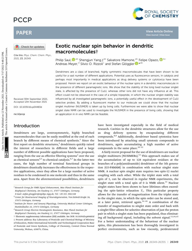
A nuclear singlet multimer (NUSIMER) G5-PAMAM-GGA-NH2,is a poly(amidoamide) dendrimer of the 5th generation(G5-PAMAM) which has been modified by attaching the gly-cin–glycin–alanin tripeptide (GGA) (1) to it to obtain a macro-molecule (2) that contains up to 128 proton pairs per moleculewith the same chemical shift dispersion in which a nuclearsinglet state can be populated13 (Fig. 1). Due to the high densityof chemically equivalent proton pairs in which a singlet statecan be populated, the use of NUSIMERs can greatly increase thesensitivity and NUSIMERs can be detected at low concentra-tions in reasonably quick experiments. The presence of amideand amine functions within the structure as well as at the endof each branch, makes them excellent candidates for theinteraction with metal ions and thus the potential use as probesfor metal ions.
In a first set of experiments we investigated the effect ofparamagnetic metal ions on T1
�1 and Ts�1 in (1) and (2) to
assess the possibility to use NUSIMERs to sense certain ions. Inan investigation of the effect of some paramagnetic ions on T1
and Ts in a simple glycine–alanine dipeptide, it has been shownthat the singlet state is less sensitive to paramagnetic relaxationthan the conventional nuclear magnetization.40 This propertycan be useful in increasing the specificity compared to thestandard NMR experiment. Metal ions play a pivotal role inmany biological processes. Especially Cu(II) has gained someattention, since high Cu(II) concentrations are associated withthe formation of Ab-plaques which can be found in Alzheimer’sdisease patients.50 Furthermore, we investigated the effect ofFe(III), Gd(III), Mn(II) and Zn(II) on T1
�1 and Ts�1.
To further explore the possibility to use NUSIMERs for thedetection of said ions in a biological environment, we decidedto investigate, if NUSIMERs can be viable for in vivo experi-ments in cells. In order to be able to trace the NUSIMER,compound (2) has been modified by attaching a fluorescenttag (Atto488) to it to obtain G5-PAMAM-GGA-Atto488 (3) (Fig. 1).
Results and discussionInfluence of paramagnetic metal ions on T1
�1 and Ts�1
In order to simulate the environment most likely to be relevantin potential in vivo applications, samples of (2) have beenprepared in aqueous HEPES buffered solutions (10 mM) andhave been titrated with Gd(III), Mn(II), Fe(III), Cu(II) and Zn(II)respectively (experiments conducted at 600 MHz). Zn(II) isdiamagnetic but also an important disease marker and washence included in our investigation. After each addition of therespective ions T1
�1 and Ts�1 have been determined (T1
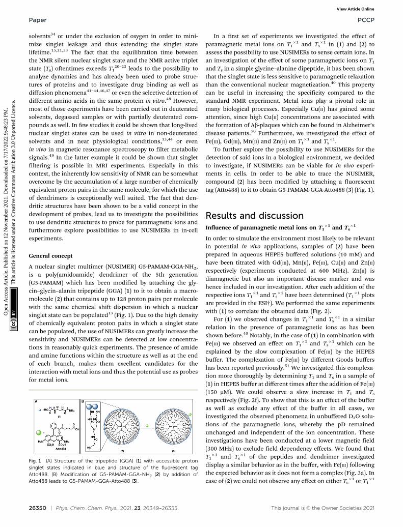
�1 plotsare provided in the ESI†). We performed the same experimentswith (1) to correlate the obtained data (Fig. 2).
For (1) we observed changes in T1�1 and Ts
�1 in a similarrelation in the presence of paramagnetic ions as has beenshown before.40 Notably, in the case of (1) in combination withFe(III) we observed an effect on T1
�1 and Ts�1 which can be
explained by the slow complexation of Fe(III) by the HEPESbuffer. The complexation of Fe(III) by different Goods buffershas been reported previously.51 We investigated this complexa-tion more thoroughly by determining T1 and Ts in a sample of(1) in HEPES buffer at different times after the addition of Fe(III)(150 mM). We could observe a slow increase in T1 and Ts
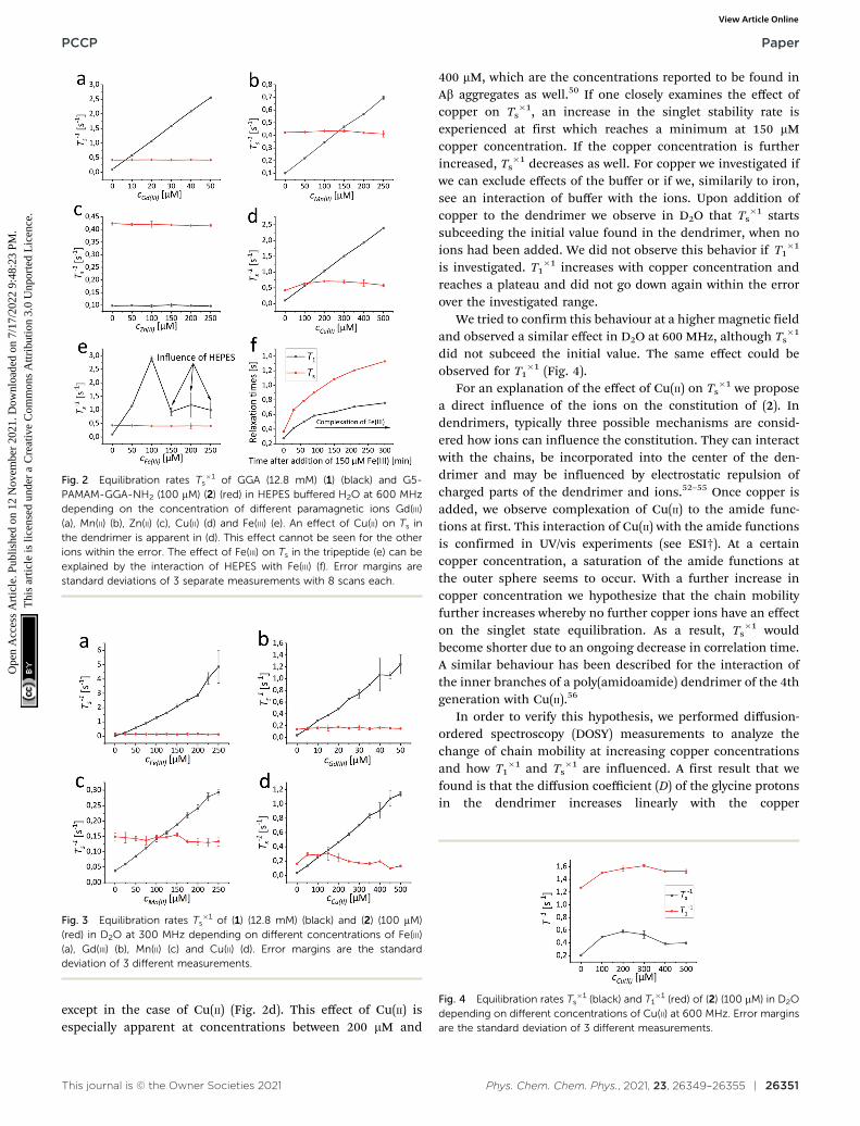
respectively (Fig. 2f). To show that this is an effect of the bufferas well as exclude any effect of the buffer in all cases, weinvestigated the observed phenomena in unbuffered D2O solu-tions of the paramagnetic ions, whereby the pD remainedunchanged and independent of the ion concentration. Theseinvestigations have been conducted at a lower magnetic field(300 MHz) to exclude field dependency effects. We found thatT1�1 and Ts
�1 of the peptides and dendrimer investigateddisplay a similar behavior as in the buffer, with Fe(III) followingthe expected behavior as it does not form a complex (Fig. 3a). Incase of (2) we could not observe any effect on either Ts
�1 or T1�1
Fig. 1 (A) Structure of the tripeptide (GGA) (1) with accessible protonsinglet states indicated in blue and structure of the fluorescent tagAtto488. (B) Modification of G5-PAMAM-GGA-NH2 (2) by addition ofAtto488 leads to G5-PAMAM-GGA-Atto488 (3).
Paper PCCP
Ope
n A
cces
s A
rtic
le. P
ublis
hed
on 1
2 N
ovem
ber
2021
. Dow
nloa
ded
on 7
/17/
2022
9:4
8:23
PM
. T
his
artic
le is
lice
nsed
und
er a
Cre
ativ
e C
omm
ons
Attr
ibut
ion
3.0
Unp
orte
d L
icen
ce.
View Article Online
This journal is © the Owner Societies 2021 Phys. Chem. Chem. Phys., 2021, 23, 26349–26355 | 26351
except in the case of Cu(II) (Fig. 2d). This effect of Cu(II) isespecially apparent at concentrations between 200 mM and
400 mM, which are the concentrations reported to be found inAb aggregates as well.50 If one closely examines the effect ofcopper on Ts
�1, an increase in the singlet stability rate isexperienced at first which reaches a minimum at 150 mMcopper concentration. If the copper concentration is furtherincreased, Ts
�1 decreases as well. For copper we investigated ifwe can exclude effects of the buffer or if we, similarily to iron,see an interaction of buffer with the ions. Upon addition ofcopper to the dendrimer we observe in D2O that Ts
�1 startssubceeding the initial value found in the dendrimer, when noions had been added. We did not observe this behavior if T1
�1
is investigated. T1�1 increases with copper concentration and
reaches a plateau and did not go down again within the errorover the investigated range.
We tried to confirm this behaviour at a higher magnetic fieldand observed a similar effect in D2O at 600 MHz, although Ts
�1
did not subceed the initial value. The same effect could beobserved for T1
�1 (Fig. 4).For an explanation of the effect of Cu(II) on Ts
�1 we proposea direct influence of the ions on the constitution of (2). Indendrimers, typically three possible mechanisms are consid-ered how ions can influence the constitution. They can interactwith the chains, be incorporated into the center of the den-drimer and may be influenced by electrostatic repulsion ofcharged parts of the dendrimer and ions.52–55 Once copper isadded, we observe complexation of Cu(II) to the amide func-tions at first. This interaction of Cu(II) with the amide functionsis confirmed in UV/vis experiments (see ESI†). At a certaincopper concentration, a saturation of the amide functions atthe outer sphere seems to occur. With a further increase incopper concentration we hypothesize that the chain mobilityfurther increases whereby no further copper ions have an effecton the singlet state equilibration. As a result, Ts
�1 wouldbecome shorter due to an ongoing decrease in correlation time.A similar behaviour has been described for the interaction ofthe inner branches of a poly(amidoamide) dendrimer of the 4thgeneration with Cu(II).56
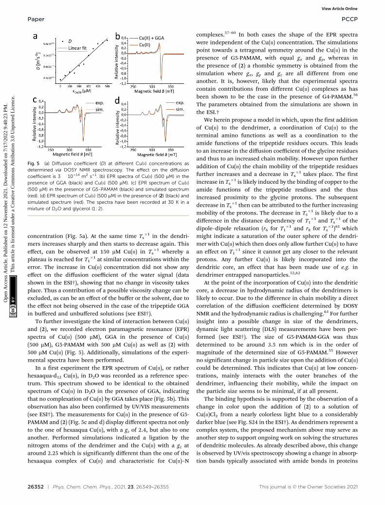
In order to verify this hypothesis, we performed diffusion-ordered spectroscopy (DOSY) measurements to analyze thechange of chain mobility at increasing copper concentrationsand how T1
�1 and Ts�1 are influenced. A first result that we
found is that the diffusion coefficient (D) of the glycine protonsin the dendrimer increases linearly with the copper
Fig. 2 Equilibration rates Ts�1 of GGA (12.8 mM) (1) (black) and G5-
PAMAM-GGA-NH2 (100 mM) (2) (red) in HEPES buffered H2O at 600 MHzdepending on the concentration of different paramagnetic ions Gd(III)(a), Mn(II) (b), Zn(II) (c), Cu(II) (d) and Fe(III) (e). An effect of Cu(II) on Ts inthe dendrimer is apparent in (d). This effect cannot be seen for the otherions within the error. The effect of Fe(III) on Ts in the tripeptide (e) can beexplained by the interaction of HEPES with Fe(III) (f). Error margins arestandard deviations of 3 separate measurements with 8 scans each.
Fig. 3 Equilibration rates Ts�1 of (1) (12.8 mM) (black) and (2) (100 mM)
(red) in D2O at 300 MHz depending on different concentrations of Fe(III)(a), Gd(III) (b), Mn(II) (c) and Cu(II) (d). Error margins are the standarddeviation of 3 different measurements.
Fig. 4 Equilibration rates Ts�1 (black) and T1
�1 (red) of (2) (100 mM) in D2Odepending on different concentrations of Cu(II) at 600 MHz. Error marginsare the standard deviation of 3 different measurements.
PCCP Paper
Ope
n A
cces
s A
rtic
le. P
ublis
hed
on 1
2 N
ovem
ber
2021
. Dow
nloa
ded
on 7
/17/
2022
9:4
8:23
PM
. T
his
artic
le is
lice
nsed
und
er a
Cre
ativ
e C
omm
ons
Attr
ibut
ion
3.0
Unp
orte
d L
icen
ce.
View Article Online
26352 | Phys. Chem. Chem. Phys., 2021, 23, 26349–26355 This journal is © the Owner Societies 2021
concentration (Fig. 5a). At the same time Ts�1 in the dendri-
mers increases sharply and then starts to decrease again. Thiseffect, can be observed at 150 mM Cu(II) in Ts
�1 whereby aplateau is reached for T1
�1 at similar concentrations within theerror. The increase in Cu(II) concentration did not show anyeffect on the diffusion coefficient of the water signal (datashown in the ESI†), showing that no change in viscosity takesplace. Thus a contribution of a possible viscosity change can beexcluded, as can be an effect of the buffer or the solvent, due tothe effect not being observed in the case of the tripeptide GGAin buffered and unbuffered solutions (see ESI†).
To further investigate the kind of interaction between Cu(II)and (2), we recorded electron paramagnetic resonance (EPR)spectra of Cu(II) (500 mM), GGA in the presence of Cu(II)(500 mM), G5-PAMAM with 500 mM Cu(II) as well as (2) with500 mM Cu(II) (Fig. 5). Additionally, simulations of the experi-mental spectra have been performed.
In a first experiment the EPR spectrum of Cu(II), or ratherhexaaqua-d12 Cu(II), in D2O was recorded as a reference spec-trum. This spectrum showed to be identical to the obtainedspectrum of Cu(II) in D2O in the presence of GGA, indicatingthat no complexation of Cu(II) by GGA takes place (Fig. 5b). Thisobservation has also been confirmed by UV/VIS measurements(see ESI†). The measurements for Cu(II) in the presence of G5-PAMAM and (2) (Fig. 5c and d) display different spectra not onlyto the one of hexaaqua Cu(II), with a gz of 2.4, but also to oneanother. Performed simulations indicated a ligation by thenitrogen atoms of the dendrimer and the Cu(II) with a gz ataround 2.25 which is significantly different than the one of thehexaaqua complex of Cu(II) and characteristic for Cu(II)–N
complexes.57–60 In both cases the shape of the EPR spectrawere independent of the Cu(II) concentration. The simulationspoint towards a tetragonal symmetry around the Cu(II) in thepresence of G5-PAMAM, with equal gx and gy, whereas inthe presence of (2) a rhombic symmetry is obtained from thesimulation where gx, gy and gz are all different from oneanother. It is, however, likely that the experimental spectracontain contributions from different Cu(II) complexes as hasbeen shown to be the case in the presence of G4-PAMAM.56
The parameters obtained from the simulations are shown inthe ESI.†
We herein propose a model in which, upon the first additionof Cu(II) to the dendrimer, a coordination of Cu(II) to theterminal amino functions as well as a coordination to theamide functions of the tripeptide residues occurs. This leadsto an increase in the diffusion coefficient of the glycine residuesand thus to an increased chain mobility. However upon furtheraddition of Cu(II) the chain mobility of the tripeptide residuesfurther increases and a decrease in Ts
�1 takes place. The firstincrease in Ts
�1 is likely induced by the binding of copper to theamide functions of the tripeptide resdiues and the thusincreased proximity to the glycine protons. The subsequentdecrease in Ts
�1 then can be attributed to the further increasingmobility of the protons. The decrease in Ts
�1 is likely due to adifference in the distance dependency of T1
�1 and Ts�1 of the
dipole–dipole relaxation (r6 for T1�1 and r8 for Ts
�1)61 whichmight indicate a saturation of the outer sphere of the dendri-mer with Cu(II) which then does only allow further Cu(II) to havean effect on T1
�1 since it cannot get any closer to the relevantprotons. Any further Cu(II) is likely incorporated into thedendritic core, an effect that has been made use of e.g. indendrimer entrapped nanoparticles.52,62
At the point of the incorporation of Cu(II) into the dendriticcore, a decrease in hydrodynamic radius of the dendrimers islikely to occur. Due to the difference in chain mobility a directcorrelation of the diffusion coefficient determined by DOSYNMR and the hydrodynamic radius is challenging.63 For furtherinsight into a possible change in size of the dendrimers,dynamic light scattering (DLS) measurements have been per-formed (see ESI†). The size of G5-PAMAM-GGA was thusdetermined to be around 3.5 nm which is in the order ofmagnitude of the determined size of G5-PAMAM.55 Howeverno significant change in particle size upon the addition of Cu(II)could be determined. This indicates that Cu(II) at low concen-trations, mainly interacts with the outer branches of thedendrimer, influencing their mobility, while the impact onthe particle size seems to be minimal, if at all present.
The binding hypothesis is supported by the observation of achange in color upon the addition of (2) to a solution ofCu(II)Cl2 from a nearly colorless light blue to a considerablydarker blue (see Fig. S24 in the ESI†). As dendrimers represent acomplex system, the proposed mechanism above may serve asanother step to support ongoing work on solving the structuresof dendritic molecules. As already described above, this changeis observed by UV/vis spectroscopy showing a change in absorp-tion bands typically associated with amide bonds in proteins
Fig. 5 (a) Diffusion coefficient (D) at different Cu(II) concentrations asdetermined via DOSY NMR spectroscopy. The effect on the diffusioncoefficient is 3 � 10�14 m2 s�1. (b) EPR spectra of Cu(II) (500 mM) in thepresence of GGA (black) and Cu(II) (500 mM). (c) EPR spectrum of Cu(II)(500 mM) in the presence of G5-PAMAM (black) and simulated spectrum(red). (d) EPR spectrum of Cu(II) (500 mM) in the presence of (2) (black) andsimulated spectrum (red). The spectra have been recorded at 30 K in amixture of D2O and glycerol (1 : 2).
Paper PCCP
Ope
n A
cces
s A
rtic
le. P
ublis
hed
on 1
2 N
ovem
ber
2021
. Dow
nloa
ded
on 7
/17/
2022
9:4
8:23
PM
. T
his
artic
le is
lice
nsed
und
er a
Cre
ativ
e C
omm
ons
Attr
ibut
ion
3.0
Unp
orte
d L
icen
ce.
View Article Online
This journal is © the Owner Societies 2021 Phys. Chem. Chem. Phys., 2021, 23, 26349–26355 | 26353
upon addition of Cu(II) to a solution of (2). We did not observethis change in the case of Gd(III), Mn(II) and Fe(III) (See Fig. S12–S23 in the ESI†).
Assessment of NUSIMERs as potential bioprobes
The observed selective effect of Cu(II) on T1�1 and Ts
�1 gives riseto a possible application of the NUSIMERs as Cu(II) probes,especially, but not limited to, the detection of Cu(II) in cells ortissue. This is especially interesting due to the central role ofCu(II) in the formation of Ab plaques. We would like to note thatchanges in Ts are already sensed below 100 mM copper con-centrations which is well in the region in what is found inextracellular Ab plaques. In order to test the viability of futuretissue experiments, we performed additional measurements ofa solution of (2) in an agarose gel which simulates the con-sistency of brain matter.64 In this case, two experiments havebeen performed: In both cases, the dendrimers were measuredat a concentration of 10 mM in gel which consisted of a HEPES(4-(2-hydroxyethyl)-1-piperazineethanesulfonic acid) bufferedsolution. One of the samples additionally contained 50 mM ofCu(II). Relaxation rate experiments gave a T1
�1 of 0.016 s�1 anda Ts
�1 of 2.42 � 0.06 s for the sample without Cu(II) and T1�1 of
2.32 s�1 and a Ts�1 of 0.69 s�1 for the sample with Cu(II). These
findings indicate that the same effect thoroughly studied in100 mM concentrations in D2O can be observed in lowerconcentrations in a high viscosity environment and in thepresence of buffered H2O solutions. Ts
�1 appears thereby tobe less sensitive to the presence of copper than T1
�1. Thosefindings strongly indicate that detection of Cu(II) using NUSI-MERs is possible in tissue.
In order to evaluate the possibility to use NUSIMERs in cellexperiments, we introduced a fluorescence tag (Atto488) to (2)to obtain the fluorescently labelled NURSIMER (3). We foundan average of 5 fluorescence tags per molecule. At first, we havestudied the molecular parameters T1
�1 and Ts�1 of (3) in D2O
and protonated phosphate saline buffer (PBS) to assess theviability for cell experiments (Table 1). We have discovered that(3) does not display a change in Ts
�1 in protonated buffer withrespect to the parent structure (2). In all cases, the equilibrationrate Ts
�1 is shorter than T1�1 and therefore represents an
excellent starting point not just to filter signals but also toprobe molecular dynamics in biological systems. Table 1 sum-marizes the results.
The observation that a long-lived state can be populatedeven in aqueous buffered solutions encouraged us to furtherinvestigate this behavior in the presence of cells by using
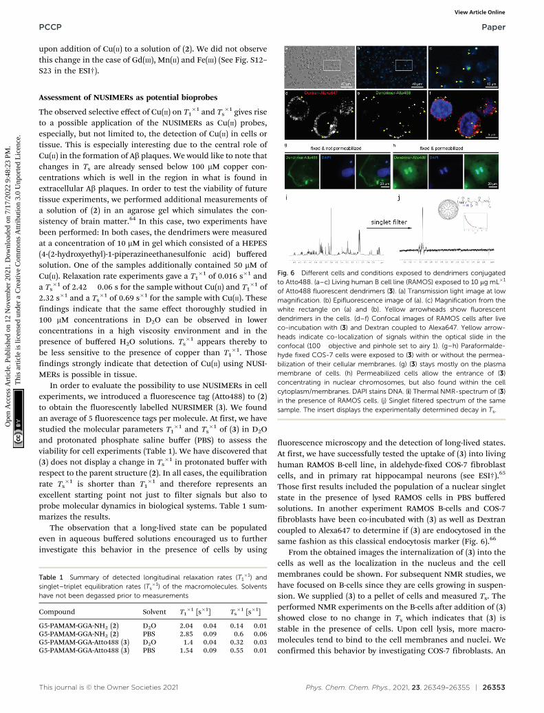
fluorescence microscopy and the detection of long-lived states.At first, we have successfully tested the uptake of (3) into livinghuman RAMOS B-cell line, in aldehyde-fixed COS-7 fibroblastcells, and in primary rat hippocampal neurons (see ESI†).65
Those first results included the population of a nuclear singletstate in the presence of lysed RAMOS cells in PBS bufferedsolutions. In another experiment RAMOS B-cells and COS-7fibroblasts have been co-incubated with (3) as well as Dextrancoupled to Alexa647 to determine if (3) are endocytosed in thesame fashion as this classical endocytosis marker (Fig. 6).66
From the obtained images the internalization of (3) into thecells as well as the localization in the nucleus and the cellmembranes could be shown. For subsequent NMR studies, wehave focused on B-cells since they are cells growing in suspen-sion. We supplied (3) to a pellet of cells and measured Ts. Theperformed NMR experiments on the B-cells after addition of (3)showed close to no change in Ts which indicates that (3) isstable in the presence of cells. Upon cell lysis, more macro-molecules tend to bind to the cell membranes and nuclei. Weconfirmed this behavior by investigating COS-7 fibroblasts. An
Table 1 Summary of detected longitudinal relaxation rates (T1�1) and
singlet–triplet equilibration rates (Ts�1) of the macromolecules. Solvents
have not been degassed prior to measurements
Compound Solvent T1�1 [s�1] Ts
�1 [s�1]
G5-PAMAM-GGA-NH2 (2) D2O 2.04 � 0.04 0.14 � 0.01G5-PAMAM-GGA-NH2 (2) PBS 2.85 � 0.09 0.6 � 0.06G5-PAMAM-GGA-Atto488 (3) D2O 1.4 � 0.04 0.32 � 0.03G5-PAMAM-GGA-Atto488 (3) PBS 1.54 � 0.09 0.55 � 0.01
Fig. 6 Different cells and conditions exposed to dendrimers conjugatedto Atto488. (a–c) Living human B cell line (RAMOS) exposed to 10 mg mL�1
of Atto488 fluorescent dendrimers (3). (a) Transmission light image at lowmagnification. (b) Epifluorescence image of (a). (c) Magnification from thewhite rectangle on (a) and (b). Yellow arrowheads show fluorescentdendrimers in the cells. (d–f) Confocal images of RAMOS cells after liveco-incubation with (3) and Dextran coupled to Alexa647. Yellow arrow-heads indicate co-localization of signals within the optical slide in theconfocal (100� objective and pinhole set to airy 1). (g–h) Paraformalde-hyde fixed COS-7 cells were exposed to (3) with or without the permea-bilization of their cellular membranes. (g) (3) stays mostly on the plasmamembrane of cells. (h) Permeabilized cells allow the entrance of (3)concentrating in nuclear chromosomes, but also found within the cellcytoplasm/membranes. DAPI stains DNA. (i) Thermal NMR-spectrum of (3)in the presence of RAMOS cells. (j) Singlet filtered spectrum of the samesample. The insert displays the experimentally determined decay in Ts.
PCCP Paper
Ope
n A
cces
s A
rtic
le. P
ublis
hed
on 1
2 N
ovem
ber
2021
. Dow
nloa
ded
on 7
/17/
2022
9:4
8:23
PM
. T
his
artic
le is
lice
nsed
und
er a
Cre
ativ
e C
omm
ons
Attr
ibut
ion
3.0
Unp
orte
d L
icen
ce.
View Article Online
26354 | Phys. Chem. Chem. Phys., 2021, 23, 26349–26355 This journal is © the Owner Societies 2021
observed broadening of the NMR-signal of (3) supports thebinding hypothesis. Despite the broadening, the signal obser-vation of (3) in the presence of lysed cells was possible using asinglet NMR sequence45 with an additional singlet filter.67 Thisfiltering experiment allows for direct observation of the glycineprotons in (3) while suppressing other proton signals from cellmembranes and water with a limit of detection of 6 mMconcentrations of (3) in a single scan at 900 MHz. Afterextensive washing of the lysed cells, we have still observed (3)due to the possibility of populating singlet states. Additionally,we confirmed the binding by observing an orange coloring ofthe cells and solid state NMR experiments (see ESI†). Furtherexperiments on cytosol of B-cells spiked with (3) after lysis andremoval of cell membranes showed the stability: we observedno change of the lineshape of (3) and in the measured Ts over aperiod of several hours. As in the previous experiments, theglycine signal can solely be obtained while suppressing othersignals (see Fig. S6, ESI†).
Conclusion
In conclusion we observed a unique behaviour of the molecularparameters T1 and Ts in NUSIMERs in the presence of differentparamagnetic metal ions. Unexpectedly, the presence of Fe(III),Mn(II) and even Gd(III) did not have any effect on neither T1 norTs, which is a behaviour that, to the best of our knowledge, hasnot been observed so far. Furthermore we were able to observea unique effect of the presence of Cu(II) on T1 and Ts, displayinga sharp decrease in both parameters at low Cu(II) concentra-tions followed by an increase at around 150 mM Cu(II). Wepropose an interaction between the terminal tripeptides of theNUSIMERs and Cu(II) to be responsible for this observation.The effect of the paramagnetic relaxation competes with achange in the mobility of the tripeptides at the outer sphereof the dendrimers which occurs upon addition of Cu(II). Basedon this observed effect we explored the possibility to useNUSIMERs in an biological environment and evaluate theviability to use NUSIMERs as Cu(II) probes. We succeeded inattaching a fluorescence tag to the NUSIMERs and observingthe uptake in different cells by fluorescence spectroscopy.Using the NUSIMER G5-PAMAM-GGA-Atto488, we were alsoable to eliminate any background signal using singlet NMRspectroscopy in the presence of cells. This is especially promis-ing, because it opens up the possibility to branch out into Cu(II)detection in cells or even in vivo, which could in the future leadto a non-invasive technique for early diagnostics of neurode-generative diseases. This prospect seems especially possiblesince it has recently be shown, that singlet filtering can beapplied in MRI.49 Our described experiments could in generalserve as an approach to design new molecular probes usingnuclear spin singlet states.
Conflicts of interest
There are no conflicts to declare.
Acknowledgements
The authors acknowledge generous funding from the MaxPlanck Society and the Max Planck Institute for BiophysicalChemistry. We thank Prof. M. Bennati for enlightening discus-sions and access to her EPR instrumentation. K. Overkamp isacknowledged for her support in analyzing the synthesizedcompounds. We furthermore thank Dr V. Belov and theChemical Synthesis Facility for a scale-up of compound (2).The authors thank Dr S. Becker and Dr N. Rezaie-Ghaleh forhelpful discussions. We thank H. Sebesse for support in mak-ing the figure. Dr L. Andreas is acknowledged for providingaccess to the solid state NMR instrumentation. Dr R. Dervisogluis acknowledged for help in preparing the samples for solidstate NMR. We are indebted to Prof. C. Griesinger for access tohis facilities. Open Access funding provided by the Max PlanckSociety.
Notes and references
1 G. M. Dykes, J. Chem. Technol. Biotechnol., 2001, 76,903–918.
2 E. Buhleier, W. Wehner and F. Vogtle, Synthesis, 1978,155–158.
3 M. S. Diallo, S. Christie, P. Swaminathan, J. H. Johnson Jr.and W. A. Goddard, Environ. Sci. Technol., 2005, 39,1366–1377.
4 V. Balzani, P. Ceroni, S. Gestermann, C. Kauffmann,M. Gorka and F. Vogtle, Chem. Commun., 2000, 853–854.
5 T. D. James, H. Shinmori, M. Takeuchi and S. Shinkai,Chem. Commun., 1996, 705–706.
6 S. B. Garber, J. S. Kingsbury, B. L. Gray and A. H. Hoveyda,J. Am. Chem. Soc., 2000, 122, 8168–8179.
7 J. W. J. Knapen, A. W. van der Made, J. C. de Wilde,P. W. N. M. van Leeuwen, P. Wijkens, D. M. Grove andG. van Koten, Nature, 1994, 372, 659–663.
8 Y. Vida, D. Collado, F. Najera, S. Claros, J. Becerra,J. A. Andrades and E. Perez-Inestrosa, RSC Adv., 2016, 6,49839–49844.
9 S. P. Kambhampati, M. K. Mishra, P. Mastorakos, Y. Oh,G. A. Lutty and R. M. Kannan, Eur. J. Pharm. Biopharm.,2015, 95, 239–249.
10 F.-H. Liu, C.-Y. Hou, D. Zhang, W.-J. Zhao, Y. Cong,Z.-Y. Duan, Z.-Y. Qiao and H. Wang, Biomater. Sci., 2018,6, 604–613.
11 A. K. Sharma, L. Gupta, H. Sahu, A. Qayum, S. K. Singh,K. T. Nakhate, Ajazuddin and U. Gupta, Pharm. Res., 2018,35, 9.
12 S. P. Kuruvilla, G. Tiruchinapally, A. C. Crouch,M. E. H. ElSayed and J. M. Greve, PLoS One, 2017,12, e0181944.
13 P. Saul, S. Mamone and S. Gloggler, Chem. Sci., 2019, 10,413–417.
14 M. H. Levitt, Annu. Rev. Phys. Chem., 2012, 63, 89–105.15 M. Carravetta and M. H. Levitt, J. Am. Chem. Soc., 2004, 126,
6228–6229.
Paper PCCP
Ope
n A
cces
s A
rtic
le. P
ublis
hed
on 1
2 N
ovem
ber
2021
. Dow
nloa
ded
on 7
/17/
2022
9:4
8:23
PM
. T
his
artic
le is
lice
nsed
und
er a
Cre
ativ
e C
omm
ons
Attr
ibut
ion
3.0
Unp
orte
d L
icen
ce.
View Article Online
This journal is © the Owner Societies 2021 Phys. Chem. Chem. Phys., 2021, 23, 26349–26355 | 26355
16 M. Carravetta, O. G. Johannessen and M. H. Levitt, Phys.Rev. Lett., 2004, 92, 153003.
17 M. Carravetta and M. H. Levitt, J. Chem. Phys., 2005,122, 214505.
18 G. Pileio and M. H. Levitt, J. Chem. Phys., 2009, 130, 214501.19 G. Pileio, M. Carravetta and M. H. Levitt, Proc. Natl. Acad.
Sci. U. S. A., 2010, 107, 17135–17139.20 G. Pileio, M. Carravetta, E. Hughes and M. H. Levitt, J. Am.
Chem. Soc., 2008, 130, 12582–12583.21 G. Stevanato, J. T. Hill-Cousins, P. Håkansson, S. S. Roy,
L. J. Brown, R. C. D. Brown, G. Pileio and M. H. Levitt,Angew. Chem., Int. Ed., 2015, 54, 3740–3743.
22 T. Theis, G. X. Ortiz Jr., A. W. J. Logan, K. E. Claytor, Y. Feng,W. P. Huhn, V. Blum, S. J. Malcolmson, E. Y. Chekmenev,Q. Wang and W. S. Warren, Sci. Adv., 2016, 2, e1501438.
23 Y. Zhang, P. C. Soon, A. Jerschow and J. W. Canary, Angew.Chem., Int. Ed., 2014, 53, 3396–3399.
24 M. C. D. Tayler and M. H. Levitt, Phys. Chem. Chem. Phys.,2011, 13, 5556–5560.
25 G. Pileio, M. Concistre, M. Carravetta and M. H. Levitt,J. Magn. Reson., 2006, 182, 353–357.
26 T. Theis, Y. Feng, T. Wu and W. S. Warren, J. Chem. Phys.,2014, 140, 014201.
27 S. J. Elliott, L. J. Brown, J.-N. Dumez and M. H. Levitt, Phys.Chem. Chem. Phys., 2016, 18, 17965–17972.
28 R. Buratto, D. Mammoli, E. Chiarparin, G. Williams andG. Bodenhausen, Angew. Chem., Int. Ed., 2014, 53, 11376–11380.
29 R. Buratto, A. Bornet, J. Milani, D. Mammoli, B. Vuichoud,N. Salvi, M. Singh, A. Laguerre, S. Passemard, S. Gerber-Lemaire, S. Jannin and G. Bodenhausen, ChemMedChem,2014, 9, 2509–2515.
30 Y. Feng, R. M. Davis and W. S. Warren, Nat. Phys., 2012, 8,831–837.
31 E. Vinogradov and A. K. Grant, J. Magn. Res., 2008, 194,46–57.
32 W. S. Warren, E. Jenista, R. T. Branca and X. Chen, Science,2009, 323, 1711–1714.
33 G. Pileio, S. Bowen, C. Laustsen, M. C. D. Wayler, J. T. Hill-Cousins, L. J. Brown, R. C. D. Brown, J. H. Ardenskjær-Larsenand M. H. Levitt, J. Am. Chem. Soc., 2013, 135, 5084–5088.
34 Y. Feng, T. Theis, X. Liang, Q. Wang, P. Zhou andW. S. Warren, J. Am. Chem. Soc., 2013, 135, 9632–9635.
35 Y. Zhang, X. Duan, P. C. Soon, V. Sychrovsky, J. W. Canaryand A. Jerschow, ChemPhysChem, 2016, 17, 2967–2971.
36 S. Cavadini, J. Dittmer, S. Antonijevic and G. Bodenhausen,J. Am. Chem. Soc., 2005, 127, 15744–15748.
37 P. Ahuja, R. Sarkar, P. R. Vasos and G. Bodenhausen, J. Am.Chem. Soc., 2009, 131, 7498–7499.
38 S. J. DeVience, R. L. Walsworth and M. S. Rosen, NMRBiomed., 2013, 26, 1204–1212.
39 R. Buratto, D. Mammoli, E. Canet and G. Bodenhausen,J. Med. Chem., 2016, 59, 1960–1966.
40 M. C. D. Tayler and M. H. Levitt, Phys. Chem. Chem. Phys.,2011, 13, 9128–9130.
41 R. Sarkar, P. R. Vasos and G. Bodenhausen, J. Am. Chem.Soc., 2007, 129, 328–334.
42 P. Ahuja, R. Sarkar, P. R. Vasos and G. Bodenhausen,J. Chem. Phys., 2007, 127, 134112.
43 N. Salvi, R. Buratto, A. Bornet, S. Ulzega, I. R. Rebollo,A. Angelini, C. Heinis and G. Bodenhausen, J. Am. Chem.Soc., 2012, 134, 11076–11079.
44 A. S. Kiryutin, A. N. Pravdivtsev, A. V. Yurkovskaya, H.-M. Viethand K. L. Ivanov, J. Phys. Chem., 2016, 120, 11978–11986.
45 S. J. DeVience, R. L. Walsworth and M. S. Rosen, Phys. Rev.Lett., 2013, 111, 173002.
46 C. Stavarache, A. Hanganu, A. Paun, C. Paraschivescu,M. Matache and P. R. Vasos, J. Magn. Res., 2017, 284, 15–19.
47 A. Bornet, P. Ahuja, R. Sarkar, L. Fernandes, S. Hadji,S. Y. Lee, A. Haririnia, D. Fushman, G. Bodenhausen andP. R. Vasos, ChemPhysChem, 2011, 12, 2729–2734.
48 S. Mamone, N. Rezaei-Ghaleh, F. Opazo, C. Griesinger andS. Gloggler, Sci. Adv., 2020, 6, eaaz1955.
49 S. Mamone, A. B. Schmidt, N. Schwaderlapp, T. Lange,D. von Elverfeldt, J. Hennig and S. Gloggler, NMR Biomed.,2021, 34, e4400.
50 M. A. Lovell, J. D. Robertson, W. J. Teesdale, J. L. Campbelland W. R. Markesbery, J. Neurol. Sci., 1998, 158, 47–52.
51 B. S. Gupta, M. Taha and M.-J. Lee, J. Solution Chem., 2013,42, 2296–2309.
52 X. Wang, X. Cai, J. Hu, N. Shao, F. Wang, Q. Zhang, J. Xiaoand Y. Cheng, J. Am. Chem. Soc., 2013, 135, 9805–9810.
53 P. Welch and M. Muthukumar, Macromolecules, 1998, 31,5892–5897.
54 S. Garcıa-Gallego, M. Cangiotti, L. Fiorani, A. Fattori,M. Angeles Munoz Fernandez, R. Gomez, M. F. Ottavianiand F. J. de la Mata, Dalton Trans., 2013, 42, 5874–5889.
55 D. Lombardo, Biochem. Res. Int., 2014, 837651.56 M. F. Ottaviani, S. Bossmann, N. J. Turro and D. A. Tomalia,
J. Am. Chem. Soc., 1994, 116, 661–671.57 H. Motschi, Colloids Surf., 1984, 9, 333–347.58 K. J. de Almeida, Z. Rinkevicius, H. W. Hugosson,
A. C. Ferreira and H. Ågren, Chem. Phys., 2007, 332, 176–187.59 J. V. Folgado, W. Henke, R. Allmann, H. Stratemeier,
D. Beltran-Porter, T. Rojo and D. Reinen, Inorg. Chem.,1990, 29, 2035–2042.
60 A. Meyer, G. Schnakenburg, R. Glaum and O. Schiemann,Inorg. Chem., 2015, 54, 8456–8464.
61 S. J. Elliott, C. Bengs, L. J. Brown, J. T. Hill-Cousins, D. J. O’Leary,G. Pileio and M. H. Levitt, J. Chem. Phys., 2019, 150, 064315.
62 J. C. Garcia-Martinez and R. M. Crooks, J. Am. Chem. Soc.,2004, 126, 16170–16178.
63 L. F. Pinto, J. Correa, M. Martin-Pastor, R. Riguera andE. Fernandez-Megia, J. Am. Chem. Soc., 2013, 135, 1972–1977.
64 Z.-J. Chen, G. T. Gillies, W. C. Broaddus, S. S. Prabhu,H. Fillmore, R. M. Mitchell, F. D. Corwin andP. P. Fatouros, J. Neurosurg., 2019, 101, 314–323.
65 M. Maidorn, A. Olichon, S. O. Rizzoli and F. Opazo, MABS,2019, 11, 305–321.
66 M. A. Gomes de Castro, C. Hobartner and F. Opazo, PLoSOne, 2017, 12, e0173050.
67 S. Mamone and S. Gloggler, Phys. Chem. Chem. Phys., 2018,20, 22463–22467.
PCCP Paper
Ope
n A
cces
s A
rtic
le. P
ublis
hed
on 1
2 N
ovem
ber
2021
. Dow
nloa
ded
on 7
/17/
2022
9:4
8:23
PM
. T
his
artic
le is
lice
nsed
und
er a
Cre
ativ
e C
omm
ons
Attr
ibut
ion
3.0
Unp
orte
d L
icen
ce.
View Article Online