examining the effect of relative humidity on mammoth molars
TRANSCRIPT

Examining the Effect of Relative Humidity on
Mammoth Molars
Daniel Doyle
Research Project submitted to the Department of Art
in conformity with the requirements for the
Degree of Master of Art Conservation
Queen’s University
Kingston, Ontario
CANADA
May, 2015

i

ii
Abstract
Museum and natural history collections are very sensitive to fluctuations in relative humidity (RH).
Due to a high degree of structural anisotropy, mammoth molars are potentially damaged by such
fluctuations. Upon excavation, mammoth molars often undergo rapid acclimatization to the
ambient RH. The main focus of this research was to quantify what occurs to mammoth molars at
low RH, and to investigate whether permanent changes take place or if previous cracking is able
to accommodate RH fluctuations. A research project conducted by Samantha Fisher in the Art
Conservation Program at Queen’s University, Kingston, Ontario, in 2014 on mammoth molars
found that cracks widen and extend after exposure to 75%RH, cracks continue to extend after a
drop to 35%RH. It was also found that cement adsorbs and desorbs faster than enamel, and that
Fourier transform infrared spectroscopy shows the presence of hydroxyapatite and water with
some carbonate and fluoride substitutions. As the molars used had already undergone humidity
cycling, there were no significant changes to cracks observed during the experiment. To further
understand the changes which occur during significant drops to low RH, such as during the initial
excavation and drying of molars, a single cycle was undertaken from ambient RH to a low 11%RH
using a lithium chloride salt. Weight changes indicating the desorption of water from the teeth
were measured to better understand the internal moisture change, and to identify when the molars
reached equilibrium. The resulting dimensional changes and crack propagation were measured
using ImageJ analysis software, as well as physically measuring the cracks.
Overall, an increase in the crack ratios and dimensions was measured, as well as a possible joining
of previously separate cracks. A measurable loss of material also occurred from the three molars,
including powder and fragments. It is certain that exposure to the 11%RH environment has
produced an irreversible change in the molars. This research is only one of a very limited number
of studies into the effects of relative humidity on natural history collections. As such, a survey of
current storage conditions for natural history collections was undertaken, to better understand
current practices and propose preventative steps. The survey found that many mammoth molar
collections undergo fluctuations of RH +/-10% outside the normal storage conditions which last
longer than one week. It is certain that these environments cause additional damage to mammoth
molars comparable to those observed in this study.

iii
Acknowledgements
Dr. Robert Waller, President and Senior Risk Analyst, Protect Heritage Corp.
Dr. Alison Murray, Associate Professor, Art Conservation, Queen’s University
Dr. H.F. (Gus) Shurvell, Professor Emeritus, Chemistry, Queen’s University
Grant Zazula, Paleontologist, Yukon Palaeontology Program
Elizabeth Hall, Assistant Paleontologist, Yukon Palaeontology Program
Dr. Tom Strang, Senior Conservation Scientist, Canadian Conservation Institute
Paul Bégin, Senior Conservation Scientist, Canadian Conservation Institute
Further Thanks
Alaska State Museum (USA)
California Academy of Sciences (USA)
Canadian Museum of Nature, Ontario (CA)
The Dr. John D. Cooper Archaeological and Paleontological Center, California (USA)
Georgia College Natural History Museum (USA)
Idaho Museum of Natural History (USA)
Illinois State Museum (USA)
Mammoth Site of Hot Springs, South Dakota (USA)
Museum of Geology, South Dakota School of Mines & Technology (USA)
Museum Village, New York (USA)
National Museum of Ireland, Dublin (IR)
National Museum Wales (UK)
Naturalis Biodiversity Center, Zuid Holland (NL)
Science Museum of Minnesota (USA)
Simon Fraser University Museum of Archaeology, British Columbia (CA)
TMM Collections at the Vertebrate Paleontology Laboratory, University of Texas, (USA)
University of Alaska Museum (USA)
University of Arkansas Museum Collections (USA)
Yale Peabody Museum of Natural History, Connecticut (USA)
iv
Table of Contents
Authorization for Copying of Queen’s University Research Projects i
Abstract ii
Acknowledgements iii
List of Figures v
List of Tables v
1. Introduction 1
1.1 Relative Humidity 1
1.2 Mammoth Molars 2
1.3 Previous Research 4
1.4 Research Goals 6
2. Experiment 7
2.1 Materials 7
2.2 Environmental Set-Up 7
2.3 Method of Analysis 9
3. Results and Discussion 11
3.1 Observations 11
3.2 Crack Measurement 16
3.3 Storage Survey 18
4. Conclusion 20
5. Bibliography 21
Appendix A: Table of Recorded Molar Weights 22
Appendix B: Binary Molar Images 24
Curriculum Vitæ 27

v
List of Figures
Fig. 1: Profiles from left of Elephas maximus, Loxodonta africana and Mammuthus columbi
Fig. 2: Anatomical features of a lower mammoth molar
Figs. 3-5: Molars 406.955, 406.956, 406.957
Fig. 6: Environmental set-up with PEM2 and HOBO14 data loggers
Figs. 7-9: Weight change for molars 406.955, 406.956, 406.957
Fig. 10: Exponential growth curve of moisture desorption
Figs. 11-16: Before and after experiment images
Figs. 17-28: Binary overall and detail before and after images
List of Tables
Table 1: Measurement for Tooth 406.955
Table 2: Measurement for Tooth 406.956
Table 3: Measurement for Tooth 406.957
Table 4: Binary Image Comparison Data
Table 5: Molar weights
1
1. Introduction
Fluctuations in relative humidity (RH) can be quite damaging to museum and natural history
collections. To better characterize the effect of such fluctuations on mammoth molars during
significant drops to low RH, a single cycle was undertaken from ambient RH to 11%RH. Weight
and dimensional changes, along with crack propagation, were used to quantify the effect of low
humidity on the molars.
1.1 Relative Humidity
Many guidelines have been developed for the preventative conservation of museum collections
through environmental controls. Some of the earliest evidence that maintaining a steady, moderate
relative humidity (RH) could prevent damage dates to the Second World War. At the time, for the
safekeeping of the objects, the collections of the National Gallery, London, were moved to mines
in Wales. While the climate in the caves was constant, the relative humidity was too high, and
therefore the air was slightly heated to lower the RH to 55-60%. This was based on findings from
earlier studies from the National Gallery which found this RH to be the effective average in the
un-air conditioned Gallery. It was noted that humidity related problems, such as cracking and
flaking, essentially disappeared while the collections were maintained in this stable environment.
Following the War, with the collection returned to the uncontrolled Gallery environment, these
problems returned (Erhardt 1995 19).
Many years of experimental and observational data has resulted in the establishment of
environmental recommendations. These recommended temperatures and RH levels vary by
material, with organic materials commonly being most sensitive. RH levels outside of these
recommendations can lead to increased deterioration and damage (Michalski 1993 624-629). As
RH changes, so does the moisture content of organic materials including wood, paper, leather,
photographs, plastics, paints, and glues (Michalski 2013). In low relative humidity, objects can
desiccate, shrinking as water is lost. If the material is constrained by other components or even its
own inner bulk, this leads to cracking and distortion. Conversely, in high relative humidity the
object may take on water, leading to swelling of the material and possible fungal colonization.

2
Again, if the material is constrained by components or its own bulk, expanding parts may become
crushed (Michalski 2013). Repeated equilibrations to high and low levels of RH might lead to
cycles of swelling and contraction and the eventual physical breakdown of objects that cannot
withstand these movements. While older humidity control regulations required costly maintaining
of a narrow range of allowable humidity, it has been noted that even a short period of a few weeks
of system failure would negate the positive effects. As such, recommendations now favour more
complex specifications with lower maintenance costs (Michalski 2013).
1.2 Mammoth Molars
Due to a high degree of structural anisotropy, mammoth molars are objects potentially damaged
by exposure to incorrect RH levels. Mammoths were once widespread across Europe, northern
Asia, North America, and central Mexico. Appearing in the middle of the Pliocene epoch (about
3-4 million years ago), the mammoth gradually became cold adapted, successfully colonizing
extreme northern climes, crossing to North America at least 1.7 million years ago (Haynes 1991).
Of the Proboscidea, the family which included mammoths (mammuthus) and mastodons
(mammut), the only living relatives are the African and Asian elephants (loxodonta africana and
elephas maximus respectively), with the mammoth disappearing from the North American fossil
record around 10,000 years ago (Haynes 1991) (Fig. 1).
Fig. 1: Profiles from left of Elephas maximus, Loxodonta africana and Mammuthus columbi
(Hayes 1991)

3
Mammoth molars are made of tightly packed enamel and dentine plates surrounded by cement to
form ridges for grinding plant matter (Lister and Sher 2001 1094-1097) (Fig. 2). All the
components of the molars contain both inorganic (mineral) and organic compounds in varying
proportions. Organic components of the molars include collagen and enamel protein. Enamel is
almost entirely mineral, containing only 0.4-0.9% organic material and 2.1-3.6% water by weight.
Dentine is mostly inorganic with 17.5-18.5% collagen, 0.2% resistant protein, and 5% water.
Cement is a mixture of around 65-70% inorganic material and 24-26% organics, of which more
than 20% may be collagen (Hillson 1986 155, 184, 193).
Fig. 2: Anatomical features of a lower mammoth molar (Ferretti 2003 385).
Upon excavation from a near wet state, mammoth molars often undergo rapid acclimatization to
the ambient RH. As mammoth molars, and teeth in general, are especially sensitive to fluctuation
in humidity, this can lead to damage. The complex, layered structure of mammoth molars leads to
cracking and delamination. The organic materials in the teeth, especially in the cement and dentine,
are hygroscopic. This allows adsorption and desorption of water leading to expansion and
contraction (Williams 1991 21). The Canadian Conservation Institute (CCI) recommends the
storage and display of ivory at around 45-55%RH (CCI 2010). Ivory and other teeth are especially
sensitive and easily damaged, with the deterioration of teeth usually being the first sign of
deterioration in skulls (Williams 1991 13). Previous research has found that desiccation of teeth
below 40%RH causes significant cracking, with sudden drops in relative humidity being more
damaging when the relative humidity was below 45% (Williams 1991 20-21).
4
1.3 Previous Research
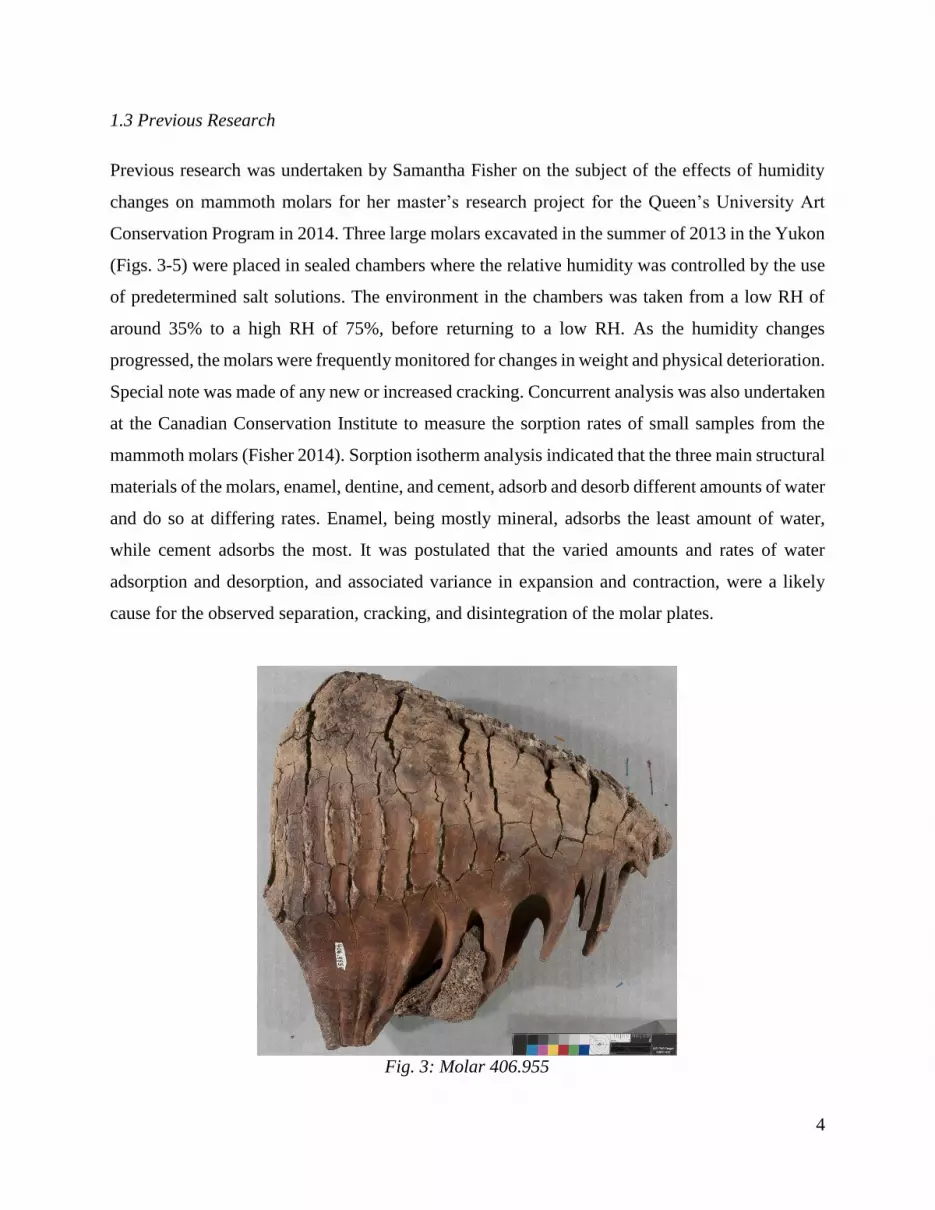
Previous research was undertaken by Samantha Fisher on the subject of the effects of humidity
changes on mammoth molars for her master’s research project for the Queen’s University Art
Conservation Program in 2014. Three large molars excavated in the summer of 2013 in the Yukon
(Figs. 3-5) were placed in sealed chambers where the relative humidity was controlled by the use
of predetermined salt solutions. The environment in the chambers was taken from a low RH of
around 35% to a high RH of 75%, before returning to a low RH. As the humidity changes
progressed, the molars were frequently monitored for changes in weight and physical deterioration.
Special note was made of any new or increased cracking. Concurrent analysis was also undertaken
at the Canadian Conservation Institute to measure the sorption rates of small samples from the
mammoth molars (Fisher 2014). Sorption isotherm analysis indicated that the three main structural
materials of the molars, enamel, dentine, and cement, adsorb and desorb different amounts of water
and do so at differing rates. Enamel, being mostly mineral, adsorbs the least amount of water,
while cement adsorbs the most. It was postulated that the varied amounts and rates of water
adsorption and desorption, and associated variance in expansion and contraction, were a likely
cause for the observed separation, cracking, and disintegration of the molar plates.
Fig. 3: Molar 406.955

5
Fig. 4: Molar 406.956
Fig. 5: Molar 406.957
6
1.4 Research Goals
Expanding on the research conducted by Samantha Fisher, this experiment studied the effects of
relative humidity on mammoth molars. By subjecting molars to lower RH than in previous
research, new insights into their post-excavation behaviour were obtained. This was used to
determine the behaviour of mammoth molars and their potential for further cracking and
degradation at low humidity. It was hypothesized that due to the differences in adsorption and
desorption rates of the various materials within mammoth molars, upon fluctuations in relative
humidity, the molar materials would differentially swell and contract leading to cracking. Once
initially cracked, further exposure to low humidity would expand these crack which, upon
returning to higher humidity, would then be permanently increased.
This research, in conjunction with data obtained from a PEM2 data logger placed in the Yukon
Paleontology Program’s storage and a survey of the environment and practices of other institutions
holding mammoth teeth, adds to the evidence-based recommendations for post-collection
environmental controls, enabling the determination of an informed method for the preventative
conservation of the molars.

7
2. Experimental
To determine the effect of low relative humidity on mammoth molars, the teeth were placed in a
controlled environment and the changing weight values of the teeth, corresponding to water loss,
measured. Initial cracking was compared with final cracking to determine any changes in the
structure of the teeth.
2.1 Materials
The three mammoth molars obtained for use in this experiment, numbered 406.955, 406.956, and
406.957, were excavated by the Yukon Paleontology Program in 2013. Further, smaller samples
from the Canadian Museum of Nature were also available. It was not possible to obtain freshly
excavated molar fragments, which would have shown what occurs during the initial drying of the
teeth. As the teeth have previously been exposed to low 35%RH during Samantha Fisher’s
research, it was possible to determine whether any further cracking occurred upon additional
desiccation of the teeth at a very low (11%) RH level, or whether the previous cracks were able to
accommodate the dimensional changes once formed. As these molars were obtained directly from
the paleontologists, it is known that these have not been consolidated with any type of adhesive or
coating.
2.2 Environmental Set-Up
The mammoth molars were placed in a chamber with a glass tray of desiccated lithium chloride
salt, with the ambient environment adjusting to 11%RH. Based on the research conducted by
Samantha Fisher, it was expected to take at least 40 days to achieve the lower RH equilibrium in
the teeth. In practice the lower RH equilibrium took 80 days to achieve. This was gauged through
frequent (minimum twice weekly) weighing of the teeth and fitting the weight vs. time data to an
appropriate growth curve. Once the weight change was consistent with having reached 90% of an
extrapolated end weight, the equilibration was considered complete for practical museum
situations and experimental purposes. The teeth were then removed and photographed, and the
presence of any newly formed cracks documented. Unfortunately, due to time restrictions, a

8
complete humidity cycle bringing the teeth to equilibrium at 75%RH was not possible, and is an
area for further research.
To make the controlled RH environment, the dry salt was placed in a tempered glass dish with as
large a surface area as possible to maximize rates of sorption or desorption. One fan was installed
in the lid of the chamber previously, and a second was installed to further aid in the circulation of
air. Fans were oriented to cause a circular motion of air through the chamber as a whole (on one
side the fan was directed upward while on the other side the fan was directed downward).
Consistent with Samantha Fisher’s experiment, a wire rack was placed over the saturated salt
solution inside the chamber, onto which the mammoth molars were placed. HOBO 14 and PEM 2
data loggers were placed in the tank to monitor the environment (Fig. 6). The molars were weighed
at intervals during the course of the experiment, with care taken that the salt solution remained
unsaturated. When this solution became saturated, it was necessary to boil the slurry to de-saturate
the salt of collected moisture.
Fig. 6: Environmental set-up with PEM2 and HOBO14 data loggers

9
2.3 Method of Analysis
Imaging and analysis of the crack development were the main challenges of the experiment.
Working with Samantha Fisher and Michael Doutre in March 2014, it was found that analysis of
standard photography of the mammoth molars through ImageJ was not successful. The dark
colouring of the teeth made for difficulties in the software’s analysis of crack changes.
Additionally, minor variations in the angle of photography and lighting led to slight inconsistencies
in the shadowing of the teeth which ImageJ was unable to differentiate from crack growth (Fisher
2014, 16).
Physical measurements of the teeth were recorded prior to photography. The pre-experiment
weight of the teeth was recorded in grams to one decimal place using an Ohaus Adventurer SL
digital scale. Length, width, and height of each tooth was measured in mm to two decimal points,
though these are expected to remain unchanged from the end results of Samantha Fisher’s report.
To increase the contrast, dusting of the surface with a lightening powder was considered but was
not undertaken as removal of the powder would not be possible without damage to the teeth. To
lessen the impact of the dark colour of the teeth on the comparison of cracks, overall photographs
optimized for crack boundary recognition were taken of the mammoth molars by use of high-
contrast in-camera settings. Colour molar photographs were taken using a Nikon D300 at f/11,
1/25s, ISO 200, while greyscale high-contrast photographs were taken at f/11, 1/3s, ISO 200. The
photos were taken on a standard copy stand setup, lit using incandescent lights at 45o to the surface.
To achieve reproducible results in the orientation of the tooth surfaces between photographs, a
rectangular piece of Plexiglas was placed on the tooth grinding surface, and a bubble level was
used to aid in orienting the teeth so as to be level with the camera lens. A standard scale and colour
reference was included in all photographs.
Using ImageJ, the high-contrast photographs were processed into binary images, with all pixels
being assigned a value of either 0 or 255 (black or white). The particles present in the images, in
this instance being the black areas representing dark areas, shadows, and cracks on the molar
surfaces, were then analyzed (Labno 2015 3-5). The summarized before and after results
represented the crack ratios (white areas representing uncracked tooth surface to black areas

10
representing cracked and dark-coloured areas) and the statistics describing the overall change in
ratio and contiguous black area size were used to test the hypothesis. In the event that the
photographic crack comparisons would not provide accurate results, a semi-manual system of
crack-tooth ratio measurement was also employed. Measurements of selected crack widths were
taken under microscopic viewing with Propoint digital calipers to an accuracy of 0.03mm. These
locations were marked on photographs of the teeth for future replication of measurements.
Under consultation with Dr. George Bevan, Queen’s University, it was felt that reflectance
transformation imaging (RTI) would not be useful in the imaging and analysis of the teeth. This
was due to the highly ridged, three-dimensional structure of the molar grinding surface. RTI is
found to be most useful when measuring cracks in flat surfaces, such as on painted canvases, and
it was thought that the standard deviation would be too great from using this technique on a more
three-dimensional structure, such as the molars.
A PEM2 data logger was sent to the Yukon Paleontology Program’s storage facility for the
recording of environmental variables during the winter. This enabled the gathering of the low
humidity values which similar mammoth molars may be exposed to post-excavation. Analysis of
this data allowed the determination of the reasonableness of the low relative humidity level of the
experiment compared with the natural environment molars are actually exposed to, and thus the
subsequent relativity of the crack development data.
A collections survey was distributed through the Foundation of the American Institute for
Conservation (FAIC) Conservation Online Distlist (CoOL) and the Yale University Natural
History Collections listserver (NHCOLL-L). This survey was comprised of ten questions aimed at
gathering a clearer understanding of current common storage conditions and practices in
collections housing mammoth molars (section 3.3).
Comparative analysis of the experimental values and the Yukon data logger information with the
collections survey allowed for the determination of the current practices and environments to
which mammoth molars are subjected. This allowed for the development of preventative steps in
the conservation and storage of mammoth molars.
11
3. Results and Discussion
3.1 Observations
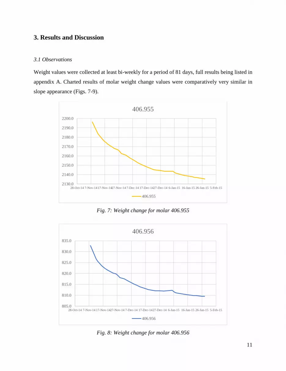
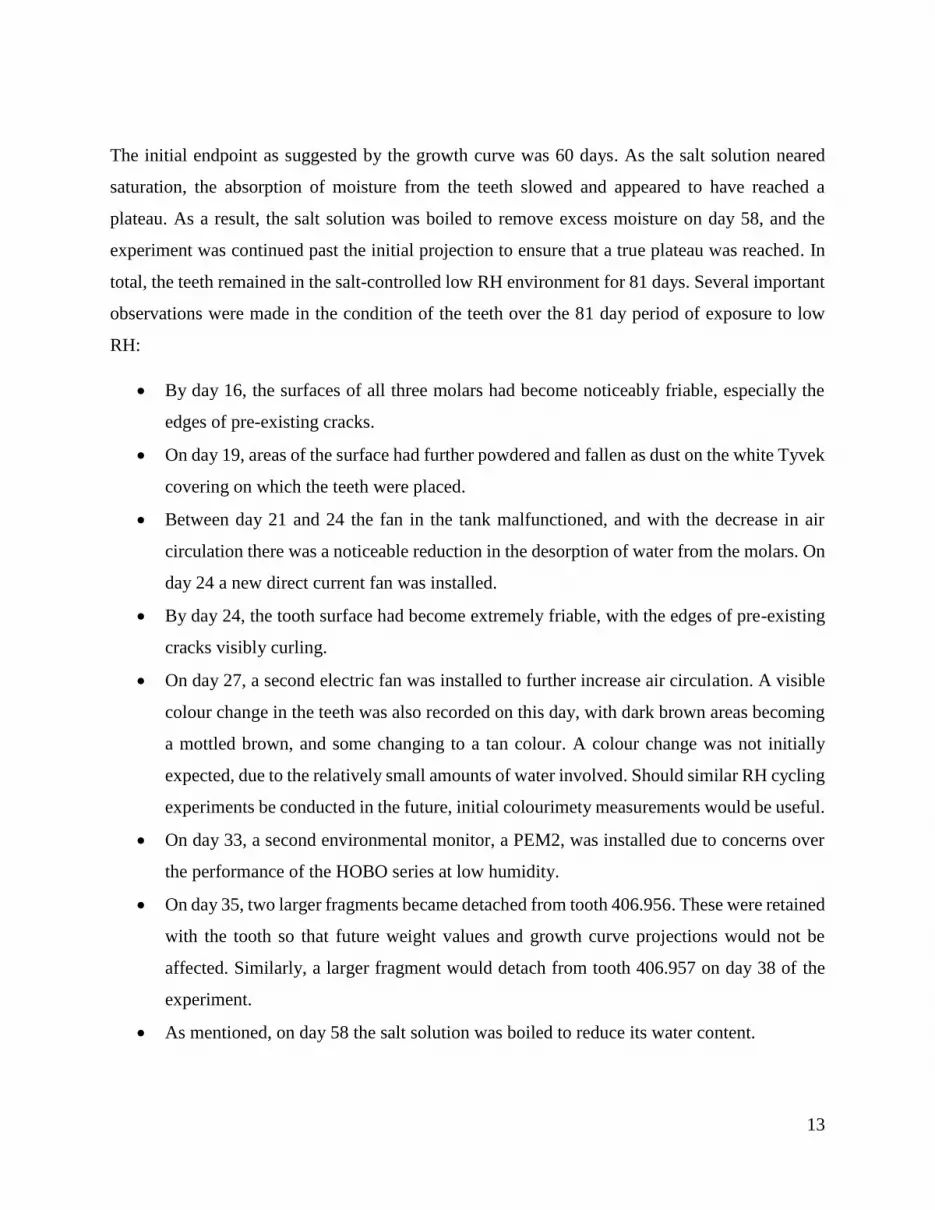
Weight values were collected at least bi-weekly for a period of 81 days, full results being listed in
appendix A. Charted results of molar weight change values were comparatively very similar in
slope appearance (Figs. 7-9).
Fig. 7: Weight change for molar 406.955
Fig. 8: Weight change for molar 406.956
2130.0
2140.0
2150.0
2160.0
2170.0
2180.0
2190.0
2200.0
28-Oct-14 7-Nov-1417-Nov-1427-Nov-14 7-Dec-14 17-Dec-1427-Dec-14 6-Jan-15 16-Jan-15 26-Jan-15 5-Feb-15
406.955
406.955
805.0
810.0
815.0
820.0
825.0
830.0
835.0
28-Oct-14 7-Nov-14 17-Nov-1427-Nov-14 7-Dec-14 17-Dec-1427-Dec-14 6-Jan-15 16-Jan-15 26-Jan-15 5-Feb-15
406.956
406.956

12
Fig. 9: Weight change for molar 406.957
This data was further adjusted to reflect the amount of weight lost, which was plotted in
CurveExpert to allow for the fitting of a growth curve (Fig. 10), guiding endpoint calculations
using the formula:
Exponential Association: y=a(1-exp(-bx)
Coefficient Data:
a = 6.16E+01
b = 3.87E-02
Fig. 10: Exponential growth curve of moisture desorption
1115.0
1120.0
1125.0
1130.0
1135.0
1140.0
1145.0
1150.0
1155.0
28-Oct-14 7-Nov-1417-Nov-1427-Nov-14 7-Dec-14 17-Dec-1427-Dec-14 6-Jan-15 16-Jan-15 26-Jan-15 5-Feb-15
406.957
406.957
S = 1.55163230
r = 0.99573469
X Axis (units)
Y A
xis
(u
nit
s)
0.0 11.0 22.0 33.0 44.0 55.0 66.00.00
10.01
20.02
30.03
40.04
50.05
60.06
13
The initial endpoint as suggested by the growth curve was 60 days. As the salt solution neared
saturation, the absorption of moisture from the teeth slowed and appeared to have reached a
plateau. As a result, the salt solution was boiled to remove excess moisture on day 58, and the
experiment was continued past the initial projection to ensure that a true plateau was reached. In
total, the teeth remained in the salt-controlled low RH environment for 81 days. Several important
observations were made in the condition of the teeth over the 81 day period of exposure to low
RH:
By day 16, the surfaces of all three molars had become noticeably friable, especially the
edges of pre-existing cracks.
On day 19, areas of the surface had further powdered and fallen as dust on the white Tyvek
covering on which the teeth were placed.
Between day 21 and 24 the fan in the tank malfunctioned, and with the decrease in air
circulation there was a noticeable reduction in the desorption of water from the molars. On
day 24 a new direct current fan was installed.
By day 24, the tooth surface had become extremely friable, with the edges of pre-existing
cracks visibly curling.
On day 27, a second electric fan was installed to further increase air circulation. A visible
colour change in the teeth was also recorded on this day, with dark brown areas becoming
a mottled brown, and some changing to a tan colour. A colour change was not initially
expected, due to the relatively small amounts of water involved. Should similar RH cycling
experiments be conducted in the future, initial colourimety measurements would be useful.
On day 33, a second environmental monitor, a PEM2, was installed due to concerns over
the performance of the HOBO series at low humidity.
On day 35, two larger fragments became detached from tooth 406.956. These were retained
with the tooth so that future weight values and growth curve projections would not be
affected. Similarly, a larger fragment would detach from tooth 406.957 on day 38 of the
experiment.
As mentioned, on day 58 the salt solution was boiled to reduce its water content.
14
Little further change was noticed, apart from the continued loss of powdered material, until
day 74 when the teeth colour appeared to further change. The tan areas of tooth 406.955
appeared even lighter in colour. Dark patches appeared more noticeable on tooth 406.956,
possibly indicating that the surrounding tooth was becoming visually lighter in colour. As
the cracks in tooth 406.957 had expanded, a reddish-brown material very similar to rust
had presented between the cracks. X-ray fluorescence of the material showed it to be iron.
This may have come from the surround burial environment and precipitated from the
slightly acidic ground water onto the slightly alkaline tooth surface.
By day 79, the teeth appeared very cracked and fragile, with the root surfaces of 406.956,
which had mostly remained dark, appearing to become lighter.
Along with the increase in surface-crack area, and the possible joining of previously separate
cracks (Figs. 11-16), definite permanent change has been noted. In total, the three mammoth
molars collectively lost 13.3g worth of powdered and fragmented material (from a total of
4064.3g). While it is difficult to accurately determine which layer constitutes the majority of the
powdering, the larger fragmentation has notably only occurred in the outer cement layer. As the
teeth have been brought back to the ambient humidity (~45%RH), fragmentation and powdering
has actively continued. It has been suggested by Dr. Rob Waller that this powdering of the surface,
which appeared to begin after only around two to three weeks of exposure to a low RH
environment, may partially collect in the pre-existing cracks. As humidity rises, and the molar
accordingly adsorbs moisture, these small amounts of material may impede the full expansion of
the tooth material, acting as fulcrums. This could cause further structural stress and contribute to
the further disintegration of the teeth. This is an area which will require further study to validate.

15
Fig. 11: 406.955 before experiment Fig. 12: 406.955 after experiment
Fig. 13: 406.956 before experiment Fig. 14: 406.956 after experiment

16
Fig. 15: 406.957 before experiment Fig. 16: 406.957 after experiment
3.2 Tooth Crack Measurements
The following initial and post-experiment measurement of crack widths were taken under
magnified viewing using Propoint digital calipers, with an accuracy of 0.03mm (Table 1-3). When
comparing the pre- and post-experiment crack measurement values it is apparent that previous
cracks have widened at differing rates, with the smallest change being an increase of 7% and the
largest being an increase of +104%.
Table 1: Measurement for Tooth 406.955
Initial End Change %Change
Weight at 21.1C and 41.3%RH 2196.2g 2135.5g 60.7g -3%
Crack 1, top face, vertical crack 1.21mm 2.11mm 0.90mm +74%
Crack 2, top face, vertical crack 1.62mm 1.79mm 0.17mm +10%
Crack 3, side face, vertical crack 2.84mm 4.23mm 1.39mm +49%
Crack 4, bottom face, vertical crack 1.11mm 1.42mm 0.31mm +28%
17
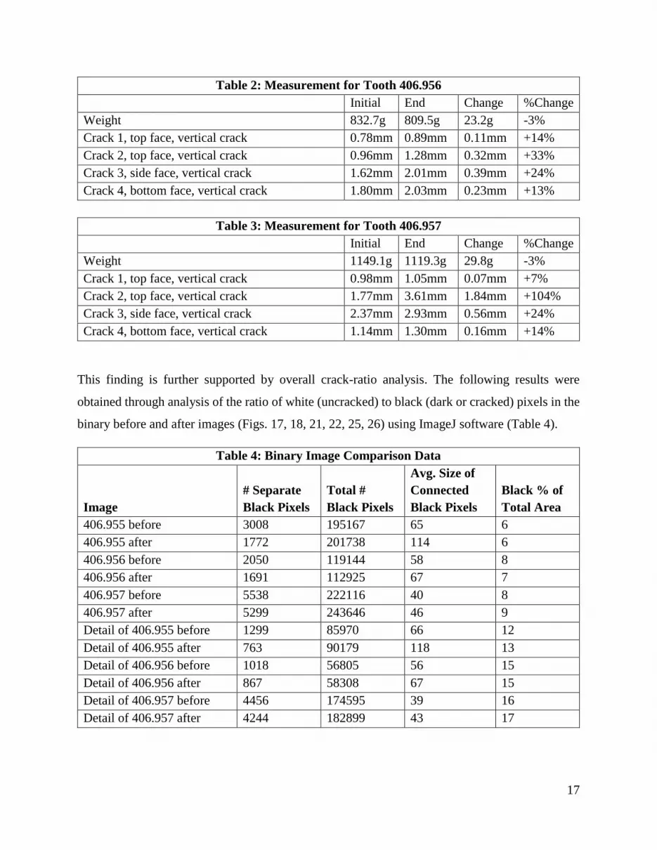
Table 2: Measurement for Tooth 406.956
Initial End Change %Change
Weight 832.7g 809.5g 23.2g -3%
Crack 1, top face, vertical crack 0.78mm 0.89mm 0.11mm +14%
Crack 2, top face, vertical crack 0.96mm 1.28mm 0.32mm +33%
Crack 3, side face, vertical crack 1.62mm 2.01mm 0.39mm +24%
Crack 4, bottom face, vertical crack 1.80mm 2.03mm 0.23mm +13%
Table 3: Measurement for Tooth 406.957
Initial End Change %Change
Weight 1149.1g 1119.3g 29.8g -3%
Crack 1, top face, vertical crack 0.98mm 1.05mm 0.07mm +7%
Crack 2, top face, vertical crack 1.77mm 3.61mm 1.84mm +104%
Crack 3, side face, vertical crack 2.37mm 2.93mm 0.56mm +24%
Crack 4, bottom face, vertical crack 1.14mm 1.30mm 0.16mm +14%
This finding is further supported by overall crack-ratio analysis. The following results were
obtained through analysis of the ratio of white (uncracked) to black (dark or cracked) pixels in the
binary before and after images (Figs. 17, 18, 21, 22, 25, 26) using ImageJ software (Table 4).
Table 4: Binary Image Comparison Data
Image
# Separate
Black Pixels
Total #
Black Pixels
Avg. Size of
Connected
Black Pixels
Black % of
Total Area
406.955 before 3008 195167 65 6
406.955 after 1772 201738 114 6
406.956 before 2050 119144 58 8
406.956 after 1691 112925 67 7
406.957 before 5538 222116 40 8
406.957 after 5299 243646 46 9
Detail of 406.955 before 1299 85970 66 12
Detail of 406.955 after 763 90179 118 13
Detail of 406.956 before 1018 56805 56 15
Detail of 406.956 after 867 58308 67 15
Detail of 406.957 before 4456 174595 39 16
Detail of 406.957 after 4244 182899 43 17
18
In general, the number of separate areas of black pixels (# Separate Black Pixels) decreased while
the overall size (Avg. Size of Connected Black Pixels) and ratio of black to white pixels (Black %
of Total Area) increased. This represents a widening and possibly a joining of cracks previously
identified as being separate. The one instance of a decrease in the total number of black pixels, and
subsequently the black percentage of the total area (overall image of tooth 406.956), was noted to
be due to a large loss from the edge of the tooth. To account for such losses, areas of detail from
the centre of the molar surfaces were compared (Figs. 19, 20, 23, 24, 27, 28) with all results
showing an increase in the percentage of black pixels (Table 4). While full quantification of crack
propagation may not be possible with this method of analysis, its correlation to the results obtained
from the semi-manual crack measurement do suggest it is a valid qualitative method of analysis.
3.3 Storage Survey
In conjunction with the experiment, a simple survey was distributed to institutions housing
mammoth molars or related collections. The anonymous answers from this survey create a more
complex overview of the current storage practices for mammoth molars. The survey questions
included:
1. Does your institution currently store mammoth molars?
2. How are the mammoth molars currently stored (open shelves, boxes, drawers, etc.)?
3. Is the temperature controlled or modified in your storage area?
4. If the temperature is controlled or modified, within what range of temperatures does your
storage area typically fall?
5. Is the humidity controlled or modified in your storage area?
6. If the humidity is controlled or modified, within what range of relative humidity does your
storage area typically (90% of the time) fall?
7. Have there been periods longer than one week in which the relative humidity was higher
than this range by more than 10%?
8. Have there been periods longer than one week in which the relative humidity was lower
than this range by more than 10%?

19
Over the course of the survey there was a total of twenty-five respondents from various locations
in North America and Europe. Participating institutions, excluding those who requested to remain
anonymous, may be found in the ‘Further Thanks’ section of the ‘Acknowledgements’ (pg. ii).
Twenty (80%) of the responding institutions reported controlling or modifying the temperature in
their storage areas, most commonly at around 18oC +/- 3oC. The remaining institutions do not
currently modify the temperature or only do so occasionally. Only fourteen (56%) of institutions
reported controlling or modifying the humidity in their storage areas (either with both
humidification and dehumidification or solely dehumidification), with reported humidity ranging
from around 20%RH to 60%RH, with 50% +/- 5% being most common. Twelve institutions (48%)
indicated that there may have been periods longer than a week in which the RH was higher than
their typical range by more than 10%, with six of these (32%) indicating that this is known to have
occurred. Similarly, thirteen institutions (52%) indicated that there may have been periods longer
than a week in which the RH was lower than their typical range by more than 10%, with six of
these (24%) indicating that this is known to have occurred. The most common methods of storage
included closed cabinet systems, drawers (usually open top), and storage on open shelves.
Through the CCI equipment loan program, a PEM2 data logger was sent to the Yukon
Palaeontology Program facility in Whitehorse to record the ambient storage environment. Data
collection occurred from January 12th, 2015 to March 3rd, 2015. The data logger was placed on
open shelving in the collection storage area, collecting temperature and relative humidity values
at twenty minute intervals. Temperature remained relatively stable at 18 ±1°C from January 12th
to February 9th, raising to 19 ±1°C for the remaining duration. Some fluctuations were recorded,
including a drop on January 30th from 18°C to 15°C before quickly raising to 20°C. On February
5th-6th another drop occurred from 18°C to 16°C lasting twenty-four hours. The relative humidity
was low, averaging 14%RH and dropping to a low of 2%RH (maximum RH values attained being
25%). This falls within the range of the low 11%RH attained in this experiment, and mammoth
molars housed in heated but not humidified storage environments may undergo conditions and
resulting damage comparable to that in this study.

20
4. Conclusion
Following acclimatization to a low 11%RH environment, ImageJ analysis of the mammoth molars
shows an overall increase in the crack ratios of the teeth, including an increase in crack dimensions
as well as a possible joining of previously separate cracks. Physical measurement of crack widths
further indicate the widening of cracks by between 10% and 100% depending on the crack location.
A measurable loss of material occurred from the three molars, including powder and fragments,
weighing in total 13.3g (of the original total weight of 4178.0g). As the molars re-equilibrated to
the ambient 45%RH, they adsorption and expansion of the teeth caused further powdering and
disintegration. This indicates that exposure to the 11%RH environment has produced an
irreversible change in the molars. Field testing in the Yukon storage facility as well as a survey of
North American and European collections show that many mammoth molar collections undergo
fluctuations of RH +/-10% outside the normal storage conditions and lasting longer than one week.
In many cases, collection storage areas are heated but not humidified. Heating of cold outdoor air
without humidification lowers the relative humidity, creating the recorded values which reached a
low of 2%RH. It is certain that these environments cause additional damage to mammoth molars
comparable to those observed in this study. If storage areas are to be heated for comfort,
humidification of the air to 45%-65%RH is recommended to prevent desiccation and damage (CCI
2010). It is also recommended that the initial drying of molars post-excavation be undertaken in
a controlled and slow manner, to prevent or minimize the initial appearance of damage.
This study demonstrates that molar deterioration significantly increases due to low relative
humidity. Although further study of the mechanisms involved is required, it was also observed
that damage continues upon raising of the relative humidity. Comparable studies have shown that
teeth are more fragile below 50%RH, making it more difficult to protect teeth under such
conditions (Williams 1991 23). Closed storage systems, which appear to be most common among
surveyed institutions, are able to provide some buffering against fluctuations in relative humidity.
However, if low relative humidity is maintained for a prolonged period, damage comparable to
that in this study may occur.

21
5. Bibliography
CCI. 2010. Care of Ivory, Bone, Horn, and Antler. CCI Note 6/1. Accessed October 2nd, 2014.
http://www.cci-icc.gc.ca/publications/notes/6-1-eng.aspx
Erhardt, D., M. Mecklenburg, C. Tumosa, M. McCormick-Goodhart. 1995. The Determination
of Allowable RH Fluctuations. WAAC Newsletter. Vol. 17. No. 1. Conservation Analytical
Laboratory, Smithsonian Institution.
Ferretti, M.P. 2003. Structure and Evolution of Mammoth Molar Enamel. Acta Palaeontol
48 (3).
Fisher, Samantha. 2014. Examining the Effect of Relative Humidity on Mammoth Molars. Art
Conservation Research Project. Kingston: Queen’s University.
Haynes, Gary. 1991. Mammoth, Mastodonts, & Elephants: Biology, Behavior, and the Fossil
Record. Cambridge: Cambridge University Press.
Hillson, S. 1986. Teeth. New York, NY: Cambridge University Press.
ImageJ: Image Processing and Analysis in Java. Accessed March 19th, 2014.
http://imagej.nih.gov/ij/index.html
Labno, C. 2015. Basic Intensity Quantification with ImageJ. Chicago: University of Chicago
Integrated Light Microscopy Core. Accessed March 28th, 2015.
https://digital.bsd.uchicago.edu/resources_files/Basic%20image%20quantification.pdf
Lister, A.M. and Sher, A.V. 2001. The Origin and Evolution of the Woolly Mammoth. Science
(November) 294.
Michalski, S. 1993. Relative Humidity: A Discussion of Correct/Incorrect Values. ICOM
Committee for Conservation 10th triennial meeting: Washington, DC, 22-27 August 1993:
preprints. ICOM Committee for Conservation, Paris, France.
Michalski, S. 2013. Agent of Deterioration: Incorrect Relative Humidity. Canadian
Conservation Institute. Accessed December 17th, 2014. http://www.cci-icc.gc.ca/resources-
ressources/agentsofdeterioration-agentsdedeterioration/chap10-eng.aspx
Williams, S.L. 1991. Investigation of the Causes of Structural Damage to Teeth in Natural
History Collections. Collection Forum 7 (1).

22
Appendix A: Table of Recorded Molar Weights
Table 5: Molar weights, collected between November 8th, 2014 and January 28th, 2015.
Date Elapsed Temp (C)
RH
(HOBO)
Weight of
Molar
955(g)
Weight of
Molar
956(g)
Weight of
Molar
957(g)
RH
(PEM2)
8-Nov-14 0 21.1 41.3 2196.2 832.7 1149.1
12-Nov-14 4 20.8 18 2184.0 826.6 1142.6
13-Nov-14 5 20.8 18 2181.8 825.6 1141.5
16-Nov-14 8 20.4 17 2176.8 823.5 1138.8
17-Nov-14 9 20.8 17 2175.4 823.0 1138.0
18-Nov-14 10 20.5 17 2174.2 822.5 1137.3
19-Nov-14 11 20.1 16 2172.8 822.0 1136.7
20-Nov-14 12 19.7 16 2171.7 821.6 1136.1
24-Nov-14 16 20.3 17 2167.8 820.1 1134.0
26-Nov-14 18 20.4 16 2166.9 819.8 1133.7
27-Nov-14 19 20.6 15 2165.8 819.3 1133.1
29-Nov-14 21 20.1 15 2162.5 818.1 1131.5
2-Dec-14 24 20.3 15 2160.9 817.5 1130.6
5-Dec-141 27 20.2 14 2158.0 816.4 1129.2
9-Dec-14 31 20.6 14 2154.3 815.1 1127.8
11-Dec-14 33 20.3 14 2152.5 814.5 1127.1 12
13-Dec-14 35 20.8 14 2150.9 813.9 1126.4 10
16-Dec-14 38 21.4 14 2149.0 813.3 1125.5 10
19-Dec-14 41 22.1 14 2147.2 812.6 1124.6 10
22-Dec-14 44 21.2 14 2145.5 812.2 1124.0 10
24-Dec-14 46 21.5 15 2144.8 812.0 1123.6 11
27-Dec-14 49 21.4 16 2144.2 812.0 1123.4 12
1 A second fan was installed in the tank

23
30-Dec-14 52 21.1 16 2143.7 811.9 1123.2 12
05-Jan-152 58 20.1 17 2143.7 812.2 1123.2 12
06-Jan-15 59 19.8 13 2142.6 811.7 1122.7 9
07-Jan-15 60 20 13 2141.6 811.3 1122.3 9
08-Jan-15 61 19.1 13 2141.2 811.1 1122.0 9
12-Jan-15 65 20.5 13 2139.6 810.6 1121.3 9
13-Jan-15 66 20.7 13 2139.2 810.5 1121.1 9
14-Jan-15 67 20.2 13 2138.9 810.4 1120.9 9
15-Jan-15 68 20.5 13 2138.7 810.3 1120.8 9
19-Jan-15 72 21.0 13 2137.6 810.0 1120.3 9
20-Jan-15 73 20.1 13 2137.3 809.9 1120.1 9
21-Jan-15 74 20.3 13 2137.0 809.9 1120.0 9
22-Jan-15 75 20.4 13 2137.0 809.9 1119.9 9
26-Jan-15 79 20.3 13 2135.9 809.5 1119.5 9
27-Jan-15 80 20.3 13 2135.7 809.5 1119.4 9
28-Jan-15 81 20.8 13 2135.5 809.5 1119.3 9
2 With the teeth gaining weight it was decided that the salt solution had become saturated. It was removed from the
tank and the absorbed moisture boiled off to dehydrate it.

24
Appendix B: Binary Molar Images
Before and after experiment binary images (overall and detail) used in ImageJ analysis of crack
ratios.
Fig. 17: 406.955 overall before image Fig. 18: 406.955 overall after image
Fig. 19: 406.955 detail before image Fig. 20: 406.955 detail after image

25
Fig. 21: 406.956 overall before image Fig. 22: 406.956 overall after image
Fig. 23: 406.956 detail before image Fig. 24: 406.956 detail after image

26
Fig. 25: 406.957 overall before image Fig. 26: 406.957 overall after image
Fig. 27: 406.957 detail before image Fig. 28: 406.957 detail after image

27
Daniel Doyle 52 Main Street,
Kingston, Ontario, Canada, K7K 3Y6
Email: [email protected]
(613) 546-0004
Education
Queen’s University (Kingston, Ont. Canada) 2013-Present
Masters of Art Conservation
•Currently in final year of studies, involving intensive research focus
•Work study during term running woodshop, photo studio, x-ray machine and chemical disposal
Caherconnell Archaeological Field School (Kilfenora, Ireland) 2012
Diploma in Archaeological Excavation (Level 1)
Algonquin College (Ottawa, Ont. Canada) 2009-2011
Diploma in Applied Museum Studies
University of Toronto (Toronto, Ont. Canada) 2003-2007
Honours Bachelor of Arts, Egyptology and Near Middle Eastern Civilizations
University of St. Michael’s College (Toronto, Ont. Canada) 2003-2007
Honours Bachelor of Arts, Celtic Studies (dual degree with UofT)
Work Experience
The Colony of Avalon Archaeological Site (Ferryland, NFLD, Canada) 2014
Graduate Conservation Intern, July 2014
•Archaeological conservation of freshly excavated metals (copper, iron, lead, silver) and organic
materials (leather, bone, cork) dating from a 17th Century colonial context, involving condition reporting,
identifying objects in consultation with the site archaeologist, proposing and undertaking treatments
The Rooms Provincial Museum (St. John’s, NFLD, Canada) 2014
Graduate Conservation Intern, June and August 2014
•Development and purchasing of disaster response kits for the museum
•Assessment, researching provenance, and treatment of ethnographic and organic (wood) objects
including re-articulation of a maritime archaic dog skeleton
•Private contract conservation of a sacred relic (human bone)
28
New Brunswick Museum (Saint John, N.B., Canada) 2013
Conservation Technician, April to September 2013
•Preparing exhibitions for travel, including condition reporting, packing artefacts (, and crate design
•Preparing artefacts for headlining exhibition (1812: The New Brunswick Story, 2013-2015), including
condition reporting, proposing treatment options, conducting treatments, photo-documentation, and
mount fabrication for various objects (military uniforms, swords, guns)
•Hanging of gallery shows and setup of exhibitions (paintings and quilts)
•Disaster response conservation of artefacts and construction work related to flood damage
Smithsonian Institution (Washington, D.C., USA) 2012
Objects Conservation Intern, June to July 2012
•Participation in public programming of the Lunder Conservation Centre, describing ongoing treatments
to the public and creating didactic displays to enhance visitor interaction with conservation treatments
•Monitoring environmental conditions and evaluating preservation concerns for objects
•Preparing artefacts for exhibition, including condition reporting, researching, proposing, and conducting
treatments on three-dimensional composite objects in consultation with the artists
•Extensive training received in analytical examination techniques, including Hirox digital microscopy,
infrared / UV examination, and digital x-ray
New Brunswick Museum (Saint John, N.B., Canada) 2011-2012
Post Graduate Conservation Intern, September 2011 to June 2012
•Working on a large scale, nationally significant art on paper conservation project
•Creation of exhibition (Behind the Scenes: Conservation of Miller Brittain's mural drawings for the
Saint John Tuberculosis Hospital (1941-1942), 2013-2014) focusing on conservation practices,
including sourcing materials, creating educational plans and interactives, and delivering content to
groups on a daily basis
•Environmental monitoring, pest control and monitoring, and preventative conservation strategies
New Brunswick Museum (Saint John, N.B., Canada) 2011-2012
Collections Move Technician, October 2011 to March 2012
•Preparing museum collection for move to off-site storage location; including condition reporting, mount
making, packing, and documenting in the ‘TMS’ database of a variety of materials (pottery, glass,
composite objects, and antiquities)
Canada Science & Technology Museum (Ottawa, Ont. Canada) 2011
Objects Conservation Intern, January to May 2011
•Working with industrial and modern collections composed mostly of metals and plastics, with work
also including pieces made of stone, wood, bone, shell, ceramic, leather, rubber, and other synthetic
materials
•Preparing artefacts for inclusion in a tri-museum exhibition, including researching artifacts, condition
reporting, proposing treatment options, conducting treatments, and documentation in the KE Emu
database
Royal Ontario Museum (Toronto, Ont. Canada) 2004-2007
Gallery Interpreter (Public Programming), October 2004 to May 2007
•Engage visitors with exhibits by experiential learning through controlled physical interaction with
artefacts, providing historical significance, and relating to objects on display

29
Conferences and Workshops Attended
Association of North American Graduate Programs in Conservation (ANAGPIC). 41st Annual
Conference. Wilmington, DE, USA.
2015
Canadian Conservation Institute (CCI). Archaeological Conservation Field Techniques Workshop. St.
John’s, NL, Canada.
2014
Association of North American Graduate Programs in Conservation (ANAGPIC). 40th Annual
Conference. Buffalo, NY, USA.
2014
Canadian Association for Conservation (CAC). 39th Annual Conference. Saint John, NB, Canada. 2013
Canadian Conservation Institute (CCI). Teaching Conservation in Native Heritage Centres. Glooscap
Mi’kmaq Heritage Centre, NS, Canada.
2011
Publications and Lectures
Doyle, D. 2015. Míle Míle i gCéin: The Irish Language in Canada. Ottawa: Borealis Press. 319 pgs.
Association of Newfoundland and Labrador Archives (ANLA). 2014. “ANLA’s Guide to the
Preservation of Scrapbooks.” Training and Development. Online video. St. John’s, NFLD.
2015
2014
Doyle, D., and R. Smart. 2014. “Care of Arsenic-Contaminated Taxidermy and Ethnographic
Collections.” Museum Association of Newfoundland and Labrador Quarterly Newsletter.
St. John’s, NFLD.
2014
Titus, C., D. Doyle, J. Beaudry Tardif, and M. Dumville. 2013. “Behind the Scenes: Conservation of
Miller Brittain's mural cartoons for the Saint John Tuberculosis Hospital (1941-1942).”
Presented by M. Dumville at the Canadian Association for Conservation (CAC) 39th Annual
Conference. Saint John, N.B.
2013
Doyle, D. 2013. “Gaeltacht Bhaile na hÉireann agus Stáid na Gaelainne i gCeanada.” Garm Lu:
Canadian Celtic Arts Journal. Vol. 34. Toronto, Ont. 3 pgs.
2013
Doyle, D. 2013. An Ghaeilge i gCeanada. Lecture presented at Éigse Cholm Cille, Colaiste Mhig Aoidh,
University of Ulster. Derry, Northern Ireland.
2013
Awards
Canadian Museum Association Awards of Outstanding Achievement in Conservation 2014
Governor General’s Canadian History Medal 2000
Skills / Assets / Community Involvement
•Bilingual Irish Gaelic and English with basic understanding of French, Ojibwe, and Egyptian Arabic
•Proficiency in both TMS and KE EMu museum database systems
•Fully licensed for the safe handling of restricted and unrestricted firearms under Canadian Law
•Involved with Comhaltas Ceoltóirí Éireann and the Permanent North American Gaeltacht Project as an
uillean piper and Irish Gaelic language teacher