evolution of beetle bioluminescence: the origin of beetle luciferin
TRANSCRIPT
8 ORIGINAL RESEARCH J. C. Day et al.
Copyright © 2004 John Wiley & Sons, Ltd. Luminescence 2004;19:8–20
Luminescence 2004; 19: 8–20Published online in Wiley InterScience (www.interscience.wiley.com). DOI: 10.1002/bio.749 ORIGINAL RESEARCH
Copyright © 2004 John Wiley & Sons, Ltd.
Evolution of beetle bioluminescence: the origin ofbeetle luciferin
John C. Day,1* Laurence C. Tisi2* and Mark J. Bailey1
1Centre for Ecology and Hydrology (CEH)–Oxford, Mansfield Road, Oxford OX1 3SR, UK2Lumora Ltd, Institute of Biotechnology, Tennis Court Road, Cambridge CB2 1QT, UK
Received 28 August 2003; revised 7 November 2003; accepted 10 November 2003
ABSTRACT: Bioluminescence, the conversion of chemical energy into light in living organisms, is dependent on two principalcomponents, an enzyme luciferase and the substrate luciferin. In beetles, the enzyme luciferase has been extensively studied, withsignificant enzymological, sequence and structural data now available. Furthermore, the enzyme has been employed in a remark-able number of important applications, from microbial detection and medical imaging to GM gene expression studies. However,there is little information regarding the biosynthesis of beetle luciferin, and here we review the literature and speculate as to itsevolutionary origins. Luciferin consists of a benzothiazole moiety attached to a thiazole carboxylic acid moiety, the former beingrarely observed in nature but the latter being observed in a broad range of biologically derived molecules. Benzothiazoles are,however, observed in melanogenesis and we speculate as to whether this may be relevant to the understanding of luciferinbiosynthesis in beetles. This review examines recent novel insights into beetle luciferin recycling and we assess a range of possiblebiosynthetic mechanisms. Copyright © 2004 John Wiley & Sons, Ltd.
KEYWORDS: luciferin; Coleoptera; luciferase; bioluminescence; imaging
luminescence, including luring of prey, communicationand aposematic signalling (4). Such complex roles arereflected in the complex physiology and control of thelight-emitting organs in these beetles. This exceptionallybright, coordinated light display, with its range of beha-vioural roles, may have evolved from a less sophis-ticated system with weaker bioluminescence, perhapswith a role at first unrelated to or incidental to aetiolo-gical considerations. However, the sequence of eventsduring evolution leading to the display of even weakbioluminescence is still not fully understood. Addressingthis question requires an understanding of not only thebiochemistry of the light emitting reaction per se, but theevolutionary origins of the luciferase and luciferin them-selves in these beetles. Presently, a substantial amountof information is available regarding the evolution ofbeetle luciferase and the reaction it catalyses. However,the biosynthetic pathway for the generation of beetleluciferin is unknown. In fact, it is not even clear to whichfamily of metabolites beetle luciferin is related. Conse-quently, understanding how this bioluminescent systemcame to evolve at the biochemical level requires anunderstanding of the biosynthetic pathway of luciferin.
Aside from the evolutionary insight that an under-standing of beetle luciferin synthesis could bring,there are also exciting practical applications that couldbenefit from this knowledge. The application of variousluciferases, from numerous organisms, in a wide varietyof imaging applications is becoming a dominant applica-tion of bioluminescent systems (5). Firefly luciferasehas proved to be a powerful tool in a wide range of
INTRODUCTION
Bioluminescence, the production of light by living organ-isms, is a biochemical reaction in which the oxygenationof a substrate, ‘luciferin’, via an enzyme, ‘luciferase’,generates light. Numerous different luciferin–luciferasereactions have been characterized from quite diverseorganisms and hence bioluminescence seems to haveevolved independently many times. The majority ofbioluminescent organisms inhabit the oceans but anumber of bioluminescent species can be found on theland. The bioluminescent members of the coleopteranfamily Lampyridae, and to a lesser extent other closelyrelated beetle families, make up the most widespreadand abundant bioluminescent terrestrial organisms onthe planet and include such familiar animals as firefliesand glow-worms. The members of the Lampyridae allshare a common bioluminescent reaction that uses anidentical luciferin and closely homologous luciferase. Itis this particular bioluminescent system that will be thesubject of the current review (1).
In the Lampyridae, bioluminescence is primarily usedfor mate attraction in adults and has evolved into anelaborate flash display in many species (2, 3), but anumber of other roles have been proposed for larval bio-
*Correspondence to: J. C. Day, Centre for Ecology and Hydrology(CEH)–Oxford, Mansfield Road, Oxford OX1 3SR, UK.Email: [email protected] to: L. C. Tisi, Lumora Ltd, Institute of Biotechnology,Tennis Court Road, Cambridge CB2 1QT, UK.Email: [email protected]
Origins of beetle luciferin ORIGINAL RESEARCH 9
Copyright © 2004 John Wiley & Sons, Ltd. Luminescence 2004;19:8–20
applications, from the rapid detection of pathogens(6) to in vivo imaging of cancers cells and organisms (5,7, 8). The discovery of genes encoding the biosyntheticpathway of beetle luciferin would enable the transforma-tion of cells or organisms already containing luciferaseto maintain bioluminescence without the continualreplenishment of the substrate, i.e. an autonomous bio-luminescent system.
Presently, the imaging of in vivo beetle luciferaseexpression systems, e.g. in carcinogenic tissue in amouse, requires the injection of the mouse with sub-stantial amounts of luciferin. This can be problematic, asthe luciferin does not easily cross membranes and, moreimportantly, introduces additional expense if continualmonitoring is required. Although prokaryotes can bemade to be autonomously bioluminescent by trans-formation with the bacterial lux genes (9), currently thereis no system that enables a eukaryotic cell to becomeautonomously bioluminescent.
Understanding the biosynthesis of eukaryotic luciferinmay thus lead to the development of a powerful reportersystem for imaging applications. However, there is a ca-veat. At present there is no evidence that any eukaryoteactually fully encodes all the genes required to make aparticular luciferin from simple, common metabolites.In other words, attempts to address the biosynthesis ofbeetle luciferin cannot assume that the beetles them-selves encode all the genes required to make luciferinfrom simple precursors. If may be necessary for them toingest particular precursors or even obtain precursorsfrom symbionts.
This review will summarize both historical andrecent data on luciferin biosynthesis and present poten-tial biosynthetic pathway models that could account forluciferin biogenesis in beetles.
EVOLUTIONARY ORIGINS OFBIOLUMINESCENCE
Light emission occurs in many different species inphylogenetically diverse organisms, and among thesedifferent groups the biochemistry, colour and method ofdisplay of light may be very different, leading researchersearly on to conclude that bioluminescent systems inmajor groups are not evolutionarily conserved (10). Theroles of bioluminescence in these various organisms arewide-ranging, including prey attraction (11), sexual com-munication (2, 3), aposematic signalling (4, 12–14) andcamouflage and protection (15–18). There are also manyexamples where the precise roles for bioluminescenceare unclear. In some such cases, the role of biolumine-scence has been suggested to have its purpose at abiochemical level rather than for any behaviouralpurpose, i.e. where the light emission itself appears to beincidental (10, 19).
Fundamentally, the evolution of bioluminescenceinvolves the coincidence of a luciferase with a luciferinin an environment where the luciferin can be oxygenated,with subsequent light production. The ubiquitous in-volvement of oxygen or reactive oxygen species (ROS)in bioluminescence is fundamentally a function of ther-modynamics, i.e. oxygenation reactions are some of theonly biochemical reactions with enough energy to pro-duce a visible photon. ROSs are highly reactive speciesderived from molecular oxygen and include the hydro-xide radical, superoxides and peroxides (20).
However, further significance has been attached tothe oxygenation of luciferins in bioluminescence asidefrom thermodynamic necessity. In particular, since bio-luminescent reactions turn-over molecular oxygen orROSs as substrates and hence diminish their abundance,it has been proposed that such reactions may haveevolved initially as oxygen/ROS detoxification strategies.From this primal function, subsequent ecological bene-fits of the light-emitting reactions could then lead toselection for brighter bioluminescence. This oxygendetoxification hypothesis was first proposed to explainthe origin of the bacterial bioluminescent system (10, 19),but has been subsequently presented as a working hypo-thesis for beetle bioluminescence (21–23). Such hypo-theses are certainly interesting; however, the fact thatoxygen must be involved in bioluminescence, for theaforementioned thermodynamic reasons, does notnecessarily mean that oxygen toxicity is a driving forcefor the evolution of bioluminescence. Furthermore,there are numerous oxygen detoxification processes thatare not bioluminescent, so clearly bioluminescence is nota prerequisite of oxygen detoxification.
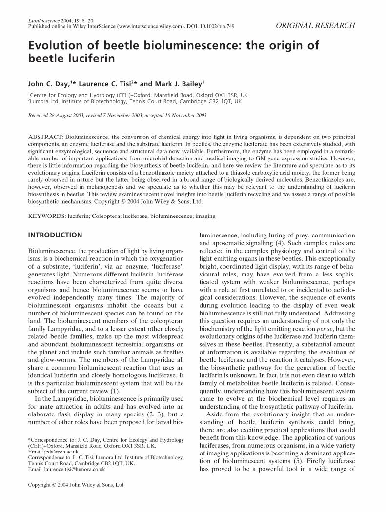
Where protein sequences are available for a particu-lar luciferase, phylogenetic analysis can indicate fromwhich enzyme family the luciferase evolved, e.g. bothcoelenterate and beetle luciferases have been found tobe homologous to proteins completely disassociated withbioluminescence (24, 25). As such, the genetic ‘pedigree’of luciferases can be easily established, given sufficientsequence data (25, 26). For luciferins, however, the situa-tion is more complicated. To begin with, it appears thata large number of different bioluminescent organismsshare a small number of luciferins (although the luciferinchemistry from several bioluminescent organisms isnot yet known; Fig. 1). In fact, of the five knownluciferin structures, two of the luciferins, Vargula andCoelenterazine, are structurally similar, with a common(guanosine-like) bicyclic heterocycle at their core, sug-gesting a possible evolutionary relationship [Fig. 1(d, e)].Otherwise, the luciferin chemistries shown in Fig. 1are extremely diverse and have clearly not all shareda common ancestor. In most cases the derivation ofcertain luciferins seems straightforward, e.g. bacterialluciferin is a flavin derivative (flavins being commonmetabolites). Further, the structure of dinoflagellate
10 ORIGINAL RESEARCH J. C. Day et al.
Copyright © 2004 John Wiley & Sons, Ltd. Luminescence 2004;19:8–20
luciferin, characterized from Pyrocystis lunula, is alinear tetrapyrrole and probably derived from chloro-phyll (27). However, for beetle luciferin, it is more diffi-cult to relate the structure to a known class of biologicalmetabolites that might suggest from which metabolicpathway it evolved.
Some bioluminescent organisms are capable ofmaking their own luciferin de novo from simple, com-mon metabolites. In these cases the bioluminescentorganism must encode all the genes required to code forthe necessary biosynthetic enzymes required for theentire biosynthetic pathway of their luciferin. For exam-ple, it has been demonstrated that the bioluminescentprokaryote Vibrio harveyi encodes all the genes on theLUX operon required to produce its luciferin de novoas well as its luciferase (28). Other bioluminescentorganisms, however, do not synthesize their luciferin atall, but rather acquire it, e.g. the hydromedusa Aequoreavictoria appears to be dependent on a dietary supply ofcoelenterazine for bioluminescence (29). Thus, there isprecedence for both in situ synthesis of a luciferin, asencoded by the genome of the bioluminescent organism,and the acquisition of a luciferin from an extraneoussource. It can be anticipated that for some bioluminesc-ent organisms there could be a mixture of the aboveapproaches, i.e. there may be a need to acquire a keycompound from another organism that is then convertedinto the appropriate luciferin by the organism itself.
ENZYMOLOGY AND EVOLUTION OF BEETLELUCIFERASE
The most studied of all beetle bioluminescent systems isthat of the North American firefly Photinus pyralis. Thisfirefly luciferase (EC 1.13.12.7) catalyses the oxidativedecarboxylation of beetle luciferin (LH2) in the presenceof ATP, O2 and Mg2+, producing light in a multi-step re-action; the same reaction is thought to occur for allknown beetle luciferases (Fig. 2).
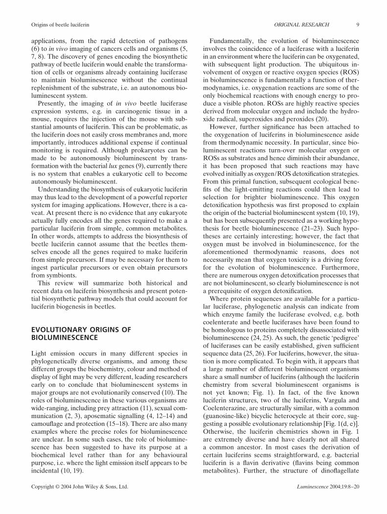
In the first stage of the reaction, both substrates,luciferin and ATP, form a ternary complex with lucife-rase. In the second step, an acid anhydride [Fig. 2(a)]is formed between the carboxylic group of luciferinand AMP (i.e. a luciferyl adenylate), with the release ofpyrophosphate. Through several intermediate stages,this adenylate is oxidized, generating the cyclic per-oxide, dioxetanone [Fig. 2(b–d)]. Decarboxylation ofthis peroxide leads to formation of oxyluciferin in asinglet electronically excited state that rapidly decays toits ground state, releasing a photon [Fig. 2(e–f)]. Thequantum yield for this reaction has been measuredat 0.88 relative to luciferin utilization and is, to date,the highest yield for any bioluminescent system (30).Although beetle luciferases use the same luciferin,depending on the luciferase sequence the light displayed
Figure 1. The chemical structures of five basic types ofluciferin: (a) beetle luciferin, found in all known biolumines-cent Coleoptera (including fireflies, click beetles and glow-worms); (b) bacterial luciferin, found in several bacteria, someof which are harboured by fish or squid; (c) dinoflagellateluciferin, found in dinoflagellates, which may also be har-boured by euphausiid shrimps; (d) Vargula luciferin, found inostracods and some fish; (e) coelenterazine, found in a diverserange of organisms, including copepods, squids, cnidarians,ctenophores, radiolarians, fish, mysid shrimps and chaetognaths.However, the luciferins from a large variety of bioluminescentorganisms, such as bioluminescent dipterans, molluscs andseveral worms, remains unknown.
Origins of beetle luciferin ORIGINAL RESEARCH 11
Copyright © 2004 John Wiley & Sons, Ltd. Luminescence 2004;19:8–20
Figure 2. Steps involved in the biochemistry of the light-emitting reaction catalysed by beetle luciferase.Initially, beetle luciferin, ATP-Mg2+ and beetle luciferase form a ternary complex, leading to the adenylationof beetle luciferin and releasing pyrophosphate (a), following proton abstraction (b), the luciferyl adenylatereacts with oxygen (c), leading to the formation of a cyclic peroxide (d), which results in decarboxylation andthe formation of excited state oxyluciferin (e), whose relaxation results in photon emission (f).
can range in colour from green (λmax 530 nm) to red (λmax
635 nm) (31–33).Firefly luciferase from Photinus pyralis is a 61 kDa
protein coded for by a mRNA encoding 550 aminoacids. After translation, firefly luciferase is localized inthe peroxisomes of the light organ cells (34) as a resultof a peroxisome targeting sequence located at theC-terminus of the protein (35–37). In 1996 the 3-D struc-ture of Photinus pyralis firefly luciferase was resolved(38). Based upon this data and the 3-D structure of theenzyme–substrate complex of gramicidin S synthetase,the 3-D structure for luciferase complexed with ATPand luciferin was proposed (39, 40). Two additionalstructures of adenylate-forming enzymes are also known,the crystal structure of DhbE, which is involved in theinitial stage of bacillibactin synthesis, along with anacetyl-CoA synthetase (41, 42).
Since the cloning and sequencing of the first beetleluciferase in 1985, (43) the amino acid sequences fromseveral beetle luciferases have been determined andfound to share high levels of identity (44–54). Theadenylating activity of firefly luciferase, along withsequence and structural homology, have grouped thisenzyme with a much larger ‘superfamily’ of homologous,adenylating enzymes. The closest family members to
beetle luciferase of this superfamily are the co-enzymeA (CoA) ligases, with homologues with strong identityfound in plants as well as invertebrates (26). Other moredistant relatives include the peptide synthetases, whichdiffer, mechanistically, in ligating their substrates toa carrier-protein bound phosphopantetheine ratherthan to the phosphopantetheine moiety of free CoA.Nonetheless, as demonstrated by a comparison of thecrystal structures of gramicidin S synthestase, a peptidesynthetase, and firefly luciferase, even these distantrelatives are structurally very similar to firefly luciferase.The bioluminescent reactions catalysed by beetle luci-ferases hence share with this superfamily a step involv-ing the adenylation of a carboxylic acid to form anactivated adenylate, in the case of beetle luciferase theformation of a luciferyl–adenylate. However, for beetleluciferases, instead of the proceeding steps involvingthe reaction of luciferyl–adenylate with CoA to form athioester of CoA and luciferin, molecular oxygen reactswith the luciferyl–adenylate, leading to the oxidativedecarboxylation of the substrate luciferin, with sub-sequent light emission.
In fact, chemically synthesized luciferyl–adenylateis capable of enzyme-free chemiluminescence undersuitable conditions, although with much lower quantum
12 ORIGINAL RESEARCH J. C. Day et al.
Copyright © 2004 John Wiley & Sons, Ltd. Luminescence 2004;19:8–20
efficiency than the bioluminescent reaction (55). Thisdemonstrates that the luciferyl–adenylate is intrinsicallyprone to oxygenation and indeed, chemiluminescence.Thus, fundamentally, beetle luciferase’s ability tocatalyse bioluminescence is a function of its abilityto adenylate beetle luciferin. An interesting question,therefore, is whether beetle luciferin was ever a produc-tive substrate for the formation of luciferin–CoA estersvia a beetle luciferin–CoA ligase activity, i.e. was therean ancestral beetle luciferin–CoA ligase where theoxygenation reaction did not significantly compete withligase activity? If so, what metabolic pathway utilized theluciferin–CoA thioester?
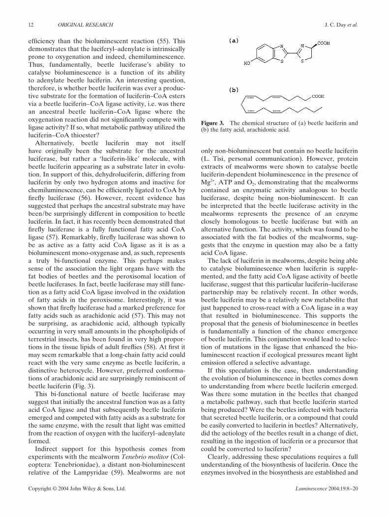
Alternatively, beetle luciferin may not itselfhave originally been the substrate for the ancestralluciferase, but rather a ‘luciferin-like’ molecule, withbeetle luciferin appearing as a substrate later in evolu-tion. In support of this, dehydroluciferin, differing fromluciferin by only two hydrogen atoms and inactive forchemiluminescence, can be efficiently ligated to CoA byfirefly luciferase (56). However, recent evidence hassuggested that perhaps the ancestral substrate may havebeen/be surprisingly different in composition to beetleluciferin. In fact, it has recently been demonstrated thatfirefly luciferase is a fully functional fatty acid CoAligase (57). Remarkably, firefly luciferase was shown tobe as active as a fatty acid CoA ligase as it is as abioluminescent mono-oxygenase and, as such, representsa truly bi-functional enzyme. This perhaps makessense of the association the light organs have with thefat bodies of beetles and the peroxisomal location ofbeetle luciferases. In fact, beetle luciferase may still func-tion as a fatty acid CoA ligase involved in the oxidationof fatty acids in the peroxisome. Interestingly, it wasshown that firefly luciferase had a marked preference forfatty acids such as arachidonic acid (57). This may notbe surprising, as arachidonic acid, although typicallyoccurring in very small amounts in the phospholipids ofterrestrial insects, has been found in very high propor-tions in the tissue lipids of adult fireflies (58). At first itmay seem remarkable that a long-chain fatty acid couldreact with the very same enzyme as beetle luciferin, adistinctive heterocycle. However, preferred conforma-tions of arachidonic acid are surprisingly reminiscent ofbeetle luciferin (Fig. 3).
This bi-functional nature of beetle luciferase maysuggest that initially the ancestral function was as a fattyacid CoA ligase and that subsequently beetle luciferinemerged and competed with fatty acids as a substrate forthe same enzyme, with the result that light was emittedfrom the reaction of oxygen with the luciferyl–adenylateformed.
Indirect support for this hypothesis comes fromexperiments with the mealworm Tenebrio molitor (Col-eoptera: Tenebrionidae), a distant non-bioluminescentrelative of the Lampyridae (59). Mealworms are not
Figure 3. The chemical structure of (a) beetle luciferin and(b) the fatty acid, arachidonic acid.
only non-bioluminescent but contain no beetle luciferin(L. Tisi, personal communication). However, proteinextracts of mealworms were shown to catalyse beetleluciferin-dependent bioluminescence in the presence ofMg2+, ATP and O2, demonstrating that the mealwormscontained an enzymatic activity analogous to beetleluciferase, despite being non-bioluminescent. It canbe interpreted that the beetle luciferase activity in themealworms represents the presence of an enzymeclosely homologous to beetle luciferase but with analternative function. The activity, which was found to beassociated with the fat bodies of the mealworms, sug-gests that the enzyme in question may also be a fattyacid CoA ligase.
The lack of luciferin in mealworms, despite being ableto catalyse bioluminescence when luciferin is supple-mented, and the fatty acid CoA ligase activity of beetleluciferase, suggest that this particular luciferin–luciferasepartnership may be relatively recent. In other words,beetle luciferin may be a relatively new metabolite thatjust happened to cross-react with a CoA ligase in a waythat resulted in bioluminescence. This supports theproposal that the genesis of bioluminescence in beetlesis fundamentally a function of the chance emergenceof beetle luciferin. This conjunction would lead to selec-tion of mutations in the ligase that enhanced the bio-luminescent reaction if ecological pressures meant lightemission offered a selective advantage.
If this speculation is the case, then understandingthe evolution of bioluminescence in beetles comes downto understanding from where beetle luciferin emerged.Was there some mutation in the beetles that changeda metabolic pathway, such that beetle luciferin startedbeing produced? Were the beetles infected with bacteriathat secreted beetle luciferin, or a compound that couldbe easily converted to luciferin in beetles? Alternatively,did the aetiology of the beetles result in a change of diet,resulting in the ingestion of luciferin or a precursor thatcould be converted to luciferin?
Clearly, addressing these speculations requires a fullunderstanding of the biosynthesis of luciferin. Once theenzymes involved in the biosynthesis are established and
Origins of beetle luciferin ORIGINAL RESEARCH 13
Copyright © 2004 John Wiley & Sons, Ltd. Luminescence 2004;19:8–20
from where the enzymes originated, luciferin biogenesismay also be resolved.
STRUCTURE AND BIOSYNTHESIS OF BEETLELUCIFERIN
Luciferin, 2-(6-hydroxybenzothiazol-2-yl)-2-thiazoline-4-carboxylic acid [Fig. 1(a)], was first isolated in crystallineform from the North American firefly Photinus pyralisin 1957 (60). Proof of its structure came from thesuccessful chemical synthesis of enzymatically activeluciferin in 1961 (61) and the structure was confirmed byX-ray crystallography [Fig. 1(a)] (62). Luciferin appearsto be conserved in structure between bioluminescentbeetle species, and even families, irrespective of meta-morphic stage or lantern location (63–65).
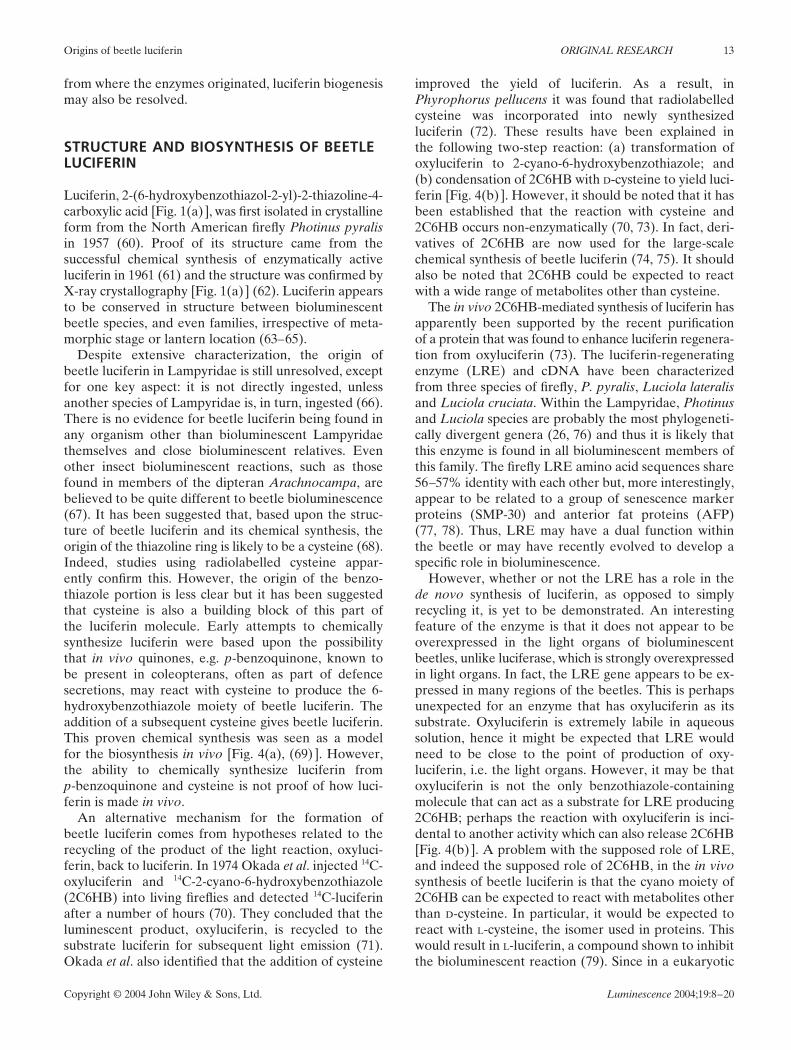
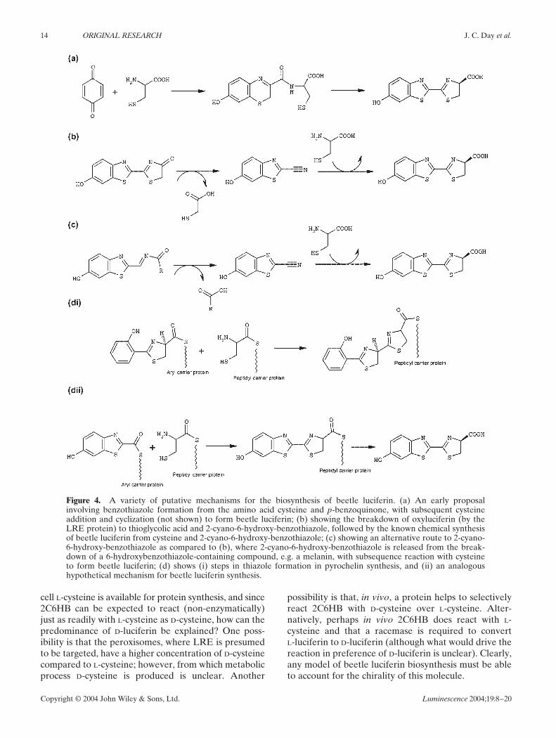
Despite extensive characterization, the origin ofbeetle luciferin in Lampyridae is still unresolved, exceptfor one key aspect: it is not directly ingested, unlessanother species of Lampyridae is, in turn, ingested (66).There is no evidence for beetle luciferin being found inany organism other than bioluminescent Lampyridaethemselves and close bioluminescent relatives. Evenother insect bioluminescent reactions, such as thosefound in members of the dipteran Arachnocampa, arebelieved to be quite different to beetle bioluminescence(67). It has been suggested that, based upon the struc-ture of beetle luciferin and its chemical synthesis, theorigin of the thiazoline ring is likely to be a cysteine (68).Indeed, studies using radiolabelled cysteine appar-ently confirm this. However, the origin of the benzo-thiazole portion is less clear but it has been suggestedthat cysteine is also a building block of this part ofthe luciferin molecule. Early attempts to chemicallysynthesize luciferin were based upon the possibilitythat in vivo quinones, e.g. p-benzoquinone, known tobe present in coleopterans, often as part of defencesecretions, may react with cysteine to produce the 6-hydroxybenzothiazole moiety of beetle luciferin. Theaddition of a subsequent cysteine gives beetle luciferin.This proven chemical synthesis was seen as a modelfor the biosynthesis in vivo [Fig. 4(a), (69) ]. However,the ability to chemically synthesize luciferin fromp-benzoquinone and cysteine is not proof of how luci-ferin is made in vivo.
An alternative mechanism for the formation ofbeetle luciferin comes from hypotheses related to therecycling of the product of the light reaction, oxyluci-ferin, back to luciferin. In 1974 Okada et al. injected 14C-oxyluciferin and 14C-2-cyano-6-hydroxybenzothiazole(2C6HB) into living fireflies and detected 14C-luciferinafter a number of hours (70). They concluded that theluminescent product, oxyluciferin, is recycled to thesubstrate luciferin for subsequent light emission (71).Okada et al. also identified that the addition of cysteine
improved the yield of luciferin. As a result, inPhyrophorus pellucens it was found that radiolabelledcysteine was incorporated into newly synthesizedluciferin (72). These results have been explained inthe following two-step reaction: (a) transformation ofoxyluciferin to 2-cyano-6-hydroxybenzothiazole; and(b) condensation of 2C6HB with D-cysteine to yield luci-ferin [Fig. 4(b) ]. However, it should be noted that it hasbeen established that the reaction with cysteine and2C6HB occurs non-enzymatically (70, 73). In fact, deri-vatives of 2C6HB are now used for the large-scalechemical synthesis of beetle luciferin (74, 75). It shouldalso be noted that 2C6HB could be expected to reactwith a wide range of metabolites other than cysteine.
The in vivo 2C6HB-mediated synthesis of luciferin hasapparently been supported by the recent purificationof a protein that was found to enhance luciferin regenera-tion from oxyluciferin (73). The luciferin-regeneratingenzyme (LRE) and cDNA have been characterizedfrom three species of firefly, P. pyralis, Luciola lateralisand Luciola cruciata. Within the Lampyridae, Photinusand Luciola species are probably the most phylogeneti-cally divergent genera (26, 76) and thus it is likely thatthis enzyme is found in all bioluminescent members ofthis family. The firefly LRE amino acid sequences share56–57% identity with each other but, more interestingly,appear to be related to a group of senescence markerproteins (SMP-30) and anterior fat proteins (AFP)(77, 78). Thus, LRE may have a dual function withinthe beetle or may have recently evolved to develop aspecific role in bioluminescence.
However, whether or not the LRE has a role in thede novo synthesis of luciferin, as opposed to simplyrecycling it, is yet to be demonstrated. An interestingfeature of the enzyme is that it does not appear to beoverexpressed in the light organs of bioluminescentbeetles, unlike luciferase, which is strongly overexpressedin light organs. In fact, the LRE gene appears to be ex-pressed in many regions of the beetles. This is perhapsunexpected for an enzyme that has oxyluciferin as itssubstrate. Oxyluciferin is extremely labile in aqueoussolution, hence it might be expected that LRE wouldneed to be close to the point of production of oxy-luciferin, i.e. the light organs. However, it may be thatoxyluciferin is not the only benzothiazole-containingmolecule that can act as a substrate for LRE producing2C6HB; perhaps the reaction with oxyluciferin is inci-dental to another activity which can also release 2C6HB[Fig. 4(b) ]. A problem with the supposed role of LRE,and indeed the supposed role of 2C6HB, in the in vivosynthesis of beetle luciferin is that the cyano moiety of2C6HB can be expected to react with metabolites otherthan D-cysteine. In particular, it would be expected toreact with L-cysteine, the isomer used in proteins. Thiswould result in L-luciferin, a compound shown to inhibitthe bioluminescent reaction (79). Since in a eukaryotic
14 ORIGINAL RESEARCH J. C. Day et al.
Copyright © 2004 John Wiley & Sons, Ltd. Luminescence 2004;19:8–20
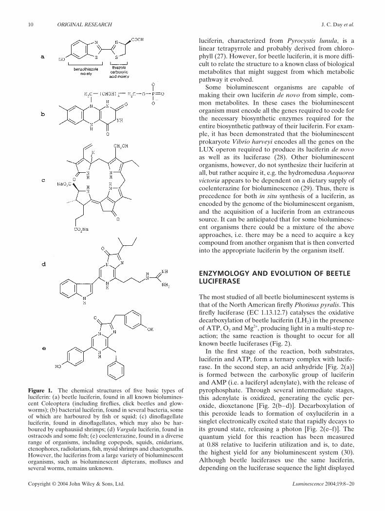
Figure 4. A variety of putative mechanisms for the biosynthesis of beetle luciferin. (a) An early proposalinvolving benzothiazole formation from the amino acid cysteine and p-benzoquinone, with subsequent cysteineaddition and cyclization (not shown) to form beetle luciferin; (b) showing the breakdown of oxyluciferin (by theLRE protein) to thioglycolic acid and 2-cyano-6-hydroxy-benzothiazole, followed by the known chemical synthesisof beetle luciferin from cysteine and 2-cyano-6-hydroxy-benzothiazole; (c) showing an alternative route to 2-cyano-6-hydroxy-benzothiazole as compared to (b), where 2-cyano-6-hydroxy-benzothiazole is released from the break-down of a 6-hydroxybenzothiazole-containing compound, e.g. a melanin, with subsequence reaction with cysteineto form beetle luciferin; (d) shows (i) steps in thiazole formation in pyrochelin synthesis, and (ii) an analogoushypothetical mechanism for beetle luciferin synthesis.
cell L-cysteine is available for protein synthesis, and since2C6HB can be expected to react (non-enzymatically)just as readily with L-cysteine as D-cysteine, how can thepredominance of D-luciferin be explained? One poss-ibility is that the peroxisomes, where LRE is presumedto be targeted, have a higher concentration of D-cysteinecompared to L-cysteine; however, from which metabolicprocess D-cysteine is produced is unclear. Another
possibility is that, in vivo, a protein helps to selectivelyreact 2C6HB with D-cysteine over L-cysteine. Alter-natively, perhaps in vivo 2C6HB does react with L-cysteine and that a racemase is required to convertL-luciferin to D-luciferin (although what would drive thereaction in preference of D-luciferin is unclear). Clearly,any model of beetle luciferin biosynthesis must be ableto account for the chirality of this molecule.
Origins of beetle luciferin ORIGINAL RESEARCH 15
Copyright © 2004 John Wiley & Sons, Ltd. Luminescence 2004;19:8–20
Thus, a rigorously proven pathway for the in vivobiosynthesis of beetle luciferin cannot as yet be claimed.Furthermore, only one enzyme to date, LRE, has in anyway been associated with luciferin synthesis. However,there is no evidence that LRE has a role in the de novosynthesis of beetle luciferin. This is pertinent to the inter-pretation of radiolabelling experiments. If oxyluciferinrecycling acts in addition to a separate de novo luciferinbiosynthetic pathway, then the appearance of radiolabelin luciferin may be unrelated to the de novo biosynthesisof luciferin in beetles. Clearly, then, there is a need toexamine possible in vivo mechanisms that could producebeetle luciferin.
BIOSYNTHESIS OF LUCIFERIN-LIKEMOLECULES
In addressing the biosynthesis of luciferin, it may beappropriate to consider the biosynthetic pathway ofmolecules that are similar to luciferin, i.e. those contain-ing either a benzothiazole moiety or a thiazole ringmoiety. This is especially so in those cases where theenzymes involved in the biosynthesis of a luciferin-likecompound have been identified and sequenced, thusproviding enzymatic precedence for the biosynthesis ofaspects of the luciferin structure. Such comparisons mayoffer clues to luciferin biosynthesis in beetles and sug-gest experiments to test for similar enzymatic functionspresent in beetles. Three examples in particular areconsidered here: siderophore synthesis, which demon-strates enzymatic precedence for thiazole ring forma-tion; melanogenesis, which exemplifies benzothiazolesynthesis; and thiamine synthesis, which demonstratesthiazole ring formation but potentially offers a route tobenzothiazoles.
The siderophores are microbial iron chelates that aresynthesized by bacteria in response to low iron availabil-ity in the environment. Recently, the biosynthetic path-ways and genes of a number of bacterial siderophoreshave been determined (80) and some enzymes involvedin siderophore biosynthesis have been shown to beadenylating enzymes homologous to firefly luciferase(81, 82). Some siderophore precursors show remarkablesimilarity in structure to beetle luciferin, with cysteine-derived thiazole ring structures such as pyochelin precur-sors [Fig. 4(di) ] (83). Analysis of siderophore synthesissuggests a possible mechanism for the formation ofluciferin by an analogous process, i.e. cysteine additionto a carboxylic acid [Fig. 4(di–ii) depicts such a path-way]. This scheme is interesting in that: (a) it shows thatthiazoline ring formation may occur via enzyme/CoA-bound thioesters; and (b) beetle luciferin could have asits precursor a carboxylic acid that uses a luciferase-likeprotein to ligate cysteine to form the thiazoline ring ofbeetle luciferin. An interesting aspect of such a pathway
is that it involves luciferase homologues. This could evensuggest that luciferase evolved from a biosynthetic en-zyme involved in the biosynthetic pathway for luciferin orluciferin-like molecule. To test whether beetle luciferincould be synthesized in vivo by a mechanism similar tothat of siderophores, we have recently synthesized 6-hydroxybenzothiazole 2-carboxylic acid (6HBC) as thefree acid, the thio-ester of which is shown in Fig. 4(dii)(84). However, we found that 6HBC rapidly decarboxy-lates to 6-hydroxybenzothiazole, so if it is an intermedi-ate, it is an unstable one. Furthermore, no evidence wasfound that it could act as a beetle luciferin presursorin experiments on firefly tail extracts (E. Law & L. Tisi,personal communication). However, as Fig. 4(d) sug-gests, if 6HBC were an intermediate, it may only ever bepresent as a bound enzyme–CoA thioester, such that thefree acid is never a substrate in itself.
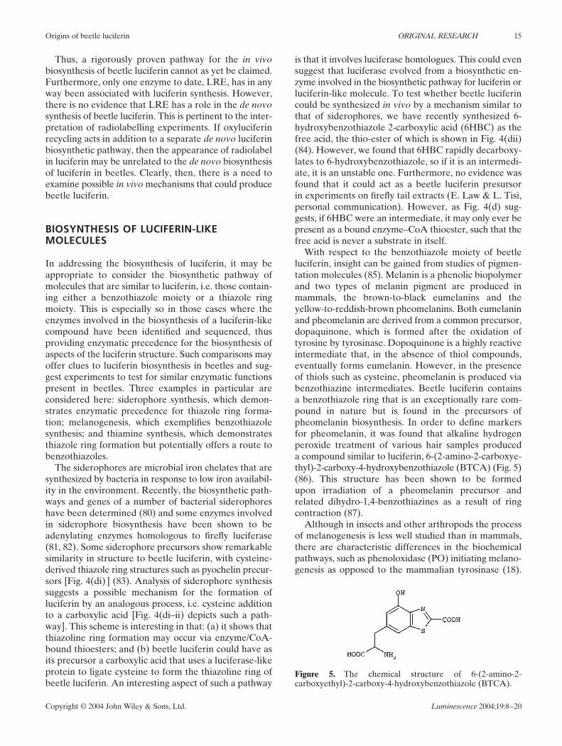
With respect to the benzothiazole moiety of beetleluciferin, insight can be gained from studies of pigmen-tation molecules (85). Melanin is a phenolic biopolymerand two types of melanin pigment are produced inmammals, the brown-to-black eumelanins and theyellow-to-reddish-brown pheomelanins. Both eumelaninand pheomelanin are derived from a common precursor,dopaquinone, which is formed after the oxidation oftyrosine by tyrosinase. Dopoquinone is a highly reactiveintermediate that, in the absence of thiol compounds,eventually forms eumelanin. However, in the presenceof thiols such as cysteine, pheomelanin is produced viabenzothiazine intermediates. Beetle luciferin containsa benzothiazole ring that is an exceptionally rare com-pound in nature but is found in the precursors ofpheomelanin biosynthesis. In order to define markersfor pheomelanin, it was found that alkaline hydrogenperoxide treatment of various hair samples produceda compound similar to luciferin, 6-(2-amino-2-carboxye-thyl)-2-carboxy-4-hydroxybenzothiazole (BTCA) (Fig. 5)(86). This structure has been shown to be formedupon irradiation of a pheomelanin precursor andrelated dihydro-1,4-benzothiazines as a result of ringcontraction (87).
Although in insects and other arthropods the processof melanogenesis is less well studied than in mammals,there are characteristic differences in the biochemicalpathways, such as phenoloxidase (PO) initiating melano-genesis as opposed to the mammalian tyrosinase (18).
Figure 5. The chemical structure of 6-(2-amino-2-carboxyethyl)-2-carboxy-4-hydroxybenzothiazole (BTCA).
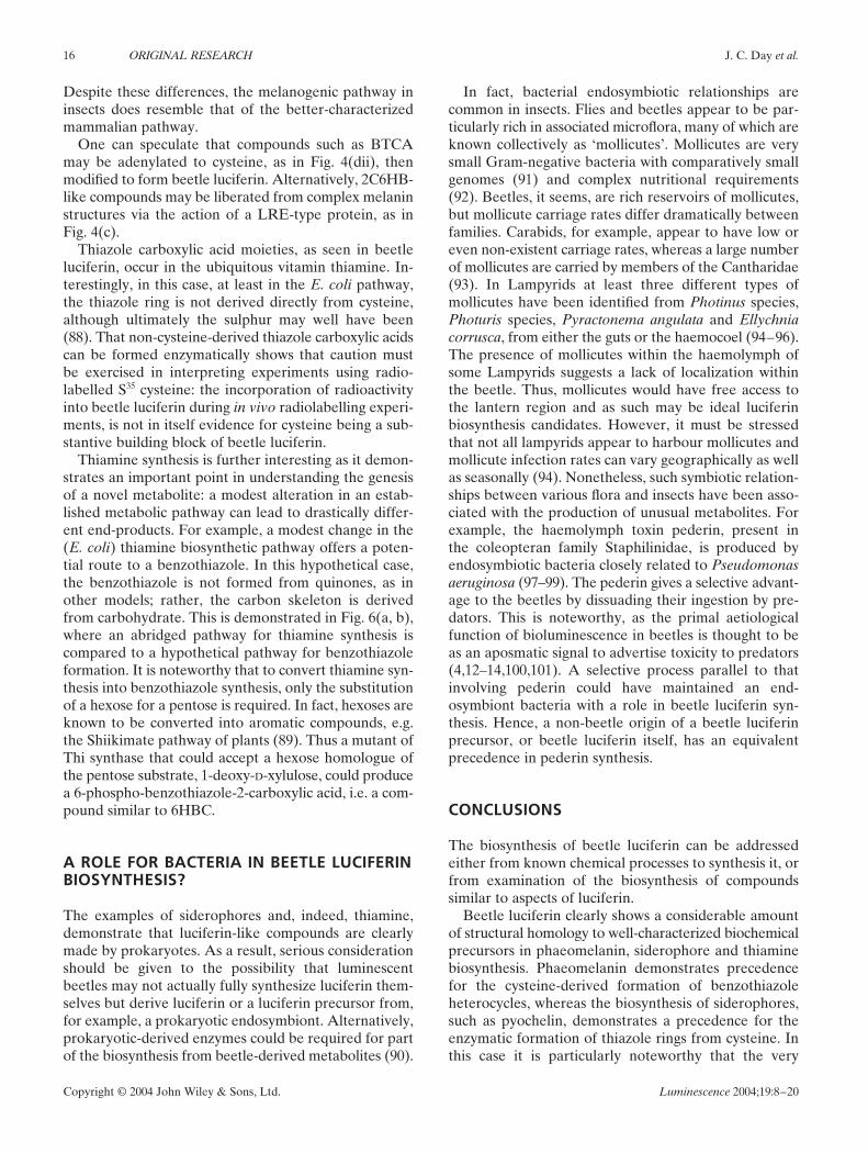
16 ORIGINAL RESEARCH J. C. Day et al.
Copyright © 2004 John Wiley & Sons, Ltd. Luminescence 2004;19:8–20
Despite these differences, the melanogenic pathway ininsects does resemble that of the better-characterizedmammalian pathway.
One can speculate that compounds such as BTCAmay be adenylated to cysteine, as in Fig. 4(dii), thenmodified to form beetle luciferin. Alternatively, 2C6HB-like compounds may be liberated from complex melaninstructures via the action of a LRE-type protein, as inFig. 4(c).
Thiazole carboxylic acid moieties, as seen in beetleluciferin, occur in the ubiquitous vitamin thiamine. In-terestingly, in this case, at least in the E. coli pathway,the thiazole ring is not derived directly from cysteine,although ultimately the sulphur may well have been(88). That non-cysteine-derived thiazole carboxylic acidscan be formed enzymatically shows that caution mustbe exercised in interpreting experiments using radio-labelled S35 cysteine: the incorporation of radioactivityinto beetle luciferin during in vivo radiolabelling experi-ments, is not in itself evidence for cysteine being a sub-stantive building block of beetle luciferin.
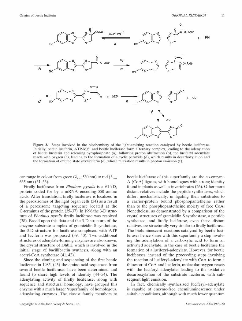
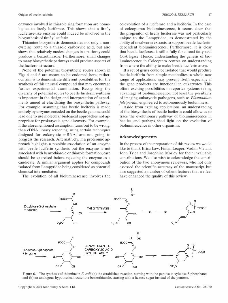
Thiamine synthesis is further interesting as it demon-strates an important point in understanding the genesisof a novel metabolite: a modest alteration in an estab-lished metabolic pathway can lead to drastically differ-ent end-products. For example, a modest change in the(E. coli) thiamine biosynthetic pathway offers a poten-tial route to a benzothiazole. In this hypothetical case,the benzothiazole is not formed from quinones, as inother models; rather, the carbon skeleton is derivedfrom carbohydrate. This is demonstrated in Fig. 6(a, b),where an abridged pathway for thiamine synthesis iscompared to a hypothetical pathway for benzothiazoleformation. It is noteworthy that to convert thiamine syn-thesis into benzothiazole synthesis, only the substitutionof a hexose for a pentose is required. In fact, hexoses areknown to be converted into aromatic compounds, e.g.the Shiikimate pathway of plants (89). Thus a mutant ofThi synthase that could accept a hexose homologue ofthe pentose substrate, 1-deoxy-D-xylulose, could producea 6-phospho-benzothiazole-2-carboxylic acid, i.e. a com-pound similar to 6HBC.
A ROLE FOR BACTERIA IN BEETLE LUCIFERINBIOSYNTHESIS?
The examples of siderophores and, indeed, thiamine,demonstrate that luciferin-like compounds are clearlymade by prokaryotes. As a result, serious considerationshould be given to the possibility that luminescentbeetles may not actually fully synthesize luciferin them-selves but derive luciferin or a luciferin precursor from,for example, a prokaryotic endosymbiont. Alternatively,prokaryotic-derived enzymes could be required for partof the biosynthesis from beetle-derived metabolites (90).
In fact, bacterial endosymbiotic relationships arecommon in insects. Flies and beetles appear to be par-ticularly rich in associated microflora, many of which areknown collectively as ‘mollicutes’. Mollicutes are verysmall Gram-negative bacteria with comparatively smallgenomes (91) and complex nutritional requirements(92). Beetles, it seems, are rich reservoirs of mollicutes,but mollicute carriage rates differ dramatically betweenfamilies. Carabids, for example, appear to have low oreven non-existent carriage rates, whereas a large numberof mollicutes are carried by members of the Cantharidae(93). In Lampyrids at least three different types ofmollicutes have been identified from Photinus species,Photuris species, Pyractonema angulata and Ellychniacorrusca, from either the guts or the haemocoel (94–96).The presence of mollicutes within the haemolymph ofsome Lampyrids suggests a lack of localization withinthe beetle. Thus, mollicutes would have free access tothe lantern region and as such may be ideal luciferinbiosynthesis candidates. However, it must be stressedthat not all lampyrids appear to harbour mollicutes andmollicute infection rates can vary geographically as wellas seasonally (94). Nonetheless, such symbiotic relation-ships between various flora and insects have been asso-ciated with the production of unusual metabolites. Forexample, the haemolymph toxin pederin, present inthe coleopteran family Staphilinidae, is produced byendosymbiotic bacteria closely related to Pseudomonasaeruginosa (97–99). The pederin gives a selective advant-age to the beetles by dissuading their ingestion by pre-dators. This is noteworthy, as the primal aetiologicalfunction of bioluminescence in beetles is thought to beas an aposmatic signal to advertise toxicity to predators(4,12–14,100,101). A selective process parallel to thatinvolving pederin could have maintained an end-osymbiont bacteria with a role in beetle luciferin syn-thesis. Hence, a non-beetle origin of a beetle luciferinprecursor, or beetle luciferin itself, has an equivalentprecedence in pederin synthesis.
CONCLUSIONS
The biosynthesis of beetle luciferin can be addressedeither from known chemical processes to synthesis it, orfrom examination of the biosynthesis of compoundssimilar to aspects of luciferin.
Beetle luciferin clearly shows a considerable amountof structural homology to well-characterized biochemicalprecursors in phaeomelanin, siderophore and thiaminebiosynthesis. Phaeomelanin demonstrates precedencefor the cysteine-derived formation of benzothiazoleheterocycles, whereas the biosynthesis of siderophores,such as pyochelin, demonstrates a precedence for theenzymatic formation of thiazole rings from cysteine. Inthis case it is particularly noteworthy that the very
Origins of beetle luciferin ORIGINAL RESEARCH 17
Copyright © 2004 John Wiley & Sons, Ltd. Luminescence 2004;19:8–20
enzymes involved in thiazole ring formation are homo-logous to firefly luciferase. This shows that a fireflyluciferase-like enzyme could indeed be involved in thebiosynthesis of firefly luciferin.
Thiamine biosynthesis demonstrates not only a non-cysteine route to a thiazole carboxylic acid, but alsoshows that relatively modest changes in a pathway couldproduce a benzothiazole. Furthermore, small changesto many biosynthetic pathways could produce aspects ofthe luciferin structure.
None of the potential biosynthetic routes shown inFigs 4 and 6 are meant to be endorsed here; rather,our aim is to demonstrate different possibilities for thesynthesis of this unusual compound that may encouragefurther experimental examination. Recognizing thediversity of potential routes to beetle luciferin synthesisis important in the design and interpretation of experi-ments aimed at elucidating the biosynthetic pathway.For example, assuming that beetle luciferin is madeentirely by enzymes encoded on the beetle genome couldlead one to use molecular biological approaches not ap-propriate for prokaryotic gene discovery. For example,if the aforementioned assumption turns out to be wrong,then cDNA library screening, using certain techniquesdesigned for eukaryotic mRNA, are not going toprogress the research. Alternatively, if a proteomic ap-proach highlights a possible association of an enzymewith beetle luciferin synthesis but the enzyme is notassociated with benzothiasole or thiazole formation, careshould be exercised before rejecting the enzyme as acandidate. A similar argument applies for compoundsisolated from Lampyridae being considered as potentialchemical intermediates.
The evolution of all bioluminescence involves the
co-evolution of a luciferase and a luciferin. In the caseof coleopteran bioluminescence, it seems clear thatthe progenitor of firefly luciferase was not particularlyunique to the Lampyridae, as demonstrated by theability of mealworm extracts to support beetle luciferin-dependent bioluminescence. Furthermore, it is clearthat beetle luciferase is still a fully functional fatty acidCoA ligase. Hence, understanding the genesis of bio-luminescence in Coleoptera centres on understandingfrom where the ability to make beetle luciferin arose.
If a set of genes could be isolated that would producebeetle luciferin from simple metabolites, a whole newrange of applications may present itself, especially ifthe gene products are functional in eukaryotes. Thisoffers exciting possibilities in reporter systems takingadvantage of bioluminescence, not least the possibilityof imaging eukaryotic pathogens, such as Plasmodiumfalciparum, engineered to autonomously bioluminess.
Aside from exciting applications, an understandingof the biosynthesis of beetle luciferin could allow us totrace the evolutionary pathway of bioluminescence inbeetles and perhaps shed light on the evolution ofbioluminescence in other organisms.
Acknowledgements
In the process of the preparation of this review we wouldlike to thank Erica Law, Finian Leaper, Vadim Viviani,John Tyler and Josephine Morley for their invaluablecontributions. We also wish to acknowledge the contri-bution of the two anonymous reviewers, who not onlyassessed the scientific accuracy of the manuscript butalso suggested a number of salient features that we feelhave enhanced the quality of this review.
Figure 6. The synthesis of thiamine in E. coli; (a) the established reaction, starting with the pentose D-xylulose-5-phosphate;and (b) an analogous hypothetical route to a benzothiazole, starting with a hexose sugar instead of the pentose.
18 ORIGINAL RESEARCH J. C. Day et al.
Copyright © 2004 John Wiley & Sons, Ltd. Luminescence 2004;19:8–20
24. Tsuji FI, Ohmiya Y, Fagan TF, Toh H, Inouye S. Molecularevolution of the Ca2+-binding photoproteins of the Hydrozoa.Photochem. Photobiol. 1995; 62: 657–661.
25. Wood KV. The chemical mechanism and evolutionary devel-opment of beetle bioluminescence. Photochem. Photobiol. 1995;62: 662–673.
26. Day JC, White PJ, Squirrell DJ, Bailey MJ. Origins ofbioluminescence in beetles: a phylogenetic perspective. InBioluminescence and Chemiluminescence: Progress and CurrentApplications, Stanley PE, Kricka LJ (eds). World ScientificPublishing: Singapore, 2002; 29–32.
27. Nakamura H, Kishi Y, Shimomura O, Morse D, Hastings JW.Structure of dinoflagellate luciferin and its enzymic andnonenzymic air-oxidation products. J. Am. Chem. Soc. 1989; 111:7607–7611.
28. Boylan M, Miyamoto C, Wall L, Graham A, Meighen E. Lux C,D and E genes of the Vibrio fischeri luminescence operon codefor the reductase, transferase, and synthetase enzymes involvedin aldehyde biosynthesis. Photochem. Photobiol. 1989; 49: 681–688.
29. Haddock SH, Rivers TJ, Robison BH. Can coelenterates makecoelenterazine? Dietary requirement for luciferin in cnidarianbioluminescence. Proc. Natl Acad. Sci. USA 2001; 98: 11148–11151.
30. McElroy WD, Seliger HH. Spectral emission and quantum yieldof firefly bioluminescence. Arch. Biochem. Biophys. 1960; 88: 136.
31. Hastings JW. Chemistries and colors of bioluminescent reactions:a review. Gene 1996; 173: 5–11.
32. McElroy WD, Seliger HH. In Molecular Architecture in CellPhysiology. Prentice Hall: Englewood Cliffs, NJ, 1966; 63–79.
33. Seliger HH, McElroy WD. The colors of firefly bioluminescence:enzyme configuration and species specificity. Proc. Natl Acad. Sci.USA 1964; 52: 75–81.
34. Keller GA, Gould S, Deluca M, Subramani S. Firefly luciferaseis targeted to peroxisomes in mammalian cells. Proc. Natl Acad.Sci. USA 1987; 84: 3264–3268.
35. Gould SJ, Keller GA, Subramani S. Identification of aperoxisomal targeting signal at the carboxy-terminus of fireflyluciferase. J. Cell Biol. 1987; 105: 2923–2931.
36. Gould SJ, Subramani S. Firefly luciferase as a tool in molecularand cell biology. Anal. Biochem. 1988; 175: 5–13.
37. Gould SJ, Keller GA, Hosken N, Wilkinson J, Subramani S. Aconserved tripeptide sorts proteins to peroxisomes. J. Cell Biol.1989; 108: 1657–1664.
38. Conti E, Franks NP, Brick P. Crystal structure of firefly luciferasethrows light on a superfamily of adenylate-forming enzymes.Structure 1996; 4: 287–298.
39. Branchini BR, Magyar RA, Murtiashaw MH, Anderson SM,Zimmer M. Site-directed mutagenesis of histidine 245 in fireflyluciferase: a proposed model of the active site. Biochemistry 1998;37: 15311–15319.
40. Sandalova TP, Ugarova NN. Model of the active site of fireflyluciferase. Biochemistry (Mosc.) 1999; 64: 962–967.
41. May JJ, Kessler N, Marahiel MA, Stubbs MT. Crystal structureof DhbE, an archetype for aryl acid activating domains of modu-lar nonribosomal peptide synthetases. Proc. Natl Acad. Sci. USA2002; 99: 12120–12125.
42. Gulick AM, Starai VJ, Horswill AR, Homick KM, Escalante-Semerena JC. The 1.75 Å crystal structure of acetyl-CoAsynthetase bound to adenosine-5′-propylphosphate and coenzymeA. Biochemistry 2003; 42: 2866–2873.
43. de Wet JR, Wood KV, Helinski DR, DeLuca M. Cloning offirefly luciferase cDNA and the expression of active luciferasein Escherichia coli. Proc. Natl Acad. Sci. USA 1985; 82: 7870–7873.
44. Choi YS, Lee KS, Bae JS et al. Molecular cloning and expressionof a cDNA encoding the luciferase from the firefly, Hotariaunmunsana. Comp. Biochem. Physiol. Biochem. Mol. Biol. 2002;132: 661–670.
45. Devine JH, Kutuzova GD, Green VA, Ugarova NN, BaldwinTO. Luciferase from the east European firefly Luciola mingrelica:cloning and nucleotide sequence of the cDNA, overexpressionin Escherichia coli and purification of the enzyme. Biochim.Biophys. Acta 1993; 1173: 121–132.
REFERENCES
1. Buck JB. Functions and evolution of bioluminescence. InBioluminescence in Action, Herring PJ (ed.). Academic Press:New York, 1978.
2. Carlson AD, Copeland J. Communication in insects. 1. Flashcommunication in fireflies. Q. Rev. Biol. 1985; 60: 415–436.
3. Lloyd JE. Firefly mating ecology, selection and evolution. In TheEvolution of Mating Systems in Insects and Arachnids, Choe JC,Crespi BJ (eds). Cambridge University Press: Cambridge, 1997;184–192.
4. Sivinski J. The nature and possible functions of luminescence inColeoptera larvae. Coleopt. Bull. 1981; 35: 167–179.
5. Greer LF III, Szalay AA. Imaging of light emission from theexpression of luciferases in living cells and organisms: a review.Luminescence 2002; 17: 43–74.
6. Kricka LJ. Prospects for chemiluminescent and biolumines-cent immunossay and nucleic acid assay in food testing andthe pharmaceutical industry. J. Biolumin. Chemilumin. 1998; 13:185–188.
7. Wang W, El-Deiry WS. Bioluminescent molecular imaging ofendogenous and exogenous p53-mediated transcription in vitroand in vivo using an HCT116 human colon carcinoma xenograftmodel. Cancer Biol. Ther. 2003; 2: 196–202.
8. Rice BW, Cable MD, Nelson MB. In vivo imaging of light-emitting probes. J. Biomed. Opt. 2001; 6: 432–440.
9. Francis KP, Yu J, Bellinger-Kawahara C et al. Visualizingpneumococcal infections in the lungs of live mice usingbioluminescent Streptococcus pneumoniae transformed with anovel Gram-positive lux transposon. Infect. Immun. 2001; 69:3350–3358.
10. Hastings JW. Biological diversity, chemical mechanisms, and theevolutionary origins of bioluminescent systems. J. Mol. Evol.1983; 19: 309–321.
11. Sivinski JM. Phototropism, bioluminenescence and the Diptera.Fla. Entomol. 1998; 81: 577–593.
12. De Cock R, Matthysen E. Aposematism and bioluminescence:experimental evidence from glow-worm larvae (Coleoptera:Lampyridae). Evol. Ecol. 1999; 13: 619–639.
13. De Cock R, Matthysen E. Glow-worm larvae bioluminescence(Coleoptera: Lampyridae) operates as an aposematic signal upontoads (Bufo bufo). Behav. Ecol. 2003; 14: 103–108.
14. Underwood TJ, Tallamy DW, Pesek JD. Bioluminescencein firefly larvae: a test of the aposematic display hypothesis(Coleoptera: Lampyridae). J. Insect Behav. 1997; 10: 365–370.
15. Clarke WD. Function of bioluminescence in mesopelagic organ-isms. Nature 1963; 198: 1244–1246.
16. Herring PJ. Aspects of bioluminescence of fishes. Oceanogr. Mar.Biol. Annu. Rev. 1982; 20: 415–470.
17. Young RE. Oceanic bioluminescence: an overview of generalfunctions. Bull. Mar. Sci. 1983; 33: 829–845.
18. Lindsay SM, Frank TM, Kent J, Partridge JC, Latz MI. Spectralsensitivity of vision and bioluminescence in the midwater shrimpSergestes similis. Biol. Bull. 1999; 197: 348–360.
19. Seliger HH. The origin of bioluminescence. Photochem.Photobiol. 1975; 21: 355–361.
20. Allen RC. Molecular oxygen (O2): reactivity and luminescence.In Bioluminescence and Chemiluminescence: Progress and Cur-rent Applications, Stanley PE, Kricka LJ (eds). World ScientificPublishing: Singapore, 2002; 223–232.
21. Barros MP, Bechara EJ. Bioluminescence as a possible auxiliaryoxygen detoxifying mechanism in elaterid larvae. Free Radic.Biol. Med. 1998; 24: 767–777.
22. Barros MP, Bechara EJ. Luciferase and urate may act as anti-oxidant defenses in larval Pyrearinus termitilluminans (Elater-idae: Coleoptera) during natural development and upon20-hydroxyecdysone treatment. Photochem. Photobiol. 2000; 71:648–654.
23. Barros MP, Bechara EJ. Daily variations of antioxidant enzymeand luciferase activities in the luminescent click-beetle Pyrearinustermitilluminans: cooperation against oxygen toxicity. InsectBiochem. Mol. Biol. 2001; 31: 393–400.
Origins of beetle luciferin ORIGINAL RESEARCH 19
Copyright © 2004 John Wiley & Sons, Ltd. Luminescence 2004;19:8–20
46. Masuda T, Tatsumi H, Nakano E. Cloning and sequence ana-lysis of cDNA for luciferase of a Japanese firefly, Luciola cruciata.Gene 1989; 77: 265–270.
47. Ohmiya Y, Ohba N, Toh H, Tsuji F. Cloning, expression andsequence analysis of cDNA for the luciferases from the Japanesefireflies, Pyrocoelia miyako and Hotaria parvula. Photochem.Photobiol. 1995; 62: 309–313.
48. Sala-Newby GB, Thomson CM, Campbell AK. Sequence andbiochemical similarities between the luciferases of the glow-wormLampyris noctiluca and the firefly Photinus pyralis. Biochem. J.1996; 313: 761–767.
49. Tatsumi H, Masuda T, Kajiyama N, Nakano E. Luciferase cDNAfrom Japanese firefly, Luciola cruciata: cloning, structure andexpression in Escherichia coli. J. Biolumin. Chemilumin. 1989; 3:75–78.
50. Tatsumi H, Kajiyama N, Nakano E. Molecular cloning andexpression in Escherichia coli of a cDNA clone encodingluciferase of a firefly, Luciola lateralis. Biochim. Biophys. Acta1992; 1131: 161–165.
51. Viviani VR, Bechara EJ, Ohmiya Y. Cloning, sequence analysis,and expression of active Phrixothrix railroad-worm luciferases:relationship between bioluminescence spectra and primary struc-tures. Biochemistry 1999; 38: 8271–8279.
52. Viviani VR, Silva AC, Perez GL et al. Cloning and molecularcharacterization of the cDNA for the Brazilian larval click-beetle Pyrearinus termitilluminans luciferase. Photochem.Photobiol. 1999; 70: 254–260.
53. Wood KV, Lam YA, Seliger HH, McElroy WD. ComplementaryDNA coding click beetle luciferases can elicit bioluminescence ofdifferent colors. Science 1989; 244: 700–702.
54. Ye L, Buck LM, Schaeffer HJ, Leach FR. Cloning andsequencing of a cDNA for firefly luciferase from Photurispennsylvanica. Biochim. Biophys. Acta 1997; 1339: 39–52.
55. White EH, Rapaport E, Seliger HH, Hopkins TA. The chemi-and bioluminescence of firefly luciferin: an efficient chemical pro-duction of electronlically excited states. Bioorg. Chem. 1971; 1:92–122.
56. Fontes R, Dukhovich A, Sillero A, Sillero MAG. Synthesis ofdehydroluciferin by firefly luciferase: effect of dehydroluciferin,coenzyme A and nucleoside triphosphates on the luminescentreaction. Biochem. Biophys. Res. Commun. 1997; 237: 445–450.
57. Oba Y, Ojika M, Inouye S. Firefly luciferase is a bifunctionalenzyme: ATP-dependent monooxygenase and a long chain fattyacyl-CoA synthetase. FEBS Lett. 2003; 540: 251–254.
58. Nor Aliza AR, Bedick JC, Rana RL et al. Arachidonic andeicosapentaenoic acids in tissues of the firefly, Photinus pyralis(Insecta: Coleoptera). Comp. Biochem. Physiol. A Mol. Integr.Physiol. 2001; 128: 251–257.
59. Viviani VR, Bechara EJV. Larval Tenebrio molitor (Coleoptera:Tenebrionidae) fat body extracts catalyze firefly D-luciferin- andATP-dependent chemiluminescence: a luciferase-like enzyme.Photochem. Photobiol. 1996; 63: 713–718.
60. Seliger HH. In Chemiluminescence and Bioluminescence,Cormier MJ, Hercules DM, Lee J (eds). Plenum: New York,1973; 461–478.
61. White EH, McCapra FM, Field GC, McElroy WD. The structureand synthesis of firefly luciferin. J. Am. Chem. Soc. 1961; 83:2402–2403.
62. Blank GE, Pletcher J, Sax M. The molecular structure of fireflyD-(−)-luciferin: a single crystal X-ray analysis. Biochem. Biophys.Res. Commun. 1971; 42: 583–588.
63. Colepicolo-Neto P, Costa C, Bechara EJH. Brazilian species ofluminescent Elateridae: luciferin identification and biolumines-cence spectra. Insect Biochem. 1986; 16: 803–810.
64. Hadj-Mohammadi MR, Chaichi MJ. Separation, identificationand determination of luciferin in the Iranian firefly, Lampyristurkestanicus, by HPLC and spectroscopic methods. Photochem.Photobiol. 1996; 64: 821–822.
65. Seliger HH, McELroy WD. Light Physical and Biological Action.Academic Press: New York, 1965.
66. Eisner T, Goetz MA, Hill DE, Smedley SR, Meinwald J. Firefly‘femmes fatales’ acquire defensive steroids (lucibufagins) fromtheir firefly prey. Proc. Natl Acad. Sci. USA 1997; 94: 9723–9728.
67. Viviani VR, Hastings JW, Wilson T. Two bioluminescent diptera:the North American Orfelia fultoni and the AustralianArachnocampa flava. Similar niche, different bioluminescencesystems. Photochem. Photobiol. 2002; 75: 22–27.
68. McCapra FM, Perring KD. Luciferin Bioluminescence. InChemiluminescence and Bioluminescence, Burr JG (ed.). MarcelDekker: New York, 1985; 359–386.
69. McCapra F, Razavi Z. A model for firefly luciferin biosynthesis.J.C.S. Chem. Commun. 1975; 2: 42–43.
70. Okada K, Iio H, Kubota L, Goto T. Firefly bioluminescence III.Conversion of oxyluciferin to luciferin in firefly. Tetrahedron Lett.1974; 15: 2771–2774.
71. Gates BJ, DeLuca M. The production of oxyluciferin during thefirefly luciferase light reaction. Arch. Biochem. Biophys. 1975;169: 616–621.
72. McCapra FM, Razavi Z. Biosynthesis of luciferin in Pyrophoruspellucens. J. Chem. Soc. Chem. Commun. 1976; 5: 153–154.
73. Gomi K, Kajiyama N. Oxyluciferin, a luminescence productof firefly luciferase, is enzymatically regenerated into luciferin.J. Biol. Chem. 2001; 276: 36508–36513.
74. Bowie LJ. Synthesis of firefly luciferin and structural analogs.Methods Enzymol. 1978; 57: 15–28.
75. Branchini BR. Chemical synthesis of firefly luciferin analogs andinhibitors. Methods Enzymol. 2000; 305: 188–195.
76. Branham MA, Wenzel JW. The evolution of bioluminescence incantharoids (Coleoptera: Elateroidea). Fla. Entomol. 2001; 84:565–586.
77. Day JC, Bailey MJ. Structure and evolution of the luciferin-regenerating enzyme (LRE) gene from the firefly Photinuspyralis. Insect Mol. Biol. 2003; 12: 365–372.
78. Gomi K, Hirokawa K, Kajiyama N. Molecular cloning andexpression of the cDNAs encoding luciferin-regenerating enzymefrom Luciola cruciata and Luciola lateralis. Gene 2002; 294: 157–166.
79. Lembert N. Firefly luciferase can use L-luciferin to produce light.Biochem J. 1996; 317: 273–277.
80. Crosa JH, Walsh CT. Genetics and assembly line enzymology ofsiderophore biosynthesis in bacteria. Microbiol. Mol. Biol. Rev.2002; 66: 223–249.
81. Rusnak F, Faraci WS, Walsh CT. Subcloning, expression, andpurification of the enterobactin biosynthetic enzyme 2,3-dihydroxybenzoate-AMP ligase: demonstration of enzyme-bound(2,3-dihydroxybenzoyl)adenylate product. Biochemistry 1989; 28:6827–6835.
82. Rusnak F, Sakaitani M, Drueckhammer D, Reichert J, Walsh CT.Biosynthesis of the Escherichia coli siderophore enterobactin:sequence of the entF gene, expression and purification of EntF,and analysis of covalent phosphopantetheine. Biochemistry 1991;30: 2916–2927.
83. Quadri LE, Keating TA, Patel HM, Walsh CT. Assembly of thePseudomonas aeruginosa non-ribosomal peptide siderophorepyochelin: in vitro reconstitution of aryl-4,2-bisthiazolinesynthetase activity from PchD, PchE, and PchF. Biochemistry1999; 38: 14941–14954.
84. Lowik DWPM, Tisi LC, Murray JAH, Lowe CR. Synthesis of6-hydroxybenzothiazole-2-carboxylic acid. Synthesis 2001; 12:1780–1783.
85. Napolitano A, Vincensi M, d’Ischia M, Prota G. A new benzo-thiazole derivative by degradation of pheomelanins with alkalinehydrogen peroxide. Tetrahedron Lett. 1996; 37: 6799–6802.
86. Costantini C, Testa G, Crescenzi O, d’Ischia M. Photochemicalring contraction of dihydro-1,4-benzothiazines. Tetrahedron Lett.1994; 35: 3365–3366.
87. Jorda L, Vera P. Local and systemic induction of two defense-related subtilisin-like protease promoters in transgenicArabidopsis plants. Luciferin induction of PR gene expression.Plant Physiol. 2000; 124: 1049–1058.
88. Jun X, Kinsland C, McLafferty FW, Begley TP. Biosynthesis ofthe thiazole moiety of thiamine in Escherichia coli: identificationof an acyldisulphide linked protein–protein conjugate that is func-tionally analogous to the ubiquitin–El complex. Proc. Natl Acad.Sci. USA 2001; 98: 8513–8518.
89. Herrmann KM, Weaver LM. The shikimate pathway. Ann. Rev.Plant Physiol. Plant Mol. Biol. 1999; 50: 473–503.
20 ORIGINAL RESEARCH J. C. Day et al.
Copyright © 2004 John Wiley & Sons, Ltd. Luminescence 2004;19:8–20
90. Tisi LC, Murray JAH. On the evolution and synthesis of beetleluciferin: clues from the similarity of bacterial siderophores tobeetle luciferin. In Bioluminescence and Chemiluminescence:Progress and Current Applications, Stanley PE, Kricka LJ (eds).World Scientific Publishing: Singapore, 2002; 53–56.
91. Carle P, Laigret F, Tully JG, Bové JM. Heterogeneity of genomesizes within the genus Spiroplasma. Int. J. Syst. Bacteriol. 1995; 45:178–181.
92. Chang C-J. Nutrition and cultivation of spiroplasmas. In TheMycoplasmas, Whitcomb RF, Tully JG (eds). Academic Press:San Diego, 1989; 113–200.
93. Tully JG, Rose DL, Whitcomb RF et al. Characterization ofsome new insect-derived acholeplasmas. Isr. J. Med. Sci. 1987; 23:699–703.
94. Hackett KJ, Whitcomb RF, Tully JG et al. Lampyridae(Coleoptera): a plethora of mollicute associations. Microb. Ecol.1992; 23: 181–193.
95. Tully JG, Rose DL, Hackett KJ et al. Mycoplasma ellychniae sp.nov., a sterol-requiring mollicute from the firefly beetle Ellychniacorrusca. Int. J. Syst. Bacteriol. 1989; 39: 284–289.
96. Williamson DL, Tully JG, Rose DL et al. Mycoplasma somniluxsp. nov., Mycoplasma luminosum, sp. nov., and Mycoplasmalucivorax, sp. nov., new sterol-requiring mollicutes from fireflybeetles (Coleoptera: Lampyridae). Int. J. Syst. Bacteriol. 1990; 40:160–164.
97. Kellner RLL, Stadium-specific transmission of endosymbiontsneeded for pederin biosynthesis in three species of Paederus rovebeetles. Entomol. Exp. Appl. 2003; 107: 115–124.
98. Kellner RLL. Suppression of pederin biosynthesis throughantibiotic elimination of endosymbionts in Paederus sabaeus.J. Insect Physiol. 2001; 47: 475–483.
99. Kellner RLL. Molecular identification of an endosymbioticbacterium associated with pederin biosynthesis in Paederussabaeus (Coleoptera: Staphylinidae). Insect Biochem. Mol. Biol.2002; 32: 389–395.
100. Lloyd JE. Firefly parasites and predators. Coleopt. Bull. 1973; 27:91–106.
101. Guilford T, Cuthill I. Aposematism and bioluminescence. Anim.Behav. 1989; 34: 286–288.