evaluación de la liberación in vivo de iones - idus
TRANSCRIPT

UNIVERSIDAD DE SEVILLA
FACULTAD DE ODONTOLOGÍA
DEPARTAMENTO DE ESTOMATOLOGIA
“EVALUACIÓN DE LA LIBERACIÓN IN VIVO DE IONES
METÁLICOS A PARTIR DE APARATOLOGÍA ORTODÓNCICA EN
DIVERSAS MATRICES Y SUS POTENCIALES EFECTOS TÓXICOS”
Memoria que presenta la Licenciada ANA MARIA MARTIN CAMEÁN
para optar al título de Doctor por la Universidad de Sevilla con la
Mención Internacional
Sevilla, 2015

ÍNDICE / INDEX

Índice / Index
I
ÍNDICE / INDEX
I. RESUMEN / Summary
II. INTRODUCCIÓN / Introduction
1. APARATOLOGIA ORTODÓNCIA: ASPECTOS GENERALES /
Orthodontic appliances: General aspects
2. CORROSIÓN. RESISTENCIA A LA CORROSIÓN / Corrosion. esistence
corrosion
2.1. Tipos de corrosión
2.2. Resistencia a la corrosión
3. LIBERACIÓN DE METALES A PARTIR DE APLICACIONES
ORTODÓNTICAS / Metals release from orthodontic appliances
3.1. Tipos de estudio: in vitro e in vivo
3.1.1. Estudios in vitro
3.1.2. Estudios in vivo
3.2. Técnicas analíticas, matrices y procedimientos para la evaluación
de la liberación de metales a partir de aparatología ortodóncica.
3.2.1 Métodos de análisis
3.2.2. Matrices y procedimientos de preparación
4. IMPLICACIONES TOXICOLÓGICAS DERIVADAS DE LA
LIBERACIÓN DE METALES A PARTIR DE APARATOLOGÍA
ORTODÓNCICA /Toxicological implications derived of the release of metals
from orthodontic appliances
4.1. Aspectos toxicológicos de los iones metálicos liberados de
aplicaciones ortodóncicas
4.1.1. Níquel 4.1.1.1. Ni y sensibilización 4.1.2. Cromo 4.1.3. Cobre

Índice / Index
II
4.1.4. Cobalto 4.1.5. Hierro
4.2. Citotoxicidad de Aparatología ortodóncica
4.3. Genotoxicidad de Aparatología ortodóncica y su evaluación
5. REFERENCIAS BIBLIOGRÁFICAS
III. JUSTIFICACIÓN Y OBJETIVOS / Significance and Purposes
IV. RESULTADOS Y DISCUSIÓN / Results and Discussion
CAPÍTULO 1/Chapter 1. Validation of a method to quantify titanium,
vanadium and zirconium in oral mucosa cells by inductively coupled plasma-
mass spectrometry (ICP-MS).
CAPÍTULO 2/Chapter 2. Development and validation of an inductively
coupled plasma mass spectrometry (ICP-MS) method for the determination of
cobalt, chromium, copper and nickel in oral mucosa cells.
CAPÍTULO 3/Chapter 3. Biomonitorization of chromium, copper,
iron,manganese and nickel in scalp hair from orthodontic patients by atomic
absorption spectrometry
CAPÍTULO 4/Chapter 4. In vivo determination of Aluminum, Cobalt,
Chromium, Copper, Nickel, Titanium and Vanadium in oral mucosa cells from
orthodontic patients with mini-implants by Inductively coupled plasma-mass
spectrometry (ICP-MS).
CAPÍTULO 5/Chapter 5. Evaluation of genotoxicity of orthodontic miniscrews
on mucosa oral cells by the alkaline comet assay
CAPÍTULO 6/Chapter 6. In vitro and in vivo evidence of the cytotoxic and
genotoxic effects of metal ions released by orthodontic appliances: A review.

Índice / Index
III
CAPÍTULO 7/ Chapter 7. Genotoxic, cytotoxic effects and gene expression
changes induced by orthodontic fixed appliances over oral mucosa cells: a
systematic review.
V. DISCUSIÓN GENERAL / General Discussion
1. DESARROLLO Y VALIDACIÓN DE MÉTODOS DE
DETERMINACIÓN DE IONES METÁLICOS EN CÉLULAS DE
LA MUCOSA ORAL DE PACIENTES EN TRATAMIENTO
ORTODÓNCICO / Development and validation of determination
methods of metallic ions in oral mucosa cells of orthodontic patients
2. LA LIBERACIÓN IÓNICA EN PELO DEL CUERO CABELLUDO
DE PACIENTES EN TRATAMIENTO DE ORTODONCIA:
IDONEIDAD DE DICHA MATRIZ Y BIOMONITORIZACIÓN DE
IONES METÁLICOS / Ionic release in scalp hair from orthodontic
patients: Matrix suitability and biomonitorizaton of metal ions
3. LIBERACIÓN DE METALES IN VIVO Y POTENCIAL
GENOTÓXICO DERIVADO DEL EMPLEO DE
MICROTORNILLOS, EN CELULAS DE LA MUCOSA ORAL / In
vivo metals release and potencial genotoxicity from miniscrews in oral
mucosa cells
VI. CONCLUSIONES / Conclusions

Índice de Figuras y Tablas / Figures and Tables Index
IV
Figura 1. Diferentes tipos de corrosión que puede sufrir la aparatología
ortodóncica (Chaturvedi, 2008)
Figura 2. Apariencia del esmalte tras el descementado de un bracket metálico (a)
y un bracket de plástico (b). La descoloración de la capa de adhesivo
se atribuye a la difusión de productos de corrosión a partir de la base
del bracket metálico o del arco de acero inoxidable, respectivamente
(Eliades y Athanasios, 2002)
Figura 3. Reacción positiva al ensayo (Pazzini y col., 2009)
Figura 4. Inflamación eritematosa de la región anterior del labio inferior tras el
cementado de brackets de acero inoxidable y la inserción de un arco
de NiTi (Eliades y Athanasios, 2002)
Figura 5. Manifestaciones extraorales de reacción alérgica al Ni (Schmalz, 2009)
Figura 6. Dermatitis vesicular tras colocación de aparatología ortodóncica
(Arenholt-Bindslev y col., 2009)
Figuras 7-9. Micrografías fluorescentes de células de la mucosa oral del ensayo
cometa. Fig. 7, célula sin daño en el ADN; Fig. 8, típica célula cometa
con ADN dañado; Fig. 9, célula apoptótica (Faccioni y col., 2003).
Tabla 1. Material ortodóncico con Ni en su composición (Eliades y col., 2002).
Tabla 2. Concentración de metales liberados in vitro en medios de inmersión
(nanogramos/mL) (tomada de Mikuliewicz y Chojnacka, 2011,
modificada y contrastada con artículos originales)

Índice de Figuras y Tablas / Figures and Tables Index
V
Tabla 3. Concentración de metales traza en fluidos de pacientes debido a la
liberación de iones metálicos a partir de diferentes aparatologías
ortodóncicas (Adaptado de Mikulewicz y Chojnacka, 2010;
Matusiewicz, 2014)
Tabla 4. Alternativas de tratamiento para aquellos pacientes con alergia al Ni
(Eliades y Athanasios, 2002)

Índice de Abreviaturas / Abbreviations Index
VI
ÍNDICE DE ABREVIATURAS / ABBREVIATIONS
INDEX
ADN: Ácido desoxirribonucleico
AAS: Espectrometría de absorción atómica
Al: Aluminio
ATSDR: Agencia para Sustancias Tóxicas y el Registro de Enfermedades
βTi: Beta Titanio
Cd: Cadmio
col: colaboradores
CONTAM: Panel de Contaminantes en la Cadena Alimentaria de EFSA
Cr: Cromo
CRMs: Materiales de referencia certificados
Cu: Cobre
DL50: Dosis Letal -50
EC: Comisión Europea
EFSA: Autoridad Europea de Seguridad Alimentaria
EGVM: Expertos en Vitaminas y Minerales de Reino Unido
Endo III: Endonucleasa III
ERO: Especies reactivas de oxígeno
Fe: Hierro
g: gramos
FPG: Formamidopirimidina glicosilasa
GFAAS: Espectrometría de absorción atómica horno de grafito
GPx: Glutatión peroxidasa

Índice de Abreviaturas / Abbreviations Index
VII
GSH: Glutatión reducido
HGF: Fibroblastos humanos gingivales
H2O2: Peróxido de hidrógeno
HNO3: Ácido nítrico
IARC: Agencia Internacional para la Investigación sobre el Cáncer
ICP-OES: Espectroscopia de emisión atómica de plasma acoplado
ICP-MS: Espectrometría de Masas con Plasma Acoplado Inductivamente
IDT: Ingesta Diaria Tolerable
IgA: Inmunoglobulina A
i.p.: intraperitoneal
LA: Disolución de ácido láctico y acético, pH 2,5
LOD: Límite de detección
LOQ: Límite de cuantificación
LPO: Peroxidación lipídica
mg: miligramo(s)
MIM brackets: Metal Injection Molding Brackets
mL: mililitro(s)
MN: Micronúcleos
Mn: Manganeso
Mo: Molibdeno
MTT: (3,-[4,5-dimetiltiazol-2-il]2,5difeniltetrazolio bromuro]
NaCl: Cloruro sódico
Ni: Níquel
NiTi: Níquel Titanio

Índice de Abreviaturas / Abbreviations Index
VIII
OH•: radical hidroxilo
OMS: Organización Mundial de la Salud
p.c.: peso corporal
RFs: Materiales de referencia
SCF: Comité Científico de la Alimentación de la Unión Europea
SRMs: Mmateriales estándar de referencia
SS: Acero inoxidable. Stainless steel
TCA: Ácidos tartárico, citrico y ascórbico, pH 2,2
TMA: Aleaciones Ti-Mo
UL: Nivel de ingesta tolerable máximo (Upper Limit)
µg: microgramo(s) / µmol: micromol(es) / µM: micromolar
V: Vanadio
Zr: Zirconio

I. RESUMEN / SUMMARY

Resumen / Summary
1
RESUMEN / SUMMARY
Resumen
La biocompatibilidad de la aparatología ortodóncica es una fuente de
investigación en los últimos años ya que está íntimamente relacionada con la
liberación de iones metálicos al organismo humano e inducción potencial de
efectos biológicos. Dentro del medio intraoral, durante un tratamiento de
ortodoncia, que habitualmente comprende 2-3 años, las células orales están en
contacto directo con la aparatología ortodóncica. Así mismo, la cavidad oral es un
medio ideal para los procesos de corrosión, dadas sus características de humedad,
pH y microbiológicas. La aparatología ortodóncica está compuesta por bandas,
brackets, arcos y muelles, entre otros dispositivos. A lo largo de los años, los
materiales ortodóncicos han evolucionado siguiendo las necesidades de los
tratamientos, apareciendo materiales como la aleación níquel-titanio en los años
70, que se caracteriza por un 55% de níquel y 43% de titanio, utilizándose en las
primeras fases de tratamiento para el alineamiento y nivelación. Así mismo,
aparecieron materiales como el acero, ßTi y TMA (titanio-molibdeno-aluminio).
Con el fin de englobar la literatura ortodóncica en relación a la liberación
de metales en la cavidad oral, se realizó una revisión de los estudios más recientes
examinando tanto la propia liberación de iones metálicos, como los efectos
tóxicos de dichos iones con especial interés en su citotoxicidad y genotoxicidad.
Estudios previos sugieren que se debe realizar un análisis de cada caso para tener
conciencia del aumento de la variabilidad de los materiales, su composición y los
procesos de manufacturación. Los estudios acerca de la toxicidad in vivo son
escasos, por lo que se deberían realizar nuevas investigaciones para intentar
clarificar los resultados contradictorios existentes hasta la actualidad, así como
para investigar los mecanismos tóxicos implicados en los efectos observados, con
especial énfasis en el daño oxidativo. Por otra parte, son necesarios nuevos
estudios de monitorización in vitro e in vivo que permitan establecer relaciones de
causa-efecto entre la liberación de iones metálicos y biomarcadores de

Resumen / Summary
2
citotoxicidad y genotoxicidad. Los resultados de esta revisión dieron lugar a la
siguiente publicación:
IN VITRO AND IN VIVO EVIDENCE OF THE CYTOTOXIC AND GENOTOXIC EFFECTS OF METAL IONS RELEASED BY ORTHODONTIC APPLIANCES: A REVIEW (Martín-Cameán A y col., 2015. Enviado a Environmental Toxicology and Pharmacology, en revisión).
Además, se ha llevado a cabo una revisión sistemática siguiendo las
directrices PRISMA (Preferred Reporting Items for Systematic Reviews and
Meta-Analyses (PRISMA). Empleando tanto fuentes electrónicas
[CENTRAL,MedLine;SCOPUS; EMBASE, Cochrane Library, ISI Web of
Science, PASCAL, OVID HealthSTAR, y EBM] y búsquedas manuales
[OpenGrey; Google Scholar] se analizaron los efectos cito/genotóxicos de este
tipo de aplicaciones ortodoncias en humanos. Se seleccionaron 17 artículos y 6
estudios finalmente cumplieron los criterios (PICOS). Se observaron diferencias
significativas en la mayoría de los estudios (5 de 6) respecto a daños citotóxicos y
genotóxicos después de un tratamiento corto (1-3 meses) y a mas largo plazo (24-
48 meses). Algunos estudios que evaluaron efectos post-tratamiento (2 de 3) no
encontraron diferencias significativas con respecto a los controles después de
eliminar las aplicaciones ortodóncicas. Se concluye por tanto, la necesidad de
llevar a cabo rigurosos ensayos clinicos aleatorios para determinar la continuidad
de los daños cito/genotóxicos inducidos durante el tratamiento ortodóncico en
población joven (12-26 años). Los resultados de esta revisión dieron lugar a la
siguiente publicación:
GENOTOXIC, CYTOTOXIC EFFECTS AND GENE EXPRESSION CHANGES INDUCED BY ORTHODONTIC FIXED APPLIANCES OVER ORAL MUCOSA CELLS: A SYSTEMATIC REVIEW. Martín-Cameán A et al., 2015. Submitted to Dental Materials (under revision)
Diversos estudios han evaluado la liberación de iones metálicos a partir de
aparatología ortodóncica en fluidos biológicos. La liberación de los elementos se
ha medido principalmente mediante espectrometría de absorción atómica (AAS),

Resumen / Summary
3
espectrometría de emisión atómica con plasma acoplado inductivamente (ICP-
AES) o a través de espectrometría de masas con plasma acoplado inductivamente
(ICP-MS). La mayoría de ellos han concluido que no se alcanzan concentraciones
tóxicas en saliva y suero. Sin embargo, concentraciones no tóxicas pueden ser
suficientes para producir cambios biológicos en la mucosa oral.
Debido a que la liberación de metales en células de mucosa oral, que han
estado en contacto prolongado con la aparatología fija, en pacientes de ortodoncia
se ha estudiado mínimamente y no existen datos de validación previa disponibles,
consideramos de importancia desarrollar y optimizar un procedimiento analítico
para determinar titanio (Ti), vanadio (V), zirconio (Zr), cobalto (Co), cromo (Cr),
cobre (Cu) y níquel (Ni) en células de mucosa oral en pacientes con y sin
tratamiento de ortodoncia mediante ICP-MS. El procedimiento analítico se basa
en la extracción y digestión de las muestras en medio ácido y cuantificación
simultánea de los elementos. El método fue validado adecuadamente: la ecuación
de regresión fue calculada a partir de los estándares preparados en la misma
matriz sin células de mucosa oral y el rango lineal fue de 0.5-50.0 ng/mL para el
Zr, de 5.0-50.0 ng/mL para Ti y V, y de 2.0-100.0 ng/mL para Co, Cr, Cu y Ni.
Los límites de detección fueron de 0.9, 2.8 y 0.4 ng/mL para Ti, V y Zr,
respectivamente; mientras que fueron de 0.10, 0.38, 0.49 y 0.67 ng/mL para Co,
Cr, Cu y Ni, respectivamente. Los límites de cuantificación fueron de 1.8, 3.4 y
0.7 ng/mL para Ti, V y Zr, respectivamente, mientras que fueron de 0.20, 1.13,
0.98 y 1.81 ng/mL para Co, Cr, Cu y Ni, respectivamente. Los porcentajes de
recuperación (%) obtenidos oscilaron entre 101-108 para el Ti, 98-111 para el V,
92-104 para el Zr, 104-109 para el Co, 103-107 para el Cr, 106-113 para el Cu y
84-110 para el Ni. Los datos de precisión intermedia (RSD%) fueron adecuados
para todos los elementos, a los tres niveles de concentración elegidos. El método
resultó robusto para los tres factores que se consideraron en el proceso de
tratamiento de la muestra: tiempo de calentamiento, volumen de agua desionizada,
y volumen de HNO3 PlasmaPure 65% utilizado para diluir las muestras, lo cual

Resumen / Summary
4
permite su validación y aplicación a células de mucosa oral en pacientes
ortodóncicos. Los métodos validados se aplicaron con éxito a la determinación de
la liberación de dichos iones metálicos en 40 pacientes, 20 de los cuales se
encontraban bajo tratamiento de ortodoncia (13-15 meses) y 20 individuos
control. Se evaluaron los resultados obtenidos, encontrándose contenidos
significativamente superiores de Co, Cr , Cu y Ni en el grupo tratado en
comparación con el grupo control, siendo las concentraciones detectadas
inferiores a las Ingestas diarias tolerables (IDT) (Ni, Co) o a sus niveles máximos
de ingesta tolerable establecidos (UL) de los elementos estudiados (Cr, Cu). Los
resultados de estos experimentos han dado lugar a las siguientes publicaciones:
VALIDATION OF A METHOD TO QUANTIFY TITANIUM, VANADIUM AND ZIRCONIUM IN ORAL MUCOSA CELLS BY INDUCTIVELY COUPLED PLASMA-MASS SPECTROMETRY (ICP-MS) (Martín Cameán A y col., 2014. Talanta 118:238-244) DEVELOPMENT AND VALIDATION OF AN INDUCTIVELY COUPLED PLASMA MASS SPECTROMETRY (ICP-MS) METHOD FOR THE DETERMINATION OF COBALT, CHROMIUM, COPPER AND NICKEL IN ORAL MUCOSA CELLS (Martín-Cameán A y col., 2014. Microchemical Journal 114:73-79)
La cavidad oral es un medio particularmente ideal para la biodegradación
de metales debido a sus propiedades iónicas, térmicas, microbiológicas y
enzimáticas. En este contexto, las aleaciones ortodóncicas emiten corrientes
electrogalvánicas tomando como medio la saliva, produciendo una liberación de
iones metálicos a la mucosa oral. Diversos factores afectan a la liberación de
iones, tales como el proceso de manufacturación, tipo de aleación, características
de la superficie del material y envejecimiento de la aleación.
Los metales no son biodegradables, y su liberación constante puede
producir efectos tóxicos irreversibles debido a su acumulación en los tejidos. El
pelo de cuero cabelludo humano es un vehículo de excreción de sustancias
diversas (metales pesados, drogas de adicción etc.,), llegando a estar considerada
esta matriz no invasiva como uno de los materiales biológicos más importantes de

Resumen / Summary
5
monitorización ambiental. A pesar de las numerosas ventajas que presenta el pelo
en biomonitorización humana, los procedimientos analíticos para la determinación
de liberación de metales en ortodoncia en dicha matriz son escasos, existiendo
únicamente un estudio preliminar (procedimiento no validado, muy escaso
número de pacientes) que demostró la no existencia de diferencias significativas
en el contenido de algunos elementos metálicos en pelo de pacientes bajo
tratamiento.
Nuestro estudio se centró en la evaluación de niveles de Cu, Cr, Fe, Mn y
Ni en pelo humano de una amplia población tratada con aparatología ortodóncica
(n=70) para determinar, si la concentración de un metal dado estaba influenciado
por el tratamiento ortodóncico en comparación a un grupo control (n=56). Los
niveles de los compuestos metálicos se determinaron a través de espectrometría de
absorción atómica (AAS), con diferentes modalidades, llama (Cu, Fe) y cámara de
grafito (GF-AAS) (Cr, Mn, Ni). Se estudió la influencia de factores individuales
(género, edad) en las concentraciones de metales, se estudiaron interacciones
interelementales mediante la evaluación de coeficientes de correlación entre
elementos, así como mediante un análisis de regresión múltiple. Las diferencias
en el contenido metálico en pelo fue significativamente mayor sólo en el caso de
Mn cuando se comparó con el grupo control, aunque los contenidos pueden
considerarse de la misma magnitud que otras poblaciones control y,
consecuentemente, no se asocian a riesgos debidos al tratamiento. El tratamiento
de ortodoncia incrementó significativamente los niveles de Mn en pacientes
jóvenes (<20 años) cuando se compararon con el grupo control. El análisis del
pelo humano es un buen método para investigar la liberación de elementos a partir
de la aparatología ortodóncica. Los resultados de este experimento dieron lugar a
la siguiente publicación:
BIOMONITORIZATION OF CHROMIUM, COPPER, IRON, MANGANESE AND NICKEL IN SCALP HAIR FROM ORTHODONTIC PATIENTS BY ATOMIC ABSORPTION SPECTROMETRY (Martín-Cameán A y col., 2014. Environmental Toxicology and Pharmacology 37:759-771)

Resumen / Summary
6
Los microtornillos se utilizan, a día de hoy, habitualmente en los
tratamientos de ortodoncia gracias a sus buenos resultados en la práctica clínica.
Estos aditamentos se han popularizado ampliamente debido a su simplicidad en el
manejo, bajo coste y la mínima necesidad de colaboración por parte del paciente.
A pesar del elevado uso de los microtornillos en ortodoncia recientemente, los
datos en la literatura ortodóncica en relación a su biocompatibilidad son muy
escasos. Algunos metales propios de su composición como Ni y Cr pueden causar
hipersensibilidad, dermatitis, asma y citotoxicidad. Así mismo, tienen un
potencial genotóxico y carcinogénico significativo, efectos no relacionados con la
dosis de exposición.
La evaluación de los agentes genotóxicos se puede llevar a cabo mediante
el análisis del daño de ADN primario, como se realiza en el ensayo cometa o
“alkaline single cell gel electrophoresis”. Este ensayo es el método de elección
para la medición de daño de ADN en células humanas en poblaciones expuestas a
agentes genotóxicos. Hasta ahora, no existían estudios acerca del potencial
genotóxico de los microtornillos en células de mucosa oral en humanos. Teniendo
en cuenta la ausencia de estudios en este campo, el objetivo de nuestro estudio fue
investigar el daño de ADN en células bucales de pacientes en tratamiento de
ortodoncia mediante el ensayo del cometa en comparación con pacientes en
tratamiento ortodóncico con microtornillos, y con respecto a un grupo control no
tratado y un grupo de personas fumadoras (grupo control positivo). En el grupo de
pacientes con tratamiento ortodóncico y en el de ortodoncia+microtornillos se
obtuvo un incremento significativo (2 veces más) de %ADN en la cola del cometa
en comparación con el grupo control. Las mujeres mostraron un aumento
significativo del %ADN en todos los tratamientos en comparación con el grupo
control, mientras que los hombres mostraron daño de forma significativa
únicamente en el grupo de ortodoncia y microtornillo. En conclusión, la
aparatología ortodóncica convencional induce genotoxicidad, y la incorporación

Resumen / Summary
7
de los microtornillos estudiados no implica un aumento adicional de daño del
ADN. Los resultados de este experimento dieron lugar a la siguiente publicación:
EVALUATION OF GENOTOXICITY OF ORTHODONTIC MINISCREWS ON MUCOSA ORAL CELLS BY THE ALKALINE COMET ASSAY (Martín-Cameán A y col., 2014. Toxicology Mechanisms and Methods, aceptado abril 2015)
Tras confirmar el efecto genotóxico de la aparatología ortodóncica y la
inocuidad de los microtornillos en cuanto al daño celular adicional, quisimos
cuantificar las diferencias en la liberación de metales como el aluminio (Al), cobre
(Cu), cromo (Cr), manganeso (Mn), níquel (Ni), titanio (Ti) y vanadio (V) en
células orales de pacientes bajo tratamiento ortodóncico convencional (brackets,
bandas y arcos) en comparación con pacientes tratados adicionalmente con
microtornillos, y con, respecto a un grupo control, utilizando ICP-MS. Las
resultados obtenidos revelaron el siguiente orden ascendente: Cr < Ni < Ti < Cu <
Al, y el Co y V fueron prácticamente no detectados. Se encontraron diferencias
significativas en comparación con el grupo control para el Cu en el grupo
ortodóncico, y para el Ni en ambos grupos, tanto grupo ortodóncico como grupo
ortodoncia+microtornillo. Se realizaron correlaciones potenciales entre los
elementos metálicos, encontrándose una correlación positiva Al/Ti, y en relación a
varios factores clínicos. Se concluye que los microtornillos no incrementan de
forma significativa la liberación de metales. Los resultados de este experimento
dieron lugar a la siguiente publicación:
IN VIVO DETERMINATION OF ALUMINIUM, COBALT, CHROMIUM, COPPER, NICKEL, TITANIUM AND VANADIUM IN ORAL MUCOSA CELLS FROM ORTHODONTIC PATIENTS WITH MINI-IMPLANTS BY INDUCTIVELY COUPLED PLASMA-MASS SPECTROMETRY (ICP-MS) (Martín-Cameán A y col., 2015. Enviado a Journal of Trace Elements in Medicine and Biology, en revisión)
Para finalizar, teniendo en cuenta los resultados derivados de los
experimentos realizados en la presente Tesis Doctoral, queda demostrada la
liberación de iones metálicos a partir de aparatología ortodóncica, incluyendo

Resumen / Summary
8
microtornillos, a partir de pelo y células de mucosa oral, y su componente
genotóxico, contribuyendo de esta forma a ampliar el conocimiento que
actualmente podemos encontrar en la bibliografía científica en relación a estos
materiales.

Resumen / Summary
9
Summary The biocompatibility of orthodontic appliances has been worth of research
in the last years as it is closely related with the release of metallic ions in the
human body and the induction of potential biologic effects. Inside the intraoral
environment, during an orthodontic treatment, that usually takes 2-3 years, oral
cells are in direct contact with the orthodontic appliance. Also, the oral cavity is
an ideal place for corrosion processes, taking into account its characteristics such
as humidity, pH and microbiology. Orthodontic appliances are composed by
bands, brackets, arches and springs, among other devices. Along the years,
orthodontic materials have evolved following the treatment needs, with the
appearance of materials such as nickel-titanium alloy in the 70s, which contains
55% Ni and 43% Ti in its composition. This is used in the early stage of the
treatment, for the alignment and leveling. Moreover, other materials have
appeared, such as steel, ßTi and TMA (titanium-molybdenum-aluminum).
With the aim of compiling the scientific orthodontic literature in relation to
the release of metallic ions in the oral cavity, a review including the most recent
studies dealing with metallic ions release and their toxic effects, with a special
focus on cytotoxicity and genotoxicity, was performed. Previous studies suggest
that a case by case evaluation is required, taking into account the increase of
material’s variability, their composition and the different manufacturing
processes. Also, in vivo toxicity studies are scarce. Therefore, further research
should be performed to try to clarify the contradictory results available nowadays,
as well as to elucidate the toxic mechanisms involved in the observed effects,
particularly oxidative stress. On the other hand, in vitro and in vivo monitoring
studies are required to establish cause-effect relationships between the release of
metallic ions and cytotoxicity and genotoxicity biomarkers. The results obtained
led to the following publication:

Resumen / Summary
10
IN VITRO AND IN VIVO EVIDENCE OF THE CYTOTOXIC AND GENOTOXIC EFFECTS OF METAL IONS RELEASED BY ORTHODONTIC APPLIANCES: A REVIEW (Martín-Cameán A et al., 2015. Submitted to Environmental Toxicology and Pharmacology, under revision) ) Moreover, a systematic review has been performed in accordance with
Preferred Reporting Items for Systematic Reviews and Meta-Analyses (PRISMA)
guidelines. Electronic [CENTRAL,MedLine;SCOPUS; EMBASE, Cochrane
Library, ISI Web of Science, PASCAL, OVID HealthSTAR, and EBM] and
manual searches [OpenGrey; Google Scholar] were employed to analyze the
genotoxic/cytotoxic effects of these types of oral appliances in humans. From the
initial electronic search (27902), 17 articles were retrieved and 6 studies [low risk
of bias(LRB)] finally met the eligibility criteria [PICOS]. Significant differences
were observed by most of the studies (5 out of 6) regarding a critically acute
detectable geno and cytotoxic effects after appliance using, in the short (at 1 and 3
months) and long term (24-48 months) evaluation. Nevertheless, some of the
studies evaluating post-removable effects (2 out of 3) conclude that this effects at
the DNA or cellular level were not statistically significant different to controls
after removing the oral aggression. In conclusion, despite no further detection of
these effects is described by a few studies after removing the appliances,
additional rigorous randomized clinical trials are needed to explore to what extent
no acquired damage is observed in the oral mucosa in the young target population
(12-26 years old). The results obtained led to the following publication
GENOTOXIC, CYTOTOXIC EFFECTS AND GENE EXPRESSION CHANGES INDUCED BY ORTHODONTIC FIXED APPLIANCES OVER ORAL MUCOSA CELLS: A SYSTEMATIC REVIEW. Martín-Cameán A et al., 2015. Submitted to Dental Materials (under revision)
Different studies have evaluated the release of metallic ions from
orthodontic appliances in biologic fluids. The release of elements has been
measured mainly by Atomic Absorption Spectrometry (AAS), Inductively
Coupled Plasma Atomic Emission Spectrometry (ICP-AES) or Inductively

Resumen / Summary
11
Coupled Plasma Mass Spectrometry (ICP-MS). Most of them concluded that toxic
concentrations are not reached in saliva and serum. However, non toxic
concentrations can be enough to induce biologic effects in the oral mucosa.
Due to the scarce research on the release of metals in oral mucosa cells, in
direct contact with fixed orthodontic appliances in orthodontic patients, and the
lack of validation data available, we considered important to develop and optimize
an analytical procedure to determine titanium (Ti), vanadium (V), zirconium (Zr),
cobalt (Co), chromium (Cr), copper (Cu) and nickel (Ni) in oral mucosa cells
from patients with and without orthodontic treatment by ICP-MS. The analytical
procedure is based on the extraction and digestion of the samples in acid
conditions and quantification of the elements simultaneously. The method was
properly validated: the regression equation was calculated from standards
prepared with the same matrix without oral mucosa cells and the linear range was
0.5-50.0 ng/mL for Zr, 5.0-50.0 ng/mL for Ti and V, and 2.0-100.0 ng/mL for Co,
Cr, Cu and Ni. Detection limits were 0.9, 2.8 and 0.4 ng/mL for Ti, V and Zr,
respectively, and 0.10, 0.38, 0.49 and 0.67 ng/mL for Co, Cr, Cu and Ni,
respectively. Quantification limits were 1.8, 3.4 and 0.7 ng/mL for Ti, V y Zr,
respectively, and 0.20, 1.13, 0.98 and 1.81 ng/mL for Co, Cr, Cu y Ni,
respectively. Recovery percentages (%) obtained ranged between 101-108 for Ti,
98-111 for V, 92-104 for Zr, 104-109 for Co, 103-107 for Cr, 106-113 for Cu and
84-110 for Ni. Intermediate precision data (RSD%) were adequate for all the
elements at the three concentration levels selected. The present method showed to
be robust for the three factors considered: heating time, volume of the deionized
water, and volume of PlasmaPure 65% HNO3 used to dilute the samples, which
permits its validation and application to oral mucosa cells from orthodontic
patients. The validated methods were successfully applied to the determination of
the above mentioned metallic ions in 40 patients, 20 of them under an orthodontic
treatment (13-15 months) and 20 control subjects. Results obtained were
evaluated and significant higher levels of Co, Cr, Cu and Ni were found in the

Resumen / Summary
12
orthodontic group in comparison to the control group. The concentrations detected
were lower than the Tolerable Daily Intake (TDI) (Ni, Co) or to the Tolerable
Upper Intake Levels (UL) established for the selected elements (Cr, Cu). The
results of these experiments led to the following publications:
VALIDATION OF A METHOD TO QUANTIFY TITANIUM, VANADIUM AND ZIRCONIUM IN ORAL MUCOSA CELLS BY INDUCTIVELY COUPLED PLASMA-MASS SPECTROMETRY (ICP-MS) (Martín Cameán A et al., 2014. Talanta 118:238-244) DEVELOPMENT AND VALIDATION OF AN INDUCTIVELY COUPLED PLASMA MASS SPECTROMETRY (ICP-MS) METHOD FOR THE DETERMINATION OF COBALT, CHROMIUM, COPPER AND NICKEL IN ORAL MUCOSA CELLS (Martín-Cameán A et al., 2014. Microchemical Journal 114:73-79)
The oral cavity is particularly an ideal environment for the biodegradation
of metals due to its ionic, thermal, microbiologic and enzymatic properties. In this
context, the orthodontic alloys emit electrogalvanic currents with saliva, and
consequently, release metal ions to the oral mucosa. Different factors influence
the ionic release, such as the manufacturing process, type of alloy, superficial
characteristics of the material and the alloy ageing.
Metals are not biodegradable substances and their constant release can lead
to irreversible toxic effects due to their accumulation into tissues. The human
scalp hair is an excretion vehicle for different compounds (heavy metals, drugs of
abuse, etc.) and this non invasive matrix is considered as one of the most
important biologic materials for environmental monitoring. In spite of the high
number of advantages that the hair has for human biomonitoring purposes, the
analytical procedures for the determination of metal release from orthodontic
appliances in this matrix are scarce. To the extent of our knowledge there is only
one preliminary study (not validated, with a short number of patients) that showed
that there were no significant differences in the content of some metallic elements
in hair from patients with orthodontic treatment.

Resumen / Summary
13
Our study focused on the evaluation of Cu, Cr, Fe, Mn and Ni contents in
the scalp hair of a wide population under orthodontic treatment (n=70) to
determine whether the concentration of a particular metal was influenced by the
orthodontic treatment in comparison to the congrol group (n=56). Metallic ions
contents were determined by Atomic Absorption Spectrometry (AAS) with
different variants, flame (Cu, Fe) and graphite furnace (GF-AAS) (Cr, Mn, Ni).
The influence of individual factors (sex, age) on the metal concentrations was
studied. Also, interelemental interactions were studied by evaluation of the
correlation coefficients between elements, and by a multiple regression analysis.
Differences in the metallic content in scalp hair were only significantly higher for
Mn in comparison to the control group, but its contents were similar to those
found in other control populations, and therefore they are not associated to the
treatment. The orthodontic treatment increased significantly Mn content in young
patients (<20 years) in comparison to the control group. The analysis of scalp hair
is a good method to investigate the release of elements from orthodontic
appliances. The results obtained in this experiment were published in the
following manuscript:
BIOMONITORIZATION OF CHROMIUM, COPPER, IRON, MANGANESE AND NICKEL IN SCALP HAIR FROM ORTHODONTIC PATIENTS BY ATOMIC ABSORPTION SPECTROMETRY (Martín-Cameán A et al., 2014. Environmental Toxicology and Pharmacology 37:759-771)
Miniscrews are frequently used nowadays in orthodontic treatments due to
their good results in the clinical practice. The use of these devices has been widely
extended due to their simple use, low cost and almost no need of the patient
cooperation. In spite of their wide use in the last years, data in the orthodontic
literature in relation to their biocompatibility are very scarce. Some characteristic
metals of their composition, such as Ni and Cr can cause hypersensitivity,
dermatitis, asthma and cytotoxicity. Also, they have a significant genotoxic and
carcinogenic potential, effects that are not related with the dose of exposure.

Resumen / Summary
14
The evaluation of genotoxic agents can be performed by the analysis of
primary DNA damage, such as the application of the comet assay or “alkaline
single cell gel electrophoresis”. This assay is the selected method for detecting
DNA damage in human cells in populations exposed to genotoxic agents. Up to
now, there were no studies regarding the genotoxic potential of miniscrews in
human oral mucosa cells. Taking into account the lack of studies in this field, the
aim of our study was to investigate the DNA damage in buccal cells from
orthodontic patients by using the comet assay, in comparison to patients with
orthodontic appliances and miniscrews and in comparison with a control (non
treated) group and a group of smokers (positive control group). The orthodontic
and the orthodontic + miniscrew groups showed a significant and similar increase
(2-fold) of the %DNA in tail in comparison to the control group. Women showed
a significant increase in the % DNA in all treatments in comparison to the control
group, whereas men showed significant changes only in the orthodontic +
miniscrew group. In conclusion, conventional orthodontic appliances induced
genotoxicity and the incorporation of the miniscrews selected to the treatment
does not result in a higher increase of DNA damage. The results of this
experiment were published in the following manuscript:
EVALUATION OF GENOTOXICITY OF ORTHODONTIC MINISCREWS ON MUCOSA ORAL CELLS BY THE ALKALINE COMET ASSAY (Martín-Cameán A et al., 2014. Submitted to Toxicology Mechanisms and Methods, under revision).
Once the genotoxic effect of orthodontic appliances was confirmed and
also that miniscrews did not increase this damage, we aimed to quantify the
differences in the released content of metals such as aluminum (Al), copper (Cu),
chromium (Cr), manganese (Mn), nickel (Ni), titanium (Ti) and vanadium (V) in
oral cells from patients with a traditional orthodontic treatment (brackets, bands
and arches) in comparison to patients treated additionally with miniscrews, and in
comparison to a control group, by ICP-MS. The results obtained showed the
following increasing trend: Cr < Ni < Ti < Cu < Al, and Co and V were

Resumen / Summary
15
practically not detected. Significant differences were found in comparison with
the control group for Cu in the orthodontic group and for Ni in both groups, the
orthodontic group and the orthodontic + miniscrew group. Potential correlations
among the metallic elements were investigated and a positive correlation Al/Ti
was found, as well as in relation to different clinical factors. It is considered that
miniscrews do not increase significantly metal release. The results obtained in this
experiment led to the following publication:
IN VIVO DETERMINATION OF ALUMINIUM, COBALT, CHROMIUM, COPPER, NICKEL, TITANIUM AND VANADIUM IN ORAL MUCOSA CELLS FROM ORTHODONTIC PATIENTS WITH MINI-IMPLANTS BY INDUCTIVELY COUPLED PLASMA-MASS SPECTROMETRY (ICP-MS) (Martín-Cameán A et al., 2015. Submitted to Journal of Trace Elements in Medicine and Biology, under revision)
Finally, taking into account the results derived from the experiments performed in
the present Doctoral Thesis, it has been proved the release of metallic ions from
orthodontic appliances, including miniscrews, from scalp hair and oral mucosa
cells, as well as their genotoxic effects. These results have contributed to increase
the scientific knowledge available in the orthodontic literature about these
materials.

II. INTRODUCCIÓN / INTRODUCTION

Introducción / Introduction
16
1. APAROTOLOGIA ORTODÓNCICA: ASPECTOS ENERALES La aparatología ortodóncica fija intraoral incluye brackets, bandas, y arcos,
que están realizados con aleaciones conteniendo níquel (Ni), cromo (Cr) en
diferentes porcentajes, así como manganeso (Mn), hierro (Fe) y cobre (Cu) (Iijima
y col., 2006, Regis y col., 2011).
Las aleaciones compuestas de Ni están presentes en un número abundante
y gran variedad de aparatos, auxiliares y utensilios ortodóncicos, llegando a
convertirse, por ello, en parte integral de la mayoría de las intervenciones diarias
ortodóncicas. En la Tabla 1, observamos las aplicaciones que poseen Ni, de las
cuales una gran parte pertenece a aleaciones de acero inoxidable tanto en arcos
como en brackets.
Categoría Material
Aparatología estándar Brackets Bandas
Utensilios de tratamiento Arcos de acero inoxidable Arcos de Níquel-Titanio (NiTi) Arcos de CoCrNi (Elgiloy)
Auxiliares mecánicos Arco lingual Barra transpalatina
Auxiliares misceláneos Ligaduras de acero inoxidable Hooks Kobayashi Coil springs
Aparatología fija de expansión Quad-helix, Disyuntores Anclaje extraoral Minitornillos
Aparatología removible Componentes de acero inoxidable de la placa de Hawley y variaciones
Intervenciones terapéuticas complejas Placas y tornillos de cirugía ortognática Aparatos de distracción osteogénica
Tabla 1. Material ortodóncico con Ni en su composición (Eliades y col., 2002).

Introducción / Introduction
17
Andreasen y Hilleman (1971) introdujeron los arcos de níquel-titanio
(NiTi) en ortodoncia a principios de 1970. Esta aleación se caracteriza por
contener un 55% de Ni y 43% de Ti (Laino y col., 2012). Las aleaciones de NiTi
se utilizan diariamente, especialmente en la fase de alineamiento y nivelación al
principio del tratamiento de ortodoncia, gracias a sus óptimas propiedades
mecánicas (Petoumeno y col., 2009). La pseudoelasticidad de los arcos de NiTi
nos permite la aplicación de fuerzas ligeras de forma continua con activaciones
prolongadas que dan lugar a la disminución de traumas tisulares y menor
incomodidad del paciente, facilitando el movimiento dentario (Gil y col., 1998).
Goldberg y Burston (Goldberg y Burston, 1979) subrayaron que es posible crear
un arco ortodóncico con propiedades elásticas interesantes procesando 11%
molibdeno (Mo), 6% zirconio (Zr), y 5% beta titanium, conteniendo vanadio (V).
Con ello, apareció la aleación multifuncional “Gum metal”.
Las aleaciones de NiTi combinan el efecto memoria de forma y la súper-
elasticidad con unas excelentes propiedades mecánicas y de corrosión, así como
gran nivel de biocompatibilidad. A pesar de estas ventajas, la ausencia de un
coeficiente de baja fricción hace difícil el uso óptimo de estos materiales en
aparatología ortodóncica (Gil y col., 1998).
Las aparatologías ortodóncicas compuestas de aleaciones de Ni son únicas
en cuanto al hecho de que no se implantan en el interior de un tejido, sino que se
colocan en una cavidad abierta. A diferencia de los materiales implantados, los
ortodóncicos poseen un continuo patrón de reacción con los factores
medioambientales presentes en la cavidad oral.
Con el fin de determinar el factor óptimo de seguridad de los materiales
ortodóncicos, se deben considerar las variaciones de las propiedades mecánicas y
su deterioro, como la fatiga por estrés continuo en un doblez durante el
movimiento dentario y la corrosión en el medio oral.
En 1997, en Japón, Kanomi utilizó los mini-implantes para anclaje de
movimientos ortodóncicos. La utilización de minitornillos en ortodoncia para

Introducción / Introduction
18
mejorar el anclaje ha evolucionado en los últimos años, con numerosas
aplicaciones incluyendo la retracción de dientes anteriores, corrección de
mordidas abiertas, distalamiento e intrusión dentaria. Los dispositivos de anclaje
temporal recientes se pueden clasificar en: biocompatibles o biológicos en la
naturaleza. Ambos grupos se pueden subclasificar en función del mecanismo de
unión al hueso, bioquímico (osteointegrado) o mecánico (estabilizado
corticalmente). Las características ideales de los materiales de la aparatología
ortodóncica son:
· No tóxico. · Biocompatible. · Excelentes propiedades mecánicas. · Resistencia al estrés y tensión. · Resistencia a la corrosión.
En un primer momento, el material de elección para la fabricación de
microtornillos fue Titanio puro comercial (cp Ti), pero para mejorar sus
propiedades mecánicas se han incorporado algunos elementos como el aluminio
(Al), vanadio (V) y el hierro (Fe) (Carvalho y Tarkany, 2014). Los microtornillos
se han popularizado extensivamente debido a su simplicidad en el manejo, bajo
coste y la mínima necesidad de colaboración del paciente (Papadopoulos y
Tarawneh, 2007; Papageorgiou y col., 2012). Aunque el desarrollo de los
dispositivos de anclaje temporal o microtornillos es satisfactorio, su
biocompatibilidad es un criterio importante que debe ser comprobado tanto en un
modelo animal como humano (Malkoc y col. 2012). A pesar del alto incremento
en la utilización de este tipo de dispositivos en los últimos años, los datos en la
literatura ortodóncica acerca de la biocompatibilidad de los microtornillos son
muy escasos (Morais y col., 2007, De Morais y col., 2009; Malkoc y col., 2012).
2. CORROSIÓN. RESISTENCIA A LA CORROSIÓN
El término “Corrosión” lo podemos definir como el proceso de interacción
entre un material sólido y el medio químico en el que se encuentra, dando lugar a

Introducción / Introduction
19
la pérdida de substancia del material, cambios de sus características estructurales
o pérdida de su integridad estructural (Chaturvedi, 2008
http://orthocj.com/journal/uploads/2008/01/0054_en.pdf).
La corrosión de una aleación ocurre cuando elementos del mismo se
ionizan, esto es, elementos que inicialmente no están cargados en su interior,
pierden electrones, se cargan positivamente y son liberados a la disolución. Desde
el punto de vista de la biocompatibilidad, la corrosión de una aleación indica que
es capaz de afectar a los tejidos de su alrededor. La liberación de elementos puede
o no causar problemas en los tejidos (Wataha, 2000). La corrosión se puede medir
de varias formas (Wataha y Schmalz, 2009):
- Observando el deterioro o alteraciones del color de su superficie (ej. tinciones).
- Observando el material en relación con alteraciones del flujo de corriente, mediante ensayos electroquímicos.
- Midiendo directamente la liberación de elementos por diferentes técnicas analíticas.
Quizás la medida más relevante de corrosión desde el punto de vista de la
biocompatibilidad es la identificación y cuantificación de los elementos liberados
(Arenhohlt-Bindslev y col., 2009).
En el medio intraoral se produce corrosión de las aleaciones ortodóncicas,
independientemente de la estructura metalúrgica de la aleación. En dicho medio
intraoral son posibles numerosos tipos de corrosiones electroquímicas, ya que la
saliva es un electrolito débil (Chaturvedi, 2008). Dentro de la cavidad oral,
durante un tratamiento de ortodoncia, las células orales están en íntimo contacto
con la aparatología metálica. Cada tratamiento de ortodoncia tiene una duración
media de entre 24-30 meses y, durante este tiempo, los procesos de corrosión
están normalmente presentes (Amini col., 2008). La asociación de diferentes
metales en el medio intraoral, donde la saliva es el medio de conexión, da lugar a
corrientes electrogalvánicas que producen una descarga de iones y compuestos
metálicos cuando se combinan con el metal químicamente corroído (Arvidson y

Introducción / Introduction
20
Johansson, 1977). Las propiedades electroquímicas de la saliva dependen de las
concentraciones de sus componentes, pH, tensión superficial y su capacidad
buffer (tampón). Con ello, podemos decir que la magnitud del proceso de
corrosión resultante puede estar controlado por estas variables (Chaturvedi, 2008).
Los productos que se liberan pueden ser tragados por el paciente, o bien, se
pueden adherir a las superficies mucosas o dentarias (Janson y col., 1998),
Una serie de factores puede influir en la corrosión de una aleación que se
utilice en materiales dentarios (Lucas y Lemons, 1992; Macedo y Cardoso 2010):
- Composición de la aleación (particularmente en la superficie) - Fases en la estructura de la aleación-estructura de la superficie (rugosidad,
presencia de óxidos) - Superficie (fisuras, hoyos, etc.) - Tratamiento térmico - Combinaciones de aleaciones - Tiempo de servicio
2.1. Tipos de Corrosión
Existen diversas formas de corrosión que afectan a las aleaciones
utilizadas en arcos con fines ortodóncicos (Eliades y Athanasios, 2002):
1. Corrosión uniforme: (“Uniform attack”)
Es el tipo de corrosión más común, que ocurre en todos los metales en
diferentes cantidades. El proceso se origina a partir de la interacción de metales
con el medio ambiente, produciendo la consecuente formación de hidróxidos y
compuestos organometálicos. Un requisito fundamental en este tipo de corrosión
es que el medio corrosivo debe tener el mismo acceso a todas las partes de la
superficie, presentando el metal una uniformidad en cuanto a su composición y
metalurgia. Este tipo de corrosión no se detecta hasta que se disuelven elevados
niveles de metal (Chaturvedi, 2008; Eliades y Athanasios, 2002).
2. Corrosión por picadura/hoyo/fosa (“Pitting Corrosion”)
Este es uno de los tipos de corrosión que tienen lugar en los brackets y
arcos. Un hoyo/picadura/fosa se considera un poro con una profundidad igual a su
anchura. Sorprendentemente, el proceso de corrosión comienza antes de ser

Introducción / Introduction
21
colocado en el medio intraoral ya que se han encontrado superficies porosas en
productos sin utilizar (Papadopoulos y col., 2000). Las superficies de arcos de
acero inoxidable y NiTi previos a ser utilizados presentan grietas y poros. Dichos
poros favorecen el ataque ya que representan superficies susceptibles a la
corrosión.
En arcos compuestos de acero inoxidable, CoCr, NiCr, NiTi, y ßTi
expuestos a corrosión electroquímica en saliva artificial se han observado
evidencias de la formación de “pitting corrosion” sobre sus superficies (Oshida y
col., 1992; Barret y col., 1993). Estudios electroquímicos han demostrado,
igualmente, que este tipo de corrosión ocurre en los arcos de NiTi en una solución
salina al 1%. Sin embargo, recordemos que muchos de los poros presentes en este
tipo de arcos proceden del proceso de fabricación (Brantley, 2000).
3. Corrosión de grietas (“Crevice Corrosion or Gasket Corrosion”): Este tipo de corrosión ocurre entre dos superficies o en zonas constreñidas
donde el intercambio de oxígeno no es posible (Chaturvedi, 2008). Tiene lugar al
poner en contacto estructuras no metálicas y un metal, como es el caso de una
ligadura elastomérica sobre un bracket, produciéndose debido a las diferencias en
iones metálicos o en concentración de oxígeno entre la grieta y sus alrededores.
En el material clínico, la profundidad de las fisuras puede alcanzar 2-5
mm, perforando la base del bracket, llegando, por tanto, a alcanzar altos niveles de
metales disueltos. El ataque puede producirse debido a la falta de oxígeno en
asociación a la formación de placa y bioproductos de la microflora, los cuales
agotan el oxígeno alterando la regeneración de la capa pasiva de óxidos de Cr
(Olefjord y Wegrelius, 1990; Eliades y Athanasios, 2002).
La reducción del pH y el incremento en la concentración de iones cloruros
son dos factores esenciales en la iniciación y en la propagación del fenómeno de
corrosión de grietas. A medida que la acidificación del medio aumenta en el
tiempo, la capa pasiva de la aleación se disuelve, acelerándose, con ello, el
proceso de corrosión local (Chaturvedi, 2008).

Introducción / Introduction
22
La corrosión de grietas y la disolución de Ni de las regiones cercanas han
sido investigadas, a su vez, en arcos de distintos materiales (Nitinol, acero
inoxidable, Elastinol) in vitro por Grimsdottir y col. (1992). Sin embargo, estos
cambios de composición se han registrado en grietas tanto en arcos de NiTi
nuevos como los usados, implicando, por ello, la posibilidad de considerar los
defectos de fabricación. Se observaron varias diferencias importantes en la
morfología de la superficie entre ambos tipos de arcos. La superficie de las
regiones acopladas a la ranura del bracket se mostraba excesivamente desgastada
y se observaron patrones característicos de delaminación. El deterioro aumentado
de esta región específica puede atribuirse a las fuerzas compresivas que se dan en
la activación del arco a través del ligado y al posible daño friccional que se
produce en el interior de la ranura.
4. Corrosión Galvánica (“Galvanic Corrosion”): Este tipo de corrosión es la disolución de metales provocada por
diferencias macroscópicas en sus potenciales electroquímicos, generalmente
ocasionado por la proximidad de dos metales diferentes (Chaturvedi, 2008).
Cuando dos o más metales distintos entran en contacto, o bien, las mismas
aleaciones pero sujetas a diferentes tratamientos mientras se exponen a fluidos
orales, tiene lugar un proceso combinado de oxidación y disolución debido a la
diferencia entre sus potenciales de corrosión (Eliades y Athanasios, 2002). El
metal menos noble se oxida y se convierte en ánodo, dando lugar a la liberación
de electrones de algunos átomos y produciendo iones solubles (Merritt y Brown,
1995). El metal más noble se convierte en cátodo y es más resistente a la
corrosión con respecto al menos noble (Eliades y Athanasios, 2002). En una
situación clínica, este fenómeno tiene lugar al poner en contacto brackets y arcos
ortodóncicos (Chaturvedi, 2008).
El acero inoxidable se caracteriza por un comportamiento activo-pasivo
dependiendo de las condiciones medioambientales en las que la capa de óxido de
cromo será eliminada (forma activa) o regenerada (forma pasiva). Por ello, la

Introducción / Introduction
23
corrosión galvánica tiene lugar dependiendo del estado del acero inoxidable
(Eliades y Athanasios, 2002).
Reed y Willman (1940) demostraron por primera vez, con detalle, la
presencia de corrientes galvánicas en la cavidad oral, estableciendo magnitudes
aproximadas de los iones liberados. Iijima y col. (2006) investigaron la corrosión
galvánica en la combinación de dos tipos de brackets (Ti y stainless steel, SS) con
diversos materiales de arcos (NiTi, CrCoTi, SS y βTi). Demostraron que la unión
de la aleación de NiTi con SUS 304 o Ti exhibía una densidad de corriente
galvánica relativamente alta incluso tras 72 horas. Se ha sugerido que la
combinación de SUS 304-NiTi y Ti-NiTi acelera marcadamente la corrosión de
aleaciones de NiTi, actuando este último como ánodo. En la combinación de
aleaciones de NiTi con Ti, el Ti actúa como ánodo y se corroe en los primeros
estadíos; sin embargo, la polaridad es inversa después de la primera hora, dando
lugar a la corrosión de NiTi. Las diferencias en las proporciones de áreas entre
ánodo-cátodo utilizados en este estudio tuvieron muy poco efecto en el
comportamiento de corrosión galvánica. Estudios previos (Iijima y col., 2001) han
demostrado que la película de óxido existente en los arcos comerciales de NiTi,
formada por el proceso de fabricación con calor, da lugar a un incremento de la
resistencia a la corrosión.
Sin embargo, otros estudios anteriores (Yuasa y col., 2004) que miden el
potencial de corrosión libre en una solución de NaCl al 0,9% en arcos
ortodóncicos, brackets y coil Springs, demostraron que los brackets fabricados a
partir de dos piezas a través de soldadura poseen un potencial de corrosión mucho
menor en comparación con aquellos brackets de una sola pieza, ya que el punto de
contacto entre la ranura y la base del bracket es susceptible de corrosión
localizada.
5. Corrosión Intergranular (“Intergranular Corrosion”): Los brackets de acero inoxidable que se encuentran bajo un rango de
temperatura, conocidos como temperaturas de sensibilización, dan lugar a una

Introducción / Introduction
24
alteración de su microestructura. Este fenómeno se debe a la precipitación de
carburo crómico en los límites de los granos (Eliades y Athanasios, 2002). Como
resultado, los alrededores de los granos y regiones adyacentes poseen a menudo
menor resistencia a la corrosión, provocando un mayor grado de corrosión en
estas zonas (Chaturvedi, 2008). A diferencia de la corrosión uniforme y de fisuras,
este tipo de corrosión afecta principalmente a la solubilidad del carburo de cromo
(Eliades y Athanasios, 2002).
6. Corrosión fretting y erosión-corrosión (“Fretting and Erosion-Corrosion”):
La combinación de un fluido corrosivo y una elevada velocidad de flujo da
lugar a fenómenos de erosión-corrosión. El propio estancamiento o la disminución
de la velocidad de fluidos causarán una tasa de corrosión baja o moderada, pero el
movimiento rápido del fluido corrosivo físicamente erosiona y elimina la película
protectora a la corrosión, exponiendo la aleación reactiva subyacente y acelerando
la corrosión.
La corrosión fretting es un tipo de erosión-corrosión. Se refiere al
proceso que ocurre en las áreas de contacto de materiales bajo carga, encontrando
su análogo en la interfase entre arco y ranura del bracket. Es la responsable de la
mayor parte de liberación de metales en los tejidos, existiendo una acción
conjunta de ataque químico y mecánico (Chaturvedi, 2008).
Resulta interesante resaltar que la aparición de esta corrosión es muy
diferente de la superficie típica del arco producida en métodos de envejecimiento
in vitro tras la aplicación de soluciones de saliva artificial o electrolíticas.
Alternativamente, la mayoría de los metales están cubiertos por una fina
capa de óxidos que se altera y produce desechos de óxidos, produciendo una
oxidación acelerada (Eliades y Athanasios, 2002).

Introducción / Introduction
25
7. Corrosión Microbiológicamente Influenciada (“Microbiologically influenced corrosion”): Matasa (1995) fue el primero en mostrar evidencias del ataque microbiano
a los adhesivos en el campo ortodóncico. Sin embargo, anteriormente se había
informado acerca del efecto de la actividad enzimática y la degradación de las
resinas de composite. La aplicación ortodóncica se caracteriza por la formación de
cráteres en la base del bracket (Eliades y Athanasios, 2002).
Diferentes estudios corroboran que las bacterias sulfato y nitrato-
reductoras son agresivas e inflamatorias para los tejidos huéspedes, así como el
hecho de que estas bacterias también afectan los procesos de corrosión de diversas
aleaciones (Margelos y col., 1991). Durante el ataque microbiano a las
aplicaciones metálicas ortodóncicas se generan diversos ácidos en el medio
intraoral. En la superficie de los dientes se crea un biofilm con la ayuda de restos
alimenticios y productos del metabolismo microbiano. Los microorganismos
aeróbicos utilizan azúcares simples en el proceso de glicolisis, liberando CO2.
Generalmente, los facultativos quimioorganotropos utilizan los azúcares y
producen ácidos orgánicos, alcoholes y CO2. En condiciones anaeróbicas, los
quimioorganotropos, como las Bacteria Reductoras de Sulfato (Sulphate Reducing
Bacteria, SRB) utilizan lactato como fuente de carbón, reduciendo el sulfato a
sulfuro. Finalmente, el sulfuro se combina con iones de hierro para formar sulfuro
ferroso como producto de corrosión final. En presencia de Bacterias Oxidativas de
Sulfato (Sulphate Oxiding Bacteria, SOB), el sulfuro se oxida a sulfato. En la
cavidad oral, el hidrógeno se combina con sulfato para formar ácido sulfúrico,
siendo éste mucho más corrosivo. El pH reducido, producido por la formación de
ácido, influye en la descalcificación de los dientes y en la corrosión de los
aparatos ortodóncicos (Chaturvedi, 2008). Debido a la deposición del biofilm, la
superficie del metal bajo éste está expuesta a diferentes cantidades de oxígeno en
comparación a otras áreas. Las zonas con menor disposición de oxígeno actúan
como ánodo, la cual da lugar a la corrosión, liberando iones metálicos a la saliva.
Estos iones metálicos se combinan con los productos finales de las bacterias en la

Introducción / Introduction
26
saliva actuando ésta como electrolito, dando lugar a productos de mayor
corrosión, como MnCl2, FeCl2, entre otros (Maruthamuthu y col., 2005).
La incidencia y la severidad de la corrosión microbiana puede reducirse
manteniendo dicho área lo más limpia posible, así como con la utilización de
sprays antibióticos, con el fin de reducir las poblaciones de microorganismos.
Chang y col., (2003) demostraron el incremento de la corrosión de los materiales
dentales metálicos en presencia de Streptococcus mutans y sus subproductos de
crecimiento. Maruthamuthu y col., (2005) estudiaron el comportamiento
electroquímico de los microorganismos en los arcos ortodóncicos en saliva
artificial con y sin saliva. De acuerdo con su estudio, las bacterias reducen
levemente la resistencia e incrementan la corriente de corrosión.
La liberación de Mn, Cr, Ni y Fe de los arcos ortodóncicos se debe a la
existencia de factores oxidativos de Mn, factores oxidativos de Fe y bacterias
heterotrópicas en la saliva (Chaturvedi, 2008).
8. Corrosión por estrés (“Stress Corrosion”):
Cuando ligamos un arco a brackets en casos de apiñamientos severos, se
incrementa el estado de reactividad de las aleaciones. La reactividad aumentada
resulta de la generación de estrés de tensión y compresión que se desarrolla
localmente debido a la sobrecarga multiaxial y tridimensional del arco. En
consecuencia, se produce una diferencia de potencial electroquímico, con unas
zonas específicas actuando como ánodos y, otras, como cátodos (Eliades y
Athanasios, 2002). Wang y col. (2007) estudiaron el agrietamiento debido a la
corrosión por estrés del NiTi en saliva artificial y demostraron que los arcos
ortodóncicos de NiTi se fracturaban por el agrietamiento de la corrosión por estrés
durante su uso.
9. Fatiga de Corrosión (“Corrosion Fatigue”): Un proceso importante en el envejecimiento de las aleaciones ortodóncicas
es la tendencia del metal a la fractura bajo situaciones de estrés cíclico de

Introducción / Introduction
27
repetición. Este proceso de fatiga se acelera debido a la reducción de la resistencia
inducida por la exposición a un medio corrosivo como la saliva. Este fenómeno
ocurre frecuentemente en arcos que permanecen en el medio intraoral durante
extensos periodos de tiempo bajo carga y, por lo general, se caracteriza por la
suavidad en las áreas de fractura, las cuales incluyen, a su vez, zonas de rugosidad
aumentada, así como de apariencia cristalina (Eliades y Athanasios, 2002). En la
Figura 1 se muestra un esquema de los distintos tipos de corrosión anteriormente
citados.
Figura 1. Diferentes tipos de corrosión que puede sufrir la
aparatología ortodóncica (Chaturvedi, 2008). 2.2. Resistencia a la corrosión Los estudios más recientes se han centrado en las alteraciones de arcos de
NiTi, así como en el análisis de su superficie interna. Se ha encontrado que las
superficies de los materiales están cubiertas de unos tegumentos proteínicos que
enmascaran la topografía de superficie de la aleación, dependiendo de las
condiciones medioambientales orales de cada paciente individualmente y del
periodo de exposición intraoral. Estos filamentos proteínicos constituyen el

Introducción / Introduction
28
denominado biofilm. Los constituyentes orgánicos de la película adquiridos sobre
la superficie de la aleación fueron amida, alcohol y carbonato, mientras que las
especies elementales predominantes fueron sodio, potasio, cloruro, calcio y
fósforo. La distribución elemental del biofilm consiste en la formación de cloruro
sódico, cloruro potásico y el fosfato cálcico cristalino que, posteriormente,
precipita en la superficie del arco. Las regiones mineralizadas proporcionan un
efecto protector sobre el sustrato de la aleación, especialmente bajo condiciones
de pH disminuido, en las que la corrosión de acero inoxidable y NiTi está
aumentada (Eliades y Athanasios, 2002).
La adsorción y calcificación del biofilm proporciona una película interna
protectora, reduciendo de este modo la incidencia de la respuesta inmune del
huésped ya que se disminuye la exposición de la superficie de la aleación al
ambiente oral. Esta formación del biofilm proporciona la denominada
“Resistencia a la Corrosión”.
El deterioro de la resistencia a la corrosión de los arcos ortodóncicos tiene
dos consecuencias:
o Pérdida de las propiedades físicas, las cuales juegan un papel muy importante en el éxito del tratamiento clínico.
o Liberación de iones de Ni, los cuales pueden ser, como ha sido demostrado, tóxicos, e incluso, causar reacciones alérgicas.
Aunque las disoluciones para los ensayos suelen tener pH 6-7, en la
cavidad oral el pH usualmente oscila entre 4-5,5, y después de una comida, en
determinadas zonas incluso se logran pH inferiores. Por ello, se ha estudiado
como varía la resistencia a la corrosión en función de la acidez del medio. En
concreto, el estudio realizado por Huang (2003) demostró que la resistencia a la
corrosión disminuye de forma significativa con la acidificación del medio en el
que se encuentren. Se analizaron cuatro arcos de NiTi inmersos en saliva artificial
Fusayama modificada, diferentes pH: 2.5, 3.75, 5.0 y 6.25, manteniéndolos a 37º
durante 1, 3, 7, 14 y 28 días, y determinaron mediante Espectroscopía de
absorción atómica (AAS) la cantidad de iones de Ni y Ti liberados. Se conluyó

Introducción / Introduction
29
que tanto la fabricación, el pH, como el tiempo de inmersión tenían una influencia
estadísticamente significativa en la liberación de iones de Ni y Ti de los arcos
NiTi sin utilizar. La liberación de los iones metálicos se incrementó con el tiempo
de inmersión en todas las soluciones estudiadas, siendo la media de iones
liberados mucho menor que en la solución a pH 2.5. En pH=2.5 la cantidad de
iones de Ni liberados era de magnitud muy similar a la de los iones de Ti
liberados; sin embargo, en la solución de pH ≥ 3, 75 la cantidad de iones de Ti
liberados fue mucho menor en comparación con los de Ni, siendo en ambos casos
inferiores a la concentración crítica considerada necesaria para producir alergia
(600-2500 µg) y a la ingesta dietética diaria de 300-500 µg.
Las diferencias en la resistencia a la corrosión entre arcos de diferentes
fabricantes pueden deberse a variaciones en la caracterización de su superficie,
tales como la topografía de su superficie y técnicas de procesado para su
producción. Los defectos preexistentes de fabricación en la superficie de los arcos
de NiTi son sitios preferentes para la corrosión, mientras que los arcos de NiTi
con una superficie más áspera no mostraron una liberación iónica mayor. Por
último, en relación a la liberación de iones de Ti, la película pasiva (generalmente
de TiO2) de los arcos de NiTi resultó muy protectora frente a la corrosión en la
saliva artificial levemente acidificada. En los arcos NiTi, los riesgos potenciales
de la corrosión están ligados a los efectos del Ni, pero un aumento de la liberación
de iones Ti indica el deterioro de la película protectora de su superficie, y ello
puede conducir a un aumento de la liberación de iones Ni (Huang, 2003).
Las aleaciones de alto contenido en Au son extremadamente resistentes a
la corrosión debido a su estabilidad termodinámica (Canay y Oktermer, 1992).
La resistencia a la corrosión en aparatología ortodóncica es importante
para la prevención de la liberación iónica en la cavidad oral. Los brackets
metálicos se diseñan con diferentes aleaciones de acero inoxidable entre la base y
las aletas, siendo posteriormente unidos a través de aleaciones de plata, oro o Ni
(Zinelis y col., 2004). La aleación de la base del bracket es un metal más suave

Introducción / Introduction
30
con el fin de que el descementado sea más fácil, mientras que el metal de las
aletas del bracket tiene mayor dureza para soportar las fuerzas aplicadas con los
arcos (Eliades y col., 2003a). Las diferencias de composición entre las estructuras,
junto con la aleación de soldado, crea diferencias en sus potenciales de corrosión
(Fontana, 1986). Con la finalidad de prevenir esta liberación iónica, se han
introducido brackets de una sola unidad creados por inyección de metales (MIM
brackets: “Metal Injection Molding Brackets”), que proporcionan una distribución
uniforme de los elementos, eliminando, así, la posibilidad de corrosión galvánica
que ocurre entre los componentes de los brackets (Siargos y col., 2007). Como el
potencial de corrosión de los brackets depende de su composición, proceso de
fabricación y microestructura, los brackets MIM se comportarán de forma
diferente a los brackets convencionales. La aparatología fabricada por inyección
metálica supone una mejora sobre los brackets convencionales ya que elimina el
potencial de corrosión galvánica que ocurre entre el acero inoxidable y las
aleaciones de soldado (Zinelis y col., 2005). Como resultado del proceso de
fabricación y la microestructura formada, se reduce la liberación iónica así como
las consecuencias biológicas adversas. En relación a la estructura interna de
ambos tipos de brackets, los brackets convencionales muestran una porosidad
menor y una consistencia más sólida, mientras que los brackets fabricados por
inyección metálica poseen una cantidad de poros interna incrementada (Siargos y
col., 2007). Por ello, aunque los brackets creados por inyección metálica están
formados por una única unidad y están libres de corrosión galvánica, su
aumentada porosidad incrementa su tendencia a la corrosión de fisuras (“Pitting
corrosion”) (Eliades y Athanasios, 2002; Zinelis y col., 2005).
Schiff y col. (2006) también estudiaron la resistencia a la corrosión de tres
tipos de brackets (CoCr, FeCrNi y Ti) en combinación con tres enjuagues bucales.
Los resultados demostraron que los materiales de los brackets se pueden dividir en
dos grupos en función de su resistencia a la corrosión: Ti y FeCrNi en un grupo, y
por otra parte CoCr, los cuales tienen propiedades parecidas al Pt. Muchos

Introducción / Introduction
31
estudios han demostrado que los iones fluoruros destruyen la capa protectora de
TiO2 en las superficies de las aleaciones de Ti, dando lugar a una morfología más
susceptible de corrosión, menor resistencia de polarización y una mayor densidad
de corriente anódica o liberación iónica (Huang y col., 2003).
3. LIBERACIÓN DE METALES A PARTIR DE APLICACIONES
ORTODÓNTICAS
Diversos estudios han investigado si las aplicaciones de ortodoncia liberan
iones metálicos a través de la emisión de corrientes electro-galvánicas, con la
saliva como medio o a través de una continua erosión a lo largo del tiempo
(Vandekereckhove y col., 1998). La liberación de un elemento es
extremadamente difícil de predecir basándose en la composición de la aleación.
Sabemos que la liberación del elemento no va en proporción a la cantidad del
mismo en la aleación. Una aleación con un alto contenido en oro no libera
necesariamente altas cantidades de dicho metal y una aleación con 1-2% de Zinc
podrá liberar cantidades significativas del metal (Sáez y col., 1999). La
biocompatibilidad de estos materiales está fuertemente relacionada con la
liberación de iones y de ahí la preocupación del paciente por conocer la posible
liberación de iones metálicos a partir de esta aparatología. La fatiga de las
aleaciones produce unas tasas de liberación aceleradas, así como aumentos de las
reacciones de desintegración (Fontana, 1986).
Las aleaciones ortodóncicas están en contacto con una variedad de
sustancias que imponen potentes efectos en su estado reactivo y en la integridad
de la superficie, entre ellas se encuentran:
·Saliva: Contiene derivados ácidos procedentes de la degradación y
descomposición de comida.
·Factores medioambientales, como el aire.

Introducción / Introduction
32
·Flora oral y sus subproductos. Se ha demostrado la colonización y la
simultánea precipitación de formaciones cristalinas, en su mayoría
compuestas de complejos cálcico-fosforados. Se depositan especies
estreptocócicas en la ligadura elastomérica tras la exposición intraoral.
La colonización microbiótica tiene dos funciones:
1. Algunas especies pueden metabolizar metales de las aleaciones. 2. Los subproductos microbianos y los procesos metabólicos pueden alterar las condiciones medioambientales.
Al implantar un material, ya sea puro o en aleación, en un medio in vivo
fisiológico complicado y corrosivo, la estabilidad de la película de óxido de la
superficie se afecta, quedando expuesta la superficie fresca de metal provocando
liberación de gran cantidad de iones metálicos, aumentando dicha liberación
(Matusiewicz, 2014).
Otro factor es la estructura de fases del material, de forma que en general,
la presencia de múltiples fases incrementa el riesgo de liberación (Arenholt-
Bindslev y col., 2009), y los elementos liberados parecen interrelacionarse en una
aleación para influir en su liberación. La rugosidad de la superficie también
incrementa la liberación de elementos porque las superficies rugosas tienen mayor
área de exposición de los átomos al ambiente externo y crean microambientes
locales que varían la exposición de la superficie a elementos como el oxígeno.
La aparatología fija (brackets, bandas, arcos y muelles) generalmente se
compone a base de acero inoxidable, Ni-Ti o aleaciones Ni-Co. Los principales
elementos que pueden liberarse son Fe, Cr, y Ni de los aceros y Ni y Ti a partir de
las aleaciones Ni-Ti. Ni y Cr son los que han recibido la mayor atención en los
últimos años por sus conocidos efectos adversos (Amini y col., 2008). Junto al Ni,
los iones Co y Cr pueden causar hipersensibilidad y dermatitis, y estos elementos
pueden inducir citotoxicidad y genotoxicidad. Los aceros inoxidables y aleaciones
conteniendo Cr realmente no se corroen fácilmente, pues se crea una película de

Introducción / Introduction
33
óxido pasiva que ofrece protección frente a los iones agresivos, retrasando la
corrosión. Sin embargo, cuando el acero se trata con calor, puede ocurrir
oxidación en su superficie, de forma que cada tratamiento de calor y método de
enfriamiento puede afectar al espesor de la película de óxido y dar lugar a varios
grados de corrosión.
Los factores que influyen en los procesos de corrosión de diferentes
aleaciones y consecuentemente en la cantidad de Ni liberada son: temperatura
intraoral, pH, composición salivar, duración de la exposición, desgaste del arco
debido a la fricción por mecanismos de deslizamiento, presencia de soldadura,
tipo de arco, entre otros (Noble y col., 2008).
Los productos generados de la liberación de metales son absorbidos por el
esmalte, como se demuestra en la incidencia de tinción dentaria que ocurre
mediante la difusión a través de la capa adhesiva. “Metallosis” es la difusión de
partículas metálicas generadas por reacciones que ocurren en el bracket (Maijer y
Smith, 1982). Este fenómeno se transmite a la capa adhesiva evidenciándose en la
Figura 2 (Eliades y Athanasios, 2002).
Figura 2. Apariencia del esmalte tras el descementado de un
bracket metálico (a) y un bracket de plástico (b). La descoloración de la capa de adhesivo se atribuye a la difusión de productos de corrosión a partir de la base del bracket metálico o del arco de
acero inoxidable, respectivamente (Eliades y Athanasios, 2002).

Introducción / Introduction
34
Las tinciones (negras o verdes), como observamos en estas situaciones, no
son un fenómeno común en el campo de la ortodoncia. Se pueden dar numerosas
posibilidades en relación a los productos de corrosión coloreados: los sulfuros y
óxido de Ni son negros, mientras que el hidróxido de Ni es verde. El sulfuro de Cr
es negro, y el fosfato de Cr es violeta. Tanto el óxido como el hidróxido de Cr son
verdes. Por último el fluouro y fosfato de Ni son ambos verdes.
En el estudio realizado por Maijer y Smith (1982) las tinciones se
observaron en la mayoría de los casos en los dientes anteriores. En todos los
casos, las bases de los brackets estaban fabricadas con acero inoxidable de tipo
304. Aquellas bases construidas a partir de acero inoxidable tipo 306L no
manifestaron tinción alguna. La escasa higiene oral que presentaron algunos
pacientes podría haber influenciado indirectamente en el desglose de la capa de
oxígeno protectora de la base del bracket. Por otra parte, podemos destacar que el
esmalte de algunos dientes puede ser más susceptible a la tinción. El mecanismo
que se produce en estos casos es la corrosión crevicular del acero inoxidable
causada por la presencia de un diferencial de concentraciones de células que
surgen de áreas con cementado defectuoso entre la base del bracket y la superficie
del diente. Maijer y Smith determinaron que la causa primaria era el tipo de
aleación, aunque otros factores, tales como la acción galvánica, el diseño de la
base del bracket y su fabricación, así como el medio oral particular también
contribuyen. El reciclado térmico de los brackets juega un papel importante como
factor etiológico, ya que produce alteraciones de la microestructura metalúrgica.
En el reciclado se aplican temperaturas excesivamente altas (600º y 800º)
produciendo una precipitación de carburo de cromo en las zonas adyacentes a los
granos de la aleación, desestabilizándola y disminuyendo la resistencia a la
corrosión.
En relación al reciclado de los brackets debemos destacar el estudio
realizado por Huang y col. (2004), que compararon la liberación de iones
metálicos de brackets nuevos y reciclados, inmersos a distintos pH (4.0 y 7.0)

Introducción / Introduction
35
durante un periodo de 48 semanas. Los brackets metálicos inmersos en una
solución tampón durante 48 semanas liberaban mayor número de iones que
aquéllos inmersos menor tiempo. Este resultado era independiente del pH de la
solución y de si el bracket era nuevo o de reciclado, demostrando que la corrosión
de la superficie del metal se incrementaba en el tiempo. Dichos autores
encontraron que los brackets metálicos inmersos en una solución a pH 4.0
liberaban más iones que aquéllos inmersos en saliva artificial a pH 7.0 y que dicha
liberación se incrementaba con el tiempo de inmersión, siendo el Ni el ión
liberado de forma predominante. Por último, determinaron que los brackets que se
utilizan en un tratamiento convencional sufren corrosión tanto en medio ácido
como neutro, siempre y cuando se encuentren inmersos durante un periodo
prolongado de tiempo.
Se ha demostrado que el empleo de pasta de dientes (Wataha y col, 2003)
y enjuagues bucales (Mueller, 1982) aumentan la liberación de iones de Ni de
aleaciones Ni. Diversos estudios in vitro sobre el efecto del cepillado dental, han
demostrado aumentos significativos en la liberación de elementos a partir de las
aleaciones de Ni cuando se utilizaban pastas dentífricas; sin embargo, cuando el
cepillado se realizaba en ausencia de pasta no se producía ningún incremento
(Schiff y col., 2004). Se ha demostrado que los arcos ortodóncicos de NiTi en
combinación con un medio de fluoruro liberaron una cantidad significativamente
mayor Ni a la saliva artificial (Cioffi y col., 2005). Sin embargo, la exposición a
fluoruros en la cavidad oral se produce durante cortos periodos de tiempo, no días.
Por ello, para mejorar la simulación de las condiciones de la cavidad oral, es
conveniente realizar exposiciones de corta duración de los arcos de NiTi en un
medio fluorado. Los arcos de NiTi, especialmente aquéllos que contienen Cu,
sufren una mayor corrosión en presencia de enjuagues fluorados (Schiff y col.,
2006). Los resultados permiten ayudar a decidir en la práctica el tipo de enjuague

Introducción / Introduction
36
bucal que se debe prescribir a los pacientes, dependiendo de la fase del
tratamiento en que se halle y de la aleación.
Los medios que contienen fluoruros penetran en los espacios estrechos
existentes entre arco y bracket, donde no se puede llevar a cabo una completa
limpieza. Las concentraciones tópicas de alto contenido en fluoruro se disponen
en dichos espacios y atacan la interfase arco/bracket, en función de la
concentración de fluoruros. Este fenómeno incrementa la fricción entre ambas
superficies. La utilización de agentes tópicos fluorados al mismo tiempo que arcos
NiTi puede disminuir las propiedades mecánicas y funcionales de los arcos y
contribuir a un mayor tiempo de tratamiento ortodóncico (Walker y col., 2005).
3.1. Tipos de Estudios: In-Vivo e In-Vitro
Numerosos ensayos in vitro e in vivo han investigado la liberación metales,
especialmente Ni y Cr.
3.1.1. Estudios IN VITRO
Los ensayos in vitro realizan mediciones de niveles de metales en medios
simulados que consisten en soluciones electrolíticas y ácidas (solución de cloruro
de sodio), saliva y sangre. El medio simulado denominado “Saliva Artificial” tipo
Fusayama modificada es uno de los medios más usualmente empleados en este
tipo de estudios. Existen una serie de factores y variables responsables de dichas
diferencias:
·La ausencia de variaciones extremas en cuanto a los parámetros que
afectan al potencial de corrosión, así como la reactividad de la aleación (pH,
temperatura, estrés). El potencial de corrosión del acero inoxidable se aumenta en
medios ácidos.
·La ausencia del ligado entre bracket y arco. Estos dos componentes son
elementos móviles que inducen la “fretting corrosion” (Fontana, 1986).

Introducción / Introduction
37
·La ausencia de la flora intraoral, acumulación de placa y los
subproductos. Esta es la diferencia más importante.
·El uso de disoluciones de almacenamiento estáticas, puede dar lugar a una
falsa evidencia. La liberación de Ni de los arcos en estas condiciones da lugar a
una rápida obtención de equilibrio en la disolución, no teniendo en cuenta la
liberación a largo plazo (Eliades y Athanasios, 2002).
Los estudios in vitro presentan varios inconvenientes, debido a sus
simplificaciones y adolecen de falta de relevancia clínica. La estimación de la
liberación iónica a través del uso de un medio de almacenaje compuesto por una
solución que no se agita (estática), que no se repone, no proporciona solidez
metodológica a la prueba, según Eliades y Athanasios (2002) En estas
condiciones, la tasa de liberación está forzada a alcanzar más rápidamente una
meseta ya que se establece un equilibrio entre los iones metálicos presentes en la
disolución y los iones metálicos en la interfase metal-disolución. Este fenómeno
nos dirige a la falsa conclusión de que la tasa de liberación se acelera al inicio y
permanece constante posteriormente. Esta observación se contrapone a los
resultados de muchos estudios que demuestran que el envejecimiento de las
aleaciones en forma de fatiga o corrosión mejora la liberación iónica (Fontana,
1986).
Por contraste con los ensayos in vivo, los ensayos in vitro indican un
potencial tóxico claramente dosis-dependiente por exposición al Ni. La exposición
en condiciones ácidas, y dinámicas, en comparación con las condiciones estáticas
(Kerosuo y col., 1995) pueden incrementar la liberación de iones Ni a partir de
aleaciones de Ni.
Revisando la literatura científica, exponemos los resultados más
significativos en relación a este tipo de estudios in vitro.
Grimsdottir y col. (1992) estudiaron la liberación de iones Ni y Cr a partir
de aparatología ortodóncica: anclaje extraoral, bandas, brackets y arcos.
Obtuvieron como resultado que la mayor cantidad de Ni liberado a los 14 días

Introducción / Introduction
38
procedía de los anclajes extraorales, los cuales contenían una elevada cantidad de
soldadura de plata. Esto concuerda con lo citado por Berge y col.(1982), quienes
introdujeron que la soldadura de plata crea una pareja galvánica, influenciando
tanto en la soldadura como en el acero inoxidable. La pareja galvánica facilita la
liberación de Ni, así como de otros metales. Hay que indicar que la sumersión en
una solución de NaCl al 0,9%, posee una salinidad bastante elevada en relación a
la saliva. Por otra parte, este estudio hizo uso de condiciones estáticas, sin
embargo, cuando dichos elementos son utilizados en la cavidad oral se produce
una activación mecánica para conseguir el movimiento dentario, es decir, es una
situación dinámica. Por lo tanto, los movimientos relativos de arcos y la fricción
que se producen dan lugar a otros tipos de corrosión, como es la “fretting
corrosion”, la cual favorece la liberación de los constituyentes de los aparatos
(Grimsdottir y col., 1992). Como se ha comentado anteriormente, se concluye que
la liberación de Ni y Cr está relacionada con la composición y el proceso de
fabricación, pero no directamente con el contenido en estos elementos.
Park y Shearer (1983) demostraron una liberación de 40 µg de Ni y 36 µg
de Cr al día a partir de una aparatología fija completa ortodóncica. En su
simulación, emplearon una solución de 0,05% de NaCl. Los signos iniciales de
corrosión fueron notables a partir del día 3. Un análisis macroscópico de los
aparatos reveló que la corrosión ocurría en las zonas de soldadura de la banda y,
una vez iniciada, aumentaba de forma severa progresivamente. Por otra parte, no
observaron evidencia de corrosión en los brackets cementados. Observaron que el
volumen de Ni liberado permanece en la solución, mientras que una gran cantidad
del Cr liberado estaba presente en un precipitado que se formaba durante la
corrosión. La cantidad de Cr analizado en el precipitado era 18 veces mayor que el
Ni precipitado. Con ello, deducen que el Ni liberado es un compuesto soluble
mientras que el Cr se libera en una forma insoluble.

Introducción / Introduction
39
En condiciones de laboratorio, se ha demostrado que los tratamientos de
calor de las aleaciones incrementan marcadamente la liberación de los iones
metálicos por factores que oscilan entre 15-60 veces (Gjerdet y Hero, 1987).
Barrett y col. (1993) evaluaron los niveles de corrosión de la aparatología
ortodóncica estándar concluyendo que libera cantidades detectables de Ni y Cr
cuando son tratadas en medio de saliva artificial; que la liberación de Ni alcanza
su máximo a la semana de la colocación de la aparatología, disminuyendo la tasa
de liberación posteriormente, con el tiempo. Sin embargo, la liberación de Cr
aumenta durante las dos primeras semanas y va disminuyendo los niveles de
liberación a lo largo de las dos siguientes semanas. Estos autores establecieron
que la liberación de Ni es 37 veces mayor que la del Cr.
Iijima y col. (2006) realizaron un estudio con el fin de cuantificar el
comportamiento de corrosión entre diversas parejas de brackets y arcos de
distintas aleaciones. Utilizaron dos tipos de materiales de brackets: SS y Ti;
mientras que de arcos ortodóncicos utilizaron NiTi, SS, CoCrNi y ®Ti,
combinándolos en distintas proporciones de superficies y sumergiéndolos por
inmersión en una solución de NaCl al 0,9% durante 3 días sucesivos. En todas las
combinaciones de parejas la densidad de corriente galvánica disminuía en el
tiempo y permanecía casi constante tras 24 horas. En la combinación de NiTi con
Ti o con ®Ti, es el Ti el que se comporta como ánodo en los estadíos iniciales y
se corroe. Sin embargo, en el tiempo de una hora la polaridad se invierte,
resultando en la corrosión del NiTi. Este fenómeno puede ser explicado por el
momento en el que el potencial de corrosión varía, ya que éste en el caso del Ti
era menor que el de NiTi inmediatamente tras la inmersión, siendo inversa tras 1
hora. Sugirieron que las combinaciones de SS 304-NiTi y NiTi-Ti aceleran la
corrosión de la aleación de NiTi, actuando éste como ánodo, ya que el NiTi
combinado con dichas aleaciones exhibe una densidad bastante mayor incluso tras
72 horas.

Introducción / Introduction
40
Gil y col. (1998) propusieron una técnica con el fin de mejorar los niveles
de fricción de los arcos de NiTi, la denominaron “Nitruración Gaseosa”. En su
investigación, estudian la liberación de iones al medio salivar, tanto de la aleación
nitrurada como del material en estado de recepción, utilizando aleaciones de NiTi
cuya composición química fue 44% Ti y 56% Ni. Los ensayos se realizaron en
saliva artificial a 37º, extrayéndose 10 mL de solución an diferentes tiempos con
el fin de analizar la liberación de iones metálicos. Comprobaron que la nitruración
gaseosa es un eficaz método de mejora del deslizamiento de los alambres
ortodóncicos de NiTi superelástico respecto al bracket. Mediante microanálisis de
energía dispersiva de Rayos X, se comprobó que la capa estaba compuesta
principalmente por nitruros de titanio que son los que le confieren una elevada
dureza y oxinitruros de titanio. Se apreció que los iones Ti y Ni liberados al
medio salivar eran menores para el caso del material que presenta la capa
nitrurada respecto al que no presentaba dicha capa. La curva que presenta la
liberación de iones era, para ambos elementos, de tipo potencial que llegaba hasta
valores casi de saturación o de crecimiento ralentizado cuando los tiempos de
exposición fueron superiores (Saez y col., 1999). La capa de nitruros de titanio
ofrecía un importante obstáculo a la liberación de los iones de Ti, ya que éstos
están enlazados de manera covalente con el nitrógeno; este tipo de enlace presenta
una mayor energía de enlace que el enlace metálico y, por tanto, habrá una mayor
estabilidad. Además, los iones Ni tienen una mayor dificultad de salir de la
aleación ya que en la superficie hay una capa de nitruros que dificulta su cinética
de liberación. Esta capa de nitruros hace que el medio fisiológico no esté en
contacto con los iones Ni ya que la capa superficial es de nitruros de Ti y por
tanto la aparición de iones Ni se produce cuando acaba la capa de nitruros de Ti.
Es de esperar, que la capa de nitruros de Ti no solamente mejore la liberación
iónica sino también la resistencia a la corrosión del metal ya que esta capa es
inerte al ataque de los electrolitos que tenemos en boca y actúa como material
cerámico (Sáez y col., 1999).

Introducción / Introduction
41
Gil y col. (2004) realizaron un nuevo estudio acerca de la influencia del
tratamiento de nitruración gaseosa en arcos NiTi y NiTiCu. En este estudio
obtuvieron un comportamiento muy similar entre ambos materiales; en ambos
casos, la concentración de iones liberados al medio se incrementó muy
rápidamente al principio y, posteriormente, alcanzó un nivel de saturación.
Eliades y col. (2004) caracterizaron in vitro, de forma cualitativa y
cuantitativa, los iones liberados a partir de series de brackets (20) de acero
inoxidable y de arcos de Ni-Ti (dos grupos de10 arcos), qu sumergieron en
disolución salina al 0,9% durante 1 mes. Se analizó el medio de inmersión
mediante Espectrometría de emisión atómica con plasma acoplado por inducción
(ICP-AES). Los resultados indicaron que no existía liberación de iones a partir de
arcos Ni-Ti y en el grupo de brackets de acero inoxidable se detectaron trazas de
Ni y de Cr. Por tanto, aunque los arcos de NI-Ti tienen un mayor contenido de Ni
(45-50% en comparación con el 8-14% del acero inoxidable) y sustancialmente
una mayor relación superficie/volumen que los brackets de acero, no se evidenció
liberación de Ni; este hecho se atribuye a la capa de precipitado de oxido de Ti
sobre la superficie de la aleación, que actúa como barrera para la difusión del Ni
sobre la superficie, minimizando su reactividad con el ambiente que le rodea.
Huang y col. (2004) demostraron que el bracket metálico Ormco fue el que
mayor concentración de Ni liberaba en medios de inmersión, y el bracket Tomy
liberó menores cantidades de Ni, Cr y Cu en comparación con el resto de brackets
metálicos.
Luft y col. (2009) investigaron en un ensayo de inmersión estático, la
liberación de iones Ni (ICP-MS), a partir de 9 sistemas de brackets de diferentes
materiales y diseños, y realizaron un estudio de las alteraciones de la superficie de
los brackets tras la corrosión mediante microscopía electrónica de barrido. La
liberación de ión Ni varió entre 0,01 µg/día hasta un máximo de 5,24 µg/día.
Todos los materiales mostraron trazas de corrosión tras el ensayo electroquímico.
Sin embargo, tras el ensayo de inmersión estática, sólo se detectaron defectos

Introducción / Introduction
42
menores de corrosión uniforme, o pitting corrosion, estando la mayoría de los
defectos localizados en las bases de los brackets y en el hook y las cantidades de
Ni liberadas fueron bajas, inferiores a la ingesta diaria dietética de Ni (300-500
µg); en algunos casos los cambios de superficie se correlacionaron con la cantidad
de Ni liberado.
De forma global, en la excelente revisión sistemática acerca de la
liberación de iones metálicos a partir de aplicaciones ortodóncicas llevada a cabo
por Mikuliewicz y Chojnacka (2011a) los autores concluyen que los ensayos in
vitro más usuales son los llevados a cabo en saliva artificial, NaCl 0,9% o bien
ácidos orgánicos (ej. ácido láctico), sumergiéndose los materiales y posterior
cuantificación de los iones metálicos por diferentes técnicas (ver apartado 3.2.).
En la Tabla 2 se exponen los estudios recogidos en dicha revisión, con indicación
del material ensayado, medio experimental, metales investigados, y
concentraciones halladas. Se puede observar que muchos de estos estudios
emplean materiales y metodologías diferentes, con variados líquidos de
inmersión, métodos analíticos de determinación diferentes, y además los
materiales ensayados tienen distinta procedencia industrial. En consecuencia,
todos estos factores hacen que sea difícil el poder comparar estos estudios entre sí.
Por ello se recomienda elaborar unos procedimientos de trabajo estandarizados
(medio inmersión-tipo y volumen, condiciones de incubación, condiciones
estáticas/dinámicas, y duración del experimento) para obtener resultados
potencialmente comparables.
Además, sería muy adecuado completar y actualizar la revisión de los
estudios in vitro existentes hasta la fecha actual.

Introducción / Introduction
43
Tabla 2. Concentración de metales liberados in vitro en medios de inmersión (nanogramos/mL) (tomada de Mikuliewicz y Chojnacka, 2011, modificada y contrastada con artículos originales).
Referencias
Material Medio de inmersión
Concentración de iones metálicos ± SD Cu Cr Fe Ni
Experimental Control Experimental Control Experimental Control Experimental Control Park and Shearer
1983
Brackets, bandas, arcos
NaCl 0,05% - - 28.000±4500 ND - - 31.250±5,500 ND
Barret y col. 1993
Brackets, bandas, arcos
Saliva artificial
- - Arcos SS*: 21,2-233,1 Arcos Nitinol: 16,4-126,9
- - - Arcos SS: 1.260-7.520 Arcos Nitinol: 702-8,408
-
Kerosuo y col. (1995)a
Brackets, bandas, arcos
NaCl 0,9% - - Estático: 4,5±2 Dinámico: 2,5±0.7
- - - Estático: 17,1±3.4 Dinámico: 44,3±22.8
-
Staffolani y col., 1999b
Brackets, bandas, arcos
HCl y ácidos orgánicos (TCA y LA)b
HCl: 0.27- 10.88 Ácidos orgánicos: 1,69-3,79
ND HCl:0,36 - 3.36 Ácidos orgánicos: 2,92-3.49
ND - - HCl: 1.62- 8.73 Ácidos orgánicos: 8.40-10.49
ND
Hwang y col. 2001
Brackets, bandas, arcos. A y B: arcos SS C y D: arcos NiTi
Saliva artificial
(C) 7,5
ND (A) 900 (B) 687±8 (C) 10,4 (D) 20,2
ND (A) ca. 7800 (B) ca. 3100 (C) ca. 720 (D) ca. 775
ND (A) ca. 800 (B) ca. 430 (C) 17,0±0.5 (D) ca. 35
ND
Eliades y col. 2004
Brackets Arcos
NaCl 0,9% - - -
1000 - -
- -
- -
11000±900 - -
Gürsoy y col. 2004c
Brackets, bandas, arcos
Saliva artificial
(A) 10±3 (B) 15±5 (C) 25±6 (D) 31±10
- (A) 4.5±0.5 (B) 4,0±0,7 (C) 7,3±0.6 (D) 7,5±0.6
- (A) 67,7±9.6 (B) 64,3±8 (C) 115±12 (D) 140±9
- (A) 10±6 (B) 15±3 (C) 20±6 (D) 20±3
-

Introducción / Introduction
44
Darabara y col. 2007d
Brackets, arcos
Ácido láctico 1M
- - (A) 1,18 (B) 6,48 (C) 1,02 (D) 1,68 (E) 0 (F) 0
- - - (A) 43,9 (B) 44,7 (C ) 53,4 (D) 45,5 (E) 44,8 (F) 16,9
Kuhta y col. 2009
Brackets, bandas, arcos: stainless steel ( SS); NiTi; y termo NiTi (T-NiTi)
Saliva artificial. pH 3,5 y pH 6,75. durante 28 días
(NiTi total3.5) 15,123 (NiTi total6.75) 546 (T-NiTi total 3.5) 18,588 (T-NiTi total6.75) 516 (SS total3.5) 19,893 (SS total6.75) 708
- (NiTi3.5) 3,758 (NiTi6.75) 57.8 (T-NiTi3.5) 3,293 (T-NiTi6.75) 31.2 (SS3.5) 4,307 (SS6.75) 76.2
- (NiTi3,.5) 15,489 (NiTi6.75) 387 (T-NiTi3.5) 13,432 (T-NiTi6.75) 232 (SS3.5) 15,150 (SS6.75) 406
- (NiTi3.5) 5,430 (NiTi6.75) 167 (T-NiTi3.5) 4,553 (T-NiTi6.75) 119 (SS3.5) 5,611 (SS6.75) 194
-
ND: No disponible; ca: valor tomado gráficamente * SS: Stainless steel a Expresado como microgramos/aparatología (inmerso en 15 ml medio, cantidades acumuladas en 8 días). TCA = disolución de ácidos tartarico, citrico y ascórbico, pH 2,2; LA= disolución de ácidos láctico y acético, pH:2,5 b Expresado como microgramos/aparatología (inmerso en 100 ml medio, cantidades acumuladas) c A: controles; B: Brackets nuevos y arcos reciclados; C: brackets reciclados y arcos nuevos; D: brackets y arcos reciclados. d A-F: Cobre NiTi arcos y seis diferentes brackets

Introducción / Introduction
45
3.1.2. Estudios IN VIVO
La humedad y las condiciones húmedas en el interior de la cavidad oral ofrecen un
medio ideal para la biodegradación de metales, facilitando consecuentemente la liberación
de iones que ocasionan efectos adversos. Ya se han indicado los inconvenientes de los
ensayos in vitro de liberación de metales, en comparación con las condiciones de una
situación clínica de rutina.
Los métodos in vivo más relevantes clínicamente suponen la estimación de
liberación del elemento metálico en cuestión (Ni, Cr, Co, etc) en fluidos biológicos, como
saliva, suero y orina, inicialmente, aunque estas matrices han ido evolucionando. La
saliva es la primera fuente de disolución del elemento metálico, y los resultados tienen
asociación directa con la cantidad de metal liberado. Las concentraciones de metal en
suero y orina, sin embargo, dependen de la tasa de excreción del elemento en cuestión,
por ej. Ni, siendo un parámetro muy individualizado así como específico de cada especie.
Eliades y Athanasios (2002) indicaron que las ecuaciones para predecir el contenido total
de metal en el organismo no tienen en cuenta la presencia de componentes
multicompartimentales en el mismo, así como tampoco la unión selectiva de los metales a
cada órgano.
Investigaciones in vivo han indicado una concentración en saliva de Ni y Fe mayor
a las tres semanas tras la colocación de aparatología fija ortodóncica (Agaoglu y col.,
2001). Sin embargo, otros autores indican que el hecho de que existan innumerables
variaciones derivadas de la alta diversidad en el número de bandas y brackets de cada
participante, hace que no existan diferencias estadísticamente significativas en las
concentraciones de Ni (Gjerdet y col., 1991). En el mismo sentido, estudios de los
contenidos de Ni y Cr en saliva no revelaron una concentración aumentada de dichos
iones en un periodo que comprendía desde el primer día hasta un mes posterior a la
colocación de aparatología fija en relación a las concentraciones previas a la inserción
(Kerosuo y col., 1997). Según estos autores, los periodos que se adoptaron en la toma de
muestras de saliva no excedían en un mes, intervalo 20 veces menor de un tratamiento

Introducción / Introduction
46
ortodóncico típico, siendo este un factor importante en la falta de tasas de liberación
significativas. Como resultado, el efecto de los procesos de corrosión y el fenómeno
mecánico como el desgaste o la fatiga en la liberación de Ni podría no ser explicado. La
toma de muestras de saliva en diversos estudios fue llevada a cabo en puntos de tiempo
discretos, dando lugar a una notable falta de datos acumulados y continuos que se
extiendan a lo largo de un amplio periodo de tiempo (Gjerdet y col., 1991; Kerosuo y col.,
1997).
Por otra parte, en ocasiones el protocolo de la toma de saliva, que involucra la
estimulación a través de la masticación de una pieza de cera de parafina, inevitablemente
restringe la colección de saliva a aquélla que se secreta casi directamente de la glándula
salival. Este efecto surge por la falta de saliva que empapa la cavidad oral, incluyendo los
dientes, limitando, por tanto, la exposición de la saliva que se segrega a la aparatología
ortodóncica. Este patrón de liberación tan corto posee un valor predictivo muy pobre para
el potencial de liberación a largo plazo.
Bishara y col. (1993) investigaron las concentraciones de Ni en sangre durante los
estadíos iniciales de un tratamiento ortodóncico, concluyendo que los pacientes con
aparatología fija ortodóncica no mostraron un aumento significativo de Ni en sangre en
los primeros 4 y 5 meses de la terapia ortodóncica.
Diversas investigaciones han fallado en demostrar un incremento significativo de
iones Ni y Cr en saliva de pacientes tratados un mes después de la inserción de la
aplicación en comparación con los niveles antes de la inserción (Gjerdet y col., 1991;
Kerosuo y col., 1997). Mientras otros estudios informan de un incremento de la
concentración en saliva de Ni y Cr tras la inserción de las aplicaciones (Kocadereli y col.,
2000; Eliades y col., 2003b; Fors y Persson, 2006).
En definitiva, son diversos los estudios in vivo realizados que han evaluado la
cantidad de metales liberados a partir de aplicaciones en ortodoncia bajo diferentes
condiciones físicas y químicas, existiendo una disparidad en los criterios de
experimentación y de obtención de resultados (Gjerdet y col., 1991; Bishara y col., 1993;
Kerosuo y col., 1997; Kocadereli y col., 2000; Agaoglu y col. 2001; Eliades y col.,

Introducción / Introduction
47
2003b; Fors y Persson, 2006; Amini y col., 2008; Petoumeno y col., 2008;Matos de Souza
y col., 2008).
Los diferentes estudios in vivo sobre liberación de metales por aparatología
ortodóncica han sido revisados de forma sistemática por Mikuliewicz y Chojnacka
(2010), y Matusiewicz y col. (2014). Estos autores estudiaron la liberación a partir de
implantes en general, con un apartado específico dedicado a las aplicaciones ortodóncicas
En la Tabla 3 se recoge un resumen de dichos artículos, con la matriz y elementos
investigados, y las concentraciones halladas. Se confirma que la mayoría de las
investigaciones se centran en la liberación de Ni y Cr, seguido de Co, Fe, Ti y molibdeno
(Mo), en matrices diversas como sangre, suero, orina y saliva, por periodos de tiempo
comprendidos entre 1 día y 1-2 meses. De forma general se concluye que los iones
metálicos se liberan al inicio del tratamiento. Y los autores indican la necesidad de llevar
a cabo estudios in vivo a largo plazo, que monitoricen de forma crónica la potencial
liberación de metales. Se indica además la inexistencia de investigaciones en matrices
como pelo, que nos indicarían posible bioacumulación a largo plazo.
3.2. Técnicas analíticas, matrices y procedimientos para la evaluación de la
liberación de metales a partir de aparatología ortodóncica.
3.2.1 Métodos de análisis.
La elección de un método apropiado de análisis depende de varios factores: el tipo
y tamaño de muestra a analizar, los elementos a medir, la cantidad y rango de
concentraciones, la exactitud y precisión, velocidad y coste del análisis, disponibilidad de
instrumentación etc. Varios de los elementos liberados a partir de la aparatología
ortodóncica van a estar presentes en los fluidos biológicos y tejidos a concentraciones o
cantidades que pueden estar ligeramente por debajo o ligeramente por encima de los
límites de detección (LODs) de la técnica en cuestión. Por ello, en estos casos, el cociente
señal/ruido puede ser bajo, lo que lleva a que los valores medidos estén sujetos a
variabilidad (Matusiewicz, 2014), de ahí la necesidad de disponer de métodos y/o

Introducción / Introduction
48
combinación de métodos que nos permitan el análisis multielemento con gran precisión y
exactitud.
La liberación concreta de elementos metálicos a partir de aplicaciones
ortodóncicas en diferentes matrices ha sido llevada a cabo de forma rápida y segura
mediante Espectroscopía de Absorción atómica (AAS) (Amini y col., 2012), incluyendo
su variedad electrotérmica, también denominada horno de grafito (GF-AAS) (Amini y
col., 2008). Asimismo, se ha aplicado de forma satisfactoria la Espectroscopia de emisión
atómica de plasma acoplado inductivamente (ICP-OES) (Eliades y col., 2004;
Mikuliewicz y col., 2014), o bien Espectrometría de Masas con Plasma Acoplado
Inductivamente (ICP-MS) (Liu y col., 2011; Reimann y col., 2012; Mikuliewicz y col.,
2012). Para muchos elementos, el poder de detección de ICP-OES no es suficiente para
determinar las concentraciones de elementos de fondo. En general, se obtienen mejores y
mas bajos LODs por ICP-MS en comparación con ICP-OES (Heitland y col., 2006), por
lo que su aplicabilidad ha sido limitada, y sobrepasada por otras técnicas más robustas,
como GF-AAS y ICP-MS. De forma general, la aplicación de ICP-MS permite una más
rápida y segura determinación de multielementos rutinaria en muestras biológicas de
forma segura, debido a las mejoras de su sensibilidad y robustez.
En las Tablas 2 y 3 se puede comprobar y consultar la aplicabilidad de las técnicas
de AAS, GF-AAS, ICP-OES e ICP-MS en la determinación de elementos metálicos
liberados, especialmente Ni, Cr, Co, Fe.
3.2.2. Matrices y procedimientos de preparación
Los tipos de matrices a los que se pueden aplicar estas técnicas, junto a los
parámetros más importantes seleccionados para la determinación de estos elementos en
fluidos biológicos han sido revisados recientemente por Matusiewicz (2014) para
implantes en general, resumiéndose las ventajas e inconvenientes de cada técnica. En las
tablas anteriormente mencionadas, se puede comprobar que las matrices empleadas en los
ensayos in vivo han sido: fundamentalmente saliva (Agaoglu y col., 2001; Eliades y col.,
2003b; Amini y col., 2008), y en menor medida suero (Agaoglu y col., 2001), orina

Introducción / Introduction
49
(Menezes y col., 2007) y células de mucosa oral (Faccioni y col., 2003).
La saliva, a pesar de ser una muestra con indudables ventajas, como son: muestra
no invasiva, de elección para niños, coste bajo, sin riesgo de infecciones y sin
requerimientos especiales de manejo y conservación (Nriagu y col., 2006), también posee
importantes inconvenientes. Su principal desventaja en relación con las células de la
mucosa oral, por ejemplo, es que su flujo está influido por muchos factores, no
afectándose por las concentraciones de las sustancias en un mismo grado, por lo que es
útil para monitorizar productos químicos que no dependan de su flujo (Esteban y Castaño,
2009). Además, la información que proporciona es exclusiva del momento del muestreo
(Hafez y col., 2011). Se considera totalmente necesario el desarrollar más métodos que
determinen la liberación de metales en células de la mucosa oral, ya que las mismas están
en contacto directo con el material ortodóncico, y su obtención es no invasiva
(Mikulewicz, y col., 2011).
En este tipo de determinaciones las fuentes potenciales de contaminación han de
identificarse y eliminarse, por lo que es imperativo mantener un control completo sobre la
alta pureza de reactivos y de agua empleados en la preparación de las muestras. Evitar la
adsorción de los metales traza en las pareces de los contenedores es otra cuestión a tener
en cuenta. Es imprescindible controlar la contaminación desde la obtención de la muestra,
ya que dicha contaminación puede originarse a partir del material del contenedor (Al en
vidrio), del uso de anticoagulantes, y del instrumental empleado en la recogida de
muestras, debiéndose utilizar jeringas o espátulas de plástico, en lugar de metálicas. Por
ejemplo, si el material esta constituido por acero inoxidable, hay un peligro potencial de
liberación de Fe, Cr y Ni. La recolección de las muestras ha de ser muy minuciosa,
debiéndose recoger en envases de Teflon, polietileno, o polipropileno, que han debido ser
tratados con medio ácido antes de su uso. Posteriormente, las muestras deben almacenarse
bien a temperaturas de refrigeración o de congelación a -20ºC.
La elección del método de preparación de la muestra es primordial en
Espectroscopía atómica (AAS, ICP-OES, ICP-MS), ya que intervienen importantes
puntos: 1) la naturaleza de la muestra biológica en los ensayos in vivo; 2) la técnica

Introducción / Introduction
50
analítica empleada para la determinación del analito; 3) el número de muestras; 4) el
analito y el rango de concentraciones esperado del mismo; 4) la certeza y precisión
requeridas; 5) el tiempo de pretratamiento de la muestra (Matusiewicz, 2014). Por ej., la
alta viscosidad de algunos fluidos, como la saliva, no permite una segura y directa
introducción en el instrumento (determinaciones previas en nuestro laboratorio así lo han
demostrado, resultados no publicados). Los principales métodos de preparación son: a)
dilución; b) disolución y c) descomposición/digestión.
Una vez que el método se ha desarrollado, debe validarse, para asegurar que los
datos derivados de su ejecución están de acuerdo con el “valor verdadero” del metal en
las muestras. Esto puede realizarse por la determinación de los elementos en cuestión en
un material de referencia, comprobando así que los resultados obtenidos son exactos y
seguros. Son por tanto necesarios los materiales certificados de referencia (CRMs), los
materiales estándar de referencia (SRMs) o materiales de referencia (RFs) con valores
certificados de numerosos metales en diferentes muestras: órganos, tejidos, fluidos
biológicos. En el caso de no existencia de materiales de referencia, el método debe
validarse frente a otro método ya instaurado de solvencia o referencia, o utilizando
criterios científicos de validación (González y Herrador, 2007). En ese sentido, resulta
totalmente necesario elaborar materiales certificados de referencia, preparados tanto en
disoluciones de saliva artificial, NaCl, ácidos orgánicos, así como fluidos biológicos, que
contengan los iones metálicos que puedan liberarse de los procesos de corrosión de los
materiales dentarios (Mikulewicz y Chojnacka, 2011a), y también resulta necesario
estandarizar procedimientos analíticos. Específicamente in vivo, se requieren estudios que
permitan la determinación de la exposición a metales a partir de aplicaciones ortodóncicas
en muestras no invasivas, sobre todo tras exposiciones a largo plazo, como faneras (pelo,
uñas) (Mikulewicz y Chojnacka, 2010), habida cuenta de que la duración media de estos
tratamientos suele ser de 1,5-2 años.

Introducción / Introduction
51
Tabla 3. Concentración de metales traza en fluidos de pacientes debido a la liberación de iones metálicos a partir de diferentes aparatologías ortodóncicas (adaptado de Mikulewicz y Chojnacka, 2010; Matusiewicz, 2014)
Referencias Material Fluido o tejido analizado
Iones metálicos medidos
Concentración (µg /L) Método de detección
Pretratamiento de la muestra Experimental Control
Kerosuo y col., 1997
Quad helix Anclaje extraoral Aparatología fija (bandas y brackets)
Saliva Cr, Ni
78-108 65-85
61 55
GF-AAS 0,5 ml de las muestras de saliva se digirieron con 0,15 ml de HCl conc. Centrifugación 3000g durante 2 min.
Kocadereli y col., 2000
Aparatología fija superior e inferior (bandas y brackets)
Saliva Cr, Ni
29-800 7-332
54 53
AAS 0.5 mL de la muestra de saliva se diluyeron con 10 mL de H2O2
Agaoglu y col. 2001
Bandas Brackets Arcos
Saliva Suero
Cr Ni Cr Ni
0,53-1,53a
4,12-11,53a
6,16-10,98a
7,87-10,27
0,76a
4,45a
6,21a
8,36
GF-AAS El suero se preparó centrifugando las muestras de sangre a 3000 rpm durante 10 min.
Meningaud y col. , 2001
Miniplacas de titanio (ASTM F67-89)
Tejidos blandos Ti 0,09-2,33 - ICP-OES Las muestras de tejidos secos fueron digeridas en un recipiente de Teflon con 65% HNO3 en horno microondas
Faccioni y col. 2003
Arcos Bandas Brackets
Células de mucosa oral
Co Ni
0,568a
2.521a 0.202a
0.725a ICP-MS 1 ml de suspensión
de células de mucosa bucal se

Introducción / Introduction
52
Arcos trató con HNO3 (2ml, 0.5%) y se diluyó con agua
Eliades y col. , 2003
Brackets de acero inoxidable
Saliva Cr Fe Ni
27 17 10
11 14 6
ICP-OES 10-15 ml de saliva se secaron y mineralizaron utilizando agua regia en un sistema cerrado
Fors y Pearsson, 2006
Arcos Brackets Bandas
Saliva Ni 0,005-25,25 µg/g 0,004 µg/g GF-AAS Las muestras de saliva se diluyeron en H2O2; 0,7g de la saliva fueron acidificadas en HNO3
Menezes y col., 2007
Aparatología ortodóncica
Orina Ni 19,89 17,67 AAS -
Petoumeno y col., 2008
Aparatología ortodóncica
Saliva Ni 28-78a 34a ICP-MS -
Matos de Souza y col., 2008
Aparatología ortodóncica
Saliva Cr Fe Ni
0,3-1,7 28-104 1,7-16
0,6 94 5,3
GF-AAS -
Amini y col., 2008
Arcos de NiTi Células de mucosa oral
Co Cr Ni
0,84 4,24 21,74a
0,44 3,46 12,26a
GF-AAS Las muestras de mucosa se diluyeron en agua y fueron acidificadas en HNO3 a 60º , 10 min
Matos de Souza y col., 2008
Brackets cementados
Saliva Cr Fe Ni
0,29-1,72 28,31-103,58 1,69-16,01
- GF-ETAAS La muestra de saliva se preparó secándola en horno eléctrico a 150º ,15 min

Introducción / Introduction
53
Petoumeno y col., 2009
Brackets Bandas Arcos
Saliva Ni 56-78 34 ICP-MS Las muestras de saliva se secaron, fueron digeridas con 0,2 ml agua regia; posterior dilución con H2O2
Amini y col., 2012
Arcos de acero inoxidable
Saliva Cr Ni
2,6 18,5
2,2 11,9
AAS 1ml de saliva se diluyó con 10 ml de H2O2
aLas diferencias entre los grupos fueron estadísticamente significativas AAS: Atomic absorption spectrometry; Espectrometria de absorción atómica GFAAS: Graphite furnace atomic absorption spectrometry; Espectrometria de absorción atómica horno de grafito
ICP-OES: Inductively coupled plasma-optical emission spectroscopy, Espectroscopia de emisión atómica de plasma acoplado ICP-MS: Inductively coupled plasma-mass spectrometry, Espectrometría de Masas con Plasma Acoplado Inductivamente.

Introducción / Introduction
54
4. IMPLICACIONES TOXICOLÓGICAS DERIVADAS DE LA
LIBERACIÓN DE METALES A PARTIR DE APARATOLOGÍA
ORTODÓNCICA
Las posibles implicaciones para la salud humana que se pueden derivar de
la liberación de iones metálicos a partir de las aplicaciones ortodóncicas
constituye un tema de enorme interés científico. En general, la mayoría de
estudios in vivo que han evaluado la liberación de iones metálicos a partir de
dichas aplicaciones en fluidos biológicos concluyen que las concentraciones
alcanzadas son inferiores a la ingesta dietética diaria de algunos de los elementos
(Kerosuo y col., 1997; Kocadereli y col., 2000; Agaoglu y col., 2001;), tales como
la Ingesta Diaria Tolerable (IDT) establecida para el Ni de 2,8 µg/kg peso
corporal /día (EFSA, 2015; http://www.efsa.europa.eu/en/search/doc/4002.pdf); o
la Ingesta máxima estimada para el Co (0,039 mg/day) (EGVM, 2003;
http://cot.food.gov.uk/sites/default/files/cot/vitmin2003.pdf); o el límite superior
de ingesta tolerable (UL) establecido para el Cu de 5 mg/día o para el Cr trivalente
de 1 mg/día, establecidos estos últimos por el Comité Científico de la
Alimentación de la Unión Europea (SCF, 2003) y por la Autoridad Europea de
Seguridad Alimentaria (EFSA, 2006;
http://www.efsa.europa.eu/en/ndatopics/docs/ndatolerableuil.pdf).
Sin embargo, no puede excluirse que concentraciones no tóxicas de estos
elementos puedan ser suficientes para producir daños biológicos/toxicológicos en
la mucosa oral. Wataha y Schmalz (2009) indican que la ingesta dietética diaria
de un elemento no es una buena regla para evaluar la seguridad de un material, ya
que por ejemplo no tiene en cuenta las concentraciones elevadas de elementos que
pueden ocurrir alrededor del mismo. Además, la concentración que se requiere
para que se produzca un efecto adverso local puede ser mucho más baja que las
concentraciones para causar efectos sistémicos a través de la vía oral. De hecho,
se ha comprobado que diversos cationes metálicos liberados a partir de estos

Introducción / Introduction
55
materiales pueden causar alteraciones biológicas importantes (síntesis ADN,
actividad fosfatasa alcalina, etc.) a concentraciones no citotóxicas (Geurtsen,
2002). Ocasionalmente, la respuesta del paciente a esta liberación de elementos
metálicos va a diferir dependiendo de la naturaleza y cantidad de dicha liberación.
Por ejemplo, las respuestas alérgicas clásicas se caracterizan porque no son dosis
dependiente, es decir, bajas dosis pueden producir inflamación a través de una
reacción tóxica que puede causarse por activación del sistema inmune (Schmalz y
col., 2000). La liberación de componentes metálicos puede desencadenar una
reacción alérgica (Leite y Nell, 2004), de forma que los tratamientos en
ortodoncia dan lugar a reacciones de hipersensibilidad (Kalimo y col., 2004),
aspecto último al que le dedicamos un apartado. Además, los efectos mutagénicos
y carcinogénicos no están relacionados con la dosis del xenobiótico en general.
4.1. Aspectos toxicológicos de los iones metálicos liberados de aplicaciones
ortodóncicas
Los principales elementos implicados en los procesos de corrosión como ya
se ha indicado son Ni, Cr y Fe a partir de aplicaciones de acero inoxidable, y Ti y
Ni a partir de aleaciones NiTi. De entre ellos, Ni y Cr han recibido particular
atención por sus propiedades alergénicas, especialmente Ni, y por sus efectos
citotóxicos, mutagénicos y carcinogénicos, que se resumen a continuación.
4.1.1. Níquel
El Ni es esencial para la actividad catalítica de algunas plantas y enzimas
bacterianos, pero sin embargo, no se han demostrado sus funciones bioquímicas
en el hombre, por lo que no es un elemento esencial para los humanos. El Ni está
presente de forma natural en suelos, agua, plantas y animales. En sus compuestos
tiene normalmente el estado de oxidación +2, pero puede existir en los siguientes
estados: 0, +1, +3, y +4. Algunas de sus sales ingeridas por vía oral causan efectos
tóxicos en riñón, médula, pulmones y sistema mieloide en animales de

Introducción / Introduction
56
experimentación.
Los porcentajes de este elemento en los materiales metálicos empleados en
ortodoncia varían entre un 8% (acero inoxidable) y mas del 50% (NiTi aleaciones)
(Railly y Price, 2003). Los humanos sensibilizados al Ni por contacto dérmico y
que tienen dermatitis alérgica por contacto (estimándose que puede ser hasta un
15% de las mujeres, pero frecuentemente no diagnosticadas) desarrollan eczema
en las manos por exposición oral o dérmica a sales del elemento. Se ha
demostrado que ingestas orales tan bajas como 500 µg/día (aproximadamente 8
µg/kg p.c./día) agravan los eczemas en manos en sujetos sensibilizados a Ni (ver
apartado Ni-sensibilización)
La absorción de las sales de Ni es bastante elevada en estado de ayuno, pero
se reduce de forma significativa en presencia de alimentos (leche, café, te, zumo
de naranja). Los picos máximos plasmáticos se alcanzan de forma rápida en 1,5-
2,5 h (15-20 µg/L) y disminuyen en los próximos 3-4 días. Se une a la albúmina,
histidina y α2-macroglobulina y se distribuye ampliamente en el organismo. Se
excreta principalmente por orina, (51-82% de la dosis en 5 días), y en menor
extensión por la bilis y el sudor. También se secreta en la leche humana, y se ha
demostrado que atraviesa la barrera placentaria en animales de experimentación.
El contenido total en el organismo humano se estima en 0,5 mg. Los niveles más
elevados ocurren en pulmón, tiroides, adrenales, y riñón. En humanos no
ocupacionalmente expuestos, la concentración de Ni en sangre total y suero se
encuentra en el rango de 1-5 µg/L y en orina es inferior a 10 µg/L.
La toxicidad aguda por vía oral de los compuestos de Ni depende de su
solubilidad. Las sales solubles, cloruro y sulfato de Ni tiene valores de Dosis Letal
50 (DL50) en ratas equivalentes a 42-129 mg Ni/kg peso corporal (pc). En
humanos, los efectos no carcinógenicos tras exposición oral al Ni incluyen efectos
gastrointestinales (GI), hematológicos, neurológicos y sobre el sistema inmune
(EFSA, 2015). La exposición a través de la piel o por vía inhalatoria puede
conducir a una reacción de sensibilización al Ni. Mientras que la exposición oral

Introducción / Introduction
57
al Ni no se conoce aún que conduzca a sensibilización, la absorción de Ni es
capaz de inducir reacciones eczematosas en la piel de individuos sensibilizados al
mismo (EFSA, 2015).
En relación con los estudios de toxicidad subcrónica, la administración por
sonda de 25 mg sulfato Ni (kg/día) equivalentes a 9,5 mg Ni/kg/día durante 120
días, produjo severas lesiones en las células germinales, observándose
espermiogénesis. En diversos estudios de dosis repetidas en ratas con sales de Ni
(carbonato, cloruro, sulfato), los principales efectos observados han sido:
disminución del peso corporal, disminución de hemoglobina, elevación de glucosa
en suero, proliferación de células linfoides y micronecrosis en intestino a las dosis
más elevadas (EFSA, 2006). En ratas, la administración repetida por vía oral de
sulfato de Ni condujo a una disminución en el peso del hígado, del timo, y
alteraciones en el sistema mieloide. Otros estudios demuestran que la
administración de cloruro de Ni conduce a la producción de efectos tóxicos en
riñón, hígado y médula, mientras que el sulfato de níquel hexahidratado ha
producido daños en pulmón de rata.
Los estudios con animales de experimentación han demostrado que algunas
sales de Ni (cloruro, sulfato) pueden producir alteraciones sobre la reproducción y
desarrollo. El Panel de Contaminantes en la Cadena Alimentaria (CONTAM) de
EFSA, ha identificado como efecto crítico para la caracterización del riesgo por
exposición oral crónica al Ni, la toxicidad sobre la reproducción y el desarrollo,
derivando recientemente la IDT de 2.8 µg Ni/kg peso corporal ya indicada (EFSA,
2015). En general, se considera necesario actualmente llevar a cabo estudios
mecanísticos para evaluar la relevancia humana de estos efectos sobre la
reproducción y desarrollo observados en animales de experimentación. En
ausencia de datos adecuados dosis-respuesta para estos efectos, no es posible
establecer un nivel de ingesta tolerable máximo (UL), según el Comité Científico
de la Alimentación de la Unión Europea y la Autoridad Europea de Seguridad
Alimentaria (EFSA) (EFSA, 2006; 2015).
Introducción / Introduction
58
La genotoxicidad del Ni y sus compuestos también ha sido revisada por
diferentes organismos, incluyendo la Agencia Internacional de Investigaciones
sobre el Cáncer (IARC, http://www.iarc.fr), la Organización Mundial de la Salud
(OMS, WHO), la ATSDR (Agency for Toxic Substances and Disease Registry,
http://www.atsdr.cdc.gov/es/) y la Comisión Europea (EC, 2004). Los estudios in
vitro han demostrado que diversas sales solubles de Ni (sulfato, cloruro) producen
aberraciones cromosómicas en células cultivadas de mamífero. También se ha
demostrado la inducción de aberraciones cromosómicas en cultivos de linfocitos
humanos con sulfato de Ni y sulfuro de Ni, y en linfocitos periféricos humanos el
sulfato de Ni indujo micronúcleos (MN). Diversas sales de Ni (sulfato, cloruro)
inducen roturas en el ADN, uniones cruzadas ADN-proteínas persistentes,
inhibición de la síntesis y reparación del ADN. Asimismo también se ha
demostrado que algunos compuestos de Ni inducen la transformación celular.
La interpretación de los resultados obtenidos en los estudios in vivo es más
complicada. Algunas sales (sulfato, cloruro, nitrato) pueden inducir aberraciones
cromosómicas en varias especies animales (rata, ratón), por diferentes vías (oral,
intratraqueal o intraperitoneal (i.p.)). Los datos de los ensayos de MN son
conflictivos, obteniéndose tanto resultados negativos como positivos. Algunos
compuestos de Ni producen daño en el ADN, como roturas de las cadenas,
uniones cruzadas ADN-proteínas, y el daño parece relacionado con procesos de
inflamación y/o apoptosis celular. Los datos humanos sobre alteraciones
cromosómicas son asimismo contradictorios.
Los mecanismos de acción genotóxica de los compuestos de Ni no están aún
dilucidados. Estudios in vivo e in vitro han demostrado que producen roturas de
las cadenas simples bien directamente o indirectamente. Se barajan diferentes
mecanismos, entre los que cabe citar el daño oxidativo por generación de especies
reactivas de oxígeno (ERO), o bien la inhibición de la síntesis del ADN ó la
inhibición de los mecanismos de reparación del ADN, como se ha demostrado
esto último in vitro a concentraciones no citotóxicas, siendo necesario profundizar

Introducción / Introduction
59
en dichos mecanismos (EFSA, 2006; 2015). La Comisión Europea (EC, 2004)
ha clasificado en la categoría 3, Mutágenos, a las siguientes sales de Ni, sulfato,
cloruro y nitrato.
La carcinogenicidad del Ni y sus compuestos ha sido evaluada por
diferentes organismos internacionales, incluyendo la IARC y la Unión Europea
(EC, 2004). Dicha agencia, evaluó globalmente a los compuestos de Ni en el
grupo 1, de Carcinógenos humanos, basándose en las evidencias suficientes de los
estudios epidemiológicos (cáncer de pulmón y nasal tras exposición por vía
inhalatoria) y las evidencias suficientes en animales de experimentación. De
acuerdo con la IARC, existe suficiente evidencia en animales de experimentación
de la carcinogenicidad de implantes de Ni metálico y de aleaciones de Ni
conteniendo aproximadamente 66-67% Ni, 13-16% Cr y 7% de Fe.
La Comisión Europea (Grupo de trabajo especializado en carcinogenicidad
y mutagenicidad) concluyó que las siguientes sales de Ni: sulfato, cloruro, nitrato
y carbonato se consideran carcinógenos humanos por inhalación (Categoría 1,
frase R49: Puede causar cáncer por inhalación). No existen evidencias
experimentales de producción de cáncer de los compuestos de Ni o Ni metálico
tras exposición por vía oral, los datos disponibles hasta la actualidad son
limitados. Existen algunas evidencias, de nuevo limitadas, que indican que los
compuestos solubles de Ni pueden actuar como promotores, por vía oral (EFSA,
2006).
En el caso de aparatotología ortodóncica, la exposición a través de
aleaciones que contengan Ni no son carcinógenas, y se considera que no deben
existir riesgos, y de hecho no existen informes de carcinogenicidad asociada al
uso intraoral de materiales que lo contengan (Wataha, 2000; Setcos y col., 2006).
Dedicamos un apartado especial a las reacciones de sensibilización
inducidas por Ni registradas en la bibliografía científica.

Introducción / Introduction
60
4.1.1.1. Ni y sensibilización
Las sales de Ni son potentes sensibilizantes de la piel en humanos, que
causan hipersensibilidad por contacto, relacionada directamente con la presencia
del metal en el ambiente y causada por ingestión o contacto directo con la piel y/o
mucosas (Volkman y col., 2007). Los iones Ni se unen a proteínas de la piel e
inducen una respuesta inmunitaria celular retardada tipo IV, cuya prevalencia,
características, etc. vemos a continuación. En individuos sensibilizados, no sólo la
exposición dérmica, sino también la ingesta oral puede provocar eczema. Las
dosis orales más bajas, administradas a sujetos sensibilizados que han provocado
eczema han sido de 0,49 mg/día con un dieta alta en Ni (Nielsen y col., 1990),
equivalentes a aproximadamente 8 µg Ni/kg p.c./día, y de 12 µg/kg p.c./día en el
agua de bebida o con el estómago vacío. Las reacciones de hipersensibilidad al Ni
en pacientes ortodóncicos han sido revisadas en la literatura científica (Bachman
1987; Hensten-Pettersen, 1989; Greppi y col., 1989; Kanerva y col., 1994;
Lindsten y Kurol, 1997; Kerosuo y col., 1996; Janson y col., 1998; Noble y col.,
2008).
Respuesta Inmunológica
De acuerdo con Coombs & Gell, existen cuatro tipos de reacciones
alérgicas. Únicamente, dos de ellas son relevantes en el campo de la medicina
dental y en la ortodoncia de forma más específica. La primera de ellas se trata de
hipersensibilidad de tipo inmediata o, también denominada alergia de tipo
humoral (Tipo I), la cual está desencadenada por Ig E específica y varios
mediadores liberados predominantemente de los mastocitos. Este tipo de reacción
se caracteriza por cursar con fiebre, asma bronquial y shock anafiláctico. El
segundo tipo es la alergia mediada por células (Tipo IV), en ella se liberan
linfoquinas procedentes de linfocitos T sensibilizados de forma específica
(Schuster y col., 2004), y de este tipo es la dermatitis alérgica por contacto
inducida por Ni. Se sugiere que los linfocitos T CD4+ y CD8+ están involucrados
en la respuesta inmune al Ni (Noble y col., 2008). Las respuestas al Ni parecen

Introducción / Introduction
61
incluir la activación de linfocitos T específicos, seguido de la proliferación e
inducción de diversas citoquinas incluyendo interferon IF-λ, así como
interleucinas, IL-2, Il-5, IL-10, etc., estimulando la proliferación del tejido, lo que
puede favorecer la hiperplasia gingival (Minang y col., 2005).
Epidemiología
Se estima que entre el 4,5-28,5% de la población presenta
hipersensibilidad al Ni, siendo mas común en las mujeres (de 2-6 veces superior
en comparación con los hombres), debido a una mayor exposición ambiental con
el metal por el contacto con detergentes, bisutería diversa como uso de pendientes,
y otros objetos metálicos, tales como botones etc. La alergia al Ni es mas común
en pacientes mas jóvenes (Volkman y col., 2007). Se estima una tasa del 20% en
el caso de mujeres en edades comprendidas entre 16-35 años, siendo menor, del
2%, en hombres debido al menor uso de bisutería (Noble y col., 2008). En los
hombres la exposición es de tipo ocupacional. La sensibilidad al Ni posee una
prevalencia mayor en personas asmáticas (Gül y col., 2007).
Los problemas de sensibilización y alergia al Ni son motivo de interés
creciente en ortodoncia, y de controversia. La frecuencia de tratamientos de
ortodoncia y el uso común del Ni en materiales de ortodoncia nos hace
cuestionarnos si estos tratamientos pueden actuar incrementando o disminuyendo
la hipersensibilidad al Ni en la población, especialmente tras constatarse un
aumento de la prevalencia de los casos de hipersensibilidad por exposición a
bisutería, piercing oral, etc., y otros materiales que contienen Ni.
Muchas aplicaciones auxiliares y utensilios empleados en ortodoncia
contienen Ni, pudiendo variar entre un 8% (aceros por ej.) a más del 50%
(aleaciones de Ni-Ti). Incluso los supuestos arcos libres de Ni como MenzaniumR,
NoniniumR contienen cantidades traza de Ni (Schuster y col., 2004). Aunque el
acero inoxidable contiene un 8% de Ni, esta aleación se considera segura para su
colocación en pacientes alérgicos al Ni debido a su estructura cristalina enrejada y

Introducción / Introduction
62
a que la liberación es mínima (Rahilly y Price, 2003), aunque su relevancia clínica
en pacientes alérgicos al Ni aún no esta clara.
Son diversos los casos reportados de hipersensibilidad al Ni ligadas a
tratamientos en ortodoncia, y los factores que pueden influir (Noble y col., 2008;
Kolokitha y Chatzistavrou, 2009). El contacto a largo plazo, el área de la piel
implicada y su integridad son cruciales en el fenómeno de sensibilización. Los
tratamientos de ortodoncia activos en niños y adolescentes generalmente duran de
2-3 años, y no sólo la aparatología intraoral, sino también las extraorales de Ni,
pueden estar en contacto durante tiempo prolongado con las mucosas y la piel
(Schuster y col., 2004). En general, aunque la presencia de iones metálicos como
el Ni se asocia con reacciones de hipersensibilidad, en el caso de las aplicaciones
de ortodoncia, la relevancia clínica es baja, existiendo casos muy contados.
Dada la controversia existente, Volkman y col., (2007) intentaron
caracterizar la relación existente entre alergia al Ni y aplicaciones en ortodoncia
sobre una población de más de 33000 pacientes, y sugirieron una prevalencia del
0,03%. Los resultados de este estudio fueron similares al llevado a cabo por
Schuster y col. (2004), estimándose una prevalencia del 0,23%. Los pacientes
presentaban signos de reacción alérgica tanto intraoral como extraoralmente. Por
tanto, ambos estudios, coinciden en que la prevalencia de reacciones adversas a
aplicaciones en ortodoncia conteniendo Ni es < 1%, baja si se compara con la
prevalencia del 10-15% de dermatitis alérgica por contacto establecida para el Ni.
Ello puede explicarse por (Rietschel y Fowler, 2001; Schuster y col., 2004;
Volkman y col., 2007):
1) dilución del Ni en la saliva 2) la saliva puede formar una barrera disminuyendo la sensibilización 3) los anticuerpos IgA pueden actuar neutralizando alérgenos 4) la mucosa oral no queratinizada, carece de la capa de queratina donde las proteínas pueden combinarse con sustancias químicas para formar alérgenos 5) la mucosa oral contiene menos células de Langerhans, que juegan un papel crucial en la sensibilización de las dermatitis por contacto 6) la extensa vascularización de la mucosa oral previene el contacto prolongado con alérgenos por absorción y dispersión de los mismos

Introducción / Introduction
63
Una revisión acerca de los síntomas, diagnóstico, tratamiento de los casos
informados de alergia motivados por materiales empleados en ortodoncia ha sido
llevada a cabo por Noble ty col., (2008) y se considera que es necesaria una mayor
concentración de Ni en la mucosa oral que en la piel para desencadenar una
reacción alérgica. Como el nivel de tolerancia de la mucosa oral es superior a la
de la piel queratinizada, se confirma con estos trabajos la validez de la práctica en
ortodoncia de ensayar la compatibilidad individual de forma gradual, empleando
inicialmente sólo un bracket o fijando solo una banda. De forma que si no se
registran reacciones alérgicas potenciales, se puede continuar el tratamiento con
materiales con Ni. En definitiva, parece que las condiciones, estructura y la
inmunología de la cavidad oral son diferentes a las de la piel, y ello hace que sea
menos reactiva en boca tras el contacto con alérgenos como el Ni. Otros estudios,
sin embargo, elevan las tasas de reacciones alérgicas al Ni en tratamientos de
ortodoncia, llegando incluso la prevalencia a ser del 17,2% tras 9 meses de
tratamiento (Pazzini y col., 2009).
Janson y col. (1998) determinaron la prevalencia de reacciones de
hipersensibilidad antes, durante y después de un tratamiento de ortodoncia con
brackets y arcos convencionales de acero inoxidable. Emplearon el ensayo de
parche al Ni y un cuestionario para evaluar la hipersensibilidad en un total de 170
pacientes, divididos en tres grupos: pacientes antes del comienzo del tratamiento
de ortodoncia, pacientes que se encuentran en tratamiento, y por último pacientes
que han tenido tratamiento previo (entre 2 meses y 6 años después). Demostraron
una tasa elevada de reacciones alérgicas, del 28,3%, superior en mujeres (23%)
que en varones (5,3%), existiendo por tanto diferencias ligadas al sexo. También
demostraron una asociación positiva entre hipersensibilidad al Ni e historial
previo de alergia al Ni, así como con el uso diario de objetos metálicos,
evidenciando una manifestación temprana de dicha reacción en personas con
hipersensibilidad al metal, corroborando estudios anteriores (Blanco-Dalmau y

Introducción / Introduction
64
col., 1984; Lamster y col., 1987; Dunlap y col., 1989). Al no encontrar diferencias
significativas entre los 3 grupos ensayados, sugieren que la terapia ortodóncica no
inicia o agrava una reacción de hipersensibilidad al Ni. Otras variables, como
historial familiar de alergia, grupo sanguíneo, presencia de restauraciones y su
número, tiempo transcurrido entre eliminación de la aplicación, no caracterizaron
la hipersensibilidad al Ni estudiada (Janson y col., 1998).
Diversos estudios indican que la sensibilidad al Ni parece menor en sujetos
que han recibido tratamiento ortodóncico, quizás porque desarrollen tolerancia
inmunológica tras un periodo largo de tratamiento (Van Hoogstraten y col., 1991y
1993; Kerosuo y col., 1996; Artik y col., 2001; Mortz y col., 2002), indicándose
que un contacto temprano con alérgenos potenciales conlleva a que la
probabilidad de reacciones alérgicas posteriores, a lo largo de la vida, sea menor
(Van Hoogstraten y col., 1991). En ortodoncia, en relación con el concepto de
tolerancia, existen estudios que indican que el tratamiento con aplicaciones que
contienen Ni previos a la sensibilización al Ni disminuye la incidencia de
hipersensibilidad al Ni (Greppi y col., 1989; Bass y col., 1993), relacionándose
dicha tolerancia con la lenta liberación a largo plazo de los aparatos ortodóncicos
(Hensten-Pettersen, 1989). Otros autores concluyen que las aplicaciones en
ortodoncia no inducen ni tolerancia ni reacciones de sensibilización (Menezes y
col., 2004). De forma general, se considera que una historia previa de alergia debe
considerarse como un factor predictivo de las manifestaciones clínicas de la
hipersensibilidad al Ni (Genelhu y col., 2005) y el tiempo de colocación del
aparato ortodóncico es realmente importante.
· Diagnóstico
El diagnóstico de una reacción de sensibilización de la mucosa oral al Ni
es más difícil, en comparación con la piel. Se debe determinar una alergia previa a
través de cuestionarios médicos apropiados, o durante la revisión oral del historial
médico, de forma que se debe alertar al paciente de una posible respuesta al Ni,
particularmente cuando se instala el arco inicial. Si se cuestiona una posible

Introducción / Introduction
65
reacción se puede confirmar mediante un ensayo de sensibilidad cutáneo
empleando sulfato de Ni 5% en petróleo (Janson y col., 1998). El ensayo del
parche cutáneo, originalmente desarrollado y descrito por Jadassohn, es el método
más empleado en materiales dentales para identificar reacciones de
hipersensibilidad tipo IV (Schmalz, 2009). El diagnóstico de una reacción de
hipersensibilidad tipo IV es posible sólo si los signos clínicos de la alergia se
asocian con la presencia de un ensayo positivo (ensayo del parche) del alérgeno
presente en la cavidad oral (Schmalz, 2009). Estudios con pacientes sujetos al
ensayo del parche, han mostrado frecuentemente reacciones positivas a metales,
como Au y Ni. Este ensayo ha sido aplicado por Pazzini y col., (2009), de forma
que todos los pacientes que fueron considerados negativos no presentaron signos
clínicos visibles, y todos los pacientes positivos (Figura 3) presentaron eritema,
edema, pápulas y ampollas.
Figura 3. Reacción positiva al ensayo (Pazzini y col., 2009)
La principal indicación para realizar un ensayo epicutáneo es la presencia
de síntomas intraorales próximos a una aplicación ortodóncica; también, como se
ha comentado, se han referido casos severos de descamaciones intraorales
generalizadas o erosiones yo eczema perioral, en las que se requiere su evaluación
alérgica. Por otro lado, un resultado negativo no es garantía de ausencia futura de
hipersensibilidad (Wataha y Schmalz, 2009; Arenholt-Bindslev y col., 2009).
· Signos y Síntomas:
Entre los signos y síntomas clínicos orales se destacan: sensación de
quemazón, hiperplasia gingival, descamación labial, quelitis angular, eritema

Introducción / Introduction
66
multiforme, periodontitis, estomatitis con eritema medio o severo, rash papular
perioral, pérdida del sabor o sabor metálico, adormecimiento y dolor en el lateral
de la lengua, como podemos observar en la Figura 4 (Eliades y Athanansios,
2002).
Figura 4. Inflamación eritematosa de la región anterior del labio
inferior tras el cementado de brackets de acero inoxidable y la inserción de un arco de NiTi (Eliades y Athanasios, 2002)
Se pueden producir reacciones eczemáticas y urticaria sobre la cara o en
zonas de la piel más distantes (Kolokitha y Chatzistavrou, 2009). Diversos autores
añaden dermatitis sistémica de contacto, así como localizada tanto intraoral como
extraoral, y conjuntivitis debido a brackets, bandas y retenedores (Mancuso y
Berdondini, 2002; de Silva y col. 2000; Pigatto y Guzzi, 2004;) En la siguiente
Figura se evidencian estas reacciones extraorales en región perioral (Schmalz,
2009).
Figura 5. Manifestaciones extraorales de reacción alérgica al Ni (Schmalz, 2009).
Se puede afirmar que todas las manifestaciones extraorales de alergia al Ni
tienen su origen intraoralmente (Schultz y col. 2004). Además de las alteraciones

Introducción / Introduction
67
a nivel de la piel, las cuales son difíciles de diagnosticar, los pacientes mencionan
síntomas generales, como dolor de cabeza, migraña, mialgia y humor depresivo,
expresándolos como síntomas de la sensibilización alérgica (Regland y col.,
2001).
En relación con el Tratamiento, existen diferentes materiales disponibles
como alternativas, recogidas en la Tabla 4 (Eliades y Athanasios, 2002)
Categoría Material Sustitutos libres de Ni y Modificaciones
Aparatología estándar Brackets Bandas
Brackets de acero inoxidable, cerámicos, plástico, Ti, Au y otros metales preciosos libres de Ni Bandas de Au
Utensilios de tratamiento Arcos de acero inoxidable Arcos de Níquel-Titanio (NiTi) Arcos de CoCrNi (Elgiloy)
No existen arcos alternativos; el desarrollo de arcos de polímeros está en proceso Arcos de β-Ti (TMA), nitride- or epoxy-coated NiTi y de ∝-Ti No existen alternativas
Auxiliares mecánicos Arco lingual Barra transpalatina Sliding yokes
Segmentos de arcos de β-Ti (TMA), plástico, o cubiertas de metal inerte (oro)
Auxiliares misceláneos Ligaduras de acero inoxidable Hooks Kobayashi Coil springs
Ligaduras cubiertas de Teflon Hooks Kobayashi de Teflon; brackets sin Ni con hooks Ligaduras elastoméricas
Aparatología fija de expansión Quad-helix, Disyuntores Anclaje extraoral Minitornillos de NiTi
Arcos de β-Ti (TMA) para el QH Arcos extraorales cubiertos de Teflon No existen alternativas
Aparatología removible Componentes de acero inoxidable de la placa de Hawley y variaciones
Retenedores de plástico o elásticos; posicionadores elásticos

Introducción / Introduction
68
Categoría Material Sustitutos libres de Ni y Modificaciones
Intervenciones terapéuticas complejas Placas y tornillos de cirugía ortognática Aparatos de distracción osteogénica
Placas y tornillos de material poliacrílico-poliglicólico reabsorbible No existen alternativas
Tabla 4. Alternativas de tratamiento para aquellos pacientes con alergia al Ni (Eliades y Athanasios, 2002).
Si el paciente tiene un historial previo de alergia, se deben evitar los
tratamientos con Ni, y en su lugar se aconsejan como tratamientos alternativos el
empleo de brackets cerámicos (alúmina policristalina, sapphire single-crystal
sapphire, y Zr), de policarbonato, de Ti o de Au-plated. Ya hemos comentado que
en relación con los arcos, el acero inoxidable es un material considerado como
seguro a emplear en componentes intraorales en pacientes sensibilizados. Se debe
eliminar el arco Ni-Ti, reemplazarlo por un arco de acero inoxidable (con un
contenido menor en Ni) y preferiblemente por aleaciones Ti-Mo (TMA) los cuales
no contienen Ni, o bien, por arcos de NiTi recubiertos de resina (Wataha y col.,
1999).
Si el paciente continuara manifestando la reacción alérgica, se deben
eliminar todos los arcos, brackets de acero, tratar al paciente con antihistamínicos,
anestésicos o corticoides tópicos. La administración de sustancias con corticoides
para contrarrestar la hipersensibilidad afecta al proceso de movimiento
ortodóncico, reduciendo la tasa de movimiento, por lo que la administración debe
evitarse si los signos no son severos (Melsen, 1978; Bachmann, 1987; Greppi y
col., 1989; Janson y col., 1998).
4.1.2. Cromo
Las aplicaciones ortodóncicas pueden contener por término medio un
17-22% de Cr, y este elemento suscita gran interés por su potencial alergénico, así
como por sus propiedades citotóxicas y mutagénicas, incluyendo carcinógenicas,

Introducción / Introduction
69
en el caso de Cr hexavalente (VI). El Cr en una aleación, puede incrementar las
propiedades de resistencia a la corrosión. Se añade a las aleaciones basadas en el
Ni para mejorar sus habilidades con el fin de formar una película de óxido
protectora en su superficie. Generalmente, la superficie de la aleación está
formada por una capa de óxido de Cr. Se ha sugerido que un contenido del 16-
27% de Cr proporciona una óptima resistencia a la corrosión en las aleaciones de
Ni (Brune, 1986).
El Cromo (Cr) es un elemento ubicuo, presente en aguas, suelo y sistemas
biológicos, que puede estar presente en diferentes estados de oxidación, desde Cro
a Cr+6. Las tres formas más estables son 0, +3, +6, que se corresponden con las
formas de metal y aleaciones, Cr trivalente (III) y Cr (VI), respectivamente. La
forma más usual presente en la naturaleza son los compuestos de Cr (III), siendo
muy diferentes sus efectos biológicos y tóxicos en comparación con las formas de
Cr (VI), como los cromatos, de mayor importancia industrial.
El Cr (III) está considerado como elemento esencial en animales y en la
nutrición humana. Influye en el metabolismo de carbohidratos, lípidos y proteínas,
a través de sus efectos sobre la insulina, aunque los mecanismos aún no están
totalmente clarificados. Los estudios limitados existentes sobre la toxicidad
subcrónica, crónica y efectos tóxicos sobre la reproducción de las sales de Cr (III),
y los escasos datos humanos, no permiten obtener datos adecuados dosis-
respuesta, por lo que no es posible establecer un nivel de ingesta tolerable máximo
(EFSA, 2006). El grupo de Expertos en Vitaminas y Minerales de Reino unido
(EGVM) ha considerado que una ingesta diaria de aproximadamente 0,15 mg de
Cr (III)/kg peso corporal/día (10 mg/persona) se espera no produzca efectos
adversos sobre la salud, exceptuando al compuesto picolinato de Cr, debido a que
in vitro se ha demostrado que puede dañar el ADN por un mecanismo no
totalmente dilucidado hasta la actualidad (EGVM, 2003).
Los compuestos de Cr (VI) son más tóxicos que los compuestos trivalentes,
siendo corrosivos y causando úlceras crónicas y otras alteraciones en la piel,

Introducción / Introduction
70
independientes de la reacciones de hipersensibilidad (Goyer y Clarkson, 2001).
Los efectos tóxicos del Cr en humanos se atribuyen a la forma hexavalente, y se
ha especulado que los efectos biológicos de Cr (VI) están relacionados con la
reducción a Cr(III) y la formación de complejos con macromoléculas
intracelulares.
La absorción de Cr (III) depende, entre otros factores, de las propiedades
químicas del compuesto ingerido, del nivel de ingesta a través de la dieta y de la
presencia de diversos componentes en la misma, por la existencia de
interacciones. El Cr se une a proteínas plasmáticas, como transferrina, afectando a
la unión del Fe con dicha proteína. El Cr (VI) es tomado selectivamente por los
eritrocitos, reducido a Cr (III) por el glutation (GSH) y se une a la hemoglobina.
Por tanto, el Cr se encuentra solo en plasma tras absorción gastrointestinal de Cr
(III), y entre plasma y eritrocitos en el caso de Cr (VI).
Sólo nos referimos a la toxicidad oral de los compuestos de Cr, que ha sido
revisada por diferentes organismos internacionales (ATSDR, 2012; EGVM,
2003).
La toxicidad aguda por vía oral de los compuestos de Cr varia en función
del compuesto, oscilando las DL50 en rata y ratón de los compuestos solubles
trivalentes entre 140 – 422 mg/kg (EGVM, 2002). Son muy escasos los estudios
de toxicidad subcrónica publicados en la literatura científica. La administración de
diferentes dosis de Cr2O3, a través de la dieta no produjo efectos tóxicos, ni
carcinógenos, probablemente por la pobre absorción del compuesto. En otro
experimento similar, sólo se detectó una ligera disminución del peso del hígado y
bazo, con la dosis más elevada.
Con respecto, al Cr y Reacciones Alérgicas, los compuestos de Cr pueden
dar reacciones alérgicas en la piel tras exposición, y de forma independiente de la
dosis. Las dermatitis alérgicas por contacto causadas por sales de cromatos fueron
notificadas por primera vez en 1925, y es un fenómeno aún común. Los
compuestos de Cr (VI) están considerados como los sensibilizantes de Cr más

Introducción / Introduction
71
potentes. De acuerdo con los ensayos de maximización, pertenecen a la categoría
de sensibilizantes de fuertes a extremos. Se acepta generalmente que el Cr metal
no actúa como un hapteno y no es sensibilizante. Teóricamente, fluidos como el
sudor y el plasma pueden transformar los compuestos metálicos de Cr en sales de
cromato alergénicas. La saliva puede tener un efecto similar sobre los aparatos
intraorales que contienen Cr. La alergia por contacto tiene alta incidencia en
trabajadores de la construcción por la posible presencia de Cr (VI) soluble en
algunos cementos; también en la industria del curtido de la piel, etc.
A menudo no está claro si los cromatos o bien otros metales y sus sales son
los agentes causales de reacciones alérgicas provocadas por aleaciones dentales.
En muchos casos en los que la reacción alérgica se ha atribuido al Cr, el
sensibilizante real ha sido el Ni. Se ha registrado un caso de dermatitis en las
manos en un paciente alérgico al Cr, y negativo al Ni en el ensayo del parche
cutáneo. En la Figura 6 se muestra la dermatitis vesicular en una chica de 15 años,
tras la inserción de aplicaciones ortodóncicas. No se observó estomatitis, y el
ensayo del parche fue positivo al cromato (Arenholt-Bindsley y col., 2009).
Figura 6. Dermatitis vesicular tras colocación de
aparatología ortodóncica (Arenholt-Bindslev y col., 2009).
La genotoxicidad de un gran número de compuestos de Cr se ha ensayado in
vitro e in vivo, estudios que se han sido revisados por diferentes organismos
(EFSA, 2006; IARC). Al evaluar los resultados es necesario tener en cuenta varias

Introducción / Introduction
72
propiedades de los compuestos en cuestión, como estado de oxidación,
solubilidad, capacidad para atravesar membranas, estabilidad intracelular, y
reactividad con componentes celulares, etc. Se ha demostrado que diversos
compuestos de Cr (VI) son genotóxicos, y los compuestos de Cr (III) aunque son
más reactivos con los ácidos nucleicos purificados produciendo uniones cruzadas
ADN-proteínas, generalmente no producen mutaciones de genes, intercambio de
cromátidas hermanas o transformaciones celulares en cultivos de células de
mamíferos. No se ha observado la inducción de daño genético o de micronúcleos
(MN) en animales de experimentación. Se ha demostrado que Cr (III) (cloruro) y
Cr (VI) (dicromato potásico) dieron resultados positivos en el ensayo cometa
realizado en linfocitos periféricos humanos aislados. Se sugiere que las especies
reactivas de oxígeno (ERO), y peróxido de hidrógeno pueden estar involucradas
en la formación de roturas de bandas del ADN por Cr (VI), pero no por Cr (III).
En humanos, Medeiros y col., (2003) estudiaron los efectos de compuestos
de Cr (III) en curtidores expuestos de forma crónica, y los de Cr (VI) en
soldadores manuales de arcos metálicos de acero inoxidable, utilizando como
marcadores la formación de uniones cruzadas ADN-proteínas (DPC) y la
presencia de MN en linfocitos periféricos. Los resultados indicaron una relación
causal entre exposición a compuestos de Cr en ambos casos y niveles de DPC en
linfocitos, siendo mayor en el caso de los soldadores. En los curtidores también se
observó un incremento de MN en linfocitos, siendo además su interpretación
compleja.
Un estudio ha revelado que el cloruro de Cr (III) disuelto en el agua de
bebida, en ratones macho y hembra, durante 1 semana, produjo una disminución
de la fertilidad de forma significativa, estimándose que las concentraciones de Cr
(III) oscilaron aproximadamente entre 250-1250 mg/kg peso corporal/día. No
existen estudios publicados acerca de la toxicidad sobre el desarrollo de los
compuestos de Cr (III) por vía oral (EFSA, 2006).
La carcinogenicidad del Cr (III) ha sido evaluada por la IARC, y tras una

Introducción / Introduction
73
revisión de los diferentes estudios experimentales con animales (rata, ratón), y por
diferentes vías, concluye que existen “evidencias limitadas en animales de
experimentación de la carcinogenicidad de Cr2O3 y de dicromato sódico”, así
como “evidencias inadecuadas de la carcinogenicidad de Cr metal, cromato de
bario y los compuestos de Cr (III)”. En cuanto a los datos humanos, la IARC
concluye que “Cr metálico y los compuestos de Cr(III) no son clasificables por su
carcinogenicidad en humanos, grupo 3”. Las especies de Cr (VI) están
ampliamente consideradas como agente responsable de un mayor riesgo de cáncer
de pulmón en trabajadores del Cr (producción de dicromato, pigmentos de
cromato, aleaciones de cromo), estando clasificadas en el grupo 1 (suficiente
evidencia en humanos) (IARC,
http://monographs.iarc.fr/ENG/Monographs/vol100C/mono100C-9.pdf), pero no
existen datos adecuados en relación con los compuestos de Cr (III) (IARC), por lo
que están clasificados en el grupo 3
(http://monographs.iarc.fr/ENG/Monographs/suppl7/Suppl7-51.pdf).
4.1.3. Cobre
El Cu se puede liberar a partir de aplicaciones ortodóncicas (bandas) y su
exceso está implicado en la formación de radicales OH. que puede iniciar
reacciones de peroxidación lipídica no específicas (Gonçalves y col., 2014).
Es un elemento traza esencial, siendo un componente imprescindible de
muchos enzimas (cuproenzimas) y proteínas. Su papel es primariamente
catalítico, ya que muchas metaloenzimas que lo contienen actúan como oxidasas,
tales como citocromo-C-oxidasa y superoxido dismutasa (SOD), esencial en
mecanismos de estrés oxidativo. Se requiere Cu para el crecimiento infantil, para
los mecanismos de defensa, maduración de los glóbulos rojos y blancos,
transporte de Fe, metabolismo de la glucosa y colesterol. También juega un papel
adicional menos conocido en la angiogénesis, mielinización de los nervios y
acción de endorfinas. Se ha estimado una ingesta segura y adecuada a través de la

Introducción / Introduction
74
dieta, entre 1,5-3,0 mg/día. Aunque los niveles de Cu están sometidos a control
homeostático, de forma que ante la ingestión de un exceso, se reduce su absorción
y se incrementa su excreción, se han dado intoxicaciones tanto agudas como
crónicas (EFSA, 2006)
La intoxicación aguda por Cu en humanos es poco frecuente, y los síntomas
incluyen salivación, dolor epigástrico, náuseas, vómitos y diarrea. Las
intoxicaciones crónicas en humanos han sido poco estudiadas, existiendo datos
que sugieren que la exposición crónica a Cu causa diarreas e irritación GI.
Aunque se ha demostrado que induce lesiones en el ADN en hígado de
pacientes con enfermedad de Wilson, no existen evidencias de que produzca
cáncer; tampoco existen evidencias de que su exceso esté ligado a una mayor
incidencia de enfermedades coronarias. Parece que juega un papel importante en
enfermedades neurológicas, y se especula que la producción de radicales hidroxi
puede contribuir a la neurodegeneración de la enfermedad de Alzheimer’s. La
enfermedad de Wilson se caracteriza por la acumulación excesiva de Cu en
hígado, cerebro, riñones y córneas, y ocurre por anomalías en la excreción biliar
del elemento, observándose en sangre un descenso de la ceruloplasmina y
elevación del Cu libre (Goyer y Clarkson, 2005). Es posible que las personas con
déficit de glucosa-6-fosfato sean más vulnerables a los efectos hematológicos del
Cu. En humanos, no existen evidencias que relacionen la ingesta oral de Cu y
toxicidad sobre la reproducción (EFSA, 2006).
El Cu posee también propiedades citotóxicas y puede inducir apoptosis
(Geurtsen, 2000; Rana, 2008).
4.1.4. Cobalto
El Co puede también producir alergias, asociado con Ni y cromato, de
forma que se puede tratar de una sensibilización concomitante, ya que suele estar
presente en los productos de Ni y Cr. En la mayoría de los casos, la alergia ocurre
en asociación con sensibilidad al Ni en mujeres, y al Cr en hombres.

Introducción / Introduction
75
Las sales de Co se absorben generalmente bien tras ingestión oral, y
aproximadamente el 80% se excreta por orina. La administración oral crónica de
altas dosis de Co para el tratamiento de anemias puede conducir a la producción
de bocio, y un exceso del metal puede producir vómitos, diarrea, y
cardiomiopatías. Es débil mutágeno y no existen evidencias de sus
carcinogenicidad por diferentes vías de exposición (Goyer y Clarkson, 2001).
Cobalto metal y sus iones son citotóxicos e inducen apoptosis, y a
elevadas concentraciones necrosis con respuesta inflamatoria (Simonsen y col.,
2012). Es conocido que es cardiotóxico en animales y en humanos. En el hombre,
por ej., se han descrito casos de cardiomiopatías en fuertes consumidores de
cerveza (años 60s) por contener cloruro de cobalto como aditivo, llegándose a
alcanzar ingestas de 6.8 mg Co/día. Pocos son los datos de toxicidad que existen
en humanos. Los casos reportados sugieren que unas ingestas agudas de 30
mg/día de Co pueden causar malestar GI, rash cutáneos y fiebre. Ingestas crónicas
de Co entre 0.17 – 0.39 mg/kg (10,2 – 23,4 mg total en un adulto de 60 kg peso)
pueden deprimir la absorción de yodo. Sus mecanismos de toxicidad aún no están
dilucidados, y los datos son insuficientes para establecer una relación dosis-
respuesta, por lo que no se existe aún un Nivel superior seguro de ingesta de este
elemento (EGVM, 2003).
4.1.5. Hierro El Fe es un elemento esencial que tiene importantes funciones metabólicas,
incluyendo el transporte y almacenamiento de O2 (hemoglobina y mioglobina), y
participa en forma hemo en numerosas reacciones redox de citocromos. También
hay que considerar los aspectos toxicológicos, que son importantes en términos de
su deficiencia (anemia, disminución de la función inmune, etc.) existiendo
diversos grupos vulnerables a dicha deficiencia, así como por intoxicaciones tras
exposiciones agudas accidentales, y sobrecarga crónica debida a hemocromatosis
idiopática o como consecuencia de un exceso de Fe en la dieta.

Introducción / Introduction
76
Su disponibilidad está regulada por un complejo mecanismo para mantener
la homeostasis. Por regla general, en el tubo digestivo se absorbe entre el 2-15%
del Fe, mientras que la eliminación supone únicamente cerca del 0,01% al día
(Goyer y Clarkson 2005). El Fe absorbido se une a la transferrina plasmática, que
los transporta hacia sus depósitos en la hemoglobina, mioglobina, las enzimas que
contienen Fe y las proteínas de depósito del Fe, como la ferritina y la
hemosiderina. En condiciones normales, el exceso de Fe ingerido se excreta.
La intoxicación aguda por Fe suele ser secundaria a la ingestión, accidental
o intencionada, de medicamentos que lo contienen, observándose
fundamentalmente efectos GI, hepáticos, pancreáticos y cardiovasculares.
También se presentan efectos adversos GI (náuseas, dolor epigástrico,
constipación) por consumo a corto plazo de suplementos de Fe no hemo (50-60
mg/día).
Como se ha indicado, la sobrecarga de Fe con síntomas clínicos (disfunción
hepática, incluyendo cirrosis hepática) puede tener tres causas principales: 1)
aporte excesivo de Fe en la dieta o tratamiento médico con altas dosis de Fe (160-
1200 mg/día); 2) individuos con hemocromatosis hereditaria (homocigotos)
incluso con niveles normales de Fe en la dieta y 3) transfusiones de sangre
frecuentes en algunas anemias resistentes al tratamiento.
Diversos estudios epidemiológicos han encontrado asociaciones entre alta
ingesta y/o almacenamientos de Fe y riesgo incrementado de enfermedades
crónicas, tales como enfermedades cardiovasculares, diabetes tipo II y cáncer del
tracto GI, aunque los datos son contradictorios y no existen evidencias
concluyentes sobre la relación causa-efecto. El panel de Productos dietéticos,
Nutrición y Alergias de EFSA ha considerado que los datos disponibles son
insuficientes para establecer una nivel máximo de ingesta tolerable para el Fe
(EFSA, 2006).
Los mecanismos exactos por los que el Fe induce estas alteraciones no están
totalmente dilucidados, aunque la toxicidad última parece resultar de su papel

Introducción / Introduction
77
catalítico en la generación de radicales libres a través de la reacción tipo Fenton,
conduciendo al daño oxidativo de biomoléculas de todo tipo (Sochaski y col.,
2002). En la relación Fe - estrés oxidativo, indicar que el ión Ferroso puede
incrementarlo, porque las ERO pueden convertir aniones superóxido en radicales
hidroxilos altamente reactivos a través de la conocida reacción de Fenton,
catalizada por Fe (McCord and Turrens, 1994).
Fe (II) + H2O2 → Fe(III) + . OH + OH-
Los radicales hidroxilo son capaces de reaccionar de forma indiscriminada
con macromoléculas celulares, originando degradación de proteínas, inactivación
de enzimas, LPO, daño en el ADN y, en última instancia, muerte celular.
De hecho, experimentalmente, se ha demostrado que la sobrecarga crónica
de Fe induce un aumento de LPO de forma dosis-dependiente, aumentando de
forma concomitante los aldehídos reactivos en plasma, modificándose las
proteínas de los tejidos, y disminuyendo la actividad de la enzima glutatión
peroxidasa (GPx) en ratón, demostrándose por tanto, la participación del estrés
oxidativo como mecanismo de acción de las alteraciones patológicas crónicas
producidas por el elemento (Sochaski y col., 2002). El exceso de Fe, al contribuir
con una mayor LPO, produce consiguientemente afectación de la membrana de
las mitocondrias, los microsomas y otros orgánulos celulares (Goyer y Clarkson,
2005).
4.2. Citotoxicidad de aparatología ortodóncica
Los ensayos de citotoxicidad constituyen la primera etapa para estudiar la
biocompatibilidad de material ortodóncico y reducir así el empleo de animales de
laboratorio (Assad y col., 1994).
Está bien documentado que algunas aleaciones de materiales empleados en
ortodoncia son citotóxicas in vitro, y que el daño puede correlacionarse con la
liberación de elementos a partir de estos materiales. Sin embargo, las
correlaciones entre liberación de iones metálicos y citotoxicidad son

Introducción / Introduction
78
extremadamente complejas, de forma que la liberación de iones metálicos (con
propiedades citotóxicas como se ha documentado anteriormente), es necesaria
para causar daño celular, pero a veces no es suficiente (Wataha y col., 2009).
Dicho de otro modo, hace falta que un elemento sea liberado para que se
manifieste citotoxicidad, aunque no todos los elementos liberados provocan un
efecto citotóxico. La citotoxicidad de una aleación en general depende del tipo de
elemento liberado, de su concentración y, probablemente, de su forma. Por
ejemplo, la aleación Ag-Pd libera Ag, Cu y algo de Pd en el medio, pero la
aleación no presenta características citotóxicas. La aleación de Oro-Cadmio libera
menos Ag y Cu y no libera Pd, pero también libera Cd. En segundo lugar, la
tendencia de un elemento a ser liberado de una aleación (labilidad) nos dice poco
de su capacidad de producir un efecto tóxico en la célula. Elementos como el Au y
el Pd, pueden ser tóxicos sólo en concentraciones relativamente moderadas a altas
y tener una labilidad baja a muy baja en la aleación. Por ello, estos elementos
representan un riesgo bajo desde el punto de vista biológico. Elementos como el
Cu, presentan una toxicidad débil a moderada, pero tienen una alta labilidad en la
aleación. Por ello, el riesgo de emplear estos elementos es relativamente más alto.
Por último, elementos tales como el Cd tienen una alta toxicidad y alta labilidad,
por lo tanto, representan el riesgo más alto biológicamente. Es importante resaltar
que no basta con ver si un elemento está presente en la composición de la
aleación, sino que también hay que verificar si está liberado y si es tóxico en las
concentraciones emitidas. La labilidad de un elemento puede verse influenciada
por la presencia de otro elemento. Por ejemplo, el Pd en concentraciones
suficientes parece reducir la labilidad de Cu en la mayoría de las aleaciones
(Wataha y col. 1991, 1992, 1995).
Los ensayos de citotoxicidad in vitro son frecuentes ya que se consideran
un índice primario de biocompatibilidad (Kao y col., 2007). Son necesarios
durante el desarrollo de un material, y son rápidos y de bajo coste en comparación

Introducción / Introduction
79
con los ensayos in vivo, aunque la extrapolación de los resultados a los pacientes
es a menudo cuestionable (Schmalz, 2009).
La calidad y especificad de los datos generados por los modelos in vitro
dependen de los siguientes factores (Castell y col., 1997):
1. El empleo de un sistema biológico que reproduzca el comportamiento metabólico del órgano diana para el efecto tóxico del xenobiótico. 2. La elección de parámetros adecuados de evaluación del efecto tóxico in vitro 3. Un correcto diseño experimental, de forma que los datos in vitro sean predictivos de los potenciales efectos in vivo.
In vitro, las células se exponen a las disoluciones de iones liberados,
extractos de los materiales, etc., y se pueden determinar diferentes parámetros,
además de la viabilidad celular, como la síntesis de proteínas, actividades
enzimáticas diversas, y mediadores inflamatorios. Uno de los ensayos más
empleados para la determinación de citotoxicidad lo constituye la captación del
colorante rojo neutro (RN), marcador de integridad lisosomal, que tiñe a las
células vivas, mientras que las células dañadas no se tiñen. Otro ensayo frecuente
es el ensayo del MTT (3,-[4,5-dimetiltiazol-2-il]2,5difeniltetrazolio bromuro] para
determinar actividad mitocondrial. Metales como Cr, Ni y Co pueden causar
apoptosis a concentraciones más bajas que las que producen necrosis celular, por
lo que son usuales los estudios morfológicos de las células expuestas. Y a nivel
molecular, para estudios de mecanismos de acción tóxica, se pueden determinar
especies reactivas de oxígeno (ERO), apoptosis, daño y reparación del ADN,
cambios o síntesis de mediadores específicos de la inflamación, etc.
En la bibliografía consultada, son diversos los estudios que evalúan in
vitro la liberación de metales a partir de aplicaciones en ortodoncia, como
brackets y arcos, y de forma simultánea sus efectos citotóxicos en diferentes
líneas celulares (Eliades y col., 2004; David y Lobner, 2004; Vande Vannet y col.,
2006, 2007; Costa y col., 2007). Mikulewicz y Chojnacka (2011b) han llevado a
cabo una revisión sistemática, enfocada a la histocompatibilidad de biomateriales
que contengan Ni en osteoblastos. Revisando la bibliografía científica, se recogen

Introducción / Introduction
80
a continuación algunos de los trabajos consultados, exponiendo los hallazgos más
significativos
Eliades y col. (2004) caracterizaron in vitro, de forma cuali y cuantitativa
los iones liberados a partir de series de brackets de acero inoxidable y de arcos de
Ni-Ti, e investigaron de forma comparativa la citotoxicidad empleando el ensayo
del MTT, y efectos sobre la síntesis del ADN sobre fibroblastos del ligamento
periodontal humanos. Costa y col. (2007) investigaron los efectos citotóxicos de
los extractos de corrosión de dos tipos de brackets sobre la línea celular L929 de
fibroblastos de ratón, empleando el ensayo de cristal violeta para la viabilidad
celular, y el ensayo del MTT para evaluar el metabolismo celular y la
proliferación celular. Los resultados mostraron que ninguno de los brackets
investigados alteraron la viabilidad celular, mientras que el bracket de
composición tipo SS AISI 304 disminuyó la actividad metabólica mitocondrial de
forma significativa a partir de los extractos obtenidos tras 42 días y de 63 días de
inmersión. Estos resultados demuestran que el metabolismo celular mitocondrial
puede afectarse a concentraciones bajas de Ni, mientras que se necesitan
concentraciones superiores para inducir la muerte celular.
Vande Vannet y col. (2006) investigaron los efectos citotóxicos de una
serie de arcos, empleando un cultivo in vitro multicapa de células humanas
epiteliales, en lugar de un cultivo monocapa (tridimensional 3D a partir de la línea
celular TR146). Ningún arco mostró citotoxicidad aguda y en concreto, los arcos
de acero inoxidable indujeron menos citotoxicidad. Posteriormente, dichos autores
investigaron en el mismo modelo tridimensional tres tipos de arcos de acero
inoxidable soldados: soldados por un punto (PW), soldados con láser (LW) y
soldados con plata (SiS), con fines de comparación. La evaluación histológica y la
medida de viabilidad (MTT) mostraron que en ningún tipo de arcos se producía
una severa toxicidad o pérdida de viabilidad (Vande Vannet y col. 2007).
Oh y col. (2005) investigaron la citotoxicidad de diferentes brackets de
acero inoxidable mediante el método de difusión sobre agar, descrito en la norma

Introducción / Introduction
81
ISO 7405, empleando un material de cobre y polietileno como controles positivo
y negativo, respectivamente. La línea celular escogida fue L-929 (fibroblastos de
ratón). Tras la adición del colorante rojo neutro, e incubación de las células con
los discos de la muestra, evaluaron las zonas de lisis y decoloración mediante
microscopio de contraste de fases. Se compararon los resultados obtenidos para
brackets de acero inoxidable, SR-50A con alto contenido de Ni, Cr y Mo, con
Mini-diamond y Archist, de forma que el bracket SR-50A fue el que mostró
mayor biocompatibilidad, con un índice de respuesta medio (0/1), en comparación
con los otros tipos, que tuvieron índices medio-positivos.
Kao y col. (2007) evaluaron los efectos citotóxicos de cuatro brackets
metálicos diferentes sobre fibroblastos humanos oral gingivales (HGF) y sobre
células de un sarcoma osteogénico humano (U2OS). No se observaron
microscópicamente cambios morfológicos en ambos tipos de células, y
concluyeron que las células de diferente origen pueden sufrir variadas respuestas
celulares tras la exposición a medios de inmersión de brackets metálicos, siendo
los cuatro tipos de brackets investigados biocompatibles en ambos tipos celulares.
al no evidenciarse cambios apoptóticos o de necrosis.
Además de la citotoxicidad, David y Lobner (2004) evaluaron in vitro los
efectos neurotóxicos de diferentes arcos empleados en ortodoncia sobre células
corticales de ratón. En concreto investigaron arcos de NiTi, CuNiTi., Ti-Mo,
Elgiloy y de acero inoxidable, empleando como bioindicador de la muerte
neuronal, la liberación de la enzima lactato deshidrogenasa tras 24 h de
exposición. Los resultados indicaron que las aleaciones de NiTi, CuNiTi y TiMo
no fueron neurotóxicas, mientras que el acero inoxidable y el material Elgiloy
fueron significativamente tóxicos. Los autores sugieren que Fe es el elemento
responsable de la toxicidad de Elgiloy, pues causa necrosis en cultivos de células
corticales, mientras que Ni y/o Cr serían los responsables de la toxicidad del acero
inoxidable, ya que ambos inducen apoptosis en diferentes tipos de células .

Introducción / Introduction
82
Sería muy conveniente realizar una puesta al día de los estudios y
resultados realizados, sin limitaciones sobre el modelo experimental y los
distintos procedimientos de exposición utilizados, conocer las deficiencias, y
proponer acciones y posibilidades de investigación futuras. Indicar, que hay que
tener precaución en la extrapolación de estos resultados obtenidos in vitro, ya que
en estos ensayos se generan productos de corrosión que se añaden a las células en
condiciones estáticas durante un tiempo prolongado y no mimetizan las
condiciones dinámicas de un escenario in vivo. Por todo ello, una vez demostrada
la mayor biocompatibilidad de un determinado tipo de bracket, o de aplicación
ortodóncica en general, se necesitan estudios posteriores sobre la
biocompatibilidad in vivo.
4.3. Genotoxicidad de aparatología ortodóncica y su evaluación
Las propiedades genotóxicas de los metales, a partir de las aplicaciones
ortodóncicas, se definen como criterio fundamental para seleccionar estos
materiales de manera segura y biológica para los pacientes (Montanaro y col.,
2005). Por ello, el estudio de la genotoxicidad de las aplicaciones en ortodoncia,
usualmente a base de aleaciones de acero inoxidable, que contienen metales como
Cr, Ni, Co, Cu, Fe, muchos de ellos con potencial genotóxico, es un criterio
fundamental a la hora de seleccionar estos materiales en relación con su
biocompatibilidad, y la seguridad de los pacientes. Los estudios in vivo sobre la
genotoxicidad de los materiales son muy escasos.
Algunos de los ensayos más empleados en la evaluación de la
genotoxicidad son el ensayo del cometa, que mide roturas de las bandas simples o
dobles del ADN, y el ensayo de micronúcleos (MN) que mide lesiones en los
cromosomas, estando ambos considerados como biomarcadores adecuados para
evaluar daños en el ADN celular. La combinación de ambos ensayos se considera
muy beneficiosa, ya que aportan características suplementarias (Van Goethem y
col., 1997).

Introducción / Introduction
83
· Ensayo Cometa:
Se trata de una prueba fundamental y determinante en la investigación de
aspectos fundamentales del daño de ADN y de respuestas celulares en relación a
dichas alteraciones. Se ha probado que es un indicador muy valioso en
epidemiología molecular. Es un ensayo atractivo, tanto por ser rápido, fiable,
sensible y económico en la medición del daño de ADN, como por sus
aplicaciones, permitiendo observaciones en células simples. Actualmente, se está
aplicando a la monitorización del daño de ADN en la población humana (Collins
y col., 1997; Azqueta y Collins, 2013).
El ensayo cometa alcalino detecta fracturas de hebra simple o doble de
ADN. El principio del ensayo cometa es que las células que poseen daño en el
ADN, que han sufrido lisis tras ser incrustadas en agarosa, proporcionan el
sustrato para las enzimas reparadoras o extractos celulares añadidos al gel. A
diferencia de una electroforesis convencional en la que la distancia que migra el
fragmento a estudiar es inversamente proporcional al tamaño de la muestra, en el
ensayo cometa, el ADN no se desplaza como fragmentos. El ensayo cometa
incluye la incubación del ADN a pH elevado antes y durante la electroforesis,
aunque el estudio original de Ostling y Johanson (1984) empleaba pH tendente a
neutro. Según Collins y col. (1997), la cantidad máxima de ADN liberado en la
cola es de un 25% en el sistema neutro, mientras que bajo condiciones alcalinas
puede ascender al 90%.
Muchos agentes genotóxicos no inducen las fracturas de las hebras de
forma directa, sino que crean regiones álcali-lábiles y es probable que sufran la
fractura del ADN mientras éste se encuentra en la solución de la electroforesis a
pH elevado. Además, las fracturas están presentes de forma transitoria, cuando las
células reparan las lesiones a través de una excisión de una base, o bien, de un
nucleótido. Por ello, un número elevado de fracturas en el ensayo cometa indicará
un daño elevado, o bien, una reparación deficiente.

Introducción / Introduction
84
Collins y col. (1997) aplicaron una modificación del ensayo cometa,
utilizando enzimas específicas de la lesión, para la monitorización de la población
humana en relación al daño oxidativo del ADN. Realizaron el ensayo cometa
basado en el de Singh y col. (1988), que fue descrito por Gedik y col., (1992) y
Collins y col., (1993), con la inclusión de la digestión con enzimas, endonucleasas
del ADN, Endonucleasa III (Endo III) y Formamidopirimidina glicosilasa
((FPG), que informan del daño oxidativo en bases primidinicas y puricas,
respectivamente (Collins, 2014).
El ensayo del cometa se denomina así por la forma característica cuando al
ADN sale de la célula y del cuerpo celular, y permite detectar alteraciones como
rotura de bandas, incompleta escisión de sitios de reparación etc., Las Figuras
siguientes (Faccioni y col., 2003) muestran una comparación entre una célula
normal, una célula cometa típica, y una célula en apoptosis.
Fig. 7 Fig. 8 Fig. 9 Figuras 7-9. Micrografías fluorescentes de células de la mucosa oral del ensayo cometa. Fig. 7, célula sin daño en el ADN; Fig. 8, típica célula cometa con ADN dañado; Fig. 9, célula apoptótica (Faccioni y col., 2003).
Hoy día se reconoce que existen cuatro áreas principales de
investigaciones en las cuales el cometa se aplica: ensayos de genotoxicidad in
vitro and in vivo, biomonitorización humana, ecogenotoxicología e investigación
básica de mecanismos de daño en el ADN y mecanismos de reparación (Collins,

Introducción / Introduction
85
2004; Azqueta y col., 2011; Azqueta y Collins, 2013). Hasta donde se ha podido
constatar en la literatura científica, el primer trabajo que evidenció la
genotoxicidad de iones derivados de las aleaciones en ortodoncia sobre células
humanas ha sido realizado por Faccioni y col. (2003). Investigaron: 1) la
concentración de metales en células de la mucosa oral, 2) la biocompatibilidad,
estudiando la viabilidad celular y apoptosis y 3) posible daño al ADN en las
células bucales por liberación de estos metales, mediante el ensayo del cometa.
Entre las ventajas de utilizar las células bucales podemos citar: el método
es el menos invasivo disponible para medir daño en el ADN in vivo, y estas
células representarían una diana adecuada para efectos genotóxicos tempranos
inducidos por agentes carcinógenos que penetran en el organismo por vía
inhalatoria y por ingestión. En el estudio, se consideraron 30 individuos control y
55 pacientes con aplicaciones fijas consistentes en 4-8 bandas y 20 brackets
cementados. Los materiales fueron American Iron and Steel Institute (AISI) tipo
304 para las bandas, y tipo 316 para brackets (Omrco). Los arcos fueron de
aleación Ni-Ti (Ni 50,8%); Acero inoxidable (Tru-Chrome, Ni 8,6%) o aleación
de Cr-Co-Ni (Elgiloy, Ni 15%, Cr 20% Co 42%, Mo 7%). Las células epiteliales
de la mucosa bucal se tomaron siguiendo el método de Besarati Nia y col. (2000).
Se realizó un contaje de las células, y su viabilidad se determinó empleando la
técnica de exclusión de azul tripan, y dos alícuotas de las células de la mucosa
bucal de cada paciente para realizar el ensayo cometa versión alcalina (Speit y
Hartmann, 1999). Se evaluó el daño al ADN determinando el porcentaje de ADN
en la cola (% ADN), la longitud de la cola (µm) y el momento de la cola, definido
como el producto de la longitud del cometa y la cantidad de ADN en la misma. El
contenido metálico se determinó mediante ICP-MS. La biocompatibilidad de los
materiales se evaluó a través de las frecuencias de células en apoptosis, células
con cometas y viabilidad celular. La viabilidad celular fue inferior en los pacientes
con ortodoncia, existiendo correlaciones negativas significativas con los niveles
de los metales. Ello indicó que las aleaciones de Ni y Co liberaron iones metálicos

Introducción / Introduction
86
en cantidades suficientes para inducir daños citotóxicos evidentes. Ambos metales
indujeron daños en el ADN, existiendo correlaciones positivas entre: contenidos
de Co en las células bucales y la longitud de las colas de los cometa, y entre el nº
de cometas y células en apoptosis. Determinaron correlación positiva entre iones
de Ni y nº de células cometa, existiendo correlación negativa entre niveles de Ni y
viabilidad celular. Este estudio corroboró los potenciales efectos tóxicos de Ni y
Co, que pueden producir rotura del ADN en células de la mucosa oral.
Normalmente, las células pueden reparar estas lesiones, pero la pérdida de su
capacidad de reparación o deficiencias enzimáticas en los procesos de reparación
puede iniciar los efectos adversos.
Algunas críticas recibidas para este trabajo y los puntos que debían
clarificarse estuvieron relacionadas con: 1) Las poblaciones de células no estaban
caracterizadas y era posible que más de un tipo de células epiteliales con
diferentes potenciales de apoptosis estuvieran agrupadas en los ensayos; 2) El
análisis estadístico de los contenidos celulares de Ni y Co y la significación de las
interacciones no quedó establecida.
Por todo ello se aconseja seguir con los estudios para establecer de forma
inequívoca la importancia crítica de estos hallazgos. Posteriormente han sido
varios los ensayos llevados a cabo, especialmente en aplicaciones ortodóncicas
clásicas, como brackets etc., siendo necesario hacer una revisión actual del tema
para evaluar la posible relación existente entre liberación de elementos metálicos
y producción de genotoxicidad in vivo, y por otro lado, existe la necesidad de
investigar diferentes aplicaciones ortodóncicas de uso más actual.
Ensayo de Micronúcleos (MN):
El ensayo de micronúcleos (MN) se basa en la frecuencia de micronúcleos,
estructuras que se originan a partir de fragmentos de cromosoma o de
cromosomas completos que no están incluidos en el núcleo principal durante la
división nuclear (Fenech y col., 1999). Así, los MN surgen a partir de la fractura

Introducción / Introduction
87
de ADN, conduciendo a un cromosoma acéntrico, así como a partir de un
cromosoma/cromatina que se encuentre rezagado en anafase. La formación de
MN está considerada un biomarcador efectivo de enfermedades y procesos
asociados con la inducción del daño de ADN.
Westpahlen y col. (2008) han investigado la genotoxicidad de algunas
aplicaciones ortodóncicas fijas, empleando la combinación de los ensayos de
Micronúcleos (MN) y del cometa sobre condiciones alcalinas en células bucales.
El procedimiento de obtención de las muestras fue similar al de Faccioni y col.
(2003), mediante cepillado de la parte interna del labio superior pero con un
cepillo citológico, siguiendo las recomendaciones de Holland y col. (2008),
después de lavado de la boca varias veces con agua tibia para eliminar las células
muertas exfoliadas. El ensayo de MN fue llevado a cabo mediante el protocolo
estandarizado de Holland y col. (2008), mediante teñido de las células y
observación al microscopio. El daño primario al ADN, evaluado por el ensayo
cometa fue bajo al principio, ó tras 10 días del tratamiento, no existiendo
diferencias significativas en ambos periodos, demostrándose que no se induce
daño genético. Sin embargo, se produjo un incremento significativo de la
frecuencia de MN 30 días después de comenzar el tratamiento. La diferencia en
los resultados obtenidos por ambos ensayos se basa en que miden indicadores
genéticos específicos, generalmente el ensayo cometa detecta más daño en el
ADN que el ensayo de MN. Los resultados positivos en el ensayo cometa no
siempre se corresponden con resultados positivos en el ensayo de MN. En este
estudio el ensayo de MN se mostró más sensible. Se recomienda la combinación
de ambos ensayos porque: permite la detección de daño primario en el ADN
(ensayo cometa), reparable, en un corto periodo de tiempo, mientras que el ensayo
de MN detecta daños cromosómicos en un estadio posterior.
A partir de estos trabajos pioneros, se han sucedido diversas
investigaciones in vivo sobre células de la mucosa oral, sobre el potencial
genotóxico de las aplicaciones ortodóncicas, cuyos resultados son contradictorios.

Introducción / Introduction
88
Son estudios no estandarizados entre si, variables en el número de pacientes, con
materiales diferentes, y con tiempos de medida del potencial genotóxico diverso
(inicio, final tratamiento, etc.). Por ello sería muy conveniente hacer una revisión
sobre el estado actual del tema, y realizar estudios experimentales in vivo que nos
permitan avanzar en el conocimiento de esta temática.

Introducción / Introduction
89
5. REFERENCIAS BIBLIOGRÁFICAS Agaoglu G, Arun T, Izgu B, Yarat A. Nickel and chromium levels in the saliva and serum of patients with fixed orthodontic appliances. Angle Orthod 2001; 71:375-379. Amini F, Borzabadi Farahani A, Jafari A, Rabbani M. In vivo study of metal content of oral mucosa cells in patients with and without fixed orthodontic appliances. Orthod Craniofac Res 2008; 11:51-56.
Amini F, Jafari A, Amini P, Sepasi S. Metal ion release from fixed orthodontic appliances –an in vivo study. Eur J Orthod 2012; 34:126-130.
Andreasen GF, Hilleman TB. An evaluation of 55 cobalt substituted nitinol wire for use in orthodontics. J Am Dent Assoc 1971; 82: 1373-1375.
Arenholt-Bindslev D, Jolanki R, Kanerva L. Diagnosis of side effects of dental materials, with special emphasis on delayed and immediate allergic reactions. En: Schmalz G, Arenholt Bindslev D., eds. Biocompatibility of Dental Materials. Berlin: Springer-Verlag; 2009; 335-366.
Artik S, Haarhuis K, Wu X, Begerow J, Gleichmann E. Tolerance to nickel: Oral nickel administration induces a high frequency of anergic T cells with persistent suppressor activity. J Immunol 2001; 167:6794-6803.
Arvidson K, Johansson EG. Galvanic series of some dental alloys. Scand J Dent Res 1977; 85: 485-491.
Assad M, Lombardi S, Bernèche S, Desrosiers EA, Yahia LH, Rivard CH. Assays of cytotoxicity of the Nickel-Titanium shape memory alloy. Ann Chir 1994; 48:731-736.
Azqueta A, Collins AR. The essential comet assay: a comprehensive guide to measuring DNA damage and repair. Arch Toxicol 2013; 87:949-968.
Azqueta A, Meier S, Priestley C, Gutzkow KB, Brunborg G, Sallette J, Soussaline F, Collins A. The influence of scoring method on variability in results obtained with the comet assay. Mutagenesis 2011; 26:393-399.
Bachmann J. New therapeutic possibilities in orthodontics in patients with nickel allergy. Fortschr Kiefeorthop 1987; 48:492-503.
Barret RD, Bishara SE, Quinn JK. Biodegradation of orthodontic appliances. Part I: Biodegradation of nickel and chromium in vitro. Am J Orthod Dentofacial Orthop 1993; 103:8-14.
Bass JK, Fine H, Cisneros GJ. Nickel hypersensitivity in the orthodontic patient. Am J Orthod Dentofacial Orthop 1993;103: 280-285.
Besarati Nia A, Van Straaten HWM, Godschalk RWL, Van Zandwijk N, Balm AJM, Kleinjans JCS, et al. Immunoperoxidase detection of polycyclic aromatic hydrocarbon-DNA adducts in mouth floor and buccal mucosa cells of smokers and non-smokers. Environ Mol Mutagen 2000; 36:127-133.

Introducción / Introduction
90
Bishara SE, Barrett RD, Selim MI. Biodegradation of orthodontic appliances. Part II. Changes in the blood level of nickel. Am J Orthod Dentofacial Orthop 1993; 103:115-119.
Blanco-Dalmau L, Carrasquillo-Alberty H, Silva-Parra J. A study of nickel allergy. J Prosth Dent 1984; 52:116-119.
Brantley WA. Orthodontic wires. En: Brantley WA, Eliades T, editores. Orthodontic Materials: Scientific and Clinical Aspects. Stuttgart Germany: Thieme; 2000; 78-100.
Brune D. Metal release from dental biomaterials. Biomaterials 1986; 7:163-175.
Canay S, Oktermer M. In vitro corrosion behaviour of 13 prosthodontic alloys. Quintessence Int 1992; 23:279-287.
Carvalho R, Tarkany R. In vitro study of humanosteoblast proliferation and morphology on orthodontic mini-implants. Angle Orthod 2014 (in press)
Castell JV, Gomez-Lechón MJ, Ponsoda X, Bort R. The use of cultured hepatocytes to investigate the mechanisms of drug hepatotoxicity. Cell Biol Toxicol 1997; 13:331-338.
Chang JC, Oshida Y, Gregory RL, Andres CJ, Brown T. Electrochemical study on microbiology-related corrosion of metallic dental materials. Biomed Mater Eng 2003; 13:281-295.
Chaturvedi TP. Corrosion behaviour of orthodontic alloys - A review. The orthodontic cyberjournal [electronic resource] 2008. http://orthocj.com/journal/uploads/2008/01/0054_en.pdf [Último acceso, Mayo 2015].
Cioffi M, Guilliland D, Ceccone G, Chiesa R, Cigada A. Electrochemical release testing of nickel-titanium orthodontic wires in artificial saliva using thin layer activation. Acta Biomater 2005;1: 717-24.
Collins AR. The Comet assay for DNA damage and repair. Principles, Applications, and Limitations. Mol Biotechnol 2004; 26:249-261.
Collins AR. Measuring oxidative damage to DNA and its repair with the comet assay. Biochim Biophys Acta 2014; 1840:794–800.
Collins AR, Duthie SJ, Dobson VL. Direct enzymic detection of endogenous oxidative base damage in human lymphocyte DNA. Carcinogenesis 1993; 14:1733-1735.
Collins AR, Dobson VL, Dusinská M, Kennedy G, Stëtina R. The comet assay: what can it really tell us? Mutat Res 1997; 375:183-193.
Costa MT, Lenza MA, Gosch CS, Costa I, Ribeiro-Dias F. In vitro evaluation of corrosionand cytotoxicity of orthodontic brackets. J Dent Res 2007; 86:441-445.
David A, Lobner D. In vitro cytotoxicity of orthodontic archwires in cortical cells cultures. Eur J Orthod 2004; 26:421-426.

Introducción / Introduction
91
Darabara MS, Bourithis LI, Zinelis S, Papadimitriou GD. Metallurgical characterization, galvanic corrosión, and ionic release of orthodontic brackets coupled with Ni-Ti archwires. J Biomed Mater Res B Appl Biomater 2007; 81:126-134.
De Morais LS, Serra GG, Palermo EFA, Andrade LR, Müller CA, Elias CN. Systematic levels of metallic ions released from orthodontic mini-implants. Am J Orthod Dentofacial Orthop 2009; 135:522-529.
De Silva BD, Doherty VR. Nickel allergy from orthodontic appliances. Contact Dermatitis 2000; 42:102-103.
Dunlap CL, Vincent SK, Barker BF. Allergic reaction to orthodontic wire: report of case. J Am Dent Assoc 1989; 118: 449-450.
EC (European Commission). Working Group of Specialized Experts in the field of Carcinogenicity and Mutagenicity. Nickel. Summary Record European Chemicals Bureau. 2004
EFSA (European Food Safety Authority) 2006. Tolerable Upper Intake Levels for Vitamins and Minerals. Committee on Food Scientific Panel on Dietetic Products, Nutrition and Allergies. pp 1-482. http://www.efsa.europa.eu/en/ndatopics/docs/ndatolerableuil.pdf. Último acceso [Mayo 2015]
EFSA CONTAM Panel (EFSA Panel on Contaminants in the Food Chain) 2015. Scientific Opinion on the risks to public health related to the presence of nickel in food and drinking water. EFSA Journal 2015; 13:4002, 202 pp. doi:10.2903/j.efsa.2015.4002
EGVM (Expert Group on Vitamins and Minerals) (2002). Review of chromium. Paper for discussion prepared by the UK Department of Health and MAFF, EVM/99/26, revised August 2002, London
EGVM (Expert Group on Vitamins and Minerals) 2003. Safe Upper Levels for Vitamins and Minerals. pp 1-360 http://cot.food.gov.uk/sites/default/files/cot/vitmin2003.pdf. Último acceso [Mayo 2015]
Eliades T, Athanasios AE. In vivo aging of orthodontic alloys: Implications for corrosion potential, nickel release, and biocompatibility. Angle Orthod 2002; 72:222-237.
Eliades T, Zinelis S, Eliades G, Athanasiou A. Characterization of as-received, retrieved, and recycled stainless steel brackets. J Orofac Orthop 2003a; 64:80-87.
Eliades T, Trapalis C, Eliades G, Katsavrias E. Salivary metal levels of orthodontic patients: a novel methodological and analytical approach. Eur J Orthod 2003b ; 25:103-106.
Eliades T, Pratsinis H, Kletsas D, Eliades G, Makou M. Characterization and cytotoxicity of ions released from stainless steel and nickel-titanium orthodontic alloys. Am J Orthod Dentofacial Orthop 2004; 125:24-29.

Introducción / Introduction
92
Esteban M, Castaño A. Non-invasive matrices in human biomonitoring: a review. Environ Int 2009; 35:438–449.
Faccioni F, Franceschetti P, Cerpelloni M, Francasso ME. In vivo study on metal release from fixed orthodontic appliances and DNA damage in oral mucosa cells. Am J Orthod Dentofacial Orthop 2003; 124:687-693.
Fenech M, Holland N, Chang WP, Zeiger E, Bonassi S. The human micronucleus project – An international collaborative study on the use of the micronucleus technique for measuring DNA damage in humans. Mutat Res 1999; 428:271-283.
Fontana MG. Corrosion Engineering. New York, NY: McGraw Hill; 1986:236.
Fors R, Persson M. Nickel in dental plaque and saliva in patients with and without orthodontic appliances. Eur J Orthod 2006; 28:292-297.
Gedik CM, Ewen SWB, Collins AR. Single-cell gel electrophoresis applied to the analysis of UV-C damage and its repair in human cells. Int J Radiat Biol 1992; 62:313-320.
Genelhu MCLS, Marigo M, Alver-Oliveira LF, Malaquias LCC, Gomez RC. Characterization to nickel-induced allergic contact stomatitis associated with fixed orthodontic appliances. Am J Orthod Dentofacial Orthop 2005; 128:378-381.
Geurtsen W. Biocompatibility of dental casting alloys. Crit Rev oral Biol Med 2002; 13:71-84.
Gil FJ, Solano E, Campos A, Boccio F, Sáez I, Alfonso MV et al. Improvement of the friction behaviour of NiTi orthodontic archwires by nitrogen diffusion. Biomed Mat Engin 1998; 8:335-342.
Gil FJ, Solano E, Mendoza A, Peña J. Inhibition of Ni release from NiTi and NiTiCu orthodontic archwires by nitrogen diffusion treatment. J Appl Biomat Biomech 2004; 2:151-155.
Gjerdet NR, Erichsen ES, Remlo HE, Evjen G. Nickel and iron in saliva of patients with fixed orthodontic appliances. Acta Odontol Scand 1991; 49:73-78.
Goldberg AJ, Burstone CJ. An evaluation of beta titanium alloys for use in orthodontic appliances. J Dent Res 1979; 58:593-600.
Gonçalves TS, de Menezes LM, Trindade C, Machado MS, Thomas P, Fenech M, Henriques JAP. Cytotoxicity and genotoxicity bands with or without silver soldered joints. Mutat Res 2014; 762:1-8.
González AG, Herrador MA. A practical guide to analytical method validation, including measurement uncertainty and accuracy profiles. Trends Anal Chem 2007; 26:227–238.
Goyer RA, Clarkson TW. Toxic effects of Metals. En: Klaassen CD, Watkins III JB (eds.). Casarett & Doull’s. Fundamentos de Toxicología. Madrid: McGraw-Hill, Interamericana; 2005; 354-379

Introducción / Introduction
93
Goyer RA, Clarkson TW. Toxic effects of Metals. En: Klaassen CD. Casarett & Doull’s. Toxicology. The Basic Science of Poisons. 6th ed. New York: McGraw-Hill. 2001; 811-867.
Greppi AL, Smith DC, Woodside DG. Nickel hypersensitivity reactions in orthodontic patients. A literature review. Univ Tor Dent J 1989; 3:11-14.
Grimsdottir MR, Gjerdet NR, Hensten-Pettersen A. Composition and in vitro corrosion of orthodontic appliances. Am J Orthod Dentofacial Orthop 1992; 101:525-532.
Gül U, Cakmak SK, Olcay I, Kiliç A, Gönül M. Nickel sensitivity in asthma patients. J Asthma 2007; 44:383-384.
Gürsoy S, Acar AG, Sesen C. Comparison of metal release from new and recycled bracket-archwire combinations. Angle Orthod 2004; 75:92-94.
Hafez HS, Selim EMN, Eid FHK, Tawfik WA, Al-Ashkar EA, Mostafa YA. Cytotoxicity, genotoxicity, and metal release in patients with fixed orthodontic appliances: A longitudinal in-vivo study. Am J Orthod Dentofacial Orthop 2011; 140:298-308.
Heitland P, Köster HD. Biomonitoring of 30 trace elements in urine of children and adults by ICP-MS. Clin Chim Acta 2006; 365:310–318.
Hensten-Pettersen A. Nickel allergy and dental treatment procedures. En: Maibach HI, Menne T, eds. Nickel and the Skin: Immunology and Toxicology. Boca Raton, Florida: CRC Press; 1989; 195-205.
Hensten-Pettersen A. Skin and mucosal reactions associated with dental materials. Eur J Oral Sci 1989; 106:707-712.
Holland N, Bolognesi C, Kirsch-Volders M, Bonassi S, Zeiger E, Knasmueller S, et al. The micronucleus assay in human buccal cells as a tool for biomonitoring DNA damage: the HUMN project perspective on current status and knowledge gaps. Mutat Res 2008; 659:93-108.
Huang HH. Effect of fluoride and albumin concentration on the corrosion behaviour of Ti-6Al-4V alloy. Biomaterials 2003; 24:275-282.
Huang T, Ding S, Min Y, Kao C. Metal ion release from new and recycled stainless steel brackets. Eur J Orthod 2004; 26:171-177.
IARC . Chromium (VI) compounds. 100C. 2012
http://monographs.iarc.fr/ENG/Monographs/vol100C/mono100C-9.pdf. Último acceso [Mayo 2015]
IARC. Chromium and Chromium Compounds.
http://monographs.iarc.fr/ENG/Monographs/suppl7/Suppl7-51.pdf Último acceso [Mayo 2015]

Introducción / Introduction
94
Iijima M, Endo K, Ohno H, Yonekura Y, Mizoguchi I. Corrosion behaviour and surface structure of orthodontic Ni-Ti alloy wires. Dent Mater J 2001; 20:103-113.
Iijima M, Endo K, Yuasac T, Ohnod H, Hayashie K, Kakizakif M, Mizoguchi I. Galvanic corrosion behavior of orthodontic archwires alloys coupled to bracket alloys. Angle Orthod 2006; 76:705-711.
Janson GR, Dainesi EA, Consolaro A, Woodside DG, de Freitas MR. Nickel hypersensitivity reaction before, during and after orthodontic therapy. Am J Orthod Dentofacial Orthop 1998 113:655-660.
Kalimo K, Mattila L, Kautiainen H. Nickel allergy and orthodontic treatment. J Eur Acad Dermatol Venereol 2004; 18:543-545.
Kanerva L, Estlander T, Jolanki R. Occupational skin allergy in the dental profession. Dermatol Clin 1994; 12:517-531.
Kao CT, Ding SJ, Min Y, Hsu TC, Chou MY, Huang TH. The cytotoxicity of orthodontic metal bracket immersion media. Eur J Orthod 2007; 29:198-203.
Kerosuo H, Moe G, Kleven E. In vitro release of nickel and chromium from different types of simulated orthodontic appliances. Angle Orthod 1995; 65:111-116.
Kerosuo H, Kullaa A, Kerosuo E, Kanerva L, Hensten-Pettersen A. Nickel allergy in adolescents in relation to orthodontic treatment and piercing of ears. Am J Orthod Dentofacial Orthop 1996; 109:148-154.
Kerosuo H, Moe G, Hensten-Pettersen A. Salivary nickel and chromium in subjects with different types of fixed orthodontic appliances. Am J Orthod Dentofacial Orthop 1997; 111: 595-598.
Kuhta M, Pavlin D, Slaj M, Varga S, Lapter-Varga M, Slaj M. Type of archwires and level of acidity: effects on the release of metal ions from orthodontic appliances. Angle Orthod 2009; 79:102-110.
Kocadereli L, Ataç PA, Kale PS, Ozer D. Salivary nickel and chromium in patients with fixed orthodontic appliances. Angle Orthod 2000; 70:431-434.
Kolokitha OE, Chatzistavrou E. A severe reaction to Ni-containing orthodontic appliances. Angle Orthod 2009; 79:186-192.
Laino G, De Santis R, Gloria A, Russo T, Suarez-Quintanilla D, Laino A, et al. Calorimetric and thermomechanical properties of titanium-based orthodontic wires: DSC-DMA relationship to predict the elastic modulus. J Biomater Appl 2012; 26:829-844.
Lamster IB, Kalfus DI, Steigerwald PJ, Chasens AI. Rapid loss of alveolar bone associated with nonprecious alloy crowns in two patients with nickel hypersensitivity. J Periodontol 1987; 58:486-492.
Leite LP, Bell RA. Adverse hypersensitivity reactions in orthodontics. Semin Orthod 2004; 10:240-243.

Introducción / Introduction
95
Lindsten R, Kurol J. Orthodontic appliances in relation to nickel hypersensitivity. A review. J Orofac Orthop 1997; 58: 100-108
Liu JK, Lee TM, Liu IH. Effect of loading force on the dissolution behaviour and surface properties of nickel-titanium orthodontic archwires in artificial saliva. Am J Orthod Dentofacial Orthop 2011; 140:166-176.
Lucas LC, Lemons JE. Biodegradation of restorative metallic systems. Adv Dent Res 1992; 6:32-37.
Macedo L, Cardoso C. The release of ions from metallic orthodontic appliances. Semin Orthod 2010; 16:282-292.
Malkoc S, Örtük F, Cörekci B, Bozkurt BS, Hakki S. Real-time cell analysis of the cytotoxicity of orthodontic mini-implants on human sgingival fibroblasts and mouse osteoblasts. Am J Orthod Dentofacial Orthop 2012; 141:419-426.
Mancuso G, Berdondini RM. Eyelid dermatitis and conjunctivitis as sole manifestations of allergy to nickel in an orthodontic appliance. Contact Dermatitis 2002; 46:245.
Margelos J, Eliades G, Palaghias G. Corrosion of endodontic silver points in vivo. J Endod 1991; 17:282-287.
Maruthamuthu S, Rajasekar A, Sathiyanarayanan S, Muthukukumar N, Palaniswamy N. Electrochemical behavior of microbes on orthodontic wires. Current Science 2005; 89:988-996.
Matasa CG. Microbial attack of orthodontic adhesives. Am J Orthod Dentofacial Orthop 1995; 108:132-141.
Matos de Souza R, Macedo de Menezes L. Nickel, chromium and iron levels in the saliva of patients with simulated fixed orthodontic appliances. Angle Orthod 2008; 78:345-350.
Matusiewicz H. Potential release of in vivo trace metals from metallic medical implants in the human body: From ions to nanoparticles –A systematic analytical review. Acta Biomater 2014; 10:2379-2403.
McCord JM, Turrens JF. Mitochondrial injury by ischemia and reperfusion. Curr Topics Bioenerg 1994; 17:173-195.
Medeiros MG, Rodrigues AS, Batoréu MC, Laires A, Rueff J, Zhitkovik A. Elevated levels of DNA protein crosslinks and micronuclei in peripheral lymphocytes of tannery workers exposed to trivalent chromium. Mutagenesis 2003; 18:19-24.
Menezes LM, Campos LC, Quintao CC, Bolognese AM. Hypersensitivity to metals in orthodontics. Am J Orthod Dentotacial Orthop 2004; 126:58-64.
Menezes LM, Quintao CA, Bolognese AM. Urinary excretion levels of nickel in orthodontic patients. Am J Orthod Dentofacial Orthop 2007; 131:635-638.

Introducción / Introduction
96
Meningaud J-P, Poupon J, Bertrand J-Ch, Chenevier C, Galliot-Guilley M, Guilbert F. Dynamic study about metal release from titanium miniplates in maxilofacial surgery. Int J Oral Maxillofac Surg 2001; 30:185-188.
Merritt K, Brown SA. Release of hexavalent chromium from corrosion of stainless steel and cobalt-chromium alloys. J Biomed Mater Res 1995; 29: 627-633. Mikulewicz M, Chojnacka K. Trace metal release form orthodontic appliances by in vivo studies: a systematic literature review. Biol Trace Elem Res 2010; 137:127-138. Mikulewicz M, Chojnacka K. Release of metal ions from orthodontic appliances by in vitro studies: a systematic literature review. Biol Trace Elem Res 2011a; 139:241-156. Mikulewicz M, Chojnacka K. Cytocompatibility of medical biomaterials containing nickel by osteoblasts: a systematic literature review. Biol Trace Elem Res 2011b; 142:865-889.
Mikulewicz M, Chojnacka K, Zielinskab A, Michalak I. Exposure to metals from orthodontic appliances by hair mineral analysis. Environ Toxicol Pharmacol 2011 ;32:10–16. Mikulewicz M, Chojnacka K, Wozniak B, Downarowicz P. Release of metal ions form orthodontic appliances: an in vitro study. Biol Trace Elem Res 2012; 146:272-280.
Mikulewicz M, Wolowiec P, Janeczek,M, Gedrange T, Chojnacka K. The release of metal ions from orthodontic appliances. Animal test. Angle Orthod 2014; 84:673-679.
Minang JT, Troye-Blomberg M, Lundeberg L, Ahlborg N. Nickel elicits concomitant and correlated in vitro production of type 1, type 2 and regulatory cytokines in subjects with contact allergy to nickel. Scand J Immunol 2005; 62:289-296.
Morais SL, Serra GG, Müller CA, Andrade LR, Palermo EFA, Elias CN, Meyers M. Titanium alloy mini-implants for orthodontic anchorage: Immediate loading and metal ion release. Acta Biomater 2007; 3:331-339.
Mortz CG, Lauritsen JM, Bindslev-Jensen C, Andersen KE. Nickel sensitization in adolescents and association with ear piercing, use of dental braces and hand eczema. The Odense Adolescense Cohort Study on Atopic Diseases and Dermatitis (TOACS). Acta Derm Venereol 2002; 82:359-364.
Mueller H. Some considerations regarding the degradational interactions between mouth rinses and silver-soldered joints. Am J Orthod Dentofacial Orthop 1982; 81:140-146.
Nielsen GD, Jepsen LV, Jorgensen PJ, Grandjean P, Brandrup F. Nickel-sensitive patients with vesicular hand eczema: oral challenge with diet naturally high in nickel. Br J Dermatol 1990; 122:299-308.
Noble J, Ahing SI, Karaiskos NE, Wiltshire WA. Nickel allergy and orthodontics, a review and report of two cases. Br Dent J 2008; 204:297-300.
Nriagu J, Burt B, Linder A, Ismail A, Sohn W. Lead levels in blood and saliva in a low-

Introducción / Introduction
97
income population of Detroit, Michigan. Int J Hyg Environ Health 2006; 209:109–121.
Oh KT, Choo SU, Kim KM, Kim KN. A stainless steel bracket for orthodontic application. Eur J Orthop 2005; 27:237-244.
Oshida Y, Sachdeva RCL, Miyazaki S. Microanalytical characterization and surface modification of TiNi orthodontic archwires. Bio-Medical Mater Eng 1992; 2:51-69.
Ostling O, Johanson KJ. Microelectrophoretic study of radiation-induced DNA damages in individual mammalian cells. Biochem Biophys Res Commun 1984; 123:291-298.
Papadopoulos MA, Eliades T, Morfaki O, Athanasiou AE. Recycling of orthodontic brackets: effects on physical properties and characteristics-ethical and legal aspects. Rev Orthop Dento Fac 2000; 34:257-276.
Papadopoulos MA, Tarawneh F. The use of miniscrew implants for temporary skeletal anchorage in orthodontics: a comprenhensive review. Oral Surg Oral Med Oral Pathol Oral Radiol Endod 2007; 103:e6-15.
Papageorgiou SN, Zogakis IP, Papadopoulos MA. Failure rates and associated risk factors of orthodontic miniscrew implants: A meta-analysis. Am J Orthod Dentofacial Orthop 2012; 11:577-595.
Park HY, Shearer TR. In vitro release of nickel and chromium from simulated orthodontic appliances. Am J Orthod 1983;84:156-159.
Pazzini CA, Júnior GO, Marques LS, Pereira CV, Pereira LJ. Prevalence of nickel allergy and longitudinal evaluation of periodontal abnormalities in orthodontic allergic patients. Angle Orthod 2009; 79:922-927.
Petoumeno E, Kislyuk M, Hoederath H, Keilig L, Bourauel C, Jäger A. Corrosion susceptibility and nickel release of nickel titanium wires during clinical application. J Orofac Orthop 2008; 69:411-423.
Petoumenou E, Arndt M, Keilig L, Reimann S, Horderath H, Eliades T, Jäger A, Bourauel C. Nickel concentration in the saliva of patients with nickel–titanium orthodontic appliances. Am J Orthod Dentofacial Orthop 2009; 135:59-65.
Pigatto PD, Guzzi G. Systemic contact dermatitis from nickel associated with orthodontic appliances. Contact Dermatitis 2004; 50:100-101.
Rahilly G, Price N. Current Products and Practice. Nickel allergy and orthodontics. J Orthod 2003; 30:171-174.
Rana SVS. Metal and apoptosis: Recent developments. J Trace Elem Med Biol 2008; 22:262-284.
Reed GJ, Willman W. Galvanism in the oral cavity. J Am Dent Assoc 1940; 27:1471.

Introducción / Introduction
98
Regis S, Soares P, Camargo ES, Guarizo Filho O, Tanaka O, Maruo H. Biodegradation of orthodontic metallic brackets and associated implications for friction. Am J Orthod Dentofacial Orthop 2011; 140:501-509.
Regland B, Zachrisson O, Stejskal V, Gottfries CG. Nickel allergy is found in a majority of women with chronic fatigue syndrome and muscle pain and may be triggered by cigarette smoke and dietary nickel intake. J Chronic Fatigue Syndrome 2001; 8:57-65.
Reimann S, Rewari A, Keilig L, Widu F, Jäger A, Bourauel C. Material testing of reconditioned orthodontic brackets. J Orofac Orthop 2012; 73:454-466.
Rietschel RL, Fowler JF Jr. Contact stomatitis and cheilitis. En: Rietschel RL, Fowler JF Jr . Fisher’s Contact Dermatitis. Philadelphia: Lippincott Williams & Wilkins; 2001; 663-685.
Sáez I, Alfonso MV, Campos A, Solano E, Cabañas M, Planell JA et al. Liberación de iones al medio salivar de la aleación NI-TI superelástica sometida a tratamiento de nitruración gaseosa en su aplicación como alambres de ortodoncia. Rev Iberoam Ortod 1999; 18:28-32.
Schiff N, Boinet M, Morgon L, Lissac M, Dalard F, Grosgogeat B. Galvanic corrosion between orthodontic wires and brackets in fluoride mouthwashes. Eur J Orthod 2006; 28:298-304.
Schiff N, Grosgogeat B, Lissac M, Dalard F. Influence of fluoridated mouthwashes on corrosion resistance of orthodontics wires. Biomaterials 2004; 25:4535-4542.
Schmalz G, Schweikl H, Hiller KA. Release of prostaglandin E2, IL-6 and IL-8 from human oral epithelial culture models after exposure to compounds of dental materials. Eur J Oral Sci 2000; 108:442–448.
Schultz JC, Connelly E, Glesne L, Warshaw EM. Cutaneous and oral eruptions from oral exposure to nickel in dental braces. Dermatitis 2004; 15:154-157.
Schuster G, Reichle R, Bauer RR, Schopf PM. Allergies induced by orthodontic alloys: Incidence and impact on treatment. Results of a survey in private orthodontic offices in the federal state of Hesse, Germany. J Orofac Orthop 2004; 65:48-59.
Scientific Committee on Food (SCF), Opinion of the Scientific Committee on Food On the Tolerable Upper Intake Level of Trivalent Chromium, SCF/CS/NUT/UPPLEV/67 Final 23 April 2003. p. 1-18. http://ec.europa.eu/food/fs/sc/scf/out197_en.pdf [Last access: 2/3/2015]
Setcos JC, Babaei-Mahani A, Silvio L, Mjör IA, Wilson NHF. The safety of nickel containing dental alloys. Dent Mater 2006; 22:1163-1168.
Siargos B, Bradley TG, Darabara M, Papadimitriou G, Zinelis S. Galvanic corrosion of metal injection molded (MIM) and conventional brackets with nickel-titanium and copper-nickel-titanium archwires. Angle Orthod 2007; 77:355-360.

Introducción / Introduction
99
Simonsen LO, Harbak H, Bennekou P. Cobalt metabolism and toxicology-A brief update. Sci Total Environ 2012; 432:210-215.
Singh NP, McCoy MT, Tice RR, Schneider EL. A simple technique for quantitation of low levels of DNA damage in individual cells. Exp Cell Res 1988; 175:184-191.
Sochaski MA, Barfay WJ, Thorpe SR, Baynes JW, Bartfay E, Lehotay DC et al. Lipid peroxidation and protein modification in a mouse model of chronic Iron overload. Metabolism 2002; 51:645-651.
Speit G, Hartmann A. The comet assay (single-cell gel test): a sensitive genotoxicity test for the detection of DNA damage and repair. Methods Mol Biol 1999; 113:203-212.
Staffolani N, Damiani F, Lilli C, Guerra M, Staffolani NJ, Belcastro S, Locci P. Ion release from orthodontic appliances. J Dent 1999; 27: 449-454.
Van Goethem F, Lison D, Kirsch-Volders M. Comparative evaluation of the in vitro micronucleus test and the alkaline single cell gel electrophoresis assay for the detection of DNA damaging agents: genotoxic effects of cobalt powder, tungsten carbide and cobalt.tungsten carbide. Mutat Res 1997; 392:31-43.
Van Hoogstraten IM, Andersen KE, Von Blomberg BM, Boden D, Bruynzeel DP, Burrows D, et al. Reduced frequency of nickel allergy upon oral nickel contact at an early age. Clin Exp Immunol 1991; 85:441-445.
Van Hoogstraten IM, Boos C, Boden D, Von Blomberg ME, Scheper RJ, Kraal G. Oral induction of tolerance to nickel sensitization in mice. J Invest Dermatol 1993; 101:26-31.
Vande Vannet B, Mohebbian N, Wehrbein H. Toxicity of used orthodontic archwires assessed by three-dimensional cell culture. Eur J Orthod 2006; 28: 426-432.
Vande Vannet B, Hanssens JL, Wehrbein H. The use of three-dimensional oral mucosa cell cultures to assess the toxicity of soldered and welded wires. Eur J Orthod 2007; 29:60-66.
Vandekerckhove R, Temmerman E, Verbeeck R. Electrochemical research on the corrosion of orthodontic nickel-titanium wires. Material Science Forum 1998; 289:1289-1298.
Volkman KK, Inda MJ, Reichl PG, Zacharisen MC. Adverse reactions to orthodontic appliances in nickel-allergic patients. Allergy Asthma Proc 2007; 28:480-484.
Walker MP, White RJ, Kula KS. Effect of fluoride prophylactic agents on the mechanical properties of nickel-titanium-based orthodontic wires. Am J Orthod Dentofacial Orthop 2005; 127: 662-669.
Wang J Li N, Rao G, Han EH, Ke W. Stress corrosion cracking of NiTi in artificial saliva. Dent Mater 2007; 23:133-137.

Introducción / Introduction
100
Wataha JC. Biocompatibility of dental casting alloys: A review. J Prosthet Dent 2000; 83:223-234.
Wataha JC, Craig RG, Hanks CT. The release of elements of dental casting alloys into cell culture medium. J Dent Res 1991; 70:1014-1018.
Wataha JC, Hanks CT, Craig RG. In vitro synergistic, antagonistic, and duration of exposure effects of metal cations on eukaryotic cells. Biomed Mater Res 1992; 26:1297-1309.
Wataha JC, Malcolm CT, Hanks CT. Correlation between cytotoxicity and the elements released by dental casting alloys. J Prostodont 1995; 8:9-14.
Wataha JC, Lockwood PE, Mettenburg D, Bouillaguet S. Tooth-brushing causes elemental release from dental casting alloys over extended intervals. J Biomed Mater Res B Appl Biomater 2003; 65:180-185.
Wataha JC, Schmalz G. Dental alloys. En: Schmalz G, Arenholt-Bindslev D (eds.). Biocompatibility of Dental Materials. Berlin: Springer-Verlag; 2009; 221-254.
Zinelis S, Annousaki O, Eliades T, Makou M. Elemental composition of brazing alloys in metallic orthodontic brackets. Angle Orthod 2004; 74:394-399.
Zinelis S, Annousaki O, Makou M, Eliades T. Metallurgical characterization of orthodontic brackets produced by metal injection molding (MIM). Angle Orthod 2005; 75:811-818.

III. JUSTIFICACIÓN Y OBJETIVOS /
SIGNIFICANT AND PURPOSES

Justificación y Objetivos/Significance and Purposes
100
A la vista de los antecedentes bibliográficos expuestos anteriormente, se
constata la necesidad de actualizar de forma global y sistemática las últimas
contribuciones al avance del conocimiento sobre la biocompatibilidad de los
materiales ortodóncicos considerando las últimas aportaciones que se hayan
publicado sobre la liberación de los elementos metálicos a partir de los mismos y
sus principales efectos tóxicos, haciendo especial hincapié en los estudios de
citotoxicidad y genotoxicidad realizados con este tipo de materiales en células de
la mucosa oral, habida cuenta del potencial citotóxico y genotóxico de muchos de
ellos. En este sentido, se plantearon revisiones bibliográficas que implican una
puesta al día de los efectos citotóxicos y genotóxicos que pueden presentarse
potencialmente tras la liberación de diversos cationes metálicos, como Ni, Cr, Co,
Fe, Cu, revisando los aspectos toxicológicos más relevantes de los mismos;
incluyendo una revisión sistemática enfocada sobre una de las matrices objeto de
este estudio, células de mucosa oral, que incluya específicamente los efectos
agudos celulares y sobre el ADN inducidos por contacto directo con las
aplicaciones ortodóncicas.
Al constatarse la escasez de métodos adecuados que permitan valorar in
vivo la liberación de elementos metálicos mediante diversas técnicas
espectroscópicas, y que los aplicados hasta la actualidad carecían de validación, se
decidió desarrollar, optimizar y validar métodos que permitieran cuantificar de
forma simultánea, exacta y reproducible los elementos que potencialmente pueden
liberarse de aplicaciones ortodoncias fijas, en células de mucosa oral de pacientes
mediante la técnica de Emisión de Plasma acoplado inductivamente-
Espectrometría de Masas (ICP-MS). Por otro lado, se ha considerado necesario
investigar la utilidad del pelo como nueva matriz para cuantificar y valorar la
exposición a metales procedentes de lixiviados a partir de aparatología
ortodóncica de forma crónica, identificando así los riesgos tóxicos a largo plazo,

Justificación y Objetivos/Significance and Purposes
101
empleando la técnica de Espectrometría de Absorción atómica (AAS).
Debido a la implantación cada vez mayor del uso de mini-implantes en
ortodoncia, nos resultó interesante identificar y valorar la posible contribución que
puede suponer su empleo en la liberación de metales iónicos en la mucosa oral de
pacientes, así como investigar sus posibles efectos genotóxicos in vivo, utilizando
para ello el ensayo cometa, siendo estas investigaciones pioneras en el caso de
mini-implantes.
Por todo ello, los objetivos específicos propuestos en la presente Tesis
Doctoral han sido:
1. Realizar una revisión de actualización de la bibliografía científica sobre
los efectos tóxicos, en particular citotóxicos, y genotóxicos inducidos por
aparatología ortodóncica in vitro, e in vivo. Además, se revisará de forma
sistemática la literatura científica relativa a las células de la mucosa oral,
una de las matrices objeto de estudio.
2. Desarrollar y validar métodos analíticos-toxicológicos, incluyendo
ensayos de robustez, que permitan la cuantificación simultánea de la
liberación de elementos metálicos diversos, como Co, Cr, Cu, Ni, Ti, V y
Zr a partir de aparatología ortodóncica in vivo, en células de mucosa oral
de pacientes tratados, por Espectroscopia de Plasma inducido acoplado a
Espectrometría de Masas ( ICP-MS).
3. Investigar la idoneidad del pelo como matriz no invasiva que permita
evaluar la potencial acumulación de metales in vivo en pacientes tratados
con aplicaciones ortodoncias fijas, en comparación con una población
control. Estudiar la posible interrelación entre dichos elementos metálicos
liberados, y valorar si las posibles diferencias de los contenidos de
cationes metálicos cuantificados en ambas poblaciones pueden estar

Justificación y Objetivos/Significance and Purposes
102
ligadas bien a factores humanos (edad, sexo), y/o factores ligados a los
tratamientos (tiempo de tratamiento, maloclusión, extracción).
4. Evaluar in vivo la contribución que puede suponer el empleo de
microtornillos en la liberación de metales en células de mucosa oral de
pacientes sometidos a un tratamiento ortodóncico. Investigar posibles
interrelaciones de los contenidos metálicos entre sí, y su relación con el
tipo de tratamiento.
5. Poner a punto y optimizar el ensayo del cometa in vivo en células de
mucosa oral humana y su aplicación para valorar daños genotóxicos
derivados de las aplicaciones ortodoncicas fijas y mini-implantes en
pacientes sometidos a dichos tratamientos.
Esta Tesis Doctoral esta estructurada como una recopilación de artículos
científicos. Los tratamientos ortodóncicos y la obtención de muestras se ha
llevado a cabo en las instalaciones de la Clínica Universitaria de la Facultad de
Odontología de la Universidad de Sevilla. Los estudios han contado con la
aprobación del Comité Ético de la Universidad de Sevilla, y con el consentimiento
escrito firmado por todos los pacientes. El trabajo experimental se ha realizado en
el Área de Toxicología de la Facultad de Farmacia de la Universidad de Sevilla,
haciendo uso así mismo de las instalaciones de los Servicios de Biología,
Microscopía y de Radioisótopos del Centro de Investigación, Tecnología e
Innovación de la Universidad de Sevilla (CITIUS). Asimismo, las investigaciones
sobre la determinación de metales en pelo se han llevado a cabo en los
laboratorios del área de Toxicología del Departamento de Medicina Legal,
Toxicología y Antropología Física de la Universidad de Granada, gracias a la
colaboración de la Dra. Molina-Villalba, y muy especialmente al trabajo y

Justificación y Objetivos/Significance and Purposes
103
supervisión del Profesor Dr. Fernando Gil Hernández, Catedrático de Toxicología
de la Universidad de Granada.
Siguiendo la normativa de la Universidad de Sevilla, el resumen, la
justificación y objetivos, y las conclusiones se redactan tanto en castellano como
en inglés para optar a la “Mención Internacional en el Título de Doctor”.

Justificación y Objetivos/Significance and Purposes
104
Taking into account the scientific literature previously described, there is
a need to up to date thoroughly the most recent contributions on the
biocompatibility of orthodontic materials considering the last reports regarding
the release of metallic elements from them and their main toxic effects, with
special focus on their cytotoxicity and genotoxicity on buccal mucosa cells, as
they are known to be cytotoxic and genotoxic. In this sense, different
bibliographic reviews were considered: on one hand the actualization of the
cytotoxic and genotoxic effects than can potentially appear after the exposure to
different metallic ions, such as Ni, Cr, Co, Fe, Cu released from orthodontic
appliances, reviewing their most relevant toxicological aspects and, on the other
hand, a systematic review focused on one of the matrices studied, oral mucosa
cells.
Once it was realized that appropriate methods to evaluate in vivo the
release of metallic elements by different spectroscopic techniques were scarce,
and that those already applied were not validated we decided to develop, optimize
and validate methods that would allow to quantify in a simultaneous, exact and
reproducible way, the elements that can be potentially released from fixed
orthodontic appliances, in oral mucosa cells of patients by Inductively Couple
Plasma-Mass Spectrometry (ICP-MS). Moreover, we considered also valuable to
investigate the applicability of scalp hair as a new matrix to quantify and evaluate
the exposure to metals from lixiviates of orthodontic appliances, identifying
potential toxic risks after long exposures, using Atomic Absorption Spectrometry
(AAS).
Due to the growing use of mini-implants in the orthodontic practice, we
considered interesting to identify and evaluate the contribution of their use in the
release of metallic ions in the oral mucosa cells of orthodontic patients, as well as

Justificación y Objetivos/Significance and Purposes
105
to investigate their potential genotoxic effects in vivo, using the Comet assay, an
issue not previously studied.
Therefore, the specific objectives of this PhD thesis were:
1. To perform a review in order to up to date the scientific bibliography
regarding to the toxic effects, particularly citotoxicity and
genotoxicity, induced by orthodontic appliances both, in vivo an in
vitro. Furthermore, a systematic review of the scientific literature
regarding oral mucosa cells, one of the matrices studied, will be
performed.
2. To develop and validate analytic-toxicological methods, including
robustness assays, that allow to quantify simultaneously the release of
different metallic elements such as Co, Cr, Cu, Ni, Ti, V and Zr from
orthodontic appliances in vivo, in oral mucosa cells of orthodontic
patients, by Inductively Coupled Plasma- Mass Spectrometry (ICP-
MS).
3. To investigate the suitability of scalp hair as non invasive matrix to
assess the potential accumulation of metals in vivo in orthodontic
patients, in comparison to a control population. To study the potential
correlation among the released metallic elements and evaluate
whether the differences found in the content of metallic cations
quantified in both populations could be linked to human factors (age,
sex) and/or factors related to the treatment (time, malocclusion,
extraction).
4. To evaluate in vivo the contribution of the use of mini-implants to the
release of metals in oral mucosa cells of orthodontic patients. Also, to

Justificación y Objetivos/Significance and Purposes
106
investigate potential interrelations between metallic elements and
their relation with the treatment.
5. To improve and optimize the in vivo Comet assay in human oral
mucosa cells and their application to evaluate the genotoxic damage
due to the use of fixed orthodontic appliances and mini-implants in
patients with such treatments.
This PhD thesis is structured as a compendium of scientific articles. The
orthodontic treatments and the sampling took place in the facilities of the
University Clinic of the School of Dentistry of the University of Sevilla. The
studies had the approval of the Ethical Committee of the University of Sevilla,
and the written consent of all participants was obtained. The experimental work
has been performed in the Area of Toxicology of the Faculty of Pharmacy
(University of Sevilla) as well as in the Services of Biology, Microscopy and
Radioisotopes from Centro de Investigación, Tecnología e Innovación from the
University of Sevilla (CITIUS). Moreover, the studies regarding the determination
of metals from hair were performed in the laboratory of the Area of Toxicology of
the Department of Legal Medicine, Toxicology and Physic Anthropology of the
University of Granada, thanks to the collaboration with Dr. Molina-Villalba and
particularly to the efforts and supervision of Dr. Fernando Gil Hernández, full
Professor of Toxicology of the University of Granada.
Following the regulations from the University of Sevilla, the summary,
significance and purposes, and conclusions are written both in Spanish and in
English to aim for a PhD with international mention.

IV. RESULTADOS Y DISCUSIÓN / RESULTS AND DISCUSSION

Resultados y Discusión / Results and Discussion
CAPÍTULO 1 / CHAPTER 1
Ana Martín-Cameán, Ángeles Jos, Ana Calleja, Fernando Gil, Alejandro
Iglesias, Enrique Solano, Ana M Cameán
VALIDATION OF A METHOD TO QUANTIFY TITANIUM, VANADIUM
AND ZIRCONIUM IN ORAL MUCOSA CELLS BY INDUCTIVELY
COUPLED PLASMA-MASS SPECTROMETRY (ICP-MS).
Talanta 118, 238-244, 2011

Validation of a method to quantify titanium, vanadium and zirconiumin oral mucosa cells by inductively coupled plasma-massspectrometry (ICP-MS)
Ana Martín-Cameán a,n, Angeles Jos b, Ana Calleja c, Fernando Gil d, Alejandro Iglesias a,Enrique Solano a, Ana M. Cameán b
a Stomatology Department, School of Dentistry, University of Seville, C/Avicena sn, C.P. 41009, Seville, Spainb Area of Toxicology, Faculty of Pharmacy, University of Seville, Spainc Investigation, Technology and Innovation Center, University of Seville, Spaind Department of Legal Medicine and Toxicology, School of Medicine, University of Granada, Spain
a r t i c l e i n f o
Article history:Received 5 July 2013Received in revised form7 October 2013Accepted 15 October 2013Available online 23 October 2013
Keywords:TitaniumVanadiumZirconiumOral mucosa cellsICP-MSOrthodontics appliances
a b s t r a c t
The release of metal ions from fixed orthodontic appliances is a source of major concern. Various studieshave evaluated the discharge of metals from these appliances in biological fluids, such as saliva or blood,overlooking the cells with prolonged contact with fixed appliances. The aim of this work is to developand optimize an analytical procedure to determine Ti, V and Zr in oral mucosa cells in patients with andwithout orthodontic appliances by Inductively Coupled Plasma Mass Spectrometry (ICP-MS). Theanalytical procedure is based on an extraction and digestion of the samples and quantification of theelements. A suitable and practical procedure for assessing the trueness and precision of the proposedmethod has been applied by using validation standards. The method has been suitably validated: theregression equation was calculated from standards prepared in the same matrix without oral mucosacells and the linear range was 0.5–50.0 ng/mL for Zr and 5.0–50.0 ng/mL for Ti and V. Limits of detectionwere 0.9, 2.8 and 0.4 ng/mL and limits of quantification 1.8, 3.4 and 0.7 ng/mL for Ti, V and Zr,respectively. The recovery percentages (%) obtained oscillated between 101 and 108 for Ti, 98 and 111 forV, and 92 and 104 for Zr. Intermediate precision (RSD%) data obtained were also adequate. The presentmethod showed to be robust for the three factors considered: heating time, volume of the deionizedwater, and volume of PlasmaPure 65% HNO3 used to dilute the samples, which permits its validation andapplication to oral mucosa cells from orthodontic patients.
& 2013 Elsevier B.V. All rights reserved.
1. Introduction
Orthodontic appliances biocompatibility is strongly related to ionicrelease. Currently there is an increasing research about the lixiviationof metal ions from biomaterials in several sites of the human body.Within the oral cavity, during an orthodontic treatment the oral cellsare in full contact with metal appliances. Each orthodontic treatmentlasts 24–30 months and during all this time, corrosion processes areusually present. Since the oral cavity has the proper conditions, suchas humidity, pH and bacterial flora, the release of metal ions isfacilitated, and that can cause adverse effects [1].
Fixed orthodontic appliances usually include brackets, bands,arch wires and springs. They are made of stainless steel, nickel–titanium or nickel–cobalt alloys [1]. Andreasen and Hilleman [2]
first introduced nickel–titanium (NiTi) wires in orthodontics in theearly 1970s. Such an alloy was characterized by 55% nickel and 43%titanium in terms of weight percent [3]. NiTi alloys are frequentlyused nowadays, especially during the levelling phase at the begin-ning of an orthodontic therapy with fixed appliances, because oftheir optimum mechanical properties [4]. Goldberg and Burstone[5] also highlighted that it is possible to make an orthodontic wirewith interesting elastic properties, by processing 11% molybdenum(Mo), 6% Zr, and 4% tin beta titanium (βTi) alloys containing V. Thesuper multifunctional titanium alloy “Gum metal” has been devel-oped. This material, belongs to a beta-type titanium alloy having abody-centered-cubic structure and is fundamentally expressed asTi3(TaþNbþV)þ(Zr, Hf)þO [6]. The common criterion for all thesefixed orthodontic materials is their permanent presence in the oralcavity for a long time without the ability to be removed by the ownpatient.
Several findings have been reported about the elementalrelease from many different dental casting alloys with different
Contents lists available at ScienceDirect
journal homepage: www.elsevier.com/locate/talanta
Talanta
0039-9140/$ - see front matter & 2013 Elsevier B.V. All rights reserved.http://dx.doi.org/10.1016/j.talanta.2013.10.035
n Corresponding author. Tel.: þ34 954 556762; fax: þ34 954 556422.E-mail address: [email protected] (A. Martín-Cameán).
Talanta 118 (2014) 238–244

compositions. However, generalization of these statements for alldental casting alloys cannot be applied because of differentreasons. First, multiple phases of the treatment will often increasethe elemental release from alloys [7]. Second, certain elementshave an inherently higher tendency to be released from dentalalloys [7,8], and third, certain environmental conditions aroundthe alloy will affect the elemental release [9,10].
Generally speaking for all fixed dental materials, elementalrelease from these materials plays a great role in their biocompat-ibility because they can induce adverse biological effects such ascytotoxicity, mutagenicity and allergy [11]. In this sense, variousstudies have evaluated the discharge of metal ions from ortho-dontic appliances in biological fluids, and most of them haveconcluded that they do not reach toxic concentrations in saliva andserum [12,13]. However, it cannot be excluded that even nontoxicconcentrations might be sufficient to produce biological changesin the oral mucosa [14]. Occasionally, the host response to theelemental release differs in the nature and amount of the releasedelements. Moreover, classically allergic responses are character-ized by dose-independence, this is, low doses that would not causeinflammation through toxicity but it would cause it by activatingimmune cells [15]. Also, mutagenicity and carcinogenic effects arenot related with the dose of the toxicant. Therefore, knowledgeabout the elemental release from these materials into the oralcavity in regards to quantification is of great importance [11,16].
The release of elements from dental casting alloys has beenmainly measured using either atomic absorption spectroscopy(AAS), inductively coupled plasma atomic emission spectrometry(ICP-AES), or inductively coupled plasma mass spectrometry (ICP-MS). Both techniques, ICP-AES and ICP-MS, are used for in vivoanalysis of metals released in saliva [4,17].While ICP-AES was usedwith artificial oral saliva [18], ICP-MS was used with artificial oralsaliva [19], cell culture medium [20], pH 3.5, pH 6 phosphate buffersolution, or pH 3.5 mixture of lactic acid and sodium chloride [21].For many elements, the power of detection of ICP-AES is notsufficient to determine elemental background concentrations. Ingeneral, lower limits of detection (LODs) are possible to obtain byICP-MS in comparison to ICP-AES [22]. Today, by application of ICP-MS the fast and accurate routine multi-element determination inbiological samples has become possible due to improved sensitivityand robustness [22].
Compared with other biological samples, such as hair, serum,blood or urine, relatively scarce work has been done on methodsfor the multi-element determination in human saliva since today[4,17]. Some authors have indicated an increase in the salivaryconcentration of nickel (Ni) and chromium (Cr) following theinsertion of fixed orthodontic appliances [12,23,24]. Saliva repre-sents an easily accessible and useful body fluid for biomonitoringhuman exposure to environmental contaminants, although thereis no consensus in its use for this aim [25]. Different advantages ofsaliva over blood collection are the following: it is non-invasive, itis the technique of choice for children and patients with limitedcoping abilities, its cost is lower, there is no risk of infection, andsamples do not require special handling or preservation [26]. Thedisadvantage of saliva is related to its flow, which is influenced bymany factors. Saliva flow does not influence all substance con-centrations to the same degree, so it can still be a useful matrix fornon-flow-dependent chemicals [25]. Moreover, saliva will giveinformation at the moment of sampling only [27].
To the extent of our knowledge, the release of metals in oralmucosa cells, with prolonged contact with fixed appliances, hasbeen scarcely investigated [1,27–30] and no previous validationdata are available, although this matrix shows the same advan-tages than saliva samples previously mentioned. Moreover, nostudies have been previously performed regarding Zr and V levelsin this matrix. Therefore, more studies are necessary to elucidate
optimal conditions to determine several metals in oral mucosacells by ICP-MS, including robustness assays, which permit theirvalidation, as it have been carried out in the present work.Classical approaches to analytical method validation rarely con-sider the stage corresponding to the robustness study, which isprimary in the sense of “method transfer”, according to harmoni-zation purposes [31].
Taking all these data into account the aim of this work was todevelop a rapid, sensitive and robust method, based on InductivelyCoupled Plasma Mass Spectrometry (ICP-MS), suitable for simul-taneously monitoring trace levels of Ti, V and Zr in oral mucosalcell samples from patients with orthodontic appliances. The proce-dure has been validated by using validation standards, according toGonzález et al. [32]. Additionally, the metallic elements present inthe orthodontic appliances employed were determined by micro-Xray fluorescence.
2. Materials and methods
2.1. Reagents and materials
High purity deionized water (418 MΩ cm) obtained by a Milli-Qwater purification system (Millipore, Bedford, MA, USA) was usedthroughout. All transfer pipettes, centrifuge tubes, plastic bottles,autosampler vials and glassware material were cleaned by soakingin 20% v/v HNO3 analytical reagent grade for 4 h, rinsing three timeswith Milli-Q water, according to EPA method 200.8 [33], and dryingin a laminar flow hood.
Blank solution consisted of 1% v/v HNO3, prepared by diluting65% PlasmaPure nitric acid (SCP Science, Courtaboeuf, France) withthe appropriate volume of Milli-Q water. A tuning solution con-taining 10 ng/mL cerium (Ce), cobalt (Co), lithium (Li), thallium (Tl)and yttrium (Y) in 1% HNO3 was prepared from single-element10000 mg/mL stock standards (AccuStandard, Inc., New Haven, CT,USA), and was used to optimize ICP-MS parameters. Rhodium1 mg/mL, prepared from a 100 mg/mL stock solution (AccuStandard,Inc., New Haven, CT, USA) was used as internal standard solutionthroughout the whole analysis.
A standard solution containing 1 mg/mL of V was prepared in100 mL Pyrex glass volumetric flask by dilution of 10 mg/mL multi-element standard solution for ICP-MS (AccuStandard, Inc., NewHaven, CT, USA). Similarly, Ti and Zr standard solutions (1 mg/mL)were prepared by dilution of 1000 mg/mL single-element standardsolutions (High-Purity Standards, Charlestone, SC, USA). The stan-dard solution of 1 mg/mL was subsequently diluted to obtainworking solutions (100 ng/mL or 50 ng/mL) in order to spike thedigestion extracts and prepare the validation standards.
2.2. Instrumentation
All ICP-MS measurements of metal contents were carried out inan Agilent 7500c ICP-MS (Agilent Technologies, Tokio, Japan),provided with and Octupole Reaction System and an IntegratedAutosampler (Agilent Technologies, Tokio, Japan). Sample introduc-tion was performed with a Babington PEEK (poly-eter-eter-ketone)nebulizer combined with a double-pass spray chamber (AgilentTechnologies, Tokio, Japan). The spray chamber was water-cooled at2 1C to ensure temperature stability and to reduce water vaporpresent in the nebulizer gas flow. The ICP torch consists of a three-cylinder assembly, with injector diameter 2.5 mm. Shield torch wasused throughout the whole analysis. All instrument parameterswere optimized daily while aspirating the tuning solution. TypicalICP-MS operating parameters are summarized in Table 1.
These parameters were optimized to obtain the highest signal-to-background ratio for 7Li, 59Co, 89Y, 140Ce and 205Tl, as well as
A. Martín-Cameán et al. / Talanta 118 (2014) 238–244 239

minimizing the oxides (140Ce16Oþ/140Ceþ), hydrides (140CeHþ/140Ceþ) and doubly-charged (140Ceþ þ/140Ceþ) signals.
Micro-X-ray fluorescence (μXRF) measurements were performedin an EAGLE III [energy-dispersive analysis by X-rays (EDAX)]energydispersive micro-X-ray fluorescence spectrometer equippedwith a Rh X-ray tube, 300-μm monocapillary optics, a charge-coupled device (CCD) camera, and an 80-mm2 Si (Li) detector.Surface scans of 0.5 cm2 were performed under a vacuum with adata acquisition time of 150 s. The quantification limit was 0.1%, andthe elements that could be measured were those between Na andPu. The apparatus was previously calibrated according to themanufacturer's specification using an aluminum–copper standardsample. Automated analyses were performed by using the funda-mental parameter quantification routine.
2.3. Sample collection and sample preparation
Forty subjects were included in this study. Twenty patientsrequired fixed orthodontic treatment (orthodontic group or testgroup), and 20 subjects served as the control group who were notundergoing orthodontic treatment. Both groups were similarregarding the sex of the components: 10 men and 10 women inthe control group, and 13 women, 7 men in the orthodontic group.The ages range was 17–46 years in the control group and 12–53 inthe orthodontic group. The time for orthodontic treatment ofpatients was 13–15 months. The orthodontic patients were alltreated with fixed orthodontic appliances in both arches. Theappliances consisted of 8 bands on the first and second molars,20 brackets and 12 patients used 0.0160.022 nickel–titaniumarchwires in upper and lower arches, whereas 8 patients used0.0160.022 stainless steel archwires in both arches. The
archwires were fixed with 0.010 stainless steel ligatures and allthe patients had long 0.012 stainless steel ligatures. The aims andthe method of cell collection were explained to all subjects, andwritten consent to participate was obtained. Treatment wasstarted after the institutional ethical committee of the Universityof Seville approved the protocol. The fixed of appliances consistedof an average of 4–8 bands or tubes and 20 bonded brackets. Thematerial used was stainless steel alloy SAF2205, AISI316L andAISI303 for the brackets, tubes and bands (DM Ceosa; Madrid,Spain). The ligatures were made of stainless steel alloy AISI304.The archwires used in this study were nickel–titanium alloy (DMCeosa, Madrid, Spain) or stainless steel (DM Ceosa, Madrid, Spain).The materials used in this study were analyzed by μXRF, and theircompositions are shown in Table 2.
The inclusion criteria for subject selection included non smo-kers; no oral diseases, no systemic diseases, no oral restorations orprosthetic; clinically healthy oral mucosa; no previous orthodontictreatment; no occupational exposure to metals, and not receivingany medication or supplements. Subjects were initially screenedwith a questionnaire to check whether they fit the criteria of thestudy. Afterwards they were clinically assessed for normal oralmucosa [27].
The participants were asked to rinse their mouth with tepidwater for 1 min to remove exfoliated dead cells. Epithelial cells ofbuccal mucosa from each patient were collected, using a mini-toothbrush (Difresh, Madrid, Spain ), according to the method ofBesaratinia et al. [34], by gentle brushing of the internal part of theoral mucosa in contact with the orthodontic appliances.
Once the samples were collected, they were digested andmeasured following the method of Natarajan et al. [29] with somemodifications regarding the water and nitric acid volumesemployed, the heating time (60 min instead of 30 min), and theuse of an internal standard. Briefly, toothbrush was introducedinto a previously cleaned (4 h in 20% v/v HNO3) 50 mL centrifugetube, together with 10 mL of deionized water and 100 mL ofPlasmaPure 65% HNO3. Then, samples were heated in a waterbath at 80 1C for 60 min. Afterwards, samples were cooled lightlyand sonicated in an ultrasonic bath for 5 min. Finally, sampleswere cooled down to room temperature, and the acid solution wasseparated from the brush. Acid solutions were stored in clean20-mL polypropylene vials at 4 1C until analysis. 5 mL of thesample volume was required for the analysis. The amounts of Ti,V and Zr in the cells were quantitatively assessed by ICP-MS. Theaddition of internal standard (103Rh), was performed on line.
Extraction efficiencies were performed in triplicate by spikingthe matrix, clean toothbrush without oral mucosal cells submittedto the same extraction procedure, with the multi-element stan-dard solution at three concentration levels: 1, 10 and 50 ng/mL forZr, and 5, 10 and 50 ng/mL for Ti and V. Besides, a robustness studywas carried out by spiking the matrix with a standard solution of25 ng/mL of each analyte.
Table 1ICP-MS instrument parameters.
Parameter Setting
RF Power (W) 1500RF Matching (V) 1.80Sampling depth (mm) 4.6Carrier gas (L/min) 1.15Spray chamber temperature (1C) 2Nebulizer pump (revolutions per second, rps) 0.1Extract (V) 3.8Einzel 1,3 (V) 100Einzel 2 (V): 22Cell entrance (V) 50Cell exit (V): 47Plate bias (V) 44QP bias (V) 4.5OctP RF (V) 190OctP bias (V) 7.0
Table 2Chemical composition of the orthodontic appliances used in the study.
Material – Product Composition (wt%)
Stainless steel – Ligature.010 18.93 Cr, 0.50 Cu, 70.37 Fe, 0.39 Mo, 9.58 Ni, and 0.23 RbStainless steel – Ligature.012 18.77 Cr, 0.30 Cu, 70.57 Fe, 0.21 Mo, 9.94 Ni, and 0.20 RbBand single tube 17.51 Cr, 0.60 Cu, 69.59 Fe, 2.06 Mo, 9.75 Ni, 0.29 Rb, and 0.19 VBand double tube 18.66 Cr, 0.31 Cu, 68.80 Fe, 2.22 Mo, 9.63 Ni, 0.19 Rb, and 0.18 VBracket BioMesh 18.42 Cr, 0.37 Cu, 66.94 Fe, 2.47 Mo, 11.57 Ni, and 0.23 RbTube 18.18 Cr, 0.50 Cu, 67.95 Fe, 2.30 Mo, 10.83 Ni, and 0.23 RbTMA arch 18.33 Cr, 0.53 Cu, 72.71 Fe, 0.27 Mo, 7.99 Ni, and 0.18 Rb0.014 Nickel–titanium arch 0.03 Cr, 56.36 Ni, 43.49 Ti, and 0.12 Zr0.016 Nickel–titanium arch 0.12 Cr, 56.33 Ni, 43.42 Ti, and 0.13 Zr0.016x.022 Stainless steel arch (AISI302 alloy) 18.74 Cr, 0.61 Cu, 72.38 Fe, 0.24 Mo, 7.41 Ni, and 0.62 Co
A. Martín-Cameán et al. / Talanta 118 (2014) 238–244240

2.4. Statistical criteria calculations for method validation
The study of intermediate precision and trueness was performedby applying an one-factor ANOVA (GraphPad InStat software Inc., LaJolla, USA) between days. Three validation standards covering theoptimal working range (0.5–50 ng/mL) were used. Each validationstandard was measured in quintuplicate for two different days. Fromthe ANOVA results, as explained in “Section 3”, both the intermedi-ate precision and the recovery were obtained. The values have beencompared with tabulated reference values.
The robustness study was carried out using an intermediatevalidation standard (25 ng/mL of each metal) according to theYouden procedure [35]. The influential factors (the heating timeemployed, the volume of the deionised water used to dilute thesamples, and volume of PlasmaPure 65% HNO3 employed) weretested according to the Student t-test as indicated below.
Data of metal content from oral mucosal cells of patients(control and orthodontic patients) are expressed as mean7stan-dard deviation. Data distribution was always found non-normal,and accordingly, non-parametric methods were applied. Dunn testwas used for comparing the individual treatments. Statisticalsignificance was inferred at Po0.05 (GraphPad InStat softwareInc., La Jolla, USA).
3. Results and discussion
3.1. General aspects
In order to develop the ICP-MS method for the detection of Ti, Vand Zr in oral mucosa cells, commercially available calibrationstandards solutions of the three elements were prepared bydiluting the appropriate volume of a 10 μg/mL mixed-elementworking standard with blank solution, to a final concentrations of0.5, 1, 5, 10, 50, 100 and 250 ng/mL of each element. Thesecalibration standards were used to assess the linear calibrationrange of the instrument,. It was found that, at least between 0.5and 250 ng/mL range, the response of the ICP-MS was linear. Theconcentration of the internal standard was 300 ng/mL Rh in allsample and calibration standard solutions.
Selected isotopes were 47Ti, 51V and 90Zr. Three-points-per-masspeak pattern was chosen, and measurements were carried out inthree replicates. Integration times per point, and per mass, were0.2 s and 0.6 s, respectively, for the three elements. Integration timefor internal standard was 0.01 s per point and 0.03 per mass.
3.2. Method validation
3.2.1. Linear rangeThe response as a function of concentration of each metal was
measured by at least 5-point calibration curve with a range within0.5–50 ng/mL. In all cases, the ratios CPS analyte/CPS internalstandard (being CPS counts per second) were recorded as signal.Response linearity was established according to Huber [36] byplotting the called response factors (signal response/analyte con-centration) against their respective concentrations. Responseswere obtained from five or six mini-toothbrush introduced inclean 50 mL centrifuge tubes and suitably spiked with workingstandards of Ti, V and Zr, and submitted to the digestion and ICP-MS proposed procedure in triplicate. The Huber plots obtained forTi, V and Zr are shown in Fig. 1. The target line has zero slope andthe intercept is just the median of the response factors obtained.Two parallel horizontal lines are drawn in the graph at 0.95 and1.05 times the median value of the response factors in a fashionsimilar to the action limits of control charts. As no intersectionswith the lines were found in the case of zirconium, the linear
range of the method applies to the full range studied, 0.5–50.0 ng/mL. In the case of Ti and V, the adequate linear range found forboth elements was 5.0–50.0 ng/mL. These values are similar tothose found by Natarajan et al. [29] when they analyzed nickel andchromium concentrations on oral mucosa cells in a range of 1–40 ng/mL of both elements, using ICP-MS. Fernández-Miñano et al.[30] evaluated in vivo metal ions release from three alloys, butthey did not provide any validation data.
3.2.2. Goodness of the fitThe linear calibration function was obtained by preparing five
or six calibration standards in the digestion extracts resulting fromthe mini-toothbrush introduced in clean 50 mL centrifuge tubes(in triplicate) from 0.5 to 50 ng/mL of Ti, V and Zr, and recordingthe signal response according to the proposed digestion and ICP-MS procedure. Here, mini-toothbrush treated with 10 mL ofdeionized water and 100 mL of PlasmaPure 65% HNO3 are takenas blank samples and the analytes (Ti, V and Zr) are spiked in orderto obtain similar conditions for future samples. So, these calibra-tion standards can be also considered as validation standards (VS).The calibration lines have correlation coefficients of 0.9965,0.9987, and 0.9994 for Ti, V, and Zr, respectively (Fig. 2), and thereis not lack-of fit and the calibration functions can be considered aslinear.
3.2.3. Detection and quantitation limitsThe limit of detection (LOD) and the LOQ were determined, by
measuring 10 independent sample blanks. Limit of detection was
Fig. 1. Linear calibration functions for the proposed procedure.
A. Martín-Cameán et al. / Talanta 118 (2014) 238–244 241

estimated using the expression YLOD¼Yblankþ3Sblank, where Yblankand Sblank are the average value of the blank signal and itscorresponding standard deviation. Limit of detection values arethen converted into concentration by using the calibration func-tion. The procedure for evaluating LOQ was equivalent to that ofLOD, but using the factor 10 instead of three for calculations. TheLOD obtained were 0.9, 2.8, and 0.4 ng/mL for Ti, V and Zr,respectively. The LOQ for three elements assayed were 1.8,3.4 and 0.7 ng/mL for Ti, V and Zr, respectively. To the extent ofour best knowledge, no data of these parameters have beenpreviously reported in the determination of trace metals in oralmucosa cells using this technique (ICP-MS). Similar detectionlimits (1 ng/mL) were found by Amini et al. [1] which analyzednickel, chromium and cobalt in oral mucosa cells using atomicabsorption spectrometry with graphite furnace (AAS-GF). As far aswe know, no LOD and LOQ data were available for the threeelements considered in the scientific literature.
3.3. Accuracy study
3.3.1. Intermediate precision and trueness studiesAccording to the International Conference on Harmonization
guidelines [37], precision may be considered at three levels: repeat-ability, intermediate precision, and reproducibility. Repeatabilityexpresses the precision evaluated under the same experimentalconditions over a short time interval, and it is termed as intra-assayor within-run. Intermediate precision applies to within-laboratoryvariations: different days, different analysts or equipments and it issometimes called between-run or inter-assay precision [32].
On the other hand, the trueness of an analytical procedureexpresses the closeness of agreement between the mean valueobtained from a series of measurements and the value, which isaccepted either a conventional value or an accepted referencevalue like validation standards [32].
Repeatability and intermediate precision were calculated ana-lyzing five replicates of mini-toothbrush spiked at three validationstandards of the three metals considered (low, medium and high)covering the dynamic working range (0.5–50.0 ng/mL) on thesame day and in two different days, respectively.
Considering two different days, as the main source of variation,an analysis of variance (ANOVA) was performed for each concen-tration, obtaining estimations of within-condition variance (S2w),also known as repeatability (S2r ), and between-condition variance(S2B). Also, the intra laboratory reproducibility or intermediateprecision, is obtained as S2IP¼S2rþS2B [31,32]. All these parametersare shown in Table 3.
From these data, the corresponding relative standard devia-tions, RSDR were calculated and compared with the acceptableRSD percentages obtained from the AOAC Peer Verified Methods(PVM) program [32,36]. As a quick rule [32], the RSDIP resultsshould be compared with one-half the corresponding RSD valuestabulated. Our results for Ti, V and Zr, at the three concentrationlevels considered, were lower or the same order than the one-half%RSDAOAC tabulated for each element (Table 3).
The assessment of trueness can be performed according thesame ANOVA results. Trueness can be expressed as the bias orrecovery obtained for each validation standards assayed [38]. Therecovery term has a more intuitive meaning and it has been testedin this work. The total recovery for any validation standards isdefined as the ratio between the observed estimation of thevalidation standards concentration, and the “true” value T, expressedas percentage or as fraction. The recoveries (%) computed for thethree validation standards considered for each element are shown inTable 3. We checked them for suitability by comparison with thepublished acceptable recovery ranges as a function of the analyteconcentration [32,36]. In our method, as the Ti and Zr concentrationsof the three validation standards ranged between 1 and 50 ng/mL,Fig. 2. Huber plots for assessing linear range.
Table 3Estimations of within-condition (repeatability), between-condition, intermediate precision (intra laboratory reproducibility) and recoveries of titanium, vanadium andzirconium assayed at three validation standards, in two different days.
Paramenters Ti concentration level V concentration level Zr concentration level
5 ng/mL 10 ng/mL 50 ng/mL 5 ng/mL 10 ng/mL 50 ng/mL 1 ng/mL 10 ng/mL 50 ng/mL
Sw 0.14 0.47 1.74 0.37 0.77 1.20 0.11 0.85 1.18SB 0.29 0.06 4.87 0.41 0.24 2.26 0.27 0.82 2.89SIP 0.20 0.39 3.15 0.38 0.64 1.63 0.18 0.84 1.93RSDIP (%) 10.00 3.55 6.20 6.90 5.80 3.30 15.00 8.10 3.90½ RSDAOAC (%)a 15 11 7.5–11 11–15 11 7.5–11 15 11 7.5–11Recovery (%) 10179
Between(40–120)b
10871Between(60–115)b
10273Between(60–110)b
11173Between(40–115)b
11171Between(60–115)b
9871.5Between(60–110)b
9278Between(40–120)b
10472Between(60–115)b
9972Between(60–110)b
a RSD values obtained from the AOAC Peer Verified Methods program according to the concentration level of analyte ([32]).b Acceptable recovery percentages according to the concentration level of analyte ([32]).
A. Martín-Cameán et al. / Talanta 118 (2014) 238–244242

the recovery ranges (%) could oscillate between 40 and 120%, 60 and115%, and 60 and 110%, for 1 ng/mL, 10 ng/mL and 50 ng/mL,respectively. The recoveries obtained oscillated between 101 and108% for Ti, and between 92 and 104% for Zr. In the case of V therecoveries oscillated between 98 and 111%. All the recovery datafulfill the rule previously mentioned, and the method can beconsidered bias-free.
In summary, this procedure has been successfully assessed fortrueness, intermediate precision and repeatability.
3.3.2. Robustness studyRobustness, considered in the sense of internal validation, deals
with the effect of experimental variables, called factors, inherent inthe analytical procedure (e.g., temperature, digestion conditions,pH, etc.) on the analytical result. A robustness study examines thealteration of these factors, as expected in a transfer betweenlaboratories, so it is of the utmost importance in the uncertaintybudget. The strategy for carrying out our robustness study is basedon a landmark procedure suggested by Youden [35], according tothe practical guide of González and Herrador [31]. Three influen-tial factors in the sample preparation procedure were identified:(X1) heating time employed; (X2) volume of the deionized waterused to dilute the samples, and (X3) volume of PlasmaPure 65%HNO3 employed. The levels are coded according to the rule: highvalue¼þ1(X1¼70 min; X2¼10.1 mL; X3¼200 mL), and low level¼1(X1¼60 min; X2¼10.0 mL; X3¼100 mL). The effect of everyfactor is estimated as the difference of the mean result obtainedat the level þ1 from that obtained at the level 1. Once effectshave been estimated, to determine whether variations have asignificant effect on the results, a significance t-test is used [39],and the t-values (Xk) are compared with the 95% confidence leveltwo-tailed tabulated value with the degrees of freedom comingfrom the precision study for each concentration. In the presentstudy, the experiments were carried out using validation standardsspiked with 25 ng/mL of each metal considered (Ti, V and Zr), andeach factor was analyzed by quintuplicate in two different days. So,for 9 degrees of freedom, the t-values obtained for X1, X2 and X3
factors are shown in Table 4. In all cases, t(Xk)ottab (2.262), andtherefore the procedure can be considered as robust against thethree factors considered (at the levels fixed in the study) for Ti, Vand Zr determination.
3.4. Evaluation of titanium, vanadium and zirconium in patientswith and without fixed orthodontic appliances
The cellular contents of the three elements from 40 patients, 20of the control group and 20 of the orthodontic group, according tothe proposed and validated method, were measured. The medianvalues obtained for titanium concentration were 3.80 and 2.50 ng/g in orthodontic and control groups, respectively. Moreover, themean value in control group (5.1473.90 ng/g) was similar to thatfound in orthodontic patients (5.2373.50 ng/g) and no significantdifferences were detected. Patients using NiTi arches showed
slightly increased Ti values in comparison to patients wearingstainless steel arches, but no significant differences were found.Only traces of Zirconium were detected in the orthodontic group(0.5470.30 ng/g) and control group (0.32, lesser than the detec-tion limit), and no significant differences were found betweenthem. Vanadium was not detected in either the orthodontic groupor the control group. These results are consistent with the minorpresence of this metal in the composition of the orthodonticmaterials employed in this study (0.18–0.19% only in the case ofbands), but the method would be suitable for monitoring emer-gent materials, such as Gum metal [6].
In comparison to other in vivo studies, our results are inagreement with the previous study by Natarajan et al. [29], inwhich the presence of Ni and Cr ions in the experimental groupwere not significantly higher than those in the control group. Bycontrast, Faccioni et al. [28] reported 3.4-fold and 2.8-foldincreases in Ni and Co concentrations in oral mucosa cells oforthodontic patients. Fernández-Miñano et al. [30] reported thatbuccal cells that had been in contact with stainless steel showedhigher concentrations of Ti47 and Mn55 than the control cells.Amini et al. [1], only found Ni contents significantly higher inmucosa cells of orthodontic patients compared with their non-appliance controls, and they did not report differences in chro-mium (Cr) and cobalt (Co) cell contents. Hafez et al. [27] reportedthat fixed orthodontic appliances for 6 months increased the Niand Cr contents of the buccal mucosa cells. All these findingsindicated that to ensure the safety of patients, further researchwould be needed to determine the long-term significance ofmetals release. Consequently, the development and validation ofmethods which permit their quantification in oral mucosa cells,which seemed advantageous because they are in direct contactwith the appliances, is of great interest.
4. Conclusions
In summary, we have developed and validated a method fortitanium, vanadium and zirconium determination in oral mucosacells from orthodontic patients in comparison to control patients,using a digestion procedure and quantification by ICP-MS. Theprocedure has been successfully assessed for trueness and preci-sion, and can be considered as robust against the three factorsconsidered in the digestion procedure, such as the heating timeemployed, the volume of the deionized water used to dilute thesamples and volume of PlasmaPure 65% HNO3 employed. Theproposed method could be suitable for monitoring of these metalsin buccal mucosa cells of orthodontic patients, as routine methodto test the biocompatibility of fixed orthodontic appliances, andfor in vivo studies focused in the discharge of metals from this kindof appliances.
Acknowledgment
The authors wish to thank the Junta de Andalucía (PAIDI CTS-358)for the financial support for the present study. We also thank the X-ray laboratory of CITIUS (University of Seville) for the microfluores-cence analysis.
References
[1] F. Amini, A. Borzabadi Farahani, A. Jafari, M. Rabbani, Orthod. Craniofacial Res.11 (2008) 51–56.
[2] G.F. Andreasen, T.B. Hilleman, J. Am. Dent. Assoc. 82 (1971) 1373–1375.[3] G. Laino, R. De Santis, A. Gloria, T. Russo, D. Suarez Quintanilla, A. Laino, et al., J.
Biomater. Appl. 26 (2012) 829–844.
Table 4Significance t-values (Xk) obtained in the robustness study assayed for the threeelements.
Elements X1 X2 X3
Ti 0.737 0.169 0.614V 0.101 0.157 0.095Zr 0.667 1.428 1.985
Critical t-value¼2.262X1: heating time employedX2: volume of the deionized water used to dilute the samplesX3: volume of PlasmaPure 65% HNO3
A. Martín-Cameán et al. / Talanta 118 (2014) 238–244 243

[4] E. Petoumenou, M. Arndt, L. Keilig, S. Reimann, H. Hoederath, T. Eliades,A. Jäger, C. Bourauel, Am. J. Orthod. Dentofacial Orthop. 135 (2009) 59–65.
[5] A.J. Goldberg, C.J. Burstone, J. Dent. Res. 58 (1979) 593–600.[6] K. Nishino, R&D Rev. Toyota CRDL 38 (2003) 50.[7] J.C. Wataha, R.G. Craig, C.T. Hanks, J. Dent. Res. 70 (1991) 1014–1018.[8] J.D. Bumgardner, L.C. Lucas, J. Dent. Res. 74 (1995) 1521–1527.[9] J.C. Wataha, P.E. Lockwoo, S.S. Khajotia, R. Turner, J. Prosthet. Dent. 80 (1998)
691–698.[10] J.C. Covington, M.A. McBride, W.F. Slagle, A.L. Disney, J. Prosthet. Dent. 54
(1985) 127–136.[11] W. Elshahawy, I. Watanabe, M. Koike, Dent. Mater. 25 (2009) 976–981.[12] I. Kocadereli, A. Atac, S. Kale, D. Ozer, Angle Orthod. 70 (2000) 431–434.[13] G. Agaoglu, T. Arun, B. Izgu, A. Yarat, Angle Orthod. 71 (2001) 375–379.[14] J. Noble, S.I. Ahing, N.E. Karaiskos, W.A. Wiltshire, Br. Dent. 204 (2008)
297–300.[15] G. Schmalz, H. Schweikl, K.A Hiller, Eur. J. Oral Sci. 108 (2000) 442–448.[16] M. Mikulewicz, K. Chojnacka, Biol. Trace Elem. Res. 137 (2010) 127–138.[17] N. Sahoo, V. Kailasam, S. Padmanabhan, B. Chitharanjan, Am. J. Orthod.
Dentofacial Orthop. 140 (2011) 340–345.[18] J.F. Lopez-Alias, J. Martinez-Gomis, J.M. Anglada, M. Peraire, Dent. Mater.
6 (2006) 836–841.[19] Y. Tai, R.D. Long, R.J. Goodking, W.H. Douglas, J. Prosthet. Dent. 68 (1992)
692–697.[20] G. Schmalz, H. Langer, H. Schweikl, J. Dent. Res. 77 (1998) 1772–1778.[21] A. Celebic, M. Baucic, J. Stipetic, I. Baucic, S. Miko, B. Momcilovic, J. Mater. Sci:
Mater. Med. 17 (2006) 301–305.[22] Heitland, H.D. Köster, Clin. Chim. Acta 365 (2006) 310–318.[23] T. Eliades, C. Trapalis, G. Eliades, E. Katsavrias, Eur. J. Orthod. 25 (2003)
103–106.[24] R. Fors, M. Persson, Eur. J. Orthod. 28 (2006) 292–297.
[25] M. Esteban, A. Castaño, Environ. Int. 35 (2009) 438–449.[26] J. Nriagu, B. Burt, A. Linder, A. Ismail, W. Sohn, Int. J. Hyg. Environ. Health 209
(2006) 109–121.[27] H.S Hafez, E.M.N. Selim, F.H.K. Eid, W.A. Tawfik, E.A. Al-Ashkar, Y.A. Mostafa,
Am. J. Orthod. Dentofacial Orthop. 140 (2011) 298–308.[28] F. Faccioni, P. Franceschetti, M. Cerpelloni, M.E. Fracasso, Am. J. Orthod.
Dentofacial Orthop. 124 (2003) 687–693.[29] M. Natarajan, S. Padmanabhan, A. Chitharanjan, M. Narasimhan, Am. J. Orthod.
Dentofacial Orthop. 140 (2011) 383–388.[30] E. Fernández-Miñano, C. Ortiz, A. Vicente, J.L. Calvo, A.J. Ortiz, Biometals 24
(2011) 935–941.[31] A.G. González, M.A. Herrador, Trends Anal. Chem. 26 (2007) 227–238.[32] A.G. González, M.A. Herrador, A.G. Asuero, Talanta 82 (2010) 1995–1998.[33] Environmental Monitoring Systems Laboratory, Office of Research and Devel-
opment, U.S. Environmental Protection Agency Cincinnati, Ohio, 45268,Method, 2008. ⟨http://www.epa.gov/⟩ Last access: May 2013.
[34] A. Besaratinia, H.W. Van Straaten, R.W. Godschalk, N. Van Zandwijk, A.J. Balm,J.C. Kleinjans, F.J. Van Schooten, Environ. Mol. Mutagenesis 36 (2000) 127–133.
[35] W.Y. Youden, Statistical Techniques for Collaborative Tests, AOAC Inter,Washington DC, USA, 1967.
[36] L. Huber (Ed.), Validation and Qualification an Analytical Laboratories, Inter-pharm Press, East Englewood, CO, USA, 1998.
[37] ICH Harmonised Tripartite Guideline, Validation of Analytical Procedures: Textand Methodology, ICH Working Group, November 2005, ⟨http://www.ich.org/LOB/media/MEDIA417.pdf⟩.
[38] AOAC peer verified methods program, Manual on Policies and Procedures,AOAC Inter., 1998, ⟨http://www.aoac.org/vmeth/PVM.pdf⟩.
[39] Y. Vander Heyden, K. Luypaert, C. Hartmann, D.L. Massart, J. Hoogmartens DeBeer, Anal. Chim. Acta 312 (1995) 245–262.
A. Martín-Cameán et al. / Talanta 118 (2014) 238–244244

Resultados y Discusión / Results and Discussion
CAPÍTULO 2 / CHAPTER 2
Ana Martín-Cameán, Ángeles Jos, Ana Calleja, Fernando Gil, Alejandro
Iglesias-Linares, Enrique Solano, Ana M Cameán
DEVELOPMENT AND VALIDATION OF AN INDUCTIVELY COUPLED
PLASMA MASS SPECTROMETRY (ICP-MS) METHOD FOR THE
DETERMINATION OF COBALT, CHROMIUM, COPPER AND NICKEL IN
ORAL MUCOSA CELLS.
Microchemical Journal 114, : 73-79, 2011

Microchemical Journal 114 (2014) 73–79
Contents lists available at ScienceDirect
Microchemical Journal
j ourna l homepage: www.e lsev ie r .com/ locate /mic roc
Development and validation of an inductively coupled plasma massspectrometry (ICP-MS) method for the determination of cobalt,chromium, copper and nickel in oral mucosa cells
Ana Martín-Cameán a,⁎, Angeles Jos b, Ana Calleja c, Fernando Gil d, Alejandro Iglesias-Linares a,Enrique Solano a, Ana M. Cameán b
a Stomatology Department, School of Dentistry, University of Seville, Spainb Area of Toxicology, Faculty of Pharmacy, University of Seville, Spainc Investigation, Technology and Innovation Center, University of Seville, Spaind Department of Legal Medicine and Toxicology, School of Medicine, University of Granada, Spain
⁎ Corresponding author at: Stomatology Department, SSeville, C/Avicena sn, C.P. 41009 Seville, Spain. Tel.: +556422.
E-mail address: [email protected] (A. Martín
0026-265X/$ – see front matter © 2013 Elsevier B.V. All rhttp://dx.doi.org/10.1016/j.microc.2013.12.009
a b s t r a c t
a r t i c l e i n f oArticle history:Received 10 November 2013Accepted 9 December 2013Available online 17 December 2013
Keywords:MetalsOral mucosa cellsICP-MSOrthodontic appliances
The oral cavity is an ideal environment for the corrosion of fixed orthodontic appliances, leading to the release ofmetal ions that, eventually, could derive in adverse effects. Therefore, it is necessary to evaluate the biocompat-ibility of thesematerials for patient's safety. In vivo, oralmucosa cells are a valuable sample for this aim, however,analytical methods to quantify the liberation of metal ions are very scarce. Thus, the purpose of this work is tooptimize and validate a sample preparation procedure to determine cobalt (Co), chromium (Cr), copper (Cu),and nickel (Ni) in oral mucosa cells in patients with andwithout orthodontic appliances, based on the extractionand digestion of the samples and quantification of the elements by inductively coupled plasma mass spectrom-etry (ICP-MS). The method has been suitably validated: the regression equation was calculated from standardsprepared in the same matrix without oral mucosa cells and the linear range was 2.0–100.0 ng mL−1 for all ele-ments. Limits of detection were 0.10, 0.38, 0.49 and 0.67 ng mL−1 and limits of quantification were 0.20, 1.13,0.98, and 1.81 ng mL−1 for Co, Cr, Cu, and Ni, respectively. The recovery percentages (%) obtained ranged be-tween 104 and 109 for Co, 103–107 for Cr, 106–113 for Cu and 84–110 for Ni. Intermediate precision (RSD%)data obtained were also adequate. The present method proved to be robust for the three factors considered:heating time, volume of deionized water, and volume of PlasmaPure 65% HNO3 used to dilute the samples.Thus, the proposed method passed in a satisfactory way the validation standards considered and could be usedto evaluate in vivo the metal ion release in oral mucosa cells from orthodontic patients.
© 2013 Elsevier B.V. All rights reserved.
1. Introduction
Orthodontic appliances are commonly used in dentistry to correctteeth and jaws that are positioned improperly. These intraoral fixed or-thodontic appliances include brackets, bands, and archwires that aremade of alloys containing nickel (Ni), cobalt (Co), and chromium (Cr)in different percentages, and also iron and copper. The different typesof orthodontic archwires contain 15% to 54% Ni, 20% to 30% Cr and40% to 60% Co [1,2].
It is well known that all metals and alloys are subject to corrosion,being electrochemical breakdown the most common type. In this con-text, orthodontic alloys emit electrogalvanic currents with saliva asthe medium, leading to a release of metal ions on the mouth's mucosa[3]. Therefore, resistance to corrosion in the mouth is a fundamental
chool of Dentistry, University of34 954 556762; fax: +34 954
-Cameán).
ights reserved.
aspect of biocompatibility. Several factors might affect, such as themanufacturing process, type of alloy, surface characteristics of thepiece, environment in which the piece is inserted, and use of the alloy(aging) [4]. Moreover, the oral environment is particularly ideal for bio-degradation of metals because of its ionic, thermal, microbiologic, andenzymatic properties [5,6].
One of the roles of Ni in the alloys is to increase the strength, ductil-ity, and resistance to general, crevice and erosion corrosion. Cr ions pro-vide an electrochemically formed passive film that offers protectionagainst aggressive ions in the oral environment and also prevents corro-sion. However, it has been reported that the major corrosion productsare Fe, Cr, and Ni for stainless steel, and Ti and Ni for nickel–titanium al-loys [7].
The general belief that there is no frank concern regarding the corro-sion by-products released in orthodontic patients is not actually sup-ported. Metals are not biodegradable, and their sustained leakagemight produce irreversible toxic effects from their accumulation in thetissues [8]. Moreover, it has been reported that metal ions are takenup by the adjacent oral tissues [1,7,9].

Table 1Typical ICP-MS instrument parameters.
Parameter Setting
RFa power (W) 1500RF matching (V) 1.80Sampling depth (mm) 4.6Carrier gas (L min−1) 1.15Spray chamber temperature (°C) 2Nebulizer pump (revolutions per second, rps) 0.1Extract (V) 3.8Einzel 1,3 (V) −100Einzel 2 (V): 22Cell entrance (V) −50Cell exit (V) −47Plate bias (V) −44QPb bias (V) −4.5OctPc RF (V) 190OctP bias (V) −7.0
a RF: Radiofrequency.b QP: Quadrupole.c OctP: Octupole.
74 A. Martín-Cameán et al. / Microchemical Journal 114 (2014) 73–79
Nickel toxic effects are well known although it is considered an es-sential trace element. It is one of the most common causes of allergiccontact dermatitis [10,11]. Ni compounds are classified as human car-cinogens (group 1) by the International Agency for Research on Cancer[12]. Chromium and Co ions can also cause hypersensivity and dermati-tis. These metals can induce cytotoxicity and genotoxicity [12–14].Copper has been reported to be more cytotoxic than Ni [15]. Also,some heavy metals used in the fabrication of archwires, such as Fe, Cu,and Coundergo redox cycling, therefore directly producing free radicals,whereas Ni produces free radicals indirectly [16]. Actually, orthodonticarchwires containing Cu induce oxidative stress in vitro, althoughcopper–nickel–titanium and rhodium–coated nickel–titanium showedrelatively lower toxicity compared with conventional nickel–titanium[2].
Various studies have evaluated the release of metal ions from ortho-dontic appliances in biologic fluids, and most have concluded that theydo not reach toxic concentrations. However, it cannot be excluded thateven nontoxic concentrations might be sufficient to produce biologicchanges in the oral mucosa [17].
Taking all these into account the monitoring of the release of metalions from orthodontic appliances is of interest. In this regard, there aredifferent available techniques including atomic absorption spectroscopy(AAS), inductively coupled plasma atomic emission spectrometry(ICP-AES), or inductively coupled plasma mass spectrometry (ICP-MS). Nowadays, by application of ICP-MS the fast and accurate routinemulti-element determination in biological samples and other matriceshas become possible due to improved sensitivity and robustness[18,19].
Hair, serum, blood and urine are the conventional biological samplesused for metal content determination. However, other samples such assaliva or mucosa cells have been reported to be useful to determine themetal release from orthodontic appliances. Thus, different authors haveindicated an increase in the salivary concentration of Ni [20] and Crfollowing the insertion of fixed orthodontic appliances [6,21–23]. How-ever, the disadvantage of saliva in comparison to oralmucosa cells is re-lated to its flow, which is influenced by many factors. Saliva flow doesnot influence all substance concentrations to the same degree, so itcan still be a useful matrix for non-flow-dependent chemicals [24].Moreover, saliva will provide information at the moment of samplingonly [8].
Regardingmetal content determination in oral mucosa cells, reportsare also very scarce [1,7,8,17,25] and results are contradictory. It is im-portant to highlight that, to the extent of our knowledge, no previousvalidation data are available on thismatrix, including robustness assays,apart from the study of our research group on the validation of an ICP-MSmethod to determine titanium, vanadium and zirconium in oralmu-cosa cells from orthodontic patients [26].
The aim of the present study was to optimize and validate a rapid,sensitive and robust sample preparation procedure to determine simul-taneously Co, Cr, Cu, and Ni in oral mucosa cells in patients with andwithout orthodontic appliances, based on the extraction and digestionof the samples and quantification of the elements by InductivelyCoupled Plasma Mass Spectrometry (ICP-MS). A suitable and practicalprocedure for assessing the trueness and precision of the proposedmethod has been applied by using validation standards, according toGonzález et al. [27]. The present procedure has been intended for eval-uating the in vivo potential release of these metals to oral mucosa cellsfrom fixed orthodontic appliances.
2. Materials and methods
2.1. Reagents and materials
High purity deionized water (N18 MΩ·cm) obtained by a Milli-Qwater purification system (Millipore, Bedford, MA, USA) was usedthroughout. All transfer pipettes, centrifuge tubes, plastic bottles,
autosampler vials and glassware material were cleaned by soaking in20% v/v HNO3 analytical reagent grade for 4 h, rinsing three timeswithMilli-Q water, according to EPAmethod 200.8 (United States Envi-ronment Protection Agency, version 5.4, 1994), and drying in a laminarflow hood.
Blank solution consisted of 1% v/v HNO3, prepared by diluting65% PlasmaPure nitric acid (SCP Science, Courtaboeuf, France) withthe appropriate volume of Milli-Q water. A tuning solution containing10 ng mL−1 cerium (Ce), cobalt (Co), lithium (Li), thallium (Tl)and yttrium (Y) in 1% HNO3 was prepared from single-element10,000 μg mL−1 stock standards (AccuStandard, Inc., New Haven, CT,USA), and was used to optimize ICP-MS parameters before each analyt-ical run. Rhodium1 μg mL−1, prepared from a 100 μg mL−1 stock solu-tion (AccuStandard, Inc., New Haven, CT, USA) was used as an internalstandard solution throughout the whole analysis.
A standard solution containing 1 μg mL−1 of Co, Cr, Cu and Ni wasprepared in 100 mL Pyrex glass volumetric flask by dilution of10 μg mL multi-element standard solution for ICP-MS (AccuStandard,Inc., New Haven, CT, USA) with the suitable amount of blank solution.The standard solution of 1 μg mL−1 was subsequently diluted withthe suitable amount of blank solution to obtain working solutions(100 ng mL−1 or 50 ng mL−1) in order to spike the digestion extractsand prepare the validation standards of 1, 2, 3, 5, 10, 25, 50, and100 ng mL−1.
2.2. Instrumentation
All ICP-MS measurements of metal contents were carried out in anAgilent 7500c ICP-MS (Agilent Technologies, Japan), provided withand Octupole Reaction System and an Integrated Autosampler (AgilentTechnologies, Japan). Sample introduction was performed with aBabington PEEK (poly-eter-eter-ketone) nebulizer combined with adouble-pass spray chamber (Agilent Technologies, Japan). The spraychamber was water-cooled at 2 °C to ensure temperature stability andto reduce water vapor present in the nebulizer gas flow. The ICP torchconsists of a three-cylinder assembly, with injector diameter 2.5 mm.Shield torchwasused throughout thewhole analysis. All instrument pa-rameters were optimized daily while aspirating the tuning solution.Typical ICP-MS operating parameters are summarized in Table 1.
These parameters were optimized to obtain the highest signal-to-background ratio for 7Li, 59Co, 89Y, 140Ce and 205Tl, aswell asminimizingthe oxides (140Ce16O+/140Ce+), hydrides (140CeH+/140Ce+) anddoubly-charged (140Ce++/140Ce+) signals. The 140Ce16O+/140Ce+ signalwas minimized to 0.8%.

75A. Martín-Cameán et al. / Microchemical Journal 114 (2014) 73–79
2.3. Sample collection and preparation
Forty subjects were included in this study. Twenty patients requiredfixed orthodontic treatment (orthodontic group or test group), and 20subjects served as the control group who were not undergoing ortho-dontic treatment. The inclusion criteria for subject selection in bothgroups included non-smokers; no oral diseases, no systemic diseases,no oral restorations or prosthetic; clinically healthy oral mucosa; noprevious orthodontic treatment; no occupational exposure to metals,and not receiving any medication or supplements.
Subjects were initially screened with a questionnaire to checkwhether theyfit the criteria of the study. Afterwards theywere clinicallyassessed for normal oral mucosa [8]. The duration of the orthodontictreatment of patients was 13–15 months. The aims and the method ofcell collectionwere explained to all subjects, andwritten consent to par-ticipate was obtained. Treatment was started after the institutional eth-ical committee of the University of Seville approved the protocol. Thefixed of appliances consisted of an average of 4–8 bands or tubes and20 bonded brackets. The material used was stainless steel alloysSAF2205, AISI316L and AISI303 for the brackets, tubes and bands, (DMCeosa; Madrid, Spain). The ligatures were made of stainless steel alloyAISI304. The archwires used in this study were nickel–titanium alloys(DM Ceosa, Madrid, Spain) or stainless steel (DM Ceosa, Madrid,Spain). The materials used in this study were analyzed by Micro-Xrayfluorescence (μXRF) [26]. The source of Cr andNi comes fromall thema-terials employed in the orthodontic treatment: stainless steel ligatures,bands, brackets, tubes, and arches (TMA, nickel–titanium and stainlesssteel). Similarly, Cu proceeds from the orthodontic appliances with theexception of nickel–titanium arches. Finally, the potential release of Cois from stainless steel arches.
The participants were asked to rinse their mouth with tepid waterfor 1 min to remove exfoliated dead cells. Epithelial cells of buccal mu-cosa from each patient were collected, using a soft rubber interdentalbrush without any metal content, as stated by the manufacturer(Sunstar Iberia S.L., San Just Desvern, Spain), and following the methodof Besarati Nia et al. [28], by gentle brushing of the internal part of theoralmucosa in contact with the orthodontic appliances. After reviewingthe scientific literature several sampling instruments were found to beof common use, such as wooden tongue depressors [8], metal spatula[17] or interdental brushes [7,28]. Preliminary analysis was carried outin our laboratory in order to assess the suitability of the different sam-pling tools for the intended determinations. Noticeable metal back-ground was found in the case of mini toothbrush and interdentalbrush with metallic parts (data not shown).
Once the sampleswere collected, the amount of cell suspension con-taining approximately 1000 cells was calculated and then they weredigested and measured following the method of Natarajan et al. [17]with some modifications regarding the water and nitric acid volumesemployed, the heating time (60 min instead of 30 min), and the useof an internal standard. Briefly, each interdental brush was introducedinto a previously cleaned (4 h in 20% v/v HNO3) 50 mL centrifugetube, together with 10 mL of deionizedwater and 100 μL of PlasmaPure65% HNO3. Then, samples were heated in a water bath at 80 °C for60 min. Afterwards, sampleswere cooled lightly and sonicated in an ul-trasonic bath for 5 min. Finally, samples were cooled down to roomtemperature, and the acid solution was separated from the brush. Acidsolutions were stored in clean 20-mL polypropylene vials at 4 °C untilanalysis. 5 mL of the sample volume was required for the analysis. Theamounts of metals assayed in the cells were quantitatively assessed byICP-MS. The addition of the internal standard (103Rh) was performedon-line. The same procedure was applied to the clean interdentalbrushes without sample in order to obtain the methodological blank.
Extraction efficiencies were performed in triplicate by spiking thematrix, clean interdental brush without oral mucosa cell submitted tothe same extraction procedure, with the multi-element standard solu-tion at three concentration levels: 5, 10 and 50 ng mL−1 for each
element. Besides, a robustness study was carried out by spiking thema-trix with a standard solution of 25 ng mL−1 of each analyte.
2.4. Statistical criteria calculations for method validation
The study of intermediate precision and trueness was performed byapplying a one-factor ANOVA (GraphPad InStat software Inc., La Jolla,USA) between days. Three validation standards covering the optimalworking range (1.0–100.0 ng mL−1) were used. Each validation stan-dard was measured in quintuplicate in two different days. From theANOVA results, as explained in the “Results and discussion” section,both the intermediate precision and the recovery were obtained. Thevalues have been compared with tabulated reference values.
The robustness study was carried out using an intermediate valida-tion standard (25 ng mL−1 of eachmetal) according to the Youden pro-cedure [29]. The influential factors (the heating time employed, thevolume of the deionized water used to dilute the samples, and the vol-ume of PlasmaPure 65% HNO3 employed) were tested according to theStudent t-test as indicated below.
Data of metal content from oral mucosa cell of patients (control andorthodontic patients) are expressed asmean ± standard deviation, andmedian and range of values obtained were also reported. Datadistribution was always found non-normal, and accordingly, non-parametric methods were applied. Dunn test was used for comparingthe individual treatments. Statistical significance was inferred atp b 0.05 (GraphPad InStat software Inc., La Jolla, USA).
3. Results and discussion
3.1. General aspects
In order to develop the ICP-MSmethod for the detection of Co, Cr, Cuand Ni in oral mucosa cells, commercially available calibration stan-dards solutions of the four elements were prepared by diluting the ap-propriate volume of a 10 μg mL−1 mixed-element working standardwith blank solution, to final concentrations of 1, 2, 3, 5, 10, 25, 50 and100 ng mL−1 of each element. These calibration standards were usedto assess the linear calibration range of the instrument. It was foundthat, at least between 1.0 and 100 ng mL−1 range, the response of theICP-MS was linear. The internal standard Rh solution (1 μg mL−1),was added online to every blank, validation standard and sample.
Selected isotopes were 59Co, 53Cr, 65Cu and 60Ni. Three-points-per-mass peak pattern was chosen, and measurements were carried out inthree replicates. Integration times per point, and per mass, were 0.2 sand 0.6 s, respectively, for the four elements. Integration time for inter-nal standard was 0.01 s per point and 0.03 per mass.
3.2. Method validation
3.2.1. Linear rangeThe response as a function of concentration of each metal was mea-
sured by at least 5-point calibration curve with a dynamic range of twoorders of magnitude (1.0–100.0 ng mL−1). In all cases, the ratios CPSanalyte/CPS internal standard (being CPS counts per second) were re-corded as signal. Response linearity was established according toHuber [30] by plotting the called response factors (signal response/ana-lyte concentration) against their respective concentrations. Responseswere obtained from eight spiked interdental brush with working stan-dards of Co, Cr, Cu and Ni, introduced in clean 50 mL centrifuge tubesand submitted to the digestion and ICP-MS proposed procedure in trip-licate. The Huber plots obtained for the metals assayed are shown inFig. 1. The target line has zero slope and the intercept is just themedianof the response factors obtained. Twoparallel horizontal lines are drawnin the graph at 0.95 and 1.05 times the median value of the responsefactors in a fashion similar to the action limits of control charts. For allelements, the adequate linear range of the method found was 2.0–

Fig. 1. Huber plots for assessing linear range.
76 A. Martín-Cameán et al. / Microchemical Journal 114 (2014) 73–79
100.0 ng mL−1. These values are similar to those found by Natarajanet al. [17] when they analyzed Ni and Cr concentrations on oral mucosacells in a range of 1 to 40 ng mL−1 of both elements, using ICP-MS.
3.2.2. Goodness of the fitTaking into account the previous section, the linear calibration func-
tionwas obtained by preparing seven calibration standards in the diges-tion extracts resulting from the interdental brush introduced in clean50 mL centrifuge tubes (in triplicate) from 2.0 to 100 ng mL−1 of eachelement assayed, and recording the signal response according to theproposed digestion and ICP-MS procedure. Here, interdental brushtreated with 10 mL of deionized water and 100 μL of PlasmaPure 65%HNO3 are taken as blank samples and the analytes (Co, Cr, Cu and Ni)are spiked in order to obtain similar conditions for future samples. So,these calibration standards can be also considered as validation stan-dards (VS). The calibration lines have correlation coefficients of0.9964, 0.9996, 0.9997 and 0.9997 for Co, Cr, Cu and Ni, respectively(Fig. 2). The corresponding ANOVA of the regression lines indicates alack-of fit F ratio of 14.11, 1.56, 1.05, and 0.12, against a critical F valueof 19.4 (Fig. 2). Consequently, there is not lack-of fit and the calibrationfunctions can be considered as linear.
3.2.3. Detection and quantitation limitsThe limit of detection (LOD) and the limit of quantification (LOQ)
were determined, by measuring 10 independent interdental brushblanks. Limit of detection was estimated using the expression YLOD =Yblank + 3Sblank, where Yblank and Sblank are the average value of theblank signal and its corresponding standard deviation. Limit of detectionvalues were then converted into concentration by using the calibration
Fig. 2. Linear calibration functions for the pro
function. The procedure for evaluating LOQ was equivalent to that ofLOD, but using the factor 10 instead of 3 for calculations. The LODs ob-tained for the four elements were 0.10 ng mL−1 for Co, 0.38 ng mL−1
for Cr, 0.49 ng mL−1 for Cu, and 0.67 ng mL−1 for Ni. Similarly, theLOQs were 0.20, 1.13, 0.98, and 1.81 ng mL−1 for Co, Cr, Cu and Ni, re-spectively. To the extent of our best knowledge, no data of these param-eters have been previously reported in the determination of tracemetals in oral mucosa cells using this technique (ICP-MS). Similar andhigher detection limits (1 ng mL−1) were found by Amini et al. [7]which analyzed Ni, Cr and Co in oral mucosa cells using atomic absorp-tion spectrometry with graphite furnace (AAS-GF).
3.3. Accuracy study
3.3.1. Intermediate precision and trueness studiesAccording to the International Conference on Harmonisation guide-
lines [31], precision may be considered at three levels: repeatability, in-termediate precision, and reproducibility. Repeatability expresses theprecision evaluated under the same experimental conditions over ashort time interval, and it is termed intra-assay or within-run. Interme-diate precision applies to within-laboratory variations: different days,different analysts or equipments and is sometimes called between-runor inter-assay precision [27].
On the other hand, the trueness of an analytical procedure expressesthe closeness of agreement between themeanvalue obtained from a se-ries of measurements and the value, which is accepted either a conven-tional value or an accepted reference value like validation standards[27].
posed procedure for the metals assayed.

Table 3Significance t-values (Xk) obtained in the robustness study for the elements assayed.
X1 X2 X3
Co 0.468 0.218 0.093Cr 1.571 0.388 0.360Cu 0.153 0.085 0.017Ni 1.760 1.407 0.822
Critical t-value = 2. 262.X1: heating time employed.X2: volume of the deionized water used to dilute the samples.X3: volume of PlasmaPure 65% HNO3.
77A. Martín-Cameán et al. / Microchemical Journal 114 (2014) 73–79
Repeatability and intermediate precision were calculated analyzingfive replicates of interdental brush spiked at three validation standardsof the four metals considered (low, medium and high) covering the dy-namicworking range (2.0–100.0 ng mL−1) on the same day and in twodifferent days, respectively.
Considering two different days, as the main source of variation, ananalysis of variance (ANOVA) was performed for each concentration,obtaining estimations of within-condition variance (S2w), also knownas repeatability (S2r), and between-condition variance (S2B). Also, theintra-laboratory reproducibility or intermediate precision, can be esti-mated as the sum S2IP = S2r + S2B [27,32]. All these parameters areshown in Table 2.
From these data, the corresponding relative standard deviations,RSDR, were calculated and compared with the acceptable RSD percent-ages obtained from the AOAC Peer Verified Methods (PVM) program[27,30]. As a quick rule [27], the RSDIP results should be comparedwith one-half the corresponding RSD values tabulated. Our results forCo, Cr, Cu and Ni, at the concentration levels considered, were loweror the same order than the one-half %RSDAOAC tabulated for each ele-ment (Table 2).
The assessment of trueness can be performed according the sameANOVA results. Trueness can be expressed as the bias or recovery ob-tained for each validation standard assayed [33]. The recovery termhas a more intuitive meaning and it has been tested in this work. Thetotal recovery for any validation standard is defined as the ratio be-tween the observed estimation of the validation standards concentra-tion, and the “true” value T, expressed as percentage or as fraction.The recoveries (%) computed for the three validation standards consid-ered for each element are shown in Table 2. We checked them for suit-ability by comparison with the published acceptable recovery ranges asa function of the analyte concentration by the AOAC Peer VerifiedMethods program [27,30]. In our case, the recoveries obtained oscillatedbetween 104 and 109% for Co, 103–107% for Cr, 106–113% for Cr, and84–110% for Ni. All the recovery data fulfill the rule previously men-tioned, and the method can be considered bias-free.
In summary, this procedure has been successfully assessed for true-ness, intermediate precision and repeatability.
3.3.2. Robustness studyRobustness, considered in the sense of internal validation, deals with
the effect of experimental variables, called factors, inherent in the ana-lytical procedure (e.g., temperature, digestion conditions, pH, etc.) onthe analytical result. A robustness study examines the alteration ofthese factors, as expected in a transfer between laboratories, so it is ofthe utmost importance in the uncertainty budget. The strategy for carry-ing out our robustness study is based on a landmark procedure sug-gested by Youden [29], according to the practical guide of Gonzálezand Herrador [32]. Three influential factors in the sample preparationprocedure were identified: (X1) heating time employed; (X2) volumeof the deionized water used to dilute the samples, and (X3) volume ofPlasmaPure 65% HNO3 employed. The levels are coded according to the
Table 2Estimations of within-condition (repeatability), between-condition, intermediate precision (assayed at three validation standards, in two different days.
Co (ng mL−1) Cr (ng mL−1)
5 10 50 5 10 50
Sw 0.34 0.36 1.32 0.28 0.37 1.14SB 0.02 0.68 3.78 0.10 0.72 3.11SIP 0.28 0.49 2.43 0.24 0.52 2.02RSDIP (%) 5.10 4.50 4.70 4.40 4.70 3.901/2 RSDAOAC (%)a 11–15 11 7.5–11 11–15 11 7.5–Recovery (%) 108 ± 1
(40–120)b109 ± 1(60–115)b
104 ± 1(60–115)b
107 ± 1(40–120)b
107 ± 1(60–115)b
103(60–
a RSD values obtained from the AOAC Peer Verified Methods program according to the concb Acceptable recovery percentages according to the concentration level of analyte [27].
rule: high value = +1 (X1 = 70 min; X2 = 10.1 mL; X3 = 200 μL),and low level = −1 (X1 = 60 min; X2 = 10.0 mL; X3 = 100 μL). Theeffect of every factor is estimated as the difference of themean result ob-tained at the level +1 from that obtained at the level −1. Once effectshave been estimated, to determinewhether variations have a significanteffect on the results, a significance t-test is used [34], and the t-values(Xk) are compared with the 95% confidence level two-tailed tabulatedvalue with the degrees of freedom coming from the precision study foreach concentration. In the present study, the experiments were carriedout using validation standards spiked with 25 ng mL−1 of each metalconsidered (Co, Cr, Cu, and Ni), and each factor was analyzed by quintu-plicate in two different days. So, for 9° of freedom, the t-values obtainedfor X1, X2 and X3 factors are shown in Table 3. In all cases, t(Xk) b ttab(2.262), and therefore the procedure can be considered as robust againstthe three factors considered (at the levels fixed in the study) for deter-mination of the metals considered.
3.4. Evaluation of cobalt, chromium, copper and nickel in patients with andwithout fixed orthodontic appliances
Themeasured cellular contents of Co, Cr, Cu and Ni from 40 patients,20 of the control group and 20 of the orthodontic group, according tothe proposed and validated method, are shown in Table 4. In all cases,the mean ion metal values showed significant increases in the ortho-dontic group in comparison with the control group. The mean valuesof metal concentrations in the oral mucosa cell of orthodontic patientswere increased approximately 13.5-fold, 9.5-fold, 2.0 fold, and 6.3-foldfor Co, Cr, Cu and Ni, respectively. Although previous studies have eval-uated metal ions in the saliva, few studies have estimated the ion con-tent in oral mucosa cell quantitatively. In comparison to these scarcein vivo studies, our results are in agreement with the results reportedby Faccioni et al. [1], which found 3.4-fold and 2.8-fold increases in Niand Co concentrations in oral mucosa cells of orthodontic patientsafter 2–4 months of treatment. By contrast, Natarajan et al. [17] studiedthe release of Ni and Cr ions from fixed orthodontic appliances (n = 20patients) at two times: at debonding and 30 days after debonding, andthe Ni and Cr concentrations were not significantly different from nor-mal patients, although the Cr ion concentrationwas higher in the exper-imental group at 30 days after debonding. Amini et al. [7], found Ni
intra laboratory reproducibility) and recoveries of cobalt, chromium, copper and nickel
Cu (ng mL−1) Ni (ng mL−1)
5 10 50 5 10 50
0.32 0.66 0.99 0.51 0.43 0.810.25 0.36 3.91 0.06 0.51 2.140.30 0.58 2.40 0.75 0.46 1.405.20 5.20 4.50 7.50 4.10 2.70
11 11–15 11 7.5–11 11–15 11 7.5–11± 1115)b
113 ± 0.1(40–120)b
111 ± 1(60–115)b
106 ± 1(60–115)b
84 ± 1(40–120)b
110 ± 1(60–115)b
103 ± 1(60–115)b
entration level of analyte [27].

Table 4Concentrations of metal ions (ng mL−1) detected in oral mucosa cells in control and orthodontic patients.
Control group (n = 20) Orthodontic group (n = 20)
Metal Mean ± SD Median Range Mean ± SD Median Range p values
Co 0.9 ± 1.1 0.6 ND–2.5 12.7 ± 6.3 11.6 4.1–25.8 0.0003⁎⁎⁎
Cr 2.9 ± 3.1 2.3 ND–7.2 28.2 ± 20.1 17.5 10.9–65.0 0.0035⁎⁎
Cu 5.1 ± 1.0 4.9 4.3–6.4 11.0 ± 7.2 8.5 5.5–30.0 0.0304⁎
Ni 4.6 ± 2.6 4.3 1.9–7.9 29.1 ± 15.4 24.9 10.6–63.0 0.0008⁎⁎⁎
⁎ Significant at p b 0.05.⁎⁎ Significant at p b 0.01.⁎⁎⁎ Significant at p b 0.001.
78 A. Martín-Cameán et al. / Microchemical Journal 114 (2014) 73–79
contents significantly higher in themucosa cells of orthodontic patientscomparedwith their non-appliance controls, but they did not report dif-ferences in Co and Cr cell contents. Fernández-Miñano et al. [25] onlydetected more Cr and Fe in mucosa cells of patients in the nickel-freegroup (n = 5) in comparison to control group (n = 15). Hafez et al.[8] reported that fixed orthodontic appliances for 6 months increasedthe Ni and Cr contents of the buccal mucosa cells. Reports on the Cu re-lease from orthodontic appliances in oral mucosa cells have been notfound in the scientific literature. However, Staffolani et al. [35] observedin vitro that Cu was released from orthodontic appliances dipped inboth inorganic and organic acid solutions. In our study, none of theorthodontics patients presented clinical symptoms, and the increasesin metal concentrations were lower when compared with their respec-tive Tolerable daily intake (TDI) in the case of Co (1.4 μg kg−1 bodyweight [36], Cu (5 mg day−1) [37] and Ni (12 μg kg−1 body weight)[38], or with the Tolerable Upper Intake level (UL) established for Cr(1 mg day−1) by the Scientific Committee of Food of the EuropeanCommission [37]. All these results indicated that to ensure the safetyof patients, further research would be needed to determine the long-term significance of metal release. Consequently, the developmentand validation ofmethodswhich permit their quantification in oralmu-cosa cells, an advantageous sample as they are in direct contact with theappliances, are of great interest.
4. Conclusions
An ICP-MS method with a previous digestion procedure has beenoptimized and validated for the simultaneous determination and quan-tification of Co, Cr, Cu and Ni in oral mucosa cells from orthodontic pa-tients in comparison to control patients. The procedure has beensuccessfully assessed for trueness and precision, and can be consideredas robust against the three factors considered in the digestion process.This method would allow to acquire more information about thein vivo corrosive potential of intraoral appliances.
Acknowledgment
The authors wish to thank the Junta de Andalucía (PAIDI CTS-358)for the financial support of the present study.
References
[1] F. Faccioni, P. Franceschetti, M. Cerpelloni, M.E. Fracasso, In vivo study on metal re-lease from fixed orthodontic appliances and DNA damage in oral mucosa cell, Am. J.Orthod. Dentofac. Orthop. 124 (2003) 687–693.
[2] S. Spalj, M.M. Zrinski, V.T. Spalj, Z.I. Buljan, In-vitro assessment of oxidative stressgenerated by orthodontic archwires, Am. J. Orthod. Dentofac. Orthop. 141 (2012)583–589.
[3] C. Hwang, J. Shin, J. Cha, Metal release from simulated fixed orthodontic appliances,Am. J. Orthod. Dentofac. Orthop. 120 (2001) 383–391.
[4] L. Macedo, C. Cardoso, The release of ions from metallic orthodontic appliances,Semin. Orthod. 16 (2010) 282–292.
[5] R.D. Barrett, S.E. Bishara, J.K. Quinn, Biodegradation of orthodontic appliances. Part I.Biodegradation of nickel and chromium, Am. J. Orthod. Dentofac. Orthop. 103(1993) 8–14.
[6] N. Sahoo, V. Kailasam, S. Padmanabhan, B. Chitharanjan, In-vivo evaluation of sali-vary nickel and chromium levels in conventional and self-ligating brackets, Am. J.Orthod. Dentofac. Orthop. 140 (2011) 340–345.
[7] F. Amini, A. Borzabadi Farahani, A. Jafari, M. Rabbani, In vivo study of metal contentof oral mucosa cells in patients with and without fixed orthodontic appliances,Orthod. Craniofac. Res. 11 (2008) 51–56.
[8] H.S. Hafez, E.M.N. Selim, F.H.K. Eid, W.A. Tawfik, E.A. Al-Ashkar, Y.A. Mostafa,Cytotoxicity, genotoxicity, and metal release in patients with fixed orthodonticappliances: a longitudinal in-vivo study, Am. J. Orthod. Dentofac. Orthop. 140(2011) 298–308.
[9] P. Garhammer, G. Schmalz, K.A. Hiller, T. Reitinger, Metal content of biopsies adja-cent to dental cast alloys, Clin. Oral Invest. 7 (2003) 92–97.
[10] C.L. Dunlap, S.K. Vincent, B.F. Barker, Allergic reaction to orthodontic wire: report ofcase, J. Am. Dent. Assoc. 118 (1989) 449–450.
[11] A.L. Greppi AL, D.C. Smith, D.G.Woodside, Nickel hypersensitivity reactions in ortho-dontic patients, Univ. Tor. Dent. J. 3 (1989) 11–14.
[12] International Agency for Research on Cancer (IARC), Monographs on Evaluation ofthe Carcinogenic Risk of Chemicals to Humans: Cr, Ni and Molding, IARC, Lyon,1990.
[13] D. Burrows, Hypersensivity to mercury, nickel and chromium in relation to dentalmaterials, Int. Dent. J. 36 (1986) 30–34.
[14] International Agency for Research on Cancer (IARC), Monographs on the evaluationof carcinogenic risk to humans: chlorinated drinking water, chlorination by-products, some other halogenated compounds, Cobalt and Cobalt Compounds,vol. 52, IARC, Lyon, 1991.
[15] R.G. Graig, C.T. Hanks, Cytotoxicity of experimental casting alloys evaluated by cellculture tests, J. Dent. Res. 69 (1990) 1539–1542.
[16] K. Jomova, M. Valko, Advances in metal-induced oxidative stress and humandisease, Toxicology 283 (2011) 65–87.
[17] M. Natarajan, S. Padmanabhan, A. Chitharanjan, M. Narasimhan, Evaluation of thegenotoxic effects of fixed appliances on oral mucosa cell and the relationship tonickel and chromium concentrations: an in-vivo study, Am. J. Orthod. Dentofac.Orthop. 140 (2011) 383–388.
[18] P. Heitland, H.D. Köster, Biomonitoring of 30 trace elements in urine of children andadults by ICP-MS, Clin. Chim. Acta 365 (2006) 310–318.
[19] I.M. Moreno, D. González-Weller, V. Gutierrez, M. Marino, A.M. Cameán, A.G.González, A. Hardisson, Determination of Al, Ba, Ca, Cu, Fe, K, Mg, Mn, Na, Sr and Znin red wine samples by inductively coupled plasma optical emission spectroscopy:evaluation of preliminary sample treatments, Microchem. J. 88 (2008) 56–61.
[20] E. Petumenou, M. Arndt, L. Keilig, S. Reimann, H. Hoederath, T. Eliades, A. Jäger, C.Bourauel, Nickel concentration in the saliva of patients with nickel–titanium ortho-dontic appliances, Am. J. Orthod. Dentofac. Orthop. 135 (2009) 59–65.
[21] I. Kocadereli, A. Atac, S. Kale, D. Ozer, Salivary nickel and chromium in patients withfixed orthodontic appliances, Angle Orthod. 70 (2000) 431–434.
[22] T. Eliades, C. Trapalis, G. Eliades, E. Katsavrias, Salivary metal levels of orthodonticpatients: a novel methodological and analytical approach, Eur. J. Orthod. 25(2003) 103–106.
[23] R. Fors, M. Persson, Nickel in dental plaque and saliva in patients with and withoutorthodontic appliances, Eur. J. Orthod. 28 (2006) 292–297.
[24] M. Esteban, A. Castaño, Non-invasive matrices in human biomonitoring: a review,Environ. Int. 35 (2009) 438–449.
[25] E. Fernández-Miñano, C. Ortiz, A. Vicente, J.L. Calvo, A.J. Ortiz, Metallic ion contentand damage to the DNA in oral mucosa cells of children with fixed orthodonticappliances, Biometals 24 (2011) 935–941.
[26] A. Jos, A. Martín-Cameán, A. Calleja, A. Iglesias, E. Solano, F. Gil, A.M. Cameán, Devel-opment of a method for titanium, vanadium and zirconium determination in oralmucosa cell by ICP-MS: intra-laboratory assessment of its accuracy by usingvalidation standards, Toxicol. Lett. 2215 (2013) S104.
[27] A.G. González, M.A. Herrador, A.G. Asuero, Intra-laboratory assessment of methodaccuracy (trueness and precision) by using validation standards, Talanta 82(2010) 1995–1998.
[28] A. Besarati Nia, H.W.M. Van Straaten, R.W.L. Godschalk, N. Van Zandwijk, A.J.M.Balm, J.C.S. Kleinjans, F.J. Van Schooten, Immunoperoxidase detection of polycyclicaromatic hydrocarbon-DNA adducts in mouth floor and buccal mucosa cells ofsmokers and nonsmokers, Environ. Mol. Mutagen. 36 (2000) 127–133.
[29] W.Y. Youden, Statistical Techniques for Collaborative Tests, AOAC Inter, WashingtonDC, USA, 1967.
[30] L. Huber, Validation and Qualification an Analytical Laboratories, Interpharm Press,East Englewood, CO, USA, 1998.

79A. Martín-Cameán et al. / Microchemical Journal 114 (2014) 73–79
[31] ICH Harmonised Tripartite Guideline, Validation of analytical procedures: text andmethodology, ICH Working Group, November 2005, (http://www.ich.org/LOB/media/MEDIA417.pdf. Last access: 2/11/2013).
[32] A.G. González, M.A. Herrador, A practical guide to analytical method validation,including measurement uncertainty and accuracy profiles, Trends Anal. Chem. 26(2007) 227–238.
[33] AOAC Peer Verified Methods Program, Manual on Policies and Procedures, AOACInter, 1998. (http://www.aoac.org/vmeth/PVM.pdf [Last access: 2/11/2013]).
[34] Y. Vander Heyden, K. Luypaert, C. Hartmann, D.L. Massart, J. Hoogmartens De Beer,Ruggedness tests on the high-performance liquid chromatography assay of theUnited States Pharmacopeia XXII for tetracycline hydrochloride. A comparison ofexperimental designs and statistical interpretations, Anal. Chim. Acta. 312 (1995)245–262.
[35] N. Staffolani, F. Damiani, C. Lilli, M. Guerra, N.J. Staffolani, S. Belcastro, P. Locci, Ionrelease from orthodontic appliances, J. Dent. 27 (1999) 449–454.
[36] A.J. Baars, R.M.C. Theelen, P.J.C.M. Janssen, J.M. hesse, M.E. van Apeldoorn, M.C.M.Meijerink, L. Verdam, M.J. Zeilmaker, Reevaluation of human toxicologicalmaximum permissible risk levels, Rijksinstituut voor Volksgezondheid en Milieu(RIVM), RIVM Report 711701 025, 2001, (http://www.rivm.nl/bibliotheek/rapporten/711701025.pdf. [Last access: 2/11/2013]).
[37] Scientific Committee on Food (SCF), Opinion of the Scientific Committee on Food onthe Tolerable Upper Intake Level of Copper, SCF/CS/NUT/UPPLEV/57 Final2003.(http://ec.europa.eu/food/fs/sc/scf/out176. [Last access: 2/11/2013]).
[38] World Health Organization (WHO), Guidelines for drinking-water quality Vol. 1,Recommendations, 3rd edition, incorporating 1st and 2nd addenda, http://www.who.int/water_sanitation_health/dwq/fulltext.pdf [Last access: 2/11/2013].

Resultados y Discusión / Results and Discussion
CAPÍTULO 3 / CHAPTER 3
Ana Martín-Cameán, Isabel Molina-Villalba, Ángeles Jos, Alejandro Iglesias-
Linares, Enrique Solano, Ana M Cameán, Fernando Gil
BIOMONITORIZATION OF CHROMIUM, COPPER, IRON,MANGANESE
AND NICKEL IN SCALP HAIR FROMORTHODONTIC PATIENTS BY
ATOMIC ABSORPTION SPECTROMETRY.
Environmental Toxicology and Pharmacology 37, :759-777, 2014

Bmos
AAFa
b
c
a
A
R
A
A
K
M
S
O
E
B
1
Tdi
1
1h
e n v i r o n m e n t a l t o x i c o l o g y a n d p h a r m a c o l o g y 3 7 ( 2 0 1 4 ) 759–771
Available online at www.sciencedirect.com
ScienceDirect
jo ur nal homepage: www.elsev ier .com/ locate /e tap
iomonitorization of chromium, copper, iron,anganese and nickel in scalp hair from
rthodontic patients by atomic absorptionpectrometry
na Martín-Cameána, Isabel Molina-Villalbab, Angeles Jos c,lejandro Iglesias-Linaresa, Enrique Solanoa, Ana M. Cameánc,ernando Gilb,∗
Stomatology Department, School of Dentistry, University of Seville, SpainDepartment of Legal Medicine and Toxicology, Faculty of Medicine, University of Granada, SpainArea of Toxicology, Faculty of Pharmacy, University of Seville, Spain
r t i c l e i n f o
rticle history:
eceived 7 January 2014
ccepted 31 January 2014
vailable online 13 February 2014
eywords:
etals
calp hair
rthodontic appliances
xposure assessment
iomonitoring
a b s t r a c t
The study was aimed to assess Cu, Cr, Fe, Mn and Ni levels in human scalp hair from a
broad population group treated with orthodontic appliances (n = 70) to determine, whether
the concentration of a given metal was significantly influenced by the orthodontic treat-
ment in comparison to control group (n = 56). Levels of metal compounds were determined
by atomic absorption spectrometry. The mean, ranges, median and 5th and 95th percentiles
of metals analyzed in hair that were hypothesized to be systemically absorbed from stainless
steel, are provided. The influence of individual factors on metal concentrations was consid-
ered (gender, age), and inter-element interactions were studied by evaluation of correlation
coefficients between elements, as well as by multiple regression analysis. Differences in the
content of metals in hair were only significantly increased for Mn when compared to the
control group, but their levels were of the same magnitude to other control populations, and
consequently, no risks linked to the treatment have been found. The orthodontic treatment
increased significantly Mn levels in young patients (<20 years old) when compared with con-
trol group. Scalp hair analysis is a good method to investigate the release of the elements
from fixed orthodontic appliances.
believed that the degradation products can elicit a foreign-
. Introduction
here is an increasing concern about the biocompatibility ofental materials and, therefore, this topic has been widely
nvestigated during recent years. The metals that are released
∗ Corresponding author at: Department of Legal Medicine and Toxicolo8071 Granada, Spain. Tel.: +34 958 24 35 46; fax: +34 958 24 61 07.
E-mail address: [email protected] (F. Gil).382-6689/$ – see front matter © 2014 Elsevier B.V. All rights reserved.ttp://dx.doi.org/10.1016/j.etap.2014.01.025
© 2014 Elsevier B.V. All rights reserved.
from biomaterials in various sites of the human body haveattracted the interest of many investigators because it is
gy, Faculty of Medicine, University of Granada, Avda. Madrid 11,
body reaction or induce pathologic processes (Macedo andCardoso, 2010). The oral environment is particularly idealfor biodegradation of metals because of its ionic, thermal,

d p h
56 subjects (32 females and 24 males) served as the control
760 e n v i r o n m e n t a l t o x i c o l o g y a n
microbiologic and enzymatic properties (Barrett et al., 1993). Inthis context, orthodontic alloys emit electrogalvanic currentswith saliva as the medium, leading to a release of metal ionson the mouth’ mucosa (Hwang et al., 2001).
The intraoral fixed orthodontic appliances include brac-kets, bands, and archwires that are made of alloys containingnickel (Ni), chromium (Cr) in different percentages, and alsomanganese (Mn), iron (Fe) and copper (Cu) (Iijimaa et al., 2006;Regis et al., 2011). The different types of orthodontic arch-wires contain 15–54% Ni, 20–30% Cr and 40–60% Co (Faccioniet al., 2003; Spalj et al., 2012). Several factors might affect metalrelease, such as the manufacturing process, type of alloy, sur-face characteristics of the piece, environment in which thepiece is inserted and use of the alloy (aging) (Macedo andCardoso, 2010).
Metals are not biodegradable, and their sustained leakagemight produce irreversible toxic effects from their accumu-lation in the tissues (Hafez et al., 2011). Various studies haveevaluated the release of metal ions from orthodontic appli-ances in biologic fluids, and most have concluded that they donot reach toxic concentrations. However, it cannot be excludedthat even nontoxic concentrations might be sufficient to pro-duce biologic changes in the oral mucosa (Natarajan et al.,2011). In fact, Ni and Cr can cause hypersensitivity, der-matitis, asthma and cytotoxicity (Goyer and Clarkson, 2001;Eliades et al., 2004; Noble et al., 2008; Pazzini et al., 2009;Arenholt-Bindsley et al., 2009). In addition, they have a signif-icant carcinogenic and mutagenic potential (de Souza and deMenezes, 2008; IARC, 1990) and at nontoxic concentrations areable to induce DNA alterations (Faccioni et al., 2003). All theseeffects are not related with the dose of exposure. Elementssuch as Cu, Fe and Mn are usually accepted as essentials, butfor their risk assessment, it is require to consider both toxicityfrom excess exposures and health consequences as a resultof deficiencies (Goyer and Clarkson, 2001). Thus, at the cel-lular level, Fe overload in adults increased lipid peroxidation,with consequent membrane damage to mitochondria, micro-somes and other cellular organelles. There are few reportedcases of Mn toxicity from oral ingestion, but chronic expo-sure produces neuropsychiatric disorders. Taking all theseinto account, the monitoring of the release of metal ions fromorthodontic appliances is of interest.
Besides serum, blood and urine are used as conventionalbiological samples for metal content determination (Gil andPla, 2001; Gil and Hernández, 2009). Saliva and hair are othernon-invasive matrices employed in human biomonitoring(Esteban and Castano, 2009; Olmedo et al., 2010; Gil et al.,2011). Investigators have indicated an increase in the salivaryconcentration of Ni (Petumenou et al., 2009) and Cr follow-ing the insertion of fixed orthodontic appliances (Kocadereliet al., 2000; Eliades et al., 2003; Fors and Persson, 2006; Sahooet al., 2011). Mucosa cells have been also reported to be usefulto determine the metal release from orthodontic appliances(Faccioni et al., 2003; Amini et al., 2008; Natarajan et al., 2011;Hafez et al., 2011; Fernández-Minano et al., 2011; Martin-Cameán et al., 2014). Human hair has proved to be a vehicle ofexcretion of substances from the human body, including heavy
metals whose concentrations are up to 10-fold higher than thelevels found in blood or urine (Bader et al., 1999; Olmedo et al.,2010). The high affinity of hair to metals is mainly due to thea r m a c o l o g y 3 7 ( 2 0 1 4 ) 759–771
presence of cystine, which makes up approximately 14% ofhuman hair, and metallic cations form bonds with the sulphurof the keratin matrix of the hair. According to the Environmen-tal Protection Agency (EPA), human hair is one of the mostimportant biological materials for worldwide environmentalmonitoring (Morton et al., 2002).
Human hair is a stable matrix that presents numerousadvantages for human biomonitoring, such as easy collection,low cost, easy transport and storage, does not show stor-age changes for the period between sampling and analysis,and provides information about short- and long-term expo-sure (Barbosa et al., 2005; Angerer et al., 2007; Zhang et al.,2007; Gil et al., 2011), and the temporal exposure pattern bysegmental analysis (Esteban and Castano, 2009). The main dis-advantages of this matrix are the difficulty in differentiatingbetween external and internal exposure, and the lack of suf-ficient information to define a normal range of metal levelstypically found in the general population because hair metalcontent vary significantly according to age, sex, hair color, andracial/ethnic factors (Barbosa et al., 2005; Angerer et al., 2007;Esteban and Castano, 2009; Gil et al., 2011).
In general, analytical procedures for the determination ofmetals in hair are very scarce (Petersen et al., 2000), andrecently, a validated method for the quantification of sev-eral heavy metals (Cd, Cr, Mn, Ni and Pb) in human hair hasbeen reported by our research group by atomic absorptionspectrometry (AAS) (Olmedo et al., 2010), and applied in occu-pationally exposed population with biomonitoring purposes(Gil et al., 2011). It is important to highlight that, to the extent ofour knowledge, only a study has investigated the exposure ofpatients to metal released from orthodontic appliances in situ,using Inductively coupled plasma (ICP-OES) (Mikulewicz et al.,2011). These authors concluded that differences in the contentof metals in hair between exposed and non-exposed groupswere not statistically significant, because it was a preliminarywork in which a small number of participants were included.
Taking all these into account, the aim of the present studywas to investigate the levels of exposure to metals (Cu, Cr,Fe, Mn and Ni) in human hair sampled from a broad popula-tion group treated with orthodontic appliances to determine,whether the concentration of a given metal was significantlyinfluenced by the orthodontic treatment in comparison tocontrol group. Also, the influence of individual factors onmetal concentrations was considered (gender, age), and inter-element interactions were studied by evaluation of correlationcoefficients between elements, as well as by multiple regres-sion analysis.
2. Materials and methods
2.1. Population study
A total number of 126 subjects were selected to participate inthe study. 70 patients (46 females and 24 males) required fixedorthodontic treatment (orthodontic group or test group) and
group who were not undergoing orthodontic treatment. Thetime for orthodontic treatment of patients was at least 24 con-secutive months. Characteristics of the sampling method used

p h a r
tdfataSbwtlwMSwooom
2
RfTkw(ta(TA23A
2
Tdh2(mcuoi
P(tAaEwioT
e n v i r o n m e n t a l t o x i c o l o g y a n d
o collect the hair samples for this study have been previouslyescribed (Olmedo et al., 2010). Written consent was obtainedrom the subjects who participated in this study and they werelso informed of their right to withdraw anytime, after whichhey all participated voluntarily. The proposal was previouslypproved by the Ethics Review Committee of the University ofevilla. The fixed of appliances consisted of an average of 4–8ands or tubes and 20 bonded brackets. The material usedas stainless steel alloy SAF2205, AISI316L and AISI303 for
he brackets, tubes and bands (DM Ceosa; Madrid, Spain). Theigatures were made of stainless steel alloy AISI304. The arch-ires used in this study were nickel–titanium alloy (DM Ceosa,adrid, Spain) or stainless steel (DM Ceosa, Madrid, Spain).
ubjects were initially screened with a questionnaire to checkhether they fit the criteria of the study (non smokers, noral and systemic diseases and not receiving any medicationr supplements, no oral restorations or prosthetic, no previ-us orthodontic treatment and non occupational exposure toetals).
.2. Hair samples
egarding human hair samples, approximately 0.1 g was cutrom the back of the head as close as possible to the scalp.he length varied between 1 and 4 cm. The hair samples wereept in acid precleaned polyethylene containers and the hairas washed by ultrasonic cleaning in a non-ionic detergent
Triton X-100, Merck, Darmstadt, Germany) solution, and thenhe detergent was removed by copious rinse with Milli-Q waternd washed by ultrasonic cleaning in an ethanol solutionMerck, Darmstadt, Germany), and again with Milli-Q water.he cleaned hair was dried at room temperature overnight.fter addition of 1 mL of HNO3 (Merck), 0.5 mL of HCl (Merck),
mL of H2O2 (Merck) and 2 mL of H2O were digested during0 min in a microwave oven Multiwave 3000 (Anton Parr, Graz,ustria) (for more details, see Olmedo et al., 2010).
.3. Determination of metals
he validation of analytical procedures developed for theetermination of metal compounds in human hair samplesas been reported elsewhere by our team (Olmedo et al.,010). This validation protocol included the limit of detectionLOD) and quantification (LOQ), linear range, precision (mini-
al, intermediate and reproducibility), accuracy, recovery andharacteristic mass. The analytical method was controlled bysing external certified reference materials NIES N 5 for hairbtained from the National Institute for Environmental Stud-
es, Japan Environment Agency (Olmedo et al., 2010).Briefly, levels of metal compounds were determined by a
erkin-Elmer Analyst 800 Atomic Absorption SpectrometerPerkin Elmer, Norwalk, USA) equipped with flame atomiza-ion (for Cu and Fe), Zeeman background correction and anS-800 autosampler by graphite furnace (for Cr, Mn and Ni)nd graphite tubes with integrated L’vov platform (Perkinlmer). Appropriate matrix modifiers (see Olmedo et al., 2010)
ere used for the selected heavy metal studied and preparedn 0.2% (v/v) nitric acid and 0.1% Triton X-100. Prior dilutionf each sample was critical in order to obtain the best results.he limit of detection (LOD) was <0.001 mg/g for Cu and 0.021,
m a c o l o g y 3 7 ( 2 0 1 4 ) 759–771 761
6.640, 0.013 and 0.026 g/g for Cr, Fe, Mn and Ni, respectively.Calibration graphs were linear until 5 mg/L for Cu and Fe, and30, 20 and 15 g/L for Cr, Mn and Ni, respectively.
2.4. Statistical analysis
Statistical analysis of the results was performed by the soft-ware Statistica (version 6.0, Statsoft, Inc., OK, USA). Descriptivestatistics (means, standard deviation, medians, range and5–95th percentiles) were reported. Normality of distributionof experimental results was assessed by Chi-Square test. Datadistribution was always found non-normal, and accordingly,non-parametric methods were applied. Thus, differences inmean levels of metal compounds as well as the poten-tial influence of classical confounders (gender and age) onthese levels were assessed by using the Mann–Whitney test.The magnitude of the correlation between metal concen-tration in human hair and these factors was carried outby determination the non-parametric technique of Spear-man. Also, a multiple regression analysis was performedincluding potential confounding clinical variables that mightinfluence the concentration of metals. Therefore a stepwiselinear regression model was run adjusted for age, gender,duration of orthodontic treatment, type of maloclussion, typeof treatment (extraction or non-extraction treatment), typeof respiration (oral or nasal respiration) as possible covari-ates. The data were analyzed by the Statistical Package SocialSciences (version 19) software (SPSS, Chicago, IL, USA) andp-values less than 0.05 were considered significant.
3. Results
3.1. Distribution of metals in human hair from controland orthodontic groups
The concentrations of Cr, Cu, Fe, Mn and Ni levels in humanhair were fitted to typical statistical distributions (Gaussian,log normal, gamma) and were shown in Fig. 1. All elementswere log normal distributed, and because their values did notfollow a normal distribution, all further statistical computa-tions were based on non-parametric techniques. Table 1 showsthe medians, ranges, 5–95th percentiles of the data for eachelement in addition to mean and standard deviation (sd) forboth groups, control and orthodontic patients. Mean values(±sd) are given to make comparisons possible with other stud-ies. Values were dispersed in control and orthodontic patients.The lower levels were found for Mn, Cr and Ni whose medianswere 0.23, 0.36 and 0.36 g/g, respectively in control group; inthe orthodontic group the following medians were obtained,0.42, 0.33 and 0.33, respectively. Fe and Cu were the predomi-nant elements with medians about 25–30 g/g in control andorthodontic groups (Table 1). When orthodontic and controlgroups were compared, only a significant increase of Mn levelswas observed (p < 0.05).
3.2. Influence of individual factors: sex and age
We have considered the influence of two individual factors onthe metals levels found in human hair from our population

762
e n
v i
r o
n m
e n
t a
l t
o x
i c
o l
o g
y a
n d
p h
a r
m a
c o
l o
g y
3
7
( 2
0 1
4 )
759–771
Fig. 1 – Log-normal distributions of metal concentrations found in human hair.

e n v i r o n m e n t a l t o x i c o l o g y a n d p h a r m a c o l o g y 3 7 ( 2 0 1 4 ) 759–771 763
Table 1 – Metal levels in hair samples from the control group (n = 56) and orthodontic group (n = 70) studied.
Metals (g/g) Control group Orthodontic group
Mean ± SD Range Median 5–95thpercentiles
Mean ± SD Range Median 5–95thpercentiles
Cr 0.36 ± 0.08 0.10–0.52 0.36 0.21–0.51 0.35 ± 0.26 0.04–1.86 0.33 0.05–0.65Cu 39.07 ± 28.37 5.00–127.00 33.00 9.00–102.00 43.61 ± 57.28 5.00–340.00 24.00 7.00–158.00Fe 27.24 ± 17.06 2.20–87.12 25.30 3.08–58.96 24.97 ± 15.00 1.76–80.08 24.86 1.98–45.76Mn 0.36 ± 0.37 0.006–1.810 0.23 0.03–1.06 0.70 ± 0.83 0.01–4.24* 0.42 0.04–2.12
3
strfogmcuNsa
aThbilp
3
Wom(c0((a
dslasb
3oc
Cbi
Ni 0.47 ± 0.37 0.008–1.810 0.36 0.06–1.1
* The significant level is p < 0.05 in comparison to control group.
tudy. Data for the bivariate analysis among metal concentra-ions in relation to sex and age are presented in Tables 2 and 3,espectively, in control and orthodontic patients. In general,emales had higher metal levels in both groups (control andrthodontic) analyzed (Table 2). Specifically, in the controlroup, females had significant (p < 0.05) higher levels of alletals, being these differences even more significant in Ni
ontent (p < 0.001) (Table 2). The differences in the median val-es were markedly larger in females for Cu (p < 0.05), Mn andi (p < 0.001) in the test group of patients. Between groups only
ignificant differences (p < 0.05) were found in Cr (decreased)nd Mn (increased) contents in females.
In order to investigate significant differences related toge, two groups were considered, namely <20, and >20 years.able 3 shows the median metal concentration in scalpair adjusted for age. No significant associations were foundetween age and metal levels in the control group. Interest-
ngly, the orthodontic treatment increased significantly Mnevels (p < 0.05) in young patients (<20 years old) when com-ared with control group.
.3. Interrelations between elements in human hair
e built a data matrix and in order to statistically prove thebserved correlations between the contents of the studied ele-ents in hair, the non-parametric technique of Spearman
Miller and Miller, 2002) was applied. The most importantorrelations (with a probability (P) of error level less than.05 are the following: Cr/Cu (r = 0.26), Cu/Mn (r = 0.38), Cu/Nir = 0.41), and Mn/Ni (r = 0.22). All correlations were positivelevels increase or decrease similarly, in the same direction)nd the sequence of correlation with Cu was: Ni > Mn > Cr.
Although typical linear correlations were inappropriateue to the non normality of the distributions of metalstudied, from a pictorial viewpoint, the element-to-elementinear regression plots for statistically significant correlationsre shown in Fig. 2. The analysis of correlation coefficientshows that there is significantly positive correlation (p < 0.05)etween Cu/Mn, Cu/Ni, and Fe/Ni.
.4. Multiple linear regression analysis of the influencef orthodontic treatment variables on the iononcentrations
orrelation matrix only provides information about relationsetween two metallic elements. Nevertheless, the interactions
nside humans are of more complex nature. We might expect
0.46 ± 0.50 0.024–2.40 0.33 0.04–1.90
that the level of a given element in hair should be influencedby the exposure to several other clinical factors; therefore,the relationships among different elements and critical clini-cal factors was examined, are better presented by a stepwiselinear multiple regression analysis (Chojnacka et al., 2005)adjusted for variables described above (Table 4). The regres-sion model showed that recorded orthodontic clinical factorsdid not influence the hair concentrations of Mn, Fe, Cr, Ni andCu. As shown in Table 4, none of the recorded variables couldserve as predictors of observed results [AdjR2: Mn: −0.023; Cr:0.001; Ni: 0.026; Fe: 0.022; Cu: −0.030 (p > .05)].
3.5. Coefficients ˛ and ˇ in hair of patients
According to Mikulewicz et al. (2011) the following coefficients˛hair and ˇhair in the assessment of bioavailability of releasedmetal ions into the organism of a patient were evaluated, being˛hair = Cwith apply/Cwithout apply and the difference (expressed asmg/kg), ˇhair: Cwith apply − Cwithout apply
Thus, the coefficient ˛hair is a measure of concentration fac-tor, and ˇhair is the difference between the content of elementsin orthodontic patients vs control. In this study, and taking intoaccount the previous significantly different results, the valuesof these coefficients were obtained for all metals consideringthe whole population, and specifically for Mn and Cr in relationto sex, and for Mn in relation to age (Table 5). The ˛hair and ˇhair
values obtained confirm marked concentrations of Mn in theorthodontic patients when the whole population is considered(94.4%), also higher contents of Cu (11.6%), and lower valuesof Cr, Fe and Ni with respect to control group. If the genderis considered, the ˛hair indicated that Mn levels were higherin the orthodontic group (≈85%) for females and males whenorthodontic and control groups are compared. In the case ofCr, a decrease and an increase for females and males, respec-tively, were observed when compared orthodontic and controlgroups. There were variations in Mn levels with age: youngpeople (<20 years old) in the orthodontic group showed near3-fold higher Mn contents than the control group, whereas1.24 increase was obtained for the >20 years group. These dif-ferences were also confirmed with the ˇhair coefficients.
4. Discussion
Metal release from orthodontic appliances has been reportedincreasing the concern about the potential consequences forhealth of patients (Danaei et al., 2011). In this matter, severalauthors have evaluated the accumulation of these metals in

764
e n
v i
r o
n m
e n
t a
l t
o x
i c
o l
o g
y a
n d
p h
a r
m a
c o
l o
g y
3
7
( 2
0 1
4 )
759–771
Table 2 – Metal levels in human hair samples and their relation with the sex.
Metals (g/g) Control group (n = 56) Orthodontic group (n = 70)
Male (n = 24) Female (n = 32) Male (n = 24) Female (n = 46)
Mean ± SD (median) Range Mean ± SD (median) Range Mean ± SD (median) Range Mean ± SD (median) Range
Cr 0.34 ± 0.09 (0.33) 0.10–0.47 0.39 ± 0.07* (0.37) 0.21–0.52 0.39 ± 0.35 (0.33) 0.04–1.86 0.32 ± 0.14# (0.33) 0.05–0.65Cu 28.70 ± 24.32 (19.36) 5.72–101.64 45.60 ± 29.50* (42.24) 4.84–127.00 22.81 ± 29.08 (14.96) 4.84–158.40 59.05 ± 67.58* (40.70) 6.16–339.68Fe 21.39 ± 11.73 (19.14) 3.08–40.26 30.55 ± 18.82* (27.83) 2.20–87.12 22.23 ± 13.95 (24.86) 1.76–69.67 26.58 ± 15.51 (25.41) 1.76–80.08Mn 0.23 ± 0.29 (0.13) 0.01–1.27 0.44 ± 0.41* (0.33) 0.04–1.81 0.45 ± 0.89 (0.14) 0.01–4.24 0.82 ± 0.77***,# (0.65) 0.02–3.29Ni 0.22 ± 0.18 (0.16) 0.01–0.77 0.63 ± 0.38*** (0.55) 0.13–1.81 0.30 ± 0.51 (0.13) 0.02–2.05 0.54 ± 0.47*** (0.43) 0.05–2.40
∗ p < 0.05 significant level observed between male and female.∗∗∗ p < 0.001 significant level observed between male and female.
# p < 0.05 significant level between control and orthodontic group.
Table 3 – Metal levels found in human hair and their relation with age.
Metals (g/g) Control group (n = 56) Orthodontic group (n = 70)
<20 years old (n = 23) >20 years old (n = 33) <20 years old (n = 42) >20 years old (n = 28)
Mean ± SD (median) Range Mean ± SD (median) Range Mean ± SD (median) Range Mean ± SD (median) Range
Cr 0.38 ± 0.11 (0.37) 0.10–0.52 0.35 ± 0.06 (0.35) 0.25–0.49 0.32 ± 0.15 (0.34) 0.04–0.65 0.40 ± 0.37 (0.33) 0.05–1.86Cu 45.02 ± 34.04 (36.52) 10.12–127.00 34.36 ± 23.83 (30.36) 4.84–113.08 44.68 ± 48.47 (28.60) 6.16–292.16 43.23 ± 72.25 (16.28) 4.84–339.68Fe 29.49 ± 18.16 (28.16) 5.06–87.12 25.08 ± 16.03 (23.10) 2.20–65.12 24.61 ± 15.09 (24.64) 1.76–80.08 25.67 ± 14.38 (25.98) 1.76–69.67Mn 0.28 ± 0.24 (0.27) 0.02–1.06 0.41 ± 0.44 (0.22) 0.01–1.80 0.81 ± 0.95# (0.47) 0.01–4.24 0.51 ± 0.50 (0.33) 0.08–2.12Ni 0.38 ± 0.24 (0.35) 0.01–0.95 0.53 ± 0.44 (0.43) 0.02–1.81 0.49 ± 0.45 (0.38) 0.04–2.05 0.42 ± 0.59 (0.26) 0.02–2.40
# The significant level observed was p < 0.05 when control and orthodontic groups were compared.

e n v i r o n m e n t a l t o x i c o l o g y a n d p h a r
Fig. 2 – Interrelation between elements determined enhuman hair: (a) Mn/Cu; (b) Ni/Cu and (c) Fe/Ni.
m a c o l o g y 3 7 ( 2 0 1 4 ) 759–771 765
different matrices, such as saliva (Eliades et al., 2003; Fors andPersson, 2006; Petumenou et al., 2009; Sahoo et al., 2011), andmore recently, mucosa cells (Martin-Cameán et al., 2014). Thedetermination of trace elements in human hair has attractedthe attention of investigators for at least 50 years, and it isconsidered one of the best choices to study environmentaland occupational exposure (Wolowiec et al., 2013). However,regarding orthodontic patients, only Mikulewicz et al. (2011)have performed a preliminary study evaluating the exposureto metals released from fixed orthodontic appliances in thepatients’ hair.
The elemental composition of hair (unlike blood or urine)reflects long-term exposure to these metals, since hair is anindicator of past changes in metabolism and environmentalexposure (Chojnacka et al., 2005). Hair content depends on age,sex (Senofonte et al., 2000; Chojnacka et al., 2006a), dietary andliving habits (Miekeley et al., 1998; Chojnacka et al., 2006b), andexposure (associated with urbanization and industrialization)(Ashraf and Jaffar, 1997; Gil et al., 2011), etc. Moreover, it is sig-nificant to take into account the geographical location of thepopulation (Amaral et al., 2008). All these factors can explainthe variability in the reference values reported in the scientificliterature. In this work, we have determined for the first timeCr, Cu, Fe, Mn and Ni contents in hair of a wide populationin our country, after undergoing orthodontic treatment, andsignificant increases of Mn content in patients were found incomparison to control group. Moreover, differences related tosex and age have been firstly reported not only in the controlgroup but also in the orthodontic group.
Metal levels found in our study population presented highvariation (Table 1), similarly to those reported in the scien-tific literature. Table 6 presents ranges reported by variousauthors, because internationally accepted reference rangesfor the content of these metals have not been established.Recently, Mikulewicz et al. (2013) have performed a system-atic review of reference values of elements in human hair andindicated that more work is this field is required. The reason ofthe lack of reference values is the absence of clear guidance onthe methodology for the selection of the population, sampling,their preparation and digestion, as well as analysis. In com-parison to Mikulewicz et al. (2011), higher Cr and Fe contentshave been found, whereas Mn and Ni values were similar;Cu was not determined previously. In our study, a significantincrease of Mn was found in orthodontic patients, whereas nodifferences between exposed and non-exposed groups werereported by Mikulewicz et al. (2011), may be due to the smallnumber of participants included.
Literature reports focus mainly on the release of Ni and Crfrom orthodontic appliances Chromium can cause hypersen-sivity and dermatitis, and induce cytotoxicity and genotoxicity(International Agency for Research on Cancer, IARC, 1990;Burrows, 1986). In relation to other values reported, our Cr val-ues are very similar to those found by Grabeklis et al. (2011) fora non exposed-population, and slightly lower than those fromworkers of a chemical plant. Cr values reported by Chojnackaet al. (2005) in urban population from Poland, or by Afridi et al.(2006) for a non-exposed population were higher, whereas
those reported by Dongarrà et al. (2012) were lower than ours.These differences could be related to the geological charac-teristics of the study areas, although, at the moment, the
766 e n v i r o n m e n t a l t o x i c o l o g y a n d p h a r m a c o l o g y 3 7 ( 2 0 1 4 ) 759–771
Table 4 – Forward stepwise multiple linear regression analysis of metal ion concentration detected in the patient’ hair.
Metal ion adjR2 F p-Value Predictor B p-Value Partial correlation
Cr 0.001 1.015 0.394 Treatment time −.033 .303 −.150Maloclussion −.008 .886 −.021Extraction treatment −.127 .144 −.212
Cu −0.030 0.291 0.832 Treatment time −2.729 .577 −.067Maloclussion −5.770 .564 −.070Extraction treatment .901 .949 .008
Fe 0.022 0.431 0.731 Treatment time −1.544 .338 −.127Maloclussion 2.108 .485 .093Extraction treatment 1.895 .648 .061
Mn −0.023 0.587 0.626 Treatment time .051 .611 .072Maloclussion .059 .739 .047Extraction treatment −.225 .374 −.125
Ni 0.026 1.543 0.213 Treatment time −.025 .589 −.071Maloclussion .134 .177 .177Extraction treatment .251 .063 .242
Table 5 – Coefficients ˛hair and ˇhair in orthodontic and control groups (whole population) for all metals, and in the case ofdifferences in metal content in relation to sex (Cr, Mn) and age (Mn).
Metals Whole population In relation to sex In relation to age
˛hair ˇhair (g/g) Female Male <20 years old >20 years old
˛hair ˇhair (g/g) ˛hair ˇhair (g/g) ˛hair ˇhair (g/g) ˛hair ˇhair (g/g)
Cr 0.972 −0.0123 0.827 −0.0666 1.175 0.0587Cu 1.116 4.5400
59
Fe 0.917 −2.2670Mn 1.944 0.3411 1.889 0.3878 1.8Ni 0.979 −0.0066
supposed association between metal profiles and geographicarea should be taken with caution (Dongarrà et al., 2012).In contrast, hair levels found by Gil et al. (2011) in workersexposed to the metal were 10-fold higher than our results.
In the literature, initially most papers have been dedicatedmainly to the detection of Cu and Zn in human hair becausethey are the most abundant elements present (Sturaro et al.,1994). The mean Cu results obtained in this study were sim-ilar to those found by Miekeley et al. (1998) in a populationfrom Brazil. In contrast, the values reported by other authors(Ward and Cambell, 1986; Ward et al., 1987; Sturaro et al., 1994;Nowak, 1998; Chojnacka et al., 2005; Dongarrà et al., 2012) werelower than ours. Similarly to Dongarrà et al. (2012) which mea-sured in children’s hair nineteen elements, copper was oneof the most abundant elements found in our study. The sec-ond mayoritary element was Fe, whose levels were within thewide ranges reported by Kamakura (1983), Ward and Cambell(1986), Ward et al. (1987), and Senofonte et al. (2000). Therefore,our Fe levels found do not exceed those previously reportedfor non-exposed populations. Both predominant elements aregenerally accepted as essentials, with potential for toxicity.Copper is a component of all living cells and is associated withmany oxidative processes, but in cases of Wilson’s disease thelow level of ceruloplasmin results in excessive accumulation.Iron takes part in oxygen transport and utilization, although
chronic iron toxicity or Fe overload in adults is a commonproblem. There is epidemiologic evidence for a relationshipbetween Fe levels and cardiovascular disease, and it may0.2060 2.872 0.5252 1.243 0.0992
contribute to lipid peroxidation in an early step in the forma-tion of atherosclerotic lesions (Goyer and Clarkson, 2001).
Nickel is a toxic metal, widely used in various industrialprocesses and the level of exposure is frequently related tothe degree of urbanization of the assayed population (Jergovicet al., 2010). One of the roles of Ni in the alloys employedin orthodontic appliances is to increase the strength, ductil-ity, and resistance to general, crevice and erosion corrosion.However, it has been reported that the major corrosion prod-ucts are Fe, Cr, and Ni for stainless steel and Ti and Ni fornickel–titanium alloys (Amini et al., 2008). Among stainlesssteel and nickel–titanium corrosion products, Ni and Cr havereceived the most attention because of their reported adverseeffects. Nickel is a known allergen, and adverse reactionsrelated to Ni-containing orthodontic devices such as arch-wires, brackets, and soldered stainless steel face-bows havebeen reported (Dunlap et al., 1989; Greppi et al., 1989). More-over it has carcinogenic and mutagenic effects (IARC, 1990).According to Sturaro et al. (1994) the analysis of Ni in hairwas a good indicator of long-term exposure, and they stud-ied the exposure to Ni of steel mill workers and found that thecontent of Ni in hair in the control group was 5.25 mg/kg andincreased significantly in workers with the time of employ-ment. Mikulewicz et al. (2011) reported in hair of control groupNi values of 0.364 ± 0.248, and higher but not significant values
in orthodontic patients (0.507 ± 0.372), although these authorsindicated that the release of Ni from orthodontic appliancescan constitute a significant resource of exposure of this
e n
v i
r o
n m
e n
t a
l t
o x
i c
o l
o g
y a
n d
p h
a r
m a
c o
l o
g y
3
7
( 2
0 1
4 )
759–771
767
Table 6 – Values in scalp hair of metal compounds selected in this study and reported in the literature.
Reference values n Cr Cu Fe Mn Ni Notes
Present study (g/g) 56 0.36 ± 0.08 39.07 ± 28.37 27.24 ± 17.06 0.36 ± 0.37 0.47 ± 0.37 Control group70 0.35 ± 0.26 43.61 ± 57.28 24.97 ± 15.00 0.70 ± 0.83 0.46 ± 0.50 Orthodontic group
(mean ± SD)Mikulewicz et al. (2011) (g/g) 18 0.129 ± 0.131 11.74 ± 8.07 0.485 ± 0.491 0.364 ± 0.248 Control group
28 0.133 ± 0.088 12.22 ± 9.91 0.574 ± 0.620 0.507 ± 0.372 Orthodontic groupHusain et al. (1980) (g/g) 102 2.45 ± 1.30 1.25 ± 0.72 Control (male)Kamakura (1983) (g/g) 1899 0.20–0.77 6.0–69.1 5.5–87.4 0.06–4.51 0.17–3.00 Control males/females
No environmental or occupationalexposure to metals
Ward and Cambell (1986) 32 0.56–1.92 3.42–8.12 18.4–52.8 0.57–1.68 1.62–4.58 Patients on maintenance with lithiumcarbonate
Ward et al. (1987) 36 0.20–1.02 7.2–19.4 12.9–96.4 0.20–4.30 0.55–3.59 Control males/femalesLeotsinidis and Kondakis (1990) (g/g) 75 0.61/0.56 0.82/1.16 Control males/femalesKono et al. (1990) 237 0.52 ± 0.28 Control
0.66 ± 0.27 WorkersGammelgaard and Veien (1990) (g/g) 16 0.149 Non-exposed
64 0.340 Hypersensitive people (median value)Sturaro et al. (1994) (g/g) 132 22 ± 10
(males)24 ± 10(females)
0.6 ± 0.5(males)0.7 ± 0.5(females)
1.6 ± 2.6 (males)2.8 ± 5.2(females)
Control males/females no subjected toany chemical treatment
Bencko (1995) (g/g) 62/102 0.124 0.286 ControlLeung and Huang (1997) (g/mL) 12–17 0.08 ± 0.11 2.34 ± 2.30 0.61 ± 0.26 Healthy people (30–50 years old)Bermejo-Barrera et al. (1998) (g/g) 25 0.03 ± 1.20 Healthy people (range)Miekeley et al. (1998) (g/g) 1091 <0.30 44.1 ± 3.5 20.8 ± 2.2 5.0 ± 0.5 0.7 ± 0.1 Rio Janeiro city (Brazil)
Nowak (1998) (g/g) 266 0.60 ± 1.13 7.96 ± 9.12 45.7 ± 37.7 2.41 ± 2.24 0.75 ± 1.15 Non-industrialized region from Poland(Mean ± SD)
Luse et al. (2000) (g/g) 46 1.83 ± 0.185.78 ± 1.60
ControlWelders
Rodushkin and Axelssom (2000) (g/g) 114 0.167 ± 0.118 9.6 ± 4.4 0.560 ± 0.550 0.430 ± 0.400 Urban population from Sweden(Mean ± SD)
Senofonte et al. (2000) (g/g) 160–412 0.08–4.56(n = 160)
7.2–82.7(n = 412)
5.9–36.8(n = 408)
0.04–0.77(n = 378)
0.07–3.40(n = 263)
School children (boys)(5th-95th values)
Schamberger (2003) (g/mL) 50 0.06 ± 0.17 0.62 ± 1.70 0.47 ± 0.31 ControlGoullé et al. (2005) (g/g) 45 0.20 0.067 0.23 Healthy people (median values)Chojnacka et al. (2005) (g/g) 83 0.57 ± 1.04 12.35 ± 12.05 15.00 ± 16.07 0.60 ± 0.59 0.84 ± 1.13 Urban population from PolandAfridi et al. (2006) (g/g) 75 3.82 ± 0.67 5.25 ± 1.46 Non-exposed
56 5.86 ± 2.21 3.43 ± 1.16 Workers steelMoreda-Pineiro et al. (2007) (g/g) 10 5.95 ± 4.30 Non-exposedRodrigues et al. (2008) (g/g) 280 1.3 ± 2.4
(0.05–6.7)Healthy Brazilian adults (range)
Chojnacka et al. (2010) 117 0.91–1.53 8.51–34.97 16.9–29.6 0.459–1.046 0.508–1.534 Poland - Urban area(Percentile 10 and 90)
Grabeklis et al. (2011) (g/g) 262 0.369 ± 0.0170.487 ± 0.043
0.934 ± 0.0651.34 ± 0.18
0.360 ± 0.0280.459 ± 0.057
Non exposedWorkers exposed chemical plant

768 e n v i r o n m e n t a l t o x i c o l o g y a n d p h
Tabl
e
6
–
(Con
tin
ued
)
Ref
eren
ce
valu
esn
Cr
Cu
FeM
n
Ni
Not
es
Gil
et
al. (
2011
) (
g/g)
178
3.43
±
7.93
0.99
2.35
±
4.66
1.19
3.29
±
16.5
90.
71M
ean
±
SD
valu
es
for
wor
kers
Med
ian
valu
es
for
wor
kers
Don
garr
à
et
al. (
2012
)33
6
Urb
an
area
(n
=
131)
0.07
5In
du
stri
alar
ea(n
=
58) 0
.139
Ru
ral a
rea
(n
=
47) 0
.260
Urb
an
area
20.0
Ind
ust
rial
area
11.7
Ru
ral a
rea
17.0
Urb
an
area
0.26
8In
du
stri
alar
ea
0.35
0R
ura
l are
a0.
400
Urb
an
area
0.42
0In
du
stri
al
area
0.40
0R
ura
l are
a
0.74
0
Med
ian
valu
es
of
Ch
ild
ren
(11–
13ye
ars
old
) fro
m
Sici
ly
Van
aels
t
et
al. (
2012
)
218
10.1
–46.
6
3.66
–17.
27
P10–
P90
Bel
gian
sch
ool g
irls
Van
aels
t
et
al. (
2013
)14
0
18
7
Med
ian
valu
es
of
sch
ool g
irls
a r m a c o l o g y 3 7 ( 2 0 1 4 ) 759–771
element. By contrast, in our study, similar contents wereobtained in control (0.47 ± 0.37) and orthodontic patients(0.46 ± 0.50). Consequently, our results are not consistent withthis hypothesis, and since our values are similar to othersfound in the scientific literature for non-exposed popula-tions (Kono et al., 1990; Rodushkin and Axelssom, 2000;Schamberger, 2003; Grabeklis et al., 2011; Dongarrà et al., 2012)the values obtained cannot be considered of health concern.
Manganese is a transitional and essential metal, and isa cofactor for a number of enzymatic reactions, particularlythose involved in phosphorylation, cholesterol, and fatty acidssynthesis. Mn is present in all living organisms. There iscurrent interest in the toxicology of manganese because ofpotential exposure from the use of Mn-containing fuel addi-tives, and because its industrial use has also expanded inrecent years (as a ferroalloy in the iron industry and as acomponent of alloys used in welding) (Goyer and Clarkson,2001). There are few reported cases of Mn toxicity from oralingestion, because homeostatic mechanisms are involved forexcreting its excess. The oral absorption of Mn is increased byFe deficiency, which may contribute to variations in individ-ual susceptibility (Goyer and Clarkson, 2001). In our study, Mnis the only element whose contents in human hair were sig-nificantly increased in orthodontic patients (0.70 ± 0.83 g/g),in comparison to control group (0.36 ± 0.37 g/g). This result isconsistent with Mikulewicz et al. (2011) which also found anincrease (18%) of this metal in orthodontic patients, althoughnot statistically significant. Danaei et al. (2011) demonstratedin vitro Mn release from orthodontic brackets kept in differ-ent mouthwashes. Also, in vitro Mikulewicz et al. (2011) found68 g/L of Mn from orthodontic appliances composed of differ-ent alloys incubated in artificial saliva, although these authorssuggested that the only possible risk of exposure would benickel. Our results obtained in the control group are in agree-ment with data reported by Dongarrà et al. (2012) in childrenliving in urban or industrial areas in Sicily. Moreover, the meanvalues for the orthodontic group were similar to those reportedby several studies in the literature for urban or control popu-lations (Sturaro et al., 1994; Chojnacka et al., 2005; Grabekliset al., 2011), and were much lower than values found for work-ers exposed to the element (Luse et al., 2000; Gil et al., 2011).Thus, these values cannot be considered of health concern.
Metal hair content also varies according to sex and age(Sturaro et al., 1994; Leung and Huang, 1997). Higher metal lev-els in hair were found for females in comparison to males inthe present study. Other reports have also confirmed that sexis an important covariate influencing trace-element content ofhuman scalp-hair with females showing higher values (Moonet al., 1986; Leotsinidis and Kondakis, 1990; Sturaro et al., 1994;Senofonte et al., 2000; Gil et al., 2011). In addition, other clin-ical variables potentially associated with observed metallicconcentrations were explored for the first time in literature.Nevertheless, as shown in Table 4, all selected predictors couldjust explain a slight influence on the overall concentration ofthese metallic ions.
We have considered two different age groups, whose choice
allowed the estimation of the concentration of these ele-ments in two different moments of human life: childhoodand adult age. In general the results obtained suggest thatthe metals concentrations are not dependent on age, with
p h a r
toehiiet(dt(C
sifCChipeWietMlpsasI
5
OgfiaacotwtsIhndtCtnfht
r
e n v i r o n m e n t a l t o x i c o l o g y a n d
he exception of Mn levels, which decreased with aging in therthodontic group. Sturaro et al. (1994) considered the influ-nce of color, age and sex on the content of several metals inuman hair, and found that Cu concentrations decreased with
ncreased age. Moreover, Mn increased in blond and decreasedn brown hair with aging, independently of sex. Senofontet al. (2000) found significant differences of the element con-ent in hair among the three age groups considered in children3–6, 7–10 and 11–13 years). Some elements showed a ten-ency to increase with age (Cu in female, Co, Zn), whereashere was little variations in others (Fe, Mn). Chojnacka et al.2006a) found that the levels of Mn, Ni, Ti, Se, Hg, Cd, As ando were not a function of age.
The studies on inter-element interactions in hair arecarce. Rodushkin and Axelssom (2000) reported synergisticnteractions between several elements, although the authorsound no significant correlations for essential elements (Zn,u, Se) and potentially toxic (Cd, Pb, As and Tl). Later,hojnacka et al. (2005) studied inter-element interactions inuman hair sampled from a population group living in urban-
zed and industrialized region of Poland. They found strongositive correlations between Ca, Mg, Mn and Sr, and all theselements were negatively correlated with another group (Pt,). A correlation Fe–Mn has been also reported, and accord-
ng to Chojnacka et al. (2006b) these correlations resultedither from the common occurrence of elements or synergis-ic effects between them. In the case of orthodontic patients,ikulewicz et al. (2011) reported statistically significant corre-
ation only between Fe and Cr (synergism). In our study, theositive correlations found between Cu/Mn, Cu/Ni an Fe/Nihowing a mutual dependence of these metals in hair, prob-bly are due to similar chemical nature: Fe and Ni lie in theame VIIIb group in periodic table of elements, Cu belongs tob group, and Mn to VII group.
. Conclusions
ur data altogether indicate that hair mineral analysis is aood method to investigate the release of the elements fromxed orthodontic appliances, and to evaluate the bioavail-bility of toxic metals. Moreover this matrix may be useds additional and/or alternative samples to saliva or mucosaells in order to know long-term exposure to metals fromrthodontic treatments. In patients, differences in the con-ent of metals in hair were only significantly increased for Mnhen compared to the control group, but their levels were of
he same magnitude to other control populations, and con-equently, no risks linked to the treatment have been found.t has been demonstrated higher accumulation of metals inair of females, and in general the metals concentrations areot dependent on age, with the exception of Mn levels, whichecreased with aging in the orthodontic group compared tohe control group. The positive correlations found betweenu/Mn, Cu/Ni and Fe/Ni showing a mutual dependence of
hese metals in hair, probably are due to similar chemical
ature. This study provides values that may be useful foruture comparative purposes with other studies focused onair mineral analysis in patients undergoing fixed appliancesreatment, and it would be worthwhile carrying out further
m a c o l o g y 3 7 ( 2 0 1 4 ) 759–771 769
studies on larger control and experimental groups and col-lecting more information about the potential health effectsderived from their accumulation.
Conflict of interest
The authors declare that there are no conflicts of interest.
Transparency document
The Transparency document associated with this article canbe found in the online version.
Acknowledgement
The authors wish to thank the Junta de Andalucía (PAIDICTS-358) for the financial support for the present study.The present article constitutes part of the doctoral thesis ofMartín-Cameán A at the University of Seville, Spain.
e f e r e n c e s
Afridi, H.I., Kazi, T.G., Jamali, M.K., Kazi, G.H., Arain, M.B., Jalbani,N., Shar, G.Q., Sarfaraz, R.A., 2006. Evaluation of toxic metalsin biological samples (scalp hair, blood and urine) of steel millworkers by electrothermal atomic absorption spectrometry.Toxicol. Ind. Health 22, 381–393.
Amaral, A.F.S., Arruda, M., Cabral, S., Rodrigues, A.S., 2008.Essential and non-essential trace metals in scalp hair of menchronically exposed to volcanogenic metals in the Azores,Portugal. Environ. Int. 34, 1104–1108.
Amini, F., Borzabadi Farahani, A., Jafari, A., Rabbani, M., 2008.In vivo study of metal content of oral mucosa cells in patientswith and without fixed orthodontic appliances. Orthod.Craniofac. Res. 11, 51–56.
Angerer, J., Ewers, U., Wilhelm, M., 2007. Human biomonitoring:state of the art. Int. J. Hyg. Environ. Health 210, 201–228.
Arenholt-Bindsley, D., Jolanki, R., Kanerva, L., 2009. Diagnosis ofside effects of dental materials, with special emphasis ondelayed and immediate allergic reactions. In: Schmalz, G.,Arenholt-Bindslev, D. (Eds.), Biocompatiblity of DentalMaterials. Springer-Verlag, Berlin, pp. 335–366.
Ashraf, W., Jaffar, M., 1997. Concentrations of selected metals inscalp hair of an occupationally exposed population segmentof Pakistan. Int. J. Environ. Stud. 51, 313–321.
Bader, M., Dietz, M.C., Ihrig, A., Triebig, G., 1999. Biomonitoring ofmanganese in blood, urine and axillary hair followinglow-dose exposure during the manufacture of dry cellbatteries. Int. Arch. Occup. Environ. Health 72, 521–527.
Barbosa Jr., F., Tanus-Santos, J.E., Gerlach, R.F., Parsons, P.J., 2005.A critical review of biomarkers used for monitoring humanexposure to lead: advantages, limitations and future needs.Environ. Health Perspect. 113, 1669–1674.
Barrett, R.D., Bishara, S.E., Quinn, J.K., 1993. Biodegradation oforthodontic appliances. Part I. Biodegradation of nickel andchromium. Am. J. Orthod. Dentofacial Orthop. 103, 8–14.
Bencko, V., 1995. Use of human hair as a biomarker in theassessment of exposure to pollutants in occupational and
environmental settings. Toxicology 101, 29–39.Bermejo-Barrera, P., Moreda-Pineiro, A., Moreda-Pineiro, J.,Bermejo-Barrera, A., 1998. Determination of aluminium andmanganese in human scalp hair by electrothermal atomic

d p h
770 e n v i r o n m e n t a l t o x i c o l o g y a nabsorption spectrometry using slurry sampling. Talanta 45,1147–1154.
Burrows, D., 1986. Hypersensivity to mercury, nickel andchromium in relation to dental materials. Int. Dent. J. 36,30–34.
Chojnacka, K., Córecka, H., Chojnacki, A., Córecki, H., 2005.Inter-element interactions in human hair. Environ. Toxicol.Pharmacol. 20, 368–374.
Chojnacka, K., Górecka, H., Górecki, H., 2006a. The effect of age,sex, smoking habit and hair color on the composition hair.Environ. Toxicol. Pharmacol. 22, 52–57.
Chojnacka, K., Górecka, H., Górecki, H., 2006b. The influence ofliving habits and family relationships on elementconcentrations in human hair. Sci. Total Environ. 366, 612–620.
Chojnacka, K., Zienliska, A., Dobrzanski, H.G.Z., Gorecki, H., 2010.Reference values for hair minerals of Polish students. Environ.Toxicol. Pharmacol. 29, 314–319.
Danaei, S.M., Safavi, A., Roeinpeikar, S.M.M., Oshagh, M.,Iranpour, S., Omidekhoda, M., 2011. Ion release fromorthodontic brackets in 3 mouthwashes: an in-vitro study.Am. J. Orthod. Dentofacial Orthop. 139, 730–734.
de Souza, R.M., de Menezes, L.M., 2008. Nickel, chromium andiron levels in the saliva of patients with simulated fixedorthodontic appliances. Angle Orthod. 78, 345–350.
Dongarrà, G., Varrica, D., Tamburo, E., D’Andrea, D., 2012. Traceelements in scalp hair of children living in differingenvironmental contexts in Sicily (Italy). Environ. Toxicol.Pharmacol. 34, 160–169.
Dunlap, C.L., Vincent, S.K., Barker, B.F., 1989. Allergic reaction toorthodontic wire: report of case. J. Am. Dent. Assoc. 118,449–450.
Eliades, T., Trapalis, C., Eliades, G., Katsavrias, E., 2003. Salivarymetal levels of orthodontic patients: a novel methodologicaland analytical approach. Eur. J. Orthod. 25, 103–106.
Eliades, T., Pratsinis, H., Kletsas, D., Eliades, G., Makou, M., 2004.Characterization and cytotoxicity of ions released fromstainless steel and nickel–titanium orthodontic alloys. Am. J.Orthod. Dentofacial Orthop. 125, 24–29.
Esteban, M., Castano, A., 2009. Non-invasive matrices in humanbiomonitoring: a review. Environ Int 35, 438–449.
Faccioni, F., Franceschetti, P., Cerpelloni, M., Fracasso, M.E., 2003.In vivo study on metal release from fixed orthodonticappliances and DNA damage in oral mucosal cells. Am. J.Orthod. Dentofacial Orthop. 124, 687–693.
Fernández-Minano, E., Ortiz, C., Vicente, A., Calvo, J.L., Ortiz, A.J.,2011. Metallic ion content and damage to the DNA in oralmucosa cells of children with fixed orthodontic appliances.Biometals 24, 935–941.
Fors, R., Persson, M., 2006. Nickel in dental plaque and saliva inpatients with and without orthodontic appliances. Eur. J.Orthod. 28, 292–297.
Gammelgaard, B., Veien, N.K., 1990. Nickel in nails, hair andplasma from nickel-hypersensitive women. Acta Derm.Venereol. 70, 417–420.
Gil, F., Hernández, A.F., 2009. Significance of biochemical markersin applied toxicology. In: Ballantyne, B., Marrs, T.C., Syversen,T. (Eds.), General and Applied Toxicology, vol. 2. John Wileyand Sons Ltd., Chichester, UK, pp. 847–858.
Gil, F., Pla, A., 2001. Biomarkers as biological indicators ofxenobiotic exposure. J. Appl. Toxicol. 21, 245–255.
Gil, F., Hernández, A.F., Márquez, C., Femia, P., Olmedo, P.,López-Guarnido, O., Pla, A., 2011. Biomonitorization ofcadmium, chromium, manganese, nickel and lead in wholeblood, urine, axillary hair and saliva in an occupationallyexposed population. Sci. Total Environ. 409,
1172–1180.Goullé, J.-P., Mahieu, L., Castermant, J., Neveu, N., Bonneau, L.,Lainé, G., et al., 2005. Metal and metalloid multielementary
a r m a c o l o g y 3 7 ( 2 0 1 4 ) 759–771
ICP-MS validation in whole blood, plasma, urine and hairreference values. Forensic Sci. Int. 153, 39–44.
Goyer, R.A., Clarkson, T.W., 2001. Toxic effects of metals. In:Klaassen, C.D. (Ed.), Casarett & Doull’s Toxicology. The BasicScience of Poisons. McGraw Hill, New York, pp. 811–867.
Grabeklis, A.R., Skalny, A.V., Nechiporenko, S.P., Lakarov, E.V.,2011. Indicator ability of biosubstances in monitoring themoderate occupational exposure to toxic metals. J. TraceElem. Med. Biol. 25, S41–S44.
Greppi, A.L., Smith, D.C., Woodside, D.G., 1989. Nickelhypersensitivity reactions in orthodontic patients. Univ. Tor.Dent. J. 3, 11–14.
Hafez, H.S., Selim, E.M.N., Eid, F.H.K., Tawfik, W.A., Al-Ashkar,E.A., Mostafa, Y.A., 2011. Cytotoxicity, genotoxicity, and metalrelease in patients with fixed orthodontic appliances: alongitudinal in vivo study. Am. J. Orthod. Dentofacial Orthop.140, 298–308.
Husain, M., Khaliquzzaman, M., Abdullah, M., Ahmed, I., Khan,A.H., 1980. Trace element concentration in hair of theBangladeshi population. Int. J. Appl. Radiat. Isot. 31, 527–533.
Hwang, C., Shin, J., Cha, J., 2001. Metal release from simulatedfixed orthodontic appliances. Am. J. Orthod. DentofacialOrthop. 120, 383–391.
Iijimaa, M., Endob, K., Yuasac, T., Ohnod, H., Hayashie, K.,Kakizakif, M., Mizoguchi, I., 2006. Galvanic corrosion behaviorof orthodontic archwires alloys coupled to bracket alloys.Angle Orthod. 76, 705–711.
International Agency for Research on Cancer (IARC), 1990.Monographs on Evaluation of the Carcinogenic Risk ofChemicals to Humans: Cr, Ni and Welding. IARC, Lyon, France,http://monographs.iarc.fr/ENG/Monographs/vol49/mono49.pdf(accessed 07.11.13).
Jergovic, M., Miskulin, M., Puntaric, D., Gmajnic, R., Milas, J.,Sipos, L., 2010. Cross-sectional biomonitoring of metals inadult populations in post-war eastern Croatia: differencesbetween areas of moderate and heavy combat. Public Health51, 451–460.
Kamakura, M., 1983. A study of the characteristics of traceelements in the hair of Japanese. Reference values, the traceelements patterns for determining normal levels. Jpn. J. Hyg.28, 823–838.
Kocadereli, I., Atac, A., Kale, S., Ozer, D., 2000. Salivary nickel andchromium in patients with fixed orthodontic appliances.Angle Orthod. 70, 431–434.
Kono, K., Yoshida, Y., Watanabe, M., Watanabe, H., Inoue, S.,Murao, M., et al., 1990. Elemental analysis of hair amonghydrofluoric acid exposed workers. Int. Arch. Occup. Environ.Health 62, 85–88.
Leotsinidis, M., Kondakis, X., 1990. Trace metals in scalp hair ofGreek agricultural workers. Sci. Total Environ. 95, 149–156.
Leung, P.L., Huang, H.M., 1997. Analysis of trace elements in thehair of volunteers suffering from Naso-pharyngeal cancer.Biol. Trace Elem. Res. 57, 19–25.
Luse, I., Bake, M.A., Bergmanis, G., Podniece, Z., 2000. Riskassessment of manganese. Cent. Eur. J. Public Health 8, 51.
Macedo, L., Cardoso, C., 2010. The release of ions from metallicorthodontic appliances. Semin. Orthod. 16, 282–292.
Martin-Cameán, A., Jos, A., Calleja, A., Gil, F., Iglesias, A., Solano,E., et al., 2014. Validation of a method to quantify titanium,vanadium and zirconium in oral mucosa cells by inductivelycoupled plasma-mass spectrometry (ICP-MS). Talanta 118,238–244.
Miekeley, N., Dias Carneiro, M.T.W., Porto da Silveira, C.L., 1998.How reliable are human hair reference intervals for traceelements. Sci. Total Environ. 218, 9–17.
Mikulewicz, M., Chojnacka, K., Zielinskab, A., Michalak, I., 2011.Exposure to metals from orthodontic appliances by hairmineral analysis. Environ. Toxicol. Pharmacol. 32, 10–16.

p h a r
M
M
M
M
M
N
N
N
O
P
P
P
R139–171.
e n v i r o n m e n t a l t o x i c o l o g y a n d
ikulewicz, M., Chojnacka, K., Gedrange, T., 2013. Referencevalues of elements in human hair: a systematic review.Environ. Toxicol. Pharmacol. 36, 1077–1086.
iller, J.N., Miller, J.C., 2002. Estadística y Quimiometría paraQuímica Analítica, 4a ed. Prentice Hall, Madrid.
oon, J., Smith, T.J., Tamaro, S., Enarson, D., Fadl, S., Davison, A.J.,et al., 1986. Trace metals in scalp hair of children and adults inthree Alberta Indian Villages. Sci. Total Environ. 54, 107–125.
oreda-Pineiro, J., Alonso-Rodríguez, E., López Mahía, P.,Muniategui-Lorenzo, S., Prada-Rodríguez, D., Moreda-Pineiro,A., Bermejo-Barrera, P., 2007. Determination of major andtrace elements in human scalp hair by pressurized-liquidextraction with acetic acid and inductively coupledplasma-optical-emission spectrometry. Anal. Bioanal. Chem.388, 441–449.
orton, J., Carolan, V.A., Gardiner, P.H.E., 2002. Removal ofexogenously bound elements from human hair by variouswashing procedures and determination by inductivelycoupled plasma mass spectrometry. Anal. Chim. Acta 455,23–34.
atarajan, M., Padmanabhan, S., Chitharanjan, A., Narasimhan,M., 2011. Evaluation of the genotoxic effects of fixedappliances on oral mucosal cells and the relationship tonickel and chromium concentrations: an in vivo study. Am. J.Orthod. Dentofacial Orthop. 140, 383–388.
oble, J., Ahing, S.I., Karaiskos, N.E., Wiltshire, W.A., 2008. Nickelallergy and orthodontics, a review and report of two cases. Br.Dent. J. 204, 297–300.
owak, B., 1998. Contents and relationship of elements in humanhair for a non-industrialized population in Poland. Sci. TotalEnviron. 209, 59–68.
lmedo, P., Pla, A., Hernández, A.F., López-Guarnido, O., Rodrigo,L., Gil, F., 2010. Validation of a method to quantify chromium,cadmium, manganese, nickel and lead in human whole blood,urine, saliva and hair samples by electrothermal atomicabsorption spectrometry. Anal. Chim. Acta 659, 60–67.
azzini, C.A., Júnior, G.O., Marques, L.S., Pereira, C.V., Pereira, L.I.,2009. Prevalence of nickel allergy and longitudinal evaluationof periodontal abnormalities in orthodontic allergic patients.Angle Orthod. 79, 922–927.
etersen, R., Thomsen, J.F., Jorgensen, N.K., Mikkelsen, S., 2000.Half life of chromium in serum and urine in a former plasmacutter of stainless steel. Occup. Environ. Med. 57, 140–142.
etumenou, E., Arndt, M., Keilig, L., Reimann, S., Hoederath, H.,Eliades, T., Jäger, A., Bourauel, C., 2009. Nickel concentrationin the saliva of patients with nickel–titanium orthodonticappliances. Am. J. Orthod. Dentofacial Orthop. 135, 59–65.
egis, S., Soares, P., Camargo, E.S., Guariza Filho, O., Tanaka, O.,Maruo, H., 2011. Biodegradation of orthodontic metallicbrackets and associated implications for friction. Am. J.Orthod. Dentofacial Orthop. 140, 501–509.
m a c o l o g y 3 7 ( 2 0 1 4 ) 759–771 771
Rodrigues, J.L., Batista, B.L., Nunes, J.A., Passos, C.J.S., Barbosa Jr.,F., 2008. Evaluation of the use of human hair forbiomonitoring the deficiency of essential and exposure totoxic elements. Sci. Total Environ. 405, 370–376.
Rodushkin, I., Axelssom, M.D., 2000. Application of doublefocusing sector field ICP-MS for multielementalcharacterization of human hair and nails. Part II. A study ofthe inhabitants of northern Sweden. Sci. Total Environ. 262,21–36.
Sahoo, N., Kailasam, V., Padmanabhan, S., Chitharanjan, B., 2011.In-vivo evaluation of salivary nickel and chromium levels inconventional and self-ligating brackets. Am. J. Orthod.Dentofacial Orthop. 140, 340–345.
Schamberger, R.J., 2003. Calcium, magnesium, and otherelements in the red blood cells and hair of normal andpatients with premenstrual syndrome. Biol. Trace Elem. Res.94, 123–129.
Senofonte, O., Violante, N., Caroli, S., 2000. Assessment ofreference values for elements in human hair of urbanschoolboys. J. Trace Elem. Med. Biol. 14, 6–13.
Spalj, S., Zrinski, M.M., Spalj, V.T., Buljan, Z.I., 2012. In-vitroassessment of oxidative stress generated by orthodonticarchwires. Am. J. Orthod. Dentofacial Orthop. 141, 583–589.
Sturaro, A., Pavoli, G., Doretti, L., Costa, C., 1994. The influence ofcolour, age, sex on the content of zinc, copper, nickel,manganese and lead in human hair. Biol. Trace Elem. Res. 40,1–8.
Vanaelst, B., Huybrechts, I., Michels, N., Vyncke, K., Sioen, I., DeVriendt, T., Flórez, M.R., Aramendía, M., Balcaen, L., Resano,M., Vanhaecke, F., De Henauw, S., 2012. Mineralconcentrations in hair of Belgian elementary school girls:reference values and relationship with food consumptionfrequencies. Biol. Trace Elem. Res. 150, 56–67.
Vanaelst, B., Huybrechts, I., Michels, N., Flórez, M.R., Aramendía,M., Balcaen, L., Resano, M., Vanhaecke, F., Bammann, K.,Bel-Serrat, S., De Henauw, S., 2013. Hair minerals andmetabolic health in Belgian elementary school girls. Biol.Trace Elem. Res. 151, 335–343.
Ward, N.I., Cambell, C., 1986. Increased bromide levels in serum,hair during lithium treatment. J. Affect. Disord. 11, 161–164.
Ward, N.I., Spyrou, N.M., Damyanova, A.A., 1987. Study of haircontent from an urban Bulgarian population using NNAassessment of environmental status. J. Radioanal. Nucl.Chem. 114, 125–135.
Wolowiec, P., Michalak, I., Chojnacka, K., Mikulewicz, M., 2013.Hair analysis in health assessment. Clin. Chim. Acta 419,
Zhang, H., Chai, Z., Sun, H., 2007. Human hair as a potentialbiomonitor for assessing persistent organic pollutants.Environ Int 33, 685–693.

Resultados y Discusión / Results and Discussion
CAPÍTULO 4 / CHAPTER 4
Ana Martín-Cameán, Ángeles Jos, María Puerto, Ana Calleja, Alejandro
Iglesias-Linares, Enrique Solano, Ana M. Cameán
IN VIVO DETERMINATION OF ALUMINUM, COBALT, CHROMIUM,
COPPER, NICKEL, TITANIUM AND VANADIUM IN ORAL MUCOSA
CELLS FROM ORTHODONTIC PATIENTS WITH MINI-IMPLANTS BY
INDUCTIVELY COUPLED PLASMA-MASS SPECTROMETRY (ICP-MS)
Journal of Trace Elements in Medicine and Biology (en segunda revisión) 2015

Elsevier Editorial System(tm) for Journal of Trace Elements in Medicine and Biology Manuscript Draft Manuscript Number: Title: In vivo determination of Aluminium, Cobalt, Chromium, Copper, Nickel, Titanium and Vanadium in oral mucosa cells from orthodontic patients with mini-implants by Inductively coupled plasma-mass spectrometry (ICP-MS) Article Type: Original Paper Section/Category: Toxicology Keywords: In vivo, metals, oral mucosa cells, orthodontic, miniscrew Corresponding Author: Prof. Ana M Cameán, Corresponding Author's Institution: First Author: Ana Martín-Cameán Order of Authors: Ana Martín-Cameán ; Angeles Jos; Maria Puerto; Ana Calleja; Alejandro Iglesias-Linares; Enrique Solano; Ana M Cameán Abstract: Miniscrews are used as orthodontic anchorage devices in the dentistry clinical practice but the in vivo metallic release from these structures has been not previously investigated. The aim of this study was to determine the content of Al, Co, Cr, Cu, Ni,Ti and V in oral mucosa cells of control subjects, patients under orthodontic treatment and with both, orthodontic treatment and miniscrew, in order to know the contribution of these mini-implants to the total metallic content. ICP-MS measurements revealed the following ascending order: Cr < Ni < Ti < Cu < Al, and Co and V were practically undetected. Significant differences in comparison to the control group were found for Cu in the orthodontic group, and for Ni in both, orthodontic and orthodontic+miniscrew groups. Potential correlations among metallic elements and with some clinical factors were also explored. These findings suggest that miniscrews do not increase significantly the metal release.

Ana M. Cameán Fernández Area of Toxicology, Department of Food Science, Toxicology and Legal Medicine
Faculty of Pharmacy University of Seville
Seville. Spain [email protected]
Dear Editor,
I would be very grateful if you consider the article entitled:
In vivo determination of Aluminium, Cobalt, Chromium, Copper, Nickel, Titanium and Vanadium in oral mucosa cells from orthodontic patients with mini-implants by Inductively coupled plasma-mass spectrometry (ICP-MS) for its publication in the journal “Journal of Trace Elements in Medicine and Biology”.
The authors of the article were: Ana Martín-Cameán, Ángeles Jos, María Puerto, Ana Calleja, Alejandro Iglesias-Linares, Enrique Solano, and Ana M. Cameán
The corresponding author’s is: Ana M. Cameán
Nowadays, miniscrews are used in orthodontics as auxiliary devices in order to provide extra anchorage. The in vivo metallic relase from these structures has not been previously investigated. The aim of this study was to determine the content of Al, Co, Cr, Cu, Ni, Ti and V in oral mucosa cells of control subjects, patients under orthodontic treatment and with both, orthodontic treatment and miniscrew, in order to know the contribution of these mini-implants to the total metallic content. ICP-MS measurements revealed the following ascending order: Cr < Ni < Ti < Cu < Al, and Co and V were practically undetected. Significant differences in comparison to the control group were found for Cu in the orthodontic group, and for Ni in both, orthodontic and orthodontic+miniscrew groups. Potential correlations among metallic elements and with some clinical factors were also explored. The little research in this field and the importance of this subject for the health status of orthodontic patients and the wide use of miniscrews are reasons that justify the interest of the results obtained.
Looking forward to hearing from you,
Ana M. Cameán
Cover Letter

Highlights
In vivo metal release from orthodontic appliances and miniscrew by ICP-MS
The following ascending order of metals was found in oral mucosa cells:
Cr<Ni<Ti<Cu<Al
Co and V values were practically undetectable in oral mucosa cells
Higher Ni levels were found in orthodontic patients and with miniscrew vs control.
Cu values were detected in orthodontic patients in comparison to control group
Highlights

0 2 4 6 8 10 12 14 16 18 20 22 24 26 28 30 32 34 36
Al (ng/mL)
0
2
4
6
8
10
12
14
16
18
Nu
mb
er o
f ca
se
s
Al
0,0 0,1 0,2 0,3 0,4 0,5 0,6 0,7 0,8
Cr (ng/mL)
0
5
10
15
20
25
30
35
40
Nu
mb
er o
f ca
se
s
Cr
0,0 0,1 0,2 0,3 0,4 0,5 0,6 0,7 0,8 0,9 1,0 1,1
Ti (ng/mL)
0
2
4
6
8
10
12
14
16
18
20N
um
be
r of c
ase
s
Ti
-0,2 0,0 0,2 0,4 0,6 0,8 1,0 1,2 1,4 1,6 1,8 2,0
Ni (ng/mL)
0
5
10
15
20
25
30
Nu
mb
er o
f ca
se
s
Ni
0,0 0,5 1,0 1,5 2,0 2,5 3,0 3,5 4,0 4,5 5,0 5,5 6,0
Cu (ng/mL)
0
5
10
15
20
25
Nu
mb
er o
f ca
se
s
Cu
Figure 1
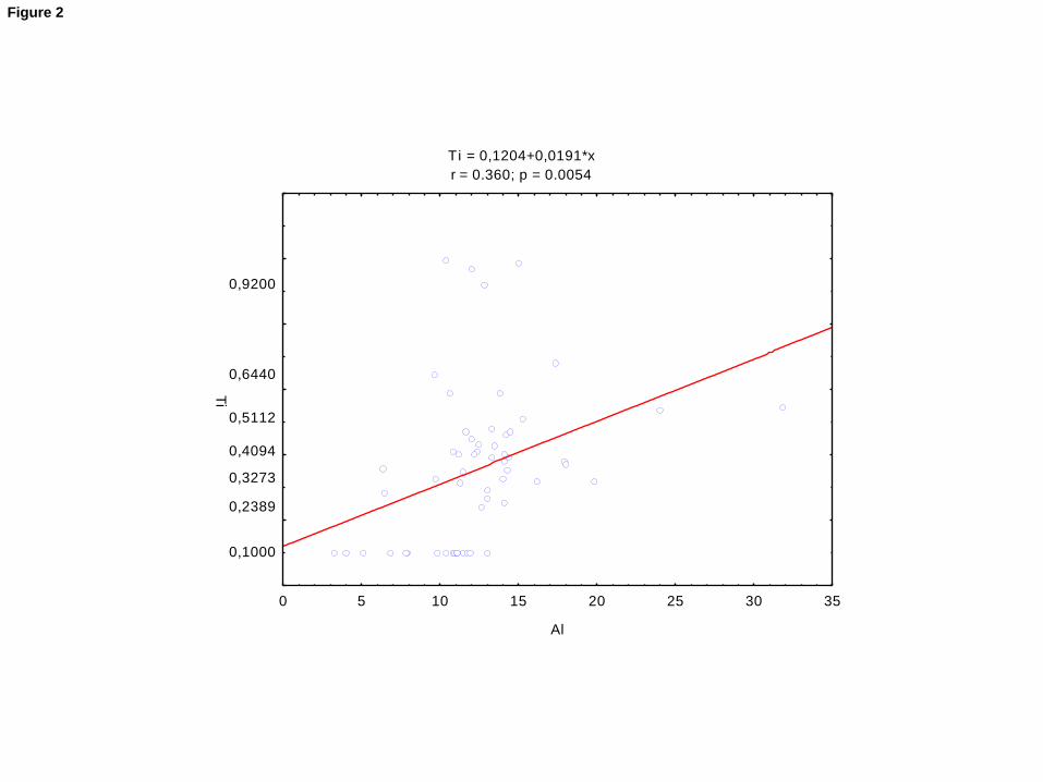
Ti = 0,1204+0,0191*x
r = 0.360; p = 0.0054
0 5 10 15 20 25 30 35
Al
0,1000
0,2389
0,3273
0,4094
0,5112
0,6440
0,9200
Ti
Figure 2

Table 1. Comparison of LODs and LOQs of the present study with previous works
Al Ti V Cr Co Ni Cu
LOD (ng/mL)
Martín-Cameán et al., 2014 a, b
- 0.90 2.8 0.38 0.10 0.67 0.49
Present study 1.0 0.20 0.08 0.23 0.38 0.14 0.13
LOQ (ng/mL)
Martín-Cameán et al., 2014 a, b
- 1.80 3.4 1.13 0.20 1.81 0.98
Present study 3.83 0.6 0.23 0.67 0.90 0.41 0.39
Table 1

Table 2. Metal levels in oral mucosa cells from the control group (n = 20), orthodontic group (n=20 ) and orthodontic+miniscrew group (n=20) studied.
Metals (ng/mL)
CONTROL GROUP ORTHODONTIC GROUP ORTHODONTIC+MINISCREW GROUP
Mean ± SD Range Median 5-95th
percentiles
Mean ± SD Range Median 5-95th
percentiles
Mean ± SD Range Median 5-95th
percentiles
Al 12.50 ± 3.37 5.12-19.81 12.34 5.12-19.81 12.21 ± 2.19 7.89-18.00 11.96 8.78-16.50 12.70 ± 6.74 3.30-31.85 12.16 3.30-31.85
Cr 0.19 ±0.04 0.12-0.27 0.18 0.12-0.27 0.19 ± 0.05 0.12-0.30 0.18 0.12-0.27 0,21 ± 0.07 0.12-0.32 0.23 0.12-0.32
Cu 0.71 ± 0.39 0.29-1.60 0.57 0.29-1.61 1.13 ±0.69* 0.14-2.30 0.89 0.24-2.28 1,054 ± 0.71 0.07-2.67 0.93 0.07-2.67
Ni 0.34 ±0.21 0.07-0.89 0.26 0.07-0.89 0.53 ± 0.17* 0.28-0.91 0.48 0.33-0.89 0.55 ± 0.32* 0.23-1.46 0.45 0.23-1.46
Ti 0.34 ± 0.12 0.10-0.51 0.38 0.10-0.51 0.40 ± 0.34 0.10-1.00 0.28 0.10-0.99 0.33 ± 0.19 0.10-0.68 0.35 0.10-0.68 * The significant level is p < 0.05 in comparison to control group
Table 2

Table 3. Forward stepwise multiple linear regression analysis of metal ion concentration
detected in the patient´s mucosa cells .
Metal
ion adjR2 F p-value Predictor B p-value
Partial
correlation
Al 0.205 1.889 0.108 Miniscrew -1.601 .259 -.240
Gender .643 .627 .105
Type of braces
-1.927
.173
-.287
Upper archwire
1.506
.022
.467
Lower archwire
.935
.438
.166
Cr 1.750 .875 .034
Ni 3.172 .199 .272
Cu .881 .320 .212
Ti 4.197 .059 .390
Cr 0.160 1.654 0.161 Miniscrew .046 .089 .354
Gender .030 .231 .254
Type of braces
.008
.775
.062
Upper archwire
-.015
.256
-.242
Lower archwire
.021
.366
.193
Al .001 .875 .034
Ni .005 .923 .021
Cu .028 .094 .349
Ti -.003 .945 -.015
Ni -0.072 0.769 0.646 Miniscrew .177 .140 .311
Table 3

Gender .069 .542 .131
Type of braces
.076 .540 .131
Upper archwire
-.061 .303 -.220
Lower archwire
.023 .822 .048
Al .023 .199 .272
Cr .092 .923 .021
Cu -.052 .498 -.145
Ti .121 .541 .131
Cu -0.001 0.996 0.471 Miniscrew -.091 .791 -.057
Gender -.297 .346 -.201
Type of braces
-.021
.952
-.013
Upper archwire
-.114
.494
-.147
Lower archwire
.205
.482
.151
Al .051 .320 .212
Cr 4.340 .094 .349
Ni -.408 .498 -.145
Ti -.393 .478 -.152
Ti 0.083 0.311 0.287 Miniscrew -.096 .471 -.155
Gender -.169 .161 -.295
Type of braces
-.091
.496
-.146
Upper archwire
-.074
.244
-.247

Lower archwire
.019
.865
.037
Al .036 .059 .390
Cr -.071 .945 -.015
Ni .142 .541 .131
Cu -.059 .478 -.152
Al: Aluminium; Cr: Chromium; Ni: Niquel; Cu: Copper; Ti: Titanium

1 2 3 4 5 6 7 8 9 10 11 12 13 14 15 16 17 18 19 20 21 22 23 24 25 26 27 28 29 30 31 32 33 34 35 36 37 38 39 40 41 42 43 44 45 46 47 48 49 50 51 52 53 54 55 56 57 58 59 60 61 62 63 64 65
In vivo determination of Aluminium, Cobalt, Chromium, Copper, Nickel, Titanium and
Vanadium in oral mucosa cells from orthodontic patients with mini-implants by
Inductively coupled plasma-mass spectrometry (ICP-MS)
Ana Martín-Cameán 1, Angeles Jos 2, Maria Puerto2,, Ana Calleja 3, Alejandro Iglesias-
Linares 4, Enrique Solano 1, Ana M. Cameán2*
1 Stomatology Department, School of Dentistry, University of Seville.
2 Area of Toxicology, Faculty of Pharmacy, University of Seville.
3 Investigation, Technology and Innovation Center, University of Seville.
4 Stomatology Department, School of Dentistry, University Complutense of Madrid,
Spain.
*Corresponding author
Ana M. Cameán Fernández
Area of Toxicology. Faculty o Pharmacy, University of Sevilla, C/Profesor García
González nº 2, CP 41018, Sevilla, Spain
E mail address: [email protected]
Tel: +34 954 556762; Fax: +34 954 556422
*Manuscript

1 2 3 4 5 6 7 8 9 10 11 12 13 14 15 16 17 18 19 20 21 22 23 24 25 26 27 28 29 30 31 32 33 34 35 36 37 38 39 40 41 42 43 44 45 46 47 48 49 50 51 52 53 54 55 56 57 58 59 60 61 62 63 64 65
Abstract
Miniscrews are used as orthodontic anchorage devices in the dentistry clinical practice
but the in vivo metallic release from these structures has been not previously
investigated. The aim of this study was to determine the content of Al, Co, Cr, Cu, Ni,Ti
and V in oral mucosa cells of control subjects, patients under orthodontic treatment
and with both, orthodontic treatment and miniscrew, in order to know the
contribution of these mini-implants to the total metallic content. ICP-MS
measurements revealed the following ascending order: Cr < Ni < Ti < Cu < Al, and Co
and V were practically undetected. Significant differences in comparison to the control
group were found for Cu in the orthodontic group, and for Ni in both, orthodontic and
orthodontic+miniscrew groups. Potential correlations among metallic elements and
with some clinical factors were also explored. These findings suggest that miniscrews do
not increase significantly the metal release.
Keywords: in vivo, metals, oral mucosa cells, orthodontic, miniscrew

1 2 3 4 5 6 7 8 9 10 11 12 13 14 15 16 17 18 19 20 21 22 23 24 25 26 27 28 29 30 31 32 33 34 35 36 37 38 39 40 41 42 43 44 45 46 47 48 49 50 51 52 53 54 55 56 57 58 59 60 61 62 63 64 65
1. Introduction
Several oral clinical manifestations in orthodontic treatment are associated
with corrosion products and ion release from the appliances employed [1]. Saliva acts
as an electrolyte for electron and ion conduction, and the fluctuation of pH and
temperature, the enzymatic and microbial activity, and the various chemicals
introduced into the oral cavity through food and drink are all corrosion conductors [2].
The metal appliances used in orthodontic treatment (brackets, tubes, bands) remain in
the mouth for an average of 2 years in this potentially corrosive environment. In spite
of the high resistance to corrosion of the alloys used for their manufacture, they may
still suffer some localized corrosion resulting from the oral cavity conditions [3]. The
inherent heterogeneity of each metal alloy and its use with other alloys, the
microsurface discontinuity, the forces acting on the appliances, and the friction
between wires and brackets also add to the corrosion process [4].
Various studies have evaluated the discharge of metal ions from orthodontic
appliances in biologic fluids, and most have concluded that they do not reach toxic
concentrations [5-7]. However, it cannot be excluded that even nontoxic
concentrations might be sufficient to produce biologic changes in the oral mucosa. In
fact, cations released from dental alloys may cause significant biological alterations
(DNA synthesis, alkaline phosphatase activity, etc) at non-cytotoxic concentrations [8].
Thus, in addition to the cytotoxic effects of metal ions, the physical and mechanical
effects of orthodontic appliances could also induce changes in the oral mucosa [9].
Moreover, it has been reported that metal ions are taken up by the adjacent oral
tissues [10-12]. Occasionally, the host response to the elemental release differs in the

1 2 3 4 5 6 7 8 9 10 11 12 13 14 15 16 17 18 19 20 21 22 23 24 25 26 27 28 29 30 31 32 33 34 35 36 37 38 39 40 41 42 43 44 45 46 47 48 49 50 51 52 53 54 55 56 57 58 59 60 61 62 63 64 65
nature and amount of the released elements. Classically allergic responses are
characterized by dose-independence, this is, low doses that would not cause
inflammation through toxicity but it would cause it by activating immune cells `13].
Also, mutagenicity and carcinogenic effects are not related with the dose of the
toxicant. Therefore, knowledge about the elemental release from these materials into
the oral cavity in regard to quantification is of great importance [14-15].
There are numerous in vitro studies that have demonstrated the release of
metals from orthodontic alloys [1, 16-18] which have been revised by Mikulewicz et al.,
[19]. Recently, these authors have evaluated the release of some metals (nickel,
chromium, copper) from fixed orthodontic appliances in vitro in a continuous- flow
system [20]. However, there has been little in vivo research into the adverse effects of
metal release, their absorption and toxicity. The few studies available involve saliva
analysis [10, 21-22]. The disadvantage of saliva is related to its flow, which is
influenced by many factors. Saliva flow does not influence all substance concentrations
to the same degree, so it can still be a useful matrix for non-flow-dependent chemicals
[23]. To the extent of our knowledge, the release of metals in oral mucosa cells, with
prolonged contact with fixed appliances, has been scarcely investigated [4, 9-10, 12,
17], although this matrix shows the same advantages than saliva samples previously
mentioned [24].
The release of elements from dental casting alloys in different matrices has
been mainly measured using either Atomic absorption spectroscopy (AAS) [25-26],
Inductively coupled plasma-optical emission spectrometry (ICP-OES) [16, 27], or
Inductively coupled plasma mass spectrometry (ICP-MS) [1, 28-30]. For many elements,
the power of detection of ICP-OES is not sufficient to determine elemental background

1 2 3 4 5 6 7 8 9 10 11 12 13 14 15 16 17 18 19 20 21 22 23 24 25 26 27 28 29 30 31 32 33 34 35 36 37 38 39 40 41 42 43 44 45 46 47 48 49 50 51 52 53 54 55 56 57 58 59 60 61 62 63 64 65
concentrations. In general, lower limits of detection (LODs) are possibly obtained by
ICP-MS in comparison to ICP-AES [31]. Currently, by application of ICP-MS the fast and
accurate routine multi-element determination in biological samples has become
possible due to improved sensitivity and robustness. In this sense, we have recently
published two validation methods to quantify some elements in oral mucosa cells
using this technique [24, 32].
The development of orthodontic treatments introduced in 1997 mini-screws by
Kanomi [33]. They were mainly used to anchor orthodontic movements. Mini-screws
became widely used to bear orthodontic and orthopaedic loads, becoming effective
orthodontic anchorage devices [34-35]. Firstly, the material of choice to manufacture
mini-implants was commercially pure Titanium (cp Ti), but in order to improve its
mechanical properties, some elements such as aluminium (Al), vanadium (V) and iron
(Fe) have been incorporated [36]. Many new systems were introduced, such as
onplants [37], palatal implants [38], bicortical screws [39], miniplates [40], miniplates
with a tube [41], and the zygoma anchorage system [42]. Although the dramatic
developments and improvements of orthodontic appliances such as brackets,
archwires and mini-implants are satisfying and amazing, little attention has been given
to metallic ion release from orthodontic implant systems. The concern about this has
been limited to orthodontic brackets and wires. To our knowledge, in vivo results
about metallic ion release from orthodontic miniscrews have not been reported until
now.
Taking into account all these facts, the aim of this study was to investigate the
possible differences in metal release of aluminium (Al), copper (Cu), chromium (Cr),
manganese (Mn), nickel (Ni), titanium (Ti) and vanadium (V), in oral mucosa cells of

1 2 3 4 5 6 7 8 9 10 11 12 13 14 15 16 17 18 19 20 21 22 23 24 25 26 27 28 29 30 31 32 33 34 35 36 37 38 39 40 41 42 43 44 45 46 47 48 49 50 51 52 53 54 55 56 57 58 59 60 61 62 63 64 65
patients subjected to conventional orthodontic appliances (brackets, archwires and
bands) in comparison to patients treated additionally with mini-screws, and also with
respect to a control group, by using Inductively Coupled Plasma Mass Spectrometry
(ICP-MS).
2. Materials and methods
2.1. Patients and orthodontic appliances
Sixty patients between 11 and 60 years old were included in this study. The
purposes of the study and the method of cell collection were explained to all subjects,
and the written consent of all participants was obtained. Previously, the protocol of
the study had the approval of the Ethical Committee of the University of Sevilla (Sevilla,
Spain). These patients were classified into three groups. The control group (a) (8 men,
12 women) was selected between volunteers under the following inclusion criteria:
nonsmokers, without oral or systemic diseases, without oral restorations, implants or
prosthetics, clinically healthy oral mucosa, no previous orthodontic treatment and no
occupational exposure to metals. The orthodontic group (b) (11 men and 9 women,
nonsmokers) was treated with fixed orthodontic appliances in both arches.
The appliances consisted of 8 bands (Ceosa, Madrid, Spain) on the first and second
permanent molars, brackets in both arches, and upper and lower 0.016 NiTi,
0.016x0.022 NiTi and/or 0.016x0.022 Stainless Steel archwires (AISI302 alloy) (Ceosa,
Madrid, Spain). The orthodontic-miniscrew group (c) (10 men and 10 women,
nonsmokers) required fixed orthodontic and miniscrew (Jeil Medical, Seoul, Korea).
The location of the miniscrew was variable: palatal, vestibular, anterior or posterior.
Each of the patients had only one miniscrew placed in mouth during sample collection.

1 2 3 4 5 6 7 8 9 10 11 12 13 14 15 16 17 18 19 20 21 22 23 24 25 26 27 28 29 30 31 32 33 34 35 36 37 38 39 40 41 42 43 44 45 46 47 48 49 50 51 52 53 54 55 56 57 58 59 60 61 62 63 64 65
A total of 20 self-drilling miniscrews (JA, Jeil Medical) were used, which had a
cylindrical screw design of 1.6 mm in diameter and 9.0 mm in length, and had a circle-
shaped head with a hole diameter of 0.9 mm. The orthodontic appliances were the
same as the orthodontic group. The sample collection took place after around 15
months of orthodontic treatment (15±3 months) in groups (b) and (c).
Micro-Xray fluorescence (μXRF) measurements of the materials used in this
study, including miniscrews, were performed according to Martin-Cameán et al. [24].
Results (wt.%) obtained for the composition of the miniscrew were: 6.78 Al, 88.68 Ti,
0.17 Fe, 0.13 Ni, 4.25 V
2.2. Buccal cell collection technique
The participants were asked to rinse their mouth with tepid water for 1 min to
remove exfoliated dead cells. Epithelial cells of buccal mucosa from each patient were
collected, using a soft rubber interdental brush without any metal content, as stated
by the manufacturer (Sunstar Iberia S.L., San Just Desvern, Spain), and following the
methods of Besarati Nia et al. [43] and Martin-Cameán et al. [32].
2.3. Reagents and materials
Type I water (>18MΩ·cm) obtained by a Milli-Q Element water purification
system (Millipore, Bedford, MA, USA) was used throughout. All transfer pipettes,
centrifuge tubes, plastic bottles, autosampler vials and laboratory ware material were
made of plastic and cleaned by soaking in 20% v/v HNO3 sub-boiling quality for 4 h,
rinsing three times with type I water, according to EPA method 200.8 (United States
Environment Protection Agency, version 5.4, 1994), and drying in a laminar flow hood.
Blank solution consisted of 1% v/v HNO3, prepared by diluting 65% nitric acid

1 2 3 4 5 6 7 8 9 10 11 12 13 14 15 16 17 18 19 20 21 22 23 24 25 26 27 28 29 30 31 32 33 34 35 36 37 38 39 40 41 42 43 44 45 46 47 48 49 50 51 52 53 54 55 56 57 58 59 60 61 62 63 64 65
with the appropriate volume of type I water. High purity nitric acid was obtained by
triple purification of 65% reagent grade nitric acid in a sub-boiling distiller purchased
from Savillex (Savillex Corp., Eden Prairie, MN , USA). A tuning solution containing
10ng/mL cerium (Ce), cobalt (Co), lithium (Li), thallium (Tl) and yttrium (Y) in 1% HNO3
was prepared from single-element (10,000 μg/mL) stock standards (AccuStandard, Inc.,
New Haven, CT, USA), and was used to optimize ICP-MS parameters before each
analytical run. Rhodium 1 μg/mL, prepared from a 100 μg/mL stock solution
(AccuStandard, Inc., New Haven, CT, USA) was used as an internal standard solution
throughout the whole analysis. A standard solution containing 1 μg/mL of Al, Co, Cr, Cu,
Ni and V was prepared in 100 mL Pyrex glass volumetric flask by dilution of 10 μg/mL
multi-element standard solution for ICP-MS (AccuStandard, Inc., New Haven, CT, USA)
with the suitable amount of blank solution. The standard solution of 1 μg/mL was
subsequently diluted with the suitable amount of blank to prepare the working
standards. A stock 10000 µg/mL Ti standard was purchased from AccuStandards as
well and subsequently diluted with the needed amount of blank solution in order to
obtain less concentrated standards.
2.4. Instrumentation and Determination of metals
All ICP-MS measurements of metal contents were carried out in an Agilent
7500c ICP-MS (Agilent Technologies, Japan). Sample introduction was performed with
a PFA (perfluoroalkoxy) microflow auto-aspirating nebulizer combined with a double-
pass spray chamber (Agilent Technologies, Japan). The validation of analytical
procedures developed for the determination of metals in oral mucosa cells has been
reported previously by our workgroup Martin-Cameán et al. [24,32], with some

1 2 3 4 5 6 7 8 9 10 11 12 13 14 15 16 17 18 19 20 21 22 23 24 25 26 27 28 29 30 31 32 33 34 35 36 37 38 39 40 41 42 43 44 45 46 47 48 49 50 51 52 53 54 55 56 57 58 59 60 61 62 63 64 65
modifications to improve the limits of detection (LODs) and quantification (LOQs). For
this purpose, nitric acid and water metallic content were reduced, plastic ware was
preferred to glassware and a more stable nebulizer was used for sample introduction.
Moreover, the present work comprises the determination of most elements previously
studied (Co, Cr, Cu, Ni, Ti, V) and is further expanded to include Al. Briefly, each
interdental brush was introduced into a previously cleaned (4 h in 20% v/v HNO3) 50
mL centrifuge tube, together with 10 mL of type I water and 100 μL of sub-boiling 65%
HNO3, resulting from the distillation of 65% reagent grade HNO3 from JT Baker
(Avantor Performance Materials B.V., Deventer, The Netherlands). Then, samples were
heated in a water bath at 80 °C for 60 min. Afterwards, samples were cooled lightly
and treated in an ultrasonic bath for 5 min. Finally, samples were cooled down to room
temperature, and the acid solution was separated from the brush. Acid solutions were
stored in clean 20-mL polypropylene vials at 4 °C until analysis. Less than 0.5mL were
needed for the analysis.. Rhodium was used as an internal standard and was spiked at
250 ng/mL to every blank, sample and standard individually. The relative standard
deviation of the concentration of Rh in all samples was calculated to be 0.85%.
2.5. Statistical analysis
Statistical analysis of the results was performed by the soft-ware Statistica
(version 6.0, Statsoft, Inc., OK, USA). Descriptive statistics (means, standard deviation,
medians, range and 5–95th percentiles) were reported. Normality of distribution of
experimental results was assessed by the Chi-Square test. Data distribution was always
found non-normal, and accordingly, non-parametric methods were applied, such as
Mann–Whitney and Spearman tests. In addition, a a stepwise linear regression was

1 2 3 4 5 6 7 8 9 10 11 12 13 14 15 16 17 18 19 20 21 22 23 24 25 26 27 28 29 30 31 32 33 34 35 36 37 38 39 40 41 42 43 44 45 46 47 48 49 50 51 52 53 54 55 56 57 58 59 60 61 62 63 64 65
run adjusted for the presence of miniscrew along the fixed appliances, just fixed
appliances or any of treatments, gender, type of braces, type of upper and lower
archwires during sample collection. The data were analyzed by the Statistical Package
Social Sciences (version 19) software (SPSS, Chicago, IL, USA) and p-values less than .05
were considered statistically significant.
3. Results
3.1. Analytical aspects
Several improvements were introduced throughout the measurements, aiming
at reducing the LODs and LOQs. The ad hoc triple distillation of the nitric acid provides
higher purity than that of available commercial solutions. Acid is distilled few hours
before its intended use, and thus impurities are kept at a minimum. Same triple-
distilled nitric acid was employed during the cleaning of the laboratory ware. Type I
water was obtained from a Milli-Q Element source, which offers one more filter than
the widespread Milli-Q sources, and reduces even further the metallic content of
water. The employment of plastic labware reduces the leaching of metallic ions from
the walls of the container into the sample, compared to widespread borosilicate glass,
as stated by Richter (Richter, 2003) [44]. The microflow nebulizer works by auto-
aspiration, taking advantage of the Venturi effect, and thus provides a more stable
signal in the ICP-MS, because mechanical disturbances from the peristaltic pump are
avoided.
Table 1 shows the LODs and LOQs attainable in this study, compared to those
obtained previously in our lab [24,32]. All elements, except for Co, show a remarkable
lowering (2-3 fold) of their LODs and LOQs, especially in the case of V.

1 2 3 4 5 6 7 8 9 10 11 12 13 14 15 16 17 18 19 20 21 22 23 24 25 26 27 28 29 30 31 32 33 34 35 36 37 38 39 40 41 42 43 44 45 46 47 48 49 50 51 52 53 54 55 56 57 58 59 60 61 62 63 64 65
3.2. Contents and distribution of metals in oral mucosa cells from patients.
The cellular content of the eight elements were measured for all the patients,
and in the case of cobalt and vanadium, only traces were detected in a small number
of samples (1-2%), being their contents lower than their respective LODs. Then, these
two elements will be not further considered. The concentration of Al, Cr, Cu, Ni and Ti
levels in oral mucosa cells were fitted to typical statistical distributions (Gaussian, log
normal, gamma) and are shown in Figure 1. None of the elements were normal
distributed, and all further statistical computations were based on non-parametric
techniques. Table 2 shows the descriptive statistics for the elements in the three
groups of patients considered (control, orthodontic and orthodontic+miniscrew). In
order to make comparisons with the scientific literature mean values (± sd) are
provided. Metal release values in oral mucosa cells was variable in all the patients
considered, mainly in the case of Cu. The lowest levels were found for Cr whose
medians were 0.18 ng/mL in control and orthodontic group, and 0.23 ng/mL in
orthodontic+miniscrew group. Ni values in control group (median=0.26 ng/mL) were
significantly lower in comparison to orthodontic patients (0.48 ng/mL) (p<0.05) and to
orthodontic+ miniscrew group (0.45 ng/mL) (p<0.05). Similar levels were found for Ti,
whose medians were 0.38, 0.28 and 0.35 ng/mL in control, orthodontic and
orthodontic+miniscrew groups, respectively. In the case of copper levels, a significant
increase was found in orthodontic group (median=0.89 ng/mL) when compared to
control group (0.57 ng/mL) (p<0.05). Aluminium was the predominant element with
medians about 12 ng/mL for all the patients considered.
3.3. Interrelations between elements

1 2 3 4 5 6 7 8 9 10 11 12 13 14 15 16 17 18 19 20 21 22 23 24 25 26 27 28 29 30 31 32 33 34 35 36 37 38 39 40 41 42 43 44 45 46 47 48 49 50 51 52 53 54 55 56 57 58 59 60 61 62 63 64 65
To statistically prove possible correlations between the contents of the studied
elements in oral mucosa cells the non-parametric technique of Spearman [45] was
employed. The only correlation (with a probability (P) of error level less than 0.05 was
Al/Ti (r = 0.469), and this correlation was positive, that is, both levels change in the
same direction.
Following the procedure described by Martin-Cameán et al. [26] and despite of
the non-normality of the distributions of metals studied in oral mucosa cells, the
element-to-element linear regression plots for statistically significant correlations were
evaluated. The analysis of correlation coefficients shows that there is significantly
positive correlations (p<0.05) only in the case of Al/Ti (r=0.36) (Figure 2).
3.4. Multiple linear regression analysis of the influence of orthodontic treatment
variables on the ion concentrations
The levels of metallic elements can be influenced by different factors such as gender,
type of treatment including type of braces and archwires and also it might be influenced not
simply by the level of another element, but by the level of the group of elements potentially
released from orthodontic appliances and miniscrew. In this regard, the relationships among
different elements and clinical factors related to the treatment, and between their
concentrations were examined by a stepwise linear multiple regression analysis [46] adjusted
for variables described above (Table 3). Results from the regressive analysis give no relevant
information regarding a linear correlation or influence of any of the included variables in the
matrix with any of the metallic ions (p >.05). Therefore as compiled in Table 3, none of the
clinical variables are useful predictors of the results of the present study (p > .05)

1 2 3 4 5 6 7 8 9 10 11 12 13 14 15 16 17 18 19 20 21 22 23 24 25 26 27 28 29 30 31 32 33 34 35 36 37 38 39 40 41 42 43 44 45 46 47 48 49 50 51 52 53 54 55 56 57 58 59 60 61 62 63 64 65
4. Discussion
In vivo metal release from orthodontic appliances has been reported using
different techniques and experimental procedures, and a systematic review of this
matter has been published by Mikulewicz and Chojnacka [15]. These reports deal with
traditional orthodontic appliances, but miniscrews are also a potential source of
metals not evaluated before.
The biological samples usually investigated have been saliva [4, 6-7,12,47-50],
there were also some works relative to serum, urine, and more recently reports in hair
[26, 51-52] and mucosa oral cells [4, 9-10, 12,17], whose advantages of these latter
matrices have been reported [24]. Regarding the ICP-MS technique employed to
determine metals from these samples, the procedure for sample pretreatment and
determination of metals in oral mucosa cells have been validated in our laboratory [24,
32]. In this work, the adopted strategy carried out to reduce the background due to
metallic contribution from reagents and instrumental instabilities, have had a positive
impact on LOD and LOQs values of the elements analysed by ICP-MS.
The release of the elements studies showed the following ascending order: Cr <
Ni < Ti < Cu < Al, and two metals, V and Co were considered as not detected in the
samples analysed. Vanadium has been not previously determined in oral mucosa cells
[4, 9-10, 12, 17] or was not detected [24]. However, although V is present in some
materials (bands 0.18-0.19%, and miniscrew 4.25%), the application of miniscrew did
not contribute to a significant release of this metal. Cobalt was only present in the
stainless steel arch (0.62%) and its null release is in agreement with Fernandez-Miñano
et al. [17] who evaluated three alloys in brackets and tubes: stainless steel, titanium
and nickel- free. Also, Amini et al. [12] did not find differences in the concentration of

1 2 3 4 5 6 7 8 9 10 11 12 13 14 15 16 17 18 19 20 21 22 23 24 25 26 27 28 29 30 31 32 33 34 35 36 37 38 39 40 41 42 43 44 45 46 47 48 49 50 51 52 53 54 55 56 57 58 59 60 61 62 63 64 65
Co of patients treated with conventional fixed appliances after 16 months. However,
Faccioni et al. [10] found a significant 2.8-fold increase in the concentration of Co
released during the 24-48 months period of treatment. In this case, some of the
archwires used were chromium-cobalt-nickel alloy and a considerable percentage of
Co (42%) was included in its composition.
In general, among stainless steel and nickel –titanium corrosion products, Ni
and Cr have received the most attention because of their reported adverse effects [26].
In relation to Ni, the increased release observed in all experimental groups with
respect the control in the present study agreed with those reported by Faccioni et al.
[10] who found 3.4-fold higher Ni values in orthodontic patients than those in the
control subjects. This finding could be explained because Ni is a common constituent
of the standard orthodontic appliances, especially in Ni-Ti arches. Similarly, Amini et al.
[12] confirmed that nickel contents in mucosa cells of orthodontic patients were
significantly higher (2-fold) f than in controls. Moreover Hafez et al. [4] studied Ni
release with the time in a longitudinal controlled clinical trial, and reported an
increase of Ni cellular content from 0.52 to 0.68 and 0.78 ng/mL in buccal mucosa cells,
after 0, 3 and 6 months of treatment, respectively. However, Natarajan et al. [9] did
not find significant differences in Ni ion contents after 30 days of debonding. Therefore,
the study period is a factor to take into account. Nickel is well-known for producing
contact dermatitis in sensitized persons that can become quite severe [53], and
adverse reactions related to Ni-containing orthodontic devices (archwires, brackets,
soldered stainless steel face-bows) have been reported [54]. Very low concentrations
of Ni2* could induce gene activation in endothelial cells, similar to pro-inflammatory

1 2 3 4 5 6 7 8 9 10 11 12 13 14 15 16 17 18 19 20 21 22 23 24 25 26 27 28 29 30 31 32 33 34 35 36 37 38 39 40 41 42 43 44 45 46 47 48 49 50 51 52 53 54 55 56 57 58 59 60 61 62 63 64 65
mediators like interleukin6 (IL-6) and interleukin 8 (IL-8) and provoke inflammatory
reactions and may modulate the immune response by activation T-and B-cells [8]. This
element has carcinogenic and mutagenic effects [55], and consequently, the exposure
to it should be as low as reasonably possible.
Regarding titanium levels, due to its mechanical properties, good resistance to
corrosion in biological fluids and very low toxicity, this metal has been the most
commonly selected material for dental implants and prostheses [56-57]. Despite its
common presence in orthodontic fixed appliances (NiTi archwires), including
miniscrews, no increased release was detected in this work. Other authors however,
showed enhanced values of Ti in oral mucosa cells [17]. This element has not been
investigated in saliva.
Data relative to Cr release in oral mucosa cells are contradictory. Some reports
indicated higher contents of this metal in oral mucosa cells in comparison to control
groups [4,17], whereas our results agreed with Amini et al. [12] and Natarajan et al. [9]
who did not find an increase of the release of this metal. In other matrices, such as
saliva, the release of Cr and Ni from orthodontic appliances was assessed by Amini et
al. [12] and these authors did not find significant release of Cr, but higher levels in the
case of Ni in 28 subjects who had undergone fixed orthodontic therapy for 12-18
months. Moreover, most of the experiments measured ion release during the
exposure to orthodontic appliances to a biological medium (blood, serum, urine, or
saliva) for periods ranging from 1 day to 1 month or even over a 10-month period.
These reports have been recently revised by Matusiewicz [58], and in general,
contradictory results have been found in salivary metal ion concentrations between
subjects with and without fixed orthodontic appliances.

1 2 3 4 5 6 7 8 9 10 11 12 13 14 15 16 17 18 19 20 21 22 23 24 25 26 27 28 29 30 31 32 33 34 35 36 37 38 39 40 41 42 43 44 45 46 47 48 49 50 51 52 53 54 55 56 57 58 59 60 61 62 63 64 65
In relation to copper contents, the only significant increase observed in the
orthodontic group and not in the orthodontic+miniscrew group could be due to the
high variability of the results. Release of Cu from orthodontic appliances has been not
extensively studied in the scientific literature, although it is commonly present in many
alloys [59] used in the orthodontic practice. Recently, Mikulewicz et al. [27] studied in
vivo the Cu release (and other elements) in non-invasive (hair) and invasive matrices in
pigs who received plates simulating the orthodontic appliance, and found significant
differences in the levels of this element in liver, kidney, aorta, lung and hair of
experimental pigs.
Aluminium is the third most common element (about 8%) of the earth’s crust,
and it is an extremely versatile metal with a wide variety of uses, and is non-essential
for humans, resulting in low-level distribution in human organs [60, 61]. Its ubiquity
could explain the highest levels observed in all the patients. However, its oral
bioavailability in humans and experimental animals is low; from drinking water it has
been estimated to be in the range of 0.3%, whereas from food and beverages
generally is considered to be lower, about 0.1% [62]. In relation to orthodontic
appliances composition, aluminium is present in the miniscrew, so this is the first time
that its potential release has been evaluated. According to the results obtained, no
differences were found in the three experimental groups studied. Therefore,
miniscrew resulted biocompatible with respect to this metal.
In the case of orthodontic patients, the studies on inter-element interactions
are scarce and only limited to levels contained in hair. Synergism has been reported
between Fe and Cr by Mikulewicz et al. [51], and also positive correlations between
Cu/Mn, Cu/Ni and Fe/Ni were found in a previous report from our laboratory [26].

1 2 3 4 5 6 7 8 9 10 11 12 13 14 15 16 17 18 19 20 21 22 23 24 25 26 27 28 29 30 31 32 33 34 35 36 37 38 39 40 41 42 43 44 45 46 47 48 49 50 51 52 53 54 55 56 57 58 59 60 61 62 63 64 65
These interactions could be explained in part by the common origin of elements, the
miniscrew, or synergistic effects between them.
Linear correlation and presence of potential confounding clinical factors affecting
the observed results were explored by a regression analysis of these clinical variables
and the metals concentrations. No evidence was found of any statistical correlation of
gender, presence of miniscrews, type of braces neither archwires in the lower and
upper jaws and between metallic ions. These results might be partially explained due
to inherent limitation in sample size for finding linear associations. However,
interestingly, these results imply that miniscrews are safe auxiliary orthodontic devices
in term of metallic ion releasing to the mouth as previous studies have concluded in
vitro [63] and in the animal model [64]. Nevertheless, no data are found to date in this
or other studies regarding the ionic releasing into the alveolar bone since those mini-
implants are directly screw into the alveolar bone without any surgical approach; and
screwing friction is a well-known factor that induces severe damage in the metallic
surface [65]. These potential effects might be of substantial interest for future studies
in the field.
Finally, no adverse effects were observed in any patient treated in our study,
and it would be explained because the quantified amounts of the elements released
are below their respective Tolerable daily intake (TDI) in the case of Cu (5 mg/day [66] ,
Ni (12 µg/Kg body weight) [67], or Tolerable weekly intake (TWI) for Al of 1 mg/Kg boy
weight/week for its cumulative nature in the organism [62], and with the Tolerable
Upper Intake Level (UL) for Cr (1 mg/day) [68]. Neither Joint of Expert Committee on
Food Additives (JECFA) nor the World Health Organization (WHO) have determined

1 2 3 4 5 6 7 8 9 10 11 12 13 14 15 16 17 18 19 20 21 22 23 24 25 26 27 28 29 30 31 32 33 34 35 36 37 38 39 40 41 42 43 44 45 46 47 48 49 50 51 52 53 54 55 56 57 58 59 60 61 62 63 64 65
relevant tolerable intake for titanium against to compare the values of metal ions
released obtained [69].
Conclusions
The modifications introduced in the pretreatment of oral mucosa cells reduce
the background due to metallic contribution from reagents and instrumental
instabilities, and have a positive impact for an adequate in vivo determination by ICP-
MS of Al, Cu, Cr, Mn, Ni, Ti and V released in patients with fixed orthodontic appliances.
Only traces of Co and V were detected, whereas the release of the elements studied in
patients showed the following ascending order: Cr < Ni < Ti < Cu < Al. Differences in the
content of metals in oral mucosa cells were only significantly increased for Ni in
orthodontic and ortohodontic+miniscrew groups, and also in Cu levels in orthodontic
group, in comparison to the control group. Nevertheless no linear correlation resulted
for none of the ions with regard to the type of orthodontic fixed appliance used, with
or without miniscrew. In general, the incorporation of miniscrews assayed did not
imply a significant increase of metal release. A positive correlation between Al/Ti was
found, probably due in part to their common origin, or synergistic effects between
them.
Acknowledgements:
This work was supported by the Junta de Andalucía (Plan Andaluz Investigación,
code CTS-358).
Conflict of interest
The authors declare that there are no conflicts of interest.

1 2 3 4 5 6 7 8 9 10 11 12 13 14 15 16 17 18 19 20 21 22 23 24 25 26 27 28 29 30 31 32 33 34 35 36 37 38 39 40 41 42 43 44 45 46 47 48 49 50 51 52 53 54 55 56 57 58 59 60 61 62 63 64 65
References
[1] Ortiz AJ, Fernández E, Vicente A, Calvo JL, Ortiz C. Metallic ions released from
stainless steel, nickel-free, and titanium orthodontic alloys: Toxicity and DNA
damage. Am J Orthod Dentofacial Orthop 2011; 140:e115-e122.
[2] Eliades T, Bourauel C. Intraoral aging of orthodontic materials: the picture we miss
and its clinical relevance. Am J Orthod Dentofacial Orthop 2005; 127:403-412.
[3] Oh K-T, Kim K-M. Iron release and cytotoxicity of stainless steel wires. Eur J Orthod
2005; 27:533–540.
[4] Hafez HS, Selim EMN, Eid FHK,Tawfik WA, Al-Ashkar EA, Mostafa YA. Cytotoxicity,
genotoxicity, and metal release in patients with fixed orthodontic appliances: A
longitudinal in-vivo study Am J Orthod Dentofacial Orthop 2011; 140:298–308.
[5] Barrett RD, Bishara SE, Quinn JK. Biodegradation of orthodontic appliances. Part I.
Biodegradation of nickel and chromium in vitro. Am J Orthod Dentofacial Orthop
1993; 103:8-14.
[6] Kocadereli I, Atac A, Kale S, Ozer D.Salivary nickel and chromium in patients with
fixed orthodontic appliances. Angle Orthod 2000; 70:431-4.
[7] Ağaoğlu G, Arun T, Izgu B, Yarat A. Nickel and chromium levels in the saliva and
serum of patients with fixed orthodontic appliances. Angle Orthod 2001; 71:375-9.
[8] Geurtsen W. Biocompatibility of dental casting alloys. Crit Rev Oral Biol Med 2002;
13:71-84.
[9] Natarajan M, Padmanabhan S, Chitharanjan A, Narasimhan M. Evaluation of the
genotoxic effects of fixed appliances on oral mucosal cells and the relationship to
nickel and chromium concentrations: An in-vivo study. Am J Orthod Dentofacial
Orthop 2011; 140:383–388.

1 2 3 4 5 6 7 8 9 10 11 12 13 14 15 16 17 18 19 20 21 22 23 24 25 26 27 28 29 30 31 32 33 34 35 36 37 38 39 40 41 42 43 44 45 46 47 48 49 50 51 52 53 54 55 56 57 58 59 60 61 62 63 64 65
[10] Faccioni F, Franceschetti P, Cerpelloni M, Fracasso M. In vivo study on metal
release from fixed orthodontic appliances and DNA damage in oral mucosa cells.
Am J Orthod Dentofacial Orthop 2003; 124:687-694.
[11] Garhammer P, Schmalz G, Hiller KA, Reitinger T. Metal content of biopsies
adjacent to dental cast alloys. Clin Oral Invest 2003; 7:92-7.
[12] Amini F, Farahani A, Jafari A, Rabbani M. In vivo study of metal content of oral
mucosa cells in patients with and without fixed orthodontic appliances. Orthod
Craniofac Res 2008; 11:51-6.
[13] Schmalz G, Schweikl H, Hiller KA.Release of prostaglandin E2, IL-6 and IL-8 from
human oral epithelial culture models after exposure to compounds of dental
materials. Eur J Oral Sci 2000; 108:442–8.
[14] Elshahawy W, Watanabe I, Koike M. Elemental ion release from four different
fixed prosthodontic materials. Dent Mater 2009; 25:976–981.
[15] Mikulewicz M, Chojnacka K.Trace element release from literature appliances by in
vivo studies: a systematic literature review. Biol Trace Elem Res 2010; 137:127–138.
[16] Eliades T, Pratsinis H, Kletsas D, Eliades G, Makou M. Characterization and
cytototxicity of ions released from stainless steel and nickel-titanium orthodontic
alloys. Am J Orthod Dentofacial Orthop 2004; 125:24-9.
[17] Fernández-Miñano E, Ortiz C, Vicente A, Calvo JL, Ortiz A.J. Metallic ion content
and damage to the DNA in oral mucosa cells of children with fixed orthodontic
appliances. Biometals 2001; 24:935-941.
[18] Kao C, Ding S, Min Y, Hsu TC, Chou M, Huang T. The cytotoxicity of orthodontic
metal bracket immersion media. Eur J Orthod 2007; 29:198-203.
[19] Mikulewicz M, Chojnacka K. Release of metal ions form orthodontic appliances by

1 2 3 4 5 6 7 8 9 10 11 12 13 14 15 16 17 18 19 20 21 22 23 24 25 26 27 28 29 30 31 32 33 34 35 36 37 38 39 40 41 42 43 44 45 46 47 48 49 50 51 52 53 54 55 56 57 58 59 60 61 62 63 64 65
in vitro studies: a systematic literature review. Biol Trace Elem Res 2011; 139:241-
256.
[20] Mikulewicz M, Chojnacka K, Wolowiec P. Release of metal ions from fixed
orthodontic appliance. An in vitro study in continuous flow system. Angle Orthod
2014; 84:140-8.
[21] Arvideon K, Cottler-Fox M, Friberg V. Cytotoxic effects of Co–Cr alloys on
fibroblast derived from human gingival. Scand J Dent Res 1986; 95:356–363.
[22] Matos de Souza R, Macedo de Menezes L. Nickel, chromium and iron levels in
saliva of patients with simulated fixed orthodontic appliances. Angle Orthod 2008:
78:345-350.
[23] Esteban M, Castaño A. Non-invasive matrices in human biomonitoring: a review.
Environ Int 2009; 35:438–449.
[24] Martín-Cameán A, Jos A, Calleja A, Gil F, Iglesias A, Solano E, Cameán A.M.
Validation of a method to quantify titanium, vanadium and zirconium in oral
mucosa cells by inductively coupled plasma-mass spectrometry (ICP-MS). Talanta
2014; 118:238–244.
[25] Amini F, Jafari A, Amini P, Sepasi S.. Metal ion release from fixed orthodontic
appliances –an in vivo study. Eur J Orthod. 2012; 34:126-130.
[26] Martín-Cameán A. Molina-Villalba I, Jos,A, Iglesias-Linares A, Solano E, Cameán
AM, Gil F. Biomonitorization of chromium, copper, iron, manganese and nickel in
scalp hair from orthodontic patients by atomic absorption spectrometry. Environ
Toxicol Pharmacol 2014; 37:759–771.
[27] Mikulewicz M, Wolowiec P, Janeczek,M, Gedrange T, Chojnacka K. The release of
metal ions from orthodontic appliances. Animal test. Angle Orthod. 2014; 84: 673-

1 2 3 4 5 6 7 8 9 10 11 12 13 14 15 16 17 18 19 20 21 22 23 24 25 26 27 28 29 30 31 32 33 34 35 36 37 38 39 40 41 42 43 44 45 46 47 48 49 50 51 52 53 54 55 56 57 58 59 60 61 62 63 64 65
9.
[28] Liu JK, Lee TM, Liu IH. Effect of loading force on the dissolution behaviour and surface
properties of nickel-titanium orthodontic archwires in artificial saliva. Am J Orthod Dentofacial
Orthop 2011; 140:166-176.
[29] Reimann S, Rewari A, Keilig L, Widu F, Jäger A, Bourauel C. Material testing of
reconditioned orthodontic brackets. J Orofac Orthop 2012; 73:454-66
[30] Mikulewicz M, Chojnacka K, Wozniak B, Downarowicz P. Release of metal ions form
orthodontic appliances: an in vitro study. Biol Trace Elem Res 2012; 146:272-280.
[31] Heitland P, Köster HD. Biomonitoring of 30 trace elements in urine of children and
adults by ICP-MS. Clin Chim Acta 2006; 365:310–318.
[32] Martín-Cameán A, Jos A, Calleja A, Gil F, Iglesias-Linares A, Solano E, Cameán,AM.
Development and validation of an inductively coupled plasma mass spectrometry
(ICP-MS) method for the determination of cobalt, chromium, copper and nickel in
oral mucosa cells. Microchem. J. 2014; 114:73–9.
[33] Kanomi R. Mini-implant for orthodontic anchorage. J. Clin. Orthod. 1997; 31:763-7.
[34] Kim JW, Ahn SJ, Chang YI Histomorphometric and mechanical analyses of the drill-
free screw as orthodontic anchorage. Am. J. Orthod. Dentofacial Orthop. 2005;
128:190-4.
[35] Melsen B, Verna C. Miniscrew implants: the Aarhus anchorage system. Semin.
Orthod. 2005; 11:24-31.
[36] Carvalho R, Tarkany R. In vitro study of human osteoblast proliferation and
morphology on orthodontic mini-implants. Angle Orthod. 2014(in press). DOI:
10.2319/100714-717.1.
[37] Hong H, Ngan P, Li HG, Qi LG, Wei SHY. Use of onplants as stable anchorage for

1 2 3 4 5 6 7 8 9 10 11 12 13 14 15 16 17 18 19 20 21 22 23 24 25 26 27 28 29 30 31 32 33 34 35 36 37 38 39 40 41 42 43 44 45 46 47 48 49 50 51 52 53 54 55 56 57 58 59 60 61 62 63 64 65
facemask treatment: a case report. Angle Orthod.2005; 75:402-9.
[38] Cousley R. Critical aspects in the use of orthodontic palatal implants. Am J Orthod
Dentofacial Orthop 2005; 127:723-9.
[39] Freudenthaler JW, Haas R, Bantleon HP.Bicortical titanium screws for critical
orthodontic anchorage in the mandible: a preliminary report on clinical
applications. Clin Oral Implants Res 2001; 12 :358-363.
[40] Choi BH, Zhu .J, Kim YH. A clinical evaluation of titanium miniplates as anchors for
orthodontic treatment. Am J Orthod Dentofacial Orthop 2005; 128:382-4.
[41] Chung KR, Kim YS, Linton JL, Lee YJ. The miniplate with tube for skeletal anchorage.
J Clin Orthod 2002; 36:407-412.
[42] Clerck H, Geerinckx V, Siciliano S. The zygoma anchorage system. J Clin Orthod
2002; 36:455-460.
[43] Besarati Nia A, Van Straaten HWM, Godschalk RWL, Van Zandwijk N, Balm AJM,
Kleinjans JCS, Van Schooten FJ. Immunoperoxidase detection of polycyclic aromatic
hydrocarbon-DNA adducts in mouth floor and buccal mucosa cells of smokers and
non smokers. Environ Mol Mutagen 2000; 36:127–133.
[44] Richter R. Clean Chemistry. Techniques for the Modern Laboratory. Milestone
Press, 2003
[45] Miller JN, Miller JC.Estadística y Quimiometría para Química Analítica, 4ª ed.
Madrid: Prentice Hall, 2002
[46] Chojnacka K, Córecka H, Chojnacki A, Córecki H. Inter-element interactions in
human hair. Environ Toxicol Pharmacol 2005; 20:368–374.
[47] Kerosuo H, Moe G, Hensten-Pettersen A.Salivary nickel and chromium in subjects
with different types of fixed orthodontic appliances. Am J Orthod Dentofacial

1 2 3 4 5 6 7 8 9 10 11 12 13 14 15 16 17 18 19 20 21 22 23 24 25 26 27 28 29 30 31 32 33 34 35 36 37 38 39 40 41 42 43 44 45 46 47 48 49 50 51 52 53 54 55 56 57 58 59 60 61 62 63 64 65
Orthop 1997; 111:595–8.
[48] Eliades T, Trapalis C, Eliades G, Katsavrias E. Salivary metal levels of orthodontic
patients: a novel methodologicaland analytical approach. Eur J Orthod 2003;
25:103–6..
[49] Petoumeno E, Arndt M, Keilig L, Reimann S, Hoederath H, Eliades T, Jäger A,
Bourauel C. Nickel concentration in the saliva of patients with nickel–titanium
orthodontic appliances. Am J Orthod Dentofacial Orthop 2009; 135:59–65.
[50] Sahoo N, Kailasam V, Padmanabhan S, Chitharanjan AB. In-vivo evaluation of
salivary nickel and chromium levels in conventional and self-ligating brackets. Am J
Orthod Orthop 2011; 140:340-5.
[51] Mikulewicz M, Chojnacka K, Zielinskab A, Michalak I. Exposure to metals from
orthodontic appliances by hair mineral analysis. Environ Toxicol Pharmacol 2011;
32:10–16.
[52] Mikulewicz M, Wolowiec P, Loster B, Chojnacka K. Metal ions released from fixed
orthodontic appliance affect hair mineral content. Biol Trace Elem Res 2015;
163:11-8.
[53] Baselt RC.,Disposition of Toxic Drugs and Chemicals in Man. Ninth Ed.
California: ,Biomedical Pub., 2011
[54] Grepp, AL, Smith DC, Woodside DG. Nickel hypersensitivity reactions in
orthodontic patients. Univ. Tor. Dent. J. 1989; 3:11-4.
[55] International Agency for Research on Cancer (IARC). Monographs on Evaluation of
the Carcinogenic Risk ofChemicals to Humans: Cr, Ni and Welding. IARC, Lyon,
France, 1990.
http://monographs.iarc.fr/ENG/Monographs/vol49/mono49.pdf(accessed

1 2 3 4 5 6 7 8 9 10 11 12 13 14 15 16 17 18 19 20 21 22 23 24 25 26 27 28 29 30 31 32 33 34 35 36 37 38 39 40 41 42 43 44 45 46 47 48 49 50 51 52 53 54 55 56 57 58 59 60 61 62 63 64 65
07.11.13).
[56] Adya N, Alam M, Ravindranath T, Mubeen A, Saluja B. Corrosion in titanium
dental implants: literature review. J Indian Prosthod Soc 2005; 5:126–131.
[57] Siddiq A, Payne AGT, De Silva RK, Duncan WJ. Titanium allergy: could it affect
dental implant integration?. Clin. Oral Impl. Res. 2011; 22:673–680.
[58] Matusiwewicz H. Potential release of in vivo trace metals from metallic medical
implants in the human body: From ions to nanoparticles – A systematic analytical
review. Acta Biomater 2014; 10:2379-2403.
[59] Aaseth J, Norseth T.Copper. In: Friberg L, Nordberg GF, Vouk V editors. Handbook
on the Toxicology of Metals. Volume II. Amsterdam:,Elsevier Science Publishers
B.V., 1986. p. 233-254.
[60] Elinder CG, Sjögren B. Aluminium. In: Friberg L, Nordberg GF, Vouk V editors.
Handbook on the Toxicology of Metals. Volume II. Amsterdam:,Elsevier Science
Publishers B.V., 1986. p. 1-25.
[61] Watanabe K, Tanaka T, Shigemi T, Saeki K, Fujita Y, Morikawa K, Nakashima H,
Takahashi S, Watanabe S, Maki K. Al and Fe levels in mixed saliva of children
related to elution behavior from teeth and restorations. J Trace Elem Med Biol
2011; 25: 143-8.
[62] European Food Safety Authority (EFSA).Safety of aluminium from dietary intake
Scientific Opinion of the Panel on Food Additives, Flavourings, Processing Aids and
Food Contact Materials (AFC). The EFSA Journal 2008; 754:1-34
[63] Malkoç S, Öztürk F, Çörekçi B, Bozkurt BS, Hakki SS. Real-time cell analysis of the
cytotoxicity of orthodontic mini-implants on human gingival fibroblasts and mouse
osteoblasts. Am J Orthod Dentofacial Orthop 2012; 141:419-426.

1 2 3 4 5 6 7 8 9 10 11 12 13 14 15 16 17 18 19 20 21 22 23 24 25 26 27 28 29 30 31 32 33 34 35 36 37 38 39 40 41 42 43 44 45 46 47 48 49 50 51 52 53 54 55 56 57 58 59 60 61 62 63 64 65
[64] Morais LS, Serra GG, Muller CA, Andrade LR, Palermo EF, Elias CN, Meyers M.
Titanium alloy mini-implants for orthodontic anchorage: immediate loading and
metal ion release. Acta Biomater 2007; 3:331-9.
[65] Yadav S, Upadhyay M, Liu S, Roberts E, Neace WP, Nanda R. Microdamage of the
cortical bone during mini-implant insertion with self-drilling and self-tapping
techniques: a randomized controlled trial. Am J Orthod Dentofacial Orthop 2012;
141:538-546.
[66] Scientific Committee on Food (SCF). Opinion of the Scientific Committee on Food
on the Tolerable Upper Intake Level of Copper, SCF/CS/NUT/UPPLEV/57 Final2003.
p.1-19 http://ec.europa.eu/food/fs/sc/scf/out176_en.pdf [Last access: 2/3/2015]).
[67] World Health Organization (WHO). Guidelines for drinking-water quality Vol. 1,
Recommendations, 3rd edition, incorporating 1st and 2nd addenda, http://www.
who.int/water_sanitation_health/dwq/fulltext.pdf [Last access: 2/11/2013].
[68] Scientific Committee on Food (SCF), Opinion of the Scientific Committee on Food
On the Tolerable Upper Intake Level of Trivalent Chromium,
SCF/CS/NUT/UPPLEV/67 Final 23 April 2003. p. 1-18.
http://ec.europa.eu/food/fs/sc/scf/out197_en.pdf [Last access: 2/3/2015]
[69] Joint Food Safety and Standards Group. Multi-element survey of wild fungi and
blackberries. Food Surveillance Information Sheet No. 199. 2000.

1 2 3 4 5 6 7 8 9 10 11 12 13 14 15 16 17 18 19 20 21 22 23 24 25 26 27 28 29 30 31 32 33 34 35 36 37 38 39 40 41 42 43 44 45 46 47 48 49 50 51 52 53 54 55 56 57 58 59 60 61 62 63 64 65
Figure captions
Figure 1. Distributions of metal concentrations determined in oral mucosa cells.
Figure 2. Interrelation between Al/Ti determined in oral mucosa cells.

1 2 3 4 5 6 7 8 9 10 11 12 13 14 15 16 17 18 19 20 21 22 23 24 25 26 27 28 29 30 31 32 33 34 35 36 37 38 39 40 41 42 43 44 45 46 47 48 49 50 51 52 53 54 55 56 57 58 59 60 61 62 63 64 65
Table captions
Table 1. Comparison of LODs and LOQs of the present study with previous works
Table 2. Metal levels in oral mucosa cells from the control group (n = 20), orthodontic
group (n=20 ) and orthodontic+miniscrew group (n=20) studied.
Table 3. Forward stepwise multiple linear regression analysis of metal ion
concentration detected in the patient´s mucosa cells.

*Conflict of Interest Statement

Resultados y Discusión / Results and Discussion
CAPÍTULO 5 / CHAPTER 5
Ana Martín-Cameán, María Puerto, Ángeles Jos, Amaya Azqueta, Alejandro
Iglesias-Linares, Enrique Solano, Ana M. Cameán
EVALUATION OF GENOTOXICITY OF ORTHODONTIC MINISCREWS
ON MUCOSA ORAL CELLS BY THE ALKALINE COMET ASSAY
Toxicology Mechanisms and Methods (en revisión). 2015

For Peer Review O
nly
Evaluation of genotoxicity of orthodontic miniscrews on
mucosa oral cells by the alkaline comet assay
Journal: Toxicology Mechanisms and Methods
Manuscript ID: Draft
Manuscript Type: Original Paper
Date Submitted by the Author: n/a
Complete List of Authors: Martin-Cameán, Ana; University of Sevilla, Puerto, Maria; University of Sevilla, JOS, ANGELES; University of Sevilla, Azqueta, Amaya; Universty of Pamplona, Iglesias-Linares, Alejandro; University Complutense, Solano, Enrique; University of Sevilla, Camean, Ana; University of Sevilla,
Keywords: Miniscrew, genotoxicity, comet assay, buccal cells
URL: http://mc.manuscriptcentral.com/utxm E-mail: TBT
Toxicology Mechanisms and Methods

For Peer Review Only
a
b
Page 1 of 28
URL: http://mc.manuscriptcentral.com/utxm E-mail: TBT
Toxicology Mechanisms and Methods
123456789101112131415161718192021222324252627282930313233343536373839404142434445464748495051525354555657585960

For Peer Review O
nlyVariable: Median %DNA in tail, Distribution: Normal
Chi-Square test = 4,81774, df = 5 (adjusted) , p = 0,43853
-5 0 5 10 15 20 25 30 35 40 45 50 55 60 65
% DNA in tail
0
1
2
3
4
5
6
7
8
9
10
11
No
. of o
bse
rva
tion
s
Page 2 of 28
URL: http://mc.manuscriptcentral.com/utxm E-mail: TBT
Toxicology Mechanisms and Methods
123456789101112131415161718192021222324252627282930313233343536373839404142434445464748495051525354555657585960

For Peer Review O
nly
0
10
20
30
40
50
60
70
80
90
Control Orthodontic Orthodontic +Miniscrew
Control (+)
% D
NA
in t
ail
*** ***
*** ### ‡‡‡
Page 3 of 28
URL: http://mc.manuscriptcentral.com/utxm E-mail: TBT
Toxicology Mechanisms and Methods
123456789101112131415161718192021222324252627282930313233343536373839404142434445464748495051525354555657585960

For Peer Review O
nly
0
5
10
15
20
25
30
35
40
45
50
Control Orthodontic Orthodontic +Miniscrew
% D
NA
in t
ail
Male
Female
* *** ***
Page 4 of 28
URL: http://mc.manuscriptcentral.com/utxm E-mail: TBT
Toxicology Mechanisms and Methods
123456789101112131415161718192021222324252627282930313233343536373839404142434445464748495051525354555657585960

For Peer Review O
nlya) b)
c) d)
Page 5 of 28
URL: http://mc.manuscriptcentral.com/utxm E-mail: TBT
Toxicology Mechanisms and Methods
123456789101112131415161718192021222324252627282930313233343536373839404142434445464748495051525354555657585960

For Peer Review O
nly
Table 1. Chemical composition of the orthodontic appliances and miniscrew.
Material - Product Composition (wt.%)
Stainless steel - Ligature .010 18.93 Cr, 0.50 Cu, 70.37 Fe, 0.39 Mo, 9.58
Ni, 0.23 Rb
Stainless steel - Ligature .012 18.77 Cr, 0.30 Cu, 70.57 Fe, 0.21 Mo, 9.94
Ni, 0.20 Rb
Band single tube 17.51 Cr, 0.60 Cu, 69.59 Fe, 2.06 Mo, 9.75
Ni, 0.29 Rb, 0,19 V
Band double tube 18.66 Cr, 0.31 Cu, 68.80 Fe, 2.22 Mo, 9.63
Ni, 0.19 Rb, 0.18 V
Bracket BioMesh 18.42 Cr, 0.37 Cu, 66.94 Fe, 2.47 Mo,
11.57 Ni, 0.23 Rb
0.016 Nickel-Titanium arch 0.12 Cr, 56.33 Ni, 43.42 Ti, 0.13 Zr
0.016x0.022 Nickel-Titanium arch 0.10 Cr, 55.9 Ni, 43.88 Ti, 0.12 Zr
0.016x0.022 Stainless Steel arch
(AISI302 alloy)
18.74 Cr, 0.61 Cu, 72.38 Fe, 0.24 Mo, 7.41
Ni, 0.62 Co.
Miniscrew 6.78 Al, 88.68 Ti, 0.17 Fe, 0.13 Ni, 4.25 V
Page 6 of 28
URL: http://mc.manuscriptcentral.com/utxm E-mail: TBT
Toxicology Mechanisms and Methods
123456789101112131415161718192021222324252627282930313233343536373839404142434445464748495051525354555657585960

For Peer Review O
nly
1
Evaluation of genotoxicity of orthodontic miniscrews on mucosa oral cells by
the alkaline comet assay
Ana Martín-Cameán1, María Puerto2, Ángeles Jos2, Amaya Azqueta3, Alejandro Iglesias-
Linares4, Enrique Solano1, Ana M. Cameán2*.
1 Stomatology Department, School of Dentistry, University of Sevilla, Spain.
2Area of Toxicology, Faculty of Pharmacy, University of Sevilla, Spain.
3 Department of Pharmacology and Toxicology, Faculty of Pharmacy, University of Navarra,
Spain.
4Stomatology Department, School of Dentistry, University Complutense of Madrid, Spain.
*Corresponding author:
Ana M. Cameán
2Area of Toxicology, Faculty of Pharmacy, University of Sevilla, c/ Profesor García
González nº 2, 41012- Sevilla, Spain
E mail address: [email protected]
Tel: +34 954 556762
Fax: +34 954 556422
Keywords: Miniscrew, genotoxicity, comet assay, buccal cells
Page 7 of 28
URL: http://mc.manuscriptcentral.com/utxm E-mail: TBT
Toxicology Mechanisms and Methods
123456789101112131415161718192021222324252627282930313233343536373839404142434445464748495051525354555657585960

For Peer Review O
nly
2
Abstract
Miniscrew implants are widely used nowadays in orthodontic treatments due to their
good results in clinical practice. However, data regarding the biocompatibility of
commercially available orthodontic miniscrews and temporary devices are very
scarce, and their role as genotoxicity inducers has been not previously evaluated
with the alkaline comet assay. The aim of this study was to investigate the DNA
damage in buccal cells of patients subjected to orthodontic treatments. The alkaline
comet assay has been applied in oral mucosa cells from patients treated with
conventional orthodontic treatment in comparison to patients treated additionally with
miniscrews, non-treated volunteers (control) and smoking volunteers (positive
control). The application of orthodontic appliances and miniscrews induced
significant and similar (2-fold) increases of % DNA in tail in comparison to control
group. Females experienced a significant increase in %DNA in all the treatments in
comparison to the control group, whereas males showed significant damage only
with the combined orthodontic and miniscrew treatment. In conclusion, conventional
orthodontic appliances induced genotoxicity, and the incorporation of miniscrews
assayed did not imply any additional increase of DNA damage.
Page 8 of 28
URL: http://mc.manuscriptcentral.com/utxm E-mail: TBT
Toxicology Mechanisms and Methods
123456789101112131415161718192021222324252627282930313233343536373839404142434445464748495051525354555657585960

For Peer Review O
nly
3
1. Introduction
Orthodontic miniscrews have been extensively popularized worldwide because
of their simplicity of management, low cost and the minimal need for patient
compliance (Papadopoulos and Tarawneh, 2007; Papageorgiou et al., 2012). The
value of miniscrew implants is in the prerequisite that they remain relatively stationary
in the bone, their ability to increase anchorage capacity, and the absence of adverse
effects or complications that could endanger health or treatment outcome (Liou et al.,
2004; Papageorgiou et al., 2012).
Although the developments and improvements of orthodontic temporary
anchorage devices (TADs) are satisfying, their biocompatibility is an important
question that remains to be fully elucidated not only in the animal model but also in
humans (Malkoc et al., 2012). Despite the high increase in the use of these type of
devices in orthodontics in recent years, data in the orthodontic literature regarding
the biocompatibility of commercially available orthodontic TADs are still very scarce
(Morais et al., 2007; De Morais et al., 2009; Malkoc et al., 2012).
In the oral cavity, many factors work together to create an environment that
makes aqueous corrosion in metals and alloys more favourable (Burrows, 1986;
Natarajan et al., 2011). Saliva acts as an electrolyte for electron and ion conduction,
and the fluctuation of pH and temperature, the enzymatic and microbial activity, and
the various chemicals introduced into the oral cavity through food and drink are all
corrosion conductors (Eliades and Bourauel, 2005; Hafez et al., 2011). The released
metals can induce adverse biologic effects, such as cytotoxicity, and they are
suspected genotoxic agents (Faccioni et al., 2003). The genotoxic properties of
Page 9 of 28
URL: http://mc.manuscriptcentral.com/utxm E-mail: TBT
Toxicology Mechanisms and Methods
123456789101112131415161718192021222324252627282930313233343536373839404142434445464748495051525354555657585960

For Peer Review O
nly
4
metals from orthodontic appliances are defined as an essential criterion to select
these materials in a safe biological manner for patients (Montanaro et al., 2005;
Westphalen et al., 2008). Genotoxicity tests can be defined as in vitro and in vivo
approaches designed to detect compounds that induce genetic damage, including
DNA lesions, gene mutation, chromosomal breakage, altered DNA repair capacity,
and cellular transformation (Angelieri et al., 2011). In the case of orthodontic
appliances, biocompatibility data from in vivo human studies are needed in order to
evaluate all potential risks (Angelieri et al., 2011). Indeed, the limited data existing on
the biocompatibility of them appear to be insufficient (de Morais et al., 2009), and has
been mainly limited to orthodontic brackets and wires (Faccioni et al., 2003;
Westphalen et al., 2008; Angelieri et al., 2011; Fernández-Miñano et al., 2011; Hafez
et al., 2011; Natarajan et al., 2011; Ortiz et al., 2011).
The assessment of genotoxic agents can be performed through the analysis of
primary DNA damage, as accessed by the comet assay or alkaline single cell gel
electrophoresis (Westphalen et al., 2008). It measures single- and double-strand
breaks and alkali-labile sites in each individual cell (Singh et al., 1988). The utility of
the comet assay lies in its requirement for very small numbers of cells and on its
ability to evaluate DNA damage in either proliferating or non-proliferating cells
(Hartmann and Speit, 1994). The comet assay is considered a quick, simple,
sensitive, reliable, and fairly inexpensive way of measuring DNA damage (Collins et
al., 2002). The use of buccal cells in the assay was reported for the first time for
biomonitoring studies of DNA damage of smokers (Rojas et al., 1996), and also to
investigate differences in the basal level of DNA damage between young adults
exposed to air pollution in different areas of Mexico city (Valverde et al., 1997).
These authors reported the feasibility of using the comet assay in the monitoring of
Page 10 of 28
URL: http://mc.manuscriptcentral.com/utxm E-mail: TBT
Toxicology Mechanisms and Methods
123456789101112131415161718192021222324252627282930313233343536373839404142434445464748495051525354555657585960

For Peer Review O
nly
5
humans potentially exposed to genotoxic pollutants (Valverde et al., 1997). The
published protocols used in these reports of buccal cells, collected easily from the
inside of the mouth, showed sustained massive damage and disintegration at the
high pH used. They were improved by Szeto et al., (2005), demonstrating that their
buccal cell comet assay was a feasible and potentially useful alternative tool to the
usual lymphocyte model in human biomonitoring and nutritional work.
The comet assay is the method of choice for measuring DNA damage in
human cells obtained in the course of population-based studies of exposure to
different to genotoxic agents (Dusinska and Collins, 2008). But, to our knowledge,
investigations into the in vivo genotoxic potential of miniscrews in mucosa oral cells
from humans using this methodology have not been reported previously. Taking into
account all these facts, the purpose of this study was to investigate inter-individual
differences in buccal cell DNA damage (as strand breaks and alkali labile sites) in
patients subjected to conventional orthodontic treatment (brackets, archwires and
bands) in comparison to patients treated additionally with miniscrews. In addition an
external control group of volunteers was used to compare analyzed data.
2. Materials and Methods
2.1. Patients and type of orthodontic appliances.
Seventy patients between 11 and 60 years old were included in this study. The
purposes of the study and the method of cell collection were explained to all subjects,
and the written consent of all participants was obtained, according to Hafez et al.,
(2011). Previously, the protocol of the study had the approval of the Ethical
Committee of the University of Sevilla (Sevilla, Spain) in compliance with the World
Medical Association Declaration of Helsinki. These patients were classified into four
Page 11 of 28
URL: http://mc.manuscriptcentral.com/utxm E-mail: TBT
Toxicology Mechanisms and Methods
123456789101112131415161718192021222324252627282930313233343536373839404142434445464748495051525354555657585960

For Peer Review O
nly
6
groups. The control group (a) (8 men, 12 women) was selected from volunteers
under the following inclusion criteria: nonsmokers, without oral or systemic diseases,
without oral restorations, implants or prosthetics, clinically healthy oral mucosa, no
previous orthodontic treatment and no occupational exposure to metals (Hafez et al.,
2011). The orthodontic group (b) (non-smokers:11 men, 9 women) was treated with
fixed orthodontic appliances in both arches. The appliances consisted of 8 bands
(Ceosa, Madrid, Spain) on the first and second permanent molars, brackets in both
arches, and upper and lower 0.016 NiTi, 0.016x0.022 NiTi and/or 0.016x0.022
Stainless Steel archwires (AISI302 alloy) (Ceosa, Madrid, Spain) (Martin-Cameán et
al., 2014). The orthodontic-miniscrew group (c) (non-smokers:10 men, 10 women)
required both fixed orthodontic and miniscrew appliances (Jeil Medical, Seoul,
Korea). The location of the miniscrew was variable: palatal, vestibular, anterior or
posterior. Each patient had only one miniscrew in the mouth at the time of sample
collection. A total of 20 self-drilling miniscrews (JA, Jeil Medical) were used, which
had a cylindrical screw design of 1.6 mm in diameter and 9.0 mm in length, and had
a circled-shaped head with a hole diameter of 0.9 mm. The orthodontic appliances in
this group (orthodontic+miniscrew) were the same as the orthodontic group. The
sample collection took place after around 15 months of orthodontic treatment (15±3
months) in groups (b) and (c). A fourth group (d) composed of 10 smoking volunteers
(5 men, 5 women) was included as positive control, as they are known to suffer
higher DNA damage in exfoliated buccal cells (Rojas et al., 1996).
The microstructure of the orthodontic miniscrew was analyzed using scanning
electron microscopy with a Jeol 6460 LV microscope (JEOL USA, Inc.) operated at
20 kV. The images obtained are shown in Figure 1. Micro-Xray fluorescence (µXRF)
measurements of the materials used in this study, including miniscrews, were
Page 12 of 28
URL: http://mc.manuscriptcentral.com/utxm E-mail: TBT
Toxicology Mechanisms and Methods
123456789101112131415161718192021222324252627282930313233343536373839404142434445464748495051525354555657585960

For Peer Review O
nly
7
performed in an EAGLE III [energy-dispersive analysis by X-rays (EDAX)] energy
dispersive micro-X-ray fluorescence spectrometer equipped with a Rh X-ray tube,
300-µm monocapillary optics, a charge-coupled device (CCD) camera, and an 80-
mm2 Si (Li) detector (Alba et al., 2010; Martin-Cameán et al., 2014). The composition
of these materials is shown in Table I.
2.2. Buccal cell sampling
A cytobrush was used for sampling, which appears to be the most effective
technique for collecting large numbers of oral mucosa cells (Holland et al., 2008).
First a soft bristle toothbrush was used to remove the first layer of the buccal mucosa
cells by scraping the inside cheek of the mouth gently after rinsing the mouth with
distilled water (Szeto et al., 2005). This sample was discarded and a second
toothbrush was used to collect buccal cells and treated following Szeto et al., (2005).
All samples were collected and rapidly processed.
2.3. Comet assay
The method used herein is similar to the reported by Collins et al. (1997),
Szeto et al. (2005), Hafez et al. (2011) and Collins and Azqueta (2012) but with some
modifications. In order to obtain nucleoids from oral mucosa cells the lysis solution
included 0.1 % DMSO and a subsequent digestion with 10 mg/ml proteinase K for
up to 1.5 h in a metal-box at 37 ºC was performed. The medium-throughput format of
12 minigels per slide (two rows of six minigels in a microscope glass slide) was used
(Shapashnikov et al., 2010; Azqueta et al., 2013) and 3 samples from the same
patient were analyzed on each slide. Briefly, cell suspension were mixed with
agarose and placed on agarose precoated slides. After the lysis of the cells (4 ºC),
Page 13 of 28
URL: http://mc.manuscriptcentral.com/utxm E-mail: TBT
Toxicology Mechanisms and Methods
123456789101112131415161718192021222324252627282930313233343536373839404142434445464748495051525354555657585960

For Peer Review O
nly
8
their digestion with proteinase K and the denaturation of the DNA in an alkaline buffer
20 min (4 ºC), the electrophoresis was carried out at approximately 1 V/cm for 20 min
(4 ºC). Finally, once the slides were washed and air dried, nucleoids were stained
with SYBR Gold and visualized according to the conditions reported by us earlier
(Maisanaba et al., 2013b). Images of at least 100 randomly selected nuclei per
patient were analyzed. Percentages of tail DNA, automatically obtained by the
software, were used to describe each of the nucleoids/comets analyzed and the
median of the scored comets was obtained to describe each patient/volunteer.
2.4. Statistical analysis
Mean ± SD of the medians were calculated for each group of patients (groups
b,c) or volunteers (groups a,d). Statistical analysis of the results was performed by
the soft-ware Statistica (version 6.0, Statsoft, Inc., USA) and GraphPad InStat3
software (La Jolla, CA, USA). Normality of distribution of experimental results was
assessed by Chi-Square test (Mikulewicz et al., 2011). Data distribution was always
found normal, and accordingly, parametric methods were applied (Martin-Cameán et
al., 2014). The percentages of DNA in tail were assessed by analysis of variance
(ANOVA) followed by TuKey-Kramer multiple comparison tests, contrasting control
and exposed groups, and male versus female. Differences were considered
significant from p < 0.05.
3. Results
The potential genotoxic effects of fixed orthodontic appliances, and specifically
miniscrews were evaluated by the alkaline comet assay. The distribution of %DNA in
Page 14 of 28
URL: http://mc.manuscriptcentral.com/utxm E-mail: TBT
Toxicology Mechanisms and Methods
123456789101112131415161718192021222324252627282930313233343536373839404142434445464748495051525354555657585960

For Peer Review O
nly
9
tail in human mucosa oral cells was fitted to a typical normal statistical distribution
(Figure 2). The application of orthodontic appliances and miniscrews induced
significant and similar (2-fold) increases of % DNA in tail in comparison to control
group (p<0.001), and no statistical differences between the two orthodontic
treatments were found (Figure 3). Moreover, the positive control group (smokers)
experienced significant increases of % DNA in tail (>4-fold) with respect to the control
group of patients and to the experimental groups of patients submitted to orthodontic
treatments (≈ 2-fold).
We have considered the possible influence of sex, and data for the bivariate
analysis among genotoxic biomarkers and sex are presented in Figure 4. In general,
females belonging to orthodontic group (n=9) and orthodontic+miniscrew group
(n=10) had significant increases in % DNA in tail (2.5-fold, p<0.001; 2.3, p<0.001)
with respect to the control group (n=12). Males experienced a slight non-significant
increase of % DNA in tail in the orthodontic group whereas a 2-fold significant
increase was observed in the orthodontic + miniscrew group (n=10) in comparison to
the control group (n=8)(p<0.05). There were no significant differences related to the
sex for any of the treatments.
Images of cells from the different experimental groups, with undetectable DNA
damage (control group) (a), and typical comets in orthodontic (b),
orthodontic+miniscrew (c), and positive control group (smokers, d) are shown in
Figure 5.
4. Discussion
Nowadays, there are four main areas of research in which the comet assay
has been adopted: in vitro and in vivo genotoxicity testing, human biomonitoring,
Page 15 of 28
URL: http://mc.manuscriptcentral.com/utxm E-mail: TBT
Toxicology Mechanisms and Methods
123456789101112131415161718192021222324252627282930313233343536373839404142434445464748495051525354555657585960

For Peer Review O
nly
10
ecogenotoxicology and basic research into mechanisms of DNA damage and DNA
repair (Collins, 2004; Azqueta et al., 2011; Azqueta and Collins, 2013). The comet
assay is a widely used biomonitoring tool for DNA damage and the buccal cell model
constitutes an attractive and potentially useful tool for investigating in vivo effects on
DNA damage of dietary agents, lifestyle choices, chemical agents and xenobiotics in
general (Szeto et al., 2005; Pereira da Silva et al., 2012; Collins et al., 2014). The
genotoxicity of orthodontic appliances is of serious concern. Persistent DNA damage
can lead to mutations and cancer (Hafez et al., 2011). In a labile tissue such as the
buccal mucosa, cellular proliferation of a damaged cell might produce many defective
cells (Hafez et al., 2011).
Material biocompatibility tested by in vitro methods lacks the simulation of the
oral cavity with its multifactorial environment (Hafez et al., 2011). Moreover, current
in vivo human studies are aimed at representing the real condition of the oral cavity
by sampling buccal cells, which are directly exposed to the appliances (Faccioni et
al., 2003; Westphalen et al., 2008). Besides, there are different reasons that support
the use of this cell type, among others: it is the least invasive method available for
measuring DNA damage, and these cells could represent a preferred target site for
early genotoxic events induced by carcinogenic agents (Holland et al., 2008;
Westphalen et al., 2008). In this regard, orthodontic appliances have been shown to
induce genotoxicity in oral mucosa cells by using the alkaline version of the comet
assay, although the number of in vivo human studies on this topic is still scarce
(Faccioni et al., 2003; Westphalen et al., 2008; Fernández-Miñano et al., 2011; Hafez
et al., 2011). These studies deal with traditional fixed orthodontic appliances
including brackets, bands, archwires, but as far as we know this is the first study that
evaluated the possible influence of miniscrews in the genotoxic response.
Page 16 of 28
URL: http://mc.manuscriptcentral.com/utxm E-mail: TBT
Toxicology Mechanisms and Methods
123456789101112131415161718192021222324252627282930313233343536373839404142434445464748495051525354555657585960

For Peer Review O
nly
11
In all these works the buccal cells were harvested by gentle scraping of the
surface of the cheek using only one soft toothbrush. In this study, we have compiled
these methods including some modifications. Thus, our first soft toothbrush was used
to remove the first layer of the buccal mucosa and exfoliated dead cells, and the
second toothbrush was used to collect the buccal cells. In this way, we assured a
better performance of the assay as dying cells do not interfere. Although exfoliated
buccal mucosa cells were used previously to evaluate DNA damage, the buccal cells
used in this study come from the prickle cell layer (or stratum spinosum), that could
show less genotoxic effect than other layers more exposed to toxic agents (Holland
et al., 2008).
Regarding to methodological aspects of the comet assay, Östling and
Johanson (Ostling and Johanson, 1984) first reported the use of lysis and
electrophoresis at pH 9.5. Later, Szeto et al. (2005) developed a neutral comet assay
(electrophoresis at pH 9) for buccal cells including a previous proteinase K treatment.
These conditions gave an acceptable comet image as reported by the authors. Singh
et al. (Singh et al., 1988) modified the method by increasing the pH of
electrophoresis to >13 (with 0.3 M NaOH). Also, Hafez et al. (2011) evaluated DNA
damage of buccal cells including an additional step with proteinase K for 45 mn to
enhance the lysis step as recommended by Szeto et al. (2005), and these conditions
stretched the supercoiling of the DNA strands and produced migration in
approximately 25% of the control cells. In the present work the addition of proteinase
K (10mg/ml) treatment produced complete lysis of buccal cells. Moreover, we have
used the alkaline version of the comet assay (pH >13 to unwind the DNA and during
the electrophoresis) – the most widely used method today (Collins, 2004; Collins and
Azqueta, 2012). All these modifications improve the quality of the nuclei obtained,
Page 17 of 28
URL: http://mc.manuscriptcentral.com/utxm E-mail: TBT
Toxicology Mechanisms and Methods
123456789101112131415161718192021222324252627282930313233343536373839404142434445464748495051525354555657585960

For Peer Review O
nly
12
and result in about 20% DNA in tail in the control group, in comparison to
approximately 25% obtained by other authors (Fernández-Miñano et al., 2011; Hafez
et al., 2011).
In this research, the comet assay results on human buccal cells revealed that
conventional orthodontic appliances (20 orthodontic patients) and the additional
employment of miniscrews (20 patients with orthodontic+miniscrew) induced genetic
damage in comparison to a control group (20 control subjects), although the
genotoxic effects of orthodontic miniscrews were similar to patients wearing
traditional fixed orthodontic appliances. These results agree with previous research
by Faccioni et al., (2003) after 2-4 years of conventional orthodontic treatment (55
orthodontic patients and 30 control subjects). By contrast, Westphalen et al. (2008)
found that after 10 days of the placement of the orthodontic appliances, the comet
assay results indicated that these appliances did not induce any genetic damage.
Similar findings were observed when the comet assay was carried out in cultured
human gingival keratinocytes exposed for up to 14 days to orthodontic brackets
(Tomakidi et al., 2000). These differences may be explained by the samples
analyzed, larger number of patients using the appliances for a much longer period
(1.5-3 years), and composition of orthodontic appliances.
According to Fernández-Miñano et al. (2011) the genotoxicity induced in
buccal cells could be related with the composition of the orthodontic appliances
employed. However, they considered that the orthodontic apparatus made with
titanium were not toxic for the oral mucosa cells. By contrast, Hafez et al. (2011) in a
longitudinal in vivo study observed that stainless steel brackets with stainless steel
archwires showed the least biological damage, whereas the titanium brackets with
nickel-titanium archwires produced the greatest cytotoxicity and genotoxicity (using
Page 18 of 28
URL: http://mc.manuscriptcentral.com/utxm E-mail: TBT
Toxicology Mechanisms and Methods
123456789101112131415161718192021222324252627282930313233343536373839404142434445464748495051525354555657585960

For Peer Review O
nly
13
the comet assay). They also reported damage to the DNA in mucosa cells at 3
months of treatment and not at 6 months, and this might imply recovery from the
initial insult and tolerance or DNA repair. Thus, regarding the time of treatment
results are contradictory, with authors indicating a recovery and others higher effects.
In this work, from a general point of view, we have not found additional DNA
damage in patients with orthodontic treatment+miniscrew, despite this being a more
invasive procedure, and in closer contact with the buccal cells. Taking into account
that the major component of miniscrews is Ti (followed by Al and V), this could not be
the main responsible of the genotoxicity observed. When the sex is considered,
females follow the same trend. Men also showed an increase in the genotoxic
damage but this was only significant in the %DNA in tail of the orthodontic+miniscrew
group. No significant differences were found between males and females in all
groups of patients assayed. No data in the scientific literature have been found
relating the influence of sex on the genotoxicity of orthodontic appliances, but from a
general point of view Slyskova et al. (2014) reported that sex is a factor significantly
associated with the level of DNA damage and that this damage is higher in women.
We would like to indicate that this is a preliminary study in which a reduced number
of patients has been considered. Therefore, Dusinska and Collins (2008) indicated
that one of the main advantages of the comet assay was that it requires far smaller
number of subjects and much less time than conventional epidemiology assays.
Nevertheless, confirmation of the differences observed in the genotoxic potential
between males and females in orthodontic and orthodontic+miniscrews groups is
necessary. The little research in this field, the importance of this subject for the health
status of orthodontic patients and the wide use of miniscrews for distal or mesial
movement of teeth, anterior retraction or protraction of teeth, intrusion or extrusion,
Page 19 of 28
URL: http://mc.manuscriptcentral.com/utxm E-mail: TBT
Toxicology Mechanisms and Methods
123456789101112131415161718192021222324252627282930313233343536373839404142434445464748495051525354555657585960

For Peer Review O
nly
14
etc., are reasons for further studies with the use of miniscrews with different
compositions, larger samples, and long-term follow-up analysis.
5. Conclusion
The modifications introduced in the comet assay are satisfactory to evaluate in
vivo the potential genotoxicity induced by fixed orthodontic appliances in oral
mucosal cells. On the basis of the results obtained, we can say that the conventional
orthodontic appliances induced genotoxicity, and the incorporation of miniscrews
assayed did not imply a significant increase of DNA damage when all the patients are
considered. In relation to sex, no differences were found between males and females
in all groups of patients considered. Females experienced a significant increase in
%DNA in all the treatments in comparison to the control group, whereas males
showed significant damage only with the combined orthodontic and miniscrew
treatment. To confirm these results further in vivo studies are required.
Acknowledgements:
This work was supported by the Junta de Andalucía (Plan Andaluz Investigación,
code CTS-358).
Declaration of interest
The authors report no declarations of interest
Page 20 of 28
URL: http://mc.manuscriptcentral.com/utxm E-mail: TBT
Toxicology Mechanisms and Methods
123456789101112131415161718192021222324252627282930313233343536373839404142434445464748495051525354555657585960

For Peer Review O
nly
15
References
Alba MD, Aparício P, Benítez JM, Castro MA, Díaz M, Orta MM. (2010). Application
of Micro-X-ray Fluorescence Analysis for the Characterization of Industrial Wastes.
Ind Eng Chem Res 49, 2348–52.
Angelieri F, Marcondes JPC, de Almeida DC, Salvadori DMF, Ribeiro DA, (2011).
Genotoxicity of corrosion eluates obtained from orthodontic brackets in vitro. Am J
Orthop Dentofacial Orthod 139, 504-9.
Azqueta A, Meier S, Priestley C, Gutzkow KB, Brunborg G, Sallette J, Soussaline F,
Collins A. (2011). The influence of scoring method on variability in results obtained
with the comet assay, Mutagenesis 26, 393-399.
Azqueta A, Collins AR. (2013). The essential comet assay: a comprehensive guide to
measuring DNA damage and repair. Arch Toxicol. 87, 949-968.
Azqueta A, Arbillaga L, Lopez de Cerain A, Collins A. (2013). Enhancing the
sensitivity of the comet assay as a genotoxicity test, by combining it with bacterial
repair enzyme FPG, Mutagenesis 2, 271-7.
Burrows D. (1986). Hypersensitivity to mercury, nickel and chromium in relation to
dental materials. Int Dent J 36, 30-34.
Collins AR. (2002). The comet assay. Principles, applications, and limitations. Meth
Mol Biol 203, 163-77.
Collins AR. (2004). The Comet assay for DNA damage and repair. Principles,
Applications, and Limitations. Mol Biotechnol. 26´, 249-61.
Collins AR, Azqueta A. (2012). Single-cell gel electrophoresis combined with lesion-
specific enzymes to measure oxidative damage to DNA. Meth Cell Biol 112, 69–92.
Page 21 of 28
URL: http://mc.manuscriptcentral.com/utxm E-mail: TBT
Toxicology Mechanisms and Methods
123456789101112131415161718192021222324252627282930313233343536373839404142434445464748495051525354555657585960

For Peer Review O
nly
16
Collins AR, Mitchell DL, Zunino A, de Wit J, Busch D. (1997). UV-sensitive rodent
mutant cell lines of complementation groups 6 and 8 differ phenotypically from their
human counterparts. Environ Mol Mutagen. 29,152–160.
Collins A, Koppen G, Valdiglesias V, Dusinska M, Kruszewski M, Møller P, Rojas E,
Dhawan A, Benzie I, Coskun E, MorettiM, Speit M, Bonassi S. (2014). The comet
assay as a tool for human biomonitoring studies: The ComNet Project . Mut Res 759,
7–39.
De Morais LS, Serra GG, Palermo EFA, Andrade LR, Müller CA, Meyers MA, Elias
CN. (2009). Systemic levels of metallic ions released from orthodontic mini-implants.
Am J Orthod Dentofacial Orthop 135, 522-29.
Dusinska M, Collins AR. (2008). The comet assay in human biomonitoring: gene-
environment interactions. Mutagenesis 23, 191-205.
Eliades T, Bourauel C. (2005). Intraoral aging of orthodontic materials: the picture we
miss and its clinical relevance. Am J Orthod Dentofacial Orthop 127, 403-12.
Faccioni F, Franceschetti P, Cerpelloni M, Fracasso ME. (2003). In vivo study on
metal release from fixed orthodontic appliances and DNA damage in oral mucosal
cells. Am J Orthod Dentofacial Orthop 12, 687-93.
Fernández-Miñano E, Ortiz C, Vicente A, Calvo JL, Ortiz AJ. (2011). Metallic ion
content and damage to the DNA in oral mucosa cells of children with fixed
orthodontic appliances. Biometals 24, 935-41.
Hafez HS, Selim EMN, Eid FHK, Tawfik WA, Al-Ashkar EA, Mostafa YA. (2011).
Cytotoxicity, genotoxicity, and metal release in patients with fixed orthodontic
appliances: A longitudinal in-vivo study. Am J Orthod Dentofacial Orthop 140, 298-
308.
Page 22 of 28
URL: http://mc.manuscriptcentral.com/utxm E-mail: TBT
Toxicology Mechanisms and Methods
123456789101112131415161718192021222324252627282930313233343536373839404142434445464748495051525354555657585960

For Peer Review O
nly
17
Hartmann A, Speit G. (1994). Comparative investigations of the genotoxic effects of
metals in the single cell gel (SCG) assay and the sister chromatid exchange test.
Environ Mol Mutagen 23, 299-305.
Holland N, Bolognesi C, Kirsch-Volders M, Bonassi S, Zeiger E, Knasmueller S,
Fenech M. (2008). The micronucleus assay in human buccal cells as a tool for
biomonitoring DNA damage: the HUMN project perspective on current status and
knowledge gaps. Mutat Res 659, 93-108.
Liou EJW, Pai BCJ, Lin JCY. (2004). Do miniscrews remain stationary under
orthodontic forces? Am J Orthod Dentofacial Orthop 16, 42-47.
Maisanaba, S, Gutiérrez-Praena D, Pichardo S, Moreno FJ, Jordá M, Cameán AM,
Aucejo S, Jos A. (2013a). Toxic effects of a modified montmorillonite clay on the
human intestinal cell line Caco-2. J Appl Toxicol 34, 714-25.
Maisanaba S, Puerto M, Pichardo S, Jordá M, Moreno FJ, Aucejo S, Jos A. (2013b).
In vitro toxicological assessment of clays for their use in food packaging applications,
Food Chem Toxicol 5, 266-75.
Malkoc S, Örtük F, Cörekci B, Bozkurt BS, Hakki S. (2012). Real-time cell analysis of
the cytotoxicity of orthodontic mini-implants on human gingival fibroblasts and mouse
osteoblasts. Am J Orthod Dentofacial Orthop 141, 419-26.
Martín-Cameán A, Jos A, Calleja A, Gil F, Iglesias A, Solano E, Cameán AM. (2014).
Validation of a method to quantify titanium, vanadium and zirconium in oral mucosa
cells by inductively coupled plasma-mass spectrometry (ICP-MS). Talanta 118, 238-
44.
Mikulewicz M, Chojnacka K, Zielinskab A, Michalak I. (2011).Exposure to metals from
orthodontic appliances by hairmineral analysis. Environ Toxicol Pharmacol 32, 10–
16.
Page 23 of 28
URL: http://mc.manuscriptcentral.com/utxm E-mail: TBT
Toxicology Mechanisms and Methods
123456789101112131415161718192021222324252627282930313233343536373839404142434445464748495051525354555657585960

For Peer Review O
nly
18
Montanaro L, Cervellati M, Campoccia D, Prati C, Breschi L, Arciola CR. (2005). No
genotoxicity of a new nickel-free stainless steel. Int J Artif Organs 28, 58-65.
Morais SL, Serra GG, Muller CA, Andrade LR, Palermo EFA, Elias CN, Meyers M
(2007). Titanium alloy mini-implants for orthodontic anchorage: Immediate loading
and metal ion release. Acta Biomateralia 3, 331-39.
Natarajan M, Padmanabhan S, Chitharanjan A, Narasimhan M. (2011). Evaluation of
the genotoxic effects of fixed appliances on oral mucosal cells and the relationship to
nickel and chromium concentrations: An in-vivo study. Am J Orthod Dentofacial
Orthop 140, 383-88.
Ortiz AJ, Fernández E, Vicente A, Calvo JL, Ortiz C. (2011). Metallic ions released
form stainless steel, nickel-free, and titanium orthodontic alloys: Toxicity and DNA
damage. Am J Orthod Dentofacial Orthop 140, e115-e122.
Ostling O, Johanson KJ. (1984). Microelectrophoretic study of radiation- induced
DNA damages in individual mammalian cells. Biochem Biophys Res Commun 123,
291-98.
Papadopoulos MA, Tarawneh F. (2007). The use of miniscrew implants for temporary
skeletal anchorage in orthodontics: a comprehensive review. Oral Surg Oral Med
Oral Pathol Oral Radiol Endod 103, e6-15.
Papageorgiou SN, Zogakis IP, Papadopoulos MA. (2012). Failure rates and
associated risk factors of orthodontic miniscrew implants: A meta-analysis. Am J
Orthod Dentofacial Orthop 11, 577-95.
Pereira da Silva VH, Gomes de Moura CF, Spadari-Bratfisch RC, Ribeiro DA. .
(2012). Cytogenetic biomonitoring of peripheral blood and oral mucosa cells from car
painters. Toxicol Mechan Meth. 22, 497-501
Page 24 of 28
URL: http://mc.manuscriptcentral.com/utxm E-mail: TBT
Toxicology Mechanisms and Methods
123456789101112131415161718192021222324252627282930313233343536373839404142434445464748495051525354555657585960

For Peer Review O
nly
19
Rojas E, Valverde M, Sordo M, Ostrosky-Wegman P. (1996). DNA damage in exfoliat
buccal cells of smokers assessed by the single cell gel electrophoresis assay. Mutat
Res 370, 115-20.
Shaposhnikov S, Azqueta A, Henriksson S, Meier S, Gaivao I, Huskisson NH, Smart
A, Brunborg G, Nilsson M, Collins AR. (2010). Twelve-gel slide format optimised for
comet assay and fluorescent in situ hybridisation. Toxicol Lett 195, 31-34.
Singh NP, McCoy MT, Tice RR, Schneider EL. (1988). A simple technique for
quantitation of low levels of DNA damage in individual cells. Exp Cell Res 175, 184–
91.
Slyskova J, Lorenzo Y, Karlsen A, Carlsen MH, Novosadova V, Blomhoff R, Vodicka
P, Collins AR. (2014). Both genetic and dietary factors underlie individual differences
in DNA damage levels and DNA repair capacity. DNA repair 16, 66-73.
Szeto YT, Benzie IFF, Collins AR, Choi SW, Cheng CY, Yow CMN, Tse MMY.
(2005). A buccal cell model comet assay: Development and evaluation for human
biomonitoring and nutritional studies. Mutat Res 578, 371-81.
Tomakidi P, Koke U, Kern R, Erdinger L, Krüger H, Kohl A, Komposch G. (2000).
Assessment of acute cyto- and genotoxicity of corrosion eluates obtained from
orthodontic materials using monolayer cultures of immortalized human gingival
keratinocytes. J Orofac Orthop 61, 2-19.
Valverde M, López MC, López I, Sánchez I, Fortoul TI, Ostrosky-Wegman P, Rojas
E. (1997). DNA damage in leukocytes and buccal and nasal epithelial cells of
individuals exposed to air pollution in Mexico City. Environ Mol Mutag 30, 147-52.
Westphalen GH, Menezes LM, Prá D, Garcia GG, Schmitt VM, Henriques JAP,
Medina-Silva R. (2008). In vivo determination of genotoxicity induced by metals from
orthodontic appliances using micronucleus and comet assays. Gen Mol Res 7, 1259-
Page 25 of 28
URL: http://mc.manuscriptcentral.com/utxm E-mail: TBT
Toxicology Mechanisms and Methods
123456789101112131415161718192021222324252627282930313233343536373839404142434445464748495051525354555657585960

For Peer Review O
nly
20
1966.
Page 26 of 28
URL: http://mc.manuscriptcentral.com/utxm E-mail: TBT
Toxicology Mechanisms and Methods
123456789101112131415161718192021222324252627282930313233343536373839404142434445464748495051525354555657585960

For Peer Review O
nly
21
Table captions
Table I. Chemical composition of the orthodontic appliances and miniscrew.
Page 27 of 28
URL: http://mc.manuscriptcentral.com/utxm E-mail: TBT
Toxicology Mechanisms and Methods
123456789101112131415161718192021222324252627282930313233343536373839404142434445464748495051525354555657585960

For Peer Review O
nly
22
Figure captions
Fig. 1. Scanning electron microscope energy-dispersive x-ray spectroscopy image of
the miniscrew selected in this study. a) Part of the mini-screw inserted in the alveolar
bone; b) part of the mini-screw exposed to the mouth corrosive environment
Fig. 2. Distribution of % DNA in tail found in buccal mucosa cells of all patients
analyzed.
Fig. 3. DNA damage in oral mucosa cells of the control group, orthodontic group,
orthodontic-miniscrew group, and positive control group (smokers). The levels of
DNA strand-breaks are expressed as % DNA in tail. All values are expressed as
mean ± s.d. *** significantly different from control group (p < 0.001). ### positive
control group in comparison to orthodontic group (p < 0.001). ‡‡‡ positive control
group in comparison to orthodontic-miniscrew group (p < 0.001).
Fig. 4. DNA damage in oral mucosa cells and their relation with the sex of subjects.
The levels of DNA strand-breaks are expressed as % DNA in tail. All values are
expressed as mean ± s.d. The significance levels observed are *** p<0.001 and
*p<0.05 in comparison to control group values.
Fig. 5. Typical buccal cell comet images from control group (a), orthodontic group (b),
orthodontic-miniscrew group (c) and positive control group (d) following lysis at pH 10,
pre-treatment with proteinase K and electrophoresis in 1mM EDTA and 0.3 M NaOH
for 20 min at 25 V.
Page 28 of 28
URL: http://mc.manuscriptcentral.com/utxm E-mail: TBT
Toxicology Mechanisms and Methods
123456789101112131415161718192021222324252627282930313233343536373839404142434445464748495051525354555657585960

Resultados y Discusión / Results and Discussion
CAPÍTULO 6 / CHAPTER 6
Ana Martín-Cameán, Angeles Jos, Pilar Mellado-García, Alejandro Iglesias-
Linares, Enrique Solano, Ana M. Cameán
IN VITRO AND IN VIVO EVIDENCE OF THE CYTOTOXIC AND
GENOTOXIC EFFECTS OF METAL IONS RELEASED BY
ORTHODONTIC APPLIANCES: A REVIEW
Environmental Toxicology and Pharmacology (en revisión) 2015

Elsevier Editorial System(tm) for Environmental Toxicology and Pharmacology Manuscript Draft Manuscript Number: Title: In vitro and in vivo evidence of the cytotoxic and genotoxic effects of metal ions released by orthodontic appliances: A review Article Type: Review Article Keywords: Orthodontic appliances, metal release, cytotoxicity, genotoxicity Corresponding Author: Ms. Ana Martin Cameán, Corresponding Author's Institution: University of Seville First Author: Ana Martin Cameán Order of Authors: Ana Martin Cameán; Ángeles Jos; Pilar Mellado-García; Alejandro Iglesias-Linares; Enrique Solano; Ana M Cameán Abstract: Intraoral fixed orthodontic appliances are frequently used in the clinical practice of dentistry. They are made from alloys containing different metals at various percentages. The use of these appliances leads to the long-term exposure of patients to these materials, and the potential toxic effects of this exposure raises concerns about patient safety. Thus, the biocompatibility (corrosion behaviour and toxicity) of these materials has to be evaluated prior to clinical use. In the present report, the most recent studies in the scientific literature examining metal ion release from orthodontic appliances and the toxic effects of these ions have been reviewed with a special focus on cytotoxicity and genotoxicity. Previous studies suggest that a case-by-case safety evaluation is required to take into account the increasing variability of materials, their composition and the manufacturing processes. Moreover, in vivo toxicity studies are still scarce. Further investigations could be performed to elucidate the toxic mechanisms involved in the observed effects with a special emphasis on oxidative damage. Additionally, in vitro and in vivo monitoring studies are needed to establish cause-effect relationships between metal ion release and biomarkers of cytotoxicity and genotoxicity. Suggested Reviewers: Fernando Gil University of Granada [email protected] Professor in toxicology and specialist in toxicology metals Guillermina Font University of Valencia [email protected] Professor in Toxicology, President of the Scientific Committee of Spanish Food Safety Agency Alexander Campos Ciimar Porto [email protected] Specialist in Environmental Toxicology

Opposed Reviewers:

Conflict of Interest StatementClick here to download Conflict of Interest Statement: coi_disclosure ANA MARTIN-CAMEAN.pdf

Conflict of Interest StatementClick here to download Conflict of Interest Statement: coi_disclosure PILAR MELLADO GARCIA.pdf

Conflict of Interest StatementClick here to download Conflict of Interest Statement: coi_disclosure ANGELES JOS.pdf

Conflict of Interest StatementClick here to download Conflict of Interest Statement: coi_disclosure ALEJANDRO IGLESIAS-LINARES.pdf

Conflict of Interest StatementClick here to download Conflict of Interest Statement: coi_disclosure ENRIQUE SOLANO.pdf

Conflict of Interest StatementClick here to download Conflict of Interest Statement: coi_disclosure ANA CAMEAN.pdf

Ana Martín-Cameán Stomatology Department, School of Dentistry University of Seville, C/Avicena sn, C.P. 41009
Seville. Spain [email protected]
Dear Editor,
I would be very grateful if you consider the article entitled:
“In vitro and in vivo evidence of the cytotoxic and genotoxic effects of metal ions released by orthodontic appliances: A review” for its publication in the journal “Environmental Toxicology and Pharmacology”.
The authors of the article were: Ana Martín-Cameán1*, Ángeles Jos2, Pilar Mellado-García1 , Alejandro Iglesias-Linares3, Enrique Solano1, Ana M. Cameán2.
The corresponding author’s is: Ana Martin-Cameán
In the present report, the most recent studies in the scientific literature examining metal ion release from orthodontic appliances and the toxic effects of these ions have been reviewed with a special focus on cytotoxicity and genotoxicity. Previous studies suggest that a case-by-case safety evaluation is required to take into account the increasing variability of materials, their composition and the manufacturing processes. Moreover, in vivo toxicity studies are still scarce. Further investigations could be performed to elucidate the toxic mechanisms involved in the observed effects with a special emphasis on oxidative damage. Additionally, in vitro and in vivo monitoring studies are needed to establish cause-effect relationships between metal ion release and biomarkers of cytotoxicity and genotoxicity.
Looking forward to hearing from you,
Ana Martín-Cameán
*Cover Letter

1 2 3 4 5 6 7 8 9 10 11 12 13 14 15 16 17 18 19 20 21 22 23 24 25 26 27 28 29 30 31 32 33 34 35 36 37 38 39 40 41 42 43 44 45 46 47 48 49 50 51 52 53 54 55 56 57 58 59 60 61 62 63 64 65
1
In vitro and in vivo evidence of the cytotoxic and genotoxic effects of metal ions released
by orthodontic appliances: A review
Concise title: Cytotoxicity and Genotoxicity of orthodontic appliances: A review
Ana Martín-Cameán1*, Ángeles Jos2, Pilar Mellado-García2, Alejandro Iglesias-Linares3,
Enrique Solano1, Ana M. Cameán2.
1 Stomatology Department, School of Dentistry, University of Sevilla, Spain.
2Area of Toxicology, Faculty of Pharmacy, University of Sevilla, Spain.
3Stomatology Department, School of Dentistry, University Complutense of Madrid, Spain.
*Corresponding author:
Ana Martín-Cameán
Stomatology Department, School of Dentistry, University of Seville, C/Avicena sn, C.P. 41009
Seville. Spain
E-mail address: [email protected]
Tel: +34 954 556762
Fax: +34 954 556422
*ManuscriptClick here to view linked References

1 2 3 4 5 6 7 8 9 10 11 12 13 14 15 16 17 18 19 20 21 22 23 24 25 26 27 28 29 30 31 32 33 34 35 36 37 38 39 40 41 42 43 44 45 46 47 48 49 50 51 52 53 54 55 56 57 58 59 60 61 62 63 64 65
2
Abstract:
Intraoral fixed orthodontic appliances are frequently used in the clinical practice of
dentistry. They are made from alloys containing different metals at various percentages. The
use of these appliances leads to the long-term exposure of patients to these materials, and
the potential toxic effects of this exposure raises concerns about patient safety. Thus, the
biocompatibility (corrosion behaviour and toxicity) of these materials has to be evaluated
prior to clinical use. In the present report, the most recent studies in the scientific literature
examining metal ion release from orthodontic appliances and the toxic effects of these ions
have been reviewed with a special focus on cytotoxicity and genotoxicity. Previous studies
suggest that a case-by-case safety evaluation is required to take into account the increasing
variability of materials, their composition and the manufacturing processes. Moreover, in
vivo toxicity studies are still scarce. Further investigations could be performed to elucidate
the toxic mechanisms involved in the observed effects with a special emphasis on oxidative
damage. Additionally, in vitro and in vivo monitoring studies are needed to establish cause-
effect relationships between metal ion release and biomarkers of cytotoxicity and
genotoxicity.
Keywords: Orthodontic appliances, metal release, cytotoxicity, genotoxicity

1 2 3 4 5 6 7 8 9 10 11 12 13 14 15 16 17 18 19 20 21 22 23 24 25 26 27 28 29 30 31 32 33 34 35 36 37 38 39 40 41 42 43 44 45 46 47 48 49 50 51 52 53 54 55 56 57 58 59 60 61 62 63 64 65
3
1. Introduction
Metal ion release during orthodontic treatment has become an important issue in the
assessment of the biosafety of orthodontic treatments (Chojnacka and Mikulewicz, 2014). Most
appliances routinely applied during orthodontic treatment are made of alloys that contain cobalt
(Co), chromium (Cr), iron (Fe), nickel (Ni), and titanium (Ti). Among them, Ni and Cr have generated
great concern (Mikulewicz and Chojnacka, 2010) because, in addition to their intrinsic toxicity, metal-
based orthodontic appliances contain 8-50% Ni and 17-22% Cr on average. Well-conducted
systematic reviews of the release of metal ions from orthodontic appliances through in vitro and in
vivo studies have been previously carried out by Mikulewicz and Chojnacka (2010, 2011a).
Many of these metallic ions are essential trace elements and are absorbed with food.
Nevertheless, a localized increase or a systemic distribution with increased deposits of these metals
in another region may produce a toxic reaction (Rose et al., 1998). Ni and Cr, in particular, have been
associated with hypersensitivity, cytotoxic and genotoxic effects (Amini et al., 2013). The extent to
which a metallic substance is tolerated biologically depends on the quantity of liberated ions (i.e., the
corrosion rate), the pathogenicity of the metallic ions released and the reaction products of the ions
(Rose et al., 1998). Certain elements have an inherently higher tendency to be released from dental
alloys based on to the lability of each metal. Copper (Cu) and Ni are labile elements (Wataha, 2000).
The extent to which the released corrosion products can produce clinically relevant effects, both
local and systemic, on the health of the patients, is not fully known (Grimsdottir and Hensten-
Pettersen, 1993; Rose et al., 1998; Wataha, 2000; Geurtsen, 2002; WesTphalen et al., 2008).
Moreover, routine clinical use and manipulation of these orthodontic appliances may interfere with
the properties of these materials and their biocompatibility. Due to potential toxic effects, the
biocompatibility of orthodontic materials has to be evaluated before their clinical use. There are
different reports available in the scientific literature on this topic, including a systematic review on
the cytocompatibility of medical biomaterials (Mikulewicz and Chojnacka, 2011b). Moreover, the
American Society for Testing and Materials (ASTM) and the International Organization for

1 2 3 4 5 6 7 8 9 10 11 12 13 14 15 16 17 18 19 20 21 22 23 24 25 26 27 28 29 30 31 32 33 34 35 36 37 38 39 40 41 42 43 44 45 46 47 48 49 50 51 52 53 54 55 56 57 58 59 60 61 62 63 64 65
4
Standardization (ISO) have standards that cover specifically dental materials (ISO 7405) and medical
devices (ISO 10993), which also include dental materials.
In the present report, three main aspects regarding the biocompatibility of orthodontic
materials have been reviewed: 1) current knowledge on the data of metal ion release from
orthodontic appliances, 2) the main toxic effects associated with ion metals frequently present in
orthodontic materials and 3) cytotoxicity and genotoxicity studies performed on buccal mucosa cells
exposed to metal materials. The main objectives were to identify state-of-the-art information about
these points and to identify possible data gaps and research needs in this field.
2. In vitro and in vivo studies on the release of metal ions from orthodontic appliances and their
toxicological implications
2.1. In vitro studies
In the systematic literature review of the release of metal ions under in vitro conditions
published by Mikulewicz and Chojnacka (2011a), the authors concluded that the most popular assays
are batch tests in which the materials are submersed in an environment of artificial saliva, 0.9% NaCl,
or organic acids (such as lactic acid) and the released metal ions are quantified by different
techniques, such as atomic absorption spectroscopy (AAS), inductively coupled plasma optical
emission spectrometry (ICP-OES), or inductively coupled plasma mass spectrometry (ICP-MS),
throughout the experiment and/or at the end of the experiment. In general, the advantages of these
in vitro tests are simplicity, elimination of other factors that are not related to the orthodontic
treatment, reproducibility, and lack of ethical problems (Chojnacka and Mikulewicz, 2014). The main
disadvantage of in vitro tests is that they do not reflect the real conditions of the oral cavity (e.g., the
presence of biofilms, which grow on the surface of the materials) and that the materials may respond
differently under laboratory conditions versus clinical conditions (Mikulewicz and Chojnacka 2011a).
Many of these in vitro studies use different methodological/material approaches, including various

1 2 3 4 5 6 7 8 9 10 11 12 13 14 15 16 17 18 19 20 21 22 23 24 25 26 27 28 29 30 31 32 33 34 35 36 37 38 39 40 41 42 43 44 45 46 47 48 49 50 51 52 53 54 55 56 57 58 59 60 61 62 63 64 65
5
types of immersion liquids, different analytical methods, and materials obtained from several
manufacturers. All of these factors make comparing studies more difficult, or even impossible
(Mikulewicz et al., 2014a). Therefore, Mikulewicz and Chojnacka (2011a) recommended that
procedures be standardized (including the type and volume of immersion media, incubation
conditions, static/dynamic conditions, and the duration of the experiment) to obtain results that are
potentially comparable.
In the present work, Table 1 summarizes more recent in vitro studies on released metals
from fixed orthodontic appliances, including information on the orthodontic material employed, the
solutions used, the duration of treatment, the method of sample collection, the instrumental
technique chosen for measuring metal ions, the median or mean concentrations of trace metals
investigated and the main findings. These new in vitro studies were performed with different goals:
1) to compare the biocompatibility of different orthodontic materials as a function of released
metallic ions (Suarez et al., 2010; Ortiz et al., 2011; Mikulewicz et al., 2012); 2) to investigate the
potential influence of several typical treatments for the patient (i.e., use of mouthwashes) on metal
release (Danaei et al., 2011); and 3) to monitor changes in metal release after the application of new
treatments or laboratory methods that could improve the biocompatibility of orthodontic appliances,
such as oxidation treatments (Espinar et al., 2011), different recycling techniques (Reimann et al.,
2012), the effects of thermocycling and pH of orthodontic archwires (Sheibaninia, 2014), or
differences between orthodontic bands with or without silver-soldered joints (Gonçalves et al.,
2014). All these experiments aimed to understand the dominant factors that could affect corrosion
resistance or adversely affect the release of elements. However, as far as we know, no
standardization of these studies has been carried out. Consequently, the problem of comparing the
data between studies has yet to be solved.
A brief description with the most important findings of these studies is shown below.
Suárez et al. (2010) investigated Ni released in 3 types of lingual orthodontic archwires
(stainless steel (SS), NiTi and CuNiTi) after 7, 14 and 30 days of immersion in saline solution. They

1 2 3 4 5 6 7 8 9 10 11 12 13 14 15 16 17 18 19 20 21 22 23 24 25 26 27 28 29 30 31 32 33 34 35 36 37 38 39 40 41 42 43 44 45 46 47 48 49 50 51 52 53 54 55 56 57 58 59 60 61 62 63 64 65
6
demonstrated that SS archwires (8% Ni) released the highest amount of Ni compared to NiTi and
NiTiCu archwires (which both have 50% Ni content). This result was also reported previously by
Hwang et al. (2001). In all conditions, the Ni amounts released were lower than 1/10 of the Ni
required to induce cell damage. Similarly, Ortiz el al., (2011) compared the amount of metallic ions
released by 3 alloys (stainless steel, nickel-free, and titanium) in a culture medium over 30 days, and
their results reinforce the idea that materials with higher Ni content are more susceptible to
corrosion because materials with high Ni content release greater quantities of Ni, Fe, Cr, and Mn into
the culture media during the first week of immersion. The titanium brackets and tubes were the
most biocompatible of the 3 alloys that were investigated. Sfondrini et al. (2010), whose results
agreed with the findings of previous studies (Huang et al., 2003; Huang et al., 2004), showed that the
release of Ni from three types of brackets (new conventional SS, recycled SS, and Ni-free brackets)
was higher for recycled brackets, whereas the lowest release of Ni was from Ni-free brackets. This
result could be explained by the high temperatures used to heat steel during recycling. A chromium
carbide precipitate is formed under high temperatures, and that precipitate is susceptible to
intragranular corrosion, which likely leads to general weakening of the structure. Moreover, for all
types of brackets, acidic conditions (pH 4.2) produced the highest Ni release, an outcome similar to
results obtained by Milheiro et al. (2012), and the high release may have occurred because acidic
conditions provide a reduced environment in which the stainless steel oxide film required for
corrosion resistance is less stable (Huang et al., 2003). A significant increase in Ni release was also
demonstrated as a function of the immersion period for all of the brackets, except for the time
interval between 24-48 hours. In contrast, Gil et al. (2012) reported a significant reduction in Ni ion
release from recycled NiTi archwires in the saliva compared to original archwires. These authors
found a reduction in Ni content in the matrix of these archwires after thermal treatment due to the
formation of very stable Ti3Ni4 precipitates. For both types of archwires, the concentration of ions
released in the medium initially increased very fast, but later in the experiment, the concentration
was saturated. This saturation has also been reported by the same authors, who demonstrated a

1 2 3 4 5 6 7 8 9 10 11 12 13 14 15 16 17 18 19 20 21 22 23 24 25 26 27 28 29 30 31 32 33 34 35 36 37 38 39 40 41 42 43 44 45 46 47 48 49 50 51 52 53 54 55 56 57 58 59 60 61 62 63 64 65
7
reduction in Ni release from NiTi archwires treated with an oxidation treatment designed to obtain
Ni-free surfaces. The treatment resulted in the formation of a titanium oxide protective layer
(Espinar et al., 2011).
Because NiTi wires are subjected to mechanical stresses and deformation from tooth
movement, the applied forces might induce damage of the oxide film on the surface of the wires and
subsequent loss of protection. Therefore, Liu et al. (2011) measured Ni release under a continuous
bending stress to simulate the intraoral environment, and they demonstrated that bending stress
induced greater Ni ion release from NiTi wires compared to unstressed archwires. They suggested
that bending stress induced damage of the passivated oxide film on NiTi wires, and the release of
ions could be explained by the exposed active metal surface rather than metal-ion transport through
the oxide film and hydrolysis of the oxides. Consequently, stress is an important factor to be
considered on the corrosion behaviour in the design and clinical use of these NiTi wires.
Orthodontic space maintainers were studied by Bhaskar and Subba Reddy (2010), who
showed in their study that the release of Cr and Ni ions from bands reached maximum quantities at
the end of 7 days. Nevertheless, these authors did not find significant differences in ion release when
different band materials were used in the bands of these space maintainers.
Danaei et al. (2011) studied the release of Cr, Cu, Fe, Mn and Ni from SS orthodontic brackets
after treatment with 3 different mouthwashes was found to reduce the risk of white-spot lesions
around the brackets. After 45 days of immersion, they concluded that the highest ion release
occurred in the presence of chlorhexidine mouthwash, and they recommended avoiding prolonged
application of chlorhexidine mouthwash in patients who have allergies.
The reconditioning of orthodontic brackets by different techniques, including direct flaming,
acid bath recycling, or commercial recycling, can degrade the most essential properties of these
materials, even though significant differences were not observed in nickel-ion release after static
immersion of reconditioned brackets in Fusayama’s artificial saliva (Reimann et al., 2012). Recently,
Sheibaninia (2014) evaluated the effect of pH changes and thermocycling, two main factors that

1 2 3 4 5 6 7 8 9 10 11 12 13 14 15 16 17 18 19 20 21 22 23 24 25 26 27 28 29 30 31 32 33 34 35 36 37 38 39 40 41 42 43 44 45 46 47 48 49 50 51 52 53 54 55 56 57 58 59 60 61 62 63 64 65
8
change many times during the day after each meal and drink, on the amount of Ni released from
orthodontic appliances. According to the results, thermocycling adversely affected the release of Ni
from NiTi wires, and an acidic environment accelerated the rate of Ni ion release. The results showed
that pH had some influence on Ni release, but thermocycling was clearly the dominant factor.
The release of Cu ions has been investigated very little in comparison to Ni. Recently, Zhang
et al. (2014) reported that composite archwires (CoAW), formed by solder connection on a NiTi
shape memory alloy and stainless steel wire, were resistant to corrosion in simple artificial saliva. The
CoAW showed the greatest weight loss and Cu release in chloric solution. The corrosion resistance of
the Cu interlayer depends primarily on the oxidation product formed on the interlayer surface after
the initial corrosion. According to these authors, damage to the oxide film would allow active metal
to react with the surrounding environment and cause further ion release. However, the greatest level
of Cu release detected was lower than the Provisional Maximum Tolerable daily Intake (PMTDI) limit
established for this metal.
A complete in vitro study on the release of 17 elements from stainless steel (SS) orthodontic
appliances has been carried out by Mikuliewicz et al. (2012). Their study reinforced the idea that Ni
and Cr are metals that require more attention. Moreover, in this study, three ion release coefficients
were defined: α (dimensionless), which give the degree of release of metal ions; β, which is the
difference between the concentrations (µg/L) of ions in artificial saliva relative to the control
solution; and γ, which is the ion release coefficient (%). These coefficients were used to show which
elements were dissolved to the highest extent and to demonstrate that the release of these metals
was proportional to their content in the alloy. The concentrations of Ni and Cr were also correlated,
which suggests that these ions were released together as a result of corrosion. When these
concentrations were compared to the maximum admissible concentrations or recommended levels,
the researchers concluded that the only possible risk for orthodontic patients would be exposure to
nickel.

1 2 3 4 5 6 7 8 9 10 11 12 13 14 15 16 17 18 19 20 21 22 23 24 25 26 27 28 29 30 31 32 33 34 35 36 37 38 39 40 41 42 43 44 45 46 47 48 49 50 51 52 53 54 55 56 57 58 59 60 61 62 63 64 65
9
In a later study, the same authors designed a new continuous-flow system for the in vitro
testing of fixed orthodontic appliances (thermostatic glass reactor) to evaluate the release of metal
ions (Mikulewicz et al., 2014a). Compared to previous investigations, this innovative study
incorporated an approach that is a greater approximation of in vivo assays due to continuous
monitoring of ion release over a prolonged period of time. The experimental conditions reflected the
human oral cavity (37ºC), and the saliva flow rate was 0.5 mL/min. Sampling was performed at
several time points during the 28-day study, and the metal ion concentrations of eight elements
were determined by ICP-OES. The ions were released in the following order:
Si>Cu>Ni>Cr>Mo>Mn>Cd. Fe ions were not released from the appliance, and there was no
correlation between the content of a given metal in an alloy and the released quantity. Statistically
significant differences were found for Ni, Cr, and Cu between the experimental and control groups.
The highest concentration of metals was released at the beginning of the experiment. After this,
point passivation probably occurred, which hindered further release. The total mass of released
metal ions over 4 weeks was as follows: Ni (18.7 µg), Cr (5.47 µg), and Cu (31.3 µg). All the values
were far below the recommended daily doses, so the treatment might not be a significant source of
exposure to these metal ions.
2.2. In vivo studies
Regarding in vivo studies, previous investigations have demonstrated the corrosion and
release of metal ions from orthodontic appliances through emission of electro-galvanic currents with
saliva acting as the medium for continuous erosion over time (Kerosuo et al., 1997; Kocaderelli et al.,
2000; Agaoglu et al., 2001; Eliades et al., 2003; Amini et al., 2008; Matos de Souza et al., 2008;
Petoumeno et al., 2008). These results were systematically reviewed by Mikuliewicz and Chojnacka
(2010). Most of these experiments measured ion release (especially Ni, Cr, Co, Fe, Ti, Mo) during
exposure to a biological medium, such as blood, serum, urine, or saliva, for periods ranging from 1
day to 1-2 months. In general, an increase in the salivary concentration of Ni and Cr followed the
1 2 3 4 5 6 7 8 9 10 11 12 13 14 15 16 17 18 19 20 21 22 23 24 25 26 27 28 29 30 31 32 33 34 35 36 37 38 39 40 41 42 43 44 45 46 47 48 49 50 51 52 53 54 55 56 57 58 59 60 61 62 63 64 65
10
insertion of fixed orthodontic appliances (Kocadereli et al., 2000; Eliades et al., 2003; Fors and
Pearson, 2006). The general conclusions of these studies (as of 2010) were that metal ions are
released only in the initial stage of the orthodontic treatment. Moreover, there is a lack of in vivo
long-term studies that monitor chronic exposure over several years of therapy (each orthodontic
treatment lasts 24-30 months). Additionally, Mikuliewicz and Chojnacka (2010) indicated that no
research using hair or nail analysis had been reported, although these techniques can be useful for
biomonitoring of exposure from dental appliances. Moreover, different authors agree that in vivo
investigations are urgently needed to study the behaviour and biocompatibility of different
commercially available dental alloys under real-life conditions (Matusiewicz et al., 2014).
With respect to the new data reported by the in vivo studies reviewed in this work (Table 2),
some research has focused on the recommendations to investigate new matrices and long-term
exposure. Several studies on saliva confirmed once more that Cr and Ni levels found in this matrix
should be monitored because of their toxicity. Amini et al. (2012) found a significant increase in Ni
levels in the saliva of patients treated with stainless steel (SS) arches, but they did not find an
increase in Cr levels. Later, the same authors (Amini et al., 2013) were the first researchers to
evaluate the effect of stress on salivary Cr and Ni contents in orthodontic patients at different times.
They concluded that induction of stress led to a significant increase in Ni release into saliva, whereas
Cr content was not significantly altered. Freitas et al. (2011) evaluated the toxicity of silver solder in
orthodontics by measuring the release of Ag, Cd, Cu, and Zn in the saliva of control and experimental
groups at 6 time points: before placement of the appliance and 10 min, 24 h, 7 days, 30 days and 60
days after placement. In the experimental group, significant concentrations of all elements were
detected 10 min after placement. The highest mean concentration was found for Cu, and the lowest
mean concentration was found for Zn. Comparison between the groups revealed significant
differences for Cu (all periods), Zn (10 min, 24 h, 7 days and 30 days), and Cd (10 min). The
researchers concluded that high concentrations of ions were released after placement of the

1 2 3 4 5 6 7 8 9 10 11 12 13 14 15 16 17 18 19 20 21 22 23 24 25 26 27 28 29 30 31 32 33 34 35 36 37 38 39 40 41 42 43 44 45 46 47 48 49 50 51 52 53 54 55 56 57 58 59 60 61 62 63 64 65
11
appliances and the metals with risk for potential toxicity, in decreasing order, were Cu, Ag, Cd and Zn.
In this first in vivo study, which lasted for 60 days, reductions in metal concentrations were observed
after 30 and 60 days. The tendency of the concentrations to stabilize and return to the values
measured before placement of the appliances was associated with the chemical passivity reached by
the metals in the mouth from biofilm formation that alters the surface characteristics of the alloy.
Despite of the advantages of saliva over blood collection (non-invasive, lower cost, no risk of
infection, no special handling or preservation conditions required), the matrix used in this in vivo
study has some disadvantages: its flow is influenced by many factors, and it only provides
information about the moments in which the samples were collected (Hafez et al., 2011). Oral
mucosa cells, with prolonged contact with fixed appliances, could function as an alternative matrix,
but this approach has not been investigated fully (Amini et al., 2008; Faccioni et al., 2003; Hafez et
al., 2011; Natarajan et al., 2011; Fernandez-Miñano et al., 2011). Recently, two methods were
developed and validated in our laboratory, which included robustness studies, for the simultaneous
determination of vanadium (V), titanium (Ti) and zirconium (Zr) release (Martin-Cameán et al.,
2014a), or Co, Cr, Cu and Ni release (Martin-Cameán et al., 2014b) in buccal mucosa cells. No
significant differences in Ti concentrations were detected in orthodontic patients compared to
control subjects. Only traces of Zr were detected and vanadium was not detected in either the
orthodontic group or the control group. The mean values of Co, Cr, Cu and Ni significantly increased
in the orthodontic group. More studies using this matrix are necessary to confirm these data and to
acquire additional information about the in vivo corrosive potential of intraoral appliances, which
should include studies that monitor appliances throughout the orthodontic treatment.
In addition to using conventional biological samples to evaluate metal release from
orthodontic appliances, some recent studies have demonstrated that human hair is an adequate
non-invasive matrix for monitoring different ion metals (Mikulewicz et al., 2011c; Martin-Cameán et
al., 2014c; Mikulewicz et al., 2015). Human hair is a stable matrix that presents numerous advantages

1 2 3 4 5 6 7 8 9 10 11 12 13 14 15 16 17 18 19 20 21 22 23 24 25 26 27 28 29 30 31 32 33 34 35 36 37 38 39 40 41 42 43 44 45 46 47 48 49 50 51 52 53 54 55 56 57 58 59 60 61 62 63 64 65
12
for human biomonitoring, including easy collection, low cost, easy transport and storage. Hair
samples also do not show storage changes from the period between sampling and analysis, and hair
samples provide information about short- and long-term exposure (Gil et al., 2011), including the
temporal exposure pattern calculated through segmental analysis (Esteban and Castaño, 2009). The
first attempt to use human hair to investigate metal release from fixed orthodontic appliances was
carried out by Mikulewicz et al. (2011c). This preliminary study (28 patients/18 controls)
demonstrated that stainless steel appliances were the source of significant exposure to Ni and noted
that 22% of patients undergoing orthodontic treatment had increased Ni levels in their hair. The
researchers applied the coefficients αhair and βhair in their assessment of the bioavailability of released
metal ions in patients. The highest difference between the groups was found for Ni (39%), Mn (18%),
Fe (4.1%) and Cr (2.5%). Furthermore, statistically significant correlations were found between Cr and
Fe, which showed that these metals had similar sources of exposure, and multiple regression analysis
determined the dependence of Ni content on the level of Co and Mg (synergism) and V (antagonism).
Later, a study from our laboratory in human scalp hair from a broad population group with
orthodontic appliances (n=70) confirmed that hair mineral analysis is a good method for investigating
long-term exposure to different elements (Cr, Cu, Fe, Mn and Ni) (Martin-Camean et al., 2014c). In
orthodontic patients, differences in the content of metals in hair were only significantly increased for
Mn compared to the control group, but their levels were of the same magnitude to other control
populations, and no risks linked to the treatment were found. The accumulation of metals in hair was
higher in females, and only Mn levels decreased with age in the orthodontic group. Moreover,
correlations found between Cu/Mn, Cu/Ni and Fe/Ni showed a mutual dependence of these
elements in hair (similar chemical structure). Mikuliewicz et al. (2015) evaluated metal ion
accumulation in the hair of patients with fixed appliances at different time points throughout their
treatment (the beginning and in the 4th, 8th, and 12th months of the treatment). These authors
reported a peak release of Cr and Fe after 4 months, and the Ni peak gradually increased throughout
the year. This study revealed that the Cr content was significantly higher during the treatment,

1 2 3 4 5 6 7 8 9 10 11 12 13 14 15 16 17 18 19 20 21 22 23 24 25 26 27 28 29 30 31 32 33 34 35 36 37 38 39 40 41 42 43 44 45 46 47 48 49 50 51 52 53 54 55 56 57 58 59 60 61 62 63 64 65
13
although the doses of the released metal ions did not pose toxicological risks, and the researchers
demonstrated that hair mineral analysis permits the study of the kinetics of metal ions and ion
transfer to hair tissue.
Other innovative work has focused on the utility of invasive matrices in in vivo experimental
models, such as pigs, to study the accumulation of metal ions released from orthodontic appliances
after long-term exposure (Mikulewicz et al., 2014b). Hair (non-invasive) and kidney, liver, lung, aorta,
and oral mucosa cells were collected from experimental pigs (with plates) and control pigs for multi-
elemental analysis. The greatest differences were found in the aorta (Ni levels were 4.8 times higher
in the experimental group vs. the control group), in the cheek (Ni levels were 3.5 times higher) and in
the hair sampled after 3 months. Metal ions were released from the appliances in low doses,
especially at the beginning of the experiment, and the doses did not reach toxic levels.
The kinetics of metal ions released under in vitro (artificial saliva) and in vivo conditions (pig,
human hair) have been recently discussed using a mathematical function (Chojnacka and Mikulewicz,
2014). The authors defined the ω coefficient to combine the in vitro and the in vivo data on animals
on one hand with the in vitro and the in vivo data from human beings. The model was positively
verified in pigs, and it can be extrapolated for use on patients to determine the dose of metal ions
that patients are exposed to during orthodontic treatment. This matter is of great interest, and
further studies are needed to develop tools that allow researchers to extrapolate the data obtained
from in vitro and in vivo procedures to clinical practice.
2.3. General toxic effects of metal ions released from orthodontic appliances
The potential implications for human health derived from the release of ions from metallic
orthodontic appliances are still a matter of substantial concern. In general, most of the in vivo studies
that have evaluated the discharge of metal ions from orthodontic appliances in biological fluids
(section 2.2) have concluded that the levels of metal ions do not reach the normal daily dietary

1 2 3 4 5 6 7 8 9 10 11 12 13 14 15 16 17 18 19 20 21 22 23 24 25 26 27 28 29 30 31 32 33 34 35 36 37 38 39 40 41 42 43 44 45 46 47 48 49 50 51 52 53 54 55 56 57 58 59 60 61 62 63 64 65
14
intake of some elements, such as the Tolerable Daily Intake (TDI) of 2.8 μg/kg body weight/day for Ni
(EFSA, 2015; http://www.efsa.europa.eu/en/search/doc/4002.pdf), the estimated maximum intake
for Co (39 µg/d) (EGVM, 2003; http://cot.food.gov.uk/sites/default/files/cot/vitmin2003.pdf)), the
Tolerable Upper Intake Level (UL) established for Cu (5 mg/day), or Cr trivalent (1 mg/day) (SCF,
2003a,b). However, the possibility remains that even nontoxic concentrations of cations released
from dental alloys might be sufficient to produce biological alterations (e.g., in DNA synthesis or
alkaline phosphatase activity) (Geurtsen, 2002). Occasionally, the host response to the elemental
release differs according to the nature of and amount of the released elements. Classical allergic
responses are characterized by dose-independence, i.e., low doses would not cause inflammation
through toxicity but would activate immune cells (Schmalz et al., 2000). Additionally, mutagenicity
and carcinogenic effects are not related to the dose of the toxicant. However, to understand dose-
dependent toxic effects, it is necessary to know the bioavailability of the metallic ions of interest. In
this regard, the scientific literature suggests that the oral bioavailability is low and ranges between
0.4-2.5% for Cr or between 30-40% for Cu (SCF, 2003). The extent of gastrointestinal absorption of Co
depends upon the dose (EGVM, 2003). Moreover, it is also important to know if the released
elements could generate localized toxicity adjacent to the restoration (Wataha, 2000).
The main elements involved in corrosion products are Ni, Cr, and Fe from stainless steel and
Ti and Ni from NiTi alloys. Ni and Cr ions are of particular interest because of their allergenic
properties, especially Ni, and these ions have mutagenic, cytotoxic and carcinogenic effects, which
are briefly summarized below.
Regarding Ni, the percentage of Ni in metallic alloys used in orthodontic treatments ranges
from 8% (stainless steel) to more than 50% (NiTi alloys) (Railly and Price, 2003). Diverse cases of
intra- or extraoral allergic reactions after exposure to Ni-containing orthodontic appliances have
been reported over the last 30 years (Volkman et al., 2007; Noble et al., 2008). Ni is a strong
immunologic sensitizer that causes allergic contact dermatitis, which is a Type IV delayed

1 2 3 4 5 6 7 8 9 10 11 12 13 14 15 16 17 18 19 20 21 22 23 24 25 26 27 28 29 30 31 32 33 34 35 36 37 38 39 40 41 42 43 44 45 46 47 48 49 50 51 52 53 54 55 56 57 58 59 60 61 62 63 64 65
15
hypersensitivity immune response occurring at least 24 hours after exposure. Tissue reactions to Ni
may consist of intraoral diffuse red zones, hyperplasia, change in colour, bleeding, blisters and
ulcerations extending to the perioral area, and eczematic as well as urticarial reactions of the face or
more distant skin areas that are sometimes severe (Pazzini et al., 2009; Kolokitha and Chatzistavrou,
2009; Menezes and Quintao, 2010). Whereas oral exposure to Ni is not known to lead to
sensitization, oral absorption of Ni can elicit eczematous flare-up reactions in the skin in Ni-sensitized
individuals. Concentrations as low as 8 and 12 μg Ni/kg body weight have provoked such reactions
(EFSA, 2006; 2015). However, the literature investigating the relationship between Ni-allergy and
orthodontic appliances is controversial, with prevalence of the allergy ranging between 17.2%
(Pazzini et al., 2009) to 0.03% (Volkman et al. 2007). In general, although adverse reactions in
patients with Ni-allergy have been observed, they are uncommon, and risks of sensitization have
been considered insignificant (Menezes et al., 2004; Setcos et al., 2006; Kolokitha et al., 2008).
Recent clinical evidence suggested that treatment with Ni-containing metallic orthodontic appliances
before sensitization to the metal (e.g., ear piercing) may reduce the frequency of Ni hypersensitivity
from full fixed appliances and the length of treatment by a factor of 1.5-2. These effects support the
concept of inducing immunological tolerance via oral administration of Ni (Fors et al., 2012).
Moreover, Ni is known to have cytotoxic effects, probably due to increased production of
intracellular lactate dehydrogenase, which affects the redox equilibrium and stimulates apoptosis in
in vitro oral human epithelium cells (Trombetta et al., 2005). Regarding the molecular mechanisms
of toxicity, oxidative stress and inhibition of Bcl-2 expression are involved in Ni-induced apoptosis
(Rana, 2008).
Some Ni-compounds have been considered as carcinogens, and the International Agency for
Research of Cancer (IARC) evaluated and classified Ni compounds as group 1 carcinogens for humans
(http://monographs.iarc.fr/ENG/Monographs/vol100C/mono100C-10.pdf). The exact mechanisms of
Ni-induced carcinogenesis are not known because Ni compounds are weakly mutagenic (in
comparison to other metals) in most assays, and Ni compounds are likely involved in genetic and

1 2 3 4 5 6 7 8 9 10 11 12 13 14 15 16 17 18 19 20 21 22 23 24 25 26 27 28 29 30 31 32 33 34 35 36 37 38 39 40 41 42 43 44 45 46 47 48 49 50 51 52 53 54 55 56 57 58 59 60 61 62 63 64 65
16
epigenetic routes. In addition to chromosomal damage, DNA-protein cross-links and oxidative DNA
base damage are observed in Ni(II)-exposed cells, and Ni also inhibits DNA repair. Nickel induces
oxidative stress that depletes glutathione and activates AP1, NF-kB and other transcription factors
sensitive to oxidation (Denkhaus and Salnikow, 2002). Moreover, the epigenetic effects of Ni include
alterations in gene expression, which are induced by DNA hypermethylation and suppression of
histone acetylation, as well as activation or silencing of certain genes and transcription factors
(Denkhaus and Salnikow, 2002; Kasprzak et al., 2003). Because the Ni-containing alloys in
orthodontic appliances are reported to be not carcinogenic, the risks of genetic damage are
considered to be minimal, and no reports of carcinogenicity associated with the intraoral use of
these dental alloys have been found (Wataha, 2000; Setcos et al., 2006). Other studies have
suggested that chronic exposure to Ni-containing dental materials may adversely affect human
monocytes and may affect cytokine secretion indirectly by modulation of the Nrf2 antioxidant
pathway (Lewis et al., 2006).
The oxidation state of chromium (Cr) has been linked to genotoxicity both in vivo and in vitro
(Beyersmann and Hartwig, 2008). The state of Cr is critical to its mutagenic activity because only Cr
(IV) is a mutagen (Wataha, 2000). Structural genetic lesions produced by the intracellular reduction
of Cr(VI) include DNA adducts, DNA stand breaks, DNA-protein crosslinks, oxidized bases, abasic sites,
and DNA inter- and intrastrand crosslinks. The damage can lead to dysfunctional DNA replication and
transcription, aberrant cell cycle checkpoints, deregulated DNA repair mechanisms, microsatellite
instability, inflammatory responses, and the disruption of key regulatory gene networks responsible
for the balance of cell survival and cell death. All these negative effects may play an important role in
Cr (VI) carcinogenesis (Nickens et al., 2010; Gonçalves et al., 2014). Moreover, reactive oxygen
species (ROS) and p53 contribute to apoptosis induced by Cr (VI) (Rana et al., 2008).

1 2 3 4 5 6 7 8 9 10 11 12 13 14 15 16 17 18 19 20 21 22 23 24 25 26 27 28 29 30 31 32 33 34 35 36 37 38 39 40 41 42 43 44 45 46 47 48 49 50 51 52 53 54 55 56 57 58 59 60 61 62 63 64 65
17
Copper ions can also be released in high amounts from some orthodontic appliances (bands),
and an excess of Cu is involved in the formation of OH- radicals generated from H2O2 via the Haber-
Weiss and Fenton reactions. These radicals can initiate nonspecific lipid peroxidation (Gonçalves et
al., 2014). Cu is also cytotoxic and induces apoptosis through p53-dependent and p53-independent
pathways (Geurtsen, 2002; Rana, 2008).
Cobalt ions and Co metal are cytotoxic and induce apoptosis and, at higher concentrations,
necrosis with an inflammatory response (Simonsen et al., 2012). The mechanisms of Co toxicity are
not clear, but some effects could be related to the high affinity of Co for sulphydryl groups, which
may cause inhibition of crucial enzymes. Co toxicity could also arise from the displacement of
divalent cations in the ion centre of metal-activated enzymes, the effects of Co as a Ca2+ channel
antagonist, and the generation of ROS in cells through Fenton-like reactions, which lead to oxidative
stress and oxidative damage in proteins, lipids and DNA (Simonsen et al., 2012). This oxidative DNA
damage together with alterations in DNA repair processes explains the genotoxic effects of Co metal
and salts that have been reported in mammalian in vitro test systems. Carcinogenic effects have also
been observed with Co (II), which increases the risk of cancer in humans, but no carcinogenicity data
are available following exposure through an oral route (EGVM, 2003). As we have stated before, no
studies have been published supporting the hypothesis that treatment with orthodontic appliances
might be a human carcinogenic hazard (Geurtsen, 2002).
3. Cytotoxic and genotoxic effects of orthodontic appliances
New dental materials for clinical use (in orthodontics, endodontics, prosthetics and
implantology) are considered medical devices, and their approval in the biomedical field depends on
the biocompatibility of the materials. For that reason, investigations into the cytotoxicity and
mutagenic/genotoxic potential of these materials by using a battery of tests from different models
are of great interest (Velasco- Ortega et al., 2010).

1 2 3 4 5 6 7 8 9 10 11 12 13 14 15 16 17 18 19 20 21 22 23 24 25 26 27 28 29 30 31 32 33 34 35 36 37 38 39 40 41 42 43 44 45 46 47 48 49 50 51 52 53 54 55 56 57 58 59 60 61 62 63 64 65
18
3.1. Cytotoxicity studies
Cytotoxicity tests constitute an efficient first step in a biocompatibility study and reduce
animal use in the laboratory (Assad et al., 1994). The term “cytotoxicity” is used to describe the
cascade of molecular events that interfere with macromolecular synthesis and lead to unequivocal
cellular, functional and structural damage (Aldridge, 1993; Murray et al., 2007). Regarding dental
treatments, it is advantageous to maintain maximal tissue vitality and cytotoxic reactions must be
prevented, which is why all dental compounds are required to be screened before they are used
clinically (Murray et al., 2007). Table 3 shows the reports available in the scientific literature
concerning the cytotoxicity of orthodontic materials from the last ten years. In the present review,
only reports focusing on orthodontic materials have been considered. There is another excellent
systematic review (Mikulewicz and Chojnacka 2011b) in which the cytocompatibility of medical
biomaterials containing Ni with osteoblasts was evaluated. The present review did not apply
limitations on the experimental model used, which allowed for a broad spectrum of studies in the
field to be analysed.
The recommended testing methods (ISO 10993; ISO 7405) use cell counting, dye-binding,
metabolic impairment or membrane integrity as end-points for the cytotoxicity test or assay (Murray
et al., 2007). Among them, the MTT test is the most popular (Rose et al., 1998; Es-Souni et al., 2001;
2002; Mockers et al., 2002; Eliades et al., 2004; Nocca et al., 2006; Kao et al., 2007; Malkoc et al.,
2010; Ortiz et al., 2011; Zhang et al., 2014; Gonçalves et al., 2014). This assay is based on the capacity
of the cells to reduce the tetrazolium dye MTT 3-(4,5-methylthiazol-2-yl)-2,5-diphenyltetrazolium
bromide to insoluble formazan through the activity of the mitochondrial succinate dehydrogenase in
living cells. Assays based on cell staining (e.g., with neutral red or trypan blue) are also frequently
used (Tomakidi et al., 2000; Faccioni et al., 2003; Oh et al., 2005; Hafez et al., 2011; Bueno & Basting,
2014) as well as tests that evaluate morphological changes through observations under a microscope
(Assad et al., 1994; Es-Souni et al., 2001; Assad et al., 2002; Bogdanski et al., 2002; Kobayashi et al.,

1 2 3 4 5 6 7 8 9 10 11 12 13 14 15 16 17 18 19 20 21 22 23 24 25 26 27 28 29 30 31 32 33 34 35 36 37 38 39 40 41 42 43 44 45 46 47 48 49 50 51 52 53 54 55 56 57 58 59 60 61 62 63 64 65
19
2007; Toy et al., 2014; Bueno & Basting, 2014). However, very few of the available reports in the
scientific literature, which are shown in Table 3, refer to ISO/ASTM standards for performing the
assays.
Several approaches have been used in addition to different methods to assay the cytotoxicity
of orthodontic materials. Thus, some studies evaluated orthodontic materials in direct contact with
cells, whereas other researchers exposed cells to eluates obtained after the orthodontic material was
immersed in cell culture medium (or lactic acid, NaCl solution, etc.). The former studies, although
they are closer to modelling real exposure because there is a direct contact between the cells and
the material, only employ the short exposure times used in cytotoxicity tests (mainly 24-72 h).
Therefore, the release of metals occurs in such a short time that these studies do not effectively
model in vivo conditions. Using these methods, cytotoxicity cannot be directly correlated to either
the types of ions or the concentrations of various ions released by the materials being tested
(Tomakidi et al., 2000). However, the exposure of the cells to the highest levels of ions released by
the test materials (using eluates obtained by immersing orthodontic materials in the proper medium
for a certain amount of time) allows researchers to simulate the worst-case scenarios with cell
cultures (Tomakidi et al., 2000).
The experimental models used to evaluate the cytotoxicity of orthodontic materials mainly
include established cell lines of human and non-human origins. Regarding human cell lines, cell types
found within the area of orthodontic appliance application (e.g., immortalized human gingival
keratinocytes IHGK, fibroblast cells from explants of human gingival, and the human osteosarcoma
cell line U2OS) and cells with no relation to orthodontic treatments (HepG2 from kidney, PK84 skin
fibroblasts, etc.) are used as models. The non-human cell line that is frequently used is the L929
mouse fibroblast cell line (Assad et al., 1994; Rose et al., 1998; Mockers et al., 2002; Assad et al.,
2002; Oh et al., 2005; Malkoc et al., 2010; Spalj et al., 2012; Zhang et al., 2014).

1 2 3 4 5 6 7 8 9 10 11 12 13 14 15 16 17 18 19 20 21 22 23 24 25 26 27 28 29 30 31 32 33 34 35 36 37 38 39 40 41 42 43 44 45 46 47 48 49 50 51 52 53 54 55 56 57 58 59 60 61 62 63 64 65
20
The results of these studies are quite variable. Some studies have reported cytotoxic effects
of the tested materials (Assad et al., 1994; Es-Souni et al., 2001; etc.), whereas other investigations
have demonstrated the biocompatibility of the tested materials (Wever et al., 1997; Ryhänen et al.,
1997; Tomakidi et al., 2000; Assad et al., 2002; Zhang et al., 2014; etc.).
There is an additional way to assess the cytotoxicity of orthodontic materials. The approach
consists of the extraction of epithelial buccal cells from orthodontic patients. In this approach, the
viability of the cells is evaluated during or after orthodontic treatment (in vivo exposure). These
studies are scarce, and the results are contradictory. Thus, Hafez et al. (2011) evaluated the
cytotoxicity of buccal mucosa cells with the trypan exclusion test in 40 orthodontic patients and 20
control subjects before orthodontic treatment as well as 3 and 6 months after the appliance
placement. They observed that cellular viability decreased, but this decrease was not evident at the
6-month time point, which possibly indicates that the cells were repaired or developed a tolerance
for the appliance. Stainless steel brackets and archwires showed the least amount of biological
damage, whereas Ti brackets and archwires produced the greatest cytotoxicity. Toy et al. (2004),
however, found morphological evidence of cytotoxicity following 6 months of orthodontic treatment.
Angelieri et al. (2011) observed that orthodontic therapy did not increase nuclear alterations after 6
months of treatment.
Cytotoxicity induced by orthodontic appliances is related to the metal ion release from
corrosion processes. In this regard, approximately half of the studies in Table 3 also include analytical
quantification of the metal ions found in orthodontic appliances, and there is a special focus on Ni
content. The data on ion metal release from these materials have been reviewed in section 2.
All these studies suggest the importance of case-by-case safety evaluations of orthodontic
appliances because the composition and the treatments performed on the materials have a role in
the resulting cytotoxicity.

1 2 3 4 5 6 7 8 9 10 11 12 13 14 15 16 17 18 19 20 21 22 23 24 25 26 27 28 29 30 31 32 33 34 35 36 37 38 39 40 41 42 43 44 45 46 47 48 49 50 51 52 53 54 55 56 57 58 59 60 61 62 63 64 65
21
3.2. Genotoxicity studies
The genotoxic properties of metals from orthodontic appliances are an essential for
determining the biological safety of these materials in patients (Montanaro et al., 2005).
Genotoxicity tests can be defined as in vitro and in vivo approaches designed to detect compounds
that induce genetic damage, including DNA lesions, gene mutation, chromosomal breakage, altered
DNA repair capacity, and cellular transformation (Angelieri et al., 2011). Various orthodontic
appliances (wires, brackets, extension screws) fabricated with different base metal alloys and cpTi
were screened for genotoxic alterations, and these reports showed that various metals are genotoxic
and/or carcinogenic. There is strong evidence that Ni, Co, Cr and Be increase the risk of cancer in
humans (Geurtsen, 2002), although no studies have been published which support the hypothesis
that dental casting alloys might be a carcinogenic hazard to man.
It is important to note that no single genotoxicity test is capable of detecting all relevant
genotoxic agents. Therefore, in accordance with current regulatory requirements, medical devices
are assessed for genotoxic potential with a battery of in vitro and in vivo genotoxicity assays.
According to the International Standard ISO-10993, the following test battery is proposed to test
genotoxicity: 1) a test for gene mutation in bacteria (Ames test, OECD 471, which is conducted with
strains of Salmonella typhimurium and Escherichia coli designed to detect all possible single base pair
changes as well as frameshift mutations); 2) an in vitro mammalian genotoxicity assay, such as one of
the following recommended tests: a) the Mouse Lymphoma gene mutation assay (MLA) (OECD 476),
which is preferred because it detects the broadest set of genotoxic mechanisms associated with
carcinogenic activity, b) an in vitro chromosomal aberration (CA) assay (OECD 473), or c) an in vitro
micronucleus assay (MN) (OECD 487); and 3) an in vivo cytogenetics assay, such as one of the
following recommended tests: a) a bone marrow micronucleus (MN) Assay (OECD 474), b) a bone
marrow chromosomal aberration (CA) assay (OECD 475), or c) a peripheral blood MN assay.
Table 4 compiles the different in vitro and in vivo genotoxicity assays available in the
scientific literature. The data from related research suggest that studies on the genotoxicity and DNA

1 2 3 4 5 6 7 8 9 10 11 12 13 14 15 16 17 18 19 20 21 22 23 24 25 26 27 28 29 30 31 32 33 34 35 36 37 38 39 40 41 42 43 44 45 46 47 48 49 50 51 52 53 54 55 56 57 58 59 60 61 62 63 64 65
22
damage resulting from orthodontic materials are rare. Regarding in vitro studies, some of the assays
mentioned above have been utilized in research studies, such as the Salmonella reverse-mutation
test (Wever et a., 1997; Montanaro et al., 2005), the chromosomal aberration (CA) test (Wever et al.,
1997; Montanaro et al., 2005), and testing for sister chromatid exchanges (Montanaro et al., 2005),
but the authors did not specify the use of OECD protocols. Moreover, the alkaline version of the
single cell gel (comet) assay (Tomakidi et al., 2000; Angelieri et al., 2011), including the modified
version using endonuclease III (Endo III) and formamidopyrimidine glycosylase (FPG) (Gonçalves et al.,
2014), has also been used by researchers.
Below, detailed information on the results and conclusions stated in the genotoxic studies
compiled in this review is provided.
In one of the pioneering in vitro studies, the researchers investigated the short-term
biological safety of the NiTi alloy (Weber et al., 1997) used in wires for orthodontic tooth alignment
as well as orthopaedic and cardiovascular applications. The previous biocompatibility results were
conflicting. Good results were found using this material in two genotoxicity tests: the Salmonella
reverse mutation test (with four S. typhimurium strains) in the presence and absence of metabolic
activation, and the CA test in mammalian cells. However, the long-term biological safety and the
biological response of corrosion products from the NiTi alloy need to be performed. Later, Angelieri
et al. (2011b) evaluated the potential genotoxicity of corrosion eluates from four different
commercial orthodontic brackets (after 1-70 days of submersion) with a comet assay in mammalian
cells (CHO). They concluded that the brackets did not induce genetic damage. Tomakidi et al., (2000)
investigated potential DNA damage from Ni-free wires, three types of brackets and a titanium
expansion screw by using the alkaline comet assay on immortalized human gingival keratinocytes
(IHGK). They observed no DNA strand breakage in cells (tail moment) in cultures exposed to the
various eluates. The tests (cytotoxicity and comet assays) were carried out using monolayer cultures.
Therefore, the results only demonstrate local effects. Consequently, further work on subtoxic effects

1 2 3 4 5 6 7 8 9 10 11 12 13 14 15 16 17 18 19 20 21 22 23 24 25 26 27 28 29 30 31 32 33 34 35 36 37 38 39 40 41 42 43 44 45 46 47 48 49 50 51 52 53 54 55 56 57 58 59 60 61 62 63 64 65
23
(such as inflammation, allergic response) is necessary to a better evaluate the biocompatibility of
these metallic orthodontic materials.
The sensitivity and specificity of the comet assay can be improved by incubating lysed cells
with lesion-specific enzymes, endonuclease III (Endo III) and formamidopyrimidine DNA glycosylase
(FPG), which allows for the measurement of oxidised pyrimidines and oxidised purines, respectively
(Collins, 2004). With these modifications, the comet assay has become a popular method for
measuring various types of DNA damage, including oxidative damage cause by ROS (Collins, 2014;
Llana-Ruiz-Cabello et al., 2014). In orthodontics, this enzyme-modified comet assay has been
employed in research only once (Gonçalves et al., 2014) to assess the induction of oxidative DNA
lesions from different stainless steel bands with silver soldered joints (SSB) or without silver soldered
joints (NSB) or) in HepG2 (human hepatocellular carcinoma) and HOC (human oral keratinocyte) cell
lines. Both bands released elevated levels of toxic metals, and the eluates from these bands
increased the number of DNA breaks after both enzyme treatments, which suggest that these bands
induced oxidative lesions in a similar manner. Because NSB eluates were less genotoxic than SSB
eluates, the higher damage frequency observed in the SSB group without enzyme treatment could be
related to a combination of oxidative DNA damage (which may be related to the interference of Cr
and the other release metals, including Fe, Zn and Ni) and other direct effects of the metals detected
in the culture medium. Moreover, in this study, the authors employed a human oral keratinocyte cell
line for the first time to investigate the induction of genome instability using the cytokinesis-block
micronucleus cytome (CBMN-cyt) assay in which cells are scored cytologically for their viability as
well as their mitotic and genomic instability (Fenech, 2007). In this test, the SSB bands induced a
significant increase in nucleoplasmic bridge formation, which is a sensitive biomarker that provides
direct evidence of genome damage from mis-repaired DNA breaks or telomere to telomere end
fusions related to metal ion release.
Taking into account the scarce number of studies published on orthodontic appliances,
further studies with individual tests and a battery of the assays recommended by the International

1 2 3 4 5 6 7 8 9 10 11 12 13 14 15 16 17 18 19 20 21 22 23 24 25 26 27 28 29 30 31 32 33 34 35 36 37 38 39 40 41 42 43 44 45 46 47 48 49 50 51 52 53 54 55 56 57 58 59 60 61 62 63 64 65
24
standards are needed because these investigations will help us to evaluate and accurately assess the
genotoxic potential of orthodontic applications. It is important to note that, to the best of our
knowledge, the mouse lymphoma tk (MLA, OECD 476) assay has not been used yet.
As we have previously discussed, the in vitro data do not necessarily reflect in vivo clinical
conditions because in vitro methods do not always simulate the conditions of an oral cavity with a
multifactorial environment (Hafez et al., 2011). Current in vivo human studies are aimed at
representing the real condition of the oral cavity by sampling buccal cells, which are directly exposed
to the appliances (Faccioni et al., 2003; Westphalen et al., 2008). Thus, further biocompatibility data
from in vivo human studies are needed to evaluate all the risks of orthodontic appliances (Angelieri
et al., 2011).
Among in vivo tests, the comet assay (or alkaline single cell gel electrophoresis), which
measures single- and double-strand breaks and alkali-labile sites in each individual cell, is considered
to be a quick, simple, sensitive, reliable, and fairly inexpensive way of measuring DNA damage
(Collins et al., 1997; Rojas et al., 2000). The comet assay is useful because it requires only a very small
numbers of cells and it can be used to evaluate DNA damage in either proliferating or non-
proliferating cells (Hartmann and Speit, 1994). The published protocols used in some reports of
buccal cells, which are collected easily from the inside of the mouth, showed massive sustained
damage and disintegration at the high pH used in the study. This protocol was improved by Szeto et
al., (2005), who demonstrated that their buccal cell comet assay was a feasible and potentially useful
alternative tool to the usual lymphocyte model employed in human biomonitoring and nutritional
work to monitor the effects of lifestyle choices and chemical agents (Dusinska and Collins, 2008;
Collins et al., 2014).
The comet assay and the Micronucleus (MN) assay (Westphalen et al., 2008; Natarajan et al.,
2011; Öztürk et al., 2012; Heravi et al., 2013; Toy et al., 2014) are probably the most widely used

1 2 3 4 5 6 7 8 9 10 11 12 13 14 15 16 17 18 19 20 21 22 23 24 25 26 27 28 29 30 31 32 33 34 35 36 37 38 39 40 41 42 43 44 45 46 47 48 49 50 51 52 53 54 55 56 57 58 59 60 61 62 63 64 65
25
tests in in vivo studies (Table 4) that have been applied to orthodontic brackets and wires (Faccioni et
al., 2003; Westphalen et al., 2008; Fernández-Miñano et al., 2011; Hafez et al., 2011; Natarajan et al.,
2011; Martin-Camean et al., 2014d). The MN assay produces a highly reliable, rapid and broad-
spectrum determination of DNA damage at the chromosome level with limited costs and minimal
time required to complete the test (Hvhannisyan et al., 2010; Natarajan et al., 2011).
Contradictory results from genotoxic tests have been reported in the scientific literature.
These results depend on several factors: the specific appliance studied (mainly wires, bands, or
brackets), appliance composition, appliance manufacturer, sample size (the number of orthodontic
patients ranged from 15 to 55), time of treatment analysis (i.e., day of debonding, or shorter periods
of 10 or 30 days, or 9 months), and genetic assays used (e.g., the comet assay or MN frequency).
Furthermore, some studies compared the experimental group to an untreated group, while others
evaluated genotoxic effects longitudinally in the same patients (Heravi et al., 2013).
The first in vivo study was performed by Faccioni et al (2003), who conducted the alkaline
comet assay in 55 orthodontic patients after they received 2-4 years of conventional orthodontic
treatment (e.g., bands, bonded brackets and archwires of different compositions). They reported
genotoxic damage and found positive correlations between the concentrations of released Co and Ni
and the number of comets as well as correlations between Co levels and comet tails. In contrast,
Westphalen et al. (2008) found that 10 days after the placement of the orthodontic appliances, the
comet assay results indicated these appliances (made of an SS alloy) did not induce any genetic
damage. These differences may be explained by the analysed samples, the larger number of patients
using the appliances for a much longer period (1.5-3 years), and the composition of the orthodontic
appliances. In contrast, there was a significant increase in MN frequency 30 days after the beginning
of the treatment. This investigation is an example of the benefits of combining two tests with
supplementary characteristics (MN and comet assays) to obtain a better understanding of the
potential genotoxic effects of orthodontic appliances. These discrepancies could be explained

1 2 3 4 5 6 7 8 9 10 11 12 13 14 15 16 17 18 19 20 21 22 23 24 25 26 27 28 29 30 31 32 33 34 35 36 37 38 39 40 41 42 43 44 45 46 47 48 49 50 51 52 53 54 55 56 57 58 59 60 61 62 63 64 65
26
because the researchers measured different specific genetic endpoints. Although the comet assay
generally detects more DNA damage than the MN assay, the latter test was shown to be more
sensitive in this study. Furthermore, the assays were applied at different time points, 10 d or 30 d for
comet assay or MN, respectively, according to the type of DNA damage that was detected (i.e.,
primary and reparable effects in a short period of time for the comet assay and chromosomal
damage at later stages).
According to Fernández-Miñano et al. (2011), genotoxicity induced in buccal cells could be
related to the composition of orthodontic appliances. They used the comet assay to examine the
effect of three different alloys (stainless steel, titanium and Ni-free) before the orthodontic
treatment was applied and 30 days later. They concluded that the orthodontic apparatus made with
Ti was not genotoxic for oral mucosa cells, whereas the SS alloy and Ni-free allow induced DNA
damage in buccal mucosa cells. Significant release of Ti and Mn ions was detected in patients treated
with SS tubes and brackets, and more Cr and Fe were detected in the Ni-free group. In contrast,
Hafez et al. (2011) conducted a prospective longitudinal in vivo study with sixty patients over a 6-
month period in which they observed that SS brackets with SS archwires produced the least
biological damage, whereas titanium brackets with nickel-titanium archwires produced the greatest
cytotoxicity and genotoxicity (according to the comet assay). The cellular ion content in buccal cell
samples also varied over time. The Ni and Cr content increased progressively at 3 and 6 months
compared to the content measure before treatment, which disagrees with previous studies that
showed only a significant initial increase in metal content that decreases or stabilizes over time.
Despite the bioaccumulation of these metal ions, Hafez et al., (2011) also reported damage to the
DNA in mucosa cells at 3 months of treatment and not at 6 months. This result might imply recovery
from the initial placement and the development tolerance or DNA repair. Therefore, regarding the
treatment time points that were considered, the results are contradictory, with some authors
indicating a recovery and others indicating increased damage.

1 2 3 4 5 6 7 8 9 10 11 12 13 14 15 16 17 18 19 20 21 22 23 24 25 26 27 28 29 30 31 32 33 34 35 36 37 38 39 40 41 42 43 44 45 46 47 48 49 50 51 52 53 54 55 56 57 58 59 60 61 62 63 64 65
27
Moreover, Natarajan et al., (2011) showed that the higher MN frequency induced by
orthodontic appliances in oral mucosa cells reverted to normal values after only 30 days, and there
was no correlation between the concentrations of released Ni and Cr ions and genotoxic damage.
These results disagree with the previous studies mentioned above (Faccioni et al., 2003; Fernández-
Miñano et al., 2011). In contrast with these results, Heravi et al., (2013) reported no significant
differences in the frequency of MN cells before or 9 months after the appliance was placed. This
result was confirmed in a study performed by Angelieri et al (2011) using the MN test. They found a
lack of clastogenic and/or aneugenic effects from orthodontic appliances before, during, and after
the therapy.
Fixed orthodontic treatment includes metallic components as well as orthodontic
composites, and because the bonding agents are generally left in close contact with oral tissue over
long periods of time, their biocompatibility is also a concern for orthodontic treatment. Thus,
genotoxic studies have been conducted for the first time to evaluate the effects of three different
light-cured orthodontic composites over a 6-month period. The researchers used the frequency of
MN formation and nuclear alterations, such as karyorrhexis (KR), karyolysis (KL), and binucleated
cells (BN) to evaluate biocompatibility (Toy et al., 2014). During the 6 months of orthodontic
treatment, the composites did not show significant differences in MN rates within the same cell type.
In contrast, morphological signs of cytotoxicity were observed, including an increase in the number
of BNs for all the composite groups and a significant increase in the KL frequency between the
beginning and the end of the study for both bonding composites. Similarly, the same authors
employed these assays to evaluate the genotoxic effects of a banding procedure with 5 different
orthodontic cements in oral buccal epithelium cells over a 1-month period (Öztürk et al., 2012). MN
analysis revealed a significant increase in chromosomal damage between the beginning and 1 month
time points for all of the tested orthodontic cements, and they also showed morphological changes.
Further research is needed to confirm all these results and to assess the long-term biocompatibility
and biological effects of these materials (i.e., composites for band cementation).

1 2 3 4 5 6 7 8 9 10 11 12 13 14 15 16 17 18 19 20 21 22 23 24 25 26 27 28 29 30 31 32 33 34 35 36 37 38 39 40 41 42 43 44 45 46 47 48 49 50 51 52 53 54 55 56 57 58 59 60 61 62 63 64 65
28
Recently, a study was carried out in our laboratory on human buccal cells by introducing
some modifications to the comet assay. Our results revealed that conventional orthodontic
appliances and the additional employment of miniscrews induced genetic damage compared to a
control group (n=20), and the genotoxic effects of orthodontic miniscrews were similar to patients
wearing traditional fixed orthodontic appliances even though applying miniscrews involves a more
invasive procedure and closer contact with buccal cells (Martin-Camean et al., 2014d). The scarce
research in this field, the importance of this subject for the health status of orthodontic patients and
the wide use of miniscrews for distal or mesial movement of teeth, anterior retraction or protraction
of teeth, intrusion or extrusion, indicate the need for further studies, with larger samples, and long-
term follow-up analysis, about the use of miniscrews with different compositions.
4. Conclusions and future prospects
Experimental data show that the biocompatibility of orthodontic appliances depends on their
composition and their corrosion behaviour, which can be improved by performing different
techniques to reduce their deterioration. Due to the diversity in the composition of the materials and
the manufacturing techniques applied to orthodontic materials, along with the variety of treatment
lengths and intraoral conditions in orthodontics and the potential effects of these treatments on
corrosion, safety evaluations of orthodontic materials on a case-by-case are required. Because a
biocompatibility assessment is mandatory prior to their clinical use, the number of in vivo toxicity
studies is small in the scientific literature compared to in vitro studies. However, in vivo studies
provide valuable information on the effects of orthodontic materials in real clinical exposure
scenarios. Therefore, further clinical studies (including studies of metal ion release, cytotoxicity, and
genotoxicity), considering larger populations and longer treatment periods, and employing
standardized methods, are necessary. Moreover, it is advisable to employ several genetic assays to
evaluate genotoxicity. Additionally, in vivo monitoring studies of metal ions (such as Ni and Cr) are
needed to investigate cause-effect relationships are required. These additional studies would allow

1 2 3 4 5 6 7 8 9 10 11 12 13 14 15 16 17 18 19 20 21 22 23 24 25 26 27 28 29 30 31 32 33 34 35 36 37 38 39 40 41 42 43 44 45 46 47 48 49 50 51 52 53 54 55 56 57 58 59 60 61 62 63 64 65
29
researchers to draw clear conclusions while also taking the contradictory results available now into
account. Considering that metal ions can induce oxidative stress, further research is needed to
determine whether the oxidative stress mechanism is involved in the toxic effects observed in the
modified version of the comet assay that identifies oxidative genetic damage. Finally, it has been
observed that most of the experimental studies have been performed with traditional orthodontic
appliances (e.g., arches or bandwires), but other appliances that are more invasive, but widely used
and relatively new (e.g., miniscrews), novel procedures and novel adhesion materials are also worthy
of investigation.
Acknowledgements:
This work was supported by the Junta de Andalucía (Plan Andaluz Investigación, code CTS-358).
Conflict of interest
The authors declare that there are no conflicts of interest.

1 2 3 4 5 6 7 8 9 10 11 12 13 14 15 16 17 18 19 20 21 22 23 24 25 26 27 28 29 30 31 32 33 34 35 36 37 38 39 40 41 42 43 44 45 46 47 48 49 50 51 52 53 54 55 56 57 58 59 60 61 62 63 64 65
30
References
Ağaoğlu, G., Arun, T., Izgu, B., Yarat, A., 2001. Nickel and chromium levels in the saliva and serum
of patients with fixed orthodontic appliances. Angle Orthod. 71, 375-379.
Amini, F., Farahani, A., Jafari, A., Rabbani, M., 2008. In vivo study of metal content of oral mucosa
cells in patients with and without fixed orthodontic appliances. Orthod. Craniofac. Res. 11, 51-56.
Amini, F., Jafari, A., Amini, P., Sepasi, S., 2012. Metal ion release from fixed orthodontic appliances –
an in vivo study. Eur. J. Orthod. 34, 126-130.
Amini, F., Rahimi, H., Morad, G., Mollaei, M., 2013. The effect of stress on salivary metal ion content
in orthodontic patients. Biol. Trace Elem. Res. 155, 339-343.
Angelieri, F., Carlin, V., Martins, R.A., Ribeiro, D.A., 2011a. Biomonitoring of mutagenicity and
cytotoxicity in patients undergoing fixed orthodontic therapy. Am. J. Orthod. Dentofacial Orthop.
139, 399-404.
Angelieri, F., Marcondes, J.P.C., de Almeida, D.C., Salvadori, D.M.F., Ribeiro, D.A., 2011b. Genotoxicity
of corrosion eluates obtained from orthodontic brackets in vitro. Am. J. Orthop. Dentofacial Orthod.
139, 504-509.
Aldridge, W.N, 1993. The biochemical principles of toxicology. Exp. Toxicol. 5,56-78.
Assad, M., Lombardi, S., Bernèche, S., Desrosiers, E.A., Yahia, L.H., Rivard, C.H., 1994. Assays of
cytotoxicity of the Nickel-Titanium shape memory alloy. Ann. Chir. 48, 731-736.
Assad, M., Chernyshov, A., Leroux, M.A., Rivard, c.H., 2002. A new porous titanium–nickel alloy: Part
1. Cytotoxicity and genotoxicity evaluation. Bio-Med. Mater. Eng. 12, 225–237.
Beyersmann, D., Hartwig, A., 2008. Carcinogenic metal compounds: recent insight into molecular and
cellular mechanisms. Arch. Toxicol. 82, 493-512.

1 2 3 4 5 6 7 8 9 10 11 12 13 14 15 16 17 18 19 20 21 22 23 24 25 26 27 28 29 30 31 32 33 34 35 36 37 38 39 40 41 42 43 44 45 46 47 48 49 50 51 52 53 54 55 56 57 58 59 60 61 62 63 64 65
31
Bhaskar, V., Subba Reddy, V.V., 2010. Biodegradation of nickel and chromium from space
maintainers: an in vitro study. J. Indian Soc. Pedod. Prev. Dent. 28, 6-12.
Bogdanski, D., Köller, M., Müller, D., Muhr, G., Bram, M., Buchkremer, H.P., Stöver, D., Choi, J., Epple,
M., 2002. Easy assessment of the biocompatibility of Ni–Ti alloys by in vitro cell culture experiments
on a functionally graded Ni–NiTi–Ti material. Biomaterials 23, 4549–4555.
Bueno, R.C., Basting, R.T., 2014. In vitro study of human osteoblast proliferation and morphology on
orthodontic mini-implants. Angle Orthod. 2014. DOI: 10.2319/100714-717.1.
Chojnacka, K., Mikulewicz, M., 2014. Modelling of Cr and Ni ions release during orthodontic
treatment in vitro and in vivo methods. Environ. Toxicol Pharmacol. 38, 932-937.
Collins, A., Koppen, G., Valdiglesias, V., Dusinska, M., Kruszewski, M., Møller, P., Rojas, E., Dhawan,
A., Benzie, I., Coskun, E., Moretti, M., Speit, M., Bonassi, S., 2014. The comet assay as a tool for
human biomonitoring studies: The ComNet Project. Mut. Res. 759, 7–39.
Collins, A.R., 2004. The comet assay for DNA damage and repair. Principles, applications, and
limitations. Mol. Biotechnol. 26, 249–261.
Collins, A.R., 2014. Measuring oxidative damage to DNA and its repair with the comet assay. Biochim.
Biophys. Acta 1840, 794–800.
Collins, A.R., Mitchell, D.L., Zunino, A., de Wit, J., Busch, D., 1997. UV-sensitive rodent mutant cell
lines of complementation groups 6 and 8 differ phenotypically from their human counterparts.
Environ. Mol. Mutagen. 29, 152–160.
Danaei, S.M., Safavi, A., Roeinpeikar, S.M.M., Oshagh, M., Iranpour, S., Omidekhoda, M., 2011. Ion
release from orthodontic brackets in 3 mouthwashes: An in-vitro study. Am. J. Orthod. Dentofacial
Orthop. 139, 730-734.
David, A., Lobner, D., 2004. In vitro cytotoxicity of orthodontic archwires in cortical cell cultures. Eur J
Orthod. 26, 421-426.

1 2 3 4 5 6 7 8 9 10 11 12 13 14 15 16 17 18 19 20 21 22 23 24 25 26 27 28 29 30 31 32 33 34 35 36 37 38 39 40 41 42 43 44 45 46 47 48 49 50 51 52 53 54 55 56 57 58 59 60 61 62 63 64 65
32
Denkhaus, E., Salnikow, K., 2002. Nickel essentiality, toxicity, and carcinogenicity. Crit. Rev. Oncol.
Hemat. 42; 35-56.
Dusinska, M., Collins, A.R., 2008. The comet assay in human biomonitoring: gene-environment
interactions. Mutagenesis 23, 191-205.
EFSA (European Food Safety Authority) 2006. Tolerable Upper Intake Levels for Vitamins and
Minerals. Committee on Food Scientific Panel on Dietetic Products, Nutrition and Allergies. pp 1-482.
http://www.efsa.europa.eu/en/ndatopics/docs/ndatolerableuil.pdf
EFSA CONTAM Panel (EFSA Panel on Contaminants in the Food Chain) 2015. Scientific Opinion on the
risks to public health related to the presence of nickel in food and drinking water. EFSA Journal 13,
4002, 202 pp. doi:10.2903/j.efsa.2015.4002.
EGVM (Expert Group on Vitamins and Minerals) 2003. Safe Upper Levels for Vitamins and Minerals.
pp 1-360 http://cot.food.gov.uk/sites/default/files/cot/vitmin2003.pdf
Eliades, T., Trapalis, C, Eliades, G., Katsavrias, E., 2003. Salivary metal levels of orthodontic patients: a
novel methodological and analytical approach. Eur. J. Orthod. 25, 103–106.
Eliades, T., Pratsinis, H., Kletsas, D., Eliades, G., Makou, M., 2004. Characterization and cytotoxicity of
ions released from stainless steel and nickel-titanium orthodontic alloys.
Am J Orthod Dentofacial Orthop. 125, 24-29.
El Medawar, L., Rocher, P., Hornez, J.C., Traisnel, M., Breme, J., Hildebrand, H.F., 2002.
Electrochemical and cytocompatibility assessment of NiTiNOL memory shape alloy for orthodontic
use. Biomol. Eng. 19, 153-160.
Espinar, E., Llamas, J.M., Michiardi, A., Ginebra, M.P., Gil, F.J. 2011. Reduction of Ni release and
improvement of the friction behaviour of NiTi orthodontic archwires by ooxidation treatments. J.
Mater. Sci. Mater. Med. 22, 1119-1125.

1 2 3 4 5 6 7 8 9 10 11 12 13 14 15 16 17 18 19 20 21 22 23 24 25 26 27 28 29 30 31 32 33 34 35 36 37 38 39 40 41 42 43 44 45 46 47 48 49 50 51 52 53 54 55 56 57 58 59 60 61 62 63 64 65
33
Es-Souni, M., Es-Souni, M., Fischer Brandies, H., 2001. On the transformation behaviour, mechanical
properties and biocompatibility of two NiTi-based shape memory alloys: NiTi42 and NiTi42Cu7.
Biomaterials 22, 2153-2161.
Es-Souni ,M., Es-Souni. M., Fischer-Brandies, H., 2002. On the properties of two binary NiTi shape
memory alloys. Effects of surface finish on the corrosion behaviour and in vitro biocompatibility.
Biomaterials 23, 2887-2894.
Esteban, M., Castaño, A., 2009. Non-invasive matrices in human biomonitoring: A review. Environ.
Int. 35, 438-449.
Faccioni, F., Franceschetti, P., Cerpelloni, M., Fracasso, M.E., 2003. In vivo study on metal release
from fixed orthodontic appliances and DNA damage in oral mucosal cells. Am. J. Orthod. Dentofacial
Orthop. 124, 687-693.
Fenech, M., 2007. Cytokinesis-block micronucleus cytome assay. Nat. Protoc. 2, 1084-1104.
Fernández-Miñano, E., Ortiz, C., Vicente, A., Calvo, J.L., Ortiz, A.J., 2011. Metallic ion content and
damage to the DNA in oral mucosa cells of children with fixed orthodontic appliances. Biometals 24,
935-941.
Fors, R., Persson, M., 2006. Nickel in dental plaque and saliva in patients with and without
orthodontic appliances. Eur. J. Orthod. 28, 292–297.
Fors, R., Stenberg, B., Stenlund, H., Persson, M., 2012. Nickel allergy in relation to piercing and
orthodontic appliances –a population study. Contact Dermatitis 67, 342-350.
Freitas, M.P.M., Oshima, H.M.S., Menezes, L.M., 2011. Release of toxic ions from silver solder used in
orthodontics: An in-situ evaluation. Am. J. Orthod. Dentofacial Orthop. 140, 177-181.
Geurtsen, W., 2002. Biocompatibility of dental casting alloys. Crit. Rev. Oral Biol. Med. 13, 71-84.

1 2 3 4 5 6 7 8 9 10 11 12 13 14 15 16 17 18 19 20 21 22 23 24 25 26 27 28 29 30 31 32 33 34 35 36 37 38 39 40 41 42 43 44 45 46 47 48 49 50 51 52 53 54 55 56 57 58 59 60 61 62 63 64 65
34
Gil, F., Hernández, A.F., Márquez, C., Femia, P., Olmedo, P., López-Guarnido, O., Pla, A., 2011.
Biomonitorization of cadmium, chromium, manganese, nickel and lead in whole blood, urine, axillary
hair and saliva in an occupationally exposed population. Sci. Total Environ. 409, 1172–1180.
Gil, F.J., Espinar, E., Llamas, J.M., Manero, J.M., Ginebra, M.P., 2012.Variation of the superelastic
properties and nickel release from original and reused NiTi orthodontic archwires. J. Mech. Behav.
Biomed. Mater. 6, 113-119.
Gonçalves, T.S., de Menezes, L.M., Trindade, C., Machado, M.S., Thomas, P., Fenech, M., Henriques,
J.A.P., 2014. Cytotoxicity and genotoxicity bands with or without silver soldered joints. Mut. Res. 762,
1-8.
Grimsdottir, M.R., Hensten-Pettersen, A.H, 1993. Cytotoxic and antibacterial effects of orthodontic
appliances. Scand. J. Dent. Res. 101, 229-231.
Hafez, H.S., Selim, E.M.N., Eid, F.H.K., Tawfik, W.A., Al-Ashkar, E.A., Mostafa, Y.A., 2011. Cytotoxicity,
genotoxicity, and metal release in patients with fixed orthodontic appliances: A longitudinal in-vivo
study. Am. J. Orthod. Dentofacial Orthop. 140, 298-308.
Hanson, M., Lobner, D., 2004. In vitro neuronal cytotoxicity of latex and nonlatex orthodontic
elastics. Am. J. Orthod. Dentofacial. Orthop. 126, 65-70.
Hartmann, A., Speit, G., 1994. Comparative investigations of the genotoxic effects of metals in the
single cell gel (SCG) assay and the sister chromatid exchange test. Environ. Mol. Mutagen. 23, 299-
305.
Heravi, F., Abbaszadegan, M.R., Merati, M., Hasanzadeh, N., Dadkhah, E., Ahrari, F., 2013. DNA
damage in oral mucosa cells of patients with fixed orthodontic appliances. J. Den. 10, 494-500.
Huang, H.H., Chiu, Y.H., Lee, T.H., Wu, S.C., Yang, H.W., Su, K.H, et al. 2003. Ion release from NiTi
orthodontic wires in artificial saliva with various acidities. Biomaterials 24, 3585-3592.

1 2 3 4 5 6 7 8 9 10 11 12 13 14 15 16 17 18 19 20 21 22 23 24 25 26 27 28 29 30 31 32 33 34 35 36 37 38 39 40 41 42 43 44 45 46 47 48 49 50 51 52 53 54 55 56 57 58 59 60 61 62 63 64 65
35
Huang, T.H., Ding, S.J., Min, Y., Kao, C.T., 2004. Metal ion release from new and recycled stainless
steel brackets. Eur. J. Orthod. 26, 171-177.
Hwang, C.J., Shin, J.S., Cha, J.Y., 2001. Metal release from simulated fixed orthodontic appliances.
Am. J. Orthod. Dentofacial Orthop. 120, 383–389.
IARC. International Agency for Research of Cancer. IARC monographs on the evaluation of
carcinogenic risk of chemicals to man. Nickel and nickel compounds.
http://monographs.iarc.fr/ENG/Monographs/vol100C/mono100C-10.pdf [Last access 06/04/2015]
ISO 7405. Dentistry - Preclinical evaluation of biocompatibility of medical devices used in dentistry -
Test methods for dental materials. International Standards Organization; 1996.
ISO 10993. Biological evaluation of dental devices. International Standards Organization. 1992.
Kao, C.T., Ding, S.J., He, H., Chou, M.Y., Huang, T.H., 2007. Cytotoxicity of orthodontic wire corroded
in fluoride solution in vitro. Angle. Orthod. 77, 349-354.
Kerosuo, H., Moe, G., Hensten-Pettersen, A., 1997. Salivary nickel and chromium in subjects with
different types of fixed orthodontic appliances. Am. J. Orthod. Dentofacial Orthop. 111, 595–598.
Kasprzak, K.S., Sunderman, F.W., Salnikow, K., 2003. Nickel carcinogenesis. Mutat. Res. 533, 67-97.
Kobayashi, S., Ohgoe, Y., Ozeki, K., Hirakuri, K., Aoki, H., 2007. Dissolution effect and cytotoxicity of
diamond-like carbon coatings on orthodontic archwires. J. Mater. Sci. Mater. Med. 18, 2263-2268.
Kocadereli, L., Ataç, P.A., Kale, P.S., Oze, D., 2000. Salivary nickel and chromium in patients with fixed
orthodontic appliances. Angle Orthod. 70, 431–434.
Kolokitha, O.E., Chatzistavroy, E., 2009. A severe reaction to Ni-containing orthodontic appliances.
Angle Orthod. 79, 186-192.
Kolokitha, O.E., Kaklamanos, E.G., Papadopoulos, M.A., 2008. Prevalence of nickel hypersensitivity in
orthodontic patients: a meta-analysis. Am. J. Orthod. Dentofacial Orthop. 134, 722.e1-722.e12.

1 2 3 4 5 6 7 8 9 10 11 12 13 14 15 16 17 18 19 20 21 22 23 24 25 26 27 28 29 30 31 32 33 34 35 36 37 38 39 40 41 42 43 44 45 46 47 48 49 50 51 52 53 54 55 56 57 58 59 60 61 62 63 64 65
36
Krishnan, V., Krishnan, A., Remya, R., Ravikumar, K.K., Nair, S.A., Shibli, S.M., Varma, J.K., Sukumaran,
K., Kumar, K.J., 2011. Development and evaluation of two PVD-coated β-titanium orthodontic
archwires for fluoride-induced corrosion protection. Acta Biomater. 7, 1913-1927.
Lewis, J.B., Messer, R.L., McCloud, V.V., Lockwood, P.E., Hsu, S.D., Wataha, J.C ., 2006. Ni(II) activates
the Nrf2 signaling pathway in human monocytic cells. Biomaterials 27, 5348-5356.
Liu, J.K., Lee, T.M., Liu, I.H., 2011. Effect of loading force on the dissolution behaviour and surface
properties of nickel-titanium orthodontic archwires in artificial saliva. Am. J. Orthod. Dentofacial
Orthop. 140, 166-176.
LLana-Ruiz-Cabello, M., Maisanaba, S., Puerto, M., Prieto, A.I., Pichardo, S., Jos, A., Cameán, A.M.,
2014. Evaluation of the mutagenicity and genotoxic potential of carvacrol and thymol using the Ames
Salmonella test and alkaline, Endo III- and FPG-modified comet assays with the human cell line Caco-
2. Food Chem. Toxicol. 72, 122-128.
Malkoc, S., Corekci, B., Ulker, H.E., Yalçin, M., Sengün, A., 2010. Cytotoxic effects of orthodontic
composites. Angle. Orthod. 80, 571-576.
Martin-Cameán, A., Jos, A., Calleja, A., Gil, F., Iglesias, A., Solano, E., Cameán, A.M., 2014a. Validation
of a method to quantify titanium, vanadium and zirconium in oral mucosa cells by inductively
coupled plasma-mass spectrometry (ICP-MS). Talanta 118, 238–244.
Martín-Cameán, A., Jos, A., Calleja, A., Gil, F., Iglesias-Linares, A., Solano, E., Cameán, A.M., 2014b.
Development and validation of an inductively coupled plasma mass spectrometry (ICP-MS) method
for the determination of cobalt, chromium, copper and nickel in oral mucosa cells. Microchem. J.
114, 73–79.
Martín-Cameán, A., Molina-Villalba, I., Jos, A., Iglesias-Linares, A., Solano, E., Cameán, A.M., Gil, F.,
2014c. Biomonitorization of chromium, copper, iron, manganese and nickel in scalp hair from
orthodontic patients by atomic absorption spectrometry. Environ. Toxicol. Pharmacol. 37,759–771.

1 2 3 4 5 6 7 8 9 10 11 12 13 14 15 16 17 18 19 20 21 22 23 24 25 26 27 28 29 30 31 32 33 34 35 36 37 38 39 40 41 42 43 44 45 46 47 48 49 50 51 52 53 54 55 56 57 58 59 60 61 62 63 64 65
37
Martin-Cameán, A., Puerto, M., Jos, A., Azqueta, A., Iglesias-Linares, A., Solano, E., Cameán, A.M.,
2014d. Utilización de microtornillos en ortodoncia: evaluación de su genotoxicidad mediante ensayo
cometa. Rev. Toxicol. 31, 93-94.
Matos de Souza, R., Macedo de Menezes, L., 2008. Nickel, chromium and iron levels in saliva of
patients with simulated fixed orthodontic appliances. Angle Orthod. 78,345-350.
Matusiewicz, H., 2014. Potential release of in vivo trace metals from metallic medical implants in the
human body: From ions to nanoparticles –A systematic analytical review. Acta Biomater. 10, 2379-
2403.
Menezes, L.M., Campos, L.C., Quintao, C.C., Bolognese, A.M., 2004. Hypersensitivity to metals in
orthodontics. Am. J. Orthod. Dentofacial Orthop. 126, 58-64.
Menezes, L.M., Quintao, C.A., 2010. The release of ions form metallic orthodontic appliances.
Semin.Orthod. 4, 282-292.
Mikulewicz, M, Chojnacka, K., 2010. Trace metal release form orthodontic appliances by in vivo
studies: a systematic literature review. Biol. Trace Elem. Res. 137, 127-138.
Mikulewicz, M., Chojnacka, K., 2011a. Release of metal ions from orthodontic appliances by in vitro
studies: a systematic literature review. Biol. Trace Elem. Res. 139, 241-156.
Mikulewicz, M., Chojnacka, K., 2011b. Cytocompatibility of medical biomaterials containing nickel by
osteoblasts: a systematic literature review. Biol. Trace Elem. Res. 142, 865-889.
Mikulewicz,, M., Chojnacka, K., Zielinskab, A., Michalak, I., 2011c. Exposure to metals from
orthodontic appliances by hair mineral analysis. Environ. Toxicol. Pharmacol. 32, 10–16.
Mikulewicz, M., Chojnacka, K., Wozniak, B., Downarowicz, P., 2012. Release of metal ions form
orthodontic appliances: an in vitro study. Biol. Trace Elem. Res. 146, 272-280.
Mikulewicz, M., Chojnacka, K., Wolowiec, P., 2014a. Release of metal ions from fixed orthodontic
appliance: an in vitro study in continuous flow system. Angle Orthod. 84, 140-148.

1 2 3 4 5 6 7 8 9 10 11 12 13 14 15 16 17 18 19 20 21 22 23 24 25 26 27 28 29 30 31 32 33 34 35 36 37 38 39 40 41 42 43 44 45 46 47 48 49 50 51 52 53 54 55 56 57 58 59 60 61 62 63 64 65
38
Mikulewicz, M., Wolowiec, P., Janeczek, M., Gedrange, T., Chojnacka, K., 2014b. The release of metal
ions from orthodontic appliances. Animal test. Angle Orthod. 84, 673-679.
Mikulewicz, M., Wolowiec, P., Loster, B., Chojnacka, K., 2015. Metal ions released from fixed
orthodontic appliance affect hair mineral content. Biol. Trace Elem. Res. 163, 11-18.
Milheiro, A., Kleverlaan, C., Muris, J., Feilzer, A., Pallav, P., 2012. Nickel release from orthodontic
retention wires-The action of mechanical loading and pH. Dent. Mater. 28, 548-553.
Mockers, O., Deroze, D., Camps, J., 2002. Cytotoxicity of orthodontic bands, brackets and archwires
in vitro. Dent. Mater. 18, 311-317.
Montanaro, L., Cervellati, M., Campoccia, D., Prati, C., Breschi, L., Arciola, C.R., 2005. No genotoxicity
of a new nickel-free stainless steel. Int. J. Artif. Organs. 28, 58-65.
Murray, P.E., García Godoy, C., García Godoy, F., 2007. How is the biocompatibilty of dental
biomaterials evaluated?. Med. Oral Patol. Oral. Cir. Bucal. 12, 258-266.
Natarajan, M., Padmanabhan, S., Chitharanjan, A., Narasimhan, M., 2011. Evaluation of the
genotoxic effects of fixed appliances on oral mucosal cells and the relationship to nickel and
chromium concentrations: An in-vivo study. Am. J. Orthod. Dentofacial Orthop. 140, 383-388.
Nickens, K.P., Patierno, S.R., Ceyak, S., 2010. Chromium genotoxicity: a double-edged word. Chem.
Biol. Interact. 188, 276-288.
Noble, J., Ahing, S.I., Karaiskos, N.E., Wiltshire, W.A., 2008. Nickel allergy and orthodontics, a review
and report of two cases. Br. Dent. J. 204,297-300.
Nocca, G., Chimenti, C., Parziale, V., Gambarini, G., Giardina, B., Lupi, A., 2006. In vitro comparison of
the cytotoxicity of two orthodontic composite resins. Minerva Stomatol. 55, 297-305.

1 2 3 4 5 6 7 8 9 10 11 12 13 14 15 16 17 18 19 20 21 22 23 24 25 26 27 28 29 30 31 32 33 34 35 36 37 38 39 40 41 42 43 44 45 46 47 48 49 50 51 52 53 54 55 56 57 58 59 60 61 62 63 64 65
39
Oh, K.T., Kim, K.N., 2005. Ion release and cytotoxicity of stainless steel wires. Eur. J. Orthod. 27, 533-
534.
Ortiz, A.J., Fernandez, E., Vicente, A., Calvo, J.L., Ortiz., C., 2011. Metallic ions released from stainless
steel, nickel-free, and titanium orthodontic alloys: Toxicity and DNA damage. Am. J. Orthod.
Dentofacial Orthop. 140, 115-122.
Ousehal, L., Lazrak, L., 2012. Change in nickel levels in the saliva of patients with fixed orthodontic
appliances. Int. Orthod. 10, 190-197.
Óztürk, F., Yüksel, S., Toy, E., Kurtoglu, E.L., Küçük, E.B., 2012. Genotoxic effects of banding procedure
with different orthodontic cements on human oral mucosa cells. Turk. J. Med. Sci. 42, 1157-1165.
Pazzini, C.A., Júnior, G.O., Marques, L.S., Pereira, C.V., Pereira, L.J., 2009. Prevalence of Nickel allergy
and longitudinal evaluation of periodontal abnormalities in orthodontic allergic patients. Angle
Orthod. 79, 922-927.
Petoumenou, E., Arndt, M., Keilig, L., Reimann,S., Hoederath H., Eliades, T., Jäger, A., Bourauel C.,
2009. Nickel concentration in the saliva of patients with nickel-titanium orthodontic appliances, Am.
J. Orthod. Dentofacial Orthop. 135: 59-65.
Rahilly, G., Price, N., 2003. Current Products and Practice. Nickel allergy and orthodontics. J. Orthod.
30, 171-174.
Rana, S.V.S., 2008. Metal and apoptosis: Recent developments. J. Trace Elem. Med. Biol. 22, 262-284.
Reimann, S., Rewari, A., Keilig, L., Widu, F., Jäger, A., Bourauel, C., 2012. Material testing of
reconditioned orthodontic brackets. J Orofac. Orthop. 73, 454-466.
Rojas, E., Valverde, M., Lopez, M.C., Naufal, I., Sanchez, I., Bizarro, P., Lopez, I., Fortoul, T.I., Ostrosky-
Wegman, P., 2000. Evaluation of DNA damage in exfoliated tear duct epithelial cells from individuals
exposed to air pollution assessed by single cell gel electrophoresis assay. Mutat. Res. 468, 11-17.

1 2 3 4 5 6 7 8 9 10 11 12 13 14 15 16 17 18 19 20 21 22 23 24 25 26 27 28 29 30 31 32 33 34 35 36 37 38 39 40 41 42 43 44 45 46 47 48 49 50 51 52 53 54 55 56 57 58 59 60 61 62 63 64 65
40
Rose, E.C., Jonas, I.E, Kappert, H. E., 1998. In Vitro Investigation into the Biological Assessment of
Orthodontic Wires. J. Orofac. Orthop. 59, 253-64.
Ryhänen, J., Niemi, E., Serlo, W., Niemela, E., Sandvik, P., Pernu, H., Salo, T., 1997. Biocompatibility of
nickel-titanium shape memory metal and its corrosion behavior in human cell cultures. J. Biomed.
Mater. Res., 35, 451–457.
Schmalz, G., Schweikl, H., Hiller, K.A., 2000. Release of prostaglandin E2, IL-6 and IL-8 from human
oral epithelial culture models after exposure to compounds of dental materials. Eur. J. Oral Sci. 108,
442–448.
Scientific Committee on Food (SCF) 2003a. Opinion of the Scientific Committee on Food on the
Tolerable Upper Intake Level of Copper, SCF/CS/NUT/UPPLEV/57 Final2003. p.1-19
http://ec.europa.eu/food/fs/sc/scf/out176_en.pdf [Last access: 6/4/2015]).
Scientific Committee on Food (SCF), 2003b. Opinion of the Scientific Committee on Food
On the Tolerable Upper Intake Level of Trivalent Chromium, SCF/CS/NUT/UPPLEV/67 Final 23 April
2003. p. 1-18. http://ec.europa.eu/food/fs/sc/scf/out197_en.pdf [Last access: 6/4/2015]
Setcos, J.C., Babaei-Mahani, A., Silvio, L., Mjör, I.A., Wilson, N.H.F., 2006. The safety of nickel
containing dental alloys. Dent. Mater. 22, 1163-1168.
Sfondrini, M.F., Cacciafesta, V., Maffia, E., Scribante, A., Alberti, G., Biesuz, R., Klersy, C., 2010. Nickel
release from new conventional stainless steel, recycled, and nickel-free orthodontic brackets: An in
vitro study. Am. J. Orthod. Dentofacial Orthop. 137, 809-815.
Sheibaninia, A., 2014. Effect of thermocycling on nickel release from orthodontic arch wires: an in
vitro study. Biol. Trace Elem. Res. 162, 353-359.
Simonsen, L.O., Harbak, H., Bennekou, P., 2012. Cobalt metabolism and toxicology-A brief update.
Sci. Total Environ. 432, 210-215.

1 2 3 4 5 6 7 8 9 10 11 12 13 14 15 16 17 18 19 20 21 22 23 24 25 26 27 28 29 30 31 32 33 34 35 36 37 38 39 40 41 42 43 44 45 46 47 48 49 50 51 52 53 54 55 56 57 58 59 60 61 62 63 64 65
41
Spalj, S., Zrinski, M.M., Spalj, v.t., Buljan, Z.I., 2012. In-vitro assessment of oxidative stress generated
by orthodontic archwires. Am. J. Orthod. Dentofacial Orthop. 141, 583-589.
Suárez, C., Vilar, T., Gil, J., Sevilla, P., 2010. In vitro evaluation of surface topographic changes and
nickel release of lingual orthodontic archwires. J. Mater. Sci: Mate. Med. 21, 675-683.
Szeto, Y.T., Benzie, I.F.F., Collins, A.R., Choi, S.W., Cheng, C.Y., Yow, C.M.N., Tse, M.M.Y., 2005. A
buccal cell model comet assay: Development and evaluation for human biomonitoring and
nutritional studies. Mutat. Res. 578, 371-381.
Tomakidi, P., Koke, U., Kern, R., Erdinger, L., Krüger, H., Kohl, A., Komposch, G., 2000. Assessment of
acute cyto- and genotoxicity of corrosion eluates obtained from orthodontic materials using
monolayer cultures of immortalized human gingival keratinocytes. J. Orofac. Orthop. 61, 2-19.
Toy, E., Yuksel, S., Ozturk, F., Karatas, O.H., Yalcin, M., 2014. Evaluation of the genotoxicity and
cytotoxicity in the buccal epithelial cells of patients undergoing orthodontic treatment with three
light-cured bonding composites by using micronlucleus testing. Korean J. Orthod. 44, 128-135.
Trombetta, D., Mondello, M.R., Cimino, F., Cristani, M., Pergolizzi, S., Saija, A., 2005. Toxic effects of
nickel in an in vitro model of human oral epithelium. Toxicol. Lett. 159,219-225.
Velasco-Ortega, E., Jos, A., Cameán, A.M., Pato-Mourelo, J., Segura-Egea, J.J., 2010. In vitro
evaluation of cytotoxicity and genotoxicity of a commercial titanium alloy for dental implantology.
Mut. Res. 702, 17–23.
Volkman, K.K., Inda, M.J., Reichl, P.G., Zacharisen, M.C., 2007. Adverse reactions to orthodontic
appliances in nickel-allergic patients. Allergy Asthma Proc. 28,480-484.
Wataha, J.C., 2000. Biocompatibility of dental casting alloys: A review. J. Prosthet. Dent. 83, 223-234.
Weber, D.J., Veldhuizen, A.G., Sanders, M.M., Schakenraad, J.M., van Horn, J.R., 1997. Cytotoxic,
allergic and genotoxic activity of a nickel-titanium alloy. Biomaterials 18, 1115-1120.

1 2 3 4 5 6 7 8 9 10 11 12 13 14 15 16 17 18 19 20 21 22 23 24 25 26 27 28 29 30 31 32 33 34 35 36 37 38 39 40 41 42 43 44 45 46 47 48 49 50 51 52 53 54 55 56 57 58 59 60 61 62 63 64 65
42
Westphalen, G.H., Menezes, L.M., Prá, D., Garcia, G.G., Schmitt, V.M., Henriques, J.A.P., Medina-
Silva, R., 2008. In vivo determination of genotoxicity induced by metals from orthodontic appliances
using micronucleus and comet assays. Gen. Mol. Res. 7, 1259-1266.
Zhang, C., Sun, X., Zhao, S., Yu, W., Sun, D., 2014. Susceptibility to corrosion an in vitro
biocompatibility of a laser-welded composite orthodontic arch wire. Ann. Biomed. Eng. 42, 222-230.

1 2 3 4 5 6 7 8 9 10 11 12 13 14 15 16 17 18 19 20 21 22 23 24 25 26 27 28 29 30 31 32 33 34 35 36 37 38 39 40 41 42 43 44 45 46 47 48 49 50 51 52 53 54 55 56 57 58 59 60 61 62 63 64 65
43
Table captions
Table 1. In vitro studies on the release of metals from orthodontic appliances: Experimental model
and conditions, metal ions and concentrations released, and main results reported.
Table 2. In vivo studies on the release of metals from orthodontic appliances: Experimental
conditions, biological matrix, metal ions and concentrations released, and main results reported.
Table 3. Cytotoxicity studies to assess the biocompatibility of orthodontic appliances: Experimental
models and conditions, assays performed, metal ions investigated and main results reported.
Table 4. In vitro mutagenicity and in vivo genotoxicity studies performed in oral mucosa cells exposed
to orthodontic appliances: experimental models and conditions, assays performed, metal ions
investigated and main results reported.

Table 1. In vitro studies on the release of metals from orthodontic appliances: Experimental model and conditions, metal ions and concentrations released,
and main results reported
References
Orthodontic appliances
Experimental model Solutions and incubation times, days
Metal ions investigated
Detection mode
Median or mean concentration of
metal ions (µg/L or ug/g) )
Main results
Suarez et al., 2010
Stainless steel (SS), Nickel-titanium (NiTi) and copper-nickel-titanium (CuNiTi) lingual orthodontic archwires
Saline solution Samples were taken at 7, 14, and 30 days after immersion.
Ni AAS After 30 d accumulated Ni release (ng/mm
2))
oscillated between 0.001303 (SS D-Rect, the lowest) and 0.00362 (SS 0.016, the highest)
SS archwires released the highest amount of Ni. The amount of Ni released for all archwires is below the levels known to cause cell damage
Bhaskar and Subba Reddy, 2010
Bands, SS wires and space maintainers
Artificial saliva 37ºC. Samples were analyzed on 1,7,14,21, and 28 d
Cr, Ni AAS Nickel released ranging from 4.95 to 7.78 ppm, and C between 1.70 to 4.54 ppm
The peak of Ni and Cr released was on 7
th day, and then the
rate of release diminished
Sfondrini et al., 2010
3 kinds of brackets: new conventional SS, recycled SS, and Ni-free brackets
Artificial saliva at various acidities (pH4.2; 6.5 and 7.6)
After 15 min, 1 hour, and 24, 48 and 120 hours samples were analyzed
Ni GFAAS and ICP-OES
Recycled brackets: 74.2 µg/g New SS brackets: 7.14 µg/g. Ni-free brackets: 0.03 µg/g
Reconditioned brackets released the most Ni, and the highest Ni release was performed at pH 4.2 and lower a t pH 6.5 and 7.6.In general, Ni release increased with the immersion time for all brackets
Ortiz et al., 2011
Stainless steel, nickel-free, and Titanium orthodontic alloys, tubes and brackets
Buccal tubes and 20 brackets were submerged in Minimal essential medium (MEM) supplemented with 0.5% fetal bovine serum
10 samples of the 3 different metallic materials submerged in MEM were taken after 30 days of incubation
Ti ICP-MS High concentrations of Ti, Cr, Mn, Co Ni, Mo Fe, Cu and Zn were detected.
The nickel-free alloy released lower amounts of ions to the medium.
Table 1

Danaei et al., 2011
SS brackets kept in 3 different mouthwashes: Oral B, chlorhexidine, and Persica
Immersion in 3 mouthwashes Immersion in the corresponding mouthwashes and deionized water , 37ºC for 45 d
Cr, Cu, Fe, Mn, Ni
ICP-OES Cr, cu, Fe, Mn, Ni The highest metals release was found with chlorhexidine compared with the other 2 mouthwashes
Espinar et al., 2011
NiTi archwires treated by oxidation for obtaining Ni-free surfaces
Artificial saliva pH 7.4, 37ºC, for 30 days. Samples were taken at 1, 5 h, and at 1,2,5, and 15 days
Ni GFAAS --- The concentration of ion Ni initially increased very sharply and later reached a saturation level. The film of Titanium oxide reduced Ni release
Liu et al., 2011
Two kinds of commercial equiatomic NiTi archwires and half of the NiTi wires were exposed to continuous bending stress
Artificial saliva 8 groups (2 materials, 2 pH values (pH 2 and 5.3) and 2 loading conditions were investigated at 37ºC. NiTi wires were immersed and sampled at 1,3,7, and 14 days
Ni ICP-MS Under bending conditions Ni release rate ranged from 4.3 to 36.8 µg/cm
2 after 1
day of immersion
Bending stress induced greater Ni ion release compared with unstressed specimens in artificial saliva at both pH 2 and 5.3
Krishnan et al., 2011
Six groups of Β titanium arch wires uncoated and PVC-coated
Artificial saliva and high fluoride ion concentration mouth rinse
Immersion cycle of 5 min three times a day., and also immersion in a commercial artificial saliva solution
Ti, Mo ICP-AES ---No data Mo release in negligible amounts, from wires immersed in fluoride mounthwash.
Reimann et al., 2012
New brackets and 4 types of recycled brackets (flame, acid bath, commercial unit Big Jane, and commercially recycled)
Artificial saliva Static immersion for one week
Ni ICP-MS 0.15 µg/day -0.18 µg/d depending on the treatment
No significant differences were observed in Ni release with the new brackets, those recycled by direct flaming and in acid bath
Gil et al., 2012 NiTi archwires heat treated at 500 and 600ºC for different
Artificial saliva Archwires were immersed in artificial saliva at pH 7.4 and 37ºC
Ni GFAAS The Ni ion releases increased very fast initially , and after

periods of time for 30 days, and Ni was measured at hours 1 and 5, and at days 1, 2, 5 and 15.
reached a saturation level, The release of Ni was higher in the original archwires than in the reused ones, due to the formation of Ni-rich precipitates
Milheiro et al., 2012
5 different multi-stranded wires: 4 of them were stainless steel and 1 Ni-free
Distilled water of lactic acid One group of wires were submersed in distilled water and another in lactic acid 90%, and each main group was divided into there subgroups: a) not exposed to cyclic loading; b) wires exposed to 1000 cyclic loading cycles , and c) exposed to 10000 cyclic loading. After 24 h, the wires were removed and samples were measured
Ni ICP-MS Non-loaded wires in water: 0.2-4.8 ppb; in lactic acid: 12.1-263.5 ppb. Wires 1000x cycles: in water: 1.1-9.3 ppb; in lactic acid: 14.8-263.4 ppb Wires 10000x cycles in water: 1.7-9 ppb; in lactic acid: 13.7-239.5 ppb
All wires released considerable amounts of Ni. Acidity has more impact in comparison to mechanical loading
Mikulewicz et al., 2012
Wires, brackets, bands and metal ligatures of stainless steel
Artificial saliva containing 0.40 g/L NaCl
Incubation for 30 days at 120 rpm, 36.6ºC
Al, Mg,, Si, P, S, K, Ca, Ti, V, Mn, Fe, Co, Cu, Zn, Ni and Cr
ICP-MS Mg: 1,699; Al:559, Si:4.491; P:31,753; S:1,446,700; K 231,917; Ca:228,100; Ti 394: V:8; Mn:68; Fe 2,382; Co 4; Cu:121; Zn:91; Ni 573; Cr 101
Experimental group contained 39 times more Ni than de control solution. A correlation between Ni and Cr concentrations in saliva was found
Zhang et al., 2014
Composite arch-wires (CoAW)
Artificial saliva (AS), chloric solution (0.9% NaCl), fluorinated artificial saliva (0.1% NaF) and protein-containing artificial saliva (0.1% BSA)
4 different solutions Cu ICP-OES Cu release (µg/mm2)
in simple artificial saliva:0.20; in chloric solution:0.56: in florinated –AS 0.40; in protein-containing AS:0.19
The CoAW samples were most susceptible to corrosion in the chlorine solution (greatest Cu release) and most resistant in the protein-

containing artificial saliva
Sheibaninia 2014
NiTi orthodontic wires 4 groups: 2 immersed in artificial saliva at pH 7.4, and 2 groups immersed in artificial saliva+ascorbic acid to achieve pH 4.5. Each group was subdivided en wires not exposed to thermocycling and wires exposed to 500 thermocycling rounds
Solutions were incubated for 48 h at 37ºC. Thermocycling process was performed between 5-55ºC for 500 cycles
Ni GFAAS Mean Ni released per group: I (no treatment:)): 2.81 µg/L II (thermocycling): 6.11 III (pH 4.5): 3.99 IV (pH 4.5 and thermocycling): 9.74
Maximum Ni was released under acidic pH in the thermocycling group
Gonçalves et al., 2014
Stainless steel metallic orthodontic bands: Non-soldered bands (NSB) and silver soldered bands (SSB)
Bands were immersed in RPMI 1640 tissue-culture medium with 10%serum
Immersion for 24 h at 37ºC under agitation
Ag, Cd, Cu, Cr, Fe, NI, Zn
FAAS, GFAAS
NSB: Cr (ND); Fe (0.1 mg/L; Ni (0.05 mg/L) SSB: Cr (0.059 mg/L); Fe (0.619); Ni (0.629); Cu (20.35); Ag (1.381); Zn (5.276); Cd (0.004)
Ag, Cd, Cr, Cu and Zn were detected in SSB medium samples, and Fe and Ni were detected in both the SSB and NSB samples
Mikulewicz et al., 2014a
Wires, brackets and bands made of stainless steel, and elastic ligatures of a polymer
Thermostatic glass reactor with artificial saliva
Artificial saliva (0.5 mL/min) during 28 days
Ni, Cr, Cd, Cu, Fe, Mn, Mo, Si
ICP-OES Total mass released during 4 weeks. Cu:31.3 µg Cr: 5.47 µg Ni:18.7 µg
The estimated doses of Cr, Cu and Ni released during the treatment were far below the toxic dose to humans.
* AAS: Atomic absorption spectrometry FAAS: flame atomic absorption spectrometry GFAAS : graphite furnace atomic absorption spectrometry ICP-MS: Inductively coupled plasma-mass spectrometry ICP-OES: Inductively coupled plasma-optical emission spectroscopy PVC-coated: cathodic arc physical vapor deposition

Table 2. In vivo studies on the release of metals from orthodontic appliances: Experimental conditions, biological matrix, metal ions and concentrations
released, and main results reported.
References Orthodontic appliances and time
treatment (sample collection) n Matrix analyzed Metal ions
investigated Detection
mode Median or mean metal
concentrations (µg/L or ug/g) )
Main results
Freitas et al., 2011 Sample collection: before placement of appliances, and 10 min, 24 h, and 7, 30, 60 days after placement
60 children: 30 control and 30 treated
Saliva Ag, Cd, Cu, Zn GFAAS In the treated group, at 10 min after placement, the highest mean was for copper (70.60 µg/L), and the lowest for Zn (0.07 µg/L).
Great amounts of these ions ere released, with the highest concentrations immediately after placement of appliances
Mikulewicz et al., 2011c Orthodontic appliances without specification
28 patients and 18 control subjects
Hair Cr, Fe, Mn, Ni ICO-OES Control (µg/g dry hair mass)
Cr: 0.1298; Fe:11.74; Mn:0.4850; Ni:0.3642 Patients (µg/g dry hair mass)
Cr:0.1331; Fe:12.22; Mn:0.5739; Ni:0.5073
No significant differences in the content of metals in hair between exposed and non-exposed groups. The highest difference between groups was found for Ni (39%) and Mn (18%).
Amini et al., 2012 Stainless steel (SS) arches from different branches
28 patients and 28 control subjects
Saliva Cr, Ni AAS Control: Cr: 2.6; Ni: 11.9 Patients Cr: 11.9; Ni:18.5
A statistical significant difference was found for Ni between both groups
Ousehal and Lazrak, 2012 Sample collection: before appliance placement, just after placement, and 8 weeks after placement.
16 orthodontic patients
Saliva Ni
Significant increase in Ni levels just after NiTi archwire insertion. However, the difference was non-significant 8 weeks
Table 2

later
Amini et al., 2013
SS brackets, bands and NiTi archwires Sample collection at 4 times: Before treatment (T1); 3 months before induction stress (T2); 15 min after stress (T3) and 30 min after stress (T4)
30 orthodontic patients
Saliva Cr, Ni GFAAS
Cr: 11.9 µg/L to 14.1 µg/L Ni: 4. to 5.1 µg/L
Significant increase in Ni release into saliva at the stress induction, but Cr content was no significantly altered
Martin-Cameán et al., 2014a
SS alloys for brackets, tubes, bands and ligatures; NiTi and SS archwires
20 orthodontic patients and 20 controls
Oral mucosa cells Ti, V, Zr ICP-MS Mean values for control group: Ti : 5.14 ng/g; Zr:<LOD Orthodontic patients: Ti: 5.23 ng/g; Zr: 0.54 ng/g
No significant differences were found in Ti concentrations in both groups
Martin-Cameán et al., 2014b
SS alloys for brackets, tubes, bands and ligatures; NiTi and SS archwires
20 orthodontic patients and 20 controls
Oral mucosa cells Co, Cr, Cu, Ni ICP-MS Control group (median). Co: 0.6; Cr:2.3; Cu:4.9; Ni:4.3 Orthodontic group (median): Co:11.6; Cr:17.5; Cu:8.5; Ni:24.9
Significant higher values of all metals considered were detected in orthodontic patients in comparison to control group
Martin-Cameán et al., 2014c
SS alloy for brackets, tubes bands and ligatures NiTi alloy or SS archwires Treatment: At least 24 consecutive months
70 orthodontic patients and 56 controls
Hair Cr, Cu, Fe, Mn, Ni
FAAS GFAAS
Median values for control group (µg/g dry hair mass
):
Cr:0.36;Cu:33; Fe:25.3; Mn:0.23; Ni:0.36 Median values for orthodontic group, Cr:0.33; Cu:24; Fe:24.86; Mn:0.42; Ni:0.33:
Only significant increased Mn levels in orthodontic patients was detected
Mikulewicz et al., 2014b
SS alloy for experimental plates, simulating orthodontic appliances. Sample collection: in the case of hair at 0, 3 and 6 months of treatment. Invasive samples at the end of experiment.(6 months)
12 pigs in the control group and 12 pigs received plates on the inner buccal side
Non invasive samples: Pig hair Invasive samples: kidneys, liver, lungs, aorta, oral mucosa from pigs
Cd, Co, Cr, Cu, Fe, Mn, Mo, Ni, Si, Zn
ICP-OES The highest differences in the content were found in aorta (Ni level was 4.8 times higher in the experimental group vs control), in the cheek (Ni 3.5 times higher) and in the hair after 3 moths (Cr 3.4 times higher).
Metals released have passed into selected tissues of pigs

Mikulewicz et al. 2015
SS alloy for brackets and ligatures NiTi for wires Sample collection: before and on 4
th, 8
th
and 12th
months of the treatment
47 orthodontic patients
Human hair Cr, Fe, Ni ICP-OES
Mean values before treatment (mg/kg) Cr:0.0201; Fe:13.2; Ni:0.275 After 1 year treatment Cr:0.158; Fe:14.2; Ni:0.422
Only the content of Cr was statistically significantly higher during the treatment than before the beginning of therapy. After 1 year of treatment, an average accumulation of 8.94 µg Cr, 7.4 µg Ni and 131 µg Fe was found,
FAAS: Flame-atomic absorption spectrometry GFAAS: Graphite furnace atomic absorption spectrometry ICP-OES: Inductively coupled plasma-optical emission spectroscopy LOD: Limit of detection

Table 3. Cytotoxicity studies to assess the biocompatibility of orthodontic appliances: Experimental models and conditions, assays performed, metal ions
investigated and main results reported.
References Experimental model
Assays performed Orthodontic appliances Solutions and incubation times
Metal ions investigated
Metal concentrations Main results
Assad et al., 1994 L-929 fibroblasts Proliferation by light microscopy
NiTi, Ti, vitallium (Co-Cr-Mo) and 316L SS discs
Cells were exposed directly or under an agar bed
None -- Decreasing order of cytotoxicity: NiTi Co-
Cr-Mo >> pure grade 4 Ti pure grade 1 Ti
Ti 6A1 4V 316L stainless steel.
Wever et al., 1997 Human skin fibroblasts (PK84)
Dilution minimal essential medium extract test
Ni-Ti alloy for wires AISI 316 LVM Control -: UHMW polyethylene Control +: RIVM-pos. Latex
100-12.5% extracts diluted in minimal essential medium (MEM) , at 24, 48 and 72 h
None -- No cellular lysis, intracellular granulation or morphological changes were found
Ryhänen et al., 1997
Human osteoblasts and fibroblasts
Cell proliferation NiTi alloy (Nitinol), stainless steel (SS) titanium (Ti), composite material (C)
Exposure to disc materials for 10 days
Ni and Ti by GFAAS
Nitinol released more Ni than SS but after 2 days concentrations were about equal
Nitinol has good in vitro biocompatibility
Rose et al., 1998 L-929 mouse fibroblasts
MTT test 23 different orthodontic wires made from 5 different alloys
Cells were exposed for 72h to eluates of the materials
Ni, Fe, Mn, Cr, Co, Zn, Mob by ICP-AES
Different degree of ions released depending on the material
The NiTi wires Nitinol, Sentalloy and Original Chinese Wire and the β-titanium alloy TMA had no effect on the rate of cell proliferation. Nor did stainless steel wires inhibit growth significantly, with the exception of Australian Wire and Wildcat Wire. The manganese-steel alloys Noninium and Mezanium caused significant reductions in growth rate. The most severe growth inhibition was caused by the Co-Cr-Ni alloy Elgiloy. In terms of the qualitative ISO assessment all the test samples investigated corresponded to a cytotoxicity grade of 0, i. e. non-cytotoxic.
Table 3

Tomakidi et al., 2000
Immortalized human gingival keratinocyes (IHGK)
Neutral red staining Trypan blue staining Propidium iodide staining Hexosaminidase assay
Ni-free wire; an UK-1 bond, Ni-free and Ni-containing brackets ; Titanium expansion screw
Lactic acid: NaCl (0.1M) 50:50 1,3,7, and 14 days
Cr, Fe, Ni by AAS
Fe>Cr>Ni. Fe and Cr release decreased from day 1 to 14. Ni showed a different pattern
Lack of damage of cell membranes was confirmed by the results obtained from the qualitative staining techniques (neutral red/trypan blue and propidium iodide assays) and using the hexosaminidase assay
Es-Souni et al., 2001
Cultured epithelial cells from explants of human gingiva
MTT tests and morphological observations by SEM
Binary NiTi42 and a ternary NiTi42Cu7 alloy
Cells were exposed to the alloys for 24, 48 and 72h
Cu by AAS in the ternary alloy
Cu is released after an incubation period of 7 days at 37ºC.
Cytotoxicity of the ternary alloy is higher than that of the binary alloy. This is related to the release of Cu.
Es-Souni et al., 2002
Fibroblast cells from explants of human gingiva
MTT test Two arch wires: - Neosent alloy F 80 (Ni57.6Ti42.4 wt%) - SE NiTi (Ni57.8Ti42.2 wt%)
24, 48, 72h None -- Neosent is better tolerated than SeNiTi. The survival rate of the cells cultured with
Neosent is 90.75% and with SeNiTi between 77 and 85%. Since the chemical composition is the same, the net difference in the biocompatibility is thought to arise from the difference in the corrosion behavior
Mockers et al., 2002
Mouse fibroblasts L929
MTT assay 28 new and 9 clinically used materials, including brackets, molar bands and archwires. The metallic materials were made of SS, gold-plated steel, pure titanium, NiTi, Ti-Mo and silver-based soldering alloy. The non-metallic materials were in polycarbonates and ceramics
Release period of the material in the culture medium (0.1 mg/mL) for 3 and 14 days
None -- The metallic and non-metallic materials were similar in terms of cytotoxicity. The cytotoxicity of clinically used samples was equivalent to that of the non-used samples, except a cytotoxic sample, at 14 days, corresponding to a soldered and clinically used molar band. The 3 days results were different from the 14 days results in 6 cases out of 37. The materials can be considered as non cytotoxic

El Medewar et al., 2002
Human epithelial embryonic cells (L132) Human Embryonic Palatal Mesenchymal cells (HEPM)
Colony forming method on L132 cells Cytoskeletal organization in HEPM cells
NiTiNOL, highly pure Nickel (hp-Ni) and commercially pure Ti (cp-Ti)
Cells were exposed to disks of each alloy for 72h
Ni and Ti by ICP-AES in the culture medium
Ni-release is very low from the NiTiNOL but rather high from hp-Ni samples. Ti release is very low from cp-Ti and increased slighty from NiTiNOL
hp-Ni is a cytotoxic material. Nevertheless, its toxic potency depends on the cell type, it is more toxic for L132 cells than for HEPM cells. cp-Ti and NiTiNOL are biocompatible.
Assad et al., 2002 L-929 mouse fibroblast cells
Elution test Morphology by microscopy Cell layer reactivity
Porous titanium–nickel (PTN) alloys Control -: polyethylene Control +: phenol
Cells exposed to extracts
None -- Porous titanium–nickel can be considered completely cytocompatible
Bogdanski et al., 2002
Osteoblast-like osteosarcoma cells (SAOS-2, MG-63), primary human osteoblasts (HOB), and murine fibroblasts (3T3)
Cell adhesion and proliferation by light microscopy
Functionally graded material: Ten mixtures from Ni:Ti=90:10 to Ni:Ti=10:90 and pure Ti
Cells were exposed directly to the material 3-4 days
None -- NiTi alloys do not have cytotoxic effects up to a Ni content of 50%.
Faccioni et al., 2003 Epithelial cells of buccal mucosa from each patient
Trypan blue exclusion test
55 orthodontic patients with fixed appliances in both arches. The fixed appliances consisted of an average of 4 to 8 bands and 20 bonded brackets; the material was American Iron and Steel Institute (AISI) type 304 for the bands and type 316 for the brackets. The archwires were NITi alloy, SS or chromium-cobalt-nickel alloy. 30 control subjects without orthodontic treatment
Non-specified. Co and Ni by ICP-MS
Co and Ni concentrations were about 2.8-fold and 3.4-fold higher, respectively, than those in the control subjects
Significant decrease in cellular viability (%), 73.43 ±12.29 and 50.40 ± 13.55 in controls and patients, respectively

Eliades et al., 2004 Human periodontal ligament fibroblasts and gingival fibroblasts
Cytotoxic effects: MTT test Cytostatic activity: DNA synthesis assays
SS brackets and 0.018 0.025 Ni-Ti archwires
Saline solution for a month Nickel chloride as a positive control
Ni and Cr by ICP-AES
Measurable Ni and traces of Cr were detected only for the SS group, but no release was identified for the Ni-Ti wires.
Concentrations of the nickel chloride solution > 2 mM reduced by more than 50% the viability and DNA synthesis of fibroblasts; however, neither orthodontic materials-derived media had any effect on the survival and DNA synthesis of either cells
Hanson and Lobner, 2004
Murine cerebral cortical cell cultures
Release of the cytosolic enzyme lactate dehydrogenase (LDH)
3 latex and 3 nonlatex orthodontic elastics
24h after placing the piece of elastic in the bathing media
None -- The nonlatex elastics did not cause significant neuronal death, but exposure to each of the latex elastics resulted in significant neuronal death. The neuronal death induced by each of the latex elastics was blocked by adding a metal chelator. This suggests that the cause of the latex cytotoxicity was zinc release
David and Lobner, 2004
Murine cerebral cortical cell cultures
Release of the cytosolic enzyme LDH Propidium iodide staining
NiTi, Cu-Ni-Ti, Ti-Mo, Elgiloy and SS archwires alloys
Samples were place on tissue culture inserts suspended above the cell cultures for 24h
None -- NiTi, Cu-Ni-Ti, Ti-Mo were not neurotoxic, while Elgiloy and stainless steel were significantly toxic. The death was free radical mediated. SS induced apoptosis
Oh and Kim, 2005 L-929 cells, a mouse fibroblast cell-line
Neutral red staining Four types of SS wires These wires were heat-treated in a vacuum, air, or argon environment, and were cooled in either a furnace or a water bath. Control +: Cu alloy. Control -: Polyethylene
Artificial saliva 24h
Ni by GF-AAS In all groups, the dissolved Ni ions in artificial saliva was lowest for the vacuum heat treatment-furnace cooling group and a significant difference was shown compared with the other experimental groups.
The cytotoxicity was mild in all the experimental groups but the response index of the air groups was slightly higher than in the other groups
Nocca et al., 2006
Mouse fibroblast cell line (3T3 Swiss)
MTT assay Self-curing and a light-curing orthodontic composite resins
24h extracts method and indirect toxicity method
None -- The chemical-cured material is more cytotoxic than the light-cured one
Kao et al., 2007 Human osteosarcoma cell line (U2OS)
MTT assay Fluoride corrosion extracts of stainless steel (SS) and nickel-
The SS and NiTi wires were corroded with the application of 3
Ni, Cr, Ti by AAS
The release of ionic Ni showed statistically significant differences
Only orthodontic wires in acidulated fluoride saliva solution can cause U2OS cell toxicity

titanium (NiTi) wires kinds of electrolytes: 0.2% pH 3.5 acidulated phosphate fluoride (NaF) in artificial saliva, and pH 4 and pH 6.75 artificial saliva solutions. Cells were exposed for 24h.
in all groups
Kobayashi et al., 2007
Squamous carcinoma cells
Cell growth Morphology by SEM
Diamond-like carbon (DLC) film coated and non coated NiTi orthodontic archwires
Materials were immersed in each well with cells directly for 96h
Ni by MIP-MS
Ni ions released in the solution was reduced by one-sixth using DLC coating compared with non-coated wire.
DLC film-coated wires showed higher cell growth rate in comparison with the non-coated wires. From SEM observation, there were no differences between DLC coated and normal NiTi wires
Malkoc et al., 2010 L929 cells MTT assay 5 different light-cured orthodontic bonding composites
Eluates of the composites in medium. 24h of exposure
None -- Only Transbond XT showed significant cytotoxicity compared with the control group
Ortiz et al., 2011 Human fibroblast cell line 142BR
MTT assay 3 different metallic materials (buccal tubes and brackets): stainless steel, Ti and Ni-free
Materials were submerged for 30 days in MEM. Cells were exposed for 7 days.
Ti, Cr, Mn, Co, Ni, Mo, Fe, Cu, Zn, As, Se, Cd, Pb
In the SS extract 7 metals were found in greater concentration (Ti, Cr, Mn, Co, Ni, Mo, Fe)
Stainless steel was the material that induced a higher decrease in cell viability (4.60%)
Hafez et al., 2011 Buccal mucosa cells in vivo
Trypan blue exclusion dye test
40 patients with orthodontic appliances and 20 subjects as control. The appliances consisted of 4 bands, brackets and archwires
Before orthodontic treatment and 3 and 6 months after appliance placement
Cr and Ni by GF-AAS
Ni and Cr increased with time
Cellular viability decreased but this was not evident at 6 months, possibly indicating tolerance for or repair of the cells. Stainless steel brackets and archwires showed the least biologic damage, whereas Ti brackets and archwires produced the greatest cytotoxicity
Angelieri et al., 2011a
Exfoliated buccal mucosa cells from adults (in vivo)
cellular death (pyknosis, karyolysis, and karyorrhexis)
23 healthy adults undergoing orthodontic therapy. The fixed appliances consisted of an average of 4 to 8 bands and 20 bonded brackets. The arch wires used were a NiTi alloy during the
Cells were collected before, during orthodontic therapy (170 days after beginning the treatment), and after therapy (ie, at least 6 months after the insertion of SS arches)
None -- Orthodontic therapy was not able to increase other nuclear alterations closely related to cytotoxicity such as karyorrhexis, pyknosis and karyolysis

treatment or SS at the end of the orthodontic therapy
Spalj et al., 2012 Mouse fibroblast cells L929
Trypan blue dye exclusion test
Six types of orthodontic archwires were tested: (1) SS, (2) NiTi, (3) copper-nickel-titanium, (4) rhodium-coated nickel-titanium, (5) cobalt-chromium Blue Elgiloy, and (6) Ti-Mo
Cells were exposed to artificial saliva eluates for 48h
None -- SS archwires have the highest and nickel-titanium the lowest biocompatibility
Zhang et al., 2014 Murine L-929 cells MTT assay Composite arch-wire (CoAW)
Cells were co-cultured with CoAW extract for 1, 2 and 4 days
Cu by ICP-OES
Cu ions released was lower than average daily dietary intake levels.
The viability of cells cultured in CoAW extract solution remained greater than 80% over 4 days of culture, indicating that the CoAW extract solution was not cytotoxic
Gonçalves et al., 2014
HepG2 cells and Human oral keratinocyte (HOK) cell line
MMT assay
SS orthodontic bands: Non-soldered bands (NSB) and silver soldered bands (SSB)
MTT assay: 3 and 24 h of exposure to NSB, SSB or negative control culture medium HOK cells: 24 h
Ag, Cd, Cu, Cr, Fe, Ni, Zn by AAS
Ag, Cd, Cr, Cu and Zn were detected in SSB medium samples, and Fe and Ni were detected in both the SSB and NSB medium samples
The SSB bands induced stronger cytotoxicity than NSB group in both cell lines
Toy et al., 2014 Exfoliated buccal ephitelial cells
Microscopy observation
Three different light-cured orthodontic bonding composites
Before the treatment, and at 1, 3 and 6 months after treatment cells were scraped
None -- Some morphological evidence of cytotoxicity was seen for the 6-months orthodontic treatment
Bueno & Basting, 2014
Human osteoblasts Proliferation by Tripan blue exclusion method Morphology by SEM
Two mini-implants (Morelli and Neodent) Control -: polystyrene
Osteoblasts were cultured on the surface of mini-implants for 24, 48, 72h
X-ray fluorescence spectrophotometry
Ti>Al>V>Fe Osteoblast proliferation was successful, which increased over time without a significant difference between commercial brands
AAS: Atomic absorption spectrometry GFAAS: Graphite furnace atomic absorption spectrophotometry ICP-OES: Inductively coupled plasma-optical emission spectrometry MIP-MS: microwave induced plasma mass spectrometry MTT : 3(4,5-dymethylthiazole-2-yl)2,5-biphenyl tetrazolium bromide SEM: Scanning Electron Microscopy SS: Stainless steel

Table 4. In vitro mutagenicity and in vivo genotoxicity studies performed in oral mucosa cells exposed to orthodontic appliances: experimental models and
conditions, assays performed, metal ions investigated and main results reported.
References Experimental models
Assays performed Orthodontic appliances and experimental exposure conditions: solutions and incubation times (in vitro ) and time treatment (in vivo)
Metal ions investigated
Metal concentrations found in cell lines (in
vitro) or in buccal mucosa cells (in vivo
studies)
Main results
Wever et al., 1997
Salmonella typhimuriun strains (TA1535, TA 100, TA 1537, TA 98) with and without S9 Chinese hamster fibroblast cell line V79
Mutagenicity: Ames test Chromosomal aberration test (CA)
Ni-Ti alloy for wires 100-12.5% extracts diluted in minimal essential medium (MEM , at 24, 48 and 72 h
Not specified
None No mutagenic effects were found with and without metabolic activation No significant increases in CA was observed
Tomakidi et al., 2000 Comet Assay
Immortalized human gingival keratinocytes (IHGK)
Ni-free wire; 3 tupes of brackets: an UK-1 bond, Ni-free and Ni-containing brackets; and also 1 Titanium expansion screw Solutions: Lactic acid: NaCl (0.1M) 50:50, and test materials were treated at 37ºC for several incubation times: 1,3,7, and 14 d
Cr, Fe, Ni measured by AAS
For all materials, the release of Fe, the most abundant corrosion product, ranged between 0.90 µg/cm
2 (Ni-free) to
123 µg/cm2 (in
brackets). Cr values ranged between 0.02-35 µg/cm
2
Values of Ni ranged from 0.02 (Ni-free) to 18 (brackets) µg/cm
2
Metal ions were released in this decreased order: Fe> Cr>Ni. None of the test eluates had any genotoxic effects (tail moment)
Table 4

Faccioni et al., 2003 55 orthodontic patients and 30 control subjects
Alkaline comet assay
Bands (type 304), bonded brackets (type 316) and archwires of Ni-Ti alloy, stainless steel (SS) or Cr-Co-Ni alloy
Co, Ni by ICP-MS
Co and Ni were significantly higher in orthodontic patients (2.8-fold and 3.4 fold) in comparison to control subjetcs
Higher frequencies of cells with comets and apoptosis were found in the patient group. Positive correlations between Co and Ni--number of comets were found, and Co levels and comet tails
Montanaro et al., 2005
Mutagenicity : Ames test Sister chromatid exchanges (SCE) CA
Ni-free stainless steel (P558) and the conventional stainless steel AISI 316 L
None The Ames test showed that the extracts of both materials are not mutagenic Both materials did not cause increases in the average number of SCE an in CA
Westphalen et al., 2008 20 orthodontic patients
Micronucleus (MN) and Comet assays
SS alloy Samples were obtained: 1) before and 30d after placement of the orthodontic appliances for MN assay, and 2) before and 10 d after treatment for the comet assay
None
None The MN assay showed increase in MN cells 30 d after the treatment The comet assay results reveal that the treatment did not induce any genetic damage
Fernández-Miñano et al., 2011
15 orthodontic patients and 15 control subjects
Comet assay Tubes and brackets of stainless steel, titanium and nickel-free Samples were taken before the treatment
Co, Cr, Fe, Mn, Mo, Ni, Ti by ICP-MS
Co, Mo and Ni were not detected Cells in contact with stainless steel alloy displayed the highest Mn and Ti concentrations Cells in contact with the Ni-free ally presented highest Fe and Cr ions
Stainless steel and nickel-free materials induced a higher DNA damage than the Ti alloy
Natarajan et al., 2011 20 orthodontic patients and 20 untreated subjects
MN assay Samples were collected at debonding and 30 d after debonding
Cr, Ni by ICP-MS Cr and Ni ion contents were no significantly different between experimental an control groups
MN frequency was higher in the treated group at debonding, whereas no significant differences 30 d after debonding were found.

Hafez et al., 2011 28 orthodontic patients and 18 control subjects
Comet assay Buccal mucosa cells Collection samples: before treatment, and 3 and 6 months after appliance placement
Cr, Ni by GF-AAS Cr and Ni increased after 6 months of treatment..
Fixed orthodontic appliances induced DNA damage, but these changes were not evident at 6 months. When compared with the control group, the changes were significantly only for cellular Cr content and DNA damage at 3 months,
Angelieri et al., 2011a 23 patients
MN assay The fixed appliances consisted of an average of 4 to 8 bands and 20 bonded brackets. The arch wires used were a NiTi alloy during the treatment or SS at the end of the orthodontic therapy. Cells were collected before, during orthodontic therapy (170 days after beginning the treatment), and after therapy (ie, at least 6 months after the insertion of SS arches)
None MN frequencies were not significantly different before, during, and even after orthodontic therapy
Angelieri et al., 2011b Chinese hamster ovary (CHO) cells
Comet assay Four different commercial orthodontic brackets wit similar chemical composition Brackets submersed in acetic acid and NaCl 0.1 M for different times: 1, 3, 7, 14, 21, 35 and 70 days. Cells were added to eluates maintained for 30 min at 37ºC.
None None The corrosion eluates did not induce genetic damage
Öztürk et al., 2012 55 patients
MN assay Patients were divided in 5 groups, according to the orthodontic cement assayed 1 month period
None None A significant increase in chromosomal damage in all groups was reported. Band cementation with conventional glass ionomer cement showed the least genotoxic effects

Heravi et al., 2013 25 orthodontic patients
MN assay Stainless steel brackets and Ni-Ti or stainless steel archwires Samples were taken before the appliance placement and 9 months later
None
None No significant differences were found in the MN counts before and 9 months after therapy.
Toy et al., 2014 30 orthodontic patients
MN assay Brackets and molar tubes were bonded with three different light-cured bonding composites Samples were taken before and at 1,3,6 months after treatment
None None MN rates did not significantly differ within the dame cell type, so, genotoxic effects were absent
Martin-Cameán et al., 2014d
20 controls, 20 orthodontic patients and 20 orthodontic+mini-implants
Comet assay None None
Gonçalves et al., 2014
Comet assay Modified comet assay: FPG and Endo III enzymes* CBMN-Cyt assay*
HepG2* cells and Human oral keratinocyte (HOK) cell line
Ss metallic orthodontic bands: Non-soldered bands (NSB) and silver soldered bands (SSB) HOK cells: 24 h
Ag, Cd, Cu, Cr, Fe, Ni, Zn by AAS
Ag, Cd, Cr, Cu and Zn were detected in SSB medium samples, and Fe and Ni were detected in both the SSB and NSB medium samples
- NSB and SSB induced genotoxicity in comet assay, and stronger effects were found in the SSB group. - Both groups induced similar increases in oxidative DNA lesions - Nucleoplasmic bridges, biomarkers of DNA misrepair and/or telomere end fusions were elevated in the SSB group.
AAS: Atomic absorption spectrometry CBMN-CYt: cytokinesis-bloc micronucleus cytome assay FPG: Formamido pyrimidine glycosylase protein; Endo III: Endonuclease III HepG-2: human hepatocellular carcinoma cell line ICP-MS: Inductively coupled plasma-mass spectrometry

Resultados y Discusión / Results and Discussion
CAPÍTULO 7 / CHAPTER 7
Ana Martín-Cameán, Angeles Jos, Ana M. Cameán, Enrique Solano, Alejandro
Iglesias-Linares
GENOTOXIC, CYTOTOXIC EFFECTS AND GENE EXPRESSION
CHANGES INDUCED BY ORTHODONTIC FIXED APPLIANCES OVER
ORAL MUCOSA CELLS: A SYSTEMATIC REVIEW.
Dental Materials (en revisión) 2015

Genotoxic, cytotoxic effects and gene expression changes induced by orthodontic
fixed appliances over oral mucosa cells: A systematic review
Martín-‐Cameán A1*, Jos A2, Cameán AM2, Solano E1, Iglesias-‐Linares A3
1Stomatology department, School of Dentistry, University of Seville, Seville. Spain
2 Area of Toxicology, Faculty of Pharmacy, University of Seville. Seville, Spain
3 Stomatology IV, School of Dentistry, Complutense University of Madrid. Madrid.
Spain
*Corresponding author
Alejandro Iglesias-‐Linares
Complutense University of Madrid, Department of Stomatology IV, Madrid
C.P. 28040
Madrid. Spain
E mail address: [email protected]
Tel: +34 636705246

ABSTRACT
Genotoxic, cytotoxic effects and gene expression changes induced by orthodontic
fixed appliances over oral mucosa cells: A systematic review
Background: Accumulated chronic or severe acute DNA and cellular damage over oral mucosal cells constitutes one of the main initiating factors contributing to a wide range of malignant lesions to occur in the oral cavity. Eventually, oral mucosa remains to be persistently disrupted by intraoral appliances used in dentistry. Specifically, the types of fixed appliances used in orthodontics exert a maintained and localized aggression to the oral mucosa for a substantial period of time. Nevertheless, to date, notable controversy is found in literature regarding the effect of such sustained damage to the oral mucosal cells at genotoxic and cytotoxic level. The present systematic review aimed to investigate the effects of these interventions and is reported in accordance with Preferred Reporting Items for Systematic Reviews and Meta-‐Analyses (PRISMA) guidelines. Material and Methods: Electronic [CENTRAL,MedLine;SCOPUS; EMBASE, Cochrane Library, ISI Web of Science, PASCAL, OVID HealthSTAR, and EBM] and manual searches [OpenGrey; Google Scholar] up to 29th April, 2015 were conducted for publications of RCTs/quassiRCTs analyzing the genotoxic/cytotoxic effects of these types of oral appliances in humans. A primary outcome (cellular and DNA damage) and a number of secondary outcomes were examined. Two reviewers performed the study selection and the risk of bias assessment [Cochrane Collaboration’s tool] and meta-‐analysis was conducted when possible on homogenous groups. Results: From the initial electronic search (27902), 17 articles were retrieved and 6 studies [low risk of bias(LRB)] finally met the eligibility criteria [PICOS]. Despite substantial heterogeneity in the methods and sampling, in most comparisons made, significant differences were observed by most of the studies (5 out of 6) regarding a critically acute detectable geno and cytotoxic effects after appliance using, in the short (at 1 and 3 months) and long term (24-‐48 months) evaluation. Nevertheless, some of the studies evaluating post-‐removable effects (2 out of 3) conclude that this effects at the DNA or cellular level were not statistically significant different to controls after removing the oral aggresion Conclusions: Acute DNA and cellular damage over oral mucosa cells are found induced by direct contact of orthodontic appliances as reported by sound selected scientific literature (LRB).Nevertheless, despite no further detection of these effects is described by a few studies after removing the appliances, additional rigorous randomized clinical trials are needed to explore to what extent no acquired damage is observed in the oral mucosa. Keywords: Systamatic Review, Metaanalysis, orthodontic appliance, DNA damage, Genotoxicity, Cytotoxicity, oral mucosa, metal release

1. Introduction
Dental materials ought to guarantee absolute safety, biocompatibility and health
compatibility in the short and long term use [1]. These qualities are of dramatic
importance in the oral cavity medium where a hostile chemical microenvironment and
high standards of mechanical resistance requirements are usually found [2]. Eventually,
oral mucosa remains to be persistently disrupted by intraoral appliances used in
dentistry. Specifically, the types of fixed appliances used in orthodontics exert a
maintained and localized aggression to the oral mucosa for a substantial period of time
[3]. To this respect, accumulated chronic or severe acute DNA and cellular damage
over oral mucosal cells constitutes one of the main initiating factors contributing to a
wide range of malignant lesions to occur in the oral cavity [4]. Not only this but also
chemical/mechanical corrosion and metallic ions releasing [Nickel, Chromium,
Titanium, Cobalt among many others], is a well-‐documented intraoral event that
occurs in this type of appliances [5]. Various studies have evaluated the discharge of
metal ions from orthodontic appliances in biologic fluids, and most have concluded
that they do not reach toxic concentrations [6,7]. However, it cannot be excluded that
even nontoxic concentrations might be sufficient to produce biologic changes in the
oral mucosa. Despite the low release of ions from metal appliances, these can be taken
up by the adjacent oral tissues [8-‐10] over the long period of orthodontic treatment
and may possibly lead to genome alteration in the oral tissues of patients wearing
them. Metals are not biodegradable, and their sustained release might produce
irreversible toxic effects from their accumulation in the tissues [11]. To this regard, it´s
been suggested that direct metallic ion exposure to Cobalt or Nickel is associated with
several types of cancer in other mucosa [12].
Nevertheless, notable controversy is found to date in literature regarding the effect of
such sustained damage to the oral mucosal cells at genotoxic and cytotoxic level. The
present review aimed to systematically make a critical and analytical report of the
available scientific evidence about the genotoxic and cytotoxic effects derived from of
the short and long term metallic exposure over oral mucosa cells reporting in
accordance with Preferred Reporting Items for Systematic Reviews and Meta-‐Analyses
(PRISMA) guidelines [13].

2. Materials and Methods
Protocol and Registration
This systematic review was conducted is reported according to the Preferred Reporting
Items for Systematic Reviews and Meta-‐Analyses (PRISMA) statement [13] and
following the Cochrane Collaboration [14] and the Centre for Reviews and
Dissemination guidance [15]. Neither a review protocol was available nor was the
review registered.
Information resources and search strategy (Figure 1. Appendix Table1)
The databases explored were the CENTRAL, MEDLINE Database (Entrez PubMed,
www.ncbi.nim.nih.gov), SCOPUS (www.scopus.com), EMBASE and The Cochrane
Library (www.thecochranelibrary.com), ISI Web of Science, PASCAL, OVID HealthSTAR,
and EBM (Evidence-‐Based Medicine) Reviews comprising of the Cochrane Central
Register of Controlled Trials, Cochrane Database of Systematic Reviews (CDSR) and
Database of Abstracts of Reviews of Effects (DARE). We included all articles published
up to 29 April 2015. No restrictions regarding language or publication year was set. In
addition, the OpenGrey database in EAGLE (European Association for Grey Literature
Exploitation) and Google Scholar was searched for grey literature. Other sources and
journals highly likely to contain studies relevant to the reviewed topic were manually
searched also up to 2 May 2015. The present research was based on the MesH terms
and combinations compiled in AppendixTable 1 for every database and all duplicates
were electronically removed. When additional information was needed, efforts were
made to contact the authors.
Eligibility / Criteria for Studies to be Considered (PICOS)
• Participants: patients undergoing orthodontic treatment with fixed appliances.
• Interventions: Assessment of DNA damage, genotoxicity and cytotoxicity on oral
mucosal cells.

• Comparators/Control: internal or external control group with no orthodontic
treatment, or placebo.
• Outcomes:
(1) Primary outcomes: quantification of DNA damage, genotoxicity and cytotoxicity in
cells obtained from oral mucosa.
(2) Secondary outcomes: additional quantification of oral mucosa cells´ damage by
including ion concentration or changes in gene expression by PCR, array or other
biomolecular techniques.
• Studies: Controlled randomized and quasi randomized clinical trials (RCTs and q-‐
RCTs).
Other types of article that did not match the targets of this review were not included
in the final selection. Studies regarding removable orthodontic appliances, acrylic-‐
based materials, descriptive studies, case reports, case series, opinion articles, reviews,
animal and in vitro studies were excluded.
Data Collection and Analysis
Study selection
Two reviewers independently selected eligible studies by reviewing the list of
titles/abstracts according to the inclusion/exclusion criteria. From eligible
titles/abstracts the full articles were retrieved and the same reviewer independently
reviewed the full articles to establish eligibility. Any discrepancies between reviewers
for inclusion of articles were addressed through discussion until consensus was
reached. Studies excluded at this stage were identified and reasons for exclusion
recorded.
Data collection process
Based on Cochrane recommendations a pre-‐piloted standardized extraction form was
used. Eligible studies’ data were extracted and recorded by the first reviewer. The
second reviewer crosschecked the accuracy and validity of all data extracted. In this

stage, authors of selected studies where directly contacted when necessary if any
missed data was found in the paper.
Quality evaluation and Risk-‐of-‐Bias Assessment
Quality of the methods used in the included studies was assessed by two reviewers by
using the Cochrane risk of bias tool [14]. Briefly, each of the keypoints in the tool was
rated and a risk of bias evaluation allowed classifying the included studies as low,
unclear, or high risk of bias. Sample size calculation was also examined.
Analysis of the numerical data
Data analysis for the present systematic review followed Cochrane guidelines for
statistics with RevMan, 2012(version 5.2) [14] that allow us to make the comparisons
of the effects observed from the different exposures to metallic ions. Mean differences
with a 95% confidence interval was used for continuous data by using the Epidat
software (3.0 version).
A meta-‐analysis was planned if relative homogeneity were observed between selected
studies and if the outcome measurements warranted a meaningful statistical
combination. Original outcome data, if possible, were subjected to meta-‐analysis
statistical pooling was calculated by using Epidat 3.1 (Consellería de Sanidade, OPS-‐
OMS, Spain). Mean difference (MD) was used for statistical pooling for continuous
data and standard error of the mean. We computed the random-‐effects method,
based on the assumption that variability in the retrieved studies could influence the
effects being investigated, as described by Borenstein et al. [16] Heterogeneity and
inconsistency across studies was assessed through the I2 statistic and DerSimonian and
Laird’s Q statistic [17], respectively with a value greater than 50 per cent being
considered substantial heterogeneity. Finally, Egger’s test [18] and Begg’s test and
funnel plot [19] were used to evaluate publication bias. Furthermore, sensitivity
analysis was conducted to assess the robustness of meta-‐analysis. A p-‐value of < 0.05
was considered significant.

3. Results
Search
The search strategy identified a total of 27902 studies from all databases explored.
After pre-‐selection the full texts of 17 potentially eligible papers were retrieved and
examined fully and final selection included 6 studies that met all required criteria.
More detailed explanation of the selection screening process is compiled in Figure 1.
Description of Studies
Six studies finally met the selection criteria for inclusion [8,20-‐24]. The main
characteristics of eligible studies are summarized in Table 2. The excluded papers and
criteria used for exclusion are available upon request.
Quality evaluation and Risk-‐of-‐Bias Assessment
None of the included studies was at low risk of bias across all keypoints/domains
(Appendix Figure 2-‐3.). All researches were not assessed as low risk of bias because fail
of reporting information in the study or after contacting the authors or because none
or the reviewers could make a clear assessment in at least one of the
keypoints/domains of the Cochrane risk for bias assessment tool. Of the six studies
that met full criteria for final inclusion, none of them described a previous sample size
calculation, detecting some concerns regarding inadequate statistical power (< 80%)
for some of them.
Effects of interventions (metal exposure) over oral mucosa
Quantification of DNA and cellular damage: gene expression changes, genotoxic,
cytotoxic effects.
The reviewed researches varied widely in the time point of exposure before sample
collection and assessment of DNA / cellular damage. For the purpose of this review,
the interventions were classified into 3 groups, based on the length of metallic
appliance exposure against the oral mucosa: (1) short term assessment [<9 months
metal exposure]; (2) long term assessment [>9 months metal exposure]; (3)

posttreatment assessment [sample collection after up to 18 months of metal exposure
and after removing of the fixed appliance].
Three comparisons were made among the effects of the different interventions. Data
obtained from the statistical analysis between-‐groups comparisons are described
along the text, with the primary outcomes results described at short, long-‐term and
post-‐treatment follow-‐up time points compiled in the AppendixFigure 4-‐6. Data on all
secondary outcomes are available upon request.
(1) Short term assessment [<9 months]
Comparison of treated subjects group 1 (short-‐term assessment) to untreated controls
was analyzed by several authors [20,22] with the main difference being the fixed
appliances (tubes, braces and archwires) in direct contact to mucosa for less than 9
months. To this respect, one study [20] showed that after 3 months the composite
score calculated for DNA damage value decreased to 108.4 (p>.05) and then
normalized to 98.8 (p<.05) after 6 months of direct contact with the fixed metallic
appliances compared to values of 125.6 observed for internal pre-‐exposed controls.
But not only ion liberation by the whole metallic fixed appliances induces this type of
results. When evaluating results from the effect of passive (no archwires) metallic
appliances (stainless steel and nickel free alloys) in contact with oral mucosa, studies
[22] showed that statistically significant differences are observed in DNA damage by
commet assay [cell olive moment: 9.35±11.68; 68.41±26.63, respectively (p<.05)] as
compared to what is observed for the unexposed controls; but did not show this trend
for titanium tubes and brackets (p>.05). Moreover, when analyzing further the DNA
damage by micronucleous counting after 9 months [23] of chromium/niquel-‐made
appliances exposure in a single group homogenous sample, it has been found that non
statistical differences are observed between previous and post-‐exposure (p>.05) [pre-‐
exposure:10.6+5.7/post-‐exposure:9.2+6.3] (Table 2) using internal controls as the
reference value. Oppositely, the cytotoxic effects represented by cellular viability in
the oral mucosa cells described in T0, T1 and T2 [20] showed worsening from the
reference point to the 3 (p<.05) and 6 months (p>.05) therapy compared to internal
and external control groups (Table 2). Pooling the data to evaluate the overall effect of

studies at short-‐term was not possible because of incomparable ‘controls’ and
statistical (chi-‐square < .05; I2 > 50%) heterogeneity.
(2) Long term evaluations (>9 months) and (3) post-‐exposure assessment (>24 months)
Results describing DNA and cellular damage after long exposure of metallic appliances
offers some kind of similar results between studies. While some researchers have
found highly significant differences regarding micronucleus frequency [5 fold change;
p<.0001] [21] and comets (percentage of DNA in the tail and tail length (µm))
[p=.0047] [8] in long term exposed subjects (>18 months) compared to controls.
Nevertheless DNA damage to oral mucosa shows a negative regression (P>.05)
compared to controls after 30 days of removing the exposure to metallic contact
indicating somehow a maintained reparative nature in oral mucosa cells after induced
damage by exposition to fixed metallic appliances [21] (Table 2) Similarly other authors
[23] have concluded that significant differences are found (p<.05) after orthodontic
therapy with fixed appliances (>15 months) reinforced with bucal miniscrews as
additional metallic appliance in direct contact with oral mucosa, DNA damage was
observed for both orthodontic and orthodontic + miniscrews by alkaline comet assay
assessment (Table 2).
As concluding from the comparisons of the metanalysis in those comparable groups in
the long term follow up an overall estimated effect that is described as compiled and
fully detailed in Appendix Figure 4-‐6.
4. Discussion
To date little available information exists regarding the long term effects of metallic
exposure over the oral mucosa cell’s DNA, reparative capabilities and cell viability [25].
This cell population might represent a site-‐location preferred target for any premature
DNA alteration induced by carcinogenic agents entering the body via absorption by
direct contact, inhalation or ingestion [26]. Accumulated chronic or severe acute DNA
and cellular damage over oral mucosal cells constitutes one of the main initiating
factors contributing to a wide range of malignant lesions to occur in the oral cavity [27].
Eventually, oral mucosa remains to be persistently disrupted and mechanically
irritated by the types of fixed appliances used in orthodontics, conventional

orthodontic fixed appliance consists of metallic bands/tubes, brackets, and archwires
composed mainly of Nickel, Titanium, Chromium and Cobalt and other trace elements
in different proportions [28]. In addition, chemical corrosion and physical damage of
this metallic devices are common events in the oral cavity where alloys undergo
different severe physical or chemical aggressions [29]. Several in vitro and in vivo
studies have shown that metals released from orthodontic appliances are actually
actually systemically distributed and may be detected in a different range in fluids and
non-‐oral related tissues [30]. Moreover, these devices exert a maintained and localized
aggression to the oral mucosa for a substantial period of time in some cases up to four
years long. Despite no reaching toxic levels in saliva, blood samples or other
peripheral target tissues [31-‐32] at systemic level it cannot be excluded that metallic
ions realising by direct contact with oral mucosa concomitant with mechanical
irritation might be provoking a critical absolute or additive DNA damage effect in cells
of oral mucosa that may contribute to higher susceptibility to suffer from future
mucosal lesions of a wide range of severity. [8]
Nevertheless, to date, notable controversy [8,20-‐23] is found in literature regarding
the effect of such sustained damage to the oral mucosa cells at genotoxic and
cytotoxic level. The present systematic review offers an analytical and critical sum up
the in vivo effects of oral mucosa cells exposure reporting results in accordance with
Preferred Reporting Items for Systematic Reviews and Meta-‐Analyses (PRISMA)
guidelines. From the initial electronic search just 6 studies fulfilled all the required
criteria for eligibility. Between them 3 categories were built up based on the time of
metallic exposure to the oral mucosa. As compiled in Table 2, to date two main
methods are reported in selected studies for assessing DNA damage, MN counting and
alkaline comet assay. Differences in sensitivity and precision of the method have been
cited in the literature [21,33].
Nevertheless, despite of the method, DNA damage analysis performed in most of the
studies included in the present systematic review (5 out of 6) (Table 2) revealed
alterations in DNA stability by MN or ACA. Results from data analysis of the
metanalysis revealed enough homogeneity for 3 of the included studies [8,21,24] [Chi-‐
squared: 1.64; p= 0.4392] determining a global substantial effect over DNA damage in
oral mucosa cells after a long-‐term exposure (>9months) to metallic fixed orthodontic

appliance. Current studies fail to follow up real reparative capabilities of those very
long exposure subjects (>48 months) and just one research gives evidence that after
30 days of appliance removing after a long term exposure (>18 months) a regression to
the basal levels is observed in DNA damage parameters. Machinery for DNA repair
activates when DNA damage occurs ensuring integrity of the DNA [34-‐36]. However,
this reparative machinery is not predictable over the persistence of DNA damage that
conducts to genomic instability and may lead to potential DNA mutations [37,38]. To
this regard metal ions, as those released by orthodontic appliances, have been
described to alter at several levels the cellular pathways that maintain integrity and
homeostasis at the genomic level [39]. Generally speaking metallic ion exposure might
induce several pathogenic effects such as a stimulated response to inflammation,
alteration of oxidation machinery, increased lipid peroxidation or fully alteration of the
DNA repair pathways [40].
The fact that no evidence is found for any reversible dramatic changes in oral mucosa
cells with current analytical methods did not justify that the releasing of metal ions
and “transitory” DNA damage may be totally harmless. That might be a limitation of
current analytical/diagnostic methods themselves as may have happened in much
other pathology along the history. Even more, the mechanisms underlying this feature
are largely unknown, but several possible pathways seem to be involved, such as the
interaction of metals with DNA (crosslinks), the generation of oxidative DNA damage,
or interference with DNA repair and replication processes [25]. Based on the observed
effects reported in this systematic review, it might be prudent to diminish any insult, in
time and severity, induced by the fixed orthodontic devices. Moreover the fact that
corrosion of the metallic appliances is the main factor contributing to current
registered cytotoxic and genotoxic effects, the improvement in manufacturing
standards and searching for novel alloys that may prevent from corrosion may be
highly desirable for ensuring a totally safe care-‐providing with this appliances. When
evaluating cytotoxic effects, the two studies analyzing such type of damage conclude
that cellular viability is highly affected (Table 2) during metallic exposure by
orthodontic appliances. Whether reparative machinery may not reverse observed DNA
changes and apoptosis is the fate of many of the cells in direct contact, is a matter of
future study. Although many studies have evaluated metal ions released in different

fluids, few studies have quantitatively determined the ion concentrations found in oral
mucosal cells [8,21]. Quantification of metallic ions concentration in oral mucosa cell
population where found to be alterated in most of the studies included in the present
review, despite no statistical significant correlation is observed in all of them between
cellular ion content and genotoxic/cytotoxic effects [8,20].
The aims of this systematic review have been to gather the data available regarding
the biological effects of exposure to metallic appliances in the orthodontic context.
The results of it, however, must be taken cautiously. There is somehow heterogeneity
of quality level among the studies make comparisons between them somehow
difficult, but illustrates the general lack of high quality results on every one of the
different studies compiled. Besides, in order to assess the quality level of the papers
included different scales and methods have been described in the literature. Among
them the Newcastle-‐Otawa scale (NOS) has been used, and although this is a widely
used scale, its use is not yet validated. Some authors [41] have expressed their concern
that NOS may provide a quality score that has unknown validity or that includes quality
items that are even invalid and therefore may produce arbitrary results. On the other
hand, in their 2007 review, Sanderson et al.[42], after identifying and analyzing the
existing tools in literature for assessing quality, concluded that there was a need to
agree on critical elements for assessing susceptibility to bias in observational
epidemiology and to develop appropriate evaluation tools. Until that consensus is
reached the present review followed current accepted guidelines by using the
Cochrane risk of bias tool and the RevMan 5.2 software for data analysis as
recommended by the the Preferred Reporting Items for Systematic Reviews and Meta-‐
Analyses (PRISMA) statement and following the Cochrane Collaboration and the
Centre for Reviews and Dissemination guidance [13,14].
To our knowledge this is the first systematic critical and analytical report compiling all
the available sound scientific literature regarding the genotoxic and cytotoxic effects of
the short and long term exposure to a metallic irritant appliance to the oral mucosa
cells.

References [1] Wataha JC. Predicting clinical biological responses to dental materials. Dent Mater. 2012 Jan;28(1):23-‐40.
[2] Cramer NB, Stansbury JW, Bowman CN. Recent advances and developments in composite dental restorative materials. J Dent Res. 2011 Apr;90(4):402-‐16. [3] Manjith CM, Karnam SK, Manglam S, Praveen MN, Mathur A. Oral Health-‐Related Quality of Life (OHQoL) among adolescents seeking orthodontic treatment. J Contemp Dent Pract. 2012 May 1;13(3):294-‐8
[4] Kardos TB. Cellular responses to metal ions released from implants. J Oral Implantol. 2014 Jun;40(3):294-‐8.
[5] Mikulewicz M, Chojnacka K. Release of metal ions from orthodontic appliances by in vitro studies: a systematic literature review. Biol Trace Elem Res. 2011 Mar;139(3):241-‐56.
[6] I. Kocadereli, A. Atac, S. Kale, D. Ozer, Salivary nickel and chromium in patients with fixed orthodontic appliances, Angle Orthod. 70 (2000) 431–434.
[7] Agaoglu G, Arun T, Izgu B, Yarat A. Nickel and chromium levels in the saliva and serum of patients with fixed orthodontic appliances. Angle Orthod 2001;71:375-‐9.
[8] Faccioni F, Franceschetti P, Cerpelloni M, Fracasso ME. In vivo study on metal release from fixed orthodontic appliances and DNA damage in oral mucosa cell. Am J Orthod Dentofacial Orthop 2003;124:687-‐693
[9] Garhammer P, Schmalz G, Hiller KA, Reitinger T. Metal content of biopsies adjacent to dental cast alloys. Clin Oral Invest 2003;7:92-‐97
[10] Amini F, Borzabadi Farahani A, Jafari A, Rabbani M. In vivo study of metal content of oral mucsa cells in patients with and without fixed orthodontic appliances. Orthod Craniofac Res 2008;11:51-‐56
[11] Rojas E, Herrera L, Poirier L, Ostrosky-‐Wegman P. Are metals dietary carcinogens? Mutat Res 1999;443:157-‐81.
[12] Merk O, Speit G. Detection of crosslinks with the comet assay in relationship to genotoxicity and cytotoxicity. Environ Mol Mutagen 1999;33:167-‐72.
[13] Moher D, Liberati A, Tetzlaff J, Altman DG, Group P (2009). Preferred reporting items for systematic reviews and meta-‐analyses: the PRISMA statement. BMJ 339:b2535. Accessed on 04/19/2015 at: http://www.prismastatement. org/.
[14] Higgins JP, Green S (2011). Cochrane Handbook for Systematic Reviews of Interventions Version 5.1.0 [updated March 2011]: The Cochrane Collaboration, 2011. Accessed on 04/19/2015 at: www.cochrane-‐handbook.org.
[15] Akers J, Aguiar-‐Ibáñez R, Sari AB, Beynon S, Booth A, Burch J, et al. (2009). Systematic Reviews Centre for Reviews and Dissemination’s (CRD) guidance for undertaking reviews in health care/ 3rd ed. York, UK: York Publishing Services Ltd.
[16] Borenstein M, Hedges L, Rothstein H. Meta-‐analysis: fixed effect vs. random effects. Available at: www.meta-‐analysis.com. Accessed April 25, 2015
[17] Dersimonian R and Laird N. Meta-‐analysis in clinical trials. Control Clin Trials 1986; 7: 177-‐88.
[18] Egger M, Davey Smith G, Schneider M, Minder C 1997 Bias in metanalysis detected by a simple, graphical test. British Medical Journal 315: 629–634.
[19] Begg C B, Mazumdar M 1994 Operating characteristics of a rank correlation test for publication bias. Biometrics 50: 1088–1101
[20] H.S. Hafez, E.M.N. Selim, F.H.K. Eid, W.A. Tawfik, E.A. Al-‐Ashkar, Y.A. Mostafa, Cytotoxicity, genotoxicity, and metal release in patients with fixed orthodontic appliances: a longitudinal in-‐vivo study, Am. J. Orthod. Dentofac. Orthop. 140 (2011) 298–308.
[21] M. Natarajan, S. Padmanabhan, A. Chitharanjan, M. Narasimhan, Evaluation of the genotoxic effects of fixed appliances on oral mucosa cell and the relationship to nickel and chromium concentrations: an in-‐vivo study, Am. J. Orthod. Dentofac. Orthop. 140 (2011) 383–388.
[22] E. Fernández-‐Miñano, C. Ortiz, A. Vicente, J.L. Calvo, A.J. Ortiz, Metallic ion content and damage to the DNA in oral mucosa cells of children with fixed orthodontic appliances, Biometals 24 (2011) 935–941.
[23] Heravi F, Abbaszadegan MR, Merati M, Hasanzadeh N, Dadkhah E, Ahrari F. DNA damage in oral mucosa cells of patients with fixed orthodontic appliances. J Dent (Tehran). 2013 Nov;10(6):494-‐500.
[24] A Martín-‐Cameán, Puerto M, Jos A, Azqueta A, Iglesias-‐Linares A, Solano E, Cameán A. Evaluation of the genotoxic potential of orthodontic miniscrews on human mucosa oral cells by alkaline comet assay. Toxicology Mechanisms and Methods. (2015) in press [25] Hartwig A, Snyder RD, Schlepegrell R, Beyersmann D. Modulation by Co (II) of UV-‐induced DNA repair, mutagenesis and sister-‐chromatid exchanges in mammalian cells. Mutat Res 1991;248:177-‐85. [26] Borthakur G, Butryee C, Stacewica-‐Sapuntzakis M, Bowen PE. Exfoliated buccal mucosa cells as a source of DNA to study oxidative stress. Cancer Epidemiol Biomarkers Prev 2008;1:212-‐9.
[27] Chua AC, Klopcic BR, Ho DS, Fu SK, Forrest CH, Croft KD, Olynyk JK, Lawrance IC, Trinder D. Dietary iron enhances colonic inflammation and IL-‐6/IL-‐11-‐Stat3 signaling promoting colonic tumor development in mice. PLoS One. 2013 Nov 6;8(11):e78850. doi: 10.1371/journal.pone.0078850. eCollection 2013.

[28] Martín-‐Cameán A, Jos A, Calleja A, Gil F, Iglesias A, Solano E, Cameán AM. Validation of a method to quantify titanium, vanadium and zirconium in oral mucosa cells by inductively coupled plasma-‐mass spectrometry (ICP-‐MS). Talanta. (2014);118:238-‐44.
[29] De Soet JJ, Gruythuysen RJ, Bosch JA, Van Amerongen WE. The effect of 6-‐monthly application of 40% chlorexidine varnish on the microflora and dental caries incidence in a population of children in Surinam. Caries Res 2002;36:449-‐55.
[30] Martín-‐Cameán A, Molina-‐Villalba I, Jos A, Iglesias-‐Linares A, Solano E, Cameán AM, Gil F. Biomonitorization of chromium, copper, iron, manganese and nickel in scalp hair from orthodontic patients by atomic absorption spectrometry. Environ Toxicol Pharmacol. 2014 Mar;37(2):759-‐71. doi: 10.1016/j.etap.2014.01.025. Epub 2014 Feb 13
[31] Kocadereli L, Atac PA, Kale PS, Ozer D. Salivary nickel and chromium in patients with fixed orthodontic appliances. Angle Orthod 2000;70:431-‐4.
[32] Burgaz S, Demircigil GC, Yilmazer M, Ertas N, Kemaloglu Y, Burgaz Y. Assessment of cytogenetic damage in lymphocytes and in exfoliated nasal cells of dental laboratory technicians exposed to chromium, cobalt, and nickel. Mutat Res 2002;521: 47-‐56.
[33] Westphalen GH, Menzes LM, Pra D, Gracia GG, Schmitt VM, Henriques JA, et al. In vivo determination of genotoxicity induced by metals from orthodontic appliances using micronucleus and comet assays. Genet Mol Res 2008;7:1259-‐66.
[34] Wataha J. Biocompatibility of dental casting alloys: a review. J Prosthet Dent 2000;83:223-‐34.
[35] Beyersmann D, Hartwig A. Carcinogenic metal compounds: recent insight into molecular and cellular mechanism. Arch Toxicol 2008; 82:493-‐512.
[36] Seiler H, Sigel A, Sigel H. Handbook on metals in clinical and analytical chemistry. New York: Marcel Dekker; 1994.
[37] Mitchell R, Cotran R. Cell injury, adaptation and death. In: Kumar V, Cotran R, Robbins S, editors. Robbins basic pathology. 7th ed. Philadelphia: W. B. Saunders; 2003. p. 4-‐11.
[38] Ribeiro D, Salvadori D, da Silva R, Darros B, Marques M. Genomic instability in non-‐neoplastic oral mucosa cells can predict risk during4-‐nitroquinoline 1-‐oxide-‐induced rat tongue carcinogenesis. Oral Oncol 2004;40:910-‐5.
[39] Danadevi K, Rozati R, Banu BS, Rao PH, Grover P (2003) DNA damage in workers exposed to lead using comet assay. Toxicology 187:183–193
[40] Iarmarcovai G, Sari Minodier I, Chaspoul F, Botta C, De Meo M, Orsiere T, Berge´-‐Lefranc JL, Gallice P, BottaA(2005) Risk assessment of welders using analysis of eight metals by ICP-‐MS in blood and urine and DNA damage evaluation by the comet and micronucleus assays; influence of XRCC1 and XRCC3 polymorphisms. Mutagenesis 20:425–432

[41] Stang A. Critical evaluation of the Newcastle-‐Ottawa scale for the assessment of the quality of nonrandomized studies in meta-‐analyses. Eur J Epidemiol 2010;25:603-‐5.
[42] Sanderson s, Tatt id, Higgins jp. Tools for assessing quality and susceptibility to bias in observational studies in epidemiology: a systematic review and annotated bibliography. Int J Epidemiol 2007;36:666-‐76.

Figure captions
Figure 1. Flow diagram of the screening process for study selection
Figure 2. (Supplemental Appendix). Summ up assessment for the overall risk of bias.
Other bias: represents any other apparent bias in the trial design or conduct other
than the already-‐assessed biases in the tool [i.e., selection, performance and detection,
attrition, and reporting biases]
Figure 3. (Supplemental Appendix). The individual domain risk of bias for each study.
Colour nomenclature: green + Low risk of bias, yellow ? Unclear risk of bias, red -‐ High
risk of bias. §:assessed using the Cochrane Tool for Assessing Risk of Bias in
Randomized Clinical Studies.
Figure 4. (Supplemental Appendix). Meta-‐analysis comparisons for the long term
exposure to metallic ions. Galbraith Graph and analysis.
Figure 5. (Supplemental Appendix). Meta-‐analysis comparisons for the long term
exposure to metallic ions. Funnel plot assessment and cumulative effect
Figure 6. (Supplemental Appendix). Meta-‐analysis comparisons for the long term
exposure to metallic ions. Publication bias
Figure 7. (Supplemental Appendix). Meta-‐analysis comparisons for the long term
exposure to metallic ions. Random effects model

#1 ("orthodontic subjects"[tiab] OR "orthodontic treatment"[tiab] OR orthodontic appliances[mh] OR "orthodontic therapy"[tiab] OR "orthodontic appliances"[tiab] OR orthodontic device*[tiab] OR orthodontic patient*[tiab] OR fixed appliance*[tiab])
2# (corrosion[mh] OR metals[mh:noexp] OR ions[mh:noexp] OR metal release*[tiab] OR metal content*[tiab] OR metal level*[tiab] OR metal concentration*[tiab] OR metal ion*[tiab] OR trace metal* OR ion release*[tiab] OR ion content*[tiab] OR corrosion[tiab])
3# (genotox*[tiab] OR mutag*[tiab] OR toxicity OR DNA damage OR cytotoxicity OR damage OR tissue OR fragmentation OR reactivity OR biocompatibility)
4# (oral mucosa OR mucosa OR lips OR jugal mucosa OR gingiv*)
5# (in vitro OR animal[filter])
(#1 AND #2 AND #3 AND #4 NOT #5)
("orthodontic subjects"[tiab] OR "orthodontic treatment"[tiab] OR orthodontic appliances[mh] OR "orthodontic therapy"[tiab] OR "orthodontic appliances"[tiab] OR orthodontic device*[tiab] OR orthodontic patient*[tiab] OR fixed appliance*[tiab]) AND (corrosion[mh] OR metals[mh:noexp] OR ions[mh:noexp] OR metal release*[tiab] OR metal content*[tiab] OR metal level*[tiab] OR metal concentration*[tiab] OR metal ion*[tiab] OR trace metal* OR ion release*[tiab] OR ion content*[tiab] OR corrosion[tiab]) AND (genotox*[tiab] OR mutag*[tiab] OR toxicity OR DNA damage OR cytotoxicity OR damage OR tissue OR fragmentation OR reactivity OR biocompatibility) AND (oral mucosa OR mucosa OR lips OR jugal mucosa OR gingiv*) NOT (in vitro OR animal[filter])


V. DISCUSIÓN GENERAL /
GENERAL DISCUSSION

Discusión General/General Discussion
Tal y como se ha expuesto previamente en la Introducción, los estudios
sobre biocompatibilidad de los materiales ortodóncicos, que incluyan tanto
estudios de liberación de iones metálicos constituyentes de los mismos a lo largo
del tratamiento, como evaluación de los efectos potenciales in vivo, son de interés
creciente.
En las revisiones llevadas a cabo, se ha realizado una puesta al día de la
literatura científica sobre la liberación in vitro e in vivo de iones metálicos
componentes de la aparatología ortodóncica y sus efectos tóxicos, con especial
interés en los estudios de citotoxicidad y genotoxicidad llevados a cabo hasta la
fecha actual. Los estudios in vitro indican que dada la diversidad en la
composición de los materiales y las técnicas de fabricación de los mismos, es
necesario una evaluación de su seguridad caso por caso. Entre las lagunas
detectadas destaca que el número de estudios de toxicidad in vivo, tanto de
liberación de iones como de citotoxicidad y genotoxicidad, es escaso (en
comparación con in vitro) siendo prioritario el emprenderlos, ya que proporcionan
una información más adecuada de los efectos de los materiales de ortodoncia en
un escenario real. En los estudios in vivo de liberación de iones metálicos no se ha
constatado la aplicación de métodos validados de determinación, y dados los
resultados contradictorios existentes, deben llevarse a cabo estudios de
monitorización (especialmente de Ni y Cr) que permitan investigar las relaciones
causa-efecto de interés, así como investigaciones encaminadas a conocer los
mecanismos involucrados en los efectos tóxicos observados (por ej., efectos sobre
el ADN por mecanismos de estrés oxidativo). Además, se ha observado que la
mayoría de los estudios experimentales se han llevado a cabo con aparatología
ortodóncica tradicional, pero sería prioritario el avance del conocimiento en
nuevos materiales, o nuevos procedimientos, o investigar nuevas matrices, sobre
todo las que permitan una evaluación a largo plazo de la potencial acumulación de

Discusión General/General Discussion
elementos metálicos en pacientes en tratamiento de ortodoncia.
La revisión sistemática llevada a cabo sobre células de la mucosa oral,
siguiendo las guías PRISMA (Preferred Reporting Items for Systematic Reviews
and Meta-Analyses) hasta fecha actual (29 abril 2015) permitió recopilar toda los
efectos cito/genotóxicos, se seleccionaron 17 artículos y 6 estudios cumplieron
los criterios adecuados. Además de las diferencias significativas observadas en el
muestreo y métodos empleados en los diversos estudios, algunos de ellos (2-3)
evaluaron efectos después de eliminar las aplicaciones ortodóncicas, concluyendo
que los efectos sobre el ADN o a nivel celular no se diferenciaron de forma
significativa con respecto a los controles. Concluimos por tanto, la necesidad de
llevar a cabo de forma adicional rigurosos estudios clínicos aleatorios para
explorar la continuidad del daño inducido por las aplicaciones ortodóncicas en la
mucosa oral, sobre todo en el tramo de población joven (12-26 años).
Es por todo ello, por lo que decidimos realizar una serie de estudios
encaminados, por una parte, al desarrollo y validación de métodos analíticos para
cuantificar la liberación de iones metálicos en células de la mucosa oral en
pacientes bajo tratamiento de ortodoncia, y por otro lado, investigar la validez del
empleo de nuevas matrices como el pelo, para evaluar la acumulación de dicha
liberación, tras exposición a largo plazo.
1. DESARROLLO Y VALIDACIÓN DE MÉTODOS DE
DETERMINACIÓN DE IONES METÁLICOS EN CÉLULAS DE LA
MUCOSA ORAL DE PACIENTES EN TRATAMIENTO ORTODÓNCICO.
La liberación de metales en células de la mucosa oral, con prolongado
contacto con las aplicaciones ortodóncicas fijas, ha sido escasamente investigada
(Faccioni y col., 2003; Amini y col., 2008; Hafez y col., 2001; Natarajan y col.,
2011; Fernández-Miñano y col., 2011), y a pesar de las ventajas de esta matriz en

Discusión General/General Discussion
comparación con la saliva, no existen métodos validados de determinación de
metales en dichas células. Además, no se han llevado a cabo estudios de
valoración de elementos como Zirconio (Zr) y Vanadio (V), constituyentes de
materiales de uso en ortodoncia (arcos), algunos muy recientes como “Gum
metal” (a base de Ti3(Ta+Nb+V) +(Zr,Hf)+O (Bishino, 2003).
De forma general, los enfoques clásicos para la validación de métodos
analíticos sólo se basaban en la comparación entre los valores medidos y los de
referencia, sin tener en cuenta la importancia de la precisión y reproducibilidad
del método intra- e interlaboratorios, incluyendo estudios de robustez, lo cual es
imprescindible para la “transferencia del método” (González col., 2010). Por todo
ello, desarrollamos un método rápido y sensible de determinación simultánea de
Ti, V y Zr en células de mucosa oral de pacientes bajo tratamiento ortodóncico,
mediante ICP-MS, que fue validado mediante estándares de validación, de
acuerdo con González y col., (2010). El método de preparación de las muestras
incluyó la digestión de las células extraídas a partir de minicepillos en tubos de
centrifuga (con modificaciones sobre métodos anteriores) con 10 mL de agua
desionizada y 100 µL de HNO3 Plasmapure 65%, calentamiento en baño de agua
(80ºC) durante 60 minutos, sonicación en baño de ultrasonidos durante 5 minutos,
y enfriamiento a temperatura ambiente. El método fue validado, de forma que las
ecuaciones de regresión se calcularon a partir de disoluciones estándares
preparadas en la misma matriz desprovistas de células de mucosa oral, de forma
que los rangos de concentraciones fueron amplios, entre 0,5-50 ng/mL para Zr y
de 5.0-50.0 ng/mL para Ti y V, obteniéndose coeficientes de correlación que
oscilaron en un rango entre 0,9965 (Ti) y 0,9994 (Zr). Los límites de detección
(LOD) (0,9, 2,8 y 0,4 ng/mL para Ti, V y Zr, respectivamente) y de cuantificación
(LOQ) (1,8, 3,4 y 0,7 ng/mL en el caso de Ti, V y Zr, respectivamente) fueron
asimismo muy adecuados, siendo la primera vez que se publican en la bibliografía

Discusión General/General Discussion
científica. Éstos fueron similares a los encontrados por Amini y col. (2008) para
Ni y Cr en células de mucosa oral, determinados por otra técnica, GF-AAS. Las
recuperaciones obtenidas a tres niveles de concentraciones ensayadas oscilaron
entre 101-108% en el caso de Ti, 92-104% para Zr y entre 98-111% para el V,
cumpliéndose en todos los casos los rangos aceptables publicados en función de
las concentración de analito (González y col., 2010; Huber, 1998). Los valores de
repetibilidad y de precisión intermedia (%RSD) fueron satisfactorios. Asimismo,
la combinación de tres factores variables en el proceso de digestión (tiempo
empleado de calentamiento; volumen de agua desionizada para diluir las
muestras; y volumen de ácido nítrico Plasmapure 65% empleado) demostró la
robustez del método para ser reproducido bajo diferentes condiciones sin sufrir
variaciones significativas en los resultados.
El método validado se aplicó con éxito a la determinación de estos tres
elementos en 40 pacientes, 20 de los cuales estaban bajo tratamiento de
ortodoncia durante un promedio de 13-15 meses, y 20 individuos controles. En el
caso del Ti, no hubo diferencias significativas en los contenidos obtenidos en los
grupos experimental y control, y solo se detectaron cantidades traza de Zr en el
grupo de ortodoncia, no existiendo tampoco diferencias con el grupo control. Los
resultados traza del Zr fueron consistentes con la propia composición de los
materiales empleados durante el tratamiento, que fue determinada mediante
Micro-fluorescencia de Rayos X (µFRX). La no existencia de diferencias
significativas de liberación de metales entre grupo control y pacientes en
tratamiento de nuestro estudio, concuerda con los resultados previos obtenidos
para Ni y Cr por Natarajan y col. (2011). Sin embargo, otros autores, sí
demuestran diferencias significativas, con una mayor liberación de Ni y Co en
pacientes ortodóncicos (Faccioni y col., 2003), de Ti y Mn (Fernández-Miñano y
col., 2011), o de Ni y Cr tras 6 meses de tratamiento (Hafez y col., 2011). La no

Discusión General/General Discussion
detección de V en ambos grupos se justifica igualmente por la composición de la
aparotología ortodóncica empleada (solo existente en un porcentaje pequeño 0,18-
1.19% en bandas), pero es muy adecuado el método para monitorizar materiales
de empleo emergente en ortodoncia.
Debido a que los elementos que más abundan en brackets, bandas y arcos
son Ni (15-54% en arcos), Co (40-60%), Cr (20-30%), en diferentes porcentajes,
así como Fe y Cu, decidimos desarrollar y validar un procedimiento de
determinación de estos elementos en células de la mucosa oral de pacientes en
tratamiento ortodóncico mediante ICP-MS, basándonos en el método de
tratamiento de muestras anterior. Comprobamos en ensayos preliminares la
lixiviación de Co, Cr, Cu y Ni a partir de minicepillos y de cepillos interdentales
con partes metálicas, por lo que empleamos en este trabajo cepillos interdentales
de goma para la toma de muestra, sin contenidos metálicos. De nuevo, el método
propuesto fue validado, de forma que las ecuaciones de regresión a partir de
estándares preparados en la misma matriz fueron lineales entre 2,0-100,0 ng/mL
para todos los elementos. Los LOD obtenidos de 0,10, 0,38, 0,49 y 0,67 ng/mL,
para Co, Cr, Cu y Ni, respectivamente, fueron muy satisfactorios, así como sus
respectivos LOQ de 0,20, 1,13, 0,98 y 1,81 ng/mL. Las recuperaciones también
fueron excelentes, variando en el caso del Co entre 104-109%, entre 103-107%
para Cr, 106-113% para Cu y 84-110% para el Ni. La precisión intermedia
(RSD%), a tres niveles de concentración, fue también muy adecuada, y el método
demostró también su robustez para la determinación de los cuatro elementos
considerados. La aplicación del método validado se llevó a cabo de forma similar
al estudio anterior, de 40 pacientes, 20 controles y 20 pacientes en tratamiento
ortodóncico, cuya aparatología ortodóncica se analizó por µFRX. Su aplicación
nos permitió tener más datos in vivo sobre la liberación de estos metales
potencialmente liberados, de forma que en este trabajo se encontraron diferencias

Discusión General/General Discussion
estadísticamente significativas en los contenidos de todos los elementos, en
comparación con el grupo control (entre 2-veces y 13,5 veces para Cu y Co,
respectivamente). Ello concuerda con los resultados de Faccioni y col. (2003) que
encontraron contenidos 3,4 veces más elevados de Ni y 2,8 veces superiores de
Co en pacientes tras 2-4 meses de tratamiento ortodóncico, y con los resultados de
Hafez y col. (2011) que informan de mayores concentraciones de Ni y Cr en
pacientes tras 6 meses de tratamiento. De nuevo, los escasos resultados
encontrados en la bibliografía resultan ser contradictorios, pues Natarajan y col.
(2011) no encontraron diferencias en las concentraciones de Ni y Cr. Asimismo,
Amini y col., (2008) informan de mayores concentraciones de Ni en pacientes
tratados, pero no de Co y de Cr, y Fernández-Miñano y col. (2011) detectaron Cr
y Fe. Indicar que en nuestro estudio ninguno de los pacientes manifestó síntomas
durante los 1,5-2 años de tratamiento, y que al igual que en los estudios in vivo
publicados hasta la fecha, las concentraciones detectadas son muy inferiores a las
ingestas diarias tolerables (IDT o TDI) publicadas para Ni, Co, o los niveles
máximos de ingesta tolerables establecidos para Cu y Cr (III) (EFSA, 2015;
EGVM, 2003; SCF, 2003 a,b).
2. LA LIBERACIÓN IÓNICA EN PELO DEL CUERO CABELLUDO DE
PACIENTES EN TRATAMIENTO DE ORTODONCIA: IDONEIDAD DE
DICHA MATRIZ Y BIOMONITORIZACIÓN DE IONES METÁLICOS
Ya que los metales no son biodegradables y se acumulan en tejidos, se ha
demostrado que el pelo humano es un vehículo adecuado de acumulación y
excreción de metales pesados (hasta concentraciones 10 veces superiores en
comparación con sangre y orina), debido a la presencia de cisteína (14%), por la
producción de enlaces entre los cationes metálicos y los grupos sulfuro de la
matriz de queratina del pelo (Olmedo y col., 2010). De hecho, se considera esta

Discusión General/General Discussion
matriz no invasiva uno de los materiales biológicos más importantes de
monitorización ambiental (Morton y col., 2002). Dicha matriz presenta numerosas
ventajas para su uso en biomonitorización humana, tales como: fácil obtención de
la muestra, bajo coste, fácil transporte y almacenamiento, no sufre cambios
durante el periodo transcurrido entre muestreo y análisis, y además, proporciona
información sobre exposición a corto y a largo plazo (Barbosa y col., 2005;
Angerer y col., 2007; Zhang y col. 2007; Gil y col., 2011). A pesar de ello, los
procedimientos analíticos para la determinación de metales en dicha matriz son
escasos, existiendo únicamente un estudio preliminar (procedimiento no validado,
muy escaso número de pacientes) que demuestra que no existían diferencias
significativas en el contenido de algunos elementos metálicos en pelo de pacientes
bajo tratamiento de ortodoncia.
En este trabajo, nos propusimos, mediante la aplicación de
procedimientos analíticos validados previamente (Olmedo y col., 2010),
determinar mediante Espectrometría de absorción atómica (AAS) los contenidos
de Cu, Fe (variedad llama), y Cr, Mn y Ni (cámara de grafito con corrección de
fondo Zeeman, GFAAS) en pelo de cuero cabelludo de 70 pacientes en
tratamiento de ortodoncia, en comparación con una población constituida por 56
controles. Sólo se demostraron incrementos significativos de Mn en los pacientes
con respecto a los controles, y siendo los contenidos de la misma magnitud que
los informados en otras poblaciones controles, concluimos que no se han
encontrado riesgos por una acumulación de iones metálicos por el tratamiento. Se
estudió la influencia de factores individuales (sexo, edad) y se demostró una
mayor acumulación en pelo de mujeres, lo que concuerda con otros estudios
anteriores en poblaciones sin tratamiento ortodóncico (Senofonte y col., 2000; Gil
y col., 2011). Además, en general no había variaciones en la acumulación
metálica con la edad, a excepción de las concentraciones superiores de Mn

Discusión General/General Discussion
encontradas en jóvenes pacientes (< de 20 años). La influencia de la edad ha sido
estudiada en poblaciones control por diversos autores y los resultados han sido
contradictorios, a veces los contenidos aumentan con la edad (Cu en mujeres, Co,
Zn), mientras que en otros elementos metálicos el perfil de acumulación no se ve
influido por la edad (Senofonte y col., 2000; Chojnacka y col., 2006). Se
detectaron correlaciones positivas entre Cu/Mn, Cu/Ni y Fe/Ni, que pueden ser
explicadas por sinergismos existentes entre ellos (Chojnacka y col., 2006),
probablemente por su similitud química; otras interacciones se pueden producir
entre Fe y Cr, de acuerdo con Mikuliewicz y col., (2011). Por tanto, este estudio
nos ha permitido generar valores de metales en esta matriz que pueden ser muy
útiles con fines comparativos, y se concluye que la determinación in vivo de iones
metálicos propuesta en cabellos proporciona un buen método de
biomonitorización de la liberación de estos elementos.
3. LIBERACIÓN DE METALES IN VIVO Y POTENCIAL GENOTÓXICO
DERIVADO DEL EMPLEO DE MICROTORNILLOS, EN CELULAS DE
LA MUCOSA ORAL.
Una vez demostrada la liberación de ciertos iones metálicos en pacientes
sometidos a aparatología ortodóncica convencional, nos interesó investigar si la
aplicación de microtornillos, ampliamente utilizados por su efectividad como
dispositivos de anclaje, pudiera aportar alguna liberación adicional de metales in
vivo en la matriz estudiada, células de la mucosa oral, dada la inexistencia de
estudios al respecto en humanos. Investigamos la liberación de Al, Cu, Cr, Mn,
Ni, Ti y V en dicha matriz, en 20 pacientes ortodóncicos (brackets, arcos y
bandas), en comparación con 20 pacientes tratados adicionalmente con
microtornillos, además de 20 individuos control, mediante ICP-MS. La elección
de los elementos se hizo en base a la composición de la aparatología empleada,

Discusión General/General Discussion
determinándose la composición de los mismos mediante µFRX, destacando los
porcentajes de Ti (88%), seguido de Al (7%) y V (4%).
Realizamos un estudio de liberación de metales a partir de los
microtornillos in vitro, siguiendo las normas ISO 10993-12, durante 24 y 72 horas
en saliva artificial. Se cuantificaron mediante ICP-MS, y se comprobó que se
podían liberar cantidades significativas de Al y Ni después de 24h y 72 horas,
por lo que pueden incluir en la potencial liberación in vivo de estos metales. Para
la evolución de la liberación de iones metálicos in vivo se modificó el
procedimiento de preparación de las células orales de los dos métodos
anteriormente validados, y se redujo el ruido de fondo de los reactivos e
instrumental (mediante triple destilación del ácido nítrico empleado, agua tipo I a
partir de agua Milli-Q, utilización de material plástico de laboratorio en lugar de
vidrio, y autoaspiración del nebulizador), lo que supuso una mejora en los LODs y
LOQs de los elementos implicados.
La liberación de iones metálicos siguió el siguiente orden ascendente: Cr
< Ni < Ti < Cu < Al, y no se detectaron V y Co. La liberación estadísticamente
significativa de ión Ni en los dos grupos de pacientes tratados (ortodóncicos y
ortodóncicos+microtornillo) en comparación con el grupo control puede
explicarse porque este metal es un constituyente común en la aparatología
ortodóncica, y como se ha comentado, diversos autores también demuestran dicha
liberación significativa en células de la mucosa oral (Amini y col., 2008; Hafez y
col., 2011), aunque Natarajan y col (2011) no encontraron diferencias después de
30 días del cementado, por lo que el periodo de estudio considerado resulta ser un
factor primordial. En relación a los contenidos de Cu, hubo incrementos del
mismo en el grupo de ortodoncia pero no en el de ortodoncia+microtornillo,
existiendo una gran variabilidad en los resultados obtenidos, y una escasez de
datos en la bibliografía consultada. Con respecto al Cr, no se demostró liberación

Discusión General/General Discussion
significativa en el tratamiento ortodóncico, ni tampoco con el empleo de
microtornillos. Los resultados recogidos en la bibliografía respecto a la liberación
de Cr son de nuevo contradictorios (Amini y col. 2008; Natarajan 2011, Hafez y
col. 2011; Fernández-Miñano y col., 2011). En este trabajo se informa por primera
vez de la liberación de Al (presente en el microtornillo), no existiendo diferencias
significativas en los tres grupos estudiados. Se demuestra una interrelación
positiva en la liberación de Al/Ti, por primera vez, que puede explicarse en parte
por el origen común de ambos elementos, el microtornillo, o efectos sinérgicos
entre ambos. No se demostraron correlaciones estadísticas entre liberación de
metales y diversos factores clínicos o de tratamiento, como: sexo, presencia del
microtornillo, tipo de arcos y su colocación etc. quizá debido en parte a las
limitaciones del tamaño muestral. En definitiva, la incorporación de
microtornillos no implica según nuestro estudio un incremento significativo de
liberación metálica, y se recomiendan nuevos estudios, con un mayor número de
pacientes y monitorización paulatina de dicha liberación.
Una vez demostrada la liberación iónica tanto a partir de aparatología
ortodóncica como con el uso de microtornillos, nos preguntamos por los efectos
potenciales que pudieran derivarse de dicha liberación (a nivel celular, ya que no
se observaron efectos en los pacientes tratados), como efectos genotóxicos, habida
cuenta de que varios elementos metálicos han demostrado genotoxicidad, como
Ni, Cr y Co. Además los limitados datos existentes in vivo se refieren a brackets,
arcos y bandas (Faccioni y col., 2003; Westphalen y col., 2008; Angelieri y col.,
2011b; Fernández-Miñano y col., 2011; Hafez y col., 2011; Natarajan y col.,
2011; Ortiz y col., 2011), no existiendo datos acerca de los microtornillos. Tras
modificar y optimizar las condiciones de aplicación del ensayo cometa alcalino en
células de la mucosa oral debidas a otros autores (Collins y col., 1997; Szeto y
col., 2005; Hafez y col., 2011; Collins y Azqueta, 2012), se aplicó nuestro método

Discusión General/General Discussion
de forma satisfactoria a los tres grupos de pacientes anteriormente mencionados,
incluyendo un grupo control positivo, constituido por fumadores. Los valores del
porcentaje de ADN en la cola analizados en los grupos tratados (ortodoncia y
ortodoncia+microtornillo) fueron significativamente superiores (2 veces) a los del
grupo control, aunque los microtornillos no aportaron un daño genotóxico
adicional. Se comprobó, dentro de las limitaciones del tamaño muestral, la no
influencia del sexo sobre la genotoxicidad de las aplicaciones ortodóncicas,
aunque Slyskova y col. (2014) han indicado que el sexo puede estar asociado con
un nivel de daño en el ADN superior en mujeres, en general. Nuevos estudios in
vivo son necesarios en esta dirección para confirmar estos resultados preliminares,
que incluyan un mayor número de pacientes, con microtornillos de distinta
composición, y monitorización del potencial daño.

VI. CONCLUSIONES / CONCLUSIONS

Conclusiones / Conclusions
De los resultados obtenidos durante el desarrollo de la presente Tesis
Doctoral se ha llegado a las siguientes conclusiones:
PRIMERA. La revisión de actualización de la bibliografía científica ha puesto de
manifiesto la necesidad de llevar a cabo estudios de liberación in vitro caso por
caso, según protocolos establecidos. El número de estudios in vivo, tanto de
liberación de iones como de citotoxicidad y genotoxicidad, es escaso (en
comparación con in vitro) siendo prioritario el emprenderlos, siguiendo métodos
validados de determinación, que incluyan estudios de monitorización durante el
tratamiento de ortodoncia, así como investigaciones encaminadas a conocer los
mecanismos involucrados en los efectos tóxicos observados. Es prioritario aplicar
dichos estudios en materiales y procedimientos emergentes, y utilizar matrices
que permitan una evaluación a largo plazo de la acumulación de metales en
pacientes en tratamiento de ortodoncia (ej., pelo). De la revisión sistemática se
concluye asimismo la necesidad de llevar a cabo estudios clínicos aleatorios para
determinar la continuidad de los daños genotóxicos inducidos durante el
tratamiento ortodóncico en población joven.
SEGUNDA. Hemos desarrollado y validado un método analítico, sensible,
reproducible, preciso y robusto, que constituye una herramienta muy útil para la
determinación de forma simultánea de la liberación de Co, Cr, Cu, Ni, Ti, V y Zr
a partir de aparatología ortodóncica in vivo, en células de mucosa oral de
pacientes tratados, por espectrometría de masas con plasma acoplado
inductivamente (ICP-MS). El método fue lineal en un rango de concentraciones
entre 0,5-100 ng/mL. Se obtuvieron LODs y LOQs aceptables, con
recuperaciones en un rango de 84- 111%, y los valores de repetibilidad y precisión
intermedia (%RSD) fueron satisfactorios.

Conclusiones / Conclusions
TERCERA. La aplicación del método validado en células de la mucosa oral de
40 pacientes, 20 en tratamiento de ortodoncia y 20 individuos controles, ha
demostrado incrementos significativos de las concentraciones de Co, Cr, Cu y Ni
en los pacientes con aparatología ortodóncica, aunque las concentraciones
halladas son muy inferiores a las Ingestas diarias tolerables de Ni y Co, o los
niveles máximos de ingesta tolerables establecidos para Cu y Cr (III).
CUARTA. La determinación mediante métodos validados por Espectrometría de
absorción atómica (AAS) de los contenidos de Cu, Cr, Fe, Mn y Ni en pelo de
cuero cabelludo de 70 pacientes en tratamiento de ortodoncia, en comparación con
una población constituida por 56 controles, demostró sólo incrementos
significativos de Mn en los pacientes con respecto a los controles. Dado que todas
las concentraciones halladas fueron de la misma magnitud que las ya informadas
en otras poblaciones controles, concluimos que no se han encontrado riesgos por
una acumulación de iones metálicos por el tratamiento de ortodoncia en la
población estudiada.
QUINTA. En el estudio de biomonitorización en pelo, se ha comprobado una
mayor acumulación del contenido metálico en pelo de mujeres, en comparación
con hombres. En relación con la edad, sólo se detectaron concentraciones
superiores de Mn en pacientes jóvenes (< de 20 años). Se detectaron correlaciones
positivas entre Cu/Mn, Cu/Ni y Fe/Ni, que pueden ser explicadas por sinergismos
existentes entre ellos, probablemente por su similitud química. El estudio nos ha
permitido generar valores de metales en esta matriz que pueden ser muy útiles con
fines comparativos, concluyéndose la validez de la determinación de iones
metálicos en cabello como método de biomonitorización de la liberación de estos
elementos.

Conclusiones / Conclusions
SEXTA. La liberación de iones metálicos, Al, Cu, Cr, Mn, Ti y V en 20 pacientes
con tratamiento de ortodoncia convencional, en comparación con 20 tratados
adicionalmente con microtornillos e individuos control, ofreció el siguiente orden
ascendente de concentraciones: Cr < Ni < Ti < Cu < Al, no detectándose V y Co.
Se demostró un incremento significativo de la liberación de ión Ni en los dos
grupos de pacientes tratados (ortodóncicos y ortodóncicos+microtornillo), y de Cu
sólo en pacientes bajo tratamiento de ortodoncia, en comparación con la población
control. Se demostró una correlación positiva en la liberación de Al/Ti, mientras
que no se pudieron establecer correlaciones estadísticas entre liberación de
metales y diversos factores clínicos o de tratamiento como: sexo, presencia del
microtornillo, tipo de arcos y su colocación.
SÉPTIMA. La optimización de las condiciones del ensayo cometa alcalino en
células de la mucosa oral y su aplicación en pacientes con tratamiento de
ortodoncia, y ortodoncia+microtornillo para valorar daños genotóxicos
potenciales, mostró incrementos significativos de los valores del porcentaje de
ADN en cola en dichos grupos tratados, en comparación con un grupo control,
aunque los microtornillos no aportaron daño genotóxico adicional. Se comprobó,
dentro de las limitaciones del tamaño muestral, la no influencia del sexo sobre la
genotoxicidad de las aplicaciones ortodóncicas.
OCTAVA. Finalmente, y en base a todo ello, se propone la biomonitorización de
metales constituyentes de aparatología ortodóncica en células de la mucosa oral y
en pelo de pacientes en tratamiento, como estrategia adecuada para evaluar
nuevos materiales y procedimientos. Igualmente, el ensayo cometa in vivo ha
demostrado ser un método válido para valorar la inducción de efectos genotóxicos
por estos materiales.

Conclusiones / Conclusions
From the results obtained in the present Doctoral Thesis, the following
conclusions have been derived:
FIRST. The review to up to date the scientific literature has shown that it is
necessary to perform, case by case, in vitro studies on the metal release from
orthodontic appliances following standardized procedures.The number of in vivo
studies, dealing with ions release as well as cytotoxicity and genotoxicity, is
scarce (in comparison to in vitro studies). Therefore, they should be performed
following validated analytical methods, including monitoring along the treatment
period. Further research in order to elucidate the toxic mechanisms involved in the
toxic effects observed is also of interest. These studies should be applied to
emerging materials and procedures. Moreover, the use of matrices that allow to
evaluate in a long term the potential accumulation of metals in orthodontic
patients (i.e. scalp hair) is valuable. The systematic revision concludes the need to
perform rigorous randomized clinical trials to explore to what extent the
genotoxic damage observed is maintained in the oral mucosa in young target
population.
SECOND. A sensitive, reproducible, accurate and robust analytical method has
been developed and validated. This method is a useful tool to determine
simultaneously the released content of Co, Cr, Cu, Ni, Ti, V and Zr from
orthodontic appliances in vivo, in oral mucosa cells of treated patients, by
Inductively Coupled Plasma Mass Spectrometry (ICP-MS). The method was
lineal in a concentration range of 0,5-100 ng/mL. Acceptable LODs and LOQs
values were obtained, with recoveries in a range of 84- 111%. Repeatability
values and intermediate precision (%RSD) were also satisfactory.

Conclusiones / Conclusions
THIRD. The application of the previously validated method in oral mucosa cells
from 40 subjects, 20 with an orthodontic treatment and 20 control subjects, has
shown significant increases in the concentrations of Co, Cr, Cu and Ni in
orthodontic patients, although the concentrations found were much lower than the
tolerable daily intakes for Ni and Co, or the upper limits of tolerable intakes
established for Cu and Cr (III).
FOURTH. The determination with validated methods by Atomic Absorption
Spectrometry (AAS) of Cu, Cr, Fe, Mn and Ni contents in scalp hair of 70
orthodontic patients, in comparison to a control population of 56 subjects, showed
a significant increase only for Mn in orthodontic patients in comparison to the
control group. Taking into account that the metallic content found was in the
range of other previously reported control populations, it is concluded that the
accumulation of metallic ions from orthodontic appliances in the selected
population does not constitute an important toxic risk.
FIFTH. The biomonitoring study on scalp hair showed an increased metallic
content in women in comparison to men. In relation with the age, only increased
concentrations of Mn were detected in young subjects (<20 years). Positive
correlations were detected between Cu/Mn, Cu/Ni and Fe/Ni and they can be
explained by synergisms, probably due to their chemical similarity. This study has
allowed to obtain data about metals content in this matrix that can be very useful
for comparative purposes. It is concluded that the determination of metallic ions
in human scalp hair is a valid method to biomonitorize the release of these
elements.
SIXTH. The release of metallic ions, Al, Cu, Cr, Mn, Ti and V in 20 patients with
a traditional orthodontic treatment, in comparison to 20 patients treated

Conclusiones / Conclusions
additionally with miniscrews, and control subjects, showed the following
increasing concentration trend: Cr < Ni < Ti < Cu < Al, whereas V and Co were
not detected. A significant increased release of Ni ions in both groups of patients
(orthodontic patients and orthodontic + miniscrew patients), and an increase of Cu
only in orthodontic patients in comparison to the control group. It was
demonstrated a positive correlation in the release of Al/Ti. On the other hand, no
correlations were found between the release of metals and different clinical or
treatment-related factors such as: gender, presence of miniscrew, type of braces
used and their placement.
SEVENTH. The optimization of the alkaline comet assay in oral mucosa cells
and its application in patients with orthodontic treatment and orthodontic +
miniscrew treatment to evaluate potential genotoxid effect, showed a significant
increase of % DNA in tail in the treated groups, in comparison to the control
group, although miniscrews do not induce a higher effect. It was observed,
although the sample size was small, that the gender does not have any influence
on the genotoxicity of the orthodontic appliances.
EIGHTH. Finally, taking into account previous conclusions, the
biomonitorization of characteristic metals from orthodontic appliances in oral
mucosa cells and scalp hair of patients under treatment is proposed as an adequate
strategy to evaluate new orthodontic materials and procedures. Also, the in vivo
comet assay has shown to be a valuable method to study the induction of
genotoxic effects by this kind of materials.