european society for paediatric endocrinology/lawson wilkins pediatric endocrine society consensus...
TRANSCRIPT

DOI: 10.1542/peds.113.2.e133 2004;113;133-140 Pediatrics
Lynne L. Levitsky, Martin O. Savage, Robert C. Tasker and Joseph I. Wolfsdorf Daneman, Thomas P.A. Danne, Nicole S. Glaser, Ragnar Hanas, Raymond L. Hintz,
David B. Dunger, Mark A. Sperling, Carlo L. Acerini, Desmond J. Bohn, Denis and Adolescents
Endocrine Society Consensus Statement on Diabetic Ketoacidosis in Children European Society for Paediatric Endocrinology/Lawson Wilkins Pediatric
This information is current as of April 18, 2005
http://www.pediatrics.org/cgi/content/full/113/2/e133located on the World Wide Web at:
The online version of this article, along with updated information and services, is
rights reserved. Print ISSN: 0031-4005. Online ISSN: 1098-4275. Grove Village, Illinois, 60007. Copyright © 2004 by the American Academy of Pediatrics. All and trademarked by the American Academy of Pediatrics, 141 Northwest Point Boulevard, Elkpublication, it has been published continuously since 1948. PEDIATRICS is owned, published, PEDIATRICS is the official journal of the American Academy of Pediatrics. A monthly
by on April 18, 2005 www.pediatrics.orgDownloaded from

SPECIAL ARTICLE
European Society for Paediatric Endocrinology/Lawson Wilkins PediatricEndocrine Society Consensus Statement on Diabetic Ketoacidosis in
Children and Adolescents
David B. Dunger, MD*‡; Mark A. Sperling, MD‡§; Carlo L. Acerini, MD*; Desmond J. Bohn, MD§;Denis Daneman, MD‡§; Thomas P.A. Danne, MD*‡; Nicole S. Glaser, MD§; Ragnar Hanas, MD*‡;
Raymond L. Hintz, MD§; Lynne L. Levitsky, MD§; Martin O. Savage, MD*‡; Robert C. Tasker, MD*;and Joseph I. Wolfsdorf, MD§
ABBREVIATIONS. DKA, diabetic ketoacidosis; TIDM, type 1 di-abetes mellitus; LWPES, Lawson Wilkins Pediatric Endocrine So-ciety; ESPE, European Society for Paediatric Endocrinology;�-OHB, �-hydroxybutyrate; CNS, central nervous system; IV, in-travenous; ICP, intracranial pressure; ECF, extracellular fluid; ICF,intracellular fluid; GFR, glomerular filtration rate.
INTRODUCTION
Diabetic ketoacidosis (DKA) is the leadingcause of morbidity and mortality in childrenwith type 1 diabetes mellitus (TIDM). Mor-
tality is predominantly related to the occurrence ofcerebral edema; only a minority of deaths in DKA areattributed to other causes. Cerebral edema occurs in
�0.3% to 1% of all episodes of DKA, and its etiology,pathophysiology, and ideal method of treatment arepoorly understood. There is debate as to whetherphysicians treating DKA can prevent or predict theoccurrence of cerebral edema and the appropriatesite(s) for children with DKA to be managed. There isagreement that prevention of DKA and reduction ofits incidence should be a goal in managing childrenwith diabetes.
To explore these issues, the Lawson Wilkins Pedi-atric Endocrine Society (LWPES) and the EuropeanSociety for Pediatric Endocrinology (ESPE) conveneda panel� of expert physicians for a consensus confer-ence. The meeting was chaired by Mark A. Sperling,MD, representing LWPES, and David B. Dunger,MD, representing ESPE. The Consensus statementwas developed with close partnership between theESPE and LWPES and the International Society forPediatric and Adolescent Diabetes, all 3 organiza-tions being represented by members who partici-pated in the writing process. The statement also wasendorsed by related organizations; the Juvenile Dia-betes Research Foundation International, the WorldFederation of Pediatric Intensive and Critical CareSocieties, the European Society for Pediatric CriticalCare, the European Society of Pediatric and NeonatalIntensive Care, and the Australian Pediatric Endo-crine Group were represented by invited partici-pants.
Each of the major topics had a presenter and re-corder, responsible for review of the literature andproviding evidence-based recommendations accord-ing to criteria used by the American Diabetes Asso-ciation (see Appendix; levels of evidence are indicatedin capital letters, in parentheses).1 Type 2 diabetes wasnot considered. All participants contributed signifi-cantly to the development of consensus.
This document summarizes the final consensusreached and represents the current “state of the art.”
DEFINITION OF DKADKA is caused by a decrease in effective circulat-
ing insulin associated with elevations in counter-regulatory hormones including glucagon, cat-echolamines, cortisol, and growth hormone. Thisleads to increased glucose production by the liver
From the *European Society for Paediatric Endocrinology, West Smithfield,London, United Kingdom; §Lawson Wilkins Pediatric Endocrine Society,Stanford, CA; and ‡International Society for Paediatric and AdolescentDiabetes, Leicester, United Kingdom.Received for publication Oct 22, 2003; accepted Oct 22, 2003.�Participants were: Carlo L. Acerini, Cambridge, United Kingdom; DorothyJ. Becker, Pittsburgh, Pennsylvania; Desmond Bohn, Toronto, Ontario, Can-ada; Stuart J. Brink, Waltham, Massachusetts; Francesco Chiarelli, Chieti,Italy; Maria Craig, Kogarth, Australia; Gisela Dahlquist, Umea, Sweden;Denis Daneman, Toronto, Ontario, Canada; Thomas Danne, Hanover, Ger-many; David B. Dunger, Cambridge, United Kingdom; Julie A. Edge, Ox-ford, United Kingdom; Irma Fiordalisi, Greenville, North Carolina; NicoleS. Glaser, Sacramento, California; John Gregory, Cardiff, United Kingdom;Mitchell Halperin, Toronto, Ontario, Canada; Ragnar Hanas, Uddevalla,Sweden; Glenn Harris, Greenville, North Carolina; Morey W. Haymond,Houston, Texas; Ray L. Hintz, Stanford, California; Carol Inward, Cardiff,United Kingdom; Chris Kelnar, Edinburgh, United Kingdom; WielandKiess, Leipzig, Germany; Mikael Knip, Helinski, Finland; Elliot J. Krane,Stanford, California; Nathan Kuppermann, Sacramento, California; SarahMuirhead Lawrence, Ottawa, Ontario, Canada; Lynne Levitsky, Boston,Massachusetts; Marc Maes, Brussels, Belgium; Henrik Mortensen, Glostrup,Denmark; Andrew Muir, Augusta, Maine; Andreas Neu, Tubingen, Ger-many; Jose Ramet, Brussels, Belgium; Robert Rapaport, New York, NewYork; Arleta Rewers, Denver, Colorado; Marian J. Rewers, Denver, Colo-rado; Arlan L. Rosenbloom, Gainesville, Florida; Martin O. Savage, London,United Kingdom; Mark A. Sperling, Pittsburgh, Pennsylvania; Peter Swift,Leicester, United Kingdom; William V. Tamborlane, New Haven, Connect-icut; Robert C. Tasker, Cambridge, United Kingdom; Nadia Tubiana-Rufi,Paris, France; Maurizio Vanelli, Parma, Italy; Diane K. Wherrett, Toronto,Ontario, Canada; Neil H. White, St Louis, Missouri; and Joseph I. Wolfsdorf,Boston, Massachusetts.Address correspondence to David B. Dunger, Department of Paediatrics,University of Cambridge, Addenbrooke’s Hospital, Level 8, Box 116, Cam-bridge CB2 2QQ, United Kingdom. E-mail: [email protected]; or Mark A.Sperling, Department of Pediatrics/Endocrinology, Children’s Hospital ofPittsburgh, 3705 Fifth Ave, Pittsburgh, PA 15213. E-mail: [email protected] (ISSN 0031 4005). Copyright © 2004 by the American Acad-emy of Pediatrics.
http://www.pediatrics.org/cgi/content/full/113/2/e133 PEDIATRICS Vol. 113 No. 2 February 2004 e133
by on April 18, 2005 www.pediatrics.orgDownloaded from

and kidney and impaired peripheral glucose utiliza-tion, with resultant hyperglycemia and hyperosmo-lality. Increased lipolysis, with ketone body (�-hy-droxybutyrate [�-OHB] and acetoacetate) productioncauses ketonemia and metabolic acidosis. Hypergly-cemia and acidosis result in osmotic diuresis, dehy-dration, and obligate loss of electrolytes. The bio-chemical criteria for the diagnosis of DKA includehyperglycemia (blood glucose: �11 mmol/L [�200mg/dL]) with a venous pH �7.3 and/or bicarbonate�15 mmol/L. There is associated glycosuria, keto-nuria, and ketonemia. Rarely, young or partiallytreated children as well as pregnant adolescents maypresent with near-normal glucose values (“euglyce-mic ketoacidosis”).2
DKA is generally categorized by the severity of theacidosis, varying from mild (venous pH: �7.30; bi-carbonate concentration: �15 mmol/L) to moderate(pH: �7.2; bicarbonate: �10) to severe (pH: �7.1;bicarbonate: �5).3,4
FREQUENCY OF DKA
At Disease OnsetThere is wide geographic variation in the fre-
quency of DKA at diabetes onset, and rates correlateinversely with regional incidence of TIDM. Reportedfrequencies range between 15% and 67% in Europeand North America and may be more common indeveloping countries (A).5,6 In Canada and Europe,hospitalization rates for DKA in established and newpatients with TIDM have remained stable at �10 per100 000 children over the past 20 years, but severitymay be decreasing (B).7,8
DKA at onset of TIDM is more common inyounger children (�4 years of age), children withouta first-degree relative with TIDM, and those fromfamilies of lower socioeconomic status (A).4,9 High-dose glucocorticoids, atypical antipsychotics, diazox-ide, and some immunosuppressive drugs have beenreported to precipitate DKA in individuals not diag-nosed previously with TIDM (B).10,11
In Children With Established TIDMThe risk of DKA in established TIDM is 1% to 10%
per patient per year (A).12–15 Risk is increased inchildren with poor metabolic control or previousepisodes of DKA, peripubertal and adolescent girls,children with psychiatric disorders (including thosewith eating disorders), and those with difficult fam-ily circumstances (including lower socioeconomicstatus and lack of appropriate health insurance).16
Inappropriate interruption of insulin-pump therapyalso leads to DKA.12,14
Children whose insulin is administered by a re-sponsible adult rarely have episodes of DKA (C),17
and 75% of episodes of DKA beyond diagnosis prob-ably are associated with insulin omission or treat-ment error.17,18 The remainder are due to inadequateinsulin therapy during intercurrent illness (B).18–20
MORBIDITY AND MORTALITY OF DKA INCHILDREN
Reported mortality rates from DKA in nationalpopulation-based studies are reasonably constant:
0.15% (C) (United States),21 0.18% (C) (Canada),70.25% (C) (Canada),22 to 0.31% (B) (United King-dom).23 In places with less developed medical facil-ities, the risk of dying from DKA is greater, andchildren may die before receiving treatment.23
Cerebral edema accounts for between 57% and87% of all DKA deaths.24,25 The incidence of cerebraledema has been fairly consistent between nationalpopulation-based studies: 0.46% (C) (Canada),22
0.68% (B) (United Kingdom),24 and 0.87% (B) (UnitedStates).25 Single-center studies often report higherfrequencies because of ascertainment bias arisingfrom secondary referral patterns: 1.1% (C) (UnitedStates)26 to 4.6% (United States).27
Reported mortality rates from cerebral edema inpopulation-based studies are 21% (C),25 25% (C),22
and 24% (B).24 Significant morbidity is evident in10% (C),22 21% (B),25 and 26% (B)24 of survivors.However, some individual centers have reportedmarkedly lower mortality and serious morbidity af-ter DKA and cerebral edema (B [United States28], C[United States29]).
Other possible causes of mortality and morbidityinclude hypokalemia, hyperkalemia, hypoglycemia,other central nervous system (CNS) complications,hematoma (C),30 thrombosis (C),31 sepsis, infections(including rhinocerebral mucormycosis) (C),32 aspi-ration pneumonia, pulmonary edema (C),33 adult re-spiratory distress syndrome (C),34 pneumomediasti-num and subcutaneous emphysema (C),35 andrhabdomyolysis (C).36 Late sequelae relate to cere-bral edema and other CNS complications includinghypothalamopituitary insufficiency,37,38 isolatedgrowth hormone deficiency,39 and combined growthhormone and thyroid-stimulating hormone deficien-cy.40
CEREBRAL EDEMA
PresentationCerebral edema typically occurs 4 to 12 hours after
treatment is activated25,41 but can be present beforetreatment has begun (B,23 C,42,43, B25) or may developany time during treatment for DKA. Symptoms andsigns of cerebral edema are variable and includeonset of headache, gradual decrease or deteriorationin level of consciousness, inappropriate slowing ofthe pulse rate, and an increase in blood pressure(C).44,45
PathophysiologyIn vitro experiments and studies in animals and
humans presenting with cerebral edema due to othercauses (eg, trauma or stroke) suggest that the etio-pathological mechanisms may be complex. A num-ber of mechanisms have been proposed, includingthe role of cerebral ischemia/hypoxia and the gener-ation of various inflammatory mediators,46,47 in-creased cerebral blood flow,48 and disruption of cellmembrane ion transport49,50 and aquaporin chan-nels.51 The generation of intracellular organic os-molytes (myoinositol and taurine) and subsequentcellular osmotic imbalance has also been implicat-ed.52 Preliminary imaging studies in children with
e134 DIABETIC KETOACIDOSIS IN CHILDREN AND ADOLESCENTS
by on April 18, 2005 www.pediatrics.orgDownloaded from

DKA using ultrasound, computed tomography, ormagnetic resonance imaging indicate that some de-gree of cerebral edema may be present even in pa-tients without clinical evidence of raised intracranialpressure (ICP).53–56
DemographicsVarious demographic factors have been associated
with an increased risk of cerebral edema including:presentation with new-onset TIDM (B, C)23,44
younger age (C),44 and longer duration of symptoms(C).26 These associations may be a consequence of thegreater likelihood of presenting with severe DKA(C).25
Risk FactorsSeveral potential risk factors, at diagnosis or dur-
ing treatment, have been identified through epide-miologic studies.
• There is evidence that an attenuated rise in mea-sured serum sodium concentrations during ther-apy for DKA may be associated with increasedrisk of cerebral edema (C).25,57,58 There is littleevidence, however, to show associations betweenthe volume or sodium content of intravenous (IV)fluids or rate of change in serum glucose and riskfor cerebral edema (C).25–27,58,59 Therefore, it isunclear whether the association between sodiumchange and cerebral edema reflects variations influid administration or the effects of cerebral in-jury on renal salt handling.
• There is some evidence to support an associationbetween severity of acidosis and risk of cerebraledema (C).60 There is also evidence for an associ-ation between bicarbonate treatment for correctionof acidosis and increased risk of cerebral edema(C).25,61
• Greater hypocapnia at presentation of DKA, afteradjusting for the degree of acidosis, has been as-sociated with cerebral edema in 2 studies (C).25,27
This association correlates well with the observeddetrimental effects of hypocapnia in other condi-tions (B).62
• Elevated serum urea nitrogen at presentation ofDKA is associated with increased risk of cerebraledema (C),25 and this association may reflectgreater dehydration in these patients.
Most studies show no association between the de-gree of hyperglycemia at presentation of DKA withrisk of cerebral edema after correcting for other co-variates (C).25,27
MANAGEMENT OF DKA
General IssuesChildren with ketosis and hyperglycemia without
vomiting or severe dehydration can be managed athome or in an outpatient health care setting (eg,emergency ward or units with similar facilities), butthe level of care needs to be reevaluated frequentlyand supervised by an experienced diabetes team(C,3,63,64 E).
A specialist/consultant pediatrician with training
and expertise in the management of DKA shoulddirect inpatient management. The child also shouldbe cared for in a unit that has experienced nursingstaff trained in monitoring and management, clearwritten guidelines, and access to laboratories for fre-quent evaluation of biochemical variables.
Children with signs of severe DKA (long durationof symptoms, compromised circulation, or depressedlevel of consciousness) or those who are at increasedrisk for cerebral edema (including �5 years of ageand new onset) should be considered immediatelyfor treatment in an intensive care unit (pediatric, ifavailable) or a children’s ward specializing in diabe-tes care with equivalent resources and supervision(C,65 E). If transfer by ambulance to another unit isrequired, caution should be exercised in the use ofsedatives and antiemetics.
MonitoringThere should be documentation of hour-by-hour
clinical observations, IV and oral medication, fluids,and laboratory results during the entire treatmentperiod (E).
Monitoring should include:
• Hourly heart rate, respiratory rate, and bloodpressure.
• Hourly (or more frequent), accurate fluid inputand output (when there is impaired level of con-sciousness, urinary catheterization may be neces-sary).
• In severe DKA, electrocardiogram monitoringmay be helpful to assess T-waves for evidence ofhyperkalemia/hypokalemia.
• Capillary blood glucose should be monitoredhourly (but must be cross-checked against labora-tory venous glucose, because capillary methodsmay be inaccurate in the presence of poor periph-eral circulation and acidosis).
• Laboratory tests: electrolytes, urea, hematocrit,blood glucose, and blood gases should be repeatedevery 2 to 4 hours. (However, electrolytes shouldbe monitored hourly as clinically indicated in themore-severe cases.) An elevated white blood cellcount may be due to stress and cannot be taken asa sign of infection.
• Hourly or more-frequent neurologic observationsfor warning signs and symptoms of cerebral ede-ma:
HeadacheInappropriate slowing of heart rateRecurrence of vomitingChange in neurologic status (restlessness, irritability, in-
creased drowsiness, or incontinence), or specific neurologicsigns (eg, cranial nerve palsies or pupillary response)
Rising blood pressureDecreased oxygen saturation
Those monitoring should be instructed to alert thephysician of any of these manifestations, because itmay be difficult to clinically discriminate cerebraledema from other causes of altered mental status.
Fluids and Salt (Table 1)The high effective osmolality of the extracellular
fluid (ECF) compartment results in a shift of water
http://www.pediatrics.org/cgi/content/full/113/2/e133 e135
by on April 18, 2005 www.pediatrics.orgDownloaded from
from the intracellular fluid (ICF) compartment to theECF. Studies performed in adults with TIDM inwhom insulin therapy was withheld have shownfluid deficits of �5 L66 together with �20% loss oftotal body sodium and potassium.67 At the time ofpresentation, patients are ECF contracted, and clini-cal estimates of the deficit are usually in the range of7% to 10%, although these estimates can be subjec-tive and may overestimate the problem.68 Shock withhemodynamic compromise is a rare event in DKA.The serum sodium measurement is an unreliablemeasure of the degree of ECF contraction due to thedilutional effect of fluid shift. The effective osmolal-ity (2 [Na � K] � glucose) at the time of presentationis frequently in the range of 300 to 350 mOsm/L.Elevated serum urea nitrogen and hematocrit may beuseful markers of severe ECF contraction.28,64
The onset of dehydration is associated with a re-duction in glomerular filtration rate (GFR), whichresults in decreased glucose and ketone clearancefrom the blood. Studies in humans have shown thatIV fluid administration alone results in substantialfalls in blood glucose levels because of an increase inGFR.69,70 The objectives of fluid and sodium replace-ment therapy in DKA are 1) restoration of circulatingvolume, 2) replacement of sodium and the ECF andICF deficit of water, 3) restoration of GFR with en-hanced clearance of glucose and ketones from theblood, and 4) avoidance of cerebral edema.
Both animal and human studies have shown thatICP rises as IV fluids are administered.71,72 There arealso animal models of DKA that show that the use ofhypotonic fluids, compared with isotonic, is associ-ated with greater rises in ICP.71 Although there areno category A studies that demonstrate superiorityof any fluid regimen over another, there are categoryC data that suggest that rapid fluid replacement withhypotonic fluid is associated with an increased riskof cerebral edema (see “Risk Factors” above). Thereare both adult (category A) and pediatric (level B)studies that show that a less-rapid fluid-deficit cor-rection with isotonic or near-isotonic solutions re-sults in earlier reversal of acidosis.29,73 However, theuse of large amounts of 0.9% saline has also beenassociated with the development of hyperchloremicmetabolic acidosis.74,75
There are no data to support the use of colloids inpreference to crystalloids in the treatment of DKA.There also are no data to support the use of solutionsmore dilute than 0.45% NaCl; the use of these solu-tions, which contain a large amount of electrolyte-free water, is likely to lead to a rapid osmolar changeand movement of fluid into the ICF compartment.
Insulin (Table 2)Although rehydration alone causes some decrease
in blood glucose concentration, insulin therapy isessential to normalize the blood glucose concentra-tion and suppress lipolysis and ketogenesis. Al-though different routes (subcutaneous, intramuscu-lar, and IV) and doses have been used, extensiveevidence indicates that “low-dose” IV insulin admin-istration should be the standard of care.76
Physiologic studies indicate that IV insulin at a
dose of 0.1 unit/kg per hour, which achieves steady-state plasma insulin levels of �100 to 200 �U/mLwithin 60 minutes, is effective.77 Such plasma insulinlevels are able to offset insulin resistance and, inmost circumstances, inhibit lipolysis and ketogene-sis, exerting maximal or near-maximal effects onsuppression of glucose production and stimulatedperipheral glucose uptake.78 The resolution of aci-demia invariably takes longer than normalization ofblood glucose concentrations.79
Potassium (Table 3)Adults with DKA have total body potassium def-
icits on the order of 3 to 6 mmol/kg; data in children
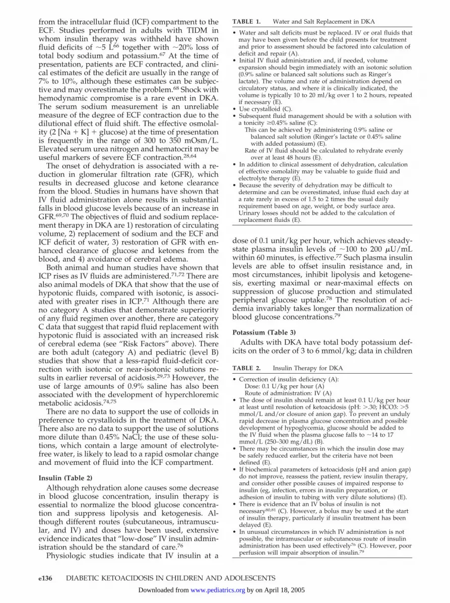
TABLE 1. Water and Salt Replacement in DKA
• Water and salt deficits must be replaced. IV or oral fluids thatmay have been given before the child presents for treatmentand prior to assessment should be factored into calculation ofdeficit and repair (A).
• Initial IV fluid administration and, if needed, volumeexpansion should begin immediately with an isotonic solution(0.9% saline or balanced salt solutions such as Ringer’slactate). The volume and rate of administration depend oncirculatory status, and where it is clinically indicated, thevolume is typically 10 to 20 ml/kg over 1 to 2 hours, repeatedif necessary (E).
• Use crystalloid (C).• Subsequent fluid management should be with a solution with
a tonicity �0.45% saline (C):This can be achieved by administering 0.9% saline or
balanced salt solution (Ringer’s lactate or 0.45% salinewith added potassium) (E).
Rate of IV fluid should be calculated to rehydrate evenlyover at least 48 hours (E).
• In addition to clinical assessment of dehydration, calculationof effective osmolality may be valuable to guide fluid andelectrolyte therapy (E).
• Because the severity of dehydration may be difficult todetermine and can be overestimated, infuse fluid each day ata rate rarely in excess of 1.5 to 2 times the usual dailyrequirement based on age, weight, or body surface area.Urinary losses should not be added to the calculation ofreplacement fluids (E).
TABLE 2. Insulin Therapy for DKA
• Correction of insulin deficiency (A):Dose: 0.1 U/kg per hour (A)Route of administration: IV (A)
• The dose of insulin should remain at least 0.1 U/kg per hourat least until resolution of ketoacidosis (pH: �.30; HCO3: �5mmol/L and/or closure of anion gap). To prevent an undulyrapid decrease in plasma glucose concentration and possibledevelopment of hypoglycemia, glucose should be added tothe IV fluid when the plasma glucose falls to �14 to 17mmol/L (250–300 mg/dL) (B).
• There may be circumstances in which the insulin dose maybe safely reduced earlier, but the criteria have not beendefined (E).
• If biochemical parameters of ketoacidosis (pH and anion gap)do not improve, reassess the patient, review insulin therapy,and consider other possible causes of impaired response toinsulin (eg, infection, errors in insulin preparation, oradhesion of insulin to tubing with very dilute solutions) (E).
• There is evidence that an IV bolus of insulin is notnecessary80,81 (C). However, a bolus may be used at the startof insulin therapy, particularly if insulin treatment has beendelayed (E).
• In unusual circumstances in which IV administration is notpossible, the intramuscular or subcutaneous route of insulinadministration has been used effectively76 (C). However, poorperfusion will impair absorption of insulin.79
e136 DIABETIC KETOACIDOSIS IN CHILDREN AND ADOLESCENTS
by on April 18, 2005 www.pediatrics.orgDownloaded from

are sparse.66,67,82–85 The major loss of potassium isfrom the intracellular pool as a result of hypertonic-ity, insulin deficiency, and buffering of hydrogenions within the cell. Serum potassium levels at thetime of presentation may be normal, increased ordecreased: Hypokalemia at presentation may be re-lated to prolonged duration of disease, whereas hy-perkalemia primarily results from reduced renalfunction.86 Administration of insulin and the correc-tion of acidosis will drive potassium back into thecells, decreasing serum levels.
PhosphateDepletion of intracellular phosphate occurs and
phosphate is lost as a result of osmotic diuresis. Inadults, deficits are in the range of 0.5 to 2.5 mmol/kg,66,67,84 but comparable data in children are un-available. The fall in plasma phosphate levels afterstarting treatment is exacerbated by insulin admin-istration as phosphate reenters cells.87 Low plasmaphosphate levels, when indicative of total body de-pletion in other conditions, have been associatedwith a wide array of metabolic disturbances; how-ever, particular interest has focused on erythrocyte2,3-diphosphoglycerate concentrations and effects ontissue oxygenation.88 Phosphate depletion persistsfor several days after resolution of DKA.66,82,84 How-ever, prospective studies have failed to show signif-icant clinical benefit from phosphate replace-ment.89–94 Nevertheless, provided that carefulmonitoring is performed to avoid hypocalcemia,95,96
potassium phosphate may be used safely in combi-nation with potassium chloride or acetate to avoidhyperchloremia.
AcidosisEven severe acidosis is reversible by fluid and
insulin replacement. Administration of insulin stops
further ketoacid synthesis and allows excess ketoac-ids to be metabolized. The metabolism of keto-anionresults in the regeneration of bicarbonate (HCO3
�)and spontaneous correction of acidemia. Also, treat-ment of hypovolemia will improve decreased tissueperfusion and renal function, thus increasing theexcretion of organic acids (see “Fluids and Salt”) andreversing any lactic acidosis, which may account for25% of the acidemia.
In DKA, there is an increased anion gap. The majorretained anions are �-OHB and acetoacetate.
anion gap � �Na�� � ��Cl��
� �HCO3��: normally 12 2 mmol/L
The indications for bicarbonate therapy in DKA areunclear. Several controlled trials of sodium bicarbon-ate in small numbers of children and adults (B, C)have been unable to demonstrate clinical benefit orany important difference in the rate of rise in theplasma bicarbonate concentration (C).25,97–100
There are potential arguments against the use ofbicarbonate.25,97,101,102 Of concern is that bicarbonatetherapy may cause paradoxical CNS acidosis andthat rapid correction of acidosis caused by bicarbon-ate will result in hypokalemia and may accentuatesodium load and contribute to serum hypertonicity.In addition, alkali therapy may increase hepatic ke-tone production, thus slowing the rate of recoveryfrom the ketosis.
These findings, however, do not address the issuethat there may be select patients who may benefitfrom cautious alkali therapy, including those withsevere acidemia (arterial pH: �6.9) in whom de-creased cardiac contractility and peripheral vasodi-latation can further impair tissue perfusion and pa-tients with potentially life-threatening hyperkalemia.
Treatment of Cerebral EdemaTreatment should be initiated as soon as the con-
dition is suspected. The rate of fluid administrationshould be reduced. Although mannitol has beenshown to have possible beneficial effects in case re-ports,103–105 there has been no definite beneficial ordetrimental effect in retrospective epidemiologicstudies.106 The response may be altered by timing ofadministration, delayed administration being less ef-fective. IV mannitol should be given (0.25–1.0 g/kgover 20 minutes) in patients with signs of cerebraledema before impending respiratory failure (C, E).Repeat in 2 hours if there is no initial response.Hypertonic saline (3%), 5 to 10 mL/kg over 30 min-utes, may be an alternative to mannitol (C).107
Intubation and ventilation may be necessary.However, aggressive hyperventilation has been as-sociated with poor outcome in one retrospectivestudy of DKA-related cerebral edema106; similarlydetrimental effects have been reported in numerousother conditions such as head trauma and high-alti-tude exposure.62 There are no data regarding glu-cocorticoid use in DKA-related cerebral edema.
TABLE 3. Potassium, Phosphate, and Acid-Base Management
Potassium• Replacement is required (A).• Replacement therapy should be based on serum potassium
measurements (E).• Start potassium replacement immediately if the patient is
hypokalemic; otherwise, start potassium concurrent withstarting insulin therapy. If the patient is hyperkalemic, deferpotassium until urine output is documented (E).
• Starting potassium concentration in the infusate should be 40mmol/L (E), and potassium replacement should continuethroughout IV fluid therapy (E).
Phosphate• There is no evidence that replacement has clinical benefit (A).
Severe hypophosphatemia should be treated (C).• Potassium phosphate salts may be used as an alternative to
or combined with potassium chloride/acetate (C).• Administration of phosphate may induce hypocalcemia (C).Acid base• Other acute resuscitation protocols no longer recommend
bicarbonate administration unless the acidosis is “profound”and “likely to affect the action of adrenaline/epinephrineduring resuscitation” (A).
• Fluid and insulin replacement without bicarbonateadministration corrects ketoacidosis (A).
• Data show that treatment with bicarbonate confers no clinicalbenefit (B).
• Repair fluids containing various buffering agents(bicarbonate, acetate, and lactate) have been used (C). Theefficacy and safety of these agents have not been established.
http://www.pediatrics.org/cgi/content/full/113/2/e133 e137
by on April 18, 2005 www.pediatrics.orgDownloaded from

PREVENTION OF DKA
Before DiagnosisEarlier diagnosis through genetic and immuno-
logic screening of high-risk children such as in therecent DPT-1 study108 decrease DKA incidence atdiabetes onset (A).14,109 High levels of awarenessrelated to the existence of other members of familieswith TIDM also reduce the risk of DKA. A schooland physician awareness campaign, targeted at 6- to14-year-olds, reduced rates of DKA from 78% toalmost 0% over a 6-year period (B).110 Increased pub-lic awareness of signs and symptoms of diabetesshould lead to earlier diagnosis, particularly in chil-dren �5 years; checking urine or blood for glucosemay prevent misdiagnosis (E). Although such strat-egies are intuitively obvious, programs to decreaseDKA at onset need to be designed and evaluated indiverse populations and age groups.
Beyond DiagnosisStudies of the effects of comprehensive diabetes
programs and telephone help lines report a reduc-tion in the rates of DKA from 15–60 to 5–5.9/100patient-years (B).19,111,112 In patients on continuoussubcutaneous insulin pumps, episodes of DKA canbe reduced with the introduction of educational al-gorithms (E). Therefore, it is likely that episodes ofDKA after diagnosis could be reduced if all childrenwith diabetes receive comprehensive diabetes healthcare and education and have access to a 24-hourdiabetes telephone help line (A).19 The value of homemeasurement of �-OHB as a mechanism for earlierdiagnosis and thus prevention of hospitalizationneeds to be assessed.
Multiple episodes of recurrent DKA are moreproblematic: In a recent United Kingdom study, 4.8%of patients accounted for 22.5% of all episodes over a3-year period.24 Insulin omission has been identifiedas the major factor in most of these cases and may beconfirmed by finding low free-insulin levels on ad-mission (C).13,113 There is no evidence that mentalhealth interventions alone can impact on the fre-quency of DKA in these children (B),14,17,111 but in-sulin omission can be prevented by sequentialschemes providing education, psychosocial evalua-tion, and treatment combined with adult supervisionof insulin administration (B).17 When responsibleadults administer insulin, a 10-fold reduction in ep-isodes of DKA has been reported (B).17
KEY ISSUES FOR FUTURE INVESTIGATION• Prevention: efficacy and cost-effectiveness of strat-
egies to reduce DKA incidence; frequency andevaluation of ketoacidosis in childhood type 2 di-abetes mellitus.
• Management: improved assessment of dehydra-tion; systematic evaluation of rehydration solu-tions such as those containing bicarbonate, acetate,lactate, and phosphate; use of lower doses of in-sulin in younger children; clarification of criteriafor reducing dose of insulin during treatment ofDKA; need for bicarbonate therapy in those withpH �6.9 and the very young.
• Cerebral edema: meta-analysis of existing epide-miologic studies to identify factors related to in-creased risk in infants and the newly diagnosed;monitoring of DKA and earlier detection of signsof cerebral edema; efficacy of hypertonic salineversus mannitol.
APPENDIXThe American Diabetes Association evidence-
grading system for clinical practice recommenda-tions is as follows1:
Level ofEvidence
Description
A Clear evidence from well-conducted, generalizable,randomized, controlled trials that are adequatelypowered, including:
• Multicenter trial• Meta-analysis incorporating quality ratings• Compelling nonexperimental evidence, (ie, “all-
or-none” rule) developed by the Center forEvidence-Based Medicine at Oxford*
Supportive evidence from well-conducted,randomized, controlled trials that are adequatelypowered, including:
• Well-conducted trials at �1 institutionsB Supportive evidence from well-conducted cohort
studies including:• Prospective cohort studies or registry• Meta-analysis of cohort studiesSupportive evidence from a well-conducted case-
control study.C Supportive evidence from poorly controlled or
uncontrolled studies including:• Randomized clinical trials with �1 major or �3
minor methodological flaws that could invalidatethe results
• Observational studies with high potential for bias• Case series or case reportsConflicting evidence with the weight of evidence
supporting the recommendation.E Expert consensus or clinical experience.
* Either all patients died before therapy and at least some survivedwith therapy or some patients died without therapy and nonedied with therapy (eg, the use of insulin in the treatment ofdiabetes ketoacidosis).
ACKNOWLEDGMENTSThe LWPES and ESPE gratefully acknowledge Aventis Phar-
maceuticals, Novo Nordisk, and the Society for Endocrinology forsupport of the consensus conference; Sten Renstad of Semser fororganizing the conference; and Emily Knight and Kathy Wypy-chowski for assistance with preparing the consensus statement.
REFERENCES1. American Diabetes Association position statement: hyperglycemic cri-
ses in patients with diabetes mellitus. Diabetes Care. 2003;26(suppl 1):S1–S2
2. International Society for Pediatric and Adolescent Diabetes. Consensusguidelines. Available at: www.ispad.org. Accessed December 11, 2003
3. Chase HP, Garg SK, Jelley DH. Diabetic ketoacidosis in children and therole of outpatient management. Pediatr Rev. 1990;11:297–304
4. Pinkey JH, Bingley PJ, Sawtell PA, Dunger DB, Gale EA. Presentationand progress of childhood diabetes mellitus: a prospective population-based study. The Bart’s-Oxford Study Group. Diabetologia. 1994;37:70–74
5. Levy-Marchal C, Papoz L, de Beaufort C, et al. Clinical and laboratoryfeatures of type 1 diabetic children at the time of diagnosis. Diabet Med.1992;9:279–284
6. Komulainen J, Lounamaa R, Knip M, Kaprio EA, Akerblom HK. Keto-acidosis at the diagnosis of type 1 (insulin dependent) diabetes mellitusis related to poor residual beta cell function. Childhood Diabetes inFinland Study Group. Arch Dis Child. 1996;75:410–415
e138 DIABETIC KETOACIDOSIS IN CHILDREN AND ADOLESCENTS
by on April 18, 2005 www.pediatrics.orgDownloaded from

7. Curtis JR, To T, Muirhead S, Cummings E, Daneman D. Recent trendsin hospitalization for diabetic ketoacidosis in Ontario children. DiabetesCare. 2002;25:1591–1596
8. Hirasing RA, Reeser HM, de Groot RR, Ruwaard D, van Buuren S,Verloove-Vanhorick SP. Trends in hospital admissions among childrenaged 0–19 years with type I diabetes in The Netherlands. Diabetes Care.1996;19:431–434
9. Komulainen J, Kulmala P, Savola K, et al. Clinical, autoimmune, andgenetic characteristics of very young children with type 1 diabetes.Childhood Diabetes in Finland (DiMe) Study Group. Diabetes Care.1999;22:1950–1955
10. Alavi IA, Sharma BK, Pillay VK. Steroid-induced diabetic ketoacidosis.Am J Med Sci. 1971;262:15–23
11. Goldstein LE, Sporn J, Brown S, et al. New-onset diabetes mellitus anddiabetic ketoacidosis associated with olanzapine treatment. Psychoso-matics. 1999;40:438–443
12. Smith CP, Firth D, Bennett S, Howard C, Chisholm P. Ketoacidosisoccurring in newly diagnosed and established diabetic children. ActaPaediatr. 1998;87:537–541
13. Morris AD, Boyle DI, McMahon AD, Greene SA, MacDonald TM,Newton RW. Adherence to insulin treatment, glycaemic control, andketoacidosis in insulin-dependent diabetes mellitus. The DARTS/MEMO Collaboration. Diabetes Audit and Research in Tayside Scot-land. Medicines Monitoring Unit [see comments]. Lancet. 1997;350:1505–1510
14. Rewers A, Chase HP, Mackenzie T, et al. Predictors of acute complica-tions in children with type 1 diabetes. JAMA. 2002;287:2511–2518
15. Rosilio M, Cotton JB, Wieliczko MC, et al. Factors associated withglycemic control. A cross-sectional nationwide study in 2, 579 Frenchchildren with type 1 diabetes. The French Pediatric Diabetes Group.Diabetes Care. 1998;21:1146–1153
16. Keenan HT, Foster CM, Bratton SL. Social factors associated with pro-longed hospitalization among diabetic children. Pediatrics. 2002;109:40–44
17. Golden MP, Herrold AJ, Orr DP. An approach to prevention of recur-rent diabetic ketoacidosis in the pediatric population. J Pediatr. 1985;107:195–200
18. Flood RG, Chiang VW. Rate and prediction of infection in children withdiabetic ketoacidosis. Am J Emerg Med. 2001;19:270–273
19. Hoffman WH, O’Neill P, Khoury C, Bernstein SS. Service and educationfor the insulin-dependent child. Diabetes Care. 1978;1:285–288
20. Glasgow AM, Weissberg-Benchell J, Tynan WD, et al. Readmissions ofchildren with diabetes mellitus to a children’s hospital. Pediatrics. 1991;88:98–104
21. Levitsky L, Ekwo E, Goselink CA, Solomon IL, Aceto T. Death fromdiabetes (DM) in hospitalized children (1970–1988) [abstract]. PediatrRes. 1991;29:A195
22. Cummings E, Lawrence S, Daneman D. Cerebral edema (CE) in pedi-atric diabetic ketoacidosis (DKA) in Canada [abstract]. Diabetes. 2003;52:A400
23. Edge JA, Ford-Adams ME, Dunger DB. Causes of death in children withinsulin dependent diabetes 1990–96. Arch Dis Child. 1999;81:318–323
24. Edge JA, Hawkins MM, Winter DL, Dunger DB. The risk and outcomeof cerebral oedema developing during diabetic ketoacidosis. Arch DisChild. 2001;85:16–22
25. Glaser N, Barnett P, McCaslin I, et al. Risk factors for cerebral edema inchildren with diabetic ketoacidosis. The Pediatric Emergency MedicineCollaborative Research Committee of the American Academy of Pedi-atrics. N Engl J Med. 2001;344:264–269
26. Bello FA, Sotos JF. Cerebral oedema in diabetic ketoacidosis in children.Lancet. 1990;336:64
27. Mahoney CP, Vlcek BW, DelAguila M. Risk factors for developing brainherniation during diabetic ketoacidosis. Pediatr Neurol. 1999;21:721–727
28. Harris GD, Fiordalisi I. Physiologic management of diabetic ketoaci-demia. A 5-year prospective pediatric experience in 231 episodes. ArchPediatr Adolesc Med. 1994;148:1046–1052
29. Felner EI, White PC. Improving management of diabetic ketoacidosis inchildren. Pediatrics. 2001;108:735–740
30. Atluru VL. Spontaneous intracerebral hematomas in juvenile diabeticketoacidosis. Pediatr Neurol. 1986;2:167–169
31. Kanter RK, Oliphant M, Zimmerman JJ, Stuart MJ. Arterial thrombosiscausing cerebral edema in association with diabetic ketoacidosis. CritCare Med. 1987;15:175–176
32. Moll GW Jr, Raila FA, Liu GC, Conerly AW Sr. Rhinocerebral mucor-mycosis in IDDM. Sequential magnetic resonance imaging of long-termsurvival with intensive therapy. Diabetes Care. 1994;17:1348–1353
33. Young MC. Simultaneous acute cerebral and pulmonary edema com-plicating diabetic ketoacidosis. Diabetes Care. 1995;18:1288–1290
34. Breidbart S, Singer L, St Louis Y, Saenger P. Adult respiratory distresssyndrome in an adolescent with diabetic ketoacidosis. J Pediatr. 1987;111:736–738
35. Watson JP, Barnett AH. Pneumomediastinum in diabetic ketoacidosis.Diabet Med. 1989;6:173–174
36. Casteels K, Beckers D, Wouters C, Van Geet C. Rhabdomyolysis indiabetic ketoacidosis. Pediatr Diabetes. 2003;4:29–31
37. Tubiana-Rufi N, Thizon-de Gaulle I, Czernichow P. Hypothalamopitu-itary deficiency and precocious puberty following hyperhydration indiabetic ketoacidosis. Horm Res. 1992;37:60–63
38. Lufkin EG, Reagan TJ, Doan DH, Yanagihara. Acute cerebral dysfunc-tion in diabetic ketoacidosis: survival followed by panhypopituitarism.Metabolism. 1977;26:363–369
39. Keller RJ, Wolfsdorf JI. Isolated growth hormone deficiency after cere-bral edema complicating diabetic ketoacidosis. N Engl J Med. 1987;316:857–859
40. Dunlop KA, Woodman D, Carson DJ. Hypopituitarism following cere-bral oedema with diabetic ketoacidosis. Arch Dis Child. 2002;87:337–338
41. Edge JA. Cerebral oedema during treatment of diabetic ketoacidosis: arewe any nearer finding a cause? Diabetes Metab Res Rev. 2000;16:316–324
42. Deeb L. Development of fatal cerebral oedema during outpatient ther-apy for diabetic ketoacidosis. Pract Diabetes Int. 1989;6:212–213
43. Glasgow AM. Devastating cerebral edema in diabetic ketoacidosis be-fore therapy. Diabetes Care. 1991;14:77–78
44. Rosenbloom AL. Intracerebral crises during treatment of diabetic keto-acidosis. Diabetes Care. 1990;13:22–33
45. Muir A. Therapeutic controversy. Cerebral edema in diabeticketoacidosis: a look beyond rehydration. J Clin Endocrinol Metab. 2000;85:509–513
46. Abbott NJ. Inflammatory mediators and modulation of blood-brainbarrier permeability. Cell Mol Neurobiol. 2000;20:131–147
47. Sarker MH, Fraser PA. The role of guanylyl cyclases in the permeabilityresponse to inflammatory mediators in pial venular capillaries in therat. J Physiol. 2002;540:209–218
48. Yang GY, Betz AL. Reperfusion-induced injury to the blood-brain bar-rier after middle cerebral artery occlusion in rats. Stroke. 1994;25:1658–1664, 1664–1665 [discussion]
49. O’Donnell ME, Martinez A, Sun D. Endothelial Na-K-Cl cotransportregulation by tonicity and hormones: phosphorylation of cotransportprotein. Am J Physiol. 1995;269:C1513–C1523
50. Kimelberg HK, Rutledge E, Goderie S, Charniga C. Astrocytic swellingdue to hypotonic or high K� medium causes inhibition of glutamateand aspartate uptake and increases their release. J Cereb Blood FlowMetab. 1995;15:409–416
51. Manley GT, Fujimura M, Ma T, et al. Aquaporin-4 deletion in micereduces brain edema after acute water intoxication and ischemic stroke.Nat Med. 2000;6:159–163
52. McManus ML, Churchwell KB, Strange K. Regulation of cell volume inhealth and disease. N Engl J Med. 1995;333:1260–1226
53. Krane EJ, Rockoff MA, Wallman JK, Wolfsdorf JI. Subclinical brainswelling in children during treatment of diabetic ketoacidosis. N EnglJ Med. 1985;312:1147–1151
54. Hoffman WH, Steinhart CM, el Gammal T, Steele S, Cuadrado AR,Morse PK. Cranial CT in children and adolescents with diabetic keto-acidosis. AJNR Am J Neuroradiol. 1988;9:733–739
55. Hoffman WH, Casanova MF, Bauza JA, Passmore GG, Sekul EA. Com-puter analysis of magnetic resonance imaging of the brain in childrenand adolescents after treatment of diabetic ketoacidosis. J Diabetes Com-plications. 1999;13:176–181
56. Hoffman WH, Pluta RM, Fisher AQ, Wagner MB, Yanovski JA. Trans-cranial Doppler ultrasound assessment of intracranial hemodynamics inchildren with diabetic ketoacidosis. J Clin Ultrasound. 1995;23:517–523
57. Harris GD, Fiordalisi I, Harris WL, Mosovich LL, Finberg L. Minimizingthe risk of brain herniation during treatment of diabetic ketoacidemia:a retrospective and prospective study [published correction appears inJ Pediatr. 1991;118:166–167] [see comments]. J Pediatr. 1990;117:22–31
58. Hale PM, Rezvani I, Braunstein AW, Lipman TH, Martinez N, GaribaldiL. Factors predicting cerebral edema in young children with diabeticketoacidosis and new onset type I diabetes. Acta Paediatr. 1997;86:626–631
59. Mel JM, Werther GA. Incidence and outcome of diabetic cerebral oe-dema in childhood: are there predictors? J Paediatr Child Health. 1995;31:17–20
60. Durr JA, Hoffman WH, Sklar AH, el Gammal T, Steinhart CM. Corre-lates of brain edema in uncontrolled IDDM. Diabetes. 1992;41:627–632
61. Bureau MA, Begin R, Berthiaume Y, Shapcott D, Khoury K, Gagnon N.Cerebral hypoxia from bicarbonate infusion in diabetic acidosis. J Pedi-atr. 1980;96:968–973
http://www.pediatrics.org/cgi/content/full/113/2/e133 e139
by on April 18, 2005 www.pediatrics.orgDownloaded from

62. Roberts I, Schierhout G. Hyperventilation therapy for acute traumaticbrain injury (Cochrane Review). The Cochrane Library; 2003. Availableat: www.cochrane.org/cochrane/revabstr/ab000566.htm. Accessed De-cember 11, 2003
63. Bonadio WA, Gutzeit MF, Losek JD, Smith DS. Outpatient managementof diabetic ketoacidosis. Am J Dis Child. 1988;142:448–450
64. Linares MY, Schunk JE, Lindsay R. Laboratory presentation in diabeticketoacidosis and duration of therapy. Pediatr Emerg Care. 1996;12:347–351
65. Monroe KW, King W, Atchison JA. Use of PRISM scores in triage ofpediatric patients with diabetic ketoacidosis. Am J Manag Care. 1997;3:253–258
66. Atchley D, Loeb, R, Richards D. On diabetic acidosis. A detailed studyof electrolyte balances following withdrawal and reestablishment oftherapy. J Clin Invest. 1933;12:297–326
67. Nabarro JDN, Spencer AG, Stowers JM. Metabolic studies in severediabetic ketosis. Q J Med. 1952;82:225–248
68. Mackenzie A, Barnes G, Shann F. Clinical signs of dehydration inchildren. Lancet. 1989;2(8663):605–607
69. Waldhausl W, Kleinberger G, Korn A, Dudczak R, Bratusch-Marrain P,Nowotny P. Severe hyperglycemia: effects of rehydration on endocrinederangements and blood glucose concentration. Diabetes. 1979;28:577–584
70. Owen OE, Licht JH, Sapir DG. Renal function and effects of partialrehydration during diabetic ketoacidosis. Diabetes. 1981;30:510–518
71. Harris GD, Fiordalisi I, Yu C. Maintaining normal intracranial pressurein a rabbit model during treatment of severe diabetic ketoacidemia. LifeSci. 1996;59:1695–1702
72. Clements RS Jr, Blumenthal SA, Morrison AD, Winegrad AI. Increasedcerebrospinal-fluid pressure during treatment of diabetic ketosis. Lan-cet. 1971(7726);2:671–675
73. Adrogue HJ, Barrero J, Eknoyan G. Salutary effects of modest fluidreplacement in the treatment of adults with diabetic ketoacidosis. Use inpatients without extreme volume deficit. JAMA. 1989;262:2108–2113
74. Adrogue HJ, Eknoyan G, Suki WK. Diabetic ketoacidosis: role of thekidney in the acid-base homeostasis re-evaluated. Kidney Int. 1984;25:591–598
75. Oh MS, Carroll HJ, Uribarri J. Mechanism of normochloremic andhyperchloremic acidosis in diabetic ketoacidosis. Nephron. 1990;54:1–6
76. Kitabchi AE. Low-dose insulin therapy in diabetic ketoacidosis: fact orfiction? Diabetes Metab Rev. 1989;5:337–363
77. Schade DS, Eaton RP. Dose response to insulin in man: differentialeffects on glucose and ketone body regulation. J Clin Endocrinol Metab.1977;44:1038–1053
78. Luzi L, Barrett EJ, Groop LC, Ferrannini E, DeFronzo RA. Metaboliceffects of low-dose insulin therapy on glucose metabolism in diabeticketoacidosis. Diabetes. 1988;37:1470–1477
79. Soler NG, FitzGerald MG, Wright AD, Malins JM. Comparative study ofdifferent insulin regimens in management of diabetic ketoacidosis. Lan-cet. 1975;2(7947):1221–1224
80. Fort P, Waters SM, Lifshitz F. Low-dose insulin infusion in the treat-ment of diabetic ketoacidosis: bolus versus no bolus. J Pediatr. 1980;96:36–40
81. Lindsay R, Bolte RG. The use of an insulin bolus in low-dose insulininfusion for pediatric diabetic ketoacidosis. Pediatr Emerg Care. 1989;5:77–79
82. Nabarro J, Spencer A, Stowers J. Treatment of diabetic ketoacidosis.Lancet. 1952;i:983–983
83. Danowski T, Peters J. Studies in diabetic acidosis and coma, withparticular emphasis on the retention of administered potassium. J ClinInvest. 1949;28:1–9
84. Butler A, Talbot N. Metabolic studies in diabetic coma. Trans Assoc AmPhysicians. 1947;60:102–109
85. Darrow DC, Pratt EL. Retention of water and electrolyte during recov-ery in a patient with diabetic acidosis. J Pediatr. 1952;41:688–696
86. Adrogue HJ, Lederer ED, Suki WN, Eknoyan G. Determinants ofplasma potassium levels in diabetic ketoacidosis. Medicine. 1986;65:163–172
87. Riley MS, Schade DS, Eaton RP. Effects of insulin infusion on plasmaphosphate in diabetic patients. Metabolism. 1979;28:191–194
88. Alberti KG, Emerson PM, Darley JH, Hockaday TD. 2,3-Diphosphoglyc-erate and tissue oxygenation in uncontrolled diabetes mellitus. Lancet.1972;2(7774):391–395
89. Gibby OM, Veale KE, Hayes TM, Jones JG, Wardrop CA. Oxygenavailability from the blood and the effect of phosphate replacement onerythrocyte 2,3-diphosphoglycerate and haemoglobin-oxygen affinityin diabetic ketoacidosis. Diabetologia. 1978;15:381–385
90. Keller U, Berger W. Prevention of hypophosphatemia by phosphateinfusion during treatment of diabetic ketoacidosis and hyperosmolarcoma. Diabetes. 1980;29:87–95
91. Wilson HK, Keuer SP, Lea AS, Boyd AE 3rd, Eknoyan G. Phosphatetherapy in diabetic ketoacidosis. Arch Intern Med. 1982;142:517–520
92. Becker DJ, Brown DR, Steranka BH, Drash AL. Phosphate replacementduring treatment of diabetic ketosis. Effects on calcium and phosphorushomeostasis. Am J Dis Child. 1983;137:241–246
93. Fisher JN, Kitabchi AE. A randomized study of phosphate therapy inthe treatment of diabetic ketoacidosis. J Clin Endocrinol Metab. 1983;57:177–180
94. Clerbaux T, Reynaert M, Willems E, Frans A. Effect of phosphate onoxygen-hemoglobin affinity, diphosphoglycerate and blood gases dur-ing recovery from diabetic ketoacidosis. Intensive Care Med. 1989;15:495–498
95. Winter RJ, Harris CJ, Phillips LS, Green OC. Diabetic ketoacidosis.Induction of hypocalcemia and hypomagnesemia by phosphate ther-apy. Am J Med. 1979;67:897–900
96. Zipf WB, Bacon GE, Spencer ML, Kelch RP, Hopwood NJ, Hawker CD.Hypocalcemia, hypomagnesemia, and transient hypoparathyroidismduring therapy with potassium phosphate in diabetic ketoacidosis.Diabetes Care. 1979;2:265–268
97. Soler NG, Bennett MA, Dixon K, FitzGerald MG, Malins JM. Potassiumbalance during treatment of diabetic ketoacidosis with special referenceto the use of bicarbonate. Lancet. 1972;2(7779):665–667
98. Hale PJ, Crase J, Nattrass M. Metabolic effects of bicarbonate in thetreatment of diabetic ketoacidosis. Br Med J (Clin Res Ed). 1984;289:1035–1038
99. Okuda Y, Adrogue HJ, Field JB, Nohara H, Yamashita K. Counterpro-ductive effects of sodium bicarbonate in diabetic ketoacidosis. J ClinEndocrinol Metab. 1996;81:314–320
100. Green SM, Rothrock SG, Ho JD, Gallant RD, Borger R, Thomas TL,Zimmerman GJ. Failure of adjunctive bicarbonate to improve outcomein severe pediatric diabetic ketoacidosis. Ann Emerg Med. 1998;31:41–48
101. Ohman JL Jr, Marliss EB, Aoki TT, Munichoodappa CS, Khanna VV,Kozak GP. The cerebrospinal fluid in diabetic ketoacidosis. N EnglJ Med. 1971;284:283–290
102. Assal JP, Aoki TT, Manzano FM, Kozak GP. Metabolic effects ofsodium bicarbonate in management of diabetic ketoacidosis. Diabetes.1974;23:405–411
103. Franklin B, Liu J, Ginsberg-Fellner F. Cerebral edema and ophthalmo-plegia reversed by mannitol in a new case of insulin-dependent dia-betes mellitus. Pediatrics. 1982;69:87–90
104. Shabbir N, Oberfield SE, Corrales R, Kairam R, Levine LS. Recoveryfrom symptomatic brain swelling in diabetic ketoacidosis. Clin Pediatr(Phila). 1992;31:570–573
105. Roberts MD, Slover RH, Chase HP. Diabetic ketoacidosis with intra-cerebral complications. Pediatr Diabetes. 2001;2:109–114
106. Marcin JP, Glaser N, Barnett P, et al. Factors associated with adverseoutcomes in children with diabetic ketoacidosis-related cerebraledema. J Pediatr. 2002;141:793–797
107. Kamat P, Vats A, Gross M, Checchia PA. Use of hypertonic saline forthe treatment of altered mental status associated with diabetic ketoac-idosis. Pediatr Crit Care Med. 2003;4:239–242
108. Diabetes Prevention Trial–Type 1 Diabetes Study Group. Effects ofinsulin in relatives of patients with type 1 diabetes mellitus. N EnglJ Med. 2002;346:1685–1691
109. Levy-Marchal C, Patterson CC, Green A; EURODIAB ACE StudyGroup. Europe and Diabetes. Geographical variation of presentation atdiagnosis of type I diabetes in children: the EURODIAB study. Euro-pean and Dibetes. Diabetologia. 2001;44(suppl 3):B75–B80
110. Vanelli M, Chiari G, Ghizzoni L, Costi G, Giacalone T, Chiarelli F.Effectiveness of a prevention program for diabetic ketoacidosis inchildren. An 8-year study in schools and private practices. DiabetesCare. 1999;22:7–9
111. Grey M, Boland EA, Davidson M, Li J, Tamborlane WV. Coping skillstraining for youth with diabetes mellitus has long-lasting effects onmetabolic control and quality of life. J Pediatr. 2000;137:107–113
112. Drozda DJ, Dawson VA, Long DJ, Freson LS, Sperling MA. Assess-ment of the effect of a comprehensive diabetes management programon hospital admission rates of children with diabetes mellitus. DiabetesEduc. 1990;16:389–393
113. Malone JI, Root AW. Root, Plasma free insulin concentrations: key-stone to effective management of diabetes mellitus in children. J Pedi-atr. 1981;99:862–867
e140 DIABETIC KETOACIDOSIS IN CHILDREN AND ADOLESCENTS
by on April 18, 2005 www.pediatrics.orgDownloaded from

DOI: 10.1542/peds.113.2.e133 2004;113;133-140 Pediatrics
Lynne L. Levitsky, Martin O. Savage, Robert C. Tasker and Joseph I. Wolfsdorf Daneman, Thomas P.A. Danne, Nicole S. Glaser, Ragnar Hanas, Raymond L. Hintz,
David B. Dunger, Mark A. Sperling, Carlo L. Acerini, Desmond J. Bohn, Denis and Adolescents
Endocrine Society Consensus Statement on Diabetic Ketoacidosis in Children European Society for Paediatric Endocrinology/Lawson Wilkins Pediatric
This information is current as of April 18, 2005
& ServicesUpdated Information
http://www.pediatrics.org/cgi/content/full/113/2/e133including high-resolution figures, can be found at:
References
http://www.pediatrics.org/cgi/content/full/113/2/e133#BIBLfree at: This article cites 111 articles, 35 of which you can access for
Citations
eshttp://www.pediatrics.org/cgi/content/full/113/2/e133#otherarticlThis article has been cited by 4 HighWire-hosted articles:
Subspecialty Collections
http://www.pediatrics.org/cgi/collection/endocrinology Endocrinology
following collection(s): This article, along with others on similar topics, appears in the
Permissions & Licensing
http://www.pediatrics.org/misc/Permissions.shtmltables) or in its entirety can be found online at: Information about reproducing this article in parts (figures,
Reprints http://www.pediatrics.org/misc/reprints.shtml
Information about ordering reprints can be found online:
by on April 18, 2005 www.pediatrics.orgDownloaded from