ets transcription factor ese1/elf3 orchestrates a positive feedback loop that constitutively...
TRANSCRIPT
Published OnlineFirst May 16, 2013.Cancer Res Nicole Longoni, Manuela Sarti, Domenico Albino, et al. prostate cancer progression.feedback loop that constitutively activates NF-kB and drives ETS transcription factor ESE1/ELF3 orchestrates a positive
Updated version
10.1158/0008-5472.CAN-12-4537doi:
Access the most recent version of this article at:
Material
Supplementary
http://cancerres.aacrjournals.org/content/suppl/2013/05/16/0008-5472.CAN-12-4537.DC1.html
Access the most recent supplemental material at:
Manuscript
Authoredited. Author manuscripts have been peer reviewed and accepted for publication but have not yet been
E-mail alerts related to this article or journal.Sign up to receive free email-alerts
Subscriptions
Reprints and
To order reprints of this article or to subscribe to the journal, contact the AACR Publications
Permissions
To request permission to re-use all or part of this article, contact the AACR Publications
on May 27, 2013. © 2013 American Association for Cancer Research. cancerres.aacrjournals.org Downloaded from
Author manuscripts have been peer reviewed and accepted for publication but have not yet been edited. Author Manuscript Published OnlineFirst on May 16, 2013; DOI: 10.1158/0008-5472.CAN-12-4537
1
ETS transcription factor ESE1/ELF3 orchestrates a positive feedback loop that constitutively activates NF-�B and drives prostate cancer progression.
Nicole Longoni1, Manuela Sarti1, Domenico Albino1,Gianluca Civenni1, Anastasia Malek1,
Erica Ortelli1, Sandra Pinton1, Maurizia Mello-Grand2, Paola Ostano2, Gioacchino
D’Ambrosio3, Fausto Sessa3-4, Ramon Garcia-Escudero5, George N. Thalmann6, Giovanna
Chiorino2, Carlo V. Catapano1 and Giuseppina M. Carbone1*
1 Institute of Oncology Research (IOR) and Oncology Institute of Southern Switzerland
(IOSI), Bellinzona, 6500 Switzerland 2 Laboratory of Cancer Genomics, Fondazione Edo ed Elvo Tempia Valenta, Biella, 13900
Italy 3 IRCCS Multimedica, Milan, Italy 4 Department of Pathology University of Insubria, Varese, Italy 5 Molecular Oncology Unit, CIEMAT, Madrid, Spain 6 Urology Research Laboratory, Department of Urology, University of Bern, Inselspital, Bern,
Switzerland
Running title: ESE1/ELF3, IL-1� and NF-κB activation in prostate cancer
Keywords: prostate cancer, ESE1/ELF3, NF-κB, IL-1�, inflammation, ETS transcription
factors
* Corresponding author: Giuseppina M. Carbone, MD. Institute of Oncology Research (IOR),
Oncology Institute of Southern Switzerland (IOSI), Via Vela 6, Bellinzona, 6500 Switzerland.
E-mail: [email protected]. Tel.: +41 (0)91 820 0366. Fax: +41 (0)91 820 0397
Conflict of interest: the authors declare that they have no conflicts of interest
on May 27, 2013. © 2013 American Association for Cancer Research. cancerres.aacrjournals.org Downloaded from
Author manuscripts have been peer reviewed and accepted for publication but have not yet been edited. Author Manuscript Published OnlineFirst on May 16, 2013; DOI: 10.1158/0008-5472.CAN-12-4537
2
Abstract
Chromosomal translocations leading to deregulated expression of ETS transcription factors
are frequent in prostate tumors. Here, we report a novel mechanism leading to oncogenic
activation of the ETS factor ESE1/ELF3 in prostate tumors. ESE1/ELF3 was overexpressed
in human primary and metastatic tumors. It mediated transforming phenotypes in vitro and in
vivo and induced an inflammatory transcriptome with changes in relevant oncogenic
pathways. ESE1/ELF3 was induced by IL-1� through NF-�B and was a crucial mediator of
the phenotypic and transcriptional changes induced by IL-1� in prostate cancer cells. This
linkage was mediated by interaction of ESE1/ELF3 with the NF-�B subunits p65 and p50,
acting by enhancing their nuclear translocation and transcriptional activity and by inducing
p50 transcription. Supporting these findings, gene expression profiling revealed an
enrichment of NF-�B effector functions in prostate cancer cells or tumors expressing high
levels of ESE1/ELF3. We observed concordant upregulation of ESE1/ELF3 and NF-�B in
human prostate tumors that was associated with adverse prognosis. Collectively, our results
define an important new mechanistic link between inflammatory signaling and the
progression of prostate cancer.
on May 27, 2013. © 2013 American Association for Cancer Research. cancerres.aacrjournals.org Downloaded from
Author manuscripts have been peer reviewed and accepted for publication but have not yet been edited. Author Manuscript Published OnlineFirst on May 16, 2013; DOI: 10.1158/0008-5472.CAN-12-4537
3
Introduction
Prostate cancer is the most common form of cancer in men and a leading cause of cancer
related death in western countries [1]. Deregulation of ETS transcription factors (TF) is very
frequent in prostate cancer, suggesting that the prostate epithelium might be highly sensitive
to unbalanced expression of these TFs [2, 3]. About 50 % of prostate tumors harbors
chromosomal translocations leading to overexpression of ETS genes, like ERG, ETV1 and
ETV4 [2, 4]. ETS TFs, including ESE3/EHF and ETV1, are also frequently deregulated in
prostate tumors and other tumor types despite the absence of chromosomal rearrangements [5-
9]. In this study, we report a novel mechanism leading to overexpression and oncogenic
activation of an additional ETS TF, ESE1/ELF3, in both primary and metastatic prostate
cancers. ESE1/ELF3 is a member of the epithelial-specific subfamily of ETS TF and has been
reported to be involved in a variety of patho-physiological processes, including cancer and
inflammatory disorders [10-12]. However, the role of ESE1/ELF3 in prostate tumorigenesis is
unknown. We found that ESE1/ELF3 functions at the crossroad between cancer and chronic
inflammation to promote prostate cancer progression. Epidemiological, genetic and
histopathological studies strongly support a connection between chronic inflammation and
prostate cancer [13]. However, the molecular mechanisms linking chronic inflammation and
prostate tumorigenesis are still unclear. Production of proinflammatory cytokines, like
interleukin-1� (IL-1�), and constitutive activation of NF-κB play an important role in cancer-
associated inflammation and tumorigenesis [14-19]. We found that ESE1/ELF3 is a target of
IL-1� and NF-κB in prostate cancer cells and an essential element in a positive feedback loop
sustaining constitutive activation of NF-κB in prostate tumors. We provide evidence that this
positive feedback loop is active in human prostate tumors and is associated with aggressive
disease and adverse prognosis. These data thus provide the rationale for patient risk
stratification and context-dependent therapeutic strategies in a specific subset of prostate
cancer patients.
Materials and Methods
Cell culture, cell transfection and selection of stable cell clones
LNCaP, 22RV1, DU145 were obtained from American Type Culture Collection (ATCC,
Manassas, VA, USA) and maintained in RPMI-1640 supplemented with 10% fetal bovine
serum. Immortalized prostate epithelial cells LHS were maintained in PrEC growth medium
(PrEGM, Cambrex, Lonza Group Ltd, Basel, Switzerland) as previously described [5, 7].
ESE1/ELF3 expressing polyclonal stable cell lines were generated by transfection of the
pESE1/ELF3 expressing vector (kindly provided by T. Libermann) [10], and negative control
on May 27, 2013. © 2013 American Association for Cancer Research. cancerres.aacrjournals.org Downloaded from
Author manuscripts have been peer reviewed and accepted for publication but have not yet been edited. Author Manuscript Published OnlineFirst on May 16, 2013; DOI: 10.1158/0008-5472.CAN-12-4537
4
cells were obtained by transfection with pcDNA3.1 as previously described [5, 7]. For
transient ESE1/ELF3 gene knock-down, cells were transfected with siRNAs directed to the
exon 3(siESE1) or to the 3’UTR (si3’UTR)(Ambion, Rotkreuz, Switzerland) and control
siRNA directed to the firefly luciferase gene (siGL3) using Lipofectamine 2000 (Invitrogen ,
Zug, Switzerland). Luciferase reporter assays were carried out as previously described [5, 7]
using the pGL4.32(luc2P/NF-�B-RE/Hygro) vector (Promega AG, Dübendorf, Switzerland).
For IL-1� treatment, cells were seeded in 6-wells plates and treated after 24 h with IL-1�
(Sigma-Aldrich Chemie GmbH, Buchs, Switzerland) diluted in 0.1% BSA in PBS.
Cell proliferation, anoikis and cell migration. Cell growth, clonogenic and anoikis assays
were performed as previously described [5, 7]. The scratch/wound healing and Boyden
chamber assays were performed and analyzed as previously described [7].
RNA extraction and quantitative RT-PCR. Total RNA was extracted and quantitative real-
time RT-PCR (qRT-PCR) was performed using custom made primers (Supplementary Table
S1) and analyzed as previously described [5].
Immunoblotting from cells and tumor xenografts. Cell lysates were prepared and analyzed
as described previously [5, 7]. Antibodies against ESE1/ELF3 (ab1392, AbCam, Cambridge,
UK), p50, p65, �-tubulin (Calbiochem, Nottingham, UK), GAPDH and Histone H3 (Millipore
AG, Zug, Switzerland) were obtained from the indicated sources. Lysate from tumor
xenografts were prepared from freshly frozen tissue. Cytoplasmic and nuclear extracts were
obtained using NE-PER Nuclear and Cytoplasmic Extraction Reagent (Thermo Scientific,
Lausanne, Switzerland).
Immunoprecipitation. Immunoprecipitation was performed as previously described [20].
Cell lysates were incubated with antibodies against ESE1/ELF3 and p50. Immunoblotting was
performed using antibodies against ESE1/ELF3, p50 and p65.
Immunofluorescence and fluorescence microscopy. Cells were grown on glass cover-slips,
as previously described [20] and incubated with antibodies for ESE1/ELF3, p50 and p65
followed by incubation with anti-rabbit Alexa Fluor 488 or anti-mouse Alexa Fluor 594
(Invitrogen) secondary antibodies. Pictures were taken as previously described [20].
on May 27, 2013. © 2013 American Association for Cancer Research. cancerres.aacrjournals.org Downloaded from
Author manuscripts have been peer reviewed and accepted for publication but have not yet been edited. Author Manuscript Published OnlineFirst on May 16, 2013; DOI: 10.1158/0008-5472.CAN-12-4537
5
Chromatin Immunoprecipitation (ChIP). ChIP was carried-out and analyzed using
quantitative real-time PCR (qPCR) as previously described [5, 7]. ChIP from fresh frozen
prostate tumors was performed as previously described [5, 7].
Animal studies. Mice were purchased from the Harlan Laboratories (San Pietro Al Natisone,
UD, Italy). Study protocols were approved by the Swiss Veterinary Authority (No.5/2011).
For subcutaneous tumor xenografts, 1 x 10⁶ cells were inoculated in the flank of athymic male
nude mice (Balb c nu/nu; n=10/group). Tumor size was monitored twice a week with a
caliper. To assay lung metastases, 1 x 10⁶ cells were injected into tail vein of athymic male
nude mice twice with a 24-h interval between injections. Animals were sacrificed after 4
weeks. Lungs were collected and a qPCR-based method that relies on selective amplification
of species-specific, unique, untranslated and conserved regions of the human and mouse
genome was used to quantify the percentage of human metastatic cells in mouse lungs [21].
Gene expression profiling. RNA from cell lines was amplified, labeled and hybridized as
described [5]. Significantly modulated transcripts were selected by applying 0.01 as cut-off
for the adjusted p-value (Benjamini-Hochberg correction) and 1 as cut-off for the log
fold-change. Data are MIAME compliant and have been deposited in the Gene Expression
Omnibus: GEO accession numbers GSE39668.
Functional annotation and transcription factor interactome analysis. For functional
annotation, gene lists were uploaded into the Database for Annotation, Visualization and
Integrated Discovery (DAVID; http://david.abcc.ncifcrf.gov/summary.jsp). Enrichment of
transcription factors interactome analysis was done using MetaCore version 6.10 (GeneGo
Inc) and Chip Enrichment Analysis (ChEA).
Gene Set Enrichment analysis (GSEA). GSEA was performed as previously described [7].
For all the datasets the comparison of ESE1/ELF3 High vs all other tumors was performed;
for the Biella dataset, the tumor vs normal tissue comparison was also made. The following
gene lists were used for GSEA: GS_3: Human NF�B Signaling Targets; GS_5: Genes
upregulated in 22RV1-pESE1 cells vs 22RV1-pcDNA.
Immunohistochemistry. Tissue microarrays (TMA) were constructed from formalin-fixed
paraffin-embedded tissue specimens as previously described [7, 22]. Tissue samples were
collected with the approval of the Institutional Ethics Committees (IRCCS Multimedica of the
on May 27, 2013. © 2013 American Association for Cancer Research. cancerres.aacrjournals.org Downloaded from
Author manuscripts have been peer reviewed and accepted for publication but have not yet been edited. Author Manuscript Published OnlineFirst on May 16, 2013; DOI: 10.1158/0008-5472.CAN-12-4537
6
Regione Lombardia, IT, and Insespital, Bern, Switzerland) and patient written informed
consent. Immunohistochemistry was carried out using antibodies against ESE1/ELF3
(ab1392, AbCam, Cambridge, UK), p50 (E-10) sc-8414 and p65 (C-20) sc-372 (Santa
Cruz,Heidelberg,Germany). Two trained investigators scored the slides and were blinded to
the study endpoints. At least two investigators scored the slides and were blinded to the study
endpoints. Score was based on the percentage of ESE1/ELF3 positive cells: Low, � 20 %;
Intermediate, >20 - <60%; High, �60%.
Survival analysis. Kaplan-Meier survival curves of patients groups were created using
survplot package of R environment.
Results
ESE1/ELF3 is over-expressed in human prostate cancers
Using qRT-PCR we found that primary tumors (n=65) had significantly increased
ESE1/ELF3 mRNA level (p-val=3.17E-04) compared to normal prostate tissue (n=14). As
previously reported [5], ESE1/ELF3 was one of the most frequently deregulated ETS genes in
prostate tumors. Specifically, 27 and 24% of tumors had, respectively, intermediate and high
level of expression (Figure 1A). Consulting additional patient microarray datasets [23, 24],
we found further evidence of ESE1/ELF3 overexpression in prostate tumors (Figure 1A and
Figure S1A). Furthermore, the level of ESE1/ELF3 was significantly higher (p-val<0.001) in
metastatic compared to primary tumors in two distinct datasets (Figure 1B), suggesting an
association of ESE1/ELF3 upregulation with tumor progression. Notably, we found also
evidence of amplification of the ESE1/ELF3 gene locus in three prostate cancer datasets with
a frequency of 2 to 6% using cancer outlier profile analysis (COPA) of DNA (Figure 1C). To
support these findings, ESE1/ELF3 protein expression was evaluated by
immunohistochemistry (IHC) in a total of 207 primary tumors and 28 normal prostate
samples in tissue microarrays (TMAs) from two patient cohorts [7]. ESE1/ELF3 was detected
in the cytoplasm and nuclei of prostate epithelial cells both in normal and cancer tissue
(Figure 1D). Consistent with gene expression data, ESE1/ELF3 protein level was low in
normal prostate and in about a third of tumors. In contrast, 35 and 28% of tumors had,
respectively, intermediate and high levels of expression. Collectively, the high frequency of
overexpression at the RNA and protein level, along with the evidence of amplification,
strongly supported an oncogenic role of ESE1/ELF3 in prostate cancer. Interestingly, when
we analyzed the relationship between ESE1/ELF3 and ERG, the most frequently rearranged
ETS gene, in patients samples previously evaluated by qRT-PCR [5] we found that
on May 27, 2013. © 2013 American Association for Cancer Research. cancerres.aacrjournals.org Downloaded from
Author manuscripts have been peer reviewed and accepted for publication but have not yet been edited. Author Manuscript Published OnlineFirst on May 16, 2013; DOI: 10.1158/0008-5472.CAN-12-4537
7
ESE1/ELF3 upregulation occurred independently of ERG overexpression (Figure S1B). On
the other hand, ESE1/ELF3 was elevated in about half of the ERG positive tumors, indicating
that the two alterations were not mutually exclusive and could co-exist in selected cases. A
similar result was obtained by comparing ESE1/ELF3 and ERG protein expression by IHC
[7], with 27% and 21 % of tumors positive for either ESE1/ELF3 or ERG alone, respectively
(Figure S1C).
ESE1/ELF3 contributes to the malignant phenotype of prostate cancer cells
To understand the biological consequences of increased expression of ESE1/ELF3, we
generated 22RV1 and LNCaP prostate cancer cell lines with stable expression of ESE1/ELF3.
LNCaP cells are ETV1 translocation positive [25], while 22RV1 cells are ETS translocation
negative [26]. LNCaP and 22RV1 cells have, respectively, low and intermediate level of
ESE1/ELF3 (Figure S2A). Stable polyclonal cell lines expressed high levels of ESE1/ELF3
comparable to the level observed in high expressing prostate tumours and in DU145 cells,
which have endogenously high level of ESE1/ELF3 (Figure S2B-D), indicating that these cell
lines could faithfully represent tumours with differential expression of ESE1/ELF3.
Furthermore, ESE1/ELF3 in the stable overexpressing cells was localized in the nucleus and
cytoplasm consistent with the distribution seen in prostate tissues (Figure S2C).
ESE1/ELF3 overexpressing cells exhibited greater ability to form colonies in soft agar (Figure
1E), increased anoikis resistance (Figure 1F) and cell migration (Figure 1G,H) than parental
cells, indicative of increased malignancy. Furthermore, ESE1/ELF3 overexpressing 22RV1
cells grew faster and formed significantly larger tumors than control cells when implanted
subcutaneously in immunodeficient mice (p-val<0.05; Figure 1I). The 22RV1-pESE1
tumours xenografts expressed high level of ESE1/ELF3, similar to the level observed in high
expressing human tumours (Figure S3). Upon intravenous injection, 22RV1-pESE1 cells
produced large metastases in the lung detected by histopathology and IHC staining, while
control 22RV1 cells did not (Figure 1J, left panel). Consistently, using a highly sensitive
qPCR method [21] we found a significantly higher load of human metastatic cells (p-
val<0.05) in the lung of mice injected with 22RV1-pESE1 cells compared to control 22RV1
cells (Figure 1J, right panel). Therefore, ESE1/ELF3 overexpression conferred increased
tumorigenicity and metastatic ability, consistent with a role in prostate cancer progression.
ESE1/ELF3 activates a proinflammatory and protumorigenic transcriptional program
To identify the gene network associated with ESE1/ELF3 upregulation we performed
genome-wide transcriptome profiling in 22RV1-pESE1 and 22RV1-pcDNA cells.
on May 27, 2013. © 2013 American Association for Cancer Research. cancerres.aacrjournals.org Downloaded from
Author manuscripts have been peer reviewed and accepted for publication but have not yet been edited. Author Manuscript Published OnlineFirst on May 16, 2013; DOI: 10.1158/0008-5472.CAN-12-4537
8
ESE1/ELF3 induced a robust signature with 602 up-regulated and 270 down-regulated genes
(p-val<0.01, log2 FC>1; Figure 2A and Supplementary Table S2). Functional annotation
analysis of the upregulated genes revealed enrichment of genes associated with relevant
oncogenic pathways, including tissue development, migration, adhesion, and apoptosis
(Figure 2A). Interestingly, genes involved in the inflammatory response constituted one of the
top functional groups among the genes induced in response to ESE1/ELF3 overexpression
(Figure 2A). Gene set enrichment analysis (GSEA) in a human prostate cancer microarray
dataset revealed that the genes induced by ESE1/ELF3 in 22RV1-pESE1 cells were
overrepresented in prostate tumors compared to normal tissue (Figure 2B), suggesting that
they were biologically relevant to prostate tumorigenesis. To further support the link between
ESE1/ELF3 and the genes upregulated in 22RV1-pESE1, we used bioinformatics tools to
identify the potential TFs regulated these genes. Using ChEA (Chip Enrichment Analysis ) we
found that the upregulated genes were highly enriched for binding of ETS TFs with over 50%
of genes showing promoter occupancy by one or more ETS factors (p-val<0.05). Similarly,
analysis of TF interactome with MetaCore confirmed that ETS factors were among the most
represented TFs associated with the genes induced in 22RV1-pESE1 (p-val<0.001).
Consistently, network interaction analysis using Ariadne Pathway Studio software showed
that a significant number of ESE1/ELF3 induced genes were targets of ETS TFs (Figure S4).
Collectively, these data implied that ESE1/ELF3 could directly regulate transcription of the
induced genes.
These findings indicated that ESE1/ELF3 could contribute directly to an inflammatory gene
signature in prostate tumors. The induction of genes known to be involved in inflammation,
invasion and metastasis [27] by ESE1/ELF3 in prostate cancer cells was confirmed by qRT-
PCR. Expression of COX2, FN1, MMP-10, ANGPTL4 and ST6GALNAC5 was significantly
higher in ESE1/ELF3 overexpressing 22RV1 and LNCaP cells compared to control cells
(Figure 2C). Furthermore, ESE1/ELF3 was bound to the promoter of COX2 and MMP10, at
the level of known ETS target sites [28-30], in 22RV1-pESE1 and LNCaP-pESE1 cells
(Figure 2D-E). We found also that ESE1/ELF3 occupied the promoters of ST6GALNAC5,
FN1 and ANGPTL4 in regions containing novel candidate ETS binding sites (EBS) that we
identified by computational analysis (Figure 2D-E and Figure S5). ESE1/ELF3 occupancy of
the COX2, MMP10 and ST6GALNAC6 promoters was demonstrated also in tissue samples of
human primary prostate tumors expressing ESE1/ELF3, while no binding was observed in
tumors with low ESE1/ELF3 expression (Figure 2F), confirming the relevance of ESE1/ELF3
for transcriptional regulation of these genes in clinical samples.
on May 27, 2013. © 2013 American Association for Cancer Research. cancerres.aacrjournals.org Downloaded from
Author manuscripts have been peer reviewed and accepted for publication but have not yet been edited. Author Manuscript Published OnlineFirst on May 16, 2013; DOI: 10.1158/0008-5472.CAN-12-4537
9
ESE1/ELF3 is induced by IL-1� and mediates its effects in prostate cancer cells
We investigated the mechanism leading to ESE1/ELF3 overexpression in prostate tumors.
Although relevant, gene amplification is likely to account only for a limited number of cases
of ESE1/ELF3 overexpression. Other mechanisms leading to ESE1/ELF3 induction would be
likely in place in the majority of prostate tumors. Because of the link between ESE1/ELF3
and inflammatory signaling, we hypothesized that ESE1/ELF3 could be the target of
proinflammatory cytokines, like interleukin-1� (IL-1�) and could mediate its effects in
prostate epithelial cells. IL-1� is frequently induced in inflammatory processes and is known
to have protumorigenic effects [15, 19, 31]. ESE1/ELF3 was previously shown to be induced
by IL-1� in other epithelial and non-epithelial cell types [28, 29, 32, 33]. However, whether
ESE1/ELF3 was induced in prostate epithelial cells and could have a role in mediating the
effects of IL-1� was not investigated. To verify experimentally the link between IL-1� and
ESE1/ELF3, we exposed 22RV1, LNCaP and immortalized prostate epithelial LHS cells to
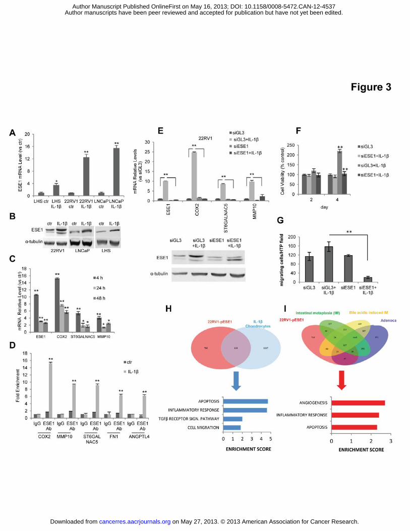
IL-1�. Treatment with IL-1� increased ESE1/ELF3 mRNA and protein level in all three cell
lines (Figure 3A,B). Interestingly, in 22RV1 cells ESE1/ELF3 mRNA increased already after
4 h of incubation with IL-1� and remained elevated compared to unstimulated cells after 24
and 48 h (Figure 3C). Concomitant with the induction of ESE1/ELF3, expression of COX2,
MMP10 and ST6GALNAC6 was also increased (Figure 3C). Consistently, IL-1� treatment of
22RV1 cells induced binding of ESE1/ELF3 to the promoters of these genes (Figure 3D). To
fully assess the contribution of ESE1/ELF3 to the response to IL-1�, we knocked down
ESE1/ELF3 prior to IL-1� induction . The level of ESE1/ELF3 was monitored at the mRNA
and protein level (Figure 3E, upper and lower panels). Notably, the transcriptional induction
of selected target genes in response to IL-1� was prevented by ESE1/ELF3 knockdown in
22RV1 (Figure 3E). Additionally, incubation with IL-1� enhanced migration and anoikis
resistance of 22RV1prostate cancer cells and knockdown of ESE1/ELF3 reduced these effect
of IL-1� (Figure 3F,G). Together, these results demonstrated that the transcriptional and
phenotypic response of prostate cancer cells to IL-1� depended on ESE1/ELF3 and mimicked
the effects of ESE1/ELF3 overexpression.
Consistent with our findings in prostate epithelial cells, we found, by analyzing the gene
expression data of chondrocytes stimulated with IL-1� [34], that ESE1/ELF3 was one of the
top genes induced by IL-1� in these cells (p-val<0.001). Similarly, we found that ESE1/ELF3
was among the genes significantly upregulated (p-val<0.01) in a IL-1� transgenic mouse
model of Barrett’s esophagus and esophageal carcinoma [35]. To assess the contribution of
ESE1/ELF3 to the IL-1� transcriptional signature in these experimental systems we looked at
the overlap with the ESE1/ELF3 gene signature in 22RV1-pESE1 cells. We observed a
on May 27, 2013. © 2013 American Association for Cancer Research. cancerres.aacrjournals.org Downloaded from
Author manuscripts have been peer reviewed and accepted for publication but have not yet been edited. Author Manuscript Published OnlineFirst on May 16, 2013; DOI: 10.1158/0008-5472.CAN-12-4537

10
significant overlap between genes induced in 22RV1-pESE1 cells and in IL -1� stimulated
chondrocytes (p-val=6.414e-08; OR, 1.8) (Figure 3H). More relevant to the cancer context,
there was significant convergence between the transcriptional signature in 22RV1-pESE1
cells and genes induced in preneoplastic and neoplastic esophageal lesions in the IL-1�
transgenic mice (p-val<0.0001) (Figure 3I and Figure S6). In all these experimental models,
the shared features were associated with relevant oncogenic pathways, particularly those
associated with activation of inflammatory trascriptome like apoptosis, migration,
angiogenesis and stress response. Furthermore, ChEA indicated that more than 40% of the
shared genes showed significant occupancy by ETS factors (p-val<0.05) and therefore could
be direct targets of ESE1/ELF3. Thus, ESE1/ELF3 is induced in several models of
inflammation and cancer and could contribute to the activation of inflammatory and
oncogenic pathways in many pre-neoplastic and neoplastic conditions.
ESE1/ELF3 is required for NF-κB activation in prostate cancer cells and tumors
IL-1� induces transcription by activating NF-κB [14, 16, 36]. NF-κB consists of five REL-
related proteins and the prototypical NF-κB complex is a heterodimer of p65/RELA and
p50/NFKB1 [37, 38]. Pro-inflammatory cytokines, like IL-1�, induce nuclear translocation of
the p65 and p50 and transcriptional activation of multiple target genes [38]. The ESE1/ELF3
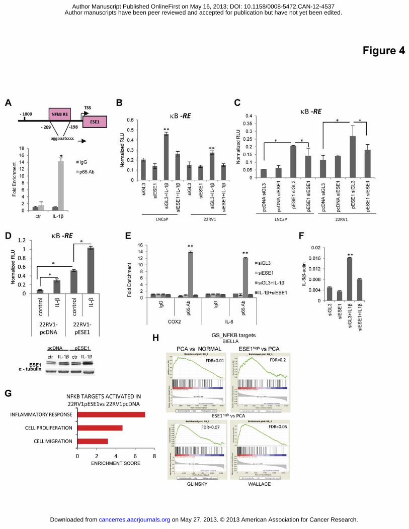
promoter contains NF-κB binding sites [33]. Consistently, we found that IL-1� induced
binding of p65 to the ESE1/ELF3 promoter, indicating that its activation occurred through
NF-κB (Figure 4A). On the other hand, the significant reversion of the transcriptional and
phenotypic effects of IL-1� by ESE1/ELF3 knockdown led us to hypothesize an active role
of ESE1/ELF3 in the transcriptional response to IL-1� and activation of NF-κB. Consistent
with this hypothesis, we found that the activity of a NF-κB responsive reporter was increased
by IL-1� and was reduced after knockdown of ESE1/ELF3 in IL-1� treated 22RV1 and
LNCaP cells (Figure 4B). Activity of the NF-κB reporter was higher also in stable
ESE1/ELF3 overexpressing cells than in control cells, indicating that ESE1/ELF3 contributed
to NF-κB activity also independently of exogenous IL-1� (Figure 4C). Consistently,
ESE1/ELF3 knockdown reduced NF-κB reporter activity in ESE1/ELF3 overexpressing
LNCaP and 22RV1 cells. Interestingly, treatment with IL-1� further increased NF-κB
reporter activity in ESE1/ELF3 overexpressing cells compared to IL-1� and ESE1/ELF3
overexpression alone, suggesting that ESE1/ELF3 led to increased responsiveness to IL-1�
along with sustained activation of NF-κB (Figure 4D). This was associated with further
increase in ESE1/ELF3 protein levels as indicated by western blotting, consistent with the
on May 27, 2013. © 2013 American Association for Cancer Research. cancerres.aacrjournals.org Downloaded from
Author manuscripts have been peer reviewed and accepted for publication but have not yet been edited. Author Manuscript Published OnlineFirst on May 16, 2013; DOI: 10.1158/0008-5472.CAN-12-4537
11
induction of a positive feedback loop by IL-1� also in ESE1/ELF3 overexpressing cells. Next,
to examine directly the contribution of ESE1/ELF3 to NF-κB transcriptional activity we
assessed the binding of p65 to COX2 and IL-6 promoters, two known NF-κB targets [29, 33]
[39] . We determined that expression of COX2 increased both in IL-1� treated cells and in
ESE1/ELF3 overexpressing cells. We found that IL-1� induced and knockdown of
ESE1/ELF3 prevented binding of p65 to the COX2 promoter (Figure 4E). Similar to COX2,
IL-6 promoter occupancy by p65 (Figure 4E) and IL-6 mRNA (Figure 4F) were induced by
IL-1� and both effects were blocked by ESE1/ELF3 knockdown. These data established for
the first time the notion that ESE1/ELF3 actively contributes to NF-κB activation by
enhancing binding of NF-κB to target gene promoters.
Bioinformatic analyses further supported a direct contribution of ESE1/ELF3 in the activation
of NF-κB target genes. TF interactome analysis with MetaCore showed that genes induced by
ESE1/ELF3 in 22RV1 cells were significantly enriched for targets of p65 (p-val= 4.4990E-
06) and p50 (p-val=0.007). Notably, NF-κB target genes induced by ESE1/ELF3 were
preferentially related to cell proliferation, migration and inflammation (Figure 4G). A
significant enrichment of p65 targets was also found among the genes induced in ESE1/ELF3
overexpressing 22RV1cells and shared with IL-1� stimulated chondrocytes (p-val=1.9690E-
11), and preneoplastic and neoplastic esophageal lesions in IL-1� transgenic mice (p-
val=3.0E-05). Ariadne Pathway analysis revealed the existence of reciprocal regulatory loops
between ESE1/ELF3 and NF-κB regulated genes (Figure S7). Furthermore, GSEA in prostate
cancer microarray datasets revealed significant enrichment of NF-κB target genes in prostate
tumors compared to normal prostate and prevalent enrichment in particular in tumors with
high ESE1/ELF3 expression (ESE1high tumors) compared to all other tumors (Figure 4H).
Thus, the transcriptional program orchestrated by ESE1/ELF3 both in prostate cell lines and
tumors involved numerous NF-κB regulated genes. Induction of these common targets
contribute to the activation of relevant oncogenic pathways as indicated by functional
annotation analysis.
ESE1/ELF3 and NF-κB constitute a positive feedback loop leading to constitutive NF-κB
activation
To further define the contribution of ESE1/ELF3 to NF-�B activation, we examined the
expression and intracellular localization of p50 and p65 in both stably overexpressing
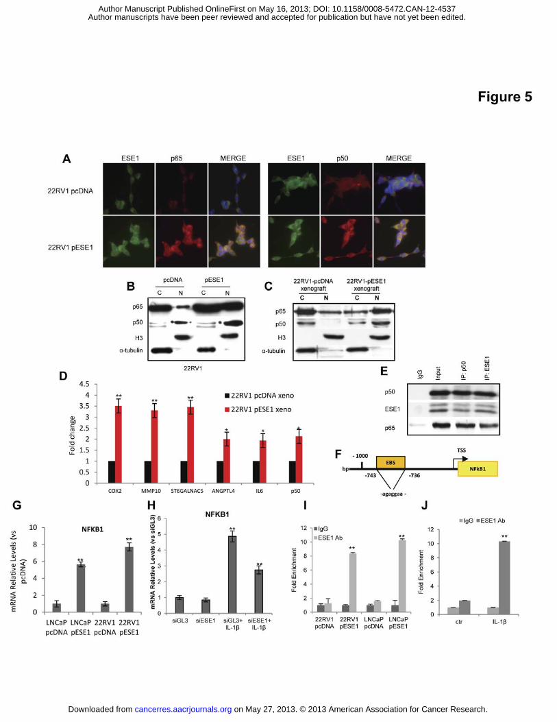
ESE1/ELF3 and IL-1� stimulated 22RV1cells. Both the level and nuclear localization of p50
and p65 were increased in ESE1/ELF3 overexpressing 22RV1 cells as revealed by
on May 27, 2013. © 2013 American Association for Cancer Research. cancerres.aacrjournals.org Downloaded from
Author manuscripts have been peer reviewed and accepted for publication but have not yet been edited. Author Manuscript Published OnlineFirst on May 16, 2013; DOI: 10.1158/0008-5472.CAN-12-4537
12
immunofluorescence (Figure 5A) and western blot (Figure 5B). Nuclear p50 and p65
increased about 2 and >6-fold, respectively, as determined by densitometric analysis of the
immunoblots. After IL-1� treatment of 22RV1 cells, we observed a similar increase of total
and nuclear level of p50 and p65 along with ESE1/ELF3 (Figure S8). Notably, knockdown of
ESE1/ELF3 reduced cytoplasmic and nuclear p50 and p65 in IL-1� treated cells (Figure S8),
indicating that ESE1/ELF3 was required for NF-κB accumulation and nuclear translocation
following IL-1� stimulation. To show that ESE1/ELF3 sustained NF-κB activation also in
vivo, we evaluated the level of p50 and p65 in tumor xenografts produced by 22RV1-pESE1
and 22RV1-pcDNA cells. There was a significant increase (> 5-fold by densitometric
analysis) of nuclear p50 and p65 in the 22RV1-pESE1 tumor xenografts compared to control
tumors (Figure 5C). Furthermore, several ESE1/ELF3 and NF-κB target genes were
overexpressed in 22RV1-pESE1tumor xenografts (Figure 5D).
Interestingly, we observed that both in stable overexpressing cell lines and in IL-1� treated
cells p50 and p65 largely co-localized with ESE1/ELF3 (Figure 5A and Figure S8).
Therefore, we tested whether ESE1/ELF3 interacted directly with p50 and p65.
Immunoprecipitation with an antibody directed to ESE1/ELF3 co-immunoprecipitated p50
and p65 in 22RV1-pESE1 cells (Figure 5E). In addition, ESE1/ELF3 was
immunoprecipitated, along with p65, by an anti-p50 antibody, confirming the physical
interaction between ESE1/ELF3 and the NF-κB subunits. Through these protein-protein
interactions, ESE1/ELF3 could affect stability, nuclear localization and promoter recruitment
of the NF-κB subunits. In addition, we hypothesized that ESE1/ELF3 could control NF-κB at
the transcriptional level. The NFKB1 gene promoter contains ETS binding sites [40],
suggesting the possibility that ESE1/ELF3 could control p50 transcription. We found higher
expression of p50 in ESE1/ELF3 overexpressing cells (Figure 5G). Furthermore, p50
increased after stimulation of 22RV1 cells with IL-1� and its induction was reduced by
ESE1/ELF3 knockdown (Figure 5H). The level of p50 was also significantly increased in the
22RV1-pESE1 compared to the 22RV1-pcDNA tumor xenografts (Figure 5D). Consistently,
ChIP demonstrated binding of ESE1/ELF3 to the NFKB1 promoter in the region containing
the predicted ETS binding site in ESE1/ELF3 overexpressing cells (Figure 5I) and in IL-1�
treated 22RV1 cells (Figure 5J). Therefore, the ability of ESE1/ELF3 to interact with the NF-
κB pathway at multiple levels results in a positive feedback loop leading to sustained
activation of NF-κB and induction of multiple oncogenic targets in prostate cancer cells.
ESE1/ELF3 sustains NF-κB activation in metastatic prostate cancer cells
on May 27, 2013. © 2013 American Association for Cancer Research. cancerres.aacrjournals.org Downloaded from
Author manuscripts have been peer reviewed and accepted for publication but have not yet been edited. Author Manuscript Published OnlineFirst on May 16, 2013; DOI: 10.1158/0008-5472.CAN-12-4537
13
Among the prostate cancer cell lines tested, we noticed that DU145 cells expressed a high
level of ESE1/ELF3, comparable to high ESE1/ELF3 expressing human tumors (Figure S2D).
DU145 cells are a metastatic prostate cancer cell line and several studies have shown
previously that NF-κB is active in these cells [41, 42]. However, the role of ESE1/ELF3 in
sustaining cell transformation and NF-κB activation in these cells in DU145 is unknown.
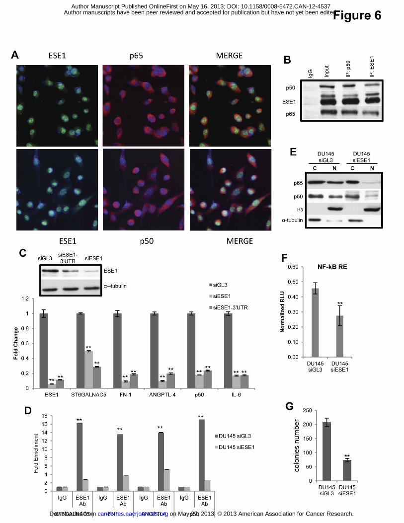
Immunofluorescence revealed that ESE1/ELF3 was highly expressed in DU145 prevalently in
the nuclear compartment but also in the cytoplasm and that it co-localized with both p50 and
p65 NF-κB subunits (Figure 6A). The physical interaction between ESE1/ELF3 and the NF-
κB subunits was also demonstrated by co-immunoprecipitation (Figure 6B). To further
understand the functional role of ESE1/ELF3 in DU145 cells, we knocked down expression
of the gene using two siRNA targeting different regions of the gene. Effective knockdown of
ESE1/ELF3 was assessed at the mRNA and protein level (Figure 6C, lower and upper left).
Concomitantly, expression of several NF-κB and ESE1/ELF3 target genes was significantly
reduced using both of the ESE1/ELF3 siRNAs (p<0.01) (Figure 6C). Relevantly, chromatin
immunoprecipitation confirmed that ESE1/ELF3 occupied the promoter of the selected target
genes and ESE1/ELF3 knockdown significantly reduced the promoter occupancy (Figure
6D). Notably, ESE1/ELF3 knockdown reduced the intranuclear levels of p65 and p50,
indicating that ESE1/ELF3 facilitated nuclear accumulation of active NF-κB complexes in
these cells (Figure 6E). Consistently, we found that NF-κB reporter activity was high in
DU145 cells and was significantly reduced by knocking down ESE1/ELF3 expression (Figure
6F). Furthermore, we found that ESE1/ELF3 knockdown significantly reduced the ability to
form anchorage-independent colonies, suggesting that it contributed to the transformed
phenotype of DU145 cells (Figure 6G). Collectively, these data point to a role of ESE1/ELF3
in sustaining constitutive activation of NF-κB independent of IL-1� stimulation in this
metastatic prostate cancer cell line.
ESE1/ELF3 and NF-κB activation are associated with poor prognosis
To determine the clinical relevance of these findings, we assessed concomitantly the protein
expression of p50, p65 and ESE1/ELF3 in TMAs of prostate cancer patients for which we
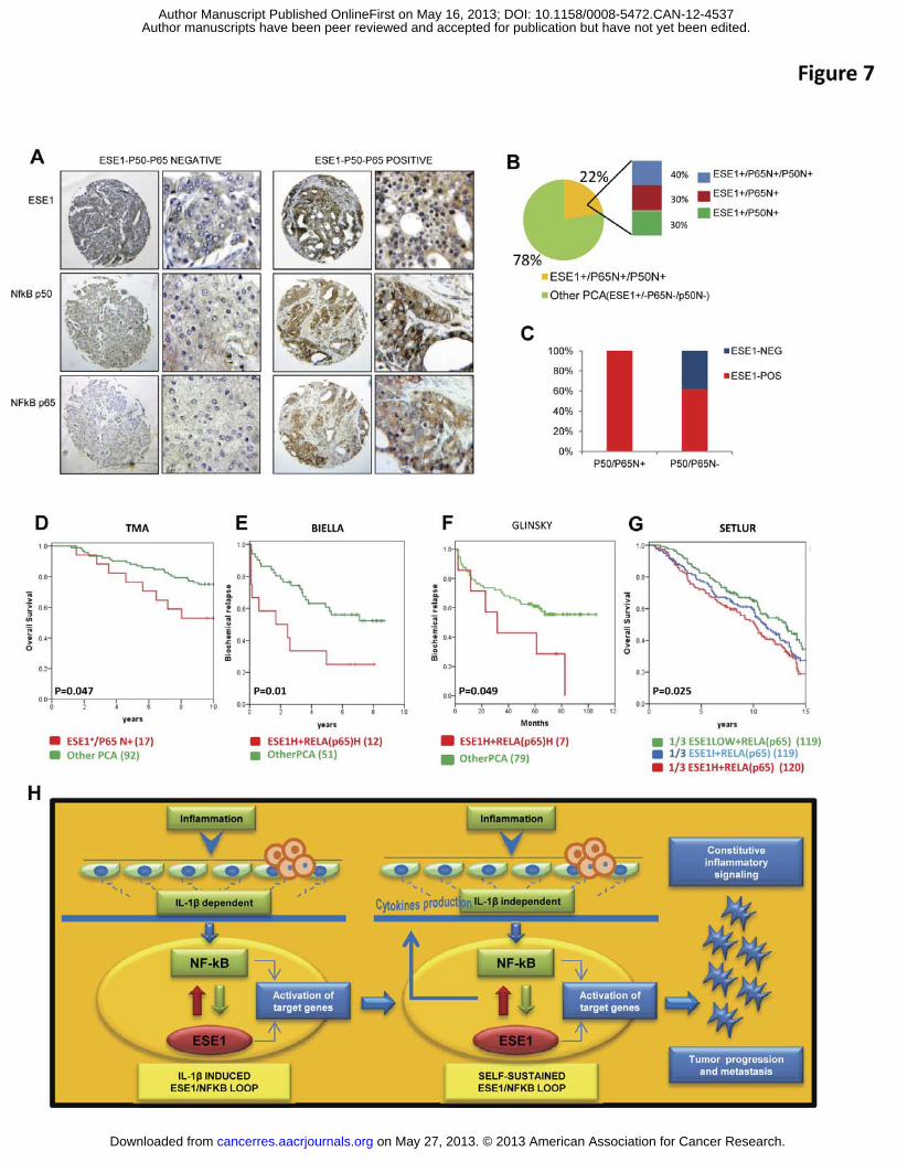
had long term clinical follow-up data [22] (Figure 7A). We found a significant association
between overexpression of ESE1/ELF3 and nuclear p50 and p65 (p-val=0.0005; OR, 14.4).
Specifically, 22% of prostate tumors exhibited strong nuclear staining for p50 and p65, with
about 40% of those being positive for both (Figure 7B). Nuclear p50 and p65 positivity was
exclusively associated with ESE1/ELF3 positive tumors of ESE1/ELF3, although not all of
on May 27, 2013. © 2013 American Association for Cancer Research. cancerres.aacrjournals.org Downloaded from
Author manuscripts have been peer reviewed and accepted for publication but have not yet been edited. Author Manuscript Published OnlineFirst on May 16, 2013; DOI: 10.1158/0008-5472.CAN-12-4537
14
the ESE1/ELF3 positive tumors showed nuclear p50 and p65 staining (Figure 7C). Thus,
concomitant expression of ESE1/ELF3 and nuclear p50 and p65 positivity were present in a
subset of prostate tumors.
Based on our biological and genomic data, we hypothesized that these features could mark
particularly clinically aggressive tumors. Consistent with this hypothesis, we found that high
expression of ESE1/ELF3 and nuclear p65 positivity [43-45] were significantly associated
with shorter survival of patients after prostatectomy (p-val=0.047; Figure 7D). Furthermore,
high ESE1/ELF3 and p65/RELA mRNA expression in patient cohorts examined by
microarrays [5] was associated with increased biochemical relapses after prostatectomy (p-
val=0.01; Figure 7E-F). A similar trend was observed when we considered the protein level of
ESE1/ELF3 and nuclear positivity for both p50 and p65 determined by IHC in TMAs (p-
val=0.06; Figure S9A) and high mRNA level of ESE1/ELF3, p65/RELA and p50/NFKB1 in
microarray data (p-val=0.038; FigureS9B). Relevantly, we found that the combined
upregulation of ESE1/ELF3 and p65/RELA mRNA was also significantly associated with
reduced overall survival after prostatectomy (p-val=0.025) in an independent gene expression
study of prostate cancer patients with 15-year clinical follow-up (Figure 7G) [46]. Together,
these findings demonstrated the prognostic value of combined ESE1/ELF3 upregulation and
NF-κB activation in prostate tumors and further reinforced the notion of their relevance for
prostate cancer progression.
DISCUSSION
This study establishes for the first time that the ETS factor ESE1/ELF3 has an oncogenic
activity and a crucial role in constitutive and cytokine-induced activation of NF-κB in
prostate tumors. Here, we report a novel mechanism leading to activation of a oncogenic ETS
TF, independent of chromosomal translocation, and linking inflammation, NF-κB activation
and prostate cancer progression. We show that ESE1/ELF3 is a key element in a positive
feedback loop involving the proinflammatory cytokine IL-1� and the NF-κB subunits p50 and
p65, and that ESE1/ELF3 expression is instrumental for the proinflammatory and
protumorigenic functions of this pathway. Chronic inflammation is an important risk factor
for prostate cancer and involves the production of multiple cytokines in response to several
inflammatory stimuli [13, 19]. IL-1� is one of the major cytokines implicated in inflammation
in the prostate [15]. NF-κB has been reported to contribute to increased proliferation,
survival, angiogenesis and metastatic progression in prostate cancer and activation of the NF-
on May 27, 2013. © 2013 American Association for Cancer Research. cancerres.aacrjournals.org Downloaded from
Author manuscripts have been peer reviewed and accepted for publication but have not yet been edited. Author Manuscript Published OnlineFirst on May 16, 2013; DOI: 10.1158/0008-5472.CAN-12-4537
15
κB pathway is associated with aggressive clinical behavior [44, 45]. We found that
ESE1/ELF3 is frequently overexpressed in human primary and metastatic prostate cancers.
ESE1/ELF3 is also amplified in a small but relevant number of cases. Consistent with an
oncogenic role, we show that ESE1/ELF3 controls a network of genes involved in cell
invasion, migration, inflammation and metastasis and its overexpression enhances the
transformed properties of prostate cancer cells and promotes tumor growth and metastasis in
mouse xenografts. We found that IL-1� induces ESE1/ELF3 in prostate epithelial cells
through activation of NF-κB and binding of p65 to the ESE1/ELF3 promoter. In turn,
ESE1/ELF3 contributes to the activation of NF-κB by transcriptional regulation of p50 and
post-transcriptional control of both p50 and p65 function. We show that ESE1/ELF3 interacts
with both p50 and p65 proteins and enhances their nuclear translocation and binding to target
gene promoters. Relevantly, we found that ESE1/ELF3 contributed to NF-κB activation also
in the absence of cytokine stimulation and that this effect was maintained in vivo in tumor
xenografts of ESE1/ELF3 overexpressing cells. ESE1/ELF3 sustained constitutive NF-κB
activation also in metastatic prostate cancer DU145 cells expressing endogenously high levels
of ESE1/ELF3. This suggests that, once a significant level of induction of ESE1/ELF3 is
reached, activation of the pathway could be self-sustained in the absence of external
inflammatory stimuli. In addition, production of cytokines like IL-6 by prostate cancer cells in
response to ESE1/ELF3 and NF-κB activation could contribute in an autocrine (cell-
autonomous) way to the positive feedback loop and inflammatory signaling. Based on these
multiple lines of evidence, we propose that the reciprocal interactions between ESE1/ELF3
and NF-κB result in sustained activation of NF-κB, greater responsiveness to pro-
inflammatory stimuli and activation of combined ESE1/ELF3 and NF-κB target genes that
accelerate prostate cancer progression (Figure 7H). Consistently, we found that the level of
ESE1/ELF3 was significantly higher in metastatic compared to primary prostate tumors
suggesting that the gene plays a role in tumor progression.
Bioinformatics analyses and functional studies further support the link between ESE1/ELF3,
IL-1� and NF-κB and their involvement in tumor progression. Notably, we found a
significant convergence between the transcriptional program observed in ESE1/ELF3
overexpressing prostate cancer cells and IL-1� induced transcriptional signatures in
experimental models of inflammatory, preneoplastic and neoplastic diseases. Intriguingly, this
convergence was observed in IL-1� transgenic mouse model of Barrett’s esophagus [35], an
established preneoplastic condition functionally related to chronic inflammation and IL-1�,
suggesting that the ESE1/ELF3-NF-κB axis could be relevant also in other types of cancers.
on May 27, 2013. © 2013 American Association for Cancer Research. cancerres.aacrjournals.org Downloaded from
Author manuscripts have been peer reviewed and accepted for publication but have not yet been edited. Author Manuscript Published OnlineFirst on May 16, 2013; DOI: 10.1158/0008-5472.CAN-12-4537
16
Moreover, both in human cell lines and prostate tumors we observed a convergence of
ESE1/ELF3 and NF-κB regulated genes. This finding was also supported by the enrichment
of NF-κB target genes by GSEA in human prostate tumors with high expression of
ESE1/ELF3. Moreover, analysis of large sets of clinical samples provided evidence that this
positive feedback loop operates in a subset of prostate cancers and could drive disease
recurrence and progression to metastatic lethal disease. About 25% of primary tumors showed
increased expression of ESE1/ELF3 and nuclear p65 by IHC. Notably, high levels of
ESE1/ELF3 and nuclear p65 positivity were associated with shorter overall survival after
prostatectomy. Similarly, high levels of ESE1/ELF3 and p65 mRNA, with and without p50,
in microarray datasets were associated with increased biochemical relapse and shorter overall
survival. These findings call for assessment of ESE1/ELF3 and p65/RELA as potential
prognostic biomarkers in prostate cancer. Furthermore, their evaluation in clinical samples
could guide the implementation of targeted treatment strategies for prostate cancer patients. In
addition to uncovering a mechanistic link between ESE1/ELF3 and NF-κB in prostate
tumorigenesis, this study opens avenues for patient risk stratification and indicates a rationale
for context-dependent therapeutic approaches in specific subsets of prostate cancer patients.
The role of ESE1/ELF3 and its association with NF-κB activation in patients with clinically
localized but aggressive and high risk prostate tumors point to the possibility that targeting the
NF-κB pathway with inhibitors that are currently in preclinical and clinical development [47]
could be a valid therapeutic strategy.
Acknowledgements
This work was supported by grants from Oncosuisse (KFS-01913-08 and KFS-02573-02-
2010), Swiss National Science Foundation (FNS-31003A-118113), Ticino Foundation for
Cancer Research, Fondazione Virginia Boeger and Fondazione Fidinam to GMC and CVC.
MMG and GC were supported by Compagnia di San Paolo, Torino, Italy. We wish to thank
Dr. Towia Libermann for the gift of the pESE1/ELF3 expressing vector.
References
1. Jemal, A., et al., Global cancer statistics. CA Cancer J Clin, 2011. 61(2): p. 69-90. 2. Rubin, M.A., C.A. Maher, and A.M. Chinnaiyan, Common gene rearrangements in
prostate cancer. J Clin Oncol, 2011. 29(27): p. 3659-68. 3. Clark, J.P. and C.S. Cooper, ETS gene fusions in prostate cancer. Nat Rev Urol, 2009.
6(8): p. 429-39. 4. Tomlins, S.A., et al., Recurrent fusion of TMPRSS2 and ETS transcription factor
genes in prostate cancer. Science, 2005. 310(5748): p. 644-8.
on May 27, 2013. © 2013 American Association for Cancer Research. cancerres.aacrjournals.org Downloaded from
Author manuscripts have been peer reviewed and accepted for publication but have not yet been edited. Author Manuscript Published OnlineFirst on May 16, 2013; DOI: 10.1158/0008-5472.CAN-12-4537
17
5. Kunderfranco, P., et al., ETS transcription factors control transcription of EZH2 and epigenetic silencing of the tumor suppressor gene Nkx3.1 in prostate cancer. PLoS One, 2010. 5(5): p. e10547.
6. Cangemi, R., et al., Reduced expression and tumor suppressor function of the ETS transcription factor ESE-3 in prostate cancer. Oncogene, 2008. 27(20): p. 2877-85.
7. Albino, D., et al., ESE3/EHF controls epithelial cell differentiation and its loss leads to prostate tumors with mesenchymal and stem-like features. Cancer Res, 2012. 72(11): p. 2889-900.
8. Vitari, A.C., et al., COP1 is a tumour suppressor that causes degradation of ETS transcription factors. Nature, 2011. 474(7351): p. 403-6.
9. Chi, P., et al., ETV1 is a lineage survival factor that cooperates with KIT in gastrointestinal stromal tumours. Nature, 2010. 467(7317): p. 849-53.
10. Oettgen, P., et al., Isolation and characterization of a novel epithelium-specific transcription factor, ESE-1, a member of the ets family. Mol Cell Biol, 1997. 17(8): p. 4419-33.
11. Oliver, J.R., R. Kushwah, and J. Hu, Multiple roles of the epithelium-specific ETS transcription factor, ESE-1, in development and disease. Lab Invest, 2012. 92(3): p. 320-30.
12. Seth, A. and D.K. Watson, ETS transcription factors and their emerging roles in human cancer. Eur J Cancer, 2005. 41(16): p. 2462-78.
13. De Marzo, A.M., et al., Inflammation in prostate carcinogenesis. Nat Rev Cancer, 2007. 7(4): p. 256-69.
14. Perkins, N.D., The diverse and complex roles of NF-kappaB subunits in cancer. Nat Rev Cancer, 2012. 12(2): p. 121-32.
15. Jerde, T.J. and W. Bushman, IL-1 induces IGF-dependent epithelial proliferation in prostate development and reactive hyperplasia. Sci Signal, 2009. 2(86): p. ra49.
16. Sims, J.E. and D.E. Smith, The IL-1 family: regulators of immunity. Nat Rev Immunol, 2010. 10(2): p. 89-102.
17. Karin, M., Nuclear factor-kappaB in cancer development and progression. Nature, 2006. 441(7092): p. 431-6.
18. Ben-Neriah, Y. and M. Karin, Inflammation meets cancer, with NF-kappaB as the matchmaker. Nat Immunol, 2011. 12(8): p. 715-23.
19. Zitvogel, L., et al., Inflammasomes in carcinogenesis and anticancer immune responses. Nat Immunol, 2012. 13(4): p. 343-51.
20. Longoni, N., et al., Aberrant expression of the neuronal-specific protein DCDC2 promotes malignant phenotypes and is associated with prostate cancer progression. Oncogene, 2012.
21. Malek, A., et al., Modulation of the activity of Sp transcription factors by mithramycin analogues as a new strategy for treatment of metastatic prostate cancer. PLoS One, 2012. 7(4): p. e35130.
22. Prtilo, A., et al., Tissue microarray analysis of hMSH2 expression predicts outcome in men with prostate cancer. J Urol, 2005. 174(5): p. 1814-8; discussion 1818.
23. Glinsky, G.V., et al., Gene expression profiling predicts clinical outcome of prostate cancer. J Clin Invest, 2004. 113(6): p. 913-23.
24. Wallace, T.A., et al., Tumor immunobiological differences in prostate cancer between African-American and European-American men. Cancer Res, 2008. 68(3): p. 927-36.
25. Tomlins, S.A., et al., Distinct classes of chromosomal rearrangements create oncogenic ETS gene fusions in prostate cancer. Nature, 2007. 448(7153): p. 595-9.
26. Ateeq, B., et al., Therapeutic targeting of SPINK1-positive prostate cancer. Sci Transl Med, 2011. 3(72): p. 72ra17.
27. Nguyen, D.X., P.D. Bos, and J. Massague, Metastasis: from dissemination to organ-specific colonization. Nat Rev Cancer, 2009. 9(4): p. 274-84.
on May 27, 2013. © 2013 American Association for Cancer Research. cancerres.aacrjournals.org Downloaded from
Author manuscripts have been peer reviewed and accepted for publication but have not yet been edited. Author Manuscript Published OnlineFirst on May 16, 2013; DOI: 10.1158/0008-5472.CAN-12-4537
18
28. Grall, F., et al., Responses to the proinflammatory cytokines interleukin-1 and tumor necrosis factor alpha in cells derived from rheumatoid synovium and other joint tissues involve nuclear factor kappaB-mediated induction of the Ets transcription factor ESE-1. Arthritis Rheum, 2003. 48(5): p. 1249-60.
29. Grall, F.T., et al., The Ets transcription factor ESE-1 mediates induction of the COX-2 gene by LPS in monocytes. FEBS J, 2005. 272(7): p. 1676-87.
30. Heo, S.H., et al., Expression profiling of ETS and MMP factors in VEGF-activated endothelial cells: role of MMP-10 in VEGF-induced angiogenesis. J Cell Physiol, 2010. 224(3): p. 734-42.
31. Tu, S., et al., Overexpression of interleukin-1beta induces gastric inflammation and cancer and mobilizes myeloid-derived suppressor cells in mice. Cancer Cell, 2008. 14(5): p. 408-19.
32. Brown, C., et al., ESE-1 is a novel transcriptional mediator of angiopoietin-1 expression in the setting of inflammation. J Biol Chem, 2004. 279(13): p. 12794-803.
33. Rudders, S., et al., ESE-1 is a novel transcriptional mediator of inflammation that interacts with NF-kappa B to regulate the inducible nitric-oxide synthase gene. J Biol Chem, 2001. 276(5): p. 3302-9.
34. Sandell, L.J., et al., Exuberant expression of chemokine genes by adult human articular chondrocytes in response to IL-1beta. Osteoarthritis Cartilage, 2008. 16(12): p. 1560-71.
35. Quante, M., et al., Bile acid and inflammation activate gastric cardia stem cells in a mouse model of Barrett-like metaplasia. Cancer Cell, 2012. 21(1): p. 36-51.
36. Chen, L.F. and W.C. Greene, Shaping the nuclear action of NF-kappaB. Nat Rev Mol Cell Biol, 2004. 5(5): p. 392-401.
37. Karin, M., et al., NF-kappaB in cancer: from innocent bystander to major culprit. Nat Rev Cancer, 2002. 2(4): p. 301-10.
38. Karin, M. and F.R. Greten, NF-kappaB: linking inflammation and immunity to cancer development and progression. Nat Rev Immunol, 2005. 5(10): p. 749-59.
39. Libermann, T.A. and D. Baltimore, Activation of interleukin-6 gene expression through the NF-kappa B transcription factor. Mol Cell Biol, 1990. 10(5): p. 2327-34.
40. Lambert, P.F., et al., The nfkb1 promoter is controlled by proteins of the Ets family. Mol Biol Cell, 1997. 8(2): p. 313-23.
41. Palayoor, S.T., et al., Constitutive activation of IkappaB kinase alpha and NF-kappaB in prostate cancer cells is inhibited by ibuprofen. Oncogene, 1999. 18(51): p. 7389-94.
42. Gasparian, A.V., et al., The role of IKK in constitutive activation of NF-kappaB transcription factor in prostate carcinoma cells. J Cell Sci, 2002. 115(Pt 1): p. 141-51.
43. Lessard, L., et al., Nuclear localisation of nuclear factor-kappaB transcription factors in prostate cancer: an immunohistochemical study. Br J Cancer, 2005. 93(9): p. 1019-23.
44. Shukla, S., et al., Nuclear factor-kappaB/p65 (Rel A) is constitutively activated in human prostate adenocarcinoma and correlates with disease progression. Neoplasia, 2004. 6(4): p. 390-400.
45. Fradet, V., et al., Nuclear factor-kappaB nuclear localization is predictive of biochemical recurrence in patients with positive margin prostate cancer. Clin Cancer Res, 2004. 10(24): p. 8460-4.
46. Setlur, S.R., et al., Estrogen-dependent signaling in a molecularly distinct subclass of aggressive prostate cancer. J Natl Cancer Inst, 2008. 100(11): p. 815-25.
47. Nakanishi, C. and M. Toi, Nuclear factor-kappaB inhibitors as sensitizers to anticancer drugs. Nat Rev Cancer, 2005. 5(4): p. 297-309.
on May 27, 2013. © 2013 American Association for Cancer Research. cancerres.aacrjournals.org Downloaded from
Author manuscripts have been peer reviewed and accepted for publication but have not yet been edited. Author Manuscript Published OnlineFirst on May 16, 2013; DOI: 10.1158/0008-5472.CAN-12-4537
19
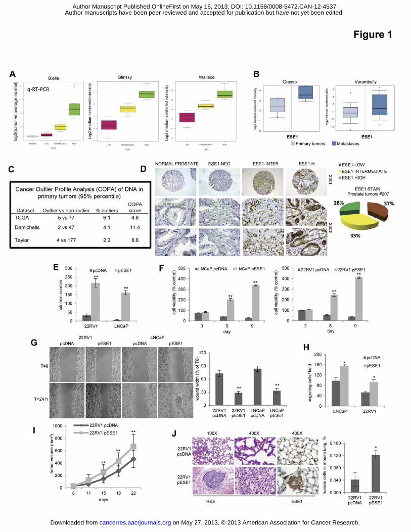
Figure legends Figure 1. ESE1/ELF3 is overexpressed in prostate cancers and promotes malignant
phenotypes. (A) Left, ESE1/ELF3 mRNA level in prostate tumors in the Biella patient cohort
determined by qRT-PCR. Middle and Right, level of ESE1/ELF3 in primary tumors in the
indicated datasets evaluated by microarrays.(B) Level of ESE1/ELF3 in primary tumors
versus metastases in the indicated datasets ( p<0.001). (C) ESE1/ELF3 amplification in
primary prostate tumors from three published datasets. (D) Immunohistochemical
determination of ESE1/ELF3 protein in normal prostate and prostate tumors. Left,
representative images; Right, distribution based on IHC score in prostate tumors. (E) Colony
formation in soft agar of control (pcDNA) and ESE1/ELF3 overexpressing (pESE1) 22RV1
and LNCaP cells. (F) Survival in anoikis of control (pcDNA) and ESE1/ELF3 overexpressing
(pESE1) 22RV1 (right) and LNCaP (left) cells. (G) Scratch-wound healing assay with control
(pcDNA) and ESE1/ELF3 overexpressing (pESE1) 22RV1 and LNCaP cells. Left,
representative images. Right, percentage of wound width relative to time 0. (H) Boyden
chamber assay with control (pcDNA) and ESE1/ELF3 overexpressing (pESE1) 22RV1 and
LNCaP cells. (I) Growth of subcutaneous xenografts (n=10/group) of 22RV1-pcDNA and
22RV1-pESE1 cells in nude mice. (J) Formation of lung metastasis upon tail vein injection
of 22RV1-pcDNA and 22RV1-pESE1 cells. Left, representative images of lung sections
stained with H&E and for ESE1/ELF3. Right, PCR quantification of human metastatic cells
in mouse lungs. P values were determined using t test. *, P < 0.01; **, P < 0.005. All data are
mean ± s.e.m.
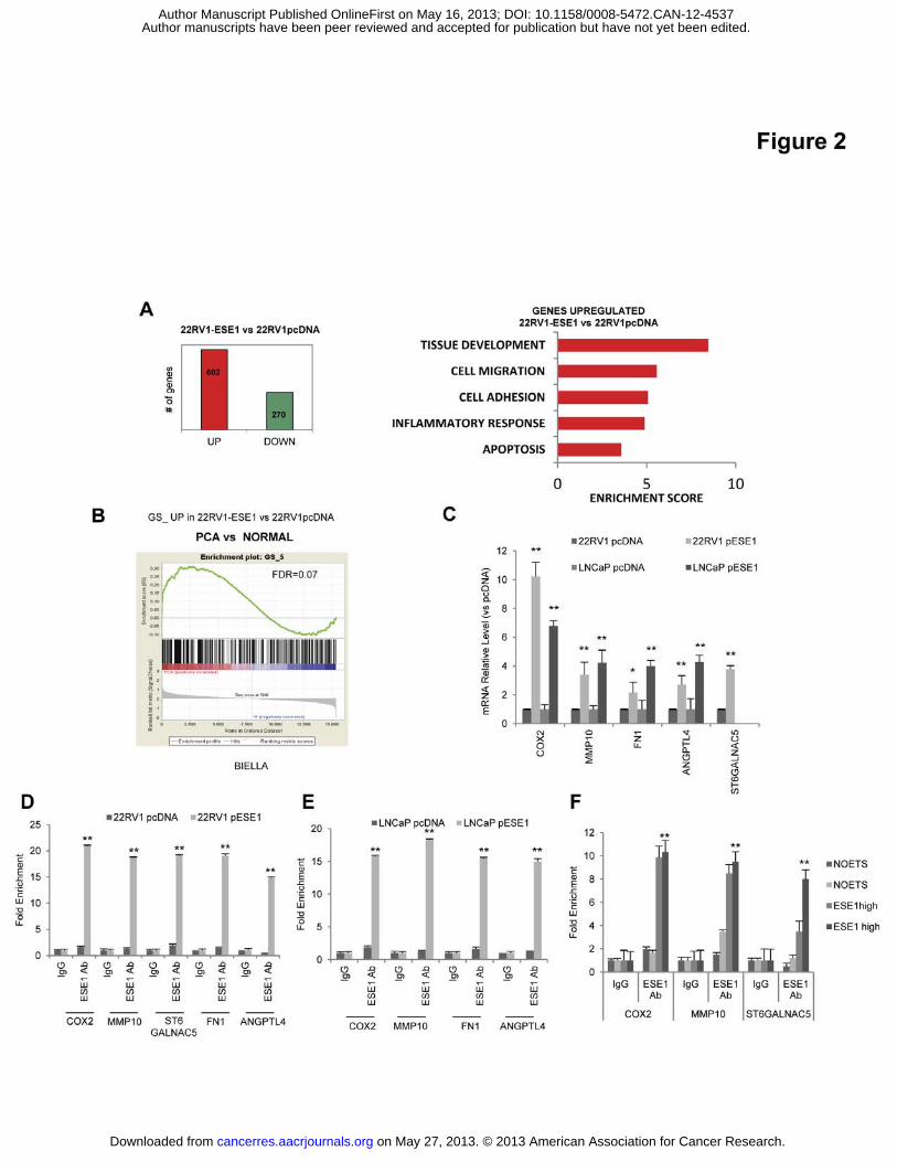
Figure 2. ESE1/ELF3 activates a transcriptional and functional program promoting
inflammation and metastatic spread. (A) Left, number of up and down-regulated genes in
22RV1-pESE1vs 22RV1-pcDNA cells determined by microarray analysis. Right, functional
annotation of the genes significantly upregulated (p<0.01) by DAVID. (B) GSEA using genes
upregulated in 22RV1-pESE1 comparing prostate tumors (PCa) with normal prostate in the
Biella dataset. (C) Expression of selected genes in control (pcDNA) and ESE1/ELF3
overexpressing (pESE1) 22RV1 and LNCaP cells by qRT-PCR. (D-E) Binding of
ESE1/ELF3 to the promoters of the indicated genes determined by chromatin
immunoprecipitation and qRT-PCR in control (pcDNA) and ESE1/ELF3 overexpressing
(pESE1) 22RV1 and LNCaP cells. (F) Binding of ESE1/ELF3 to the indicated gene
promoters in prostate tumors with high (ESE1high) or low (NOETS) expression of
ESE1/ELF3.
on May 27, 2013. © 2013 American Association for Cancer Research. cancerres.aacrjournals.org Downloaded from
Author manuscripts have been peer reviewed and accepted for publication but have not yet been edited. Author Manuscript Published OnlineFirst on May 16, 2013; DOI: 10.1158/0008-5472.CAN-12-4537
20
Figure 3. ESE1/ELF3 is induced by IL-1� and mediates the transforming effects of IL-
1�. (A) Cells were exposed to IL-1� for 4 h and ESE1/ELF3 mRNA was evaluated by qRT-
PCR. (B) Cells were exposed to IL-1� as above and ESE1/ELF3 level determined by western
blot. (C) Expression of ESE1/ELF3 and selected target genes determined by qRT-PCR in
22RV1 cells incubated with IL-1� for 4 h and analyzed at the indicated time points. (D)
Binding of ESE1/ELF3 to the promoters of the indicated genes following 4 h treatment with
IL-1�. (E) Top, Expression of ESE1/ELF3 and the indicated target genes determined by qRT-
PCR in 22RV1cells transfected with control (siGL3) or ESE1/ELF3 targeting (siESE1)
siRNA and exposed to IL-1� for 4 h. Bottom, Protein level of ESE1/ELF3 evaluated by
western-blot in the indicated experimental conditions. (F) Survival in anoikis of 22RV1 cells
transfected with control (siGL3) or ESE1/ELF3 targeting (siESE1) siRNA and exposed to IL-
1� for 4 h. (G) Boyden chamber assay with 22RV1 cells transfected with control (siGL3) or
ESE1/ELF3 targeting (siESE1) siRNA and exposed for 4 h to IL-1�. (H) Venn diagram
showing the overlap between genes up-regulated in ESE1/ELF3 overexpressing 22RV1 cells
and genes induced by IL-1� in chondrocytes (Upper panel) and functional annotation of the
common genes (Lower panel). (I) Venn diagram showing the overlap between genes up-
regulated in ESE1/ELF3 overexpressing 22RV1 cells and prenoplastic (Intestinal metaplasia
and Bile acidic metaplasia) and esophageal adenocarcinoma) lesions in the IL-1� transgenic
mice (Upper) and functional annotation of the genes in common (Lower). P values were
determined using t test. *, P < 0.01; **, P < 0.005. All data are mean ± s.e.m.
Figure 4. ESE1/ELF3 promotes NF-κB activation. (A) Top, ESE1/ELF3 promoter region
and position of the NF-κB binding site (NF-κB RE). Bottom, binding of p65 to ESE1/ELF3
promoter after IL-1� treatment in 22RV1 cells. (B) NF-κB reporter activity following IL-1�
treatment and ESE1/ELF3 down-regulation in LNCaP and 22RV1 cells. (C) NF-κB reporter
activity in control (pcDNA) and ESE1/ELF3 over-expressing (pESE1) cells following
ESE1/ELF3 down-regulation. (D) NF-κB reporter activity in control (pcDNA) and
ESE1/ELF3 over-expressing (pESE1) 22RV1 cells after 4 h exposure to IL-1�. Lower panel,
level of ESE1/ELF3 assessed by western blot in 22RV1-pcDNA and 22RV1-pESE1
following IL-1� treatment. (E) p65 binding to the COX2 and IL-6 promoter in 22RV1 cells
transfected with control (siGL3) or ESE1/ELF3 targeting (siESE1) siRNA and exposed to IL-
1� for 4 h. (F) IL-6 mRNA determined by qRT-PCR in 22RV1 cells transfected with control
(siGL3) or ESE1/ELF3 targeting (siESE1) siRNA and exposed to IL-1� for 4 h. P values
on May 27, 2013. © 2013 American Association for Cancer Research. cancerres.aacrjournals.org Downloaded from
Author manuscripts have been peer reviewed and accepted for publication but have not yet been edited. Author Manuscript Published OnlineFirst on May 16, 2013; DOI: 10.1158/0008-5472.CAN-12-4537
21
were determined using t test. (G) Functional annotation analysis by DAVID of NF-κB targets
activated in 22RV1-pESE1 cells (H) GSEA using gene sets of NF-κB regulated genes
comparing prostate tumors with normal prostate samples in the Biella microarray dataset and
ESE1high with all the other tumours (ESE1high vs. PCa,) in the indicated microarray datasets *,
P < 0.01; **, P < 0.005. All data are mean ± s.e.m.
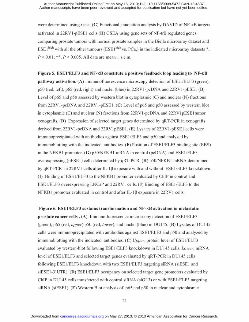
Figure 5. ESE1/ELF3 and NF-κB constitute a positive feedback loop leading to NF-κB
pathway activation. (A) Immunofluorescence microscopy detection of ESE1/ELF3 (green),
p50 (red, left), p65 (red, right) and nuclei (blue) in 22RV1-pcDNA and 22RV1-pESE1 (B)
Level of p65 and p50 assessed by western blot in cytoplasmic (C) and nuclear (N) fractions
from 22RV1-pcDNA and 22RV1-pESE1. (C) Level of p65 and p50 assessed by western blot
in cytoplasmic (C) and nuclear (N) fractions from 22RV1-pcDNA and 22RV1pESE1tumor
xenografts. (D) Expression of selected target genes determined by qRT-PCR in xenografts
derived from 22RV1-pcDNA and 22RV1pESE1. (E) Lysates of 22RV1-pESE1 cells were
immunoprecipitated with antibodies against ESE1/ELF3 and p50 and analyzed by
immunoblotting with the indicated antibodies. (F) Position of ESE1/ELF3 binding site (EBS)
in the NFKB1 promoter. (G) p50/NFKB1 mRNA in control (pcDNA) and ESE1/ELF3
overexpressing (pESE1) cells determined by qRT-PCR. (H) p50/NFKB1 mRNA determined
by qRT-PCR in 22RV1 cells after IL-1� exposure with and without ESE1/ELF3 knockdown.
(I) Binding of ESE1/ELF3 to the NFKB1 promoter evaluated by ChIP in control and
ESE1/ELF3 overexpressing LNCaP and 22RV1 cells. (J) Binding of ESE1/ELF3 to the
NFKB1 promoter evaluated in control and after IL-1� exposure in 22RV1 cells.
Figure 6. ESE1/ELF3 sustains transformation and NF-κB activation in metastatic
prostate cancer cells . (A) Immunofluorescence microscopy detection of ESE1/ELF3
(green), p65 (red, upper) p50 (red, lower), and nuclei (blue) in DU145. (B) Lysates of DU145
cells were immunoprecipitated with antibodies against ESE1/ELF3 and p50 and analyzed by
immunoblotting with the indicated antibodies. (C) Upper, protein level of ESE1/ELF3
evaluated by western-blot following ESE1/ELF3 knockdown in DU145 cells. Lower, mRNA
level of ESE1/ELF3 and selected target genes evaluated by qRT-PCR in DU145 cells
following ESE1/ELF3 knockdown with two ESE1/ELF3 targeting siRNA (siESE1 and
siESE1-3’UTR). (D) ESE1/ELF3 occupancy on selected target gene promoters evaluated by
ChIP in DU145 cells transfetcted with control siRNA (siGL3) or with ESE1/ELF3 targeting
siRNA (siESE1). (E) Western Blot analysis of p65 and p50 in nuclear and cytoplasmic
on May 27, 2013. © 2013 American Association for Cancer Research. cancerres.aacrjournals.org Downloaded from
Author manuscripts have been peer reviewed and accepted for publication but have not yet been edited. Author Manuscript Published OnlineFirst on May 16, 2013; DOI: 10.1158/0008-5472.CAN-12-4537
22
fractions following ESE1/ELF3 knockdown in DU145 cells. (F) NF-κB reporter activity
following ESE1/ELF3 knockdown in DU145 cells. (G) Colony formation in soft agar
following ESE1/ELF3 knockdown in DU145. P values were determined using t test. *, P <
0.01; All data are mean ± s.e.m.
Figure 7. Expression of ESE1/ELF3 and NF-κB activation are associated with poor
prognosis. (A) Representative images of immunohistochemical staining for ESE1/ELF3 and
NF-κB subunits p50 and p65 in prostate tumors. (B) Distribution of ESE1/ELF3, nuclear p65
and p50 staining in prostate tumors (n= 186). N+, positive nuclear stain. (C) Percentage of
ESE1/ELF3 positive and negative tumors according to nuclear p50 and p65 staining
evaluated by IHC as described above. (D) Kaplan-Meyer analysis of overall survival of the
patients cohort analyzed by TMA divided according to ELF3/ESE1 and nuclear p65 staining.
(E-F) Kaplan-Meyer analysis of biochemical relapse free survival of patients in the Biella and
Glinsky cohort analyzed by microarrays divided according to ELF3/ESE1 and p65/RELA
mRNA level. (G) Kaplan-Meyer analysis of overall survival of patients in the Setlur cohort
divided according to ESE1/ELF3 and p65/RELA mRNA level. p-values determined by Log
rank test (Mantel-Cox). Number of patients is indicated in parenthesis. (H) Proposed model
for the induction of ESE1/ELF3 by IL-1� and establishment of a positive feedback loop
leading to constitutive activation of NF-κB and inflammatory signaling in prostate tumors.
on May 27, 2013. © 2013 American Association for Cancer Research. cancerres.aacrjournals.org Downloaded from
Author manuscripts have been peer reviewed and accepted for publication but have not yet been edited. Author Manuscript Published OnlineFirst on May 16, 2013; DOI: 10.1158/0008-5472.CAN-12-4537