constitutively active ccr5 chemokine receptors differ in mediating hiv envelope-dependent fusion
TRANSCRIPT
Constitutively Active CCR5 Chemokine Receptors Differin Mediating HIV Envelope-dependent FusionAlex de Voux1¤a, Mei-Chi Chan1, Asongna T. Folefoc1¤b, Michael T. Madziva2, Colleen A. Flanagan2*
1Medical Research Council Receptor Biology Research Unit, Division of Medical Biochemistry, Faculty of Health Sciences, University of Cape Town, Cape Town, South
Africa, 2Medical Research Council Receptor Biology Research Unit, School of Physiology, University of the Witwatersrand, Johannesburg, South Africa
Abstract
The CCR5 chemokine receptor is a rhodopsin-like G protein-coupled receptor that mediates the effects of pro-inflammatoryb-chemokines. CCR5 is also the major co-receptor for entry of human immunodeficiency virus (HIV) into human cells. Gprotein-coupled receptors exist in ensembles of active and inactive conformations. Active receptor conformations can bestabilized by mutations. Although binding of the HIV envelope protein to CCR5 stimulates cellular signaling, the CCR5conformation that induces fusion of the viral membrane with cellular membranes is not known. We mutated conservedamino acids to generate constitutively active CCR5 receptors, which are stabilized in active conformations, and tested theability of constitutively active CCR5 receptors to mediate HIV envelope-directed membrane fusion. Mutation of theAsp3.49(125) and Arg6.32(225) residues of CCR5 did not cause constitutive activity, but Lys or Pro substitutions for Thr2.56(82), inthe TxP motif, caused high basal inositol phosphate signaling. Signaling did not increase in response to MIP-1b, suggestingthat the Thr2.56(82) mutants were fully stabilized in active conformations. The Thr2.56(82)Lys mutation severely decreased cellsurface CCR5 expression. Combining the Thr2.56(82)Lys mutation with an Arg6.32(225)Gln mutation partially reversed thedecrease in expression. Mutants with Thr2.56(82)Lys substitutions were poor mediators of HIV envelope-directed membranefusion, but mutants with the Thr2.65(82)Pro substitution exhibited full co-receptor function. Our results suggest that theThr2.65(82)Lys and Thr2.65(82)Pro mutations stabilize distinct constitutively active CCR5 conformations. Lys in position 2.65(82)stabilizes activated receptor conformations that appear to be constitutively internalized and do not induce envelope-dependent membrane fusion, whereas Pro stabilizes activated conformations that are not constitutively internalized andfully mediate envelope-directed membrane fusion.
Citation: de Voux A, Chan M-C, Folefoc AT, Madziva MT, Flanagan CA (2013) Constitutively Active CCR5 Chemokine Receptors Differ in Mediating HIV Envelope-dependent Fusion. PLoS ONE 8(1): e54532. doi:10.1371/journal.pone.0054532
Editor: Roland Seifert, Medical School of Hannover, United States of America
Received October 29, 2012; Accepted December 12, 2012; Published January 23, 2013
Copyright: � 2013 de Voux et al. This is an open-access article distributed under the terms of the Creative Commons Attribution License, which permitsunrestricted use, distribution, and reproduction in any medium, provided the original author and source are credited.
Funding: This work was supported by grants from the South African Medical Research Council, the National Research Foundation and the Polio researchFoundation. Alex de Voux was supported by scholarships from the Mandela Rhodes Foundation and the National Research Foundation. The funders had no role instudy design, data collection and analysis, decision to publish, or preparation of the manuscript.
Competing Interests: The authors have declared that no competing interests exist.
* E-mail: [email protected]
¤a Current address: Department of Epidemiology, Rollins School of Public Health and Laney Graduate School, Emory University, Atlanta, Georgia, United States ofAmerica¤b Current address: Department of Pathology and Laboratory Medicine, Faculty of Medicine, University of Calgary, Alberta, Canada
Introduction
The CCR5 chemokine receptor is a G protein-coupled receptor
(GPCR) that mediates leukocyte chemotaxis and recruitment to
sites of inflammation in response to pro-inflammatory b-chemokines, including macrophage inflammatory protein 1b(MIP-1b, CCL4) [1,2]. CCR5 is also the major co-receptor for
human immunodeficiency virus (HIV) infection. Sequential
binding of the surface gp120 subunit of the HIV envelope
glycoprotein (Env) to cellular CD4 and CCR5 induces a ‘‘fuso-
genic’’ Env conformation that penetrates the cell membrane and
fuses the viral and cellular membranes. The CCR5 chemokine
receptor is an attractive target for treatment and prevention of
HIV infection and the first CCR5-blocking drug, maraviroc, was
approved in 2007.
GPCR proteins exist in ensembles of inactive conformations,
which are stabilized by inverse agonists and do not support
intracellular signaling, and active receptor conformations, which
are stabilized by agonists and activate corresponding ensembles of
cellular signaling pathways. Ligands may selectively stabilize
ensembles of receptor conformations that activate subsets of
cellular signaling pathways [3,4]. For example, chemokines
stabilize CCR5 receptor conformations that activate G protein
signaling and conformations that are recognized by G protein-
coupled receptor kinases and arrestins, which promote receptor
internalization. Some chemokine ligands have distinct efficacies
for stimulating intracellular signaling and internalization of CCR5
[5]. HIV binding to CCR5 must stabilize a receptor conformation
that induces the fusion conformation of Env. HIV also stimulates
CCR5-dependent cellular signaling [6,7,8].
The structures of a small number of GPCR proteins have been
determined in inverse agonist-bound inactive conformations
[9,10,11,12] and in complexes with agonist and a G protein or
G protein mimetic, which stabilize active receptor conformations
[13,14,15]. The crystal structures support hypotheses that amino
acids that are highly conserved among GPCRs form distinct
intramolecular interactions in active and inactive receptor
conformations and act as activation ‘‘switches’’ [4,16,17,18].
PLOS ONE | www.plosone.org 1 January 2013 | Volume 8 | Issue 1 | e54532

Supporting the switch hypothesis, mutation of the Asp3.49 and
Arg3.50 residues of the conserved DRY (Asp-Arg-Tyr) motif, in
transmembrane segment (TMS) 3, stabilizes mutant receptors in
activated conformations, which stimulate cellular signaling in the
absence of agonist [19]. Different mutations of the Thr2.56(82) and
Pro2.58(84) residues of the conserved TxP motif, stabilized CCR5
mutants in inactive [20] or constitutively active conformations
[21]. A naturally-occurring Arg6.32(225)Gln mutation causes partial
constitutive activity in CCR5 [22].
The CCR5 conformation(s) that induce the fusogenic changes
in Env are not known. Binding of the gp120 subunit of Env to
CCR5 stimulates intracellular signaling [6,7,8], suggesting that
HIV stabilizes activated CCR5 conformations that activate G
proteins and other cytosolic signaling proteins. On the other hand,
CCR5 receptors with inactivating mutations, which uncouple
CCR5 from activation of G protein and other signaling pathways,
mediated Env-dependent membrane fusion [23,24,25], suggesting
that inactive CCR5 conformations mediate HIV entry. Small
molecule CCR5-binding anti-HIV drugs are inverse agonists. HIV
strains that are resistant to CCR5 ‘‘blockers’’ use drug-bound
CCR5 to infect cells [26,27,28,29], suggesting that a drug-
stabilized, inactive receptor conformation mediates infection.
Thus, inactive CCR5 conformation(s) mediate HIV infection
and we hypothesized that activated conformations that stimulate
G protein signaling would be poor mediators of Env-directed
membrane fusion.
We have investigated the ability of activated conformations of
CCR5 to mediate Env-directed membrane fusion by mutating
conserved ‘‘switch’’ residues of the human CCR5 chemokine
receptor. Mutation of Asp3.49(125) and Arg6.32(225) did not increase
constitutive activity. CCR5 mutants with Pro or Lys substituted for
Thr2.56(82) showed high basal cellular signaling, which was not
increased by stimulation with MIP-1b. The Thr2.56(82)Lys muta-
tion decreased cell surface CCR5 protein, whereas the
Thr2.56(82)Pro mutation did not. Constitutively active CCR5
receptors differed in their ability to mediate Env-directed
membrane fusion. Our results suggest that Pro and Lys
substitutions in position 2.56(82) stabilize distinct activated
CCR5 conformations that differ in their localization at the cell
surface and in their ability to induce HIV Env-dependent
membrane fusion.
Materials and Methods
DNA Constructs, Cell Lines and ProteinsThe chimeric G protein construct, Gaqi, which allows receptors
that usually activate the Gi/o family of G proteins to stimulate
inositol phosphate (IP) signaling [30] was prepared by site-directed
mutagenesis, cloned into the pcDNA3.1(+) expression vector
(Invitrogen, Carlsbad, CA) and stably expressed in HEK 293 cells
(HEK-Gqi) as previously described [22]. The HIV-1C env
construct pTHr.gp150CT [31] was a gift from Carolyn William-
son (University of Cape Town). The codon-optimized, carboxy-
terminally truncated Du151 env, Du151 gp150, was subcloned into
the pcDNA3.1(+) expression vector (Invitrogen). The HIV-1 tat
(GenBank Accession number X07861) cloned into pcDNA3.1,
HIV-1 rev (GenBank Accession No. M34378) cloned into
pcDNA3.1/Hygro (Invitrogen) and the pHIV-1LTR-Luc reporter
construct [32] were gifts from Steven Jenkinson, GlaxoSmithK-
line. The following cell lines were obtained from the AIDS
Research and Reference Reagent Program, Division of AIDS,
NIAID, NIH: Human osteosarcoma cells stably expressing CD4
(HOS-CD4.pBABE-puro) or CD4 and CCR5 (HOS-CD4-CCR5)
from Dr Nathaniel Landau [33]. The pHIV-1LTR-Luc construct
was stably transfected into both of these cell lines. Recombinant
human chemokine MIP-1b (CCL4) was purchased from Peprotec
(Rocky Hill, NJ).
Generation of Mutant CCR5 Receptor ConstructsMutant CCR5 receptor constructs were generated by PCR
using Deep Vent high fidelity DNA polymerase (New England
Biolabs, Ipswich, MA) and the wild type human CCR5 chemokine
receptor cDNA, cloned into the pcDNA3.1(+) expression vector
(Invitrogen, Carlsbad, CA), as template. The Ballesteros and
Weinstein amino acid numbering system [34] is used to facilitate
comparison of CCR5 with other rhodopsin-like GPCRs. The
generic residue number consists of the TMS, 1 to 7, in which the
residue is located, followed by the position relative to the most
conserved residue of the TMS, which is designated number 50.
The generic number is followed by the number of the residue in
the sequence of the CCR5 receptor. For example, the Asp125
residue in the conserved DRY motif of the CCR5 receptor is
designated Asp3.49(125), because it immediately precedes the most
conserved residue in TMS3, Arg3.50(126). Asp3.49(125) was mutated
to Ala (Asp3.49(125)Ala) and Asn (Asp3.49(125)Asn), whereas the
Thr2.56(82) residue in TMS2 of CCR5 was mutated to Pro
(Thr2.56(82)Pro), Lys (Thr2.56(82)Lys) and Arg (Thr2.56(82)Arg) and
Arg6.32(225), in the third intracellular loop, was mutated to Gln,
Ala, Asp and Glu. The Arg6.32(225)Gln construct was used as the
template for the double mutants, Thr2.56(82)Lys/Arg6.32(225)Gln
and Thr2.56(82)Pro/Arg6.32(225)Gln. Mutant constructs were se-
quenced and subcloned into the pcDNA3.1(+) and pcDNA3.1/
Hygro(+) expression vectors.
Cell Culture and TransfectionHEK 293 cells (ATCC) were maintained in Dulbecco’s
Modified Eagle’s Medium (DMEM, Gibco, Invitrogen, Paisley,
Scotland) containing fetal bovine serum (FBS, 10%, Highveld
Biologicals, Johannesburg, South Africa) and cultured at 37uCwith 10% CO2. HEK-Gqi cells were maintained in DMEM
supplemented with FBS (10%) and G418 (200 mg/ml). HOS-
CD4.pBABE-puro and HOS-CD4-CCR5 cells were maintained
in DMEM supplemented with FBS (10%) and puromycin (1 mg/ml), whereas the same cell lines stably transfected with pHIV-
1LTR-Luc to generate the cell lines, HOS-CD4-Luc and HOS-
CD4-CCR5-Luc, were maintained with FBS, puromycin (1 mg/ml) and G418 (400 mg/ml).
Cells were plated into 10 cm2 dishes (3–66106 cells, Corning,
Cambridge, USA) in a final volume of 10 ml DMEM with FBS
(10%) 24 h before transfection. DNA constructs (6 mg) were
incubated with FuGene HD (30 ml, Roche Diagnostics Corp.,
Indianapolis, USA) in serum-free DMEM (room temperature,
30 min) and added directly to the 10 ml medium in the 10 cm
dishes. Cells were incubated overnight (37uC; 5% CO2). For stable
transfections, selection antibiotics were added two days later and
individual colonies of antibiotic-resistant cells were harvested and
propagated. Attempts to stably transfect CCR5 constructs into
HOS-CD4-Luc cells were unsuccessful. HOS-CD4-Luc cells
transiently transfected with wild type and mutant CCR5
constructs were cultured in the presence of hygromycin B
(200 mg/ml) for two days to increase the proportion of receptor-
expressing cells and thus compensate for low transfection
efficiency.
IP ProductionBasal and MIP-1b-stimulation of IP second messenger pro-
duction was assessed as previously described [22,35]. Briefly,
HEK-Gqi cells (36106 per 10 cm dish), transfected with wild type
Constitutively Active CCR5 Receptor Conformations
PLOS ONE | www.plosone.org 2 January 2013 | Volume 8 | Issue 1 | e54532

or mutant CCR5 receptor constructs, were distributed into 12-well
plates (Corning, 2 plates/10 cm dish), incubated overnight and
then incubated with 3[H]myo-inositol (1 mCi/ml, Amersham Life
Sciences, Buckinghamshire, England, 16–18 h). The resulting
radio-labeled cells were pre-incubated with buffer I (40 mM NaCl,
4 mM KCl, 20 mM HEPES, 8.3 mM glucose, 1 mM CaCl2,
1 mMMgCl2, 10 mM LiCl, 0.1% BSA, 0.4% phenol red, 15 min,
37uC) and then incubated in duplicate with buffer I containing
various concentrations of MIP-1b (0–1027 M, 60 min, 37uC), afterwhich the medium was replaced with pre-cooled formic acid (1 ml,
10 mM, 30 min, 4uC). The resulting cell lysates were applied to
ion exchange columns (DOWEX-1, Sigma, Bellefonte, USA) and
[3H]IP was eluted (1 M ammonium formate, 0.1 M formic acid)
into vials containing scintillation fluid (16 ml, Quicksafe; Zinsser
Analytical, Frankfurt, Germany) and counted. MIP-1b concentra-
tions that stimulated half-maximal IP production (EC50 values)
were calculated using GraphPad Prism software (GraphPad
Software Inc., La Jolla, CA). Data are presented as means 6
SEM and statistical significance was assessed using unpaired T-
tests (GraphPad Prism).
Chemokine Competition BindingMIP-1b was radio-iodinated using the chloramine T method as
previously described [36,37]. HEK 293 cells (36106/10 cm dish),
transiently transfected with wild type or mutant CCR5 receptor
constructs were detached (5 mM EDTA, 50 mMHEPES, pH 7.4,
100 mM NaCl), re-suspended (36105 cells/tube) in binding buffer
(50 mM HEPES, pH 7.4, 1 mM CaCl2, 5 mM MgCl2, 0.5%
BSA) and incubated, in triplicate, with [125I]-MIP-1b (50
000 cpm, approximately 0.05 pmol) and increasing concentrations
of unlabelled MIP-1b (0 to 1027 M) in a total volume of 0.2 ml
(60 min, 27uC), as previously described [22,37]. Bound tracer was
separated by filtration through glass-fiber filters (GF/C, Whatman,
Maidstone, England) presoaked in 1% BSA. Filters were washed
twice with washing buffer (50 mM HEPES, pH 7.4, 1 mM CaCl2,
5 mM MgCl2 and 0.5 M NaCl) and radioactivity was counted in
a c-counter. Total binding (B0) of [125I]-MIP-1b to the receptor
was determined in the absence of unlabeled ligand, whereas non-
specific binding (NSB) was determined as the amount of radio-
labeled ligand bound in the presence of 1027 M unlabeled MIP-1bor bound to untransfected cells. Specific binding of [125I]-MIP-1bwas calculated as the difference between B0 and NSB. Concentra-
tions of MIP-1b that displaced 50% of total specific [125I]-MIP-1bbinding (IC50 values) were calculated using GraphPad Prism and
nonlinear regression for one-site competition curves. Data are
presented as means 6 SEM and statistical analysis of pIC50 values
was performed using unpaired two-tailed T-tests.
Fluorescence-Activated Cell Sorting (FACS) Analysis ofCCR5 Receptor ExpressionHEK 293 or HOS-CD4-Luc cells transfected with wild type or
mutant CCR5 constructs were detached from the 10 cm2 dishes,
suspended in 10 ml of phosphate-buffered saline containing BSA
(PBS-BSA, 137 mM NaCl, 2.7 mM KCl, 1.4 mM KH2PO4 and
4.3 mM Na2HPO4.7H2O, pH 7.3, 0.5% BSA) and centrifuged
(1000 rpm, 10 min). The cell pellet was re-suspended in PBS-BSA
(0.5 ml) and re-suspended cells (20 ml) were incubated with
phycoerythrin-labeled 2D7 mouse anti-hCCR5 antibody (PE-
2D7, BD BioSciences Pharmingen, Franklin Lakes, NJ, 50 ng,
21uC, 60 min) in the dark. Samples were centrifuged (2000 rpm,
10 min), washed in PBS-BSA (1.5 ml) and re-suspended in PBS-
BSA (500 ml) for FACS analysis using a FACScalibur flow
cytometer (Becton-Dickinson, Franklin Lakes, NJ). Untransfected
HEK 293 cells stained with PE-2D7 were used as a negative
control to set the gating threshold and the mean fluorescence of
gated cells transfected with the wild type construct was defined as
100% for each experiment.
Env-Directed Cell Fusion AssayA cell fusion assay that models the interaction of the host cell
receptors with the Env protein expressed on the membrane of the
HIV-1 virion [32] was used to assess the ability of mutant
receptors to mediate Env-dependent membrane fusion. In this
assay, HEK 293 cells expressing HIV Env protein and the HIV
transcription factor, Tat, were mixed with HOS-CD4-Luc re-
porter cells expressing CCR5 receptors. Binding of Env on the
HEK 293 cells to CD4 and CCR5 on the transfected HOS-CD4-
Luc cells allows fusion of the cells and Tat expressed in HEK 293
cells is able to activate Luc expression via the LTR promoter in the
HOS-CD4-Luc cells.
HOS-CD4-Luc cells were transiently transfected with wild type
or mutant CCR5 receptor cDNA cloned into the hygromycin
resistant vector, pcDNA3.1/Hygro(+) (Invitrogen), cultured over-
night and then cultured (48 h) in DMEM supplemented with FCS
(10%), G418 (400 mg/ml) and hygromycin (200 mg/ml, Sigma, St.
Louis, Missouri) to select for transfected cells. Expression of CCR5
was assessed by FACS analysis and HOS-CD4-Luc cells
expressing wild type or mutant CCR5 constructs were seeded
into 96-well plates (Corning, 6 000 cells/well). HEK 293 cells
transfected with Du151 gp150 env [31], rev and tat 24 h after
transfection of HOS-CD4-Luc cells were layered at increasing
densities (30 cells/well –48 000 cells/well in triplicate) onto
transfected HOS-CD4-Luc cells and co-cultured overnight to
allow cell fusion. Luciferase activity was determined using the
luciferase assay system (Promega, Madison, WI) according to the
manufacturer’s instructions and a Veritas luminometer (Promega).
Results
Effects of Amino Acid Substitutions on CCR5 ReceptorSignalingEight mutant CCR5 receptor constructs that were predicted to
be constitutively active were prepared and examined for consti-
tutive and agonist-stimulated IP production in HEK-Gqi cells.
Cells expressing the wild type CCR5 receptor displayed increased
basal IP production compared to vector-transfected cells (data not
shown) and showed enhanced IP production in response to MIP-
1b (1027 M, Fig. 1A, Table 1). All mutants with substitutions of
the Thr2.56(82) residue displayed enhanced basal IP production
compared with the wild type receptor (Fig. 1A, Table 1), consistent
with a previous report that these mutants are constitutively active
[21]. All three mutants showed no further increase in IP
production in response to MIP-1b (Fig. 1A, Table 1). Basal IP
production in cells transfected with wild type CCR5 or mutant
receptors varied with transfection efficiency (compare Figs. 1A and
2A), which resulted in relatively large SEM values (Table 1). The
‘‘DRY’’ motif mutants, Asp3.49(125)Ala and Asp3.49(125)Asn, dis-
played basal IP production that was similar to wild type levels, but
displayed decreased IP production in response to MIP-1b (Fig. 1A,
Table 1), suggesting that these mutants may be either poorly
expressed or uncoupled from G protein activation. The third
intracellular loop mutants, Arg6.32(225)Ala, Arg6.32(225)Asp and
Arg6.32(225)Glu, displayed basal IP production that was comparable
with wild type IP production and decreased MIP-1b-stimulated IP
production (Fig. 1A, Table 1), showing that they also were not
more constitutively active than wild type CCR5.
Constitutively Active CCR5 Receptor Conformations
PLOS ONE | www.plosone.org 3 January 2013 | Volume 8 | Issue 1 | e54532

Effects of Amino Acid Substitutions on CCR5 ReceptorExpressionFACS analysis of cell surface CCR5 expression was used to
distinguish changes in receptor expression levels and increased
constitutive activity as potential causes of altered IP production in
cells transfected with mutant CCR5 constructs. Mean fluorescence
was used as a measure of the relative density of receptors expressed
on individual cells, while the percentage of cells gated indicates the
number of cells expressing more than the threshold level of
receptor protein. HEK 293 cells were transiently transfected with
CCR5 receptor constructs and the mean fluorescence of gated
wild type-transfected cells was defined as 100% for each
experiment. 86% of cells transfected with the wild type were
gated (Table 1), indicating high transfection efficiency for HEK
293 cells. The Thr2.56(82)Pro mutant, which showed the highest
basal IP production, exhibited mean fluorescence comparable with
that of the wild type receptor (Fig. 1B, Table 1). In contrast, the
Thr2.56(82)Lys mutant receptor, which also showed increased basal
IP production, was poorly expressed, exhibiting low mean
fluorescence (661.5% of wild type levels, Fig. 1B, Table 1) and
a low proportion of cells gated (860.5%, Table 1). This low
expression combined with a high level of ligand-independent IP
production suggests that the Thr2.56(82)Lys mutant receptor is
highly constitutively active. The Thr2.56(82)Arg mutant receptor
showed intermediate expression levels (Fig. 1B, Table 1). Mutation
of Asp3.49(125) to Ala decreased receptor expression, whereas
mutation of Asp3.49(125) to Asn or mutation of Arg6.32(225)
(Arg6.32(225)Asp, Arg6.32(225)Ala or Arg6.32(225)Glu) had less marked
effects on expression of receptor protein (Fig. 1B, Table 1).
Double Amino Acid Substitutions Enhance Expression ofConstitutively Active CCR5 Mutants in HEK 293 CellsAs it is well established that efficiency of Env-dependent HIV
fusion with host cells is affected by the number of co-receptors
expressed on the cell surface [38,39,40,41,42], decreased expres-
sion of constitutively active CCR5 receptors is a potential
confounding factor in using these mutant receptors to assess the
role of receptor conformation in Env-directed membrane fusion.
Thus, it was necessary to enhance expression of mutant receptors
to wild type levels. We initially tried to use the inverse agonist,
TAK 779, as a molecular chaperone to increase expression of
mutant receptors, but its effects were inconsistent and residual
drug was a concern for subsequent analyses. We were also unable
to stably express CCR5 constructs in the HOS-CD4-Luc cells. An
alternative approach was to combine the mutations that resulted in
constitutive activity with the Arg6.32(225)Gln mutation, which
previously yielded partial constitutive activity without decreasing
receptor expression [22]. Cells transfected with the Thr2.56(82)Lys/
Arg6.32(225)Gln double-mutant receptor produced basal IP levels
7.7-fold higher than the wild type receptor and the Thr2.56(82)Pro/
Arg6.32(225)Gln mutant receptor displayed basal IP production 9.3-
fold higher than that of the wild type receptor (Fig. 2A, Table 1).
MIP-1b did not further increase IP production in cells expressing
either mutant (Fig. 2A, Table 1). Basal IP production stimulated
by the double mutant receptors was higher than the basal IP
production of the single mutants and comparable to the maximum
MIP-1b-stimulated IP production of the wild type receptor. FACS
analysis confirmed that expression of the Thr2.56(82)Lys/
Arg6.32(225)Gln double-mutant receptor was increased compared
with the Thr2.56(82)Lys receptor (Fig. 2B, Table 1).
Constitutively active GPCRs often have enhanced affinity for
agonist ligands [43]. Homologous competition-binding assays were
used to assess the affinity of wild type and mutant receptors for the
chemokine MIP-1b. In cells expressing the wild type CCR5
receptor, unlabelled MIP-1b displaced the 125I-MIP-1b with an
IC50 value of 32.6 nM 66.5 nM (Fig. 2C). The Thr2.56(82)Lys
receptor showed specific binding that was too low for calculation
of an IC50 value, consistent with poor expression of this mutant. In
contrast, the double mutant, Thr2.56(82)Lys/Arg6.32(225)Gln, dis-
played total binding comparable to the wild type receptor with an
IC50 value of 20.6764 nM (Fig. 2C). Similarly, both mutants with
Pro in position 82, Thr2.56(82)Pro and Thr2.56(82)Pro/
Arg6.32(225)Gln, displayed total binding and affinity comparable
to the wild type receptor with IC50 values of 31.967.4 nM and
30.6613 nM respectively (Fig. 2C). IC50 values for the mutant
receptors were not significantly different from the wild type
receptor.
Env-Directed Cell FusionTo assess the ability of the constitutively active CCR5 mutant
receptors to mediate fusion with cells expressing HIV Env protein,
cell fusion assays were performed, using dose-response curves in
which Env concentration was varied by varying the numbers of
Figure 1. IP production and expression of wild type andmutant CCR5 receptors. HEK-Gqi cells were transiently transfectedwith wild type or mutant CCR5 receptors, labeled with [3H]myo-inositoland incubated without (basal) or with chemokine agonist, MIP-1b(1027 M). Specific CPM denotes the CPM determined for receptorexpressing-cells minus the CPM for vector-transfected cells. Data arefrom a representative experiment performed at least three times induplicate. B, HEK 293 cells transiently transfected with wild type ormutant CCR5 receptors were stained with a PE-2D7 anti-CCR5 antibodyand analyzed by FACS. Data are representative of at least threeindependent experiments performed in duplicate.doi:10.1371/journal.pone.0054532.g001
Constitutively Active CCR5 Receptor Conformations
PLOS ONE | www.plosone.org 4 January 2013 | Volume 8 | Issue 1 | e54532
Env-expressing HEK 293 cells, while the concentrations of
receptor-expressing HOS-CD4-Luc cells were held constant. This
is analogous to standard dose-response experiments with the Env-
expressing cells constituting the agonist ligand. Cells expressing the
wild type CCR5 receptor fused well with Env-expressing cells
(Fig. 3A) and exhibited a mean EC50 value of 14,70564591 Env-
expressing cells/well (Table 2). Mutant receptors with Lys in
position 82, Thr2.56(82)Lys and Thr2.56(82)Lys/Arg6.32(225)Gln, both
mediated very low levels of Env-directed fusion (Fig. 3A, Table 2).
In contrast, cells expressing mutants with Pro in position 82,
Thr2.56(82)Pro and Thr2.56(82)Pro/Arg6.32(225)Gln, displayed high
levels of Env-directed fusion that were comparable with that
mediated by the wild type receptor (Fig. 3A, Table 2). The EC50
value for the Thr2.56(82)Pro mutant was similar to wild type
(Table 2) and the EC50 value for the Thr2.56(82)Pro/Arg6.32(225)Gln
double mutant was lower (Table 2).
FACS analysis showed that mutant CCR5 receptors were
expressed at levels lower than wild type CCR5 in HOS-CD4-Luc
cells (Fig. 3B). As we were unable to generate HOS-CD4-Luc cell
lines stably expressing mutant CCR5 receptors, we calculated
a fusion efficiency coefficient to take account of differences in
receptor expression (Fig. 3C, Table 2). The wild type CCR5
receptor showed a maximum fusion coefficient of 11.862.2. The
Pro-containing mutants, Thr2.56(82)Pro and Thr2.56(82)Pro/
Arg6.32(225)Gln, showed high maximum fusion coefficients of
16.564.1 and 18.865.6 respectively (Fig. 3C, Table 2). In
contrast, the Lys-containing mutants, Thr2.56(82)Lys and
Thr2.56(82)Lys/Arg6.32(225)Gln, both showed very low maximum
fusion coefficients (Fig. 3C, Table 2). These results show that
CCR5 mutants that constitutively activate IP signaling fall into
two categories, those with Lys in position 82 are poor mediators of
fusion, whereas those with Pro in position 82 are good mediators
of fusion. The two classes of constitutively active mutants may
define distinct activated-receptor conformations that differ in their
interactions with HIV Env protein.
In summary, we generated four CCR5 mutants that constitu-
tively activate IP signaling. The Thr2.56(82)Pro and Thr2.56(82)Pro/
Arg6.32(225)Gln mutants, which were expressed at levels similar to
the wild type receptor in HEK 293 cells, the Thr2.56(82)Lys mutant,
which was poorly expressed, and the double mutant,
Thr2.56(82)Lys/Arg6.32(225)Gln, which showed enhanced expression
relative to the Thr2.56(82)Lys mutant. Constitutively active mutants
with Lys in position 82 showed very low fusion efficiency, but
mutants with Pro in position 82 showed good fusion efficiency that
was comparable to the wild type receptor.
Discussion
We have investigated the ability of activated CCR5 conforma-
tions to mediate HIV Env-directed membrane fusion by gener-
ating constitutively active mutant CCR5 receptors. Charge-
neutralizing substitutions for Asp3.49(125) in the DRY motif and
substitutions of the naturally occurring Arg6.32(225)Gln mutation of
CCR5 did not increase constitutive activation of IP signaling.
However, substitution of the Thr2.56(82) residue of the TxP motif
caused high levels of ligand-independent cellular signaling. The
Thr2.56(82)Lys mutation also decreased cell surface CCR5 protein.
Severely decreased expression of mutants with Lys, but not Pro, in
position 82 suggests that the conformations of the constitutively
active mutant receptors differ. Mutant CCR5 receptors with Lys
in position 82, which constitutively activated IP signaling, were
poor mediators of Env-directed membrane fusion, suggesting that
HIV might not enter cells via the activated receptor conformation.
However, constitutively active receptors with Pro substituted into
the TxP motif mediated Env-directed membrane fusion very
efficiently. The differential effects on receptor expression and
membrane fusion suggest that Lys and Pro substitutions in position
82 stabilize distinct activated conformations of CCR5 that vary in
their ability to mediate Env-dependent membrane fusion.
Constitutively active GPCR mutants are defined by increased
ligand-independent (basal) signaling activity. The increased
signaling results from an increased population of activated
receptor conformations by mutant receptors. Many constitutively
active mutants exhibit decreased cell surface expression, which
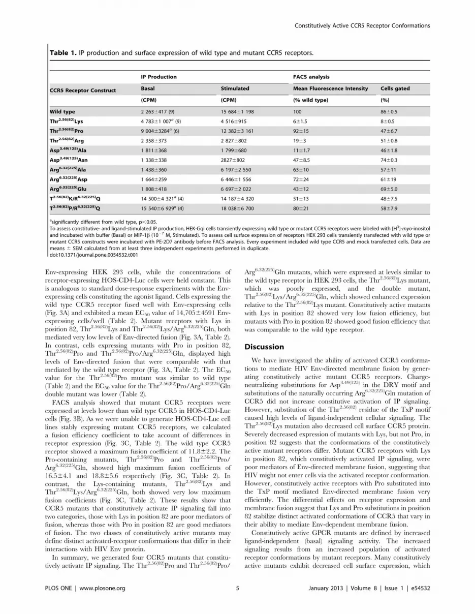
Table 1. IP production and surface expression of wild type and mutant CCR5 receptors.
IP Production FACS analysis
CCR5 Receptor Construct Basal Stimulated Mean Fluorescence Intensity Cells gated
(CPM) (CPM) (% wild type) (%)
Wild type 2 2636417 (9) 15 68461 198 100 8660.5
Thr2.56(82)Lys 4 78361 007a (9) 4 5166915 661.5 860.5
Thr2.56(82)Pro 9 00463284a (6) 12 38263 161 92615 4766.7
Thr2.56(82)Arg 2 3586373 2 8276802 1963 5160.8
Asp3.49(125)Ala 1 8116368 1 7996680 1161.7 4661.8
Asp3.49(125)Asn 1 3386338 28276802 4768.5 7460.3
Arg6.32(225)Ala 1 4386360 6 19762 550 63610 57611
Arg6.32(225)Asp 1 6646259 6 44661 556 72624 61619
Arg6.32(225)Glu 1 8086418 6 69762 022 43612 6965.0
T2.56(82)K/R6.32(225)Q 14 50064 321a (4) 14 18764 320 51613 4867.5
T2.56(82)P/R6.32(225)Q 15 54066 929a (4) 18 03866 700 80621 5867.9
asignificantly different from wild type, p,0.05.To assess constitutive- and ligand-stimulated IP production, HEK-Gqi cells transiently expressing wild type or mutant CCR5 receptors were labeled with [H3]-myo-inositoland incubated with buffer (Basal) or MIP-1b (1027 M, Stimulated). To assess cell surface expression of receptors HEK 293 cells transiently transfected with wild type ormutant CCR5 constructs were incubated with PE-2D7 antibody before FACS analysis. Every experiment included wild type CCR5 and mock transfected cells. Data aremeans 6 SEM calculated from at least three independent experiments performed in duplicate.doi:10.1371/journal.pone.0054532.t001
Constitutively Active CCR5 Receptor Conformations
PLOS ONE | www.plosone.org 5 January 2013 | Volume 8 | Issue 1 | e54532

Figure 2. IP production, expression and competition binding ofCCR5 receptors with mutations of Thr2.56(82) and Arg6.32(225). A,HEK-Gqi cells were transfected with the wild type (&) or mutant CCR5receptors Thr2.56(82)Lys (N), Thr2.56(82)Pro (m), Thr2.56(82)Lys/Arg6.32(225)Gln (#) or Thr2.56(82)Pro/Arg6.32(225)Gln (D). Untransfectedcells (%) were used as a negative control. Cells pre-labeled with[3H]myo-inositol were incubated with increasing concentrations of MIP-1b. Data are from a single experiment that is representative of at leastthree independent experiments performed in duplicate. B, HEK cellswere transfected with wild type or mutant CCR5 receptors and stainedwith PE-2D7 for FACS analysis. Results are mean values 6 SEM from atleast three independent experiments performed in duplicate. C, HEK293 cells were transiently transfected with wild type (&) or mutantCCR5 receptors, Thr2.56(82)Lys (N), Thr2.56(82)Pro (m), Thr2.56(82)Lys/Arg6.32(225)Gln (#) or Thr2.56(82)Pro/Arg6.32(225)Gln (D) and incubatedwith 125I-MIP-1b and various concentrations of unlabelled MIP-1b. Cell-bound radioactivity was collected by filtration and counted. Data arefrom a single experiment, representative of at least three independentexperiments performed in triplicate.doi:10.1371/journal.pone.0054532.g002
Figure 3. Fusion activity of wild type and mutant CCR5receptors. A, HOS cells stably expressing CD4 and the luciferasereporter gene were transiently transfected with wild type (&) or mutantCCR5 receptors Thr2.56(82)Lys (N), Thr2.56(82)Pro (m), Thr2.56(82)Lys/Arg6.32(225)Gln (#) or Thr2.56(82)Pro/Arg6.32(225)Gln (D). CCR5-expressingHOS-CD4-Luc cells were co-cultured overnight with HEK cells transientlyexpressing tat, rev and Env and luciferase activity was assessed. B,CCR5-expressing HOS-CD4-Luc cells were labeled with PE-2D7 andanalyzed by FACS analysis. C, To compare fusion efficiency amongmutant receptors that were expressed at different levels the fusioncoefficient was derived by dividing the luciferase activity by the meanfluorescence of each construct.doi:10.1371/journal.pone.0054532.g003
Constitutively Active CCR5 Receptor Conformations
PLOS ONE | www.plosone.org 6 January 2013 | Volume 8 | Issue 1 | e54532

may result from constitutive internalization of the activated
receptor, or from increased flexibility of activated receptor
conformations that results in protein instability [44,45,46,47].
Inactive and agonist-stabilized activated conformations of
CCR5 are likely to be broadly similar to those of other family A
GPCRs for which crystal structures are known, including the
closely related CXCR4 chemokine receptor [10,48]. Highly
conserved amino acid motifs are likely to form intramolecular
interactions in CCR5 that are similar to the interactions formed in
the inactive and active conformations of GPCRs that have been
crystallized [17,49,50].
The ‘‘DRY’’ motif, at the cytosolic end of TMS3, is one of the
most conserved sequences among class A GPCRs. According to
the ‘‘consensus’’ view of its function, the basic side-chain of Arg3.50
interacts simultaneously with the adjacent acidic Asp3.49 (Glu3.49 in
rhodopsin) and with Glu6.30 at the cytosolic end of TMS6, forming
an ‘‘ionic lock’’ that stabilizes inactive receptor conformations. In
activated receptors the ionic lock is broken and the guanidino
group of Arg3.50 moves to interact with Tyr5.58 in TMS5
[17,49,50,51]. The switch function of the DRY motif is supported
by charge-neutralizing mutations of Asp3.49 or Arg3.50, which
cause constitutive activity in many GPCRs [19,51]. However, our
charge-neutralizing mutations of Asp3.49(125) did not cause
constitutive activation of IP signaling in CCR5. Decreased
expression of the mutant receptors suggests that substitution of
Asp3.49(125) decreases receptor protein stability or increases re-
ceptor internalization and down-regulation, as has been described
for the Arg3.50(126)Asn CCR5 mutant [52]. This suggests that the
role of the DRY motif in activation of CCR5 does not comply with
the consensus view [51,53].
The Glu6.30 residue in intracellular loop 3 forms part of the
ionic lock in rhodopsin, but many GPCRs, including CCR5, have
basic residues in position 6.30 [53]. Crystal structures of the
inactive CXCR4 chemokine receptor show no interaction between
Arg3.50 and Arg6.30 [10,50]. The naturally-occurring
Arg6.32(225)Gln CCR5 mutant is partially constitutively active
and we hypothesized that Arg6.32(225), which is two residues away
from Arg6.30(223), might form alternative interactions that stabilize
the inactive CCR5 conformation. Other mutations of Arg6.32(225)
did not increase constitutive activity. Decreased expression of these
mutants is consistent with the role of basic amino acids in
stabilizing membrane-spanning helices [54] although the natural-
ly-occurring Arg6.32(225)Gln mutation did not decrease receptor
expression [22]. Furthermore, combining the Thr2.56(82)Lys and
Thr2.56(82)Pro mutations with the Arg6.32(225)Gln mutation in-
creased expression of constitutively active mutant CCR5 recep-
tors. The Arg6.32(225)Gln mutation may stabilize a receptor
conformation that is less susceptible to internalization or to
degradation. The Arg6.32(225)Gln double mutation enhanced
expression of constitutively active receptors more effectively in
HEK 293 cells than in HOS-CD4-Luc cells. This may result from
different receptor trafficking in the two cell lines or it may reflect
the generally lower transfection efficiency and receptor expression
in HOS-CD4-Luc cells.
A proposal that the TxP motif acts as a switch that activates
CCR5 was supported by mutations that uncoupled the CCR5
receptor from cellular signaling [20,55] or increased constitutive
cellular signaling [21]. The Thr2.56(82)Lys and Thr2.56(82)Pro
CCR5 mutants that we tested displayed increased basal IP
production and could not be further stimulated by MIP-1b. Thesame mutants were constitutively active and showed no further
response to chemokine treatment in a yeast reporter system [21],
suggesting that they are fully stabilized in activated conformations.
They also constitutively stimulated GTPcS binding in stably
transfected CHO cells. However, agonist treatment enhanced
GTPcS binding [21], suggesting that the Thr2.56(82)Lys and
Thr2.56(82)Pro mutations do not fully stabilize the CCR5 confor-
mation that activates the cognate Gai protein. The double
mutants, Thr2.56(82)Lys/Arg6.32(225)Gln and Thr2.56(82)Pro/
Arg6.32(225)Gln, both showed basal IP production that was similar
to the maximum MIP-1b-stimulated IP production mediated by
wild type CCR5, suggesting that they are fully stabilized in
activated conformations. However, it is not known whether the
CCR5 conformations that activate native Gai signaling pathways
are fully stabilized in the double mutant receptors. Mutant
receptors with Lys substituted for Thr2.56(82) showed decreased cell
surface protein, which may result from decreased receptor stability
or stabilization of receptor conformations that constitutively
expose cytosolic Ser residues to G protein-coupled receptor
kinases, leading to constitutive internalization [44,45,46,47,56].
In contrast, the Thr2.56(82)Pro mutation may stabilize receptor
conformations that are not recognized by receptor kinases or are
less flexible. The differential expression suggests that constitutively
active CCR5 mutants with Pro or Lys in position 2.56(82) may be
stabilized in distinct conformations that are differentially sensitive
to internalization and/or degradation. Distinct receptor confor-
mations of the Thr2.56(82)Lys and Thr2.56(82)Pro CCR5 mutants is
supported by the report that CHO cells expressing the
Thr2.56(82)Pro CCR5 mutant exhibited a wild type-like chemotac-
tic response to the chemokine ligand, RANTES, whereas cells
expressing the Thr2.56(82)Lys mutant showed no chemotactic
response [21].
The extended ternary complex model of receptor activation
predicts that constitutively active receptors have increased agonist
Table 2. Env-directed membrane fusion mediated by wild type and mutant CCR5 receptors.
CCR5 Receptor Construct Maximum Fusion Maximum Fusion Efficiency EC50
(% wild type) (fusion coefficient) (Env-expressing cells/well)
Wild Type 100 11.862.2 14,70564,591
T2.69(82)K 4.661.7 0.8460.2 NDa
T2.69(82)P 135610.9 16.564.1 15,38464,818
T2.69(82)K/R6.32(225)Q 16.863.1 1.960.4 NDa
T2.69(82)P/R6.32(225)Q 134.8631.1 18.865.6 5,5456957
aND, not determined because maximum fusion was too low to allow determination of EC50.HOS-CD4-Luc cells expressing wild type or mutant CCR5 receptor constructs were co-cultured with increasing concentrations of HEK 293 cells expressing HIV Env andthe HIV transactivator, tat, and luciferase activity was measured. Data are means 6 SEM of at least five experiments performed in triplicate.doi:10.1371/journal.pone.0054532.t002
Constitutively Active CCR5 Receptor Conformations
PLOS ONE | www.plosone.org 7 January 2013 | Volume 8 | Issue 1 | e54532

binding affinity, even in the absence of G protein [43]. However,
some constitutively active receptors do not exhibit this phenotype
[57,58]. We did not find significant changes in IC50 values for
MIP-1b binding to constitutively active CCR5 mutants. Arias et al
reported similar results for MIP-1b binding, but found that the
Thr2.56(82)Lys mutation decreased affinity for the agonist chemo-
kines, MIP-1a and RANTES, whereas the Thr2.56(82)Pro mutation
had less effect [21]. Studies with small molecule drugs have
suggested that the different chemokine ligands interact with
distinct CCR5 conformations [59,60]. The Thr2.56(82)Lys mutation
may selectively destabilize the ensembles of CCR5 conformations
that preferentially bind MIP-1a and RANTES.
The gp120 subunit of HIV Env is a CCR5 receptor agonist
[6,7,8]. However, Env mediates membrane fusion in cells
expressing mutant CCR5 receptors that do not support chemo-
kine-stimulated signaling [23,24,25], suggesting that inactive
conformations of CCR5 mediate membrane fusion. Furthermore,
HIV isolates that are resistant to CCR5 blockers use drug-
occupied CCR5 that is stabilized, by the inverse agonist drug, in
the inactive conformation to infect cells. We therefore hypothe-
sized that an inactive CCR5 conformation mediates HIV infection
and that activated conformations of CCR5 may not support HIV
Env-directed membrane fusion.
Consistent with our hypothesis, both of the constitutively active
mutants with Lys in position 82 showed low Env-directed
membrane fusion efficiency. The decreased fusion may result
from decreased expression, as the Thr2.56(82)Lys/Arg6.32(225)Gln
double mutation did not fully recover expression in the HOS-
CD4-Luc cells used for the fusion assay. Fusion remained lower
than that mediated by wild type CCR5 after correction for
receptor expression, but we cannot exclude threshold effects of
receptor protein levels. In contrast, constitutively active CCR5
receptors with Pro in position 82 mediated membrane fusion
similar to that mediated by wild type CCR5. Our results suggest
that CCR5 receptors that constitutively activate IP signaling exist
in at least two distinct conformations. One conformation,
stabilized by Pro in position 82, supports Env-directed membrane
fusion, whereas the other conformation, stabilized by Lys in
position 82, does not.
The different capacities of constitutively active CCR5 receptors
to mediate membrane fusion may relate to the nature of their
constitutive activity. Decreased expression of mutants with Lys in
position 82 suggests constitutive receptor phosphorylation and
activation of receptor sequestration pathways [61]. Constitutive
internalization of CCR5 may target CCR5-Env complexes for
degradation and thus inhibit the membrane fusion pathway.
Alternatively, receptor conformations that are stabilized by Lys in
position 82 may have decreased affinity for HIV Env or decreased
ability to induce the fusogenic Env protein conformation that
mediates membrane fusion. In terms of the ensemble model of
receptor conformation [3,62], mutation of Thr2.56(82) to Lys, may
stabilize an ensemble of CCR5 conformations that includes the
micro-conformations that activate G proteins and receptor
internalization, but not the micro-conformations that induce
Env-directed membrane fusion. In contrast, mutation of Thr2.56(82)
to Pro appears to stabilize an ensemble of receptor conformations
that activate G protein and mediate the co-receptor functions of
CCR5, but do not activate internalization (Fig. 4).
Distinct activated conformations of CCR5 with differential
abilities to support HIV Env-directed membrane fusion opens the
possibility of developing CCR5 ligands that select specific receptor
conformations. Indeed, a recent comparison of the CCR5
blockers, TAK 779 and maraviroc, has shown that maraviroc
has higher antiviral potency that does not correlate with inverse
agonist activity or ability to block gp120 binding. It was suggested
that maraviroc may selectively destabilize CCR5 conformations
that trigger Env penetration of cell membranes [63]. Furthermore,
it has been shown that CCR5 heterodimerizes with the CXCR4
co-receptor and that antagonists specific for one receptor
allosterically cross-inhibit ligand binding and agonist function at
the other receptor [64]. This raises the potential that CCR5-
blocking drugs may be developed to cross-inhibit infection by X4-
tropic viruses in cells where both receptors are expressed.
In conclusion, we have shown that charge-neutralizing muta-
tions of the Asp3.49(125) residue of the DRY motif do not result in
constitutive activity of CCR5, confirming that the CCR5 receptor
does not conform to the consensus mechanism of receptor
activation. We have confirmed that Lys or Pro substitutions for
the Thr2.56(82) residue of the TxP motif cause constitutive activity
of CCR5, but we have shown that mutants have distinct
properties. Constitutively active mutants with Lys in position 82
show decreased cell surface expression and decreased HIV co-
receptor function, whereas mutants with Pro in position 82 were
well expressed and fully functional as HIV co-receptors. These
distinct properties suggest that the mutations stabilize ensembles of
receptor conformations that differ in their ability to induce the
fusogenic HIV Env conformation. Our results suggest that drugs
that stimulate internalization of CCR5 may effectively inhibit HIV
infection, both by decreasing cell surface expression of CCR5 and
by stabilizing receptor conformations that inhibit fusion of virus
that binds to drug-occupied receptor.
Acknowledgments
The HIV-1C env construct pTHr.gp150CT [31] was a generous gift from
Carolyn Williamson (University of Cape Town). The HIV-1 tat, HIV-1 rev
and the pHIV-1LTR-Luc reporter construct were generously provided by
Figure. 4. Venn diagram depicting ensembles of CCR5 receptorconformations stabilized by mutation of Thr2.56(82). Trianglesrepresent receptor conformations stabilized by mutation of Thr2.56(82) toLys or Pro. Circles represent receptor conformations that mediate Gprotein activation, receptor internalization or HIV Env-directed mem-brane fusion. Mutation of Thr2.56(82) to Lys stabilizes an ensemble ofreceptor conformations that activate G protein-mediated signaling andconformations with increased susceptibility to internalization, but notconformations that support HIV Env dependent membrane fusion. TheThr2.56(82)Pro mutation stabilizes an ensemble of receptor conforma-tions that activate the G protein and conformations that support HIV-1fusion, but it does not appear to increase population of receptorconformations that result in decreased membrane expression of CCR5.doi:10.1371/journal.pone.0054532.g004
Constitutively Active CCR5 Receptor Conformations
PLOS ONE | www.plosone.org 8 January 2013 | Volume 8 | Issue 1 | e54532

Steven Jenkinson, GlaxoSmithKline. The following cell lines were obtained
from the AIDS Research and Reference Reagent Program, Division of
AIDS, NIAID, NIH: Human osteosarcoma cells stably expressing CD4
(HOS-CD4.pBABE-puro) or CD4 and CCR5 (HOS-CD4-CCR5) from
Dr Nathaniel Landau [33].
Author Contributions
Conceived and designed the experiments: AdV CAF. Performed the
experiments: AdV CAF. Analyzed the data: AdV CAF. Contributed
reagents/materials/analysis tools: ATF MCC. Wrote the paper: AdV
MTM CAF.
References
1. Oppermann M (2004) Chemokine receptor CCR5: insights into structure,
function, and regulation. Cell Signal 16: 1201–1210.
2. Lederman MM, Penn-Nicholson A, Cho M, Mosier D (2006) Biology of CCR5
and its role in HIV infection and treatment. JAMA 296: 815–826.
3. Kenakin T (2002) Drug efficacy at G protein-coupled receptors. Annu Rev
Pharmacol Toxicol 42: 349–379.
4. Deupi X, Kobilka BK (2010) Energy landscapes as a tool to integrate GPCR
structure, dynamics, and function. Physiology (Bethesda) 25: 293–303.
5. Mack M, Luckow B, Nelson PJ, Cihak J, Simmons G, et al. (1998)
Aminooxypentane-RANTES induces CCR5 internalization but inhibits recy-cling: a novel inhibitory mechanism of HIV infectivity. J Exp Med 187: 1215–
1224.
6. Freedman BD, Liu QH, Del Corno M, Collman RG (2003) HIV-1 gp120
chemokine receptor-mediated signaling in human macrophages. Immunol Res
27: 261–276.
7. Weissman D, Rabin RL, Arthos J, Rubbert A, Dybul M, et al. (1997)
Macrophage-tropic HIV and SIV envelope proteins induce a signal through the
CCR5 chemokine receptor. Nature 389: 981–985.
8. Wu Y, Yoder A (2009) Chemokine coreceptor signaling in HIV-1 infection and
pathogenesis. PLoS Pathog 5: e1000520.
9. Palczewski K, Kumasaka T, Hori T, Behnke CA, Motoshima H, et al. (2000)
Crystal structure of rhodopsin: A G protein-coupled receptor. Science 289: 739–
745.
10. Wu B, Chien EY, Mol CD, Fenalti G, Liu W, et al. (2010) Structures of theCXCR4 chemokine GPCR with small-molecule and cyclic peptide antagonists.
Science 330: 1066–1071.
11. Cherezov V, Rosenbaum DM, Hanson MA, Rasmussen SG, Thian FS, et al.
(2007) High-resolution crystal structure of an engineered human beta2-
adrenergic G protein-coupled receptor. Science 318: 1258–1265.
12. Rasmussen SG, Choi HJ, Rosenbaum DM, Kobilka TS, Thian FS, et al. (2007)
Crystal structure of the human beta2 adrenergic G-protein-coupled receptor.
Nature 450: 383–387.
13. Choe HW, Kim YJ, Park JH, Morizumi T, Pai EF, et al. (2011) Crystal structure
of metarhodopsin II. Nature 471: 651–655.
14. Scheerer P, Park JH, Hildebrand PW, Kim YJ, Krauss N, et al. (2008) Crystal
structure of opsin in its G-protein-interacting conformation. Nature 455: 497–
502.
15. Rasmussen SG, DeVree BT, Zou Y, Kruse AC, Chung KY, et al. (2011) Crystalstructure of the beta2 adrenergic receptor-Gs protein complex. Nature 477:
549–555.
16. Hofmann KP, Scheerer P, Hildebrand PW, Choe HW, Park JH, et al. (2009) A
G protein-coupled receptor at work: the rhodopsin model. Trends Biochem Sci
34: 540–552.
17. Ahuja S, Smith SO (2009) Multiple switches in G protein-coupled receptor
activation. Trends Pharmacol Sci 30: 494–502.
18. Nygaard R, Frimurer TM, Holst B, Rosenkilde MM, Schwartz TW (2009)
Ligand binding and micro-switches in 7TM receptor structures. Trends
Pharmacol Sci 30: 249–259.
19. Flanagan CA (2005) A GPCR that is not "DRY". Mol Pharmacol 68: 1–3.
20. Govaerts C, Blanpain C, Deupi X, Ballet S, Ballesteros JA, et al. (2001) The
TXP motif in the second transmembrane helix of CCR5. A structural
determinant of chemokine-induced activation. J Biol Chem 276: 13217–13225.
21. Arias DA, Navenot JM, Zhang WB, Broach J, Peiper SC (2003) Constitutive
activation of CCR5 and CCR2 induced by conformational changes in the
conserved TXP motif in transmembrane helix 2. J Biol Chem 278: 36513–
36521.
22. Folefoc AT, Fromme BJ, Katz AA, Flanagan CA (2010) South African
mutations of the CCR5 coreceptor for HIV modify interaction with chemokines
and HIV Envelope protein. J Acquir Immune Defic Syndr 54: 352–359.
23. Gosling J, Monteclaro FS, Atchison RE, Arai H, Tsou CL, et al. (1997)
Molecular uncoupling of C-C chemokine receptor 5-induced chemotaxis and
signal transduction from HIV-1 coreceptor activity. Proc Natl Acad Sci U S A94: 5061–5066.
24. Farzan M, Choe H, Martin KA, Sun Y, Sidelko M, et al. (1997) HIV-1 entry
and macrophage inflammatory protein-1beta-mediated signaling are indepen-
dent functions of the chemokine receptor CCR5. J Biol Chem 272: 6854–6857.
25. Amara A, Vidy A, Boulla G, Mollier K, Garcia-Perez J, et al. (2003) G protein-
dependent CCR5 signaling is not required for efficient infection of primary T
lymphocytes and macrophages by R5 human immunodeficiency virus type 1
isolates. J Virol 77: 2550–2558.
26. Buontempo PJ, Wojcik L, Buontempo CA, Ogert RA, Strizki JM, et al. (2009)
Quantifying the relationship between HIV-1 susceptibility to CCR5 antagonistsand virus affinity for antagonist-occupied co-receptor. Virology 395: 268–279.
27. Westby M, Smith-Burchnell C, Mori J, Lewis M, Mosley M, et al. (2007)
Reduced maximal inhibition in phenotypic susceptibility assays indicates thatviral strains resistant to the CCR5 antagonist maraviroc utilize inhibitor-bound
receptor for entry. J Virol 81: 2359–2371.
28. Tilton JC, Amrine-Madsen H, Miamidian JL, Kitrinos KM, Pfaff J, et al. (2010)HIV type 1 from a patient with baseline resistance to CCR5 antagonists uses
drug-bound receptor for entry. AIDS Res Hum Retroviruses 26: 13–24.
29. Moore JP, Kuritzkes DR (2009) A piece de resistance: how HIV-1 escapes smallmolecule CCR5 inhibitors. Curr Opin HIV AIDS 4: 118–124.
30. Kostenis E (2001) Is Galpha16 the optimal tool for fishing ligands of orphan G-
protein- coupled receptors? Trends Pharmacol Sci 22: 560–564.
31. Burgers WA, van Harmelen JH, Shephard E, Adams C, Mgwebi T, et al. (2006)
Design and preclinical evaluation of a multigene human immunodeficiency virustype 1 subtype C DNA vaccine for clinical trial. J Gen Virol 87: 399–410.
32. Jenkinson S, McCoy D, Kerner S, Ferris R, Lawrence W, et al. (2003)
Development of a high-throughput viral-free assay for the measurement ofCCR5-mediated HIV/cell fusion. Receptors Channels 9: 117–123.
33. Deng H, Liu R, Ellmeier W, Choe S, Unutmaz D, et al. (1996) Identification of
a major co-receptor for primary isolates of HIV-1. Nature 381: 661–666.
34. Ballesteros J, Weinstein W (1995) Integrated methods for the construction of
three-dimensional models and computational probing of structure-function
relations in G protein-coupled receptors. Methods Neurosci 25: 366–428.
35. Millar RP, Davidson J, Flanagan C, Wakefield I (1995) Ligand Binding and
Second-Messenger Assays for Cloned Gq/G11-Coupled Neuropeptide Recep-tors: The GnRH Receptor. Methods in Neuroscience 25: 145–162.
36. Flanagan CA, Fromme BJ, Davidson JS, Millar RP (1998) A high affinity
gonadotropin-releasing hormone (GnRH) tracer, radioiodinated at position 6,facilitates analysis of mutant GnRH receptors. Endocrinology 139: 4115–4119.
37. Fromme BJ, Coetsee M, Van Der Watt P, Chan MC, Sperling KM, et al. (2008)
High-affinity binding of southern African HIV type 1 subtype C envelopeprotein, gp120, to the CCR5 coreceptor. AIDS Res Hum Retroviruses 24:
1527–1536.
38. Reynes J, Portales P, Segondy M, Baillat V, Andre P, et al. (2000) CD4+ T cellsurface CCR5 density as a determining factor of virus load in persons infected
with human immunodeficiency virus type 1. J Infect Dis 181: 927–932.
39. Platt EJ, Wehrly K, Kuhmann SE, Chesebro B, Kabat D (1998) Effects of
CCR5 and CD4 cell surface concentrations on infections by macrophagetropic
isolates of human immunodeficiency virus type 1. J Virol 72: 2855–2864.
40. Gaertner H, Cerini F, Escola JM, Kuenzi G, Melotti A, et al. (2008) Highly
potent, fully recombinant anti-HIV chemokines: reengineering a low-cost
microbicide. Proc Natl Acad Sci U S A 105: 17706–17711.
41. Doms RW (2000) Beyond receptor expression: the influence of receptor
conformation, density, and affinity in HIV-1 infection. Virology 276: 229–237.
42. Lin YL, Mettling C, Portales P, Reynes J, Clot J, et al. (2002) Cell surface CCR5density determines the postentry efficiency of R5 HIV-1 infection. Proc Natl
Acad Sci U S A 99: 15590–15595.
43. Samama P, Cotecchia S, Costa T, Lefkowitz RJ (1993) A mutation-induced
activated state of the beta 2-adrenergic receptor. Extending the ternary complex
model. J Biol Chem 268: 4625–4636.
44. Milligan G, Bond RA (1997) Inverse agonism and the regulation of receptor
number. Trends Pharmacol Sci 18: 468–474.
45. Gether U, Ballesteros JA, Seifert R, Sanders-Bush E, Weinstein H, et al. (1997)Structural instability of a constitutively active G protein-coupled receptor.
Agonist-independent activation due to conformational flexibility. J Biol Chem272: 2587–2590.
46. Rasmussen SG, Jensen AD, Liapakis G, Ghanouni P, Javitch JA, et al. (1999)
Mutation of a highly conserved aspartic acid in the beta2 adrenergic receptor:constitutive activation, structural instability, and conformational rearrangement
of transmembrane segment 6. Mol Pharmacol 56: 175–184.
47. Alewijnse AE, Timmerman H, Jacobs EH, Smit MJ, Roovers E, et al. (2000)The effect of mutations in the DRY motif on the constitutive activity and
structural instability of the histamine H(2) receptor. Mol Pharmacol 57: 890–898.
48. Garcia-Perez J, Rueda P, Alcami J, Rognan D, Arenzana-Seisdedos F, et al.
(2011) Allosteric Model of Maraviroc Binding to CC Chemokine Receptor 5(CCR5). J Biol Chem 286: 33409–33421.
49. Deupi X, Standfuss J (2011) Structural insights into agonist-induced activation of
G-protein-coupled receptors. Curr Opin Struct Biol 21: 541–551.
50. Trzaskowski B, Latek D, Yuan S, Ghoshdastider U, Debinski A, et al. (2012)
Action of molecular switches in GPCRs-theoretical and experimental studies.Curr Med Chem 19: 1090–1109.
51. Rovati GE, Capra V, Neubig RR (2007) The highly conserved DRY motif of
class A G protein-coupled receptors: beyond the ground state. Mol Pharmacol71: 959–964.
Constitutively Active CCR5 Receptor Conformations
PLOS ONE | www.plosone.org 9 January 2013 | Volume 8 | Issue 1 | e54532
52. Lagane B, Ballet S, Planchenault T, Balabanian K, Le Poul E, et al. (2005)
Mutation of the DRY motif reveals different structural requirements for the CC
chemokine receptor 5-mediated signaling and receptor endocytosis. Mol
Pharmacol 67: 1966–1976.
53. Springael JY, de Poorter C, Deupi X, Van Durme J, Pardo L, et al. (2007) The
activation mechanism of chemokine receptor CCR5 involves common structural
changes but a different network of interhelical interactions relative to rhodopsin.
Cell Signal 19: 1446–1456.
54. Dalbey RE (1990) Positively charged residues are important determinants of
membrane protein topology. Trends Biochem Sci 15: 253–257.
55. Govaerts C, Bondue A, Springael JY, Olivella M, Deupi X, et al. (2003)
Activation of CCR5 by chemokines involves an aromatic cluster between
transmembrane helices 2 and 3. J Biol Chem 278: 1892–1903.
56. Pei G, Samama P, Lohse M, Wang M, Codina J, et al. (1994) A constitutively
active mutant beta 2-adrenergic receptor is constitutively desensitized and
phosphorylated. Proc Natl Acad Sci U S A 91: 2699–2702.
57. Zhang WB, Navenot JM, Haribabu B, Tamamura H, Hiramatu K, et al. (2002)
A point mutation that confers constitutive activity to CXCR4 reveals that T140
is an inverse agonist and that AMD3100 and ALX40–4C are weak partial
agonists. J Biol Chem 277: 24515–24521.
58. Beinborn M, Ren Y, Blaker M, Chen C, Kopin AS (2004) Ligand function at
constitutively active receptor mutants is affected by two distinct yet interactingmechanisms. Mol Pharmacol 65: 753–760.
59. Maeda K, Nakata H, Koh Y, Miyakawa T, Ogata H, et al. (2004)
Spirodiketopiperazine-based CCR5 inhibitor which preserves CC-chemokine/CCR5 interactions and exerts potent activity against R5 human immunodefi-
ciency virus type 1 in vitro. J Virol 78: 8654–8662.60. Watson C, Jenkinson S, Kazmierski W, Kenakin T (2005) The CCR5 receptor-
based mechanism of action of 873140, a potent allosteric noncompetitive HIV
entry inhibitor. Mol Pharmacol 67: 1268–1282.61. Parnot C, Miserey-Lenkei S, Bardin S, Corvol P, Clauser E (2002) Lessons from
constitutively active mutants of G protein-coupled receptors. Trends EndocrinolMetab 13: 336–343.
62. Vaidehi N, Kenakin T (2010) The role of conformational ensembles of seventransmembrane receptors in functional selectivity. Curr Opin Pharmacol 10:
775–781.
63. Garcia-Perez J, Rueda P, Staropoli I, Kellenberger E, Alcami J, et al. (2011)New insights into the mechanisms whereby low molecular weight CCR5 ligands
inhibit HIV-1 infection. J Biol Chem 286: 4978–4990.64. Sohy D, Yano H, de Nadai P, Urizar E, Guillabert A, et al. (2009) Hetero-
oligomerization of CCR2, CCR5, and CXCR4 and the protean effects of
‘‘selective’’ antagonists. J Biol Chem 284: 31270–31279.
Constitutively Active CCR5 Receptor Conformations
PLOS ONE | www.plosone.org 10 January 2013 | Volume 8 | Issue 1 | e54532