estimation of gestational age using humeral length in
TRANSCRIPT

Sudan University of Science and Technology
Collage of Graduate Studies
Estimation of Gestational Age using Humeral
Length in Ultrasonography
التصوير تقدير عمر الجنين بطول عظم العضد باستخدام
بالموجات فوق الصوتية
Thesis Submitted for Partial Fulfillment for the Requirements of
M.sc Degree in Medical Diagnostic Ultrasound
By:
Kamal Eldein Mustafa Ali Saran
Supervisor:
Dr. babiker Abd Alwahab Awaddlla
2019

اآلية
تعالى : هللا قال
ثم خلقنا النطفة علقة فخلقنا العلقة مضغة فخلقنا )
المضغة عظاما فكسونا العظام لحما ثم أنشأناه خلقا
أحسن الخالقين ( آخر فتبارك الل
صدق هللا العظيم
( 41ةي )اآلمؤمنون سورة ال

I
Dedication
Many thanks to my greatest parents for their helps and support in my
scientific career and to my mother, brother and sister.

II
Acknowledgement
My gratefulness to my supervisor Dr. Bakier Abdelwahab for his great
effort in the advising and correcting me during this research.
Special acknowledgement to the ultrasound department of Omdurman
Maternity hospital and Altohami medical Center for giving me a chance to
collect the research data.

List of Contents
2 ................................................................................................................................................................. اآلةي
Dedication ................................................................................................................................................ 3
Acknowledgement ............................................................................................................................... 4
List of Contents ..................................................................................................................................... 8
List of Tables ........................................................................................................................................ 11
List of Figures ...................................................................................................................................... 12
List of Abbreviations ....................................................................................................................... 13
Abstract .................................................................................................................................................... 14
Abstract Arabic ................................................................................................................................... 15
Chapter One
Introduction
1-1 Introduction: ................................................................................................................................. 16
1-2: Problem of the study: ............................................................................................................ 19
1-3: Objectives: ................................................................................................................................... 19
1-3-1 General objective: ................................................................................................................ 19
1-3-2 Specific objective: ................................................................................................................ 19
Chapter Two
Literature Reviews
2-1: Anatomy: ...................................................................................................................................... 20
2-1-1: anatomy of Humerus: ....................................................................................................... 20
2-1-2: Anatomy of the Femur: ................................................................................................... 21
2-2 Emberology: ................................................................................................................................. 23
2-2-1 limb growth and development: ..................................................................................... 23
2-3: Congenital anomalies of the fetal bones: ................................................................... 10
2-3-1 lethal skeletal dysplasia: ................................................................................................... 11
2-3-1-1Thanatophoric Dysplasia: ............................................................................................. 11
2-3-1-2: Thanatophoric dysplasia: ........................................................................................... 12
2-3-1-3 Platyspondyly: ................................................................................................................... 12

2-3-1-4: Achondrogenesis: ........................................................................................................... 12
2-3-1-5 Osteogenesis Imperfecta (OI): ................................................................................. 13
2-3-1-6 Hypophosphatasia: .......................................................................................................... 13
2-3-1-7 Campomelic Dysplasia: ............................................................................................... 14
2-3-1-8: Short-Rib Polydactyly Syndromes: ..................................................................... 14
2-3-1-9: Fibrochondrogenesis: ................................................................................................... 15
2-3-2 Nonlethal skeletal dysplasia: ......................................................................................... 15
2-3-2-1 Heterozygous Achondroplasia: ................................................................................ 15
2-3-2-2 Diastrophic Dysplasia: .................................................................................................. 15
2-3-2-3 Asphyxiating Thoracic Dysplasia: ......................................................................... 16
2-3-2-4 Ellis–van Creveld Syndrome: ................................................................................... 16
2-3-2-5 ChondrodysplasiaPunctata: ........................................................................................ 16
2-3-2-6 Dyssegmental Dysplasia: ............................................................................................ 17
2-3-2-7 Osteogenesis Imperfecta (OI) Types I, III, IV-Nonlethal Types: ....... 17
2-3-2-7-1Osteogenesis imperfecta type I: ........................................................................... 17
2-3-2-7-2 Osteogenesis imperfecta Type III: .................................................................... 18
2-3-2-7-2 Osteogenesis imperfecta Type IV: .................................................................... 18
2-3-2-7-3Down syndrome: ........................................................................................................... 18
2-4 Physiology: ................................................................................................................................... 18
2-4-1 Deposition and Absorption of Bone-Remodeling of Bone: ........................ 18
2-4-2 Absorption of Bone-Function of the Osteoclasts: ............................................. 19
2-5 Routine Parameter Scanning: ............................................................................................. 20
2-5-1 Femur length (FL): .............................................................................................................. 21
2.6. Previous studies: ....................................................................................................................... 22
Chapter Three
Material and Method
3.1 Materials: ........................................................................................................................................ 26
3.1.1 Subject: ........................................................................................................................................ 26
3.1.2Machine used ............................................................................................................................. 26

3.2 Method: ............................................................................................................................................ 26
3.2.1 Technique used: ...................................................................................................................... 26
3.2.2 Data collection: ....................................................................................................................... 28
32.3 Data analysis: ............................................................................................................................ 28
Chapter Four
Data analysis and result
Data analysis and result .................................................................................................................. 29
Chapter Five
Discussion, Conclusion and Recommendations
5.1 Discussion ...................................................................................................................................... 32
5.2 Conclusion:.................................................................................................................................... 34
5.3 Recommendations: ................................................................................................................... 35
References: ............................................................................................................................................ 36

List of Tables
Table (4. 1) Comparison between the femoral length and the humeral length
in association with age of pregnant women. .................................................. 29
Table (4. 1) Present the correlation of the gestational age with femoral
length and humeral length. ............................................................................. 29

List of Figures
Figure (2. 1) Show the anatomy of humerus, with presentation of muscle
attachment and insertion (Kijowski, et. al., 2004). ........................................ 21
Figure (2. 2) Show the anatomy of femur, with presentation of muscle
attachment and insertion (Kijowski, et. al., 2004). ........................................ 23
Figure (2. 3) Thanatophoric dysplasia at 22 weeks, curved femur (Wyatt-
Ashmead, J., 2014). ....................................................................................... 12
Figure (2. 4) Hypophophatasia at 20 weeks .................................................. 13
Figure (2. 5) Bowing of femur at 30 weeks (Wyatt-Ashmead, J., 2014). ... 14
Figure (2. 6) polydactyl syndrome. http://www.google com ....................... 15
Figure (2. 7) Type I. Nonlethal variant of OI with a mildly angulated femur
of normal length. (Wyatt-Ashmead, J., 2014). .............................................. 17
Figure (4. 1) Scatter plot shows the predication system to estimate the
gestational age in weeks by using the femur length in mm. .......................... 30
Figure (4. 2) Scatter plot shows the predication system to estimate the
gestational age in weeks using the humeral length in mm. ........................... 30

List of Abbreviations
Abbreviations Meaning
AC Abdominal circumference
ADD Actual date of delivery
BPD Biparietal diameter
CRL Crown rump length
EDD Expected date of delivery
FL Femur length
GA Gestational age
HC Had circumference
HL Humeral length
IUGR Intrauterine growth retardation
IVF In vitro fertilization
LFD Large for date
LMP Last menstrual period
SD Standard deviation
SFD Small for date
MHz Mega hertz
U.S Ultrasound

Abstract
This is a descriptive, cross sectional study was carried out on ultrasound
department in the Omdurman maternity hospital, the main problem for this
research is an accurate measurement of gestational age in third trimester ,
the study aim to assess the reliability of humeral length and the femur length
in predicating gestational age and prediction of menstrual age. 50 healthy
pregnant women were enrolled in the study; their age ranged 18-35 year is a
safe period for the pregnancy, delivery and have less chance of congenital
anomalies. Ultrasound devices with option of measuring humeral length
were used.
The result revealed that there was no significant difference between
gestational age obtained with femur length and that obtained with humeral
length. Furthermore, a significant correlation was noticed between
gestational age and HL, this correlation also seen between FL and gestational
age.
The ultrasound plays a great role to assess fetal bone biometrics as it is
sensitive and accurate. The study concluded that both femoral and humeral
length s were similar and reliable to estimate the gestational age.
The study recommended that the humeral length must be used as routine
measurement in calculation the gestational age in the third trimester .

15
ملخص الدراسةهذه دراسة وصفية مقطعية ، تم عملها في قسم الموجات الصوتية بمستشفى أم درمان ألمراض النساء والتوليد ومركز التهامي الطبي ) المهندسين( هدف هذه الدراسة دقة قياس عمر الجنين باستخدام
شهرية ، طول عظم العضد مقارنةبعمر الجنين باستخدام طول عظم الفخذ وعمر الجنين بزمن الدورة ال – 81المواصفات التي تم عليها تحديد النساء الحوامل لتشملهم الدراسة ، هي أن يكون عمر المرأة ما بين
الخلقية سنة وهو العمر المناسب للحمل السليم ، والوالدة اآلمنة ، واقل احتمالية لإلصابات بالتشوهات 53اة ال تشكو من أي امراض وان يكون الجنين واحد للجنين ، الصفة الثانية هي ان تكون السيدة الحامل معاف
داخل الرحم وسليم ليس لديه تشوهات خلقية ، الصفة الثالثة هي أن تكون السيدة في الشهر الرابع حتى التاسع ، تم إستخدام اجهزة موجات فوق صوتية بها خاصية تحديد عمر الجنين باستخدام طول عظم العضد
ين سيدة حامل حضرت الى قسم الموجات الصوتية بمركز التهامي الطبي بالجنين ، عينة البحث هي خمسالمهندسين ومستشفى الوالدة أم درمان ثم تم تحليل البيانات بواسطة برنامج التحليل االحصائي ، نتيجة
البحث هي انه لم يوجد فرق بين عمر الجنين المأخوذ بواسطة طول عظم العضد وطول عظم الفخذ.

16
Chapter One
Introduction
1-1 Introduction:
Obstetric ultrasound introduced since 1950, obstetric ultrasound has evolved
into a major specialty of sonography. Ultrasound is safe non ionizing easy
operative relatively inexpensive techniques painless procedure that offers
invaluable information to the obstetrician, The use of ultrasound in
pregnancy greatly increase in the last 10 years at present 40 to 60
percent of pregnancy are evaluated by ultrasound. Current technology
offers complete assessment of the fetus with high resolution images
sophisticated measurements and computations, and the ability to analyzed
fetal hemodynamic (Donofrio, et. al., 2014).
In the past gestational age was established by a combination of the historical
information and the physical examination. Reliance was placed on the
menstrual history and the maternal sensation of fetal movement
(“quickening”). Other factors include assessment of uterine size by
bimanual examination in the first trimester, initial detection of fetal heart
tones by Doppler (10–12 weeks) or auscultation (19–21 weeks), and uterine
fundal height measurement. However, both the history and the findings on
physical examination are fraught with error, even in the best of
circumstances, It has been estimated that 20% to 40% of women cannot
relate the LMP with certainty.6,7 Some of the reasons for this uncertainty
include oligomenorrhea, metrorrhagia, bleeding in the first trimester of
pregnancy, pregnancy following use of oral contraceptives or intrauterine
devices, and becoming pregnant in the postpartum period. Hertz and
co- workers9 reported that menstrual history was considered reliable in only
18% of women. In another report, even among women with known LMP,
neonatal age assessment differed markedly from that assigned by certain
menstrual dates in

17
15%.8 Physical examination also tends to be inaccurate, especially with
advancing gestational age.10 Bimanual examination in the first trimester
may be accurate within ±2 weeks; however, fundal height measurement,
which is more commonly used to assess gestational age, is only accurate
within ±4 to 6 weeks.
Clearly, the inaccuracies of history and physical examination may limit their
usefulness in assessment of gestational age. Methods that assess the time of
ovulation or conception can accurately establish gestational age.
Timed ovulation, either by basal body temperature recording or semi
quantitative assessment of luteinizing hormone surge, predicts gestational
age within ±4 to 6 days. Ovulation induction with agents such as clomiphene
citrate and Personal, also accurately predicts gestational age. In vitro
fertilization, with known date of conception, is likely the most accurate
means of predicting gestational age (±1 day). However, in most pregnancies,
the date of ovulation or conception cannot be as accurately predicted as
outlined above and gestational age must be established by other methods.
a real time scanner using endovaginally in first 5 weeks of gestation or
transabdominally is used. Normal pregnancy 40 weeks divided to first
trimester 13weeks diagnosed by yolk sac gestational sac and crown rump
length at least 7weeks or 10mm should be determine the gestational age
which is an accurate indicator of fetal age fetal cardiac activity start 6weeks
vaginally 7weeks transabdominally, after 13weeks CRL un reliable because
of spine affect flexibility so biparietal dimeter is accurate up to
24weeks, second trimester 14weeks to 27weeks the standard level of
measuring biparietal diameter ( BPD), is include cavil septum pellucidum
and thalami head circumference is the same level but is accurate till delivery
associated with abdominal circumference (AC) which is detected by
junction of the umbilical vein and portal sinus AC may allow detection
of growth retardation and macrosomia when compared with BPD. After

18
24weeks estimation of fetal age is accurate by fetal limb femur length
(FL), humeral length (HL), tibia, and ulna length, the estimation of fetal age
at the second trimester is more accurate it is prediction approximatly7 Days
before 20weeks and 10 days after 20 weeks and 21 Days in 3rd trimester
femur length is very useful biometric parameters in the second and third
trimester of pregnancy it grows linear throughout an is best measured
after 14weeks of gestation (Mukherjee, 2014).
The FL measured at the long axis of femoral shaft when the ultrasound
beam is perpendicular to the shaft these parameters are help full in the
estimation of fetal age in patients whose fundal height on abdominal
examination does not corresponding to the last menstrual period (LMP).The
fetal humeral length is not widely used as biometric parameters for
determination the gestational age also it easy to be imaged with ultrasound
and measured (Donofrio, et. al., 2014).
The study aim to clarify the role of humeral length in determining
gestational age with comparison with FL and HL may be useful when BPD
unreliable or abnormal to be used in BPD: FL ratio, and BPD: HL ratio as
categorical variable in Down syndrome or as comparison with HC and AC in
Achondroplasia. A few studies were done in Sudan concerning the
estimation of GA using fetal humeral length HL.
This research is conducted to introduce a new way in measuring the
gestational age in the third trimester using the humors length, by the
proving the accuracy by comparing the obtained gestational age by that
which obtained from the femur length. The benefit of this option is its use at
when the fetal position is unreliable for easiest way to measure the femur
length so instead we use the length of humerus.

19
1-2: Problem of the study:
The main problems deliver from an inaccurate measurement of
biparietal diameter after 24 weeks and femur length (FL) due to combination
of fetal movement and delay use of freeze button, as well as an abnormal
fetal presentation can affect measuring the gestational age by using the .
1-3: Objectives:
1-3-1 General objective:
To estimate of gestational age using humeral length in ultrasonography
1-3-2 Specific objective:
To estimate the accurate measurement in the third trimester using
humeral length and femoral length.
To assess gestational age correlated to HL and FL.

20
Chapter Two
Literature Reviews
2-1: Anatomy:
2-1-1: anatomy of Humerus:
The humerus articulates with the scapula at the shoulder joint and with the
radius and ulna at the elbow joint. The upper end of the humerus has a head,
which forms about one third of a sphere and articulates with the glenoid
cavity of the scapula. Immediately below the head is the anatomic neck.
Below the neck are the greater and lesser tuberosities, separated from each
other by the bicipital groove. Where the upper end of the humerus joins the
shaft is a narrow surgical neck. About halfway down the lateral aspect of the
shaft is a roughened elevation called the deltoid tuberosity. Behind and
below the tuberosity is a spiral groove, which accommodates the radial
nerve .The lower end of the humerus possesses the medial and lateral
epicondyles for the attachment of muscles and ligaments, the rounded
capitulum for articulation with the head of the radius, and the pulley-shaped
trochlea for articulation with the trochlear notch of the ulna. Above the
capitulum is the radial fossa, which receives the head of the radius when the
elbow is flexed. Above the trochlea anteriorly is the coronoid fossa, which
during the same movement receives the coronoid process of the ulna. Above
the trochlea posteriorly is the olecranon fossa, which receives the olecranon
process of the ulna when the elbow joint is extended (Kijowski, et. al., 2004).

21
Figure (2. 1) Show the anatomy of humerus, with presentation of muscle
attachment and insertion (Kijowski, et. al., 2004).
2-1-2: Anatomy of the Femur:
The femur articulates above with the acetabulum to form the hip joint
and below with the tibia and the patella to form the knee joint. The upper end
of the femur has a head, a neck, and greater and lesser trochanters. The head
forms about two thirds of a sphere and articulates with the acetabulum of the
hip bone to form the hip joint. In the center of the head is a small depression,
called the fovea capitis, for the attachment of the ligament of the head. Part
of the blood supply to the head of the femur from the obturator artery is
conveyed along this ligament and enters the bone at the fovea. The neck,
which connects the head to the shaft, passes downward, backward, and
laterally and makes an angle of about 125° (slightly less in the female) with
the long axis of the shaft. The size of this angle can be altered by disease.
The greater and lesser trochanters are large eminences situated at the junction

22
of the neck and the shaft. Connecting the two trochanters are the inter
trochanteric line anteriorly, where the iliofemoral ligament is attached, and a
prominent intertrochanteric crest posteriorly, on which is the quadrate
tubercle .The shaft of the femur is smooth and rounded on its anterior
surface but posteriorly has a ridge, the linea aspera, to which are attached
muscles and intramuscular septa. The margins of the linea aspera diverge
above and below. The medial margin continues below as the medial
supracondylar ridge to the adductor tubercle on the medial condyle. The
lateral margin becomes continuous below with the lateral supracondylar
ridge. On the posterior surface of the shaft below the greater trochanter is the
gluteal tuberosity for the attachment of the gluteus maximus muscle. The
shaft becomes broader toward its distal end and forms a flat, triangular area
on its posterior surface called the popliteal surface. The lower end of the
femur has lateral and medial condyles, separated posteriorly by the
intercondylar notch. The anterior surfaces of the condyles are joined by an
articular surface for the patella. The two condyles take part in the formation
of the knee joint. Above the condyles are the medial and lateral epicondyles.
The adductor tubercle is continuous with the medial epicondyle (Kijowski,
et. al., 2004).

23
Figure (2. 2) Show the anatomy of femur, with presentation of muscle
attachment and insertion (Kijowski, et. al., 2004).
2-2 Emberology:
2-2-1 limb growth and development:
The limbs, including the shoulder and pelvic girdles, comprise the
appendicular skeleton. At the end of the fourth week of development, limb
buds become visible as outpocketings from the ventrolateral body wall. The
fore-limb appears first followed by the hind limb 1 to 2 days later. Initially,
the limb buds consist of a mesenchymal core derived from the parietal
(somatic) layer of lateral plate mesoderm that will form the bones and

9
connective tissues of the limb, covered by a layer of cuboidal ectoderm.
Ectoderm at the distal border of the limb thickens and forms the apical
ectodermal ridge (AER). This ridge exerts an inductive influence on adjacent
mesenchyme, causing it to remain as a population of undifferentiated, rapidly
proliferating cells, and the progress zone. As the limb grows, cells farther
from the influence of the AER begin to differentiate into cartilage and
muscle. In this manner, development of the limb proceeds proximodistally.
By the sixth week of development, the first hyaline cartilage models,
foreshadowing the bones of the extremities, are formed by these
chondrocytes. Joints are formed in the cartilaginous condensations when
chondrogenesis is arrested, and a joint interzone is induced. Cells in this
region increase in number and density, and then a joint cavity is formed by
cell death. Surrounding cells differentiate into a joint capsule. Factors
regulating the positioning of joints are not clear, but the secreted molecule
WNT14 appears to be the inductive signal. Ossification of the bones of the
extremities, endochondral ossification, begins by the end of the embryonic
period. Primary ossification centers are present in all long bones of the limbs
by the 12th week of development. From the primary center in the shaft or
diaphysis of the bone, endochondral ossification gradually progresses toward
the ends of the cartilaginous model. At birth, the diaphysis of the bone is
usually completely ossified, but the two ends, the epiphyses, are still
cartilaginous. Shortly thereafter, however, ossification centers arise in the
epiphyses. Temporarily, a cartilage plate remains between the diaphyseal and
epiphyseal ossification centers. This plate, the epiphyseal plate, plays an
important role in growth in the length of the bones. Endochondral
ossification proceeds on both sides of the plate. When the bone has acquired
its full length, the epiphyseal plates disappear, and the epiphyses unite with
the shaft of the bone. In long bones, an epiphyseal plate is found on each
extremity; in smaller bones, such as the phalanges, it is found only at one

10
extremity; and in irregular bones, such as the vertebrae, one or more primary
centers of ossification and usually several secondary centers are present.
Synovial joints between bones begin to form at the same time that
mesenchymal condensations initiate the process of forming cartilage.Thus, in
the region between two chondrifying bone primordia, called the interzone
(for example between the tibia and femur at the knee joint), the condensed
mesenchyme differentiates into dense fibrous tissue. This fibrous tissue then
forms articular cartilage, covering the ends of the two adjacent bones; the
synovial membranes; and the menisci and ligaments within the joint capsule
(e.g., the anterior and posterior cruciate ligaments in the knee). The joint
capsule itself is derived from mesenchyme cells surrounding the interzone
region. Fibrous joints (e.g., the sutures in the skull) also form from interzone
regions, but in this case the interzone remains as a dense fibrous structure
(Mohammed, A., 2019).
2-3: Congenital anomalies of the fetal bones:
Congenital bone disorders are a heterogeneous group of disorders primarily
affecting the growth and development of the musculoskeletal system. There
are three major categories. The skeletal dysplasias are developmental
disorders of chondro-osseous tissue caused by single gene disorders with
prenatal and postnatal manifestations. thedysostoses are single-gene
disorders resulting in mal formations of individual bones caused by
transient abnormalities of signaling factors. Disruptions are morphologic
defects of an organ or larger region resulting from extrinsic breakdown or
interference with an originally normal developmental process.The prevalence
of skeletal dysplasias, also called osteochondro-dysplasias, diagnosed
prenatally or during the neonatal period, excluding limb amputations, is
2.4 to 4.5 per 10,000 births.

11
Nonetheless, the majority of lethal skeletal dysplasias, including
thanatophoric dysplasia, a chondrogenesis, and osteogenesis imperfect (OI),
type II, can be diagnosed solely on the basis of prenatal ultrasound.
Lethal skeletal dysplasias were identified correctly by prenatal
ultrasound; however, only 13 of 27 (48%) received an accurate specific
antenatal diagnosis (Wyatt-Ashmead, J., 2014).
2-3-1 lethal skeletal dysplasia:
The lethal skeletal dysplasias are characterized by severe micromelia and
small thoracic circumference with pulmonary hypoplasia.The most
important determinant of lethality is the presence and degree of
pulmonary hypoplasia. The three most common lethal skeletal dysplasias
are thanatophoric dysplasia; achondrogenesis, and osteogenesisimperfecta
type II, overall accounting for 40% to 60% of all lethal skeletal
dysplasias (Wyatt-Ashmead, J., 2014).
2-3-1-1Thanatophoric Dysplasia:
Thanatophoric dysplasia is the most common lethal skeletal dysplasia, with
a prevalence of 0.24 to 0.69 per10,000 births. The key features are
severe micro melia with rhizo melic predominance and macrocrania
(disproportionately large head) in association with decreased thoracic
circumference but a normal trunk length. Mineralization is normal, with no
fractures present. Typically, the extremities are so foreshortened that they
protrude at right angles to the body. The skin folds are thickened and
redundant secondary to a relatively greater rate of growth of the skin and
subcutaneous layers than the bones. Clinical presentation is usually caused
by large-for-date measurements secondary to poly hydramnios (Wyatt-
Ashmead, J., 2014).

12
2-3-1-2: Thanatophoric dysplasia:
Thanatophoric dysplasia has many phenotypic similarities to homozygous
achondroplasia. Both conditions may appear identical from ultrasound and
radiographic perspectives. They can be distinguished by the positive family
history, in which both parents are affected with the heterozygous form
of achondroplasia (Wyatt-Ashmead, J., 2014).
Figure (2. 3) Thanatophoric dysplasia at 22 weeks, curved femur (Wyatt-
Ashmead, J., 2014).
2-3-1-3 Platyspondyly:
Platyspondyly, or flattened vertebral bodies, is one of the most characteristic
features on AP radiographs of a thanatophoric dwarf. There is a U or H
configuration of the vertebral bodies and a relatively increased height of the
disc spaces. Platyspondyly appears on ultrasound as a wafer-thin vertebral
body with a relatively larger, hypo echoic disc space on either side of the
vertebral body (Wyatt-Ashmead, J., 2014).
2-3-1-4: Achondrogenesis:
Achondrogenesis is the second most common lethal skeletal dysplasia, with a
prevalence of 0.09 to 0.23 per10,000 births. It is a phenotypically and
genetically diverse group of chondrodysplasias characterized by severe

13
micromelia, macrocranium, decreased thoracic circumference and trunk
length, and decreased mineralization (Wyatt-Ashmead, J., 2014).
2-3-1-5 Osteogenesis Imperfecta (OI):
Osteogenesis imperfecta is a clinically and genetically heterogeneous
group of collagen disorders characterized by brittle bones resulting in
fractures. The incidence is1: 60,000 births. Until recently there were four
types of OI, all with an autosomal dominant mode of inheritance and
associated with mutations in the COL1A1 orCOL1A2 genes. In the past
several years a few more conditions that can be categorized
phenotypically into one of the four categories, but of a different etiologies
and some with autosomal recessive modes of inheritance, have been
detected (Wyatt-Ashmead, J., 2014).
2-3-1-6 Hypophosphatasia:
Hypophosphatasia congenita, the lethal neonatal form of hypophosphatasia,
is an autosomal recessive skeletal dysplasia caused by a deficiency of
tissue-nonspecific alkaline phosphatase. Frequency of hypo
phosphatasiacongenita is approximately 1 in 100,000 births. The key features
are severe micromelia, decreased thoracic circumference with normal trunk
length, and decreased mineralization with occasional fractures. Cranial vault
size remains normal. The demineralized long bones may be bowed with
occasional angulations caused by fractures. The bones appear thin and
delicate and may appear entirely absent (Wyatt-Ashmead, J., 2014).
Figure (2. 4) Hypophophatasia at 20 weeks

14
2-3-1-7 Campomelic Dysplasia:
Campomelic dysplasia, or bent-limb dysplasia, is a rare autosomal-
dominant condition that usually results from a new dominant mutation in
the SOX9 gene (sex-determining protein homeobox 9 mapped to 17q24.3).
The incidence is 0.5 to 1.0 per 100,000 births. Most cases are lethal because
of respiratory insufficiency from laryngotracheomalacia in combination
with a mildly narrowed thorax.The characteristic skeletal features of
campomelicdys-plasia are a short and ventrally bowed tibia and femur,
a hypoplastic or absent fibula, talipesequinovarus (club-foot), and
hypoplastic scapulae (Wyatt-Ashmead, J., 2014).
Figure (2. 5) Bowing of femur at 30 weeks (Wyatt-Ashmead, J., 2014).
2-3-1-8: Short-Rib Polydactyly Syndromes:
Short-rib polydactylydysplasias are a heterogeneousgroup of rare and lethal
skeletal dysplasias with an autosomal recessive mode of inheritance. All
forms are characterized by severe micromelia and decreased thoracic
circumference. The cranial vault measurements and bone mineralization are
normal. Polydactyl, cardiac, and genitourinary abnormalities are found in
most cases (Wyatt-Ashmead, J., 2014).

15
Figure (2. 6) polydactyl syndrome. http://www.google com
2-3-1-9: Fibrochondrogenesis:
Fibrochondrogenesisis a rare, lethal, autosomal recessive
rhizomelicchondrodysplasia. The typical features include narrow chest
(short ribs with cupping), short long bones with irregular metaphyses with
peripheral spurs, and extra-articular calcifications giving the appearance
of stippling, platyspondyly with decreased ossification (particularly cervical
vertebrae), and vertebral midline clefts. Other features include flat facies and
cleft palate (Wyatt-Ashmead, J., 2014).
2-3-2 Nonlethal skeletal dysplasia:
2-3-2-1 Heterozygous Achondroplasia:
Heterozygous achondroplasia is the most common nonlethal skeletal
dysplasia. About 80% of cases are the result of a spontaneous dominant
mutation associated with advanced paternal age, and the remainder is
inherited from parental heterozygous achondroplasia. The incidence is
approximately 1 in 26,000 births. Previously considered a diagnosis of the
third trimester, recent studies have shown that a second trimester diagnosis is
possible (Wyatt-Ashmead, J., 2014).
2-3-2-2 Diastrophic Dysplasia:
Diastrophic dysplasia is an autosomal recessive disorder with variable
expression and a predominantly rhizomelicform of micromelia. The term
diastrophic implies “twisted,’’ which reflects the multiple postural

16
deformities, dislocations, joint contractures, and kyphoscoliosis present. The
most characteristic feature is the “hitch-hiker thumb’’ caused by a lateral
positioning of the thumb in association with a hypoplastic first metacarpal
(Wyatt-Ashmead, J., 2014).
2-3-2-3 Asphyxiating Thoracic Dysplasia:
Asphyxiating Thoracic Dysplasia, or Jeune syndrome, is an autosomal
recessive disorder with variable expressivity. The incidence is 1 in 70,000
to 130,000 births. The perinatal mortality is high as a result of pulmonary
hypo-plasia. Those who survive may develop renal and hepatic fibrosis. The
key features are a mild to moderate form of micromelia (60%) with
rhizomelic predominance, a long narrow thorax with short horizontal ribs,
inverted “handlebar” appearance of the clavicles, renal dysplasia and cysts,
and postaxial polydactyly in 14% (Wyatt-Ashmead, J., 2014).
2-3-2-4 Ellis–van Creveld Syndrome:
Ellis–van Creveld syndrome, or chondroectodermal dysplasia, is an
autosomal recessive disorder with an incidence of 1 per 150,000 births. The
condition has a high prevalence among inbred populations, such as the
Amish and the Arabs of the Gaza strip.It is generally a nonlethal disorder,
but death can result from pulmonary hypoplasia. Key features include mild
to moderate form of micromelia with a mesomelic predominance, short
horizontal ribs, postaxial or ulnar polydactyly that is almost 100% in the
hands and 25% in the feet,and CHD (50%), most often atrial septal defect
(Wyatt-Ashmead, J., 2014).
2-3-2-5 ChondrodysplasiaPunctata:
Chondrodysplasiapunctata, or stippled epiphyses, is a heterogeneous group
of disorders with many small calcifications (ossification centers) in the
cartilage, in the ends of bones, and around the spine. Known associated
conditions include single -gene disorders such as rhizomelic

17
chondrodysplasiapunctata, ConradiHünermann syndrome, and Zellweger
syndrome (cerebrohepatorenal syndrome); chromosomal abnormalities such
as trisomy 21 and 18; maternal autoimmune diseases; and teratogen exposure
(e.g., warfarin, alcohol) (Wyatt-Ashmead, J., 2014).
2-3-2-6 Dyssegmental Dysplasia:
Dyssegmental dysplasia is a rare autosomal recessive skeletal dysplasia
characterized by gross vertebral disorganization. The findings typically
include micromelia, short narrow thorax, joint rigidity, anisospondyly
(gross irregularity of the size and shape of the vertebral bodies) which may
include malsegmentation, clefting or “over-size” bodies, kyphoscoliosis,
and multiple ossification centers (Wyatt-Ashmead, J., 2014).
2-3-2-7 Osteogenesis Imperfecta (OI) Types I, III, IV-Nonlethal Types:
2-3-2-7-1Osteogenesis imperfecta type I:
Is a mild, “tarda’’ variant inherited in an autosomal dominant manner as a
result of mutation in the COL1A1 (on chromosome 17) or COL1A2 (on
chromosome 7) and possibly in other collagen genes. OI type I is a
generalized connective tissue disorder characterized by bone fragility and
blue sclerae. The bones are of normal length, and only 5% present at birth
with fractures. Most fractures occur from child-hood to puberty. There is
progressive hearing loss in approximately 50% of type I cases (Wyatt-
Ashmead, J., 2014).
Figure (2. 7) Type I. Nonlethal variant of OI with a mildly angulated femur
of normal length. (Wyatt-Ashmead, J., 2014).

18
2-3-2-7-2 Osteogenesis imperfecta Type III:
Has a heterogeneous mode of inheritance. This is a nonlethal, progressively
deforming variety of OI that often spares the humeri, vertebrae, and
pelvis. Rib involvement is variable. The blue sclerae will normalize, and
there is no associated hearing impairment (Wyatt-Ashmead, J., 2014).
2-3-2-7-2 Osteogenesis imperfecta Type IV:
Is an autosomal dominant form of OI. It is the mildest form, involving
isolated fractures. The sclera is blue at birth but normalize over time. There
is no associated hearing impairment (Wyatt-Ashmead, J., 2014).
2-3-2-7-3Down syndrome:
Down syndrome (DS or DNS, known as Down's syndrome in the UK), also
known as trisomy 21, is a genetic disorder caused by the presence of all, or
part of a third copy of chromosome 21. It is typically associated with
physical growth delays, characteristic facial features, and mild to moderate
intellectual disability. The average IQ of a young adult with Down syndrome
is 50, equivalent to the mental age of an 8- or 9-year-old child, but this can
vary widely.
2-4 Physiology:
2-4-1 Deposition and Absorption of Bone-Remodeling of Bone:
Deposition of Bone by the Osteoblasts. Bone is continually being deposited
by osteoblasts, and it is continually being absorbed where osteoclasts are
active. Osteoblasts are found on the outer surfaces of the bones and in the
bone cavities. A small amount of osteoblastic activity occurs continually in
all living bones (on about 4 per cent of all surfaces at any given time in an
adult), so that at least some new bone is being formed constantly (Rodan,
G.A., 2003).

19
2-4-2 Absorption of Bone-Function of the Osteoclasts:
Bone is also being continually absorbed in the presence of osteoclasts, which
are large phagocytic, multinucleated cells (as many as 50 nuclei), derivatives
of monocytes ormonocyte-like cells formed in the bone marrow. The
osteoclasts are normally active on less than 1 per cent of the bone surfaces of
an adult. Later in the chapter we see that PTH controls the bone absorptive
activity of osteoclasts. Histologically, bone absorption occurs immediately
adjacent to the osteoclasts. The mechanism of this absorption is believed to
be the following: The osteoclasts send out villus like projections toward the
bone, forming a so called ruffled border adjacent to the bone.The villi secrete
two types of substances: (1) proteolytic enzymes, released from the
lysosomes of the osteoclasts, and (2) several acids, including citric acid and
lactic acid, released from the mitochondria and secretory vesicles.The
enzymes digest or dissolve the organic matrix of the bone, and the acids
cause solution of the bone salts. The osteoclastic cells also imbibe by
phagocytosis minute particles of bone matrix and crystals, eventually also
dissoluting these and releasing the products into the blood (Rodan, G.A.,
2003).
2-4-3 Bone deposition and absorption are normally in equilibrium:
Normally, except in growing bones, the rates of bone deposition and
absorption are equal to each other, so that the total mass of bone remains
constant. Osteo-clasts usually exist in small but concentrated masses, and
once a mass of osteoclasts begins to develop, it usually eats away at the bone
for about 3 weeks, creating a tunnel that ranges in diameter from 0.2 to 1
millimeter and is several millimeters long. At the end of this time, the
osteoclasts disappear and the tunnel is invaded by osteoblasts instead; then
new bone begins to develop. Bone deposition then continues for several
months, the new bone being laid down in successive layers of con-centric
circles (lamellae) on the inner surfaces of the cavity until the tunnel is filled.

20
Deposition of new bone ceases when the bone begins to encroach on the
blood vessels supplying the area. The canal through which these vessels run,
called the Haversiancanal, is all that remains of the original cavity. Each new
area of bone deposited in this way is called an osteon (Rodan, G.A., 2003).
2-5 Routine Parameter Scanning:
After ascertaining the fetal head position, serial scans were made in a plane
transverse to the fetal head. The BPD was measured in a scan that shows the
widest diameter at the level of midline echo complex; two lateral ventricle,
thalami, and cavum septum pellucidum . The three measurements were made
using freeze frames with electronic calipers. The reference point for BPD is
the measurement from the inner margin of distal skull interface to the outer
margin of proximal skull interface . The fetal spine was traced from the skull
downward till a large anechoic area (fetal bladder) identified anterior to the
sacral spine. The transducer was then placed at right angle to mid of heart
and bladder to get to the level of AC which was completely circular and
included the liver ,horizontal portion of portal sinus, as well as the stomach
bubble and the fetal spine. The AC was measured with maximum diameter
using outer to outer technique (Patre, V., et. al., 2015).
For measuring the FL, the transducer was placed at right angles to the fetal
spine and passed down the fetus maintaining this angle to the caudal end.
Since the distal femur is usually flexed, the transducer was rotated from this
position through 30-45 degrees toward the abdomen until the full length of
the femur was visualized. An attempt was made to define both ends of the
calcified portion of the femur which was measured when the maximum
length was obtained (Patre, V., et. al., 2015).
The HC measurements were taken after obtaining a horizontal section of the
fetal head which included both the BPD (corona plane) and the
occipitofrontal diameter (sagittal plane, the measurements were taken when
the head appeared as an ovoid and echoes from the third ventricle were

21
detected in the midline. After noting BPD, HC, AC, HL, and FL, the
complete information was recorded, and then each parameter was compared
with its respective standard chart. The graph was plotted between GA and
individual and the accuracy of each parameter evaluated and compared
amongst each other (Patre, V., et. al., 2015).
2-5-1 Femur length (FL):
It is a standard practice to assess femur length (FL) as part of the
evaluation of fetal size and morphology. Although measurement of all
the long bones is not required in a routine obstetric ultrasound, an
overall evaluation of the fetal skeleton should be performed to ensure the
presence and bilateral symmetry of the tubular bones. The longest femur
measurement, excluding both proximal and distal epiphyses, is usually
chosen. The inclusion of the distal femur point, or the specular
reflection of the lateral aspect of the distal femoral epiphysis cartilage,
is the most common reason for overestimating FL. An oblique FL
measurement will result in under measurement. The lateral border of the
femur in the near field of the transducer appears straight, whereas the
medial border of the femur in the far field has a curved appearance
(Wyatt-Ashmead, J., 2014).

22
2.6. Previous studies:
Gameraddin et.al, in 2015 studied The Role of Fetal Humeral Length in
Determination of Gestational Age Compared with Femoral Length Using
Ultrasonography.
Assessment of fetal gestational age with ultrasound provides high accuracy
and reliability, as ultrasound is safe, easy operating and cheap imaging
modality.
Objectives: to estimate the GA with HL and FL, to establish the role of HL
which could be applied to determine the fetal GA, to compare between FL
and HL?
Methods: there were 113 normal pregnancies (singleton) had been selected
for the study during the second and third trimesters. They were scanned with
ultrasound using 3.5MHz probe applying the obstetrics protocol to measure
the fetal bony biometrics. The length of femoral diaphysis was measured
from upper end to lower end excluding epiphysis. The humeral length was
measured from upper to lower end of diaphysis. The fetal humerus was
identified by the region of the chest in which the pumping heart is a gross
marker. The fetal femur is visualized at the region of fetal pelvis when the
fetal urinary bladder is clear, then the probe is swept at various degrees and
motions.
Results: Statistical tests such as correlation and T-test had been used between
humeral length and fetal length to analyze and get the correlation coefficients
and significant values. There was a strong positive correlation between
gestational age (last menstrual period) and humeral length (r=0.80).
Also strong correlation exists between gestational age and femoral
length(r=0.89). There was no significant difference between humeral length
and femoral length (p-value=0.630).
Conclusion: The estimation of gestational age with fetal humeral length and
femoral length still remain the most common measurements to assess the

23
fetal growth. The fetal humeral length is an accurate biometry as well as
femoral length. Evaluation of gestational age with humeral length and
femoral length joined together is more accurate than using femoral length
alone (Gameraddin et.al, 2015).
Patre, et.al in 2015 studied Ultrasonographic Evaluation of Fetal Humerus
Length for Assessment of Gestational Age and Its Comparison with Other
Conventional Parameters.
Introduction: Ultrasonography is proved to be an ideal imaging method, as it
is safe for the mother and fetus. It being a painless, non-invasive, non-
ionizing, and relatively inexpensive technique used to evaluate fetal growth
parameter many times during pregnancy.
Purpose: To estimate the gestational age (GA) with humerus length (HL) and
establish the accuracy of it as a reliable indicator for prediction of GA in
comparison with other routine parameters.
Materials and Methods: Prospective study was performed on 100 normal
singleton pregnancies at second and third trimesters. The study was
conducted on a gray scale real-time ultrasound scanner using linear and
sector transducers to measure the fetal biometrics. After visualizing the heart,
the transducer is moved to image the scapular spine located on the dorsal
surface to the head of the humerus. A straight measurement was made from
the one end of the diaphysis to the other.
Results: Biparietal diameter (BPD), head circumference (HC), abdominal
circumference (AC), and femur length (FL) were compared with standard
charts and scatter graphs were plotted. Coefficient of correlation were
calculated which were 0.9620, 0.8632, 0.8208, 0.9853 for BPD, HC, AC, and
FL, respectively, proving them reliable indicators except for AC. HL
measured in the present study was compared with standard nomogram. A
statistically significant curvilinear correlation was found between the HL and

24
GA indicating it to be a reliable indicator of GA. Significant coefficient of
correlation (0.9704) was observed between HL and GA indicating it to be a
reliable parameter.
Conclusion: The HL was most accurate parameter next to FL in assessing
GA.The study also indicates that combination of BPD, HC, AC, FL, HL is
more accurate in predicting GA than any single parameter, particularly in the
third trimester of pregnancy. HL would contribute to maximum accuracy
next to FL amongst all the parameters (Patre, et. al., 2015).
Nagesh, et al, 2016 studied Ultrasonographic Estimation of Foetal
Gestational Age by Humerus Length and Its Comparison with Femur
Length.Aims and Objectives: To estimate foetal gestational age by
measuring humerus length on ultrasonography in second and third trimesters
of normal pregnancies. To compare it with conventional parameter femur
length for verification of its accuracy and usefulness in foetalbiometry.
Material and Methods: this prospective cross-sectional study includes 100
healthy women with uncomplicated singleton gestations in the period of 14
to 40 weeks. The average gestational age of the foetus was calculated by
using routine biometric parameters. The foetal femur length and humerus
length were measured and are compared with standard tables. The gestational
age was correlated with femur and humerus lengths. The humerus length was
compared with femur length.
Results:Data obtained from 100 normal singleton gestations pertaining to
gestational age, femur length and humerus length were statistically
analysedand compared. Correlation coefficients and p-values were
calculated. The association of GA with FL and HL showed positive
correlation and are significant. [Gestational age and femur length: r-0.995,
p<0.001**, Gestational age and humerus length: r-0.993, p<0.001**, Femur
length and Humerus length: r-0.998, p<0.001**] Scatter graphs for GA and

25
FL, GA and HL, FL and HL also shown good correlation between the
variables.
Conclusions: Humerus length is a good parameter for estimation of foetal
gestational age. Compared with femur length, humerus length is similar and
reliable in estimation of foetal gestational age and there is no much
difference between the two parameters (Nagesh, et. al., 2016).

26
Chapter Three
Material and Method
3.1 Materials:
3.1.1 Subject:
A comparative observational study to estimate the GA by HL and FL in the
third trimester with B-mode gray scale ultrasonography. It has been carried
out in ultrasound department of Omdurman maternity hospital.50 pregnant
female aged between 18-35 year in the third trimester presented to the
ultrasound department of Omdurman maternity hospital with previous
normal ultrasound finding, or with current normal ultrasound finding, are
included in the research sample. Pregnant women with previous abnormal
ultrasound finding (fetal growth retardation, small gestational age, large
gestational age, bleeding and women with age out of the range of 18-35 year
old were excluded.
3.1.2Machine used
The study was performed on gray scale real time scanner Shimadzu -
355SDU and Shimadzu - Aspire with a 3.5 MHz linear and 5.3MHz
sector transducers. These devices have a facility to estimate the gestational
age by measuring humeral length.
3.2 Method:
3.2.1 Technique used:
By convex probe 3.5 MHz the fetus examined in the following sequences,
firstly the patient lie supine with exposed abdomen, then the probe is applied
to the center of the abdomen vertically, to determine the fetal lie, and
presentation, then the FL is measured and its gestional age is calculated, and
HL and its estimated gestional age is obtained.
After visualizing the heart, the transducer is moved to image the scapular
spine which is dorsal to the humerus head. The full length of the humerus

27
was then obtained in a plane as close as possible to right angles of the
ultrasound beam. A straight measurement was made from the center of one
end of the diaphysis to the other, disregarding any curvature.
All the patients were examined in supine position using 3.5 MHz convex
transducer. Fetal head was identified to determine presentation of the fetus
and then heart was located to confirm viability. A general survey of the fetus
was done to rule out any anomalies. Liquor quantity was assessed. Placental
location and maturity was noted. The measurement of femur and humerus
were done as follows: To locate fetal femur, the transducer was moved
transversely across the abdomen till iliac bones and bladder were seen. Then,
turning the probe sagittaly, the long femur bone was identified manipulating
the probe depending upon the position of thigh. Both the calcified ends of the
femur were defined in long axis. Ultrasound cursers were placed at both ends
of the diaphysis and the length was measured in mms
To locate humerus bone, the transducer was slided upwards transversely
towards thorax of the fetus to locate beating heart of the fetus. Then, with a
probe rotation of 90 degrees, probe was moved side wards to identify scapula
and then the adjoining long bone, the humerus, with probe movements
depending upon the position of fetal arm. The ends of the diaphysis of
humerus in long axis were imaged. By placing the ultrasound cursers on both
ends of the diaphysis, the length was measured in mms.
It is measured in a plane such that the bone was as close as possible to a right
angle to the ultrasound beam. Care was taken to ensure that the full length of
the bone was visualised and the view was not obscured by shadowing from
adjacent bony parts. The foetal gestational age was calculated by using BPD,
HC, AC and FL measurements in weeks. Femur length measurements were
compared with standard nomogram by Hadlock et al. The humerus length
measurements were compared with standard nomogram by Jeanty et al to
obtain gestational age

28
3.2.2 Data collection:
The collected data include the age of pregnant women, gestational age using
LMP, gestational age using FL, gestational age using HL, average gestational
age from the previous estimated gestational ages, femur length and Humeral
length in millimeter.
32.3 Data analysis:
The data is analyzed by using Statistical Package for the Social Sciences
(SPSS). Descriptive statistic, Paired samples t-test and Pearson correlation
test were used.
29
Chapter Four
Data analysis and result
4.1 Result
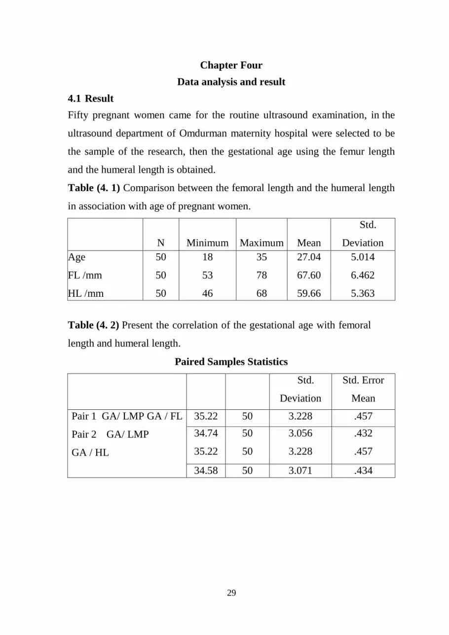
Fifty pregnant women came for the routine ultrasound examination, in the
ultrasound department of Omdurman maternity hospital were selected to be
the sample of the research, then the gestational age using the femur length
and the humeral length is obtained.
Table (4. 1) Comparison between the femoral length and the humeral length
in association with age of pregnant women.
N
Minimum
Maximum
Mean
Std.
Deviation
Age
FL /mm
HL /mm
50
50
50
,
18
53
46
35
78
68
27.04
67.60
59.66
5.014
6.462
5.363
Table (4. 2) Present the correlation of the gestational age with femoral
length and humeral length.
Paired Samples Statistics
Mean
N
Std.
Deviation
Std. Error
Mean
Pair 1 GA/ LMP GA / FL
Pair 2 GA/ LMP
GA / HL
35.22 50 3.228 .457
34.74
35.22
50
50
3.056
3.228
.432
.457
34.58 50 3.071 .434

30
Figure (4. 1) Scatter plot shows the predication system to estimate the
gestational age in weeks by using the femur length in mm.
Figure (4. 2) Scatter plot shows the predication system to estimate the
gestational age in weeks using the humeral length in mm.

31
Table 4-3. Show the mean and the stander deviation of gestational age
measured using the FL and the HL.
Paired Samples Statistics
Mean
N
Std. Deviation
Std. Error
Mean
Pair 1 GA / FL
GA / HL
34.74 50 3.056 .432
34.58 50 3.071 .434
Table 4-4. Show the correlation between the gestational age using the LMP
versus FL, LMP versus HL, and FL versus HL.
Correlations
GA/ LMP GA / FL GA / HL
GA/ Pearson Correlation
LMP Sig. (2-tailed) N
1 .941** .905**
.000 .000
50 50 50
GA / FL Pearson Correlation
Sig. (2-tailed) N
.941** 1 .962**
.000 .000
bs350 50 50
GA / HL Pearson Correlation
Sig. (2-tailed) N
.905** .962** 1
.000 .000
50 50 50
**. Correlation is significant at the 0.01 level (2-tailed).

32
Chapter Five
Discussion, Conclusion and Recommendations
5.1 Discussion
Fifty pregnant women came for the routine ultrasound examination, in the
ultrasound department of Omdurman maternity hospital.
FL is used as standard method to estimate gestational age to be compared
with the other gestational age that obtained with HL and the LMP.
The study revealed that the mean gestational age measured using the HL is
34.58 which is less than week difference from the gestational age calculated
from the FL , so this clarify that there is no significant difference between the
both measured gestational age in the third trimester by using the FL and
HL.The result is similar to previous study (Moawia et.al, 2015). However, A
study conducted by Rosati et al., (2002) revealed that the differences in
accuracy in predicting bone length using formulae derived from data
acquired during early pregnancy can be related to the fact that systematic
variations in the measurement of FL and HL occur particularly during the
first trimester of pregnancy, a period in which the upper and lower bones are
quite difficult to measure accurately (Rosati et al, 2002). In this study, the
humeral length and femur length were measured to confirm the role of fetal
humeral length as biometric parameter which could be used to determine the
gestational age.
Use of the multiple parameters method of assessing gestational age is valid
when the gestational age estimates of the various ultrasound parameters are
similar. If the gestational age estimates of one or several parameters is
greater than 2 weeks different than the estimates of the other parameters,
either the abnormal ultrasound parameters should be excluded or a different
method should be used to estimate gestational age. When the various
ultrasound parameters predict different gestational ages the fetus should be

33
further evaluated to explain these differences. In this study a significant
correlation was noticed between gestational age and HL, this correlation also
seen between FL and gestational age. Thus HL can be added to the formulae
that used in the estimation of gestational age. In the pregnancy with unknown
menstrual dates or a discrepancy between menstrual dates and mean
gestational age predicted by multiple parameters of more than 3 weeks, fetal
age should be estimated by the multiple parameters method. However, the
potential error of this method in the third trimester of pregnancy may not be
acceptable.
In conclusion, assessment of gestational age is fundamental to obstetric care
and should be a carefully thought. Furthermore, HL could be used in
estimation of gestational age.

34
5.2 Conclusion:
From this result we can conclude that the humeral length measurement in the
third trimester is accurate in calculation of the gestational age in the single
fetus with no fetal anomalies, of the healthy pregnant women with normal
uncomplicated pregnancy.
The gestational age calculated using the humeral length is another standard
parameter used in the third trimester to precise measurement of the
gestational age.
There is no significant difference between the gestational age calculated
using the femur length, last menstrual period, and the humeral length.

35
5.3 Recommendations:
1. From the above result we can see that in this research the
gestational age that calculated by the measuring the humeral
length is another accurate measurement for the fetal age in the
third trimester and should be used as standard measure when the
femur length is difficult to be obtain.
2. The humeral length must be used as routine measurement in
calculation the gestational age in the third trimester, to confirm the
accuracy of the femur length measurement, and the precise of the last
menstrual period in the calculation of the third trimester gestational
age.
3. The expected date of the delivery (EDD) should never be
reported, unless is confirmed by using the EDD that obtained
from measuring the HL.
4. Machine to use HL to be used as routine ultrasound .
5. High quality.

36
References:
1. Donofrio, M.T., Moon-Grady, A.J., Hornberger, L.K., Copel, J.A., Sklansky,
M.S., Abuhamad, A., Cuneo, B.F., Huhta, J.C., Jonas, R.A., Krishnan, A.
and Lacey, S., 2014. Diagnosis and treatment of fetal cardiac disease: a
scientific statement from the American Heart
Association. Circulation, 129(21), pp.2183-2242.
2. Gameraddin, et al, 2015, http://www.iosrjournals.org/iosr-
jdms/papers/Vol14-issue5/Version-2/Q014526568.pdf, October, 2016.
3. Kijowski, R., Tuite, M. and Sanford, M., 2004. Magnetic resonance imaging
of the elbow. Part I: normal anatomy, imaging technique, and osseous
abnormalities. Skeletal radiology, 33(12), pp.685-697.
4. Mohammed, A.M.E., 2019. Prediction Of Gestational Age Using Humeral
Length In Third Trimester Using Ultrasonography (Doctoral dissertation,
Sudan University of Science and Technology).
5. Mukherjee, S., 2014. Timing of gestational arrest prior to miscarriage
(Doctoral dissertation, Vanderbilt University).
6. Nagesh, et al, 2016, http://www.jebmh.com/latest-
articles.php?atid=95044, October, 2016.
7. Patre, et al, 2015, http://www.ijsssn.com/uploads/, October 2016.
8. Patre, V., Aryan, A.K., Sahu, P. and Patre, V., 2015. Ultrasonographic
Evaluation of Fetal Humerus Length for Assessment of Gestational Age and
Its Comparison with Other Conventional Parameters. INTERNATIONAL
JOURNAL OF SCIENTIFIC STUDY, 3(7), pp.58-64.
9. Rodan, G.A., 2003. The development and function of the skeleton and bone
metastases. Cancer: Interdisciplinary International Journal of the American
Cancer Society, 97(S3), pp.726-732.
10. Wyatt-Ashmead, J., Konstantinidou, A. and Offiah, A.C., 2014. Skeletal
dysplasias. The Pediatric and Perinatal Autopsy Manual with DVD-ROM,
p.235.

Data collection sheet
NO Maternal age LMP G P BPD(mm) FL(mm) HL(mm) Gestational age(wk)

38