comparison of diurnal variations, gestational age and gender
TRANSCRIPT
RESEARCH ARTICLE
Comparison of diurnal variations, gestational
age and gender related differences in fetal
heart rate (FHR) parameters between
appropriate-for-gestational-age (AGA) and
small-for-gestational-age (SGA) fetuses in the
home environment
Habiba Kapaya1☯*, Richard Jacques2☯, Dilly Anumba1☯
1 Department of Oncology and Metabolism, Academic Unit of Reproductive & Developmental Medicine, The
University of Sheffield, Sheffield, United Kingdom, 2 Medical Statistics Group, School of Health and Related
Research (ScHARR), University of Sheffield, Sheffield, United Kingdom
☯ These authors contributed equally to this work.
Abstract
Objective
To assess the influence of gender, time of the day and gestational age on fetal heart rate
(FHR) parameters between appropriate-for-gestational-age (AGA) and small-for-gestational
age (SGA) fetuses using a portable fetal ECG monitor employed in the home setting.
Methods
We analysed and compared the antenatal FHR data collected in the home setting on 61
healthy pregnant women with singleton pregnancies from 24 weeks gestation. Of the 61
women, 31 had SGA fetuses (estimated fetal weight below the tenth gestational centile) and
30 were pregnant with AGA fetuses. FHR recordings were collected for up to 20 h. Two 90
min intervals were deliberately chosen retrospectively with respect to signal recording qual-
ity, one during day-time and one at night-time for comparison.
Results
Overall, success rate of the fetal abdominal ECG in the AGA fetuses was 75.7% compared
to 48.6% in the SGA group. Based on randomly selected episodes of heart rate traces
where recording quality exceeded 80% we were able to show a marginal difference between
day and night-time recordings in AGA vs. SGA fetuses beyond 32 weeks of gestation. A
selection bias in terms of covering different representation periods of fetal behavioural
states cannot be excluded. In contrast to previous studies, we neither controlled maternal
diet and activity nor measured maternal blood hormone and heart rate as all mothers were
monitored in the home environment.
PLOS ONE | https://doi.org/10.1371/journal.pone.0193908 March 9, 2018 1 / 12
a1111111111
a1111111111
a1111111111
a1111111111
a1111111111
OPENACCESS
Citation: Kapaya H, Jacques R, Anumba D (2018)
Comparison of diurnal variations, gestational age
and gender related differences in fetal heart rate
(FHR) parameters between appropriate-for-
gestational-age (AGA) and small-for-gestational-
age (SGA) fetuses in the home environment. PLoS
ONE 13(3): e0193908. https://doi.org/10.1371/
journal.pone.0193908
Editor: Martin Gerbert Frasch, University of
Washington, UNITED STATES
Received: July 20, 2017
Accepted: February 21, 2018
Published: March 9, 2018
Copyright: © 2018 Kapaya et al. This is an open
access article distributed under the terms of the
Creative Commons Attribution License, which
permits unrestricted use, distribution, and
reproduction in any medium, provided the original
author and source are credited.
Data Availability Statement: All relevant data are
within the paper and its Supporting Information
files.
Funding: The study was funded by Jessop Wing
Small Grant Scheme and sponsored by Sheffield
Teaching Hospitals NHS Foundation Trust, STH
Ref: 18636.
Competing interests: The authors have declared
that no competing interests exist.
Conclusion
Based on clinically unremarkable, but statistically significant differences in the FHR parame-
ters between the AGA and SGA group we suggest that further studies with large sample
size are required to assess the clinical value of antenatal fetal ECG monitoring.
Introduction
Antepartum cardiotocography (CTG) is widely used for the assessment of fetal well-being,
although there is no high-quality evidence to indicate that it improves perinatal outcomes [1,
2]. Such lack of evidence may be partly explained by the limited understanding of the influence
of gestational age, fetal weight, gender and time of the day on the interpretation of CTG pat-
terns. Nonetheless, some evidence of improved predictive performance has been reported with
the use of computer analysis, which is more objective and consistent [3].
Studies have shown changes in CTG parameters with gestation in appropriate-for-gesta-
tional-age (AGA) and small-for-gestational-age (SGA) fetuses [3–7]. In addition, some studies
suggest differences in fetal heart rate (FHR) patterns between male and female fetuses [1, 8]
and between the day or night recordings [9–12]. However, all previous studies were carried
out in hospital in clinical settings in which maternal activity is controlled rather than under
domestic conditions which are more likely to reflect normal day time maternal activities. Fur-
thermore, there is limited data on the FHR variables as most studies utilised visual rather than
computerised system analysis of FHR tracings. Furthermore, to our knowledge, no previous
study has compared the influence of gestational age, time of day and fetal gender on FHR pat-
terns between AGA and SGA fetuses concurrently.
The aim of our present study was to explore the effects of diurnal variation, gestational age
and gender on computerized CTG in a cohort of healthy pregnant women carrying AGA and
SGA fetuses in their home environment using a portable abdominal fetal ECG monitor (Mon-
ica Healthcare, Nottingham, UK).
Material and methods
This work was undertaken to analyze and compare FHR data published separately on normal
[6] and SGA fetuses [7] that were monitored in the home environment. Both prospective stud-
ies were carried out according to ethics approval from the University and Affiliated Teaching
Hospitals Research Ethic Board. The study on small for gestational age fetuses was approved
by North-West Preston NRES Research Ethics committee (15/NW/0278) and on appropriate
for gestational age fetuses was approved by Research Ethics Committee (REC reference num-
ber: 07/Q0108/127). A total of 61 maternal-fetal pairs between 24 and 40 weeks gestation pro-
vided the data for this study. Inclusion criteria for both groups were a maternal age of�18
years with a singleton pregnancy >24 weeks gestation confirmed by early ultrasound scan.
For the purpose of this study, we used the definition by the Royal College of Obstetricians
and Gynaecologists (RCOG) [13] which informs UK clinical practice, based on sonographic
estimated fetal weight (EFW) measurement <10th percentile to describe a fetus that has not
reached its target weight. Patients were divided into two groups for comparison; fetuses with
EFW below the 10th percentile for gestational age (SGA) and fetuses with EFW >10th percen-
tile for gestation (AGA). Exclusion criteria were known fetal malformation, alcohol depen-
dence and any pharmacologically treated co-morbid conditions (e.g. diabetes, hypertension,
Comparison of fetal-heart-rate variation between normal and small fetuses
PLOS ONE | https://doi.org/10.1371/journal.pone.0193908 March 9, 2018 2 / 12
thyroid diseases, and cardiac problems). None of the participants used medications including
β-blockers, central nervous system depressants, tocolytics or steroids which could possibly
affect the FHR. Information about each pregnancy, the characteristics of deliveries and new-
born outcomes were collected from the medical records.
Women provided informed, voluntary, written consent prior to participation. The fetal
ECG monitor (Monica AN24) was attached to the participant’s abdomen by placing five skin
electrodes in standardized positions [14]. Participants went home, carried on with day-to-
day activities whilst wearing the monitor and removed the monitor after 20 hours had
elapsed. They were advised to take off the monitor at any time if they experienced any dis-
comfort. The monitor was either collected from participant’s home by the research team or
returned by the participant the following day. Daily activity was to the discretion of the par-
ticipants. No protocol was followed nor were the times of maternal resting and activity
recorded.
Fetal electrophysiological data recorded by the monitor were downloaded later via USB
connection. The methodology used for signal extraction and analysis has been described in
detail by Cohen et al [15]. In order to investigate changes in FHR pattern between the day and
night times, three consecutive 30 min frames (90 min data) with FHR acquisition success rate
of>80% were selected randomly during the “day” (7:00–23:00) and “night” (23:00–7:00) peri-
ods of the same individual. The FHR parameters studied from the Dawes Redman analysis for
this study were:
• Basal FHR: baseline heart rate
• Long-term FHR variation (LTV): overall: minute-by-minute range of pulse intervals
• Short-term FHR variation (STV) overall: sequential epoch-to-epoch variation. Both the LTV
overall and the STV overall are measured in milliseconds (ms).
• Accelerations: increase in FHR above the baseline that lasted longer than 15 s and had an
amplitude greater than 10 bpm.
• Episodes of high FHR variation (corresponding to active fetal sleep cycles): episodes in
which the LTV in at least 5 out of 6 consecutive minutes was equal to or greater than 32 ms.
In contrast to the ultrasound-based CTG monitors, although the fetal ECG monitor (Mon-
ica AN24) uses different signals to acquire FHR data, the monitor relies on the same averaging
algorithm as introduced by Dawes and Redman [16] to calculate STV and other automated
FHR parameters. In all cases, FHR data selected for the analysis fulfilled the Dawes Redman
criteria.
Statistical analysis
Independent sample t-tests were used to compare the characteristics of mothers and babies
between the two groups.
Mean differences in day and night time measurements of FHR parameters were calculated
within the AGA and the SGA group. To compare differences between groups, linear models
were fitted with FHR as the dependent variable and fixed factors for time of measurement and
fetus size as independent variables. An interaction term between the two fixed factors was
included to test if there was a difference in the day and night time measurements between the
two groups. Generalised estimating equations were used to fit the models to allow for repeated
measurements on individuals [17]. Differences between groups and time of recording are visu-
ally displayed using marginal means from the fitted models.
Comparison of fetal-heart-rate variation between normal and small fetuses
PLOS ONE | https://doi.org/10.1371/journal.pone.0193908 March 9, 2018 3 / 12
Mean differences between genders were calculated within the AGA and the SGA group. To
compare differences between genders, linear models were fitted with FHR measurement as the
dependent variable and fixed factors for gender and fetus size as independent variable. An
interaction term between the two fixed factors was included to test if there was a difference in
measurements between genders in the normal and the SGA group. Generalised estimating
equations were used to fit the models to allow for repeated measurements on individuals.
Linear models were used to examine the relationship between heart rate parameters mea-
sured at night and gestational age at time of recording. Models were fitted with FHR as the
dependent variable and a fixed factor for fetus size and gestational age covariate as indepen-
dent variables. An interaction term between the two independent variables was included to
allow for different relationships in the normal and the SGA group. Step wise model selection
was used to first test for the interaction, the difference between the two groups and finally the
linear trend. At each step, non-significant terms were removed.
Results
A total of 61 maternal-fetal pairs were studied; 30 participants carried AGA and 31 carried
SGA fetuses. The baseline characteristics of the participants and their fetuses are given in
Table 1. Although, the two groups were broadly comparable in terms of demographics and
gestations both at the time of recording and delivery, gender distribution was significantly dif-
ferent between the two groups with more male fetuses in the AGA group and a significantly
higher number of females in the SGA group (P = 0.027). As expected, birth weight was signifi-
cantly lower in the SGA group (P<0.001).
One hundred and twenty-two recordings (61 day and 61 night) made over 90min segments
were analysed from 30 AGA and 31 SGA fetuses. All the parameters from both AGA and SGA
fetuses were in the normal range at any times of the observations. Exact time of the day and
maternal activity during the recordings could not be reconstructed retrospectively.
Table 2 summarizes and compares day: night differences between the two groups. A signifi-
cant variation in day: night recording was observed in AGA fetuses with a fall in basal FHR
and an increase in STV, LTV, number of accelerations and time spent in high variation epi-
sode at night time (P� 0.05). The differences in STV, though, are within 1 millisecond of vari-
ation. However, SGA fetuses exhibited day: night difference only in basal FHR (P<0.001),
whereas STV, LTV, acceleration and time spent in HV remained unchanged (P>0.05). This
comparison is further illustrated in Fig 1.
With respect to gender, even though, the difference in STV between male and female
fetuses was higher both in the AGA and the SGA groups than the diurnal variation described
above, this observation did not reach statistical significance (Table 3).
Further analysis of the effect of gestational age on FHR parameters (Table 4) showed no sig-
nificant difference between AGA and SGA fetuses.
Table 1. Summary statistics for maternal and fetal demographic and obstetric characteristics between the AGA and the SGA.
AGA (N = 30) SGA (N = 31) Difference (95% CI) P-Value
Age (years) 28.9 (5.6) 30.0 (6.1) -1.1(-4.1, 1.9) 0.456
Body mass index (kg/m2) 26.3 (5.7) 25.2 (6.3) 1.1(-2.0, 4.2) 0.479
Gestational age at recording (weeks) 33.8 (3.9) 35.1 (3.2) -1.3(-3.1, 0.5) 0.158
Gestational age at delivery (weeks) 38.9 (2.4) 39.0 (1.8) -0.1(-1.1, 1.0) 0.918
Weight of baby (kg) 3.46 (0.55) 2.93 (0.51) 0.53(0.25, 0.80) <0.001
Sex of babyMaleFemale 19 (63.3%)11 (36.7%) 10 (34.5%)19 (65.5%) 31.1(6.0, 51.2) 0.027
https://doi.org/10.1371/journal.pone.0193908.t001
Comparison of fetal-heart-rate variation between normal and small fetuses
PLOS ONE | https://doi.org/10.1371/journal.pone.0193908 March 9, 2018 4 / 12
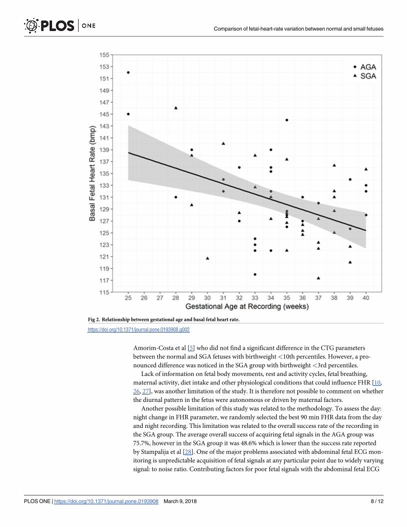
A significant negative correlation between gestational age and basal FHR (Fig 2; P = 0.003)
and a weak non-significant positive correlation between LTV, accelerations and time spent in
HV and gestational age was observed in both groups (P>0.05).
Discussion
AGA fetuses exhibited a clinically unremarkable, but in our data statistically significant differ-
ence of the Dawes and Redman parameter set between daytime and nighttime recordings.
This difference could not be demonstrated in fetuses with known SGA. SGA fetuses showed
similar change in FHR pattern with gestational age as AGA fetuses. Gender did not influence
CTG interpretation in either group.
This is the first study to compare and report changes in day: night FHR parameters, gesta-
tional age and gender related differences in CTG in AGA and SGA fetuses in home environ-
ment. Our data was retrieved collecting recordings of the FHR in a home environment over 20
hours. Data quality was as such, that overall 3x30 min both during daytime and night-time
met the quality criteria for computerized Dawes and Redman analysis. This is most likely due
to the fact, that muscular activity during maternal movement is likely to obscure the fetal ECG
from the trans-abdominal recording to a significant proportion.
There are several limitations of this study. There is no consensus on the terminology and
diagnostic criteria of fetal growth restriction. The most widely used definition for fetal growth
restriction in the United States is an estimated fetal weight (EFW) <10th percentile for gesta-
tional age [18]. However, the Royal College of Obstetricians and Gynaecologists (RCOG) con-
siders that fetuses with an EFW or abdominal circumference (AC)<10th percentile, as well as
infants with a birthweight <10th percentile are SGA, which is different from fetal growth
restriction [13]. SGA includes: 1) babies that have not reached their target growth thorough
pathological restriction of the genetic growth potential and possible manifestations of fetal
compromise (Doppler abnormalities) and 2) babies that are constitutionally small [19]. Serial
ultrasounds can allow identification and differentiation between the two to assess severity and
timing of growth restriction. The likelihood of fetal growth restriction is higher in severe SGA
cases, defined as an EFW, AC or birthweight <3rd percentile [13]. In the present study,
although our SGA cohort did not show any evidence of Doppler changes nor was the EFW
below the 3rd percentile at the time of recruitment, our study did not take into account the lon-
gitudinal evolution of each fetus, and the existence of a single sonographic finding of EFW
measurement <10th centile with normal Doppler studies per fetus was employed for study
inclusion.
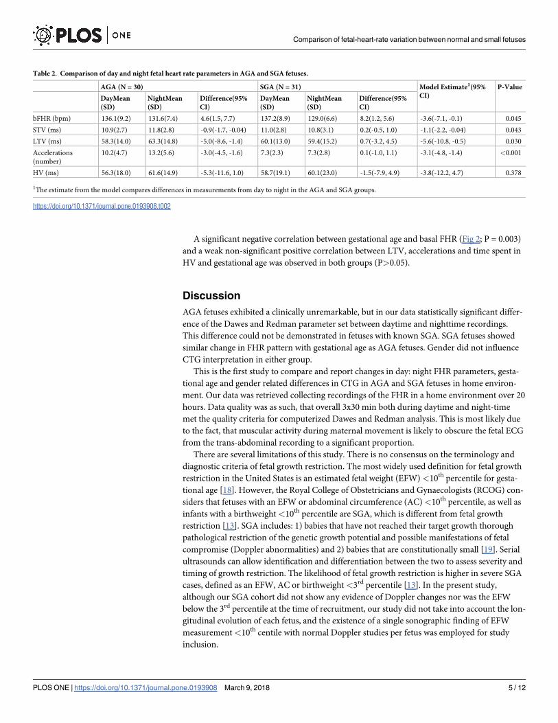
Table 2. Comparison of day and night fetal heart rate parameters in AGA and SGA fetuses.
AGA (N = 30) SGA (N = 31) Model Estimate1(95%
CI)
P-Value
DayMean
(SD)
NightMean
(SD)
Difference(95%
CI)
DayMean
(SD)
NightMean
(SD)
Difference(95%
CI)
bFHR (bpm) 136.1(9.2) 131.6(7.4) 4.6(1.5, 7.7) 137.2(8.9) 129.0(6.6) 8.2(1.2, 5.6) -3.6(-7.1, -0.1) 0.045
STV (ms) 10.9(2.7) 11.8(2.8) -0.9(-1.7, -0.04) 11.0(2.8) 10.8(3.1) 0.2(-0.5, 1.0) -1.1(-2.2, -0.04) 0.043
LTV (ms) 58.3(14.0) 63.3(14.8) -5.0(-8.6, -1.4) 60.1(13.0) 59.4(15.2) 0.7(-3.2, 4.5) -5.6(-10.8, -0.5) 0.030
Accelerations
(number)
10.2(4.7) 13.2(5.6) -3.0(-4.5, -1.6) 7.3(2.3) 7.3(2.8) 0.1(-1.0, 1.1) -3.1(-4.8, -1.4) <0.001
HV (ms) 56.3(18.0) 61.6(14.9) -5.3(-11.6, 1.0) 58.7(19.1) 60.1(23.0) -1.5(-7.9, 4.9) -3.8(-12.2, 4.7) 0.378
1The estimate from the model compares differences in measurements from day to night in the AGA and SGA groups.
https://doi.org/10.1371/journal.pone.0193908.t002
Comparison of fetal-heart-rate variation between normal and small fetuses
PLOS ONE | https://doi.org/10.1371/journal.pone.0193908 March 9, 2018 5 / 12
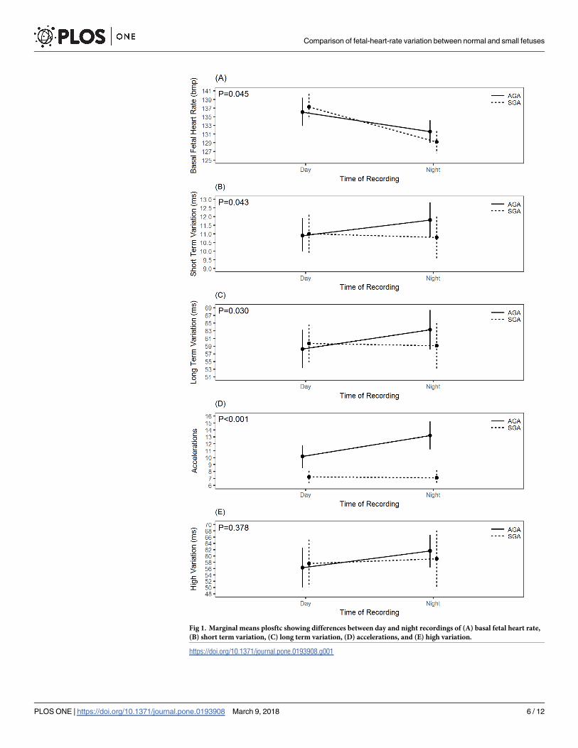
Fig 1. Marginal means plosftc showing differences between day and night recordings of (A) basal fetal heart rate,
(B) short term variation, (C) long term variation, (D) accelerations, and (E) high variation.
https://doi.org/10.1371/journal.pone.0193908.g001
Comparison of fetal-heart-rate variation between normal and small fetuses
PLOS ONE | https://doi.org/10.1371/journal.pone.0193908 March 9, 2018 6 / 12
In the current study, SGA fetuses demonstrated the widely reported phenomenon of matu-
rational change and a similar trend of CTG parameters with increasing gestational age as do
the normal fetus. Over the last couple of decades, it has become clear that fetal growth restric-
tion can start early in pregnancy when it is termed as early onset of fetal growth restriction
[20]. Figueras and Gratacos [21] and Baschat [22] have reported different behaviors in fetuses
with growth restriction before and after 32 weeks gestation [23]. Early onset fetal growth
restriction follows a more severe trajectory in terms of neonatal outcome [20] and more pro-
nounced differences in the CTG parameters compared to the late onset growth restricted [24].
In this context, our study was limited and under-representative of SGA fetuses before 32
weeks gestation (only six in the study cohort) which could mean our observations may have
occurred by chance or due to selection bias. Due to small sample size we did not undertake
separate analysis on SGA<32 week’s gestation. However, when six SGA fetuses under 32
weeks were removed from the analysis, results did not change, implying that our study was
representative of less affected SGA that were not truly compromised and therefore remained
in utero until later gestational ages. This may explain our observation of a greater overlap in
CTG parameters between the normal and SGA fetuses, i.e. a significant decrease in basal FHR
and a progressive increase in FHR variation and acceleration with advancing gestation. This is
in agreement with the observation made by Soncini et al [25] who found a progressive increase
in FHR variability and accelerations with advancing gestational age in intra-uterine-growth
restricted (IUGR) fetuses with mild or no Doppler velocimetry abnormalities. However, in
more compromised fetuses that showed a greater impairment of placental perfusion, this mat-
uration process of FHR pattern was not observed [25]. A similar observation was made by
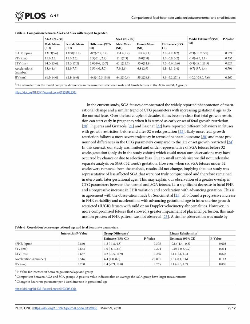
Table 3. Comparison between AGA and SGA with respect to gender.
AGA (N = 30) SGA (N = 29) Model Estimate1(95%
CI)
P-Value
Male Mean
(SD)
Female Mean
(SD)
Difference(95%
CI)
Male Mean
(SD)
FemaleMean
(SD)
Difference(95%
CI)
bFHR (bpm) 131.3(5.6) 132.0(10.0) -0.7(-7.7, 6.4) 131.4(5.2) 128.4(7.1) 3.0(-2.2, 8.2) -2.3(-10.2, 5.7) 0.574
STV (ms) 11.9(2.6) 11.6(2.6) 0.3(-2.1, 2.8) 11.1(2.3) 10.0(2.8) 1.0(-0.9, 3.2) -1.0(-4.0, 2.1) 0.535
LTV (ms) 64.0(13.6) 62.0(17.2) 2.0(-9.6, 13.7) 61.1(11.7) 55.6(14.8) 5.5(-5.6,16.6) -3.8(-19.1,11.5) 0.627
Accelerations
(number)
13.4(4.4) 12.9(7.7) 0.5(-4.0, 5.0) 7.9(2.6) 6.8 (2.9) 1.1(-1.1, 3.4) -0.7(-5.7, 4.4) 0.796
HV (ms) 61.3(14.0) 62.1(16.6) -0.8(-12.3,10.8) 64.2(10.4) 55.2(26.8) 8.9(-9.2,27.1) -10.2(-28.0, 7.6) 0.260
1The estimate from the model compares differences in measurements between male and female fetuses in the AGA and SGA groups
https://doi.org/10.1371/journal.pone.0193908.t003
Table 4. Correlation between gestational age and fetal heart rate parameters.
InteractionP-Value1 Group Difference2 Linear Relationship3
Estimate (95% CI) P-Value Estimate (95% CI) P-Value
bFHR (bpm) 0.840 1.5 (-1.8, 4.8) 0.375 -0.8 (-1.4, -0.3) 0.003
STV (ms) 0.653 1.0 (-6.1, 2.6) 0.224 -0.03 (-0.3, 0.2) 0.814
LTV (ms) 0.687 4.2 (-3.5, 11.9) 0.286 0.1 (-1.1, 1.3) 0.828
Accelerations (number) 0.516 6.4 (4.0, 8.6) <0.001 0.3 (-0.1, 0.6) 0.113
HV (ms) 0.700 1.4 (-7.9, 10.8) 0.765 0.1 (-1.5, 1.7) 0.896
1 P-Value for interaction between gestational age and group2 Comparison between AGA and SGA groups. A positive value indicates that on average the AGA group have larger measurements3 Change in heart rate parameter per 1 week increase in gestational age
https://doi.org/10.1371/journal.pone.0193908.t004
Comparison of fetal-heart-rate variation between normal and small fetuses
PLOS ONE | https://doi.org/10.1371/journal.pone.0193908 March 9, 2018 7 / 12
Amorim-Costa et al [5] who did not find a significant difference in the CTG parameters
between the normal and SGA fetuses with birthweight <10th percentiles. However, a pro-
nounced difference was noticed in the SGA group with birthweight <3rd percentiles.
Lack of information on fetal body movements, rest and activity cycles, fetal breathing,
maternal activity, diet intake and other physiological conditions that could influence FHR [10,
26, 27], was another limitation of the study. It is therefore not possible to comment on whether
the diurnal pattern in the fetus were autonomous or driven by maternal factors.
Another possible limitation of this study was related to the methodology. To assess the day:
night change in FHR parameter, we randomly selected the best 90 min FHR data from the day
and night recording. This limitation was related to the overall success rate of the recording in
the SGA group. The average overall success of acquiring fetal signals in the AGA group was
75.7%, however in the SGA group it was 48.6% which is lower than the success rate reported
by Stampalija et al [28]. One of the major problems associated with abdominal fetal ECG mon-
itoring is unpredictable acquisition of fetal signals at any particular point due to widely varying
signal: to noise ratio. Contributing factors for poor fetal signals with the abdominal fetal ECG
Fig 2. Relationship between gestational age and basal fetal heart rate.
https://doi.org/10.1371/journal.pone.0193908.g002
Comparison of fetal-heart-rate variation between normal and small fetuses
PLOS ONE | https://doi.org/10.1371/journal.pone.0193908 March 9, 2018 8 / 12
include, maternal activity, environmental electrical noise, gestational age, insulating vernix
caseosa and amniotic fluid [14, 29–31]. A lower success rate meant that it was not possible to
analyse the whole 20hr recording, therefore we randomly selected the best 90 min data with
fetal signals >80% both during the day and night, implying that our study may have suffered
selection bias. A healthy fetus cycles between episodes of active and quiet sleep. Active sleep is
associated with high variation and quiet sleep is associated with low variation episode. Studies
have shown quiet sleep can last between 50–90 min, therefore selection of 90 min intervals
guaranteed the analysis of high variation episode [5, 32]. One of the Dawes and Redman crite-
ria for normality is the presence of at least one episode of high variation [33] and this normal-
ity criteria was met for all the selected FHR traces.
The diurnal pattern of FHR and its variation exists from 20 to 22 weeks onwards [11]. We
observed a significant day: night difference in basal FHR in both AGA and SGA fetuses. How-
ever, unlike AGA fetuses, SGA fetuses showed blunting of the diurnal pattern for STV, LTV,
accelerations and time spent in HV episode. The reason for the difference in behavior of the
SGA fetuses compared to their AGA counterparts is unclear. Our study predominantly com-
prised of SGA fetuses >32 weeks gestation, implying that gestational age did not affect the
altered diurnal pattern observed in SGA group. In addition, our study was designed to exclude
other conditions and treatments which could influence diurnal variation of FHR patterns. For
instance, studies have shown that in pregnancies complicated by pre-eclampsia, the develop-
ment of diurnal rhythm in the fetus is hampered [34, 35]. For the purpose of this study, we
therefore excluded women with SGA who had pre-eclampsia. In addition, during monitoring,
no woman received any medication that could have affected the CTG parameters [36, 37].
However, there were 10 smokers within the SGA group. Although, the information they pro-
vided indicates that they did not smoke during the monitoring session, the study relied on the
women to provide information on their smoking status rather than making an objective assess-
ment of tobacco exposure. Nicotine consumption has not only been shown to produce greater
effect in women but delays the diurnal peak of heart rate pattern [38]. This, may explain why
SGA fetuses did not demonstrate the same diurnal pattern in FHR parameters as the normal
fetuses’.
To date, only a few studies have evaluated the influence of gender on CTG parameters and
these have produced conflicting results. Lange et al [39] suggested that gender does not need
to be considered when assessing FHR variability, whereas Fleisher et al [40] and Amorim-
Costa et al [1] reported significant gender-related differences in FHR parameters at different
gestations. These studies were undertaken on normal fetuses. With regard to SGA pregnancies,
there is insufficient data. Kwon et al [8] assessed the influence of gender on FHR between 29
SGA and 385 non-SGA fetuses. Their data suggested no influence of gender in the normal
group, but a significant gender-related difference in FHR variability in the SGA group. Our
results differ from them, as we did not observe any influence of gender on either of the two
groups. This discrepancy can be explained by experimental setting and sample size. We exam-
ined the influence of gender on the fetal recordings during the antenatal period whereas,
Kwon et al [8] carried out their investigation during labour.
Conclusion
This study confirms that although SGA fetus with no evidence of Doppler velocimetry abnor-
malities exhibit similar changes in FHR pattern with gestation as do AGA fetuses, the CTG
pattern seems to be differing between the two groups when the time of day is taken into con-
sideration. Nonetheless, all our observations are in the range of normal findings. We did not
follow up on perinatal outcome, nor we analysed beat-to-beat information on the acquired
Comparison of fetal-heart-rate variation between normal and small fetuses
PLOS ONE | https://doi.org/10.1371/journal.pone.0193908 March 9, 2018 9 / 12

FHR data, implying that may be the underlying difference in the STV between the AGA and
the SGA were masked by the use of averaged FHR data provided by the Monica AN24 leading
to the loss in temporal resolution. We suggest that further studies should be undertaken to
explore the value of true beat-to-beat information on FHR variability by employing Monica
AN24 before its clinical utility and more widespread adoption can be ascertained.
Supporting information
S1 Fig. Data file Normal_Small babies.
(XLSX)
Acknowledgments
We gratefully acknowledge Monica Healthcare, Nottingham, UK for providing electrodes in
kind for the study.
Author Contributions
Conceptualization: Habiba Kapaya, Dilly Anumba.
Data curation: Habiba Kapaya, Richard Jacques.
Formal analysis: Habiba Kapaya, Richard Jacques.
Investigation: Habiba Kapaya.
Methodology: Habiba Kapaya.
Project administration: Habiba Kapaya.
Supervision: Dilly Anumba.
Visualization: Dilly Anumba.
Writing – original draft: Habiba Kapaya.
Writing – review & editing: Richard Jacques, Dilly Anumba.
References1. Amorim-Costa C, Cruz J, Ayres-de-Campos D, Bernardes J: Gender-specific reference charts for cardi-
otocographic parameters throughout normal pregnancy: a retrospective cross-sectional study of 9701
fetuses. Eur J Obstet Gynecol Reprod Biol 2016, 199:102–107. https://doi.org/10.1016/j.ejogrb.2016.
01.036 PMID: 26921476
2. Grivell RM, Alfirevic Z, Gyte GM, Devane D: Antenatal cardiotocography for fetal assessment.
Cochrane Database Syst Rev 2012, 12:CD007863. https://doi.org/10.1002/14651858.CD007863.
pub3 PMID: 23235650
3. Serra V, Bellver J, Moulden M, Redman CW: Computerized analysis of normal fetal heart rate pattern
throughout gestation. Ultrasound Obstet Gynecol 2009, 34(1):74–79. https://doi.org/10.1002/uog.6365
PMID: 19489020
4. Yanagihara T, Hata T: Comparison of late-second-trimester nonstress test characteristics between
small for gestational age and appropriate for gestational age infants. Obstet Gynecol 1999, 94(6):921–
924. PMID: 10576176
5. Amorim-Costa C, de Campos DA, Bernardes J: Cardiotocographic parameters in small-for-gestational-
age fetuses: How do they vary from normal at different gestational ages? A study of 11687 fetuses from
25 to 40 weeks of pregnancy. J Obstet Gynaecol Res 2017, 43(3):476–485. https://doi.org/10.1111/
jog.13235 PMID: 28165176
6. Kapaya H, Broughton Pipkin F, Hayes-Gill B, Loughna PV: Circadian changes and sex-related differ-
ences in fetal heart rate parameters. Matern Health Neonatol Perinatol 2016, 2(1):9. https://doi.org/10.
1186/s40748-016-0037-6 PMID: 27595008
Comparison of fetal-heart-rate variation between normal and small fetuses
PLOS ONE | https://doi.org/10.1371/journal.pone.0193908 March 9, 2018 10 / 12
7. K H, D ER, A D: Do small for gestational age foetuses’ exhibit circadian changes in fetal heart rate
parameters as do appropriate for gestational age foetuses? Edorium J Matern Child Health 2017,
2:1–8.
8. Kwon JY, Park IY, Lim J, Shin JC: Changes in spectral power of fetal heart rate variability in small-for-
gestational-age fetuses are associated with fetal sex. Early Hum Dev 2014, 90(1):9–13. https://doi.org/
10.1016/j.earlhumdev.2013.11.005 PMID: 24332839
9. Babazadeh R, Abdali K, Lotfalizadeh M, Tabatabaie HR, Kaviani M: Diurnal nonstress test variations in
the human fetus at risk. Int J Gynaecol Obstet 2005, 90(3):189–192. https://doi.org/10.1016/j.ijgo.
2005.05.011 PMID: 16043174
10. Lunshof S, Boer K, Wolf H, van Hoffen G, Bayram N, Mirmiran M: Fetal and maternal diurnal rhythms
during the third trimester of normal pregnancy: outcomes of computerized analysis of continuous
twenty-four-hour fetal heart rate recordings. Am J Obstet Gynecol 1998, 178(2):247–254. PMID:
9500482
11. de Vries JI, Visser GH, Mulder EJ, Prechtl HF: Diurnal and other variations in fetal movement and heart
rate patterns at 20–22 weeks. Early Hum Dev 1987, 15(6):333–348. PMID: 3436277
12. Visser GH, Goodman JD, Levine DH, Dawes GS: Diurnal and other cyclic variations in human fetal
heart rate near term. Am J Obstet Gynecol 1982, 142(5):535–544. PMID: 7199260
13. Royal College of Obstetricians and Gynaecologists. The investigation and management of the small-
for-gestational-age fetus (guideline no. 31). London: Royal College of Obstetricians and Gynaecolo-
gists; 2002. In.
14. Graatsma EM, Jacod BC, van Egmond LA, Mulder EJ, Visser GH: Fetal electrocardiography: feasibility
of long-term fetal heart rate recordings. BJOG 2009, 116(2):334–337; discussion 337–338. https://doi.
org/10.1111/j.1471-0528.2008.01951.x PMID: 19076966
15. Cohen WR, Ommani S, Hassan S, Mirza FG, Solomon M, Brown R, Schifrin BS, Himsworth JM,
Hayes-Gill BR: Accuracy and reliability of fetal heart rate monitoring using maternal abdominal surface
electrodes. Acta Obstet Gynecol Scand 2012, 91(11):1306–1313. https://doi.org/10.1111/j.1600-0412.
2012.01533.x PMID: 22924738
16. Seliger G, Petroff D, Seeger S, Hoyer D, Tchirikov M, Schneider U: Diurnal variations of short-term vari-
ation and the impact of multiple recordings on measurement accuracy. J Perinatol 2017, 37(3):231–
235. https://doi.org/10.1038/jp.2016.202 PMID: 27831546
17. Burton P, Gurrin L, Sly P. Tutorial in Biostatistics. Extending the simple linear regression model to
account for correlated responses: an introduction to generalized estimating equations and multi-level
mixed modelling. Statistics in Medicine 1998, 17:1261–1291. PMID: 9670414
18. American College of Obstetricians and Gynecologists. Intrauterine growth restriction. Washington, DC:
American College of Obstetricians and Gynecologists; 2000. In.
19. Grivell RM, Wong L, Bhatia V: Regimens of fetal surveillance for impaired fetal growth. Cochrane Data-
base Syst Rev 2012(6):CD007113. https://doi.org/10.1002/14651858.CD007113.pub3 PMID:
22696366
20. Nawathe A, Lees C: Early onset fetal growth restriction. Best Pract Res Clin Obstet Gynaecol 2017,
38:24–37. https://doi.org/10.1016/j.bpobgyn.2016.08.005 PMID: 27693119
21. Figueras F, Gratacos E: Stage-based approach to the management of fetal growth restriction. Prenat
Diagn 2014, 34(7):655–659. https://doi.org/10.1002/pd.4412 PMID: 24839087
22. Baschat AA: Neurodevelopment following fetal growth restriction and its relationship with antepartum
parameters of placental dysfunction. Ultrasound Obstet Gynecol 2011, 37(5):501–514. https://doi.org/
10.1002/uog.9008 PMID: 21520312
23. Nardozza LM, Caetano AC, Zamarian AC, Mazzola JB, Silva CP, Marcal VM, Lobo TF, Peixoto AB,
Araujo E Junior: Fetal growth restriction: current knowledge. Arch Gynecol Obstet 2017, 295(5):1061–
1077. https://doi.org/10.1007/s00404-017-4341-9 PMID: 28285426
24. Amorim-Costa C, Gaio AR, Ayres-de-Campos D, Bernardes J: Longitudinal changes of cardiotoco-
graphic parameters throughout pregnancy: a prospective cohort study comparing small-for-gestational-
age and normal fetuses from 24 to 40 weeks. J Perinat Med 2017, 45(4):493–501. https://doi.org/10.
1515/jpm-2016-0065 PMID: 27474837
25. Soncini E, Ronzoni E, Macovei D, Grignaffini A: Integrated monitoring of fetal growth restriction by com-
puterized cardiotocography and Doppler flow velocimetry. Eur J Obstet Gynecol Reprod Biol 2006, 128
(1–2):222–230. https://doi.org/10.1016/j.ejogrb.2006.01.001 PMID: 16431011
26. Ozkaya E, Baser E, Cinar M, Korkmaz V, Kucukozkan T: Does diurnal rhythm have an impact on fetal
biophysical profile? J Matern Fetal Neonatal Med 2012, 25(4):335–338. https://doi.org/10.3109/
14767058.2011.576721 PMID: 21696335
Comparison of fetal-heart-rate variation between normal and small fetuses
PLOS ONE | https://doi.org/10.1371/journal.pone.0193908 March 9, 2018 11 / 12
27. Marzbanrad F, Kimura Y, Palaniswami M, Khandoker AH: Quantifying the Interactions between Mater-
nal and Fetal Heart Rates by Transfer Entropy. PLoS One 2015, 10(12):e0145672. https://doi.org/10.
1371/journal.pone.0145672 PMID: 26701122
28. Stampalija T, Casati D, Montico M, Sassi R, Rivolta MW, Maggi V, Bauer A, Ferrazzi E: Parameters
influence on acceleration and deceleration capacity based on trans-abdominal ECG in early fetal growth
restriction at different gestational age epochs. Eur J Obstet Gynecol Reprod Biol 2015, 188:104–112.
https://doi.org/10.1016/j.ejogrb.2015.03.003 PMID: 25801726
29. Hasan MA, Reaz MB, Ibrahimy MI, Hussain MS, Uddin J: Detection and Processing Techniques of
FECG Signal for Fetal Monitoring. Biol Proced Online 2009, 11:263–295. https://doi.org/10.1007/
s12575-009-9006-z PMID: 19495912
30. Stinstra JG, Peters MJ: The influence of fetoabdominal tissues on fetal ECGs and MCGs. Arch Physiol
Biochem 2002, 110(3):165–176. https://doi.org/10.1076/apab.110.3.165.8293 PMID: 12221516
31. Sameni R, Clifford GD: A Review of Fetal ECG Signal Processing; Issues and Promising Directions.
Open Pacing Electrophysiol Ther J 2010, 3:4–20. https://doi.org/10.2174/1876536X01003010004
PMID: 21614148
32. Muro M, Shono H, Kohara M, Ito Y, Uchiyama A, Sugimori H: Diurnal variations in resting-active cycles
in full-term fetal heart rate changes. Early Hum Dev 1996, 44(1):51–58. PMID: 8821895
33. Pardey J, Moulden M, Redman CW: A computer system for the numerical analysis of nonstress tests.
Am J Obstet Gynecol 2002, 186(5):1095–1103. PMID: 12015543
34. Koenen SV, Franx A, Mulder EJ, Bruinse HW, Visser GH: Fetal and maternal cardiovascular diurnal
rhythms in pregnancies complicated by pre-eclampsia and intrauterine growth restriction. J Matern
Fetal Neonatal Med 2002, 11(5):313–320. https://doi.org/10.1080/jmf.11.5.313.320 PMID: 12389672
35. Ditisheim AJ, Dibner C, Philippe J, Pechère-Bertschi A: Biological rhythms and preeclampsia. Front
Endocrinol (Lausanne) 2013, 4:47.
36. Piazze J, Dillon KC, Albana C: Full-term-pregnancy effects of antenatal betamethasone administration
on short-term variation as assessed by computerized cardiotocography. J Prenat Med 2012, 6(2):18–
21. PMID: 22905307
37. Nensi A, De Silva DA, von Dadelszen P, Sawchuck D, Synnes AR, Crane J, Magee LA: Effect of mag-
nesium sulphate on fetal heart rate parameters: a systematic review. J Obstet Gynaecol Can 2014, 36
(12):1055–1064. https://doi.org/10.1016/S1701-2163(15)30382-0 PMID: 25668040
38. Adan A, Sanchez-Turet M: Influence of smoking and gender on diurnal variations of heart rate reactivity
in humans. Neurosci Lett 2001, 297(2):109–112. PMID: 11121882
39. Lange S, Van Leeuwen P, Geue D, Hatzmann W, Gronemeyer D: Influence of gestational age, heart
rate, gender and time of day on fetal heart rate variability. Med Biol Eng Comput 2005, 43(4):481–486.
PMID: 16255430
40. Fleisher LA, Dipietro JA, Johnson TR, Pincus S: Complementary and non-coincident increases in heart
rate variability and irregularity during fetal development. Clin Sci (Lond) 1997, 92(4):345–349.
Comparison of fetal-heart-rate variation between normal and small fetuses
PLOS ONE | https://doi.org/10.1371/journal.pone.0193908 March 9, 2018 12 / 12