ergot alkaloid biosynthesis in aspergillus fumigatus - the
TRANSCRIPT
Graduate Theses, Dissertations, and Problem Reports
2009
Ergot alkaloid biosynthesis in Aspergillus fumigatus : Association Ergot alkaloid biosynthesis in Aspergillus fumigatus : Association
with sporulation and clustered genes common among ergot fungi with sporulation and clustered genes common among ergot fungi
Christine M. Coyle West Virginia University
Follow this and additional works at: https://researchrepository.wvu.edu/etd
Recommended Citation Recommended Citation Coyle, Christine M., "Ergot alkaloid biosynthesis in Aspergillus fumigatus : Association with sporulation and clustered genes common among ergot fungi" (2009). Graduate Theses, Dissertations, and Problem Reports. 4453. https://researchrepository.wvu.edu/etd/4453
This Dissertation is protected by copyright and/or related rights. It has been brought to you by the The Research Repository @ WVU with permission from the rights-holder(s). You are free to use this Dissertation in any way that is permitted by the copyright and related rights legislation that applies to your use. For other uses you must obtain permission from the rights-holder(s) directly, unless additional rights are indicated by a Creative Commons license in the record and/ or on the work itself. This Dissertation has been accepted for inclusion in WVU Graduate Theses, Dissertations, and Problem Reports collection by an authorized administrator of The Research Repository @ WVU. For more information, please contact [email protected].
Ergot alkaloid biosynthesis in Aspergillus fumigatus: Association with sporulation and clustered genes common among ergot fungi
Christine M. Coyle
Dissertation submitted to the Davis College of Agriculture, Forestry, and Consumer Sciences
at West Virginia University in partial fulfillment of the requirements
for the degree of
Doctor of Philosophy in
Genetics and Developmental Biology
Daniel G. Panaccione, Ph.D., Chair Kenneth P. Blemings, Ph.D.
Joseph B. Morton, Ph.D. Alan J. Sexstone, Ph.D.
Sharon L. Wenger, Ph.D.
Division of Plant and Soil Sciences
Morgantown, West Virginia 2009
Keywords: Aspergillus fumigatus, ergot alkaloid biosynthesis, sporulation
Copyright 2009 Christine M. Coyle
ABSTRACT
Ergot alkaloid biosynthesis in Aspergillus fumigatus: Association with sporulation and clustered genes common among ergot fungi
Christine M. Coyle
Ergot alkaloids, indole-derived mycotoxins, interact with multiple monoamine neurotransmitter receptors and cause disease in exposed individuals. They have been well studied in the ergot fungus, Claviceps purpurea, and have been reported in some closely related grass endophytes, as well as the distantly related opportunistic human pathogen Aspergillus fumigatus. A. fumigatus, which sporulates prolifically, produces clavines, specifically festuclavine and fumigaclavines A, B, and C in association with asexual spores and the total mass of alkaloids constitutes over 1% of the spore mass. These alkaloids differ from those of most clavicipitaceous fungi, which consist of different clavines and often more complex lysergic acid derivatives. However, the ergot alkaloid pathways of A. fumigatus and ergot fungi are hypothesized to share early biosynthetic steps, diverging at some point after the formation of the intermediate chanoclavine. A homologue of dmaW, a gene encoding dimethylallyltryptophan synthase in Neotyphodium endophytes, was found in the A. fumigatus genome. By gene knockout analysis, dmaW was shown to be required for ergot alkaloid production in A. fumigatus. Comparison of genes clustered around A. fumigatus dmaW to those clustered with dmaW in the ergot fungi revealed potential homologues that could encode proteins controlling early, shared steps in the pathway. Functional analysis via gene knockout of three additional A. fumigatus genes (easA, easE, and easF) rendered mutants with altered alkaloid profiles, demonstrating their involvement in ergot alkaloid biosynthesis. All mutants lacked normal ergot alkaloid production from the latter part of the pathway; complementation with a functional copy of the respective gene, restored normal ergot alkaloid production in each mutant. Analyses of intermediates positioned the products of easF and easE as the second and third enzymatic steps of the pathway. Knockout of easA caused accumulation of multiple intermediates, including chanoclavine; complementation with the C. purpurea easA gene resulted in accumulation of agroclavine, setoclavine, and its isomer isosetoclavine. These data confirm easA involvement post chanoclavine synthesis and more specifically assign its role to reduction of chanoclavine aldehyde, the branch point of the two lineage-specific pathways. These mutants, due to their differing ergot alkaloid profiles, are valuable for testing the role of specific ergot alkaloids in animal pathogenesis and toxicoses. Elucidation of ergot alkaloid biosynthesis, along with the capacity to control the spectrum of alkaloids produced, may be beneficial to agriculture and medicine. Additional studies demonstrated that production of ergot alkaloids was restricted to conidiating cultures. Disruption of brlA, a regulatory gene involved in conidiation, interfered with conidiophore development, as well as ergot alkaloid production. The association of these toxins with sporulation may offer insight into their ecological significance and utility to the fungus.
iii
DEDICATION I would like to dedicate this research to my advisor, Dr. Dan Panaccione. His vision and guidance has made this research possible. He has approached the project with overwhelming insight and enthusiasm, providing encouragement and enlightenment along the way. I appreciate the time and energy he has devoted to teaching and mentoring me as a graduate student. I am extremely grateful for his support, patience, and understanding during my years here at WVU, which made it possible to pursue this degree while balancing life as a mother and wife. It truly has been a privilege to work with Dan. He has helped lay my foundation for success and for this I will be forever thankful.
iv
ACKNOWLEDGEMENTS It is through the support of others that we can accomplish great things. First, I would like to thank my advisor, Dr. Dan Panaccione, and my committee members, Dr. Sharon Wenger, Dr. Ken Blemings, Dr. Joe Morton, and Dr. Alan Sexstone for their assistance and guidance throughout my Ph.D. pursuit. Dr. Dale Karlson, Dr. Jed Doelling, Dr. Joginder Nath, and Dr. Barton Baker have also provided assistance during my time at WVU, along with the Plant and Soil Science administrative assistants. Many thanks to Sherie Edenborn, Kerry Goetz, Shanthi Mulinti, Dawn Chamberlain, Mike Hamstead, and Brian Lewis for their laboratory assistance. I truly cherish the moments I have spent with my dear colleagues and friends within Environmental Microbiology and Plant Pathology. I am grateful for their support, advice, and camaraderie. I would especially like to recognize Bill Wheeler, Mark Double, Dr. Gary Bissonnette, Sherie Edenborn, Kerry Goetz, Shanthi Mulinti, Dawn Chamberlain, Tiara King, Emily Markle, Zola Msiska, Kelly Mayhew, Brian Lewis, Shawn Kenaley, and Bill Rittenour. In addition, I will never forget the lunch bunch, (This includes you, Dr. Gallegly) for the exhilarating and often enlightening conversations. I have been blessed with the constant love and support of my family and friends. My parents have provided me with endless guidance and encouragement. My siblings have enriched my life through their friendship. I feel fortunate for the encouragement and support of my in-laws over the last several years. Their willingness to care for my daughter, Grace, has made this experience easier. Grace has been a blessing throughout this endeavor. Her sweet and loving nature is a wonderful welcome, especially after a long day. And last, but definitely not least, my husband Andrew. I am so grateful for his support, love, and encouragement, especially this last year. Keeping life together, especially in the last few months, has been a challenge and his support has made it possible.
I have had a wonderful experience at WVU and will cherish these memories always!
v
Table of Contents
ABSTRACT ............................................................................................................................................. ii
DEDICATION ......................................................................................................................................... iii
ACKNOWLEDGEMENTS ......................................................................................................................... iv
LIST OF TABLES ................................................................................................................................... viii
LIST OF FIGURES ................................................................................................................................. viii
REVIEW OF LITERATURE ........................................................................................................................ 1
OVERVIEW ................................................................................................................................................. 1
ERGOT ALKALOIDS AND THEIR HISTORICAL SIGNIFICANCE ....................................................................... 1
CLASSES OF ERGOT ALKALOIDS .................................................................................................................. 4
ERGOT ALKALOID PRODUCING FUNGI ....................................................................................................... 6
ERGOT ALKALOID SYNTHESIS GENE CLUSTERS .......................................................................................... 8
SECONDARY METABOLISM AND SPORULATION ...................................................................................... 11
ERGOT ALKALOIDS IN ASPERGILLUS FUMIGATUS .................................................................................... 13
OBJECTIVES AND POTENTIAL IMPACT OF CURRENT STUDY .................................................................... 13
REFERENCES ............................................................................................................................................. 15
FIGURE LEGENDS ..................................................................................................................................... 24
CHAPTER 1: An ergot alkaloid biosynthesis gene and clustered hypothetical genes from Aspergillus fumigatus ............................................................................................................................................ 28
ABSTRACT ................................................................................................................................................ 28
INTRODUCTION ........................................................................................................................................ 29
MATERIALS AND METHODS ..................................................................................................................... 31
RESULTS ................................................................................................................................................... 36
DISCUSSION ............................................................................................................................................. 38
ACKNOWLEDGEMENTS ............................................................................................................................ 42
REFERENCES ............................................................................................................................................. 43
FIGURE LEGENDS ..................................................................................................................................... 47
CHAPTER 2: Clustered genes, easE and easF, common to Aspergillus fumigatus and ergot fungi control early steps in ergot alkaloid biosynthesis ............................................................................................. 54
ABSTRACT ................................................................................................................................................ 54
INTRODUCTION ........................................................................................................................................ 55
MATERIALS AND METHODS ..................................................................................................................... 56
RESULTS ................................................................................................................................................... 62
vi
DISCUSSION ............................................................................................................................................. 64
ACKNOWLEDGEMENTS ............................................................................................................................ 66
REFERENCES ............................................................................................................................................. 67
FIGURE LEGENDS ..................................................................................................................................... 70
CHAPTER 3: An old yellow enzyme gene is required for ergot alkaloid biosynthesis and controls the branch point between Aspergillus fumigatus and Claviceps purpurea ergot alkaloid pathways ............ 78
ABSTRACT ................................................................................................................................................ 78
INTRODUCTION ........................................................................................................................................ 79
MATERIALS AND METHODS ..................................................................................................................... 80
RESULTS ................................................................................................................................................... 87
DISCUSSION ............................................................................................................................................. 88
ACKNOWLEDGEMENTS ............................................................................................................................ 92
REFERENCES ............................................................................................................................................. 93
FIGURE LEGENDS ..................................................................................................................................... 96
CHAPTER 4: Association of ergot alkaloids with conidiation in Aspergillus fumigatus ......................... 109
ABSTRACT .............................................................................................................................................. 109
INTRODUCTION ...................................................................................................................................... 110
MATERIALS AND METHODS ................................................................................................................... 112
RESULTS ................................................................................................................................................. 116
DISCUSSION ........................................................................................................................................... 119
ACKNOWLEDGEMENTS .......................................................................................................................... 122
REFERENCES ........................................................................................................................................... 123
FIGURE LEGENDS ................................................................................................................................... 128
SUMMARY AnD FUTURE DIRECTIONS ................................................................................................ 137
REFERENCES ........................................................................................................................................... 141
APPENDIX 1: Abundant respirable ergot alkaloids from the common airborne fungus Aspergillus fumigatus .......................................................................................................................................... 142
ABSTRACT .............................................................................................................................................. 142
INTRODUCTION ...................................................................................................................................... 143
MATERIALS AND METHODS ................................................................................................................... 144
RESULTS ................................................................................................................................................. 149
DISCUSSION ........................................................................................................................................... 152
ACKNOWLEDGEMENTS .......................................................................................................................... 156
REFERENCES ........................................................................................................................................... 158
FIGURE LEGEND ..................................................................................................................................... 162
vii
APPENDIX 2: Analysis of ergot alkaloids in spore only samples versus entire fungal (plate) cultures in the fungus, Aspergillus fumigatus ............................................................................................................ 166
INTRODUCTION ...................................................................................................................................... 166
MATERIALS AND METHODS ................................................................................................................... 166
RESULTS ................................................................................................................................................. 167
DISCUSSION ........................................................................................................................................... 168
REFERENCES ........................................................................................................................................... 169
FIGURE LEGEND ..................................................................................................................................... 170
CURRICULUM VITAE .......................................................................................................................... 172
viii
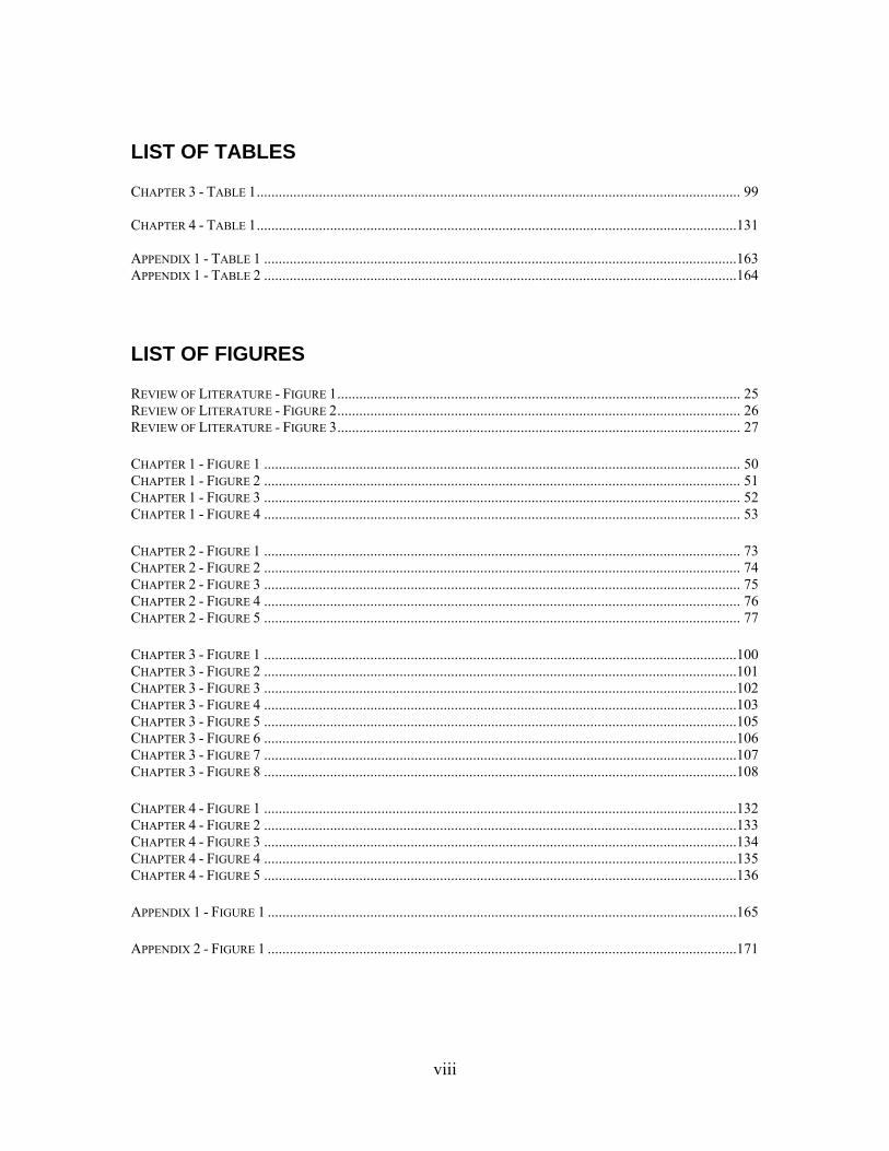
LIST OF TABLES CHAPTER 3 - TABLE 1 .................................................................................................................................... 99 CHAPTER 4 - TABLE 1 ...................................................................................................................................131 APPENDIX 1 - TABLE 1 .................................................................................................................................163 APPENDIX 1 - TABLE 2 .................................................................................................................................164
LIST OF FIGURES REVIEW OF LITERATURE - FIGURE 1 .............................................................................................................. 25 REVIEW OF LITERATURE - FIGURE 2 .............................................................................................................. 26 REVIEW OF LITERATURE - FIGURE 3 .............................................................................................................. 27 CHAPTER 1 - FIGURE 1 .................................................................................................................................. 50 CHAPTER 1 - FIGURE 2 .................................................................................................................................. 51 CHAPTER 1 - FIGURE 3 .................................................................................................................................. 52 CHAPTER 1 - FIGURE 4 .................................................................................................................................. 53 CHAPTER 2 - FIGURE 1 .................................................................................................................................. 73 CHAPTER 2 - FIGURE 2 .................................................................................................................................. 74 CHAPTER 2 - FIGURE 3 .................................................................................................................................. 75 CHAPTER 2 - FIGURE 4 .................................................................................................................................. 76 CHAPTER 2 - FIGURE 5 .................................................................................................................................. 77 CHAPTER 3 - FIGURE 1 .................................................................................................................................100 CHAPTER 3 - FIGURE 2 .................................................................................................................................101 CHAPTER 3 - FIGURE 3 .................................................................................................................................102 CHAPTER 3 - FIGURE 4 .................................................................................................................................103 CHAPTER 3 - FIGURE 5 .................................................................................................................................105 CHAPTER 3 - FIGURE 6 .................................................................................................................................106 CHAPTER 3 - FIGURE 7 .................................................................................................................................107 CHAPTER 3 - FIGURE 8 .................................................................................................................................108 CHAPTER 4 - FIGURE 1 .................................................................................................................................132 CHAPTER 4 - FIGURE 2 .................................................................................................................................133 CHAPTER 4 - FIGURE 3 .................................................................................................................................134 CHAPTER 4 - FIGURE 4 .................................................................................................................................135 CHAPTER 4 - FIGURE 5 .................................................................................................................................136 APPENDIX 1 - FIGURE 1 ................................................................................................................................165 APPENDIX 2 - FIGURE 1 ................................................................................................................................171
1
REVIEW OF LITERATURE
OVERVIEW
Ergot alkaloids are mycotoxins that can cause ill effects in humans and animals; however,
the medical community has also found advantageous uses for these compounds as
treatments for some diseases and other clinical conditions. They were first discovered in
the sclerotia of the ergot fungus, Claviceps purpurea. Ingestion of grain contaminated
with ergot sclerotia causes ergotism (44). Ergot alkaloids have been extensively studied
in C. purpurea, although the pathway by which ergot alkaloids are synthesized has yet to
be fully determined (21, 57). Ergot alkaloids also have been reported in other species,
such as Neotyphodium spp. which are endophytes of grasses, Penicillium spp. and
Aspergillus fumigatus (15, 30). A. fumigatus is a common fungus found in a variety of
indoor environments, including ventilation systems, and generates airborne spores that
are a potential danger to the health of humans. The spores can embed and survive in the
lungs of humans and animals, causing ill health to the affected individual and significant
lung disease in immunocompromised persons. Whether ergot alkaloids contribute to the
virulence of the fungus or cause toxicoses in the absence of fungal colonization is
unknown.
ERGOT ALKALOIDS AND THEIR HISTORICAL SIGNIFICANCE
Ergot alkaloids are a family of indole-derived mycotoxins that were first discovered in
the ergot fungus, Claviceps purpurea and are known to cause a wide range of effects on
exposed individuals due to their nonselective interactions with several monoamine
neurotransmitter receptors. Unnecessary for the survival of the fungus, at least under
laboratory conditions, these secondary metabolites clearly impact individuals that
2
consume them. Typically these metabolites affect the central nervous, circulatory,
immune, and reproductive systems (20, 42, 47, 58). In mammals, they can act as agonists
and/ or antagonists of dopamine, 5-hydroxytryptamine (5-HT; serotonin), and
noradrenaline (norepinephrine) receptors (46, 47). The severity to which humans and
animals are affected is dependent upon multiple factors such as the toxicity of the
particular ergot alkaloid, exposure time, and the health status of the individual. In the
past, people ingested elevated amounts of ergot alkaloids through consumption of grain
contaminated with sclerotia of the ergot fungus, C. purpurea (44), which led to ergotism,
a condition then known as St. Anthony’s fire. Exposure to alkaloids can cause
hallucinations, gangrene, and even death. It has been hypothesized that these toxins were
responsible for the Salem witch trials in Massachusetts in the seventeenth century (50).
Different types of alkaloids affect the clinical manifestations of ergotism.
However, there is significant overlap in the types of symptoms induced by different ergot
alkaloid sources. Ergopeptine alkaloids such as ergotamine or ergocristine, peptide
derivatives of lysergic acid, are frequently associated with a gangrenous form of ergotism
due to their vasoconstrictive activity. They cause painful edema of the legs and gangrene
at the tendons. The last documented outbreak of this form of ergotism was seen in
Ethiopia in 1977-1978 where 34% of the affected individuals died. Ergotism due to
intoxication with ergot clavines or simple amides of lysergic acid is more typically
associated with a convulsive form of ergotism. Affected individuals experience
gastrointestinal symptoms as well as problems within their central nervous system. The
last documented outbreak of ergotism due to ergot clavines was seen in India in 1975.
All affected individuals survived (44). Today, ergotism in humans is a rare occurrence
3
due to grain cleaning before milling which removes most of the sclerotia; however, it is
still a major concern for animal husbandry of domestic animals, such as cattle, horses,
sheep, pigs, and chickens, which feed on contaminated grasses or grains (1, 16).
Lambs fed a diet containing endophyte infected fescue seed (E+) showed a linear
reduction in feed intake, skin temperature, thermocirculation index (TCI), and prolactin
concentration when fed diets rich in E+ (17). Prolactin concentration was significantly
lowered by E+ intake, presumably due to the presence of ergot alkaloids.
Supplementation of feed with ergovaline caused a similar effect, but not to the same
extent as E+, suggesting that the other ergot alkaloids also play a role in toxicoses. In a
different study, ergovaline and lysergic acid could be detected in ruminal fluid, urine
(lysergic acid only), and feces in lambs fed an E+ diet (10).
Research has shown cattle that graze E+ tall fescue have lowered average daily
weight gains, increased body temperatures, rough coats, lowered prolactin production,
and reduced conception rates (16). Cattle grazing on mixed grasses containing E+ and E-
showed intermediate weight gain compared to cattle that grazed E- only and E+ only
pastures. A Norwegian study reported the occurrence of ergotism in free-living moose
and roe deer that fed on wild grasses (23). Ergocristine was the major alkaloid found
among the sampled wild grasses; this ergopeptine alkaloid, along with other unknown
alkaloids, could account for the gangrenous symptoms in the wild Norwegian cervids
(61). Ergot alkaloids have also been shown to affect rabbits, insects, and bacteria (6, 12,
37, 42, 48, 49).
Because of their high affinity for neurotransmitter receptors, there are also
advantageous effects of alkaloid consumption. The medical community uses these
4
compounds as a treatment for migraines, and they are also effective against Parkinsonism
and to induce uterine contractions in childbirth (26). However, because of their lack of
specificity for individual neurotransmitter receptors, negative side effects limit the utility
of these drugs (30, 42). During the twentieth century, a Swiss chemist named Albert
Hofmann began creating semi-synthetic ergot alkaloids. Among his many concoctions,
arose two important drugs, Methergine, which was responsible for preventing
hemorrhage after childbirth, and lysergic acid diethylamide (LSD), a hallucinogenic drug
used by psychiatrists and the CIA, as well as a popular recreational drug of the 60’s and
early 70’s (1). Use of LSD or dihydroergotamine has been known to cause a condition
known as serotonin syndrome, which is a condition that shares similar clinical features
with convulsive ergotism (11, 18).
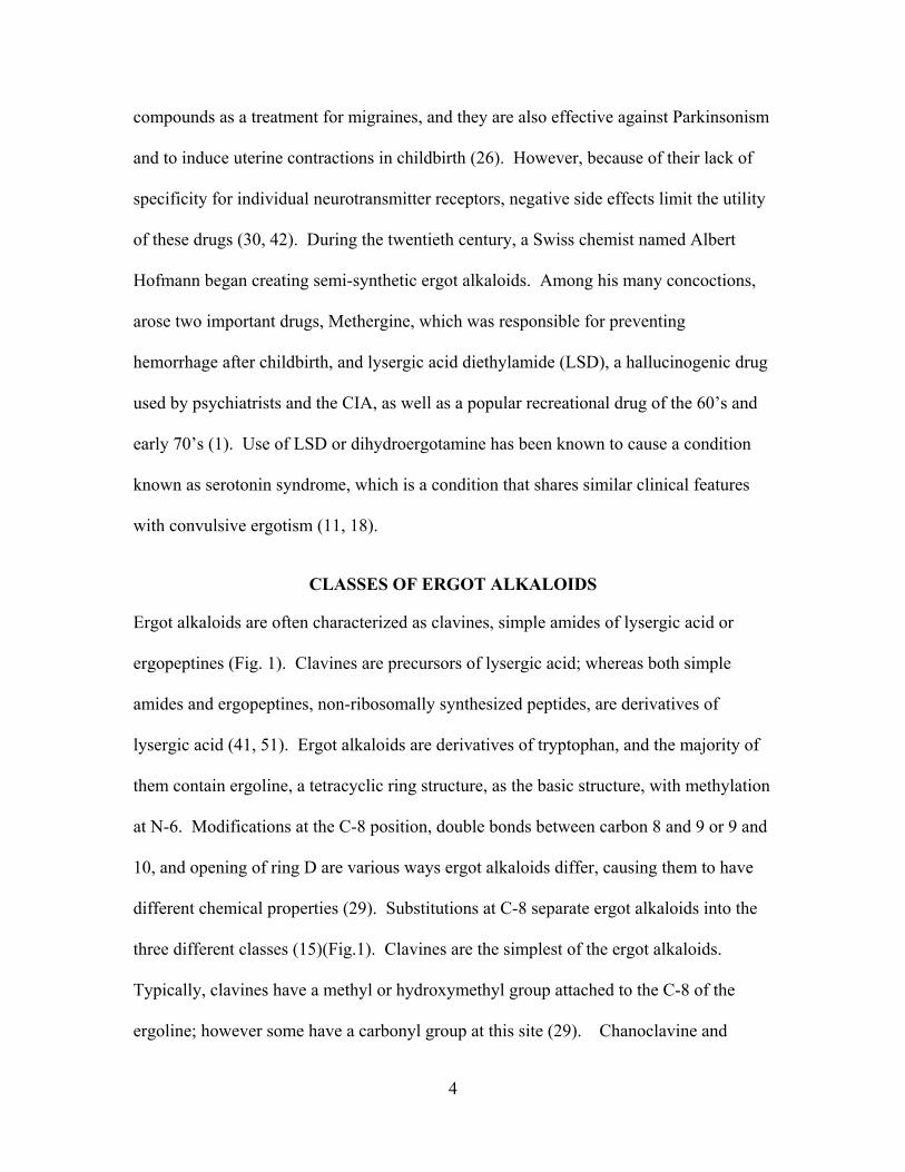
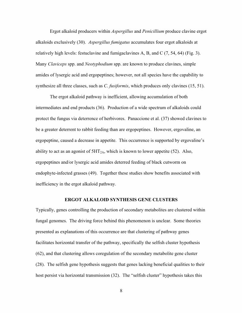
CLASSES OF ERGOT ALKALOIDS
Ergot alkaloids are often characterized as clavines, simple amides of lysergic acid or
ergopeptines (Fig. 1). Clavines are precursors of lysergic acid; whereas both simple
amides and ergopeptines, non-ribosomally synthesized peptides, are derivatives of
lysergic acid (41, 51). Ergot alkaloids are derivatives of tryptophan, and the majority of
them contain ergoline, a tetracyclic ring structure, as the basic structure, with methylation
at N-6. Modifications at the C-8 position, double bonds between carbon 8 and 9 or 9 and
10, and opening of ring D are various ways ergot alkaloids differ, causing them to have
different chemical properties (29). Substitutions at C-8 separate ergot alkaloids into the
three different classes (15)(Fig.1). Clavines are the simplest of the ergot alkaloids.
Typically, clavines have a methyl or hydroxymethyl group attached to the C-8 of the
ergoline; however some have a carbonyl group at this site (29). Chanoclavine and
5
members within the subclass 6,7-secoergolines have an open D ring, which differentiates
them from other clavines. Clavines are the ultimate end products of the ergot alkaloid
pathway in some fungi (e.g., Claviceps fusiformis and A. fumigatus); however, they
accumulate in other ergot alkaloid-producing fungi as products of shunts off the pathway
or are intermediates to more complex ergot alkaloids. Besides causing toxicoses in
exposed individuals, clavines also affect food preference by acting as a deterrent in rabbit
feeding in experiments using endophyte infected grasses (37).
Simple amides of lysergic acid include lysergic acid amide (ergine), ergonovine,
lysergic acid-2-hydroxyethylamide, and lysergyl alanine. The observation that lysergyl
peptide synthetase 1 is required for synthesis of ergine and lysergyl alanine suggests that
these simple amides are by-products of ergopeptine synthesis (43). However, in some
cases simple amides accumulate as the ultimate product of the ergot alkaloid pathways,
such as ergine and lysergic acid-2-hydroxyethyl amide in C. paspali.
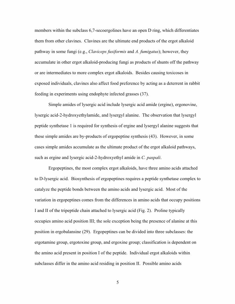
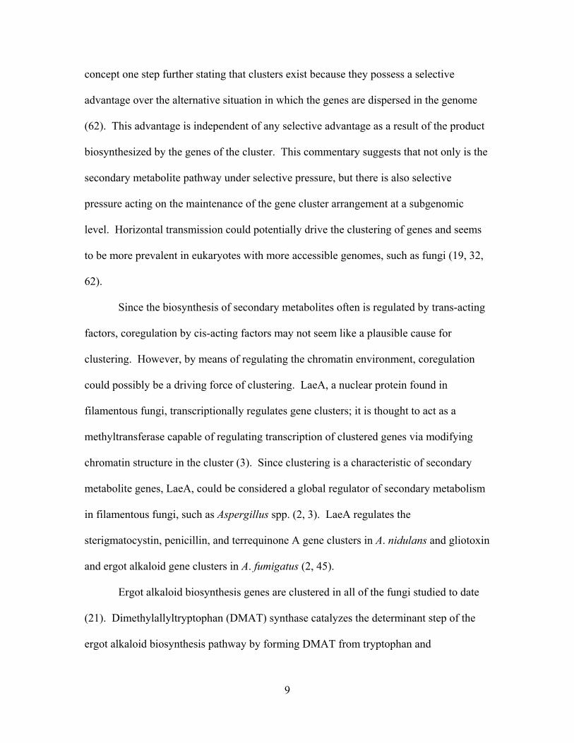
Ergopeptines, the most complex ergot alkaloids, have three amino acids attached
to D-lysergic acid. Biosynthesis of ergopeptines requires a peptide synthetase complex to
catalyze the peptide bonds between the amino acids and lysergic acid. Most of the
variation in ergopeptines comes from the differences in amino acids that occupy positions
I and II of the tripeptide chain attached to lysergic acid (Fig. 2). Proline typically
occupies amino acid position III; the sole exception being the presence of alanine at this
position in ergobalansine (29). Ergopeptines can be divided into three subclasses: the
ergotamine group, ergotoxine group, and ergoxine group; classification is dependent on
the amino acid present in position I of the peptide. Individual ergot alkaloids within
subclasses differ in the amino acid residing in position II. Possible amino acids
6
occupying this position are phenylalanine, leucine, isoleucine, norleucine, valine,
methionine and alpha-amino butyric acid (51). Ergovaline is a major alkaloid found
within Neotyphodium spp., whereas ergosine and ergotamine are examples of abundant
ergopeptine within Claviceps spp. (34, 36).
Ergopeptams, also known as lactam ergot alkaloids, are non-cyclol ergopeptines,
due to the lack of hydroxylation of the amino acid in position I; hence a cyclol bridge is
unable to form. The proline in these alkaloids is in D-configuration (15, 29).
Ergopeptams are further characterized into the subclasses, ergotamams, ergoxams,
ergotoxams, and ergoannams.
ERGOT ALKALOID PRODUCING FUNGI
Claviceps and the Neotyphodium endophytes (42, 51) are clavicipitaceous genera
belonging to the order Hypocreales. Claviceps spp. parasitize flowers of rye and other
cereals; sclerotia, fungal overwintering bodies, replace the infected plant host’s seeds.
Ergot alkaloid-rich sclerotia may be harvested along with the grain, and so grain cleaning
is essential to eliminate toxicoses. Neotyphodium spp. and their teleomorphs, Epichloë
spp., are endophytes of cool-season grasses, such as tall fescue and perennial ryegrass
(51). These fungi live symbiotically with their host species, grow intercellularly within
the plant, and disseminate in seeds (15, 41). The fungus provides many benefits to its
host, such as drought tolerance and protection from pests. However, the animals that
graze on these E+ grasses are susceptible to toxicoses due to mycotoxin production (16,
35, 48). Ergot alkaloids are typically found in the pseudostems and leaf blades of these
forages but are either undetectable or only found in extremely low concentrations
(relative to pseudostem) within the roots (40, 41). These clavicipitaceous fungi have the
7
capacity to biosynthesize a wide spectrum of alkaloids, especially lysergic acid amides
and ergopeptines (42, 51). However, due to the slow growth of these fungi and their
dependency on their plant hosts for reliable production of alkaloids, biochemical and
molecular aspects of the pathway have been elucidated slowly (39, 63).
Several other species, besides those within the genera Claviceps and
Neotyphodium, produce ergot alkaloids. These include species within the genera
Aspergillus and Penicillium, which are common fungi in the order Eurotiales (30).
These fungi produce only the clavine class of ergot alkaloids. Among these is A.
fumigatus, a thermotolerant opportunistic pathogen. This fungus is capable of growing at
temperatures up to 55 ºC and surviving at 70 ºC. It colonizes domestic and public
buildings, increasing exposure by humans (31). The fungus grows in moist areas, such as
basements and ventilation system ducts; it also is ubiquitous outdoors, with higher
concentrations in areas surrounding compost and waste removal facilities. The conidia
(asexual spores) can become airborne and inhaled by humans and animals, which can
infect and cause disease, act as an allergen, cause toxicoses, or cause inflammatory
responses (13). Contamination of ventilation systems with A. fumigatus in hospitals
poses a serious threat to immune-deficient individuals of acquiring aspergillosis. For the
general population, a common question is: are indoor molds truly causing a “new”
disease and if so, why is this more of an issue today than in the past? Many studies
correlate disease with indoor air quality, specifically regarding mold concentrations, in
buildings and homes. However, many of these claims have insufficient evidence and
lack consistent clinical diagnoses. Future studies need to correct the flaws of past
research to address these questions (56).
8
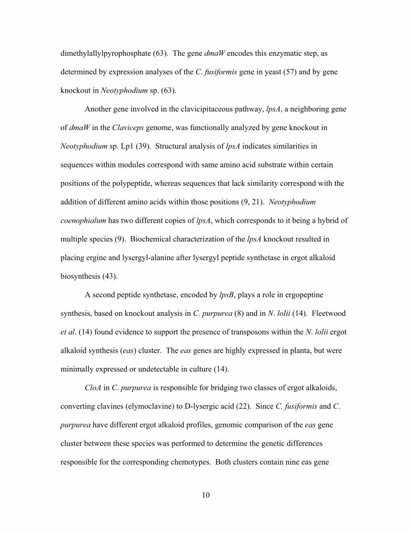
Ergot alkaloid producers within Aspergillus and Penicillium produce clavine ergot
alkaloids exclusively (30). Aspergillus fumigatus accumulates four ergot alkaloids at
relatively high levels: festuclavine and fumigaclavines A, B, and C (7, 54, 64) (Fig. 3).
Many Claviceps spp. and Neotyphodium spp. are known to produce clavines, simple
amides of lysergic acid and ergopeptines; however, not all species have the capability to
synthesize all three classes, such as C. fusiformis, which produces only clavines (15, 51).
The ergot alkaloid pathway is inefficient, allowing accumulation of both
intermediates and end products (36). Production of a wide spectrum of alkaloids could
protect the fungus via deterrence of herbivores. Panaccione et al. (37) showed clavines to
be a greater deterrent to rabbit feeding than are ergopeptines. However, ergovaline, an
ergopeptine, caused a decrease in appetite. This occurrence is supported by ergovaline’s
ability to act as an agonist of 5HT2A, which is known to lower appetite (52). Also,
ergopeptines and/or lysergic acid amides deterred feeding of black cutworm on
endophyte-infected grasses (49). Together these studies show benefits associated with
inefficiency in the ergot alkaloid pathway.
ERGOT ALKALOID SYNTHESIS GENE CLUSTERS
Typically, genes controlling the production of secondary metabolites are clustered within
fungal genomes. The driving force behind this phenomenon is unclear. Some theories
presented as explanations of this occurrence are that clustering of pathway genes
facilitates horizontal transfer of the pathway, specifically the selfish cluster hypothesis
(62), and that clustering allows coregulation of the secondary metabolite gene cluster
(28). The selfish gene hypothesis suggests that genes lacking beneficial qualities to their
host persist via horizontal transmission (32). The “selfish cluster” hypothesis takes this
9
concept one step further stating that clusters exist because they possess a selective
advantage over the alternative situation in which the genes are dispersed in the genome
(62). This advantage is independent of any selective advantage as a result of the product
biosynthesized by the genes of the cluster. This commentary suggests that not only is the
secondary metabolite pathway under selective pressure, but there is also selective
pressure acting on the maintenance of the gene cluster arrangement at a subgenomic
level. Horizontal transmission could potentially drive the clustering of genes and seems
to be more prevalent in eukaryotes with more accessible genomes, such as fungi (19, 32,
62).
Since the biosynthesis of secondary metabolites often is regulated by trans-acting
factors, coregulation by cis-acting factors may not seem like a plausible cause for
clustering. However, by means of regulating the chromatin environment, coregulation
could possibly be a driving force of clustering. LaeA, a nuclear protein found in
filamentous fungi, transcriptionally regulates gene clusters; it is thought to act as a
methyltransferase capable of regulating transcription of clustered genes via modifying
chromatin structure in the cluster (3). Since clustering is a characteristic of secondary
metabolite genes, LaeA, could be considered a global regulator of secondary metabolism
in filamentous fungi, such as Aspergillus spp. (2, 3). LaeA regulates the
sterigmatocystin, penicillin, and terrequinone A gene clusters in A. nidulans and gliotoxin
and ergot alkaloid gene clusters in A. fumigatus (2, 45).
Ergot alkaloid biosynthesis genes are clustered in all of the fungi studied to date
(21). Dimethylallyltryptophan (DMAT) synthase catalyzes the determinant step of the
ergot alkaloid biosynthesis pathway by forming DMAT from tryptophan and
10
dimethylallylpyrophosphate (63). The gene dmaW encodes this enzymatic step, as
determined by expression analyses of the C. fusiformis gene in yeast (57) and by gene
knockout in Neotyphodium sp. (63).
Another gene involved in the clavicipitaceous pathway, lpsA, a neighboring gene
of dmaW in the Claviceps genome, was functionally analyzed by gene knockout in
Neotyphodium sp. Lp1 (39). Structural analysis of lpsA indicates similarities in
sequences within modules correspond with same amino acid substrate within certain
positions of the polypeptide, whereas sequences that lack similarity correspond with the
addition of different amino acids within those positions (9, 21). Neotyphodium
coenophialum has two different copies of lpsA, which corresponds to it being a hybrid of
multiple species (9). Biochemical characterization of the lpsA knockout resulted in
placing ergine and lysergyl-alanine after lysergyl peptide synthetase in ergot alkaloid
biosynthesis (43).
A second peptide synthetase, encoded by lpsB, plays a role in ergopeptine
synthesis, based on knockout analysis in C. purpurea (8) and in N. lolii (14). Fleetwood
et al. (14) found evidence to support the presence of transposons within the N. lolii ergot
alkaloid synthesis (eas) cluster. The eas genes are highly expressed in planta, but were
minimally expressed or undetectable in culture (14).
CloA in C. purpurea is responsible for bridging two classes of ergot alkaloids,
converting clavines (elymoclavine) to D-lysergic acid (22). Since C. fusiformis and C.
purpurea have different ergot alkaloid profiles, genomic comparison of the eas gene
cluster between these species was performed to determine the genetic differences
responsible for the corresponding chemotypes. Both clusters contain nine eas gene
11
homologues, including cloA and lpsB, both of which are involved in biosynthesis of
ergopeptines. All nine eas genes were expressed in C. fusiformis. However, CloA and
LpsB were enzymatically inactive, and therefore C. fusiformis is unable to convert
clavines to ergopeptines (33). These genes involved in ergot alkaloid biosynthesis are
clustered in the genome of C. purpurea (21, 59). Several additional genes clustered
around dmaW, lpsA, lpsB, and cloA have functions typical of genes involved in secondary
metabolism.
SECONDARY METABOLISM AND SPORULATION
There is an apparent association between morphological differentiation and secondary
metabolism in microorganisms (5, 25). Hopwood (25) demonstrated that sporulation and
secondary metabolite biosynthesis in the filamentous bacterium, Streptomyces coelicolor
A3, was translationally regulated by bldA, a gene that encodes for a tRNA corresponding
to a leucine codon. The mRNA involved in vegetative growth does not require this
codon during translation, but it is essential for formation of proteins responsible for
differential growth and antibiotic synthesis. Typically in fungi, secondary metabolites
associate with sporulation either as activators of sporulation, pigments necessary for
sporulation structures, or as toxic metabolites whose production coincides with
sporulation in the growing organism (5). Environmental conditions are similar for
secondary metabolite synthesis and sporulation; environmental factors such as, pH,
temperature, air-surface interface, and nutrient supply have been found to regulate both
sporulation and secondary metabolite production in Aspergillus spp. (5).
Studies functionally analyzing genes regulating sporulation also have indicated a
corresponding involvement in mycotoxin production in fungi. In Aspergillus spp.,
12
asexual sporulation and mycotoxin biosynthesis are regulated by a common G-protein
signaling pathway (4). In A. nidulans, FadA is responsible for vegetative growth through
activation of cyclic AMP-dependent protein kinase A (PkaA). FadA activation or knock
out of flbA inhibits the production of conidia and sterigmatocystin (ST), a precursor of
aflatoxin biosynthesis (24). In the presence of FlbA, the FadA pathway is inhibited, at
least partially, which allows ST synthesis and asexual sporulation to occur. FlbA is a
protein with a RGS (Regulator of G protein Signaling) domain. Proteins in this family
negatively regulate G-protein mediated pathways (24). Mutations in the sec gene within
A. parasiticus greatly reduce conidiophore production, and also eliminate aflatoxin
biosynthesis and expression of aflR, a regulatory gene involved in the aflatoxin pathway
(27). Knockout analysis of the regulatory gene, stuA, in A. fumigatus again demonstrates
the relationship between conidiation and secondary metabolism. The transcription factor,
StuAp, is responsible for proper conidiophore development and regulation of potentially
six secondary metabolite clusters, one being ergot alkaloid biosynthesis (53, 60). This
result exemplifies the apparent association between sporulation and secondary
metabolism, both of which are governed by a complex regulatory pathway.
13
ERGOT ALKALOIDS IN ASPERGILLUS FUMIGATUS
Spilsbury and Wilkinson (55) were the first to document production of the ergot alkaloid,
festuclavine, in A. fumigatus. They also characterized two novel festuclavine derivatives,
fumigaclavines A and B, produced by this fungus. Soon after, Yamano et al. (64) noted
an additional ergot alkaloid, fumigaclavine C, was also synthesized by A. fumigatus.
Years later, Cole et al. (7) further characterized fumigaclavine C using broth cultures of
A. fumigatus that had been isolated from moldy silage.
More recently Panaccione and Coyle (38) demonstrated that A. fumigatus
accumulates ergot alkaloids in association with its asexual spores (conidia), although
exact location has not been discerned. Accumulated ergot alkaloids were fumigaclavine
C, festuclavine, fumigaclavine A, and fumigaclavine B in the order of abundance
(Appendix 1, Table 2). Under environmentally relevant conditions, the total mass of
ergot alkaloids often exceeded 1% of the mass of a conidium. Extraction of ergot
alkaloids from conidia of A. fumigatus cultured on various media, revealed that extracts
from cultures grown on latex paint or cultured maize seedlings accumulated the greatest
quantities of ergot alkaloids. Important physical attributes of conidia likely to affect their
respirability, i.e. diameter, mass, and specific gravity, were significantly lower in A.
fumigatus when compared to Aspergillus nidulans, Aspergillus niger, and Stachybotrys
chartarum (Appendix 1, Table 1).
OBJECTIVES AND POTENTIAL IMPACT OF CURRENT STUDY
In efforts to further understand ergot alkaloid biosynthesis, the objectives of this study are
to:
14
1. Characterize the ergot alkaloids produced by A. fumigatus, complete a structural
analysis of the genes within the pathway
2. Functionally analyze ergot alkaloid synthesis genes via gene knockouts,
determine the spectrum of ergot alkaloids produced by each mutant strain and
compare it to the production of the wild type
3. Investigate the association of sporulation and ergot alkaloid production in A.
fumigatus.
Several of the early steps in ergot alkaloid biosynthesis are just hypothesized and
through this investigation they may be characterized experimentally. Analysis of the
gene cluster in A. fumigatus will aid in identifying genes involved in ergot alkaloid
production in other species, especially genes involved in the early steps of the pathway
thought to be shared by multiple species. Ergot alkaloids are important metabolites in
agriculture and medicine. These metabolites possess a number of beneficial qualities,
despite their many negative side effects. Characterization of the ergot alkaloid pathway
and the ability to control the spectrum of alkaloids produced may lead to advances in
these fields by providing a potential means of eliminating the negative side effects.
Likewise, production of alkaloids could be associated with cause of illness when A.
fumigatus infects an individual. Mutant strains developed from this research could be
used for testing effects of ergot alkaloids on virulence and determining their role in
mycoses and mycotoxicoses.
15
REFERENCES
1. Bennett, J. W., and M. Klich. 2003. Mycotoxins. Clin. Microbiol. Rev.
16:497–516.
2. Bok, J., D. Hoffmeister, L. Maggio-Hall, R. Murillo, J. Glasner, and N.
Keller. 2006. Genomic mining for Aspergillus natural products. Chem. Biol.
13:31–37.
3. Bok, J. W., and N. P. Keller. 2004. LaeA, a regulator of secondary metabolism
in Aspergillus spp. Euk. Cell 3:527–535.
4. Brodhagen, M., and N. P. Keller. 2006. Signalling pathways connecting
mycotoxin production and sporulation Mol. Plant Path. 7:285–301.
5. Calvo, A. M., R. A. Wilson, J. W. Bok, and N. P. Keller. 2002. Relationship
between secondary metabolism and fungal development. Microbiol. Mol. Biol.
Rev. 66:447–459.
6. Clay, K., and C. Schardl. 2002. Evolutionary origins and ecological
consequences of endophyte symbiosis with grasses. Am. Nat. 160:S199–S127.
7. Cole, R. J., J. W. Kirksey, J. W. Dorner, D. M. Wilson, J. C. Johnson, A. N.
Johnson, D. M. Bedell, J. P. Springer, and K. K. Chexal. 1977. Mycotoxins
produced by Aspergillus fumigatus species isolated from molded silage. J. Agric.
Food Chem. 25:826–830.
8. Correia, T., N. Grammel, I. Ortel, U. Keller, and P. Tudzynski. 2003.
Molecular cloning and analysis of the ergopeptine assembly system in the ergot
fungus Claviceps purpurea. Chem. Biol. 10:1281–1292.
16
9. Damrongkool, P., A. B. Sedlock, C. A. Young, R. D. Johnson, K. E. Goetz, B.
Scott, C. L. Schardl, and D. G. Panaccione. 2005. Structural analysis of a
peptide synthetase gene required for ergopeptine production in the endophytic
fungus Neotyphodium lolii. DNA Sequ. 16:379–385.
10. De Lorme, M. J. M., S. L. Lodge-Ivey, and A. M. Craig. 2007. Physiological
and digestive effects of Neotyphodium coenophialum-infected tall fescue fed to
lambs. J. Anim. Sci. 85:1199–1206.
11. Eadie, M. J. 2003. Convulsive ergotism: epidemics of the serotonin syndrome?
Lancet Neurol. 2:429–434.
12. Eich, E., and H. Pertz. 1999. Antimicrobial and antitumor effects of ergot
alkaloids and their derivatives, p. 411–440. In V. Kren and L. Cvak (ed.), Ergot:
The Genus Claviceps. Harwood Academic Publishers, The Netherlands.
13. Fischer, G., and W. Dott. 2003. Relevance of airborne fungi and their
secondary metabolites for environmental, occupational and indoor hygiene. Arch.
Microbiol. 179:75–82.
14. Fleetwood, D. J., B. Scott, G. A. Lane, A. Tanaka, and R. D. Johnson. 2007.
A complex ergovaline gene cluster in Epichloë endophytes of grasses. Appl.
Environ. Microbiol. 73:2571–2579.
15. Flieger, M., M. Wurst, and R. Shelby. 1997. Ergot alkaloids -- sources,
structures and analytical methods. Folia Microbiol. 42:3–30.
16. Fribourg, H. A., and J. C. Waller. 2005. Neotyphodium research and application
in the USA, p. 3–22. In C. A. Roberts, C. P. West, and D. E. Spiers (ed.),
17
Neotyphodium in cool-season grasses, 1st ed. Blackwell Publishing Professional,
Ames, Iowa.
17. Gadberry, M. S., T. M. Denard, D. E. Spiers, and E. L. Piper. 2003. Effects
of feeding ergovaline on lamb performance in a heat stress environment. J. Anim.
Sci. 81:1538–1545.
18. Ganetsky, M., and D. E. Brush. 2005. Serotonin syndrome—What have we
learned? Clin. Ped. Emerg. Med. 6:103–108.
19. Goddard, M. R., and A. Burt. 1999. Recurrent invasion and extinction of a
selfish gene. Proc. Natl. Acad. Sci. USA 96:13880–13885.
20. Gröger, D., and H. G. Floss. 1998. Biochemistry of ergot alkaloids –
achievements and challenges. Alkaloids 50:171–218.
21. Haarmann, T., C. Machado, Y. Lübbe, T. Correia, C. L. Schardl, D. G.
Panaccione, and P. Tudzynski. 2005. The ergot alkaloid gene cluster in
Claviceps purpurea: extension of the cluster sequence and intra species evolution.
Phytochemistry 66:1312–1320.
22. Haarmann, T., I. Ortel, P. Tudzynski, and U. Keller. 2006. Identification of
the cytochrome P450 monooxygenase that bridges the clavine and ergoline
alkaloid pathways. ChemBioChem 7:645–652.
23. Handeland, K., and T. Vikoren. 2005. Presumptive gangrenous ergotism in
free-living moose and a roe deer. J. Wildl. Dis. 41:636–642.
24. Hicks, J. K., J.-H. Yu, N. P. Keller, and T. H. Adams. 1997. Aspergillus
sporulation and mycotoxin production both require inactivation of the FadA G
protein-dependent signaling pathway. EMBO J. 16:4916–4923.
18
25. Hopwood, D. A. 1988. The Leeuwenhoek lecture, 1987. Towards an
understanding of gene switching in Streptomyces, the basis of sporulation and
antibiotic production. Proc. R. Soc. Lond. Biol. 235:121–138.
26. Houghton, P. J., and M.-J. Howes. 2005. Natural products and derivatives
affecting neurotransmission relevant to Alzheimer's and Parkinson's disease.
Neurosignals 14:6–22.
27. Kale, S. P., J. W. Cary, D. Bhatnagar, and J. W. Bennett. 1996.
Characterization of experimentally induced, nonaflatoxigenic variant strains of
Aspergillus parasiticus. Appl. Environ. Microbiol. 62:3399–3404.
28. Keller, N. P., and T. M. Hohn. 1997. Metabolic pathway gene clusters in
filamentous fungi. Fungal Genet. Biol. 21:17–29.
29. Keller, U. 1999. Biosynthesis of ergot alkaloids, p. 95–163. In V. Kren and L.
Cvak (ed.), Ergot: The Genus Claviceps. Harwood Academic Publishers, The
Netherlands.
30. Kozlovsky, A. G. 1999. Producers of ergot alkaloids out of Claviceps genus, p.
479–499. In V. Kren and L. Cvak (ed.), In Ergot: The Genus Claviceps. Harwood
Academic Publishers, The Netherlands.
31. Latge, J.-P. 1999. Aspergillus fumigatus and Aspergillosis. Clin. Microbiol.
Rev. 12:310–350.
32. Lawrence, J. G., and J. R. Roth. 1996. Selfish operons: horizontal transfer may
drive the evolution of gene clusters. Genetics 143:1843–1860.
33. Lorenz, N., E. V. Wilson, C. Machado, C. L. Schardl, and P. Tudzynski.
2007. Comparison of ergot alkaloid biosynthesis gene clusters in Claviceps
19
species indicates loss of late pathway steps in evolution of C. fusiformis. Appl.
Environ. Microbiol. 73:7185–7191.
34. Lyons, P. C., R. D. Plattner, and C. W. Bacon. 1986. Occurrence of peptide
and clavine ergot alkaloids in tall fescue grass Science 232:487–489.
35. Malinowski, D. P., D. P. Belesky, and G. C. Lewis. 2005. Abiotic stresses in
endophytic grasses, p. 187–199. In C. P. W. C. A. Roberts, and D. E. Spiers (ed.),
Neotyphodium in cool-season grasses, 1 ed. Blackwell Publishing Professional,
Ames, Iowa.
36. Panaccione, D. G. 2005. Origins and significance of ergot alkaloid diversity in
fungi. FEMS Microbiol. Lett. 251:9–17.
37. Panaccione, D. G., J. R. Cipoletti, A. B. Sedlock, K. P. Blemings, C. L.
Schardl, C. Machado, and G. E. Seidel. 2006. Effects of ergot alkaloids on
food preference and satiety in rabbits, as assessed with gene-knockout endophytes
in perennial ryegrass (Lolium perenne). J. Agric. Food Chem. 54:4582–4587.
38. Panaccione, D. G., and C. M. Coyle. 2005. Abundant respirable ergot alkaloids
from the common airborne fungus Aspergillus fumigatus. Appl. Environ.
Microbiol. 71:3106–3111.
39. Panaccione, D. G., R. D. Johnson, J. Wang, C. A. Young, P. Damrongkool, B.
Scott, and C. L. Schardl. 2001. Elimination of ergovaline from a grass-
Neotyphodium endophyte symbiosis by genetic modification of the endophyte.
Proc. Natl. Acad. Sci. USA 98:12820–12825.
40. Panaccione, D. G., J. B. Kotcon, C. L. Schardl, R. J. Johnson, and J. B.
Morton. 2006. Ergot alkaloids are not essential for endophytic fungus-
20
associated population suppression of the lesion nematode, Pratylenchus scribneri,
on perennial ryegrass. Nematology 8:583–590.
41. Panaccione, D. G., C. Schardl, and C. M. Coyle. 2006. Pathways to diverse
ergot alkaloid profiles in fungi, p. 23–52 In J. T. Romeo (ed.), Recent Advances
in Phytochemistry, vol. 40. Elsevier, Amsterdam, The Netherlands.
42. Panaccione, D. G., and C. L. Schardl. 2003. Molecular genetics of ergot
alkaloid biosynthesis, p. 399–424. In J. F. W. Jr., C. W. Bacon, N. L. Hywel-
Jones, and J. W. Spatafora (ed.), The Clavicipitalean Fungi: Evolutionary
Biology, Chemistry, Biocontrol, and Cultural Impacts. Marcel-Dekker, New
York.
43. Panaccione, D. G., B. A. Tapper, G. A. Lane, E. Davies, and K. Fraser. 2003.
Biochemical outcome of blocking the ergot alkaloid pathway of a grass
endophyte. J. Agric. Food Chem. 51:6429–6437.
44. Peraica, M., B. Radic, A. Lucic, and M. Pavlovic. 1999. Toxic effects of
mycotoxins in humans. Bull. World Health Org. 77:754–766.
45. Perrin, R. M., N. D. Fedorova, J. W. Bok, R. A. Cramer, J. R. Wortman, H.
S. Kim, W. C. Nierman, and N. P. Keller. 2007. Transcriptional regulation of
chemical diversity in Aspergillus fumigatus by LaeA. PLOS Pathogens 3:508–
517.
46. Pertz, H. 1996. Naturally occurring clavines: antagonism/partial agonism at 5-
HT2A receptors and antagonism at α1-adrenoceptors in blood vessels. Planta
Med. 62:387–392.
21
47. Pertz, H., and E. Eich. 1999. Ergot alkaloids and their derivatives as ligands for
serotoninergic, dopaminergic, and adrenergic receptors, p. 411–440. In V. Kren
and L. Cvak (ed.), Ergot: The Genus Claviceps. Harwood Academic Publishers,
The Netherlands.
48. Popay, A. J., and S. A. Bonos. 2005. Biotic responses in endophytic grasses, p.
163–185. In C. P. W. C. A. Roberts, and D. E. Spiers (ed.), Neotyphodium in
cool-season grasses, 1st ed. Blackwell Publishing Professional, Ames, Iowa.
49. Potter, D. A., J. T. Stokes, C. T. Redmond, C. L. Schardl, and D. G.
Panaccione. 2008. Contribution of ergot alkaloids to suppression of a grass-
feeding caterpillar assessed with gene knockout endophytes in perennial ryegrass.
Entomol. Experiment. Applic. 126:138–147.
50. Rusyniak, D. E., R. B. Furbee, and R. Pascuzzi. 2005. Historical neurotoxins:
What we have learned from toxins of the past about diseases of the present.
Neurol. Clin. 23:337–352.
51. Schardl, C., D. G. Panaccione, and P. Tudzynski. 2006. Ergot alkaloids--
biology and molecular biology, p. 45–86. In G. A. Cordell (ed.), Alkaloids Chem.
Biol., vol. 63. Academic Press, San Diego, CA.
52. Schöning, C., M. Flieger, and H. H. Pertz. 2001. Complex interaction of
ergovaline with 5-HT2A, 5-HT1B/1D, and alpha1 receptors in isolated arteries of
rat and guinea pig. J. Anim. Sci. 79:2202–2209.
53. Sheppard, D. C., T. Doedt, L. Y. Chiang, H. S. Kim, D. Chen, W. C.
Nierman, and S. G. Filler. 2005. The Aspergillus fumigatus StuA protein
22
governs the up-regulation of a discrete transcriptional program during the
acquisition of developmental competence. Mol. Biol. Cell 16:5866–5879.
54. Spilsbury, J. F., and S. Wilkinson. 1961. The isolation of festuclavine and two
new clavine alkaloids from Aspergillus fumigatus Fres. J. Chem. Soc 5:2085–
2091.
55. Spilsbury, J. F., and S. Wilkinson. 1961. The isolation of festuclavine and two
new clavine alkaloids from Aspergillus fumigatus Fres. J. Chem. Soc. 5:2085–
2091.
56. Terr, A. I. 2004. Are indoor molds causing a new disease? J. Allergy Clin.
Immun. 113:221–226
57. Tsai, H.-F., H. Wang, J. C. Gebler, C. D. Poulter, and C. L. Schardl. 1995.
The Claviceps purpurea gene encoding dimethylallyltryptophan synthase, the
committed step for ergot alkaloid biosynthesis. Biochem. Bioph. Res. Co.
216:119–125.
58. Tudzynski, P., T. Correia, and U. Keller. 2001. Biotechnology and genetics of
ergot alkaloids. Appl. Microbiol. Biotechnol. 57:593–605.
59. Tudzynski, P., K. Holter, T. Correia, C. Arntz, N. Grammel, and U. Keller.
1999. Evidence for an ergot alkaloid gene cluster in Claviceps purpurea. Mol.
Gen. Genet. 261:133–141.
60. Twumasi-Boateng, K., Y. Yu, D. Chen, F. N. Gravelat, W. C. Nierman, and
D. C. Sheppard. 2009. Transcriptional profiling identifies a role for BrlA in the
response to nitrogen depletion and for StuA in the regulation of secondary
metabolite clusters in Aspergillus fumigatus. Euk. Cell 8:104–115.
23
61. Uhlig, S., T. Vikøren, L. Ivanova, and K. Handeland. 2007. Ergot alkaloids in
Norwegian wild grasses: A mass spectrometric approach. Rapid Commun. Mass
Sp. 21:1651–1660.
62. Walton, J. D. 2000. Horizontal gene transfer and the origin of secondary
metabolite gene clusters in fungi: An hypothesis. Fungal Genet. Biol. 30:167–
171.
63. Wang, J., C. Machado, D. G. Panaccione, H.-F. Tsai, and C. L. Schardl.
2004. The determinant step in ergot alkaloid biosynthesis by an endophyte of
perennial ryegrass. Fungal Genet. Biol. 41:189–198.
64. Yamano, T., K. Kishino, S. Yamantodani, and M. Abe. 1962. Investigation
on ergot alkaloids found in cultures of Aspergillus fumigatus. Takeda Kenkyusho
Nempo (Annual Report Takeda Research Laboratories) 21:95–101.
24
FIGURE LEGENDS
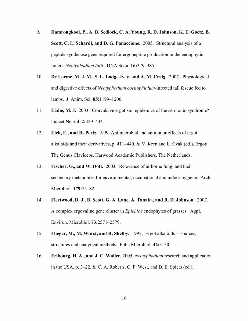
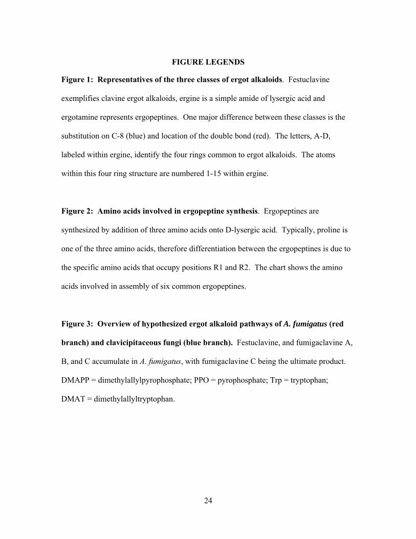
Figure 1: Representatives of the three classes of ergot alkaloids. Festuclavine
exemplifies clavine ergot alkaloids, ergine is a simple amide of lysergic acid and
ergotamine represents ergopeptines. One major difference between these classes is the
substitution on C-8 (blue) and location of the double bond (red). The letters, A-D,
labeled within ergine, identify the four rings common to ergot alkaloids. The atoms
within this four ring structure are numbered 1-15 within ergine.
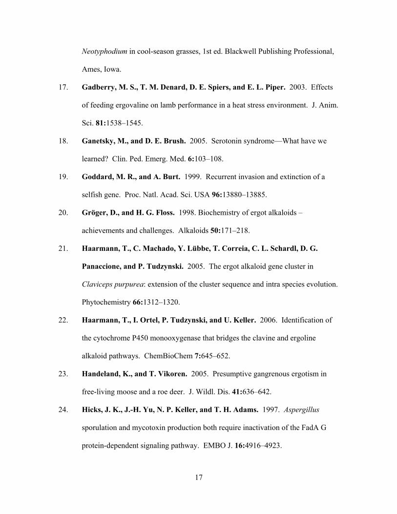
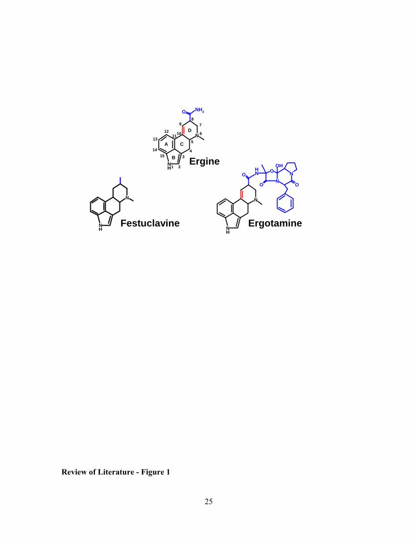
Figure 2: Amino acids involved in ergopeptine synthesis. Ergopeptines are
synthesized by addition of three amino acids onto D-lysergic acid. Typically, proline is
one of the three amino acids, therefore differentiation between the ergopeptines is due to
the specific amino acids that occupy positions R1 and R2. The chart shows the amino
acids involved in assembly of six common ergopeptines.
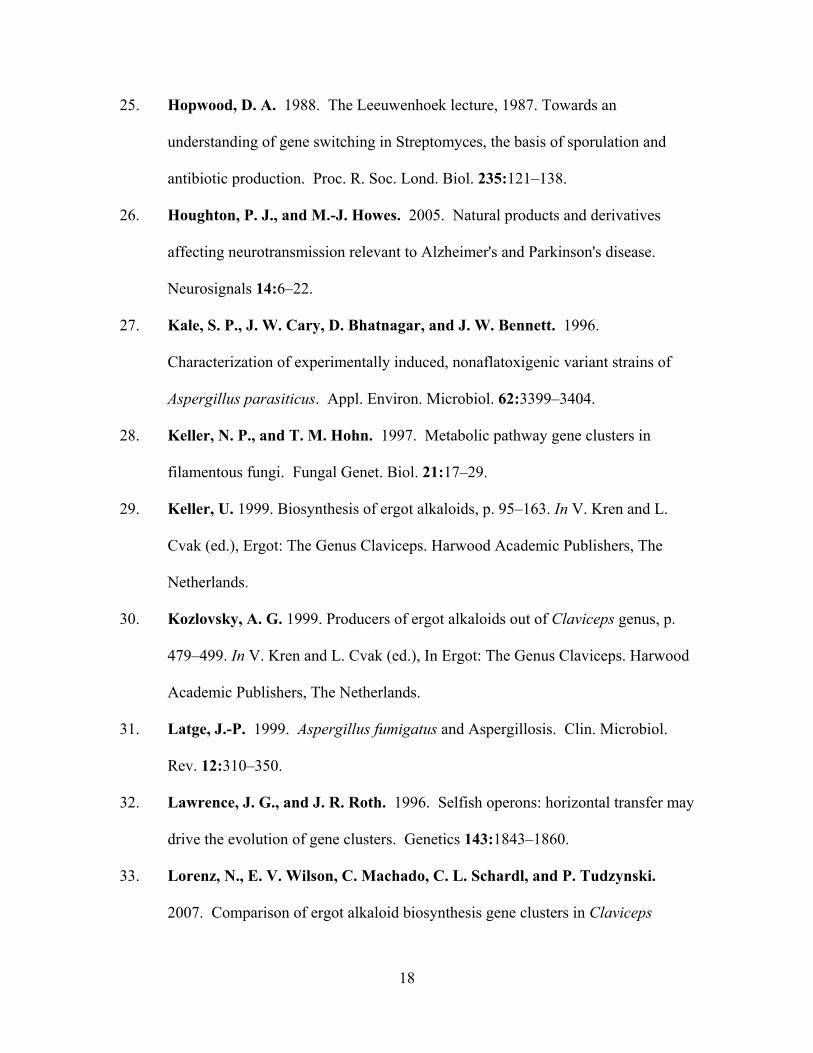
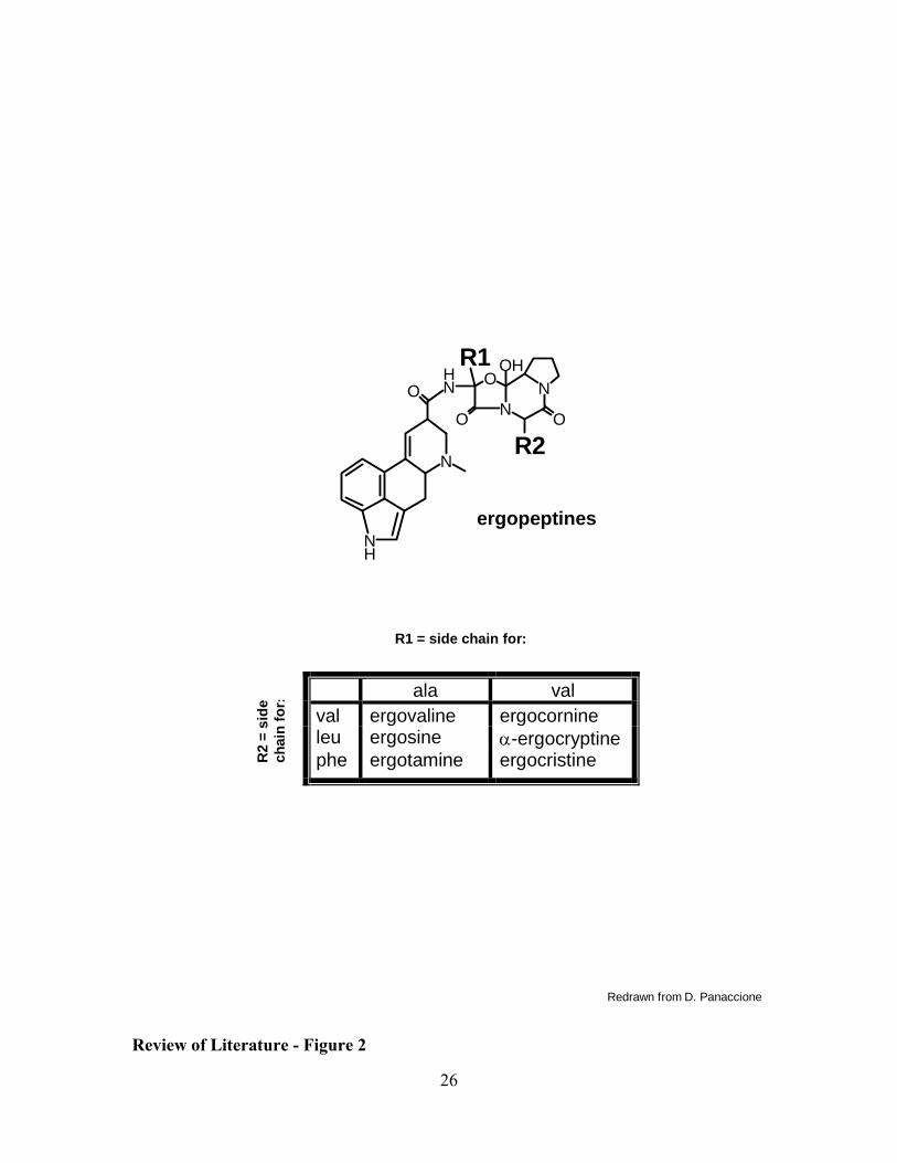
Figure 3: Overview of hypothesized ergot alkaloid pathways of A. fumigatus (red
branch) and clavicipitaceous fungi (blue branch). Festuclavine, and fumigaclavine A,
B, and C accumulate in A. fumigatus, with fumigaclavine C being the ultimate product.
DMAPP = dimethylallylpyrophosphate; PPO = pyrophosphate; Trp = tryptophan;
DMAT = dimethylallyltryptophan.
25
OO
O NH
NNO
OH
NH
N
ErgotamineNH
N
Festuclavine
O NH2
N
NH
Ergine
A
B
C
D
1
43
6
7
5
2
8
10
9
1211
15
13
14
Review of Literature - Figure 1
26
Redrawn from D. Panaccione
ergopeptines
OO
O
NH
NH
N
NNO
OHR1
R2
R1 = side chain for:
R2
= si
de
chai
n fo
r:
ala val val ergovaline ergocornine leu ergosine α-ergocryptine phe ergotamine ergocristine
Review of Literature - Figure 2
27
+
COO-
NH
NH3+
ergopeptinesand lysergic acid
amides
NH
NH
OH
chanoclavine
Trp
COO -
NH
NH3+
DMAPP
PPO
DMAT
NH
N
OH
NH
N
O
NH
N
O
O
NH
N
O
festuclavine
fumigaclavine B
fumigaclavine C
fumigaclavine A
O OH
N
NH lysergic
acid
NH
N
dmaW
agro-clavine
Review of Literature - Figure 3
28
CHAPTER 1: An ergot alkaloid biosynthesis gene and clustered hypothetical genes from Aspergillus fumigatus Published as: Coyle, C. M., and D. G. Panaccione. 2005. Appl. Environ. Microbiol. 71:3112–3118.
ABSTRACT
Ergot alkaloids are mycotoxins that interact with several monoamine receptors,
negatively affecting cardiovascular, nervous, reproductive, and immune systems of
exposed humans and animals. Aspergillus fumigatus, a common airborne fungus and
opportunistic human pathogen, contains at least four ergot alkaloids associated with its
conidia. The A. fumigatus genome contains a gene encoding dimethylallyltryptophan
(DMAT) synthase, the enzyme that catalyzes the first committed step in ergot alkaloid
biosynthesis. Knockout of the DMAT synthase gene eliminated the ergot alkaloids from
conidia, and complementation of the mutation restored ergot alkaloid production.
Clustered with the DMAT synthase-encoding gene are sequences corresponding to five
genes previously proposed to encode steps in the ergot alkaloid pathway of Claviceps
purpurea, as well as additional sequences whose deduced protein products are consistent
with involvement in the ergot alkaloid pathway. The data indicate a common, but
ancient, origin of ergot alkaloid biosynthetic capability with the ergot alkaloid producers
in the distantly related Clavicipitaceae.
29
INTRODUCTION
Ergot alkaloids are a complex family of indole-derived mycotoxins with varied and
significant biological activities (6, 7, 14, 25). Fungi derived from two relatively
divergent ascomycete orders (Hypocreales and Eurotiales) produce ergot alkaloids.
Several fungi in the family Clavicipitaceae (order Hypocreales) including the ergot fungi
in the genus Claviceps and certain grass endophytes in the genera Epichloë (including
their imperfect relatives in the genus Neotyphodium) and Balansia produce ergot
alkaloids in association with their grass hosts (3, 6, 7, 14, 25). The rather distantly
related imperfect fungus Aspergillus fumigatus, a common saprophyte and opportunistic
human pathogen with close relatives in the order Eurotiales, produces ergot alkaloids in
broth culture (4, 23) and in association with its airborne conidia (13).
Ergot alkaloid-producing members of the Clavicipitaceae (clavicipitaceous fungi)
produce a wide variety of clavine and lysergyl-derived ergot alkaloids but commonly
accumulate ergopeptines (nonribosomally synthesized peptides containing lysergic acid
and three amino acids) or simple amides of lysergic acid such as ergine and ergonovine
(6, 7, 8, 14). Aspergillus fumigatus produces several clavine ergot alkaloids including
festuclavine and fumigaclavines A, B, and C (4, 13, 23). Most of the clavine and
lysergyl-derived ergot alkaloids contain the same four-membered ergoline ring system
but differ in the number, type, and position of side chains (6, 7, 8, 14).
Festuclavine, one of the ergot alkaloids produced by A. fumigatus, was first
described from C. purpurea (6, 7) and also has been detected in Neotyphodium spp. (15,
19). However, none of the fumigaclavines produced by A. fumigatus has been detected in
a member of the Clavicipitaceae. Likewise the end products of the Claviceps spp. and
30
Neotyphodium spp. pathways (ergopeptines and lysergic acid amides) have not been
found in A. fumigatus. This distribution of alkaloids is consistent with a hypothesis that
early steps of the ergot alkaloid pathway are shared by these diverse fungal species but
that the later portions of the pathway differ between A. fumigatus and the clavicipitaceous
fungi.
The ergot alkaloid pathway is relatively long and may have alternate branches or
spurs in some ergot alkaloid producers (7, 8, 14, 15). Functions of three genes involved
in the ergot alkaloid pathway have been demonstrated by gene knockout. The gene
(dmaW) encoding dimethylallyltryptophan synthase (DMAT synthase) was knocked out
in Neotyphodium sp. Lp1, an endophyte of perennial ryegrass (29). Loss of all ergot
alkaloids in this knockout mutant indicated that dmaW controls the determinant step in
the ergot alkaloid pathway. A gene encoding lysergyl peptide synthetase 1 (LPS1), one
of two components of the lysergyl peptide synthetase complex responsible for assembly
of ergopeptines from lysergic acid and three amino acids (20, 28), was knocked out in
Neotyphodium sp. Lp1 (16). LPS1-deficient knockout mutants failed to produce
ergopeptines and simple amides of lysergic acid but still produced clavine ergot alkaloids
(15, 16). A gene encoding lysergyl peptide synthetase 2 (LPS2), the lysergic acid-
activating component of the lysergyl peptide synthetase complex, has been cloned and
knocked out in C. purpurea, resulting in the loss of ergopeptines from the knockout
mutant (5).
Genes encoding DMAT synthase, LPS1, and LPS2 are clustered within ~32 kb of
each other in the genome of C. purpurea (5, 25). Moreover, at least six additional genes
that encode activities likely to be required for ergot alkaloid production are interspersed
31
in this cluster with the ergot alkaloid biosynthesis genes (5, 25, 26). Analysis of mRNAs
from five of the clustered genes indicate they are expressed in cultures grown under
conditions conducive to ergot alkaloid production but not under repressive conditions (5).
Such clustering and common regulation of secondary metabolite biosynthetic pathway
genes is typical of fungal genomes (9, 27).
Our objective in this study was to investigate whether the ergot alkaloids of A.
fumigatus share a common biosynthetic origin with those of the clavicipitaceous fungi by
identification and functional analysis of the gene (dmaW) controlling the determinant step
in the pathway and through comparison of sequences flanking dmaW in A. fumigatus and
C. purpurea.
MATERIALS AND METHODS
Fungi and culture conditions. Aspergillus fumigatus isolate WVU1943 from parakeet
lung was described previously (13) and was manipulated in a class II biosafety cabinet.
Isolates were maintained on potato dextrose agar [PDA; 20 g/L dehydrated mashed
potatoes (Idaho Spuds, Pillsbury, Minneapolis, MN), 20 g/L glucose, 15 g/L agar (Bacto-
Agar, Difco, Detroit, MI)] at room temperature or at 37 ºC. Cultures for analysis of ergot
alkaloids were grown on PDA at 37 ºC. For the preparation of DNA or protoplasts, the
fungus was cultured overnight in 15 mL of potato dextrose broth (Difco) in a deep Petri
dish (20-mm depth, 100-mm diameter; Fisher Scientific, Pittsburgh, PA) in an orbital
shaker set at 37 ºC and 150 rpm. Cultures were inoculated by lightly dusting the medium
with conidia that had adhered to a Petri dish lid formerly covering a mature culture. In
mock inoculations, an average of 2.1 × 10⁵ conidia per cm2 was deposited by this
inoculation method.
32
Analysis of the A. fumigatus genome for ergot alkaloid biosynthesis genes.
Aspergillus fumigatus preliminary sequence data were obtained from The Institute for
Genomic Research website at http://www.tigr.org. All A. fumigatus sequences described
in this paper are located on assembly 92 (long arm of chromosome 2, formerly assembly
72) between nucleotides 2,894,958 to 2,926,335. The presence of sequences in the A.
fumigatus genome that are similar to known or putative ergot alkaloid biosynthesis genes
was assessed by application of the tblastn algorithm (1) at the National Center for
Biotechnology Information (NCBI) website (www.ncbi.nlm.nih.gov) with the deduced
protein sequences of DMAT synthase from Claviceps fusiformis (24) and LPS1 of N. lolii
(16) or C. purpurea (26) as query sequences. The nature of genes linked to the ergot
alkaloid biosynthesis genes or candidate genes was investigated by retrieving contiguous
2-kb segments of DNA from the A. fumigatus genome and querying the nonredundant
protein database of NCBI with these sequences by application of the blastx search
algorithm (1).
Knockout of dmaW in A. fumigatus. Fungal DNA for amplification by PCR
was isolated following the Gene Clean Spin protocol (Bio 101, Vista, CA). An internal
fragment of A. fumigatus dmaW (encoding amino acids 136 to 352 of the predicted 497
amino acid product) was amplified by PCR from primers dmaW forward (5'-
TTGATCTGGAGTGGTTCCGC-3') and dmaW reverse (5'-
CGTTCATGCCGAGGTTGTG-3'). A 50 µL PCR reaction contained 50 mM KCl, 10
mM Tris-HCl (pH 9.0), 0.1% (v/v) Triton X-100, 1.5 mM MgCl2, 200 µM of each
deoxyribonucleotide triphosphate, 1 µM of each primer, and 2.5 units of Taq DNA
polymerase (Promega, Madison, WI), which was added once the thermocycler reached 95
33
ºC in the initial denaturing period. The reaction began with an initial denaturing time of 2
min 30 s at 95 ºC, followed by 35 cycles of 1 min at 94 ºC, 1 min at 55 ºC, and 1 min at
72 ºC. The 651-bp dmaW PCR product was ligated into pCR2.1 (Invitrogen, Carlsbad,
CA) based on T/A overhangs and transformed into chemically competent Escherichia
coli cells. The resulting 4.5-kb gene knockout construct, pDMAT1, was linearized at a
unique PpuMI site within the dmaW coding sequences prior to transformation of A.
fumigatus.
Protoplasts of A. fumigatus were prepared according to Murray et al. (11), except
that 5 mg/mL of Driselase (InterSpex, Foster City, CA), 1.3 mg/ml Novozyme 234
(InterSpex) and 25 µg/mL chitinase (Sigma, St. Louis, MO) were used as lysing
enzymes. Cotransformation of the protoplasts with PpuMI-linearized pDMAT1 and the
hygromycin resistance-conferring plasmid pMOcosX (12)(linearized at a unique NotI
site) was performed as previously described by Panaccione et al. (16), but with 4 µg of
each DNA in 10 µL of water. Considering the relative differences in the lengths of the
respective cotransformed molecules, the equal masses introduced corresponded to an ~2-
fold molar excess of the gene knockout construct (pDMAT1, 4.5 kb) relative to the
selectable marker (pMOcosX, 8.8 kb). The transformation mixture was divided into 6
aliquots and plated on regeneration medium containing 300 µg/mL hygromycin
(Calbiochem, La Jolla, CA) as previously described (16). Transformation plates were
incubated at 37 ºC for two days and hygromycin-resistant colonies were transferred onto
PDA plates containing 400 µg/mL hygromycin.
Transformants were screened for homologous recombination of pDMAT1 with
the native dmaW gene by PCR (same conditions as above, except annealing temperature
34
was 57 ºC) from primers UF (5'-TGTAAAACGACGGCCAGTGAAT-3'; which anneals
to vector sequences near the universal primer annealing site pCR2.1) and 3S (5'-
AAGTAAGTCCCGAGCTGTTCAT-3'; which anneals to dmaW sequences near the 3'-
end of the gene and flanking the intended site of integration)(Fig. 1A). Transformants
yielding a fragment of the expected size for a gene knockout at dmaW (1.2 kb) were
purified to nuclear homogeneity by culturing from individual, germinated conidia.
Single-spored colonies were again screened by PCR (conditions as described
immediately above but with annealing temperature of 60 ºC) but with primers UR (5'-
AGCTATGACCATGATTACGCCA-3'), which anneals to vector sequences near the
reverse primer annealing site of pCR2.1, and primer 5S (5'-
TAGTGAGATACACATTCGAGCC-3'), which anneals to dmaW sequences near the 5'-
end of the gene and flanking the intended site of integration (Fig. 1A). In this way, both
the 5' and 3' borders of the integration site were analyzed by PCR. Candidate gene
knockout strains also were screened with primers 5S and 3S under PCR conditions
described above and with an annealing temperature of 57 ºC. Finally, transformants
producing fragments of expected sizes in the PCR analyses were analyzed further by
Southern hybridization as described below.
Fungal DNA for Southern blot hybridization was digested with SalI, size-
fractionated by electrophoresis through 0.8% agarose, and transferred to Biodyne A
nylon membrane (Pall-Gelman Sciences, Ann Arbor, MI) by capillary action with 20×
SSC (1× SSC is 150 mM NaCl + 15 mM trisodium citrate) as the transfer medium. A
dmaW probe was labeled with DIG-dUTP by amplifying wild type genomic DNA by
PCR from primers dmaW forward and dmaW reverse (described above). PCR conditions
35
were as described above except with 1× DIG DNA Labeling Mix (Roche Applied
Science, Indianapolis, IN) replacing standard dNTPs. Hybridization was performed at 65
ºC in 5× SSC, 0.1% (w/v) N-lauroylsarcosine, 0.2% (w/v) sodium dodecyl sulfate, 1%
(w/v) blocking reagent (Roche), 10 mM maleic acid, and 15 mM NaCl (pH 7.5). Final
washes were with 0.5× SSC + 0.1% SDS at 65 ºC. Bound probe was detected with anti-
DIG antibody followed by chemiluminescence by standard methodology (DIG Manual,
Roche Applied Science, Indianapolis, IN).
An ergot alkaloid-deficient dmaW knockout strain was complemented with a full-
length dmaW-containing fragment produced by PCR from primers 5'-
GAGAGTGGTGTTGAGAGCTGCC-3' and 5'-CCTTCAGGCTTCATGAACGGAC-3',
which prime amplification of a 3.4-kb fragment containing the entire dmaW coding
sequence along with 784 bp of 5'-flanking sequence and 1,123 bp of 3'-flanking
sequence. PCR conditions were as described above except the annealing temperature
was 57 ºC and the extension time at 72 ºC was 3 min 20 s. The PCR product was co-
transformed into a dmaW knockout strain along with phleomycin resistance-conferring
plasmid pBC-phleo (22; Fungal Genetics Stock Center, University of Missouri-Kansas
City, Kansas City, MO). Selection was on complete regeneration medium (16)
containing phleomycin at 100 µg/mL.
Analysis of ergot alkaloids. Ergot alkaloids were analyzed from conidia
produced on PDA by extraction with 80% methanol and analyzed by HPLC with
fluorescence detection as described previously (13).
36
RESULTS
Identification and analysis of a dmaW homolog in A. fumigatus. A search of the A.
fumigatus genome database revealed an excellent match for C. fusiformis dmaW, which
encodes the enzyme dimethylallyltryptophan synthase (DMAT synthase) that catalyzes
the first step in the ergot alkaloid pathway in clavicipitaceous fungi. The deduced A.
fumigatus DMAT synthase was 50% identical to DMAT synthase from C. fusiformis
(24), 53% identical to DMAT synthase from C. purpurea (26), and 59% identical to the
DMAT synthase of Neotyphodium sp. Lp1 (29). The A. fumigatus dmaW sequence
contained a single intron at an identical position to the single intron in the dmaW genes
isolated from the Claviceps and Neotyphodium spp. (data not shown).
A gene knockout construct, designed to integrate at dmaW by a single crossover
(Fig. 1) was targeted to the dmaW locus in 3 of 25 hygromycin-resistant transformants
analyzed (Figs. 1 and 2). PCR analysis of the 5' and 3' junctions of the native dmaW gene
and the integrated gene knockout construct yielded fragments of the expected length for
homologous recombination of such a construct at dmaW (Figs. 1B and 1C). PCR
amplification of the gene knockout strains from primers 5S and 3S, spanning the site of
recombination, failed to yield the 1.2-kb product amplified from the wild-type isolate
(data not shown). Southern hybridization of DNA from the putative knockout strains
digested with SalI, which has sites flanking the targeted integration site and not within
the knockout construct, indicated that three or more copies of the knockout construct
integrated in tandem at dmaW in these transformants (Fig. 2).
The dmaW knockout strains did not produce any of the known ergot alkaloids
(Fig. 3). Additional peaks, including uncharacterized molecules eluting at 55.5 min and
37
56 min (Fig. 3) and a minor peak eluting at 32.8 min (evident only upon viewing the
traces at much higher sensitivity), also were eliminated by knockout of dmaW. The peak
eluting at 32.8 minutes is proposed to contain one or more of the isomers of chanoclavine
based on a measured mass of 256 and its co-elution with a chanoclavine standard. The
later-eluting molecules have not been characterized further.
Transformation of strain dmaW ko6 with a DNA fragment containing the entire
dmaW coding sequences plus 784 bp of 5'-flanking sequences and 1,123 bp of 3' flanking
sequences restored the ability to produce ergot alkaloids (Fig. 3). The complementing
fragment was determined to have integrated ectopically, because the gene knockout
construct was retained at the native dmaW locus (data not shown). All known A.
fumigatus ergot alkaloids were produced in the complemented strains (Fig. 3).
Genes clustered with dmaW in A. fumigatus. Database searches with five C.
purpurea gene sequences, which in addition to dmaW have been proposed to be involved
in ergot alkaloid biosynthesis (but which have not yet been functionally analyzed),
identified potential homologs clustered with each other and with the dmaW homolog on
A. fumigatus (Fig. 4). These included two putative oxidoreductase-encoding genes
identified as cpox1 and cpox2, as well as a catalase-encoding gene, and two unidentified
open reading frames (labeled orfA Cp and orfB Cp) whose sequence and close linkage
with dmaW are conserved in C. purpurea (25, 26) and C. fusiformis (10). Despite the
similarity in the composition of the clusters of genes associated with dmaW in A.
fumigatus and C. purpurea, the physical arrangement in terms of the orientation and
order of genes within the cluster differed in the two fungi (Fig. 4).
38
Analysis of additional sequences clustered on the dmaW contig of A. fumigatus
identified several other genes that may encode enzymes in the ergot alkaloid pathway
(Fig. 4). These include sequences similar to cytochrome P450 monooxygenases, a FAD-
dependent monooxygenase, and short chain alcohol dehydrogenases. Additional
sequences whose deduced functions are consistent with roles in formation of the side
chains that vary among the three fumigaclavines also were identified. These include any
of several monooxygenases or hydroxylases, an O-acetyl transferase, and a divergent
dmaW-like (prenyl transferase) gene. The predicted product of the divergent dmaW-like
gene had 25% identity with the deduced product of A. fumigatus dmaW required for ergot
alkaloid biosynthesis and had 26%, 25%, and 27% identity with the deduced products of
dmaW from C. purpurea, C. fusiformis, and Neotyphodium sp. Lp1, respectively.
DISCUSSION
Our results demonstrate that the genome of the A. fumigatus contains a homolog of
dmaW that is required for ergot alkaloid biosynthesis. The loss of all known ergot
alkaloids upon inactivation of dmaW in A. fumigatus indicates that this gene controls the
determinant step in the ergot alkaloid pathway of A. fumigatus, as it does in the
clavicipitaceous fungus Neotyphodium sp. Lp1 (29). Moreover, in the A. fumigatus
genome, dmaW is clustered with genes similar to, and extending, the ergot alkaloid gene
cluster observed in the ergot fungus C. purpurea. Collectively the data indicate a
common biosynthetic and genetic origin for the ergot alkaloids of A. fumigatus with those
of the ergot alkaloid-producing members of the Clavicipitaceae.
Each of the three gene knockout strains investigated contained three or more
copies of the knockout construct integrated at dmaW. The multiple integrations may have
39
resulted from either or both of two modifications of the more typical gene knockout
strategy that we applied in this experiment. First, the knockout construct was introduced
by cotransformation at two-times the molar concentration of the selectable marker
construct. Second, the knockout was created by integrating an internal fragment of the
gene via a single crossover, a strategy that has resulted in multiple integrations in
previous studies with other fungi (e.g., 2, 17, 18, 21). This particular approach was
chosen for two reasons. Knockout construct can be easily prepared in a single cloning
step, which should facilitate similar analyses on many of the genes in the cluster. Also,
since we intended to complement the mutation in a subsequent step, any phenotype
associated with ectopic integration of the cotransformed selectable marker construct
would have been revealed at that stage. The observations that three independent
transformants containing the integration at dmaW lacked ergot alkaloids and the
complementation restored ergot alkaloid production indicate that the strategy provides a
reasonable means for rapid analysis of gene function.
Aspergillus fumigatus produces at least four ergot alkaloids, all belonging to the
clavine class of ergot alkaloids (4, 13, 23). Close analysis of peaks eliminated in the gene
knockout versus wild-type strains in this current study indicated that conidia of A.
fumigatus also contain small quantities of chanoclavine. This is not surprising since
chanoclavine is an early pathway intermediate in the biosynthesis of all known ergot
alkaloids and it accumulates to varying degrees in different ergot alkaloid producers (7, 8,
14, 15, 25).
Among the ergot alkaloids produced by A. fumigatus, only festuclavine and
chanoclavine have been detected in any of the studied clavicipitaceous ergot alkaloid
40
producers (6, 7, 15, 19). Conversely, the ergot alkaloid-producing members of the
Clavicipitaceae produce numerous ergot alkaloids that are not found in A. fumigatus.
These include the ergopeptines (tripeptide derivatives of lysergic acid) and simple amides
of lysergic acid (such as ergonovine and ergine). The production of both the ergopeptine
and simple amide classes of ergot alkaloids is dependent upon the activities or products
of lysergyl peptide synthetases (5, 15, 16). Based on the ergot alkaloid profiles and
sequences clustered with dmaW in A. fumigatus versus C. purpurea, we hypothesize that
the early stages of the pathway are shared in the A. fumigatus and C. purpurea but that
later steps in the pathways differ in the respective fungi.
The ergot alkaloid pathways of A. fumigatus and C. purpurea probably share
several steps in addition to the determinant step catalyzed by DMAT synthase. The
cluster of genes immediately around dmaW in A. fumigatus contains five hypothetical
genes that are highly similar to genes previously hypothesized to be involved in the early
stages of the ergot alkaloid pathway of C. purpurea (5, 25, 26). These include the
following: a sequence labeled orfB, which encodes a product similar to a methyl
transferase, an activity required for the second step in the pathway; two genes (cpox1 and
cpox2) encoding oxidoreductase that may catalyze early steps in the pathway (7, 14, 25);
a sequence labeled orfA, for which a biochemical function has not been proposed; and, a
catalase-encoding gene. A function for the catalase in ergot alkaloid biosynthesis is not
obvious. One possibility is that it may act peroxidatively to oxidize the primary alcohol
of chanoclavine to the corresponding aldehyde, which is a step required prior to
cyclization of the fourth ring of the basic four-membered ergoline ring system.
Interestingly, the orientation and position of three members of this putative core set of
41
genes differ in the clusters of the two fungi, suggesting that numerous recombination
events occurred after divergence of the lineages of these two fungi. Alternatively, if the
cluster had been acquired by horizontal gene transfer, then the dissimilarities indicate
than any such transfer would have been ancient.
We propose that after a shared early stage, the ergot alkaloid pathways of A.
fumigatus and the clavicipitaceous fungi diverge to yield different end products. Cultures
of A. fumigatus do not contain detectable levels of the later products of the pathway such
as lysergic acid or lysergyl derivatives such as ergopeptines (13). Consistent with this
hypothesis, searches with fragments of genes encoding LPS1 (an enzyme required for the
assembly of the ergopeptine class of ergot alkaloids from lysergic acid and three amino
acids) from N. lolii (16) or C. purpurea (26) produced only low-level matches to
miscellaneous peptide synthetase genes in the A. fumigatus genome. The degree of
identity observed was typical of the identity among peptide synthetases catalyzing the
assembly of unrelated nonribosomally synthesized peptides in fungi (27), and none of
these low-level matches was located near the dmaW gene.
Conversely, the A. fumigatus cluster contained genes not found in C. purpurea
cluster. Some of these hypothetical genes encode functions that we hypothesize to be
involved in the later steps of the A. fumigatus pathway leading from festuclavine to the
fumigaclavines, which are not produced by C. purpurea. For example, product of the
gene in the A. fumigatus cluster predicted to encode an O-acetyltransferase may function
in acetylating fumigaclavine B to fumigaclavine A. The unusual, alternate dmaW gene
(labeled alt dmaW in Fig. 4) may control the prenylation of fumigaclavine A to
fumigaclavine C. The lack of ergot alkaloids in the mutant containing a knockout at the
42
typical dmaW demonstrates that the highly divergent dmaW-like prenyl transferase gene
found in the cluster is not functionally redundant with dmaW and, thus, may catalyze
another prenylation step.
The available data for A. fumigatus extend further than the published ergot
alkaloid cluster data from C. purpurea, revealing several additional genes potentially
involved in ergot alkaloid production. Systematic gene knockouts and their
characterization should allow definition of the early (and probably shared) steps of the
ergot alkaloid pathway, several of which have been difficult to characterize
biochemically (7, 8, 14). Ergot alkaloid-deficient mutants such as the dmaW knockout
may be useful for assessing the contribution of ergot alkaloids to virulence towards
animals or potential contributions of the alkaloids to the success of A. fumigatus.
ACKNOWLEDGEMENTS
This work was supported by USDA-NRI grant No. 2001-35319-10930 and Hatch funds.
Sequencing of the A. fumigatus genome was funded by National Institute of Allergy and
Infectious Disease grant U01 AI 48830 to David Denning and William Nierman, the
Wellcome Trust, and Fondo de Investigaciones Sanitarias.
We thank Caroline Machado, Christopher Schardl, and Mark Farman (University
of Kentucky) for access to data and helpful suggestions, and Brian Tapper (AgResearch
Limited, New Zealand) and Miroslav Flieger (Czech Academy of Sciences) for the
chanoclavine standard. Preliminary sequence data from the A. fumigatus genome were
obtained from The Institute for Genomic Research at http://www.tigr.org. The assistance
of Bill Nierman and Natalie Federova with questions concerning the data is greatly
appreciated.
43
REFERENCES
1. Altschul, S. F., T. L. Madden, A. A. Schaffer, J. Zhang, Z. Zhang, W. Miller, and
D. J. Lipman. 1997. Gapped BLAST and PSI-BLAST: a new generation of protein
database search programs. Nucleic Acids Res. 25:3389–3402.
2. Apel, P. A., D. G. Panaccione, F. R. Holden, and J. D. Walton. 1993. Cloning
and targeted gene disruption of XYL1, a β1,4-xylanase gene from the maize pathogen
Cochliobolus carbonum. Mol. Plant-Microbe Interact. 6:467–473.
3. Clay, K., and C. Schardl. 2002. Evolutionary origins and ecological consequences
of endophyte symbiosis with grasses. Am. Nat. 160:S199–S127.
4. Cole, R. J., J. W. Kirksey, J. W. Dorner, D. M. Wilson, J. C. Johnson Jr., A. N.
Johnson, D. M. Bedell, J. P. Springer, K. K. Chexal, J. C. Clardy, and R. H. Cox.
1977. Mycotoxins produced by Aspergillus fumigatus species isolated from molded
silage. J. Agric. Food Chem. 25:826–830.
5. Correia, T., Grammel, N., Ortel, I., Keller, U. and Tudzynski, P. 2003.
Molecular cloning and analysis of the ergopeptine assembly system in the ergot
fungus Claviceps purpurea. Chem. Biol. 10:1281–1292.
6. Flieger, M., M. Wurst, and R. Shelby. 1997. Ergot alkaloids –– sources, structures,
and analytical methods. Folia Microbiol. 42:3–30.
7. Floss, H. G. 1976. Biosynthesis of ergot alkaloids and related compounds.
Tetrahedron 32:873–912.
8. Gröger, D., and H. G. Floss. 1998. Biochemistry of ergot alkaloids – achievements
and challenges. Alkaloids 50:171–218.
44
9. Keller, N. P., and T. M. Hohn. 1997. Metabolic pathway gene clusters in
filamentous fungi. Fungal Genet. Biol. 21:17–29.
10. Machado, C. 2004. Studies of ergot alkaloid biosynthesis genes in clavicipitaceous
fungi. Ph.D. Thesis. University of Kentucky, Lexington.
11. Murray, F. R., G. C. M. Latch, and D. B. Scott. 1992. Surrogate transformation of
perennial ryegrass, Lolium perenne, using genetically modified Acremonium
endophyte. Mol. Gen. Genet. 233:1–9.
12. Orbach, M. J. 1994. A cosmid with a HyR marker for fungal library construction
and screening. Gene 150:159–162.
13. Panaccione, D. G., and C. M. Coyle. 2005. Abundant respirable ergot alkaloids
from the common airborne fungus Aspergillus fumigatus. Appl. Environ. Microbiol.
71:3106–3111.
14. Panaccione, D. G., and C. L. Schardl. 2003. Molecular genetics of ergot alkaloid
biosynthesis, p. 399–424. In J. F. White Jr., C. W. Bacon, N. L. Hywel-Jones, and J.
W. Spatafora (ed.), The Clavicipitalean Fungi: Evolutionary Biology, Chemistry,
Biocontrol, and Cultural Impacts. Marcel-Dekker, New York.
15. Panaccione, D. G., B. A. Tapper, G. A. Lane, E. Davies, and K. Fraser. 2003.
Biochemical outcome of blocking the ergot alkaloid pathway of a grass endophyte. J.
Agric. Food Chem. 51:6429–6437.
16. Panaccione, D. G., R. D. Johnson, J. Wang, C. A. Young, P. Damrongkool, B.
Scott, and C. L. Schardl. 2001. Elimination of ergovaline from a grass-
Neotyphodium endophyte symbiosis by genetic modification of the endophyte. Proc.
Natl. Acad. Sci. USA 98:12820–12825.
45
17. Panaccione, D. G., J. S. Scott-Craig, J.-A. Pocard, and J. D. Walton. 1992. A
cyclic peptide synthetase gene required for pathogenicity of the fungus Cochliobolus
carbonum on maize. Proc. Natl. Acad. Sci. USA 89:6590–6594
18. Panaccione, D. G., M. McKiernan, and R. M. Hanau. 1988. Colletotrichum
graminicola transformed with homologous and heterologous benomyl resistance
genes retains expected pathogenicity to corn. Mol. Plant-Microbe Interact. 1:113–
120.
19. Porter, J. K., C. W. Bacon, J. D. Robbins, and D. Betowski. 1981. Ergot alkaloid
identification in Clavicipitaceae systemic fungi of pasture grasses. J. Agric. Food
Chem. 29:653–657.
20. Riederer, B., Han, M., and Keller, U. 1996. D-Lysergyl peptide synthetase from
the ergot fungus Claviceps purpurea. J. Biol. Chem. 271:27524-27530.
21. Scott-Craig, J. S., D. G. Panaccione, F. Cervone, and J. D. Walton. 1991.
Endopolygalacturonase is not required for pathogenicity of Cochliobolus carbonum
on maize. Plant Cell 2:1191–1200.
22. Silar, P. 1995. Two new easy to use vectors for transformation. Fungal Gen. Newsl.
42:73.
23. Spilsbury, J. F., and S. Wilkinson. 1961. The isolation of festuclavine and two
new clavine alkaloids from Aspergillus fumigatus Fres. J. Chem. Soc. 5:2085–2091.
24. Tsai, H.-F., H. Wang, J. C. Gebler, C. D. Poulter, and C. L. Schardl. 1995. The
Claviceps purpurea gene encoding dimethylallyltryptophan synthase, the committed
step for ergot alkaloid biosynthesis. Biochem. Biophys. Res. Co. 216:119–125.
46
25. Tudzynski, P., T. Correia, and U. Keller. 2001. Biotechnology and genetics of
ergot alkaloids. Appl. Microbiol. Biotechnol. 57:593–605.
26. Tudzynski, P., K. Holter, T. Correia, C. Arntz, N. Grammel, and U. Keller. 1999.
Evidence for an ergot alkaloid gene cluster in Claviceps purpurea. Mol. Gen. Genet.
261:133–141.
27. Walton, J. D., D. G. Panaccione, and H. Hallen. 2004. Peptide synthesis without
ribosomes, p. 127–162. In J. Tkacz, and L. Lange (ed.), Advances in Fungal
Biotechnology for Industry, Agriculture, and Medicine. Kluwer Academic, New
York.
28. Walzel, B., Riederer, B., and Keller, U. 1997. Mechanism of alkaloid cyclopeptide
synthesis in the ergot fungus Claviceps purpurea. Chem. Biol. 4:223-230.
29. Wang, J., C. Machado, D. G. Panaccione, H.-F. Tsai, and C. L. Schardl. 2004.
The determinant step in ergot alkaloid biosynthesis by an endophyte of perennial
ryegrass. Fungal Genet. Biol. 41:189–198.
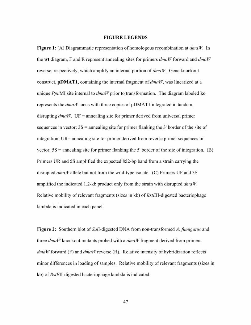
47
FIGURE LEGENDS
Figure 1: (A) Diagrammatic representation of homologous recombination at dmaW. In
the wt diagram, F and R represent annealing sites for primers dmaW forward and dmaW
reverse, respectively, which amplify an internal portion of dmaW. Gene knockout
construct, pDMAT1, containing the internal fragment of dmaW, was linearized at a
unique PpuMI site internal to dmaW prior to transformation. The diagram labeled ko
represents the dmaW locus with three copies of pDMAT1 integrated in tandem,
disrupting dmaW. UF = annealing site for primer derived from universal primer
sequences in vector; 3S = annealing site for primer flanking the 3' border of the site of
integration; UR= annealing site for primer derived from reverse primer sequences in
vector; 5S = annealing site for primer flanking the 5' border of the site of integration. (B)
Primers UR and 5S amplified the expected 852-bp band from a strain carrying the
disrupted dmaW allele but not from the wild-type isolate. (C) Primers UF and 3S
amplified the indicated 1.2-kb product only from the strain with disrupted dmaW.
Relative mobility of relevant fragments (sizes in kb) of BstEII-digested bacteriophage
lambda is indicated in each panel.
Figure 2: Southern blot of SalI-digested DNA from non-transformed A. fumigatus and
three dmaW knockout mutants probed with a dmaW fragment derived from primers
dmaW forward (F) and dmaW reverse (R). Relative intensity of hybridization reflects
minor differences in loading of samples. Relative mobility of relevant fragments (sizes in
kb) of BstEII-digested bacteriophage lambda is indicated.
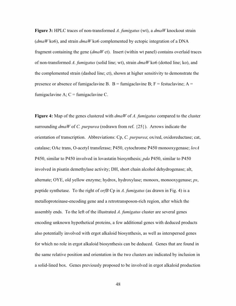
48
Figure 3: HPLC traces of non-transformed A. fumigatus (wt), a dmaW knockout strain
(dmaW ko6), and strain dmaW ko6 complemented by ectopic integration of a DNA
fragment containing the gene (dmaW ct). Insert (within wt panel) contains overlaid traces
of non-transformed A. fumigatus (solid line; wt), strain dmaW ko6 (dotted line; ko), and
the complemented strain (dashed line; ct), shown at higher sensitivity to demonstrate the
presence or absence of fumigaclavine B. B = fumigaclavine B; F = festuclavine; A =
fumigaclavine A; C = fumigaclavine C.
Figure 4: Map of the genes clustered with dmaW of A. fumigatus compared to the cluster
surrounding dmaW of C. purpurea (redrawn from ref. {25}). Arrows indicate the
orientation of transcription. Abbreviations: Cp, C. purpurea; ox/red, oxidoreductase; cat,
catalase; OAc trans, O-acetyl transferase; P450, cytochrome P450 monooxygenase; lovA
P450, similar to P450 involved in lovastatin biosynthesis; pda P450, similar to P450
involved in pisatin demethylase activity; DH, short chain alcohol dehydrogenase; alt,
alternate; OYE, old yellow enzyme; hydrox, hydroxylase; monoox, monooxygenase; ps,
peptide synthetase. To the right of orfB Cp in A. fumigatus (as drawn in Fig. 4) is a
metalloproteinase-encoding gene and a retrotransposon-rich region, after which the
assembly ends. To the left of the illustrated A. fumigatus cluster are several genes
encoding unknown hypothetical proteins, a few additional genes with deduced products
also potentially involved with ergot alkaloid biosynthesis, as well as interspersed genes
for which no role in ergot alkaloid biosynthesis can be deduced. Genes that are found in
the same relative position and orientation in the two clusters are indicated by inclusion in
a solid-lined box. Genes previously proposed to be involved in ergot alkaloid production
49
that are present in both clusters but differ in their relative position and orientation are
enclosed in dashed-line boxes. Genes for which a function has been demonstrated by
gene knockout (5, 16, 29) are indicated with an asterisk. The A. fumigatus sequences
illustrated span from nucleotides 2,894,958 to 2,926,335 of assembly 92 (long arm of
chromosome 2; formerly assembly 72) of the A. fumigatus genome. Identifiers for the A.
fumigatus sequences (as presented from left to right) are m19874, m19875, m19876,
m19877, m19878, m19880, m19881, m19882, m19883, m19884, m19885, m19886,
m19888, and m19891, respectively. Data are available from The Institute for Genomic
Research (www.tigr.org). Accession number for C. purpurea orfA = AY836771, and for
C. purpurea orfB = AY836772. Accession numbers for the remainder of the genes found
in the C. purpurea cluster are reported in refs. 5, 25, and 26.
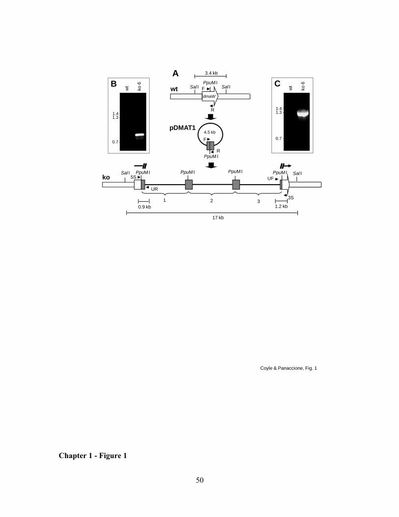
50
Coyle & Panaccione, Fig. 1
PpuMI
dmaW
F
R
PpuMI
4.5 kb
F
R
3.4 kb
Sal ISal Iwt
PpuMI PpuMI Sal IUF
3S2
PpuMISal I
17 kb
1
koPpuMI
31.2 kb
A
wt
ko 6
1.3 -1.4 -
0.7 -
C
pDMAT1
UR
5S
0.9 kb
wt
ko 6
1.3 -1.4 -
0.7 -
B
Chapter 1 - Figure 1
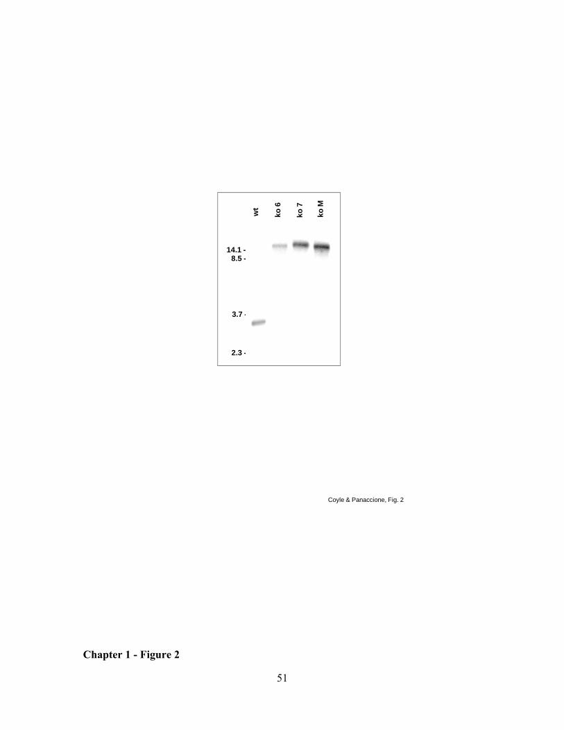
51
ko 6
ko 7
ko M
14.1 -8.5 -
3.7 -
2.3 -
wt
Coyle & Panaccione, Fig. 2
Chapter 1 - Figure 2
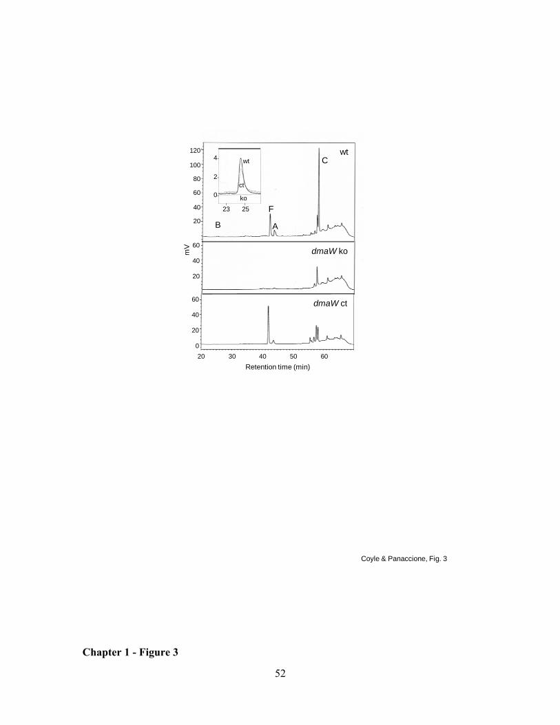
52
Coyle & Panaccione, Fig. 3
wt
F
C
AB
dmaW ko
dmaW ct
mV
4-
2-
0-I I
23 25
wt
ko
ct
20 30 40 50 60Retention time (min)
120
100
80
60
40
20
60
40
20
60
40
20
0
Chapter 1 - Figure 3
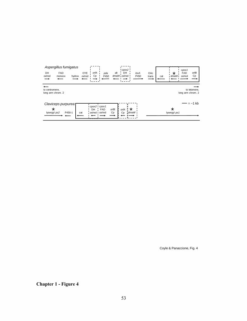
53
Coyle & Panaccione, Fig. 4
dmaW lysergyl ps1lysergyl ps2 P450-1 cat
cpox2DH
ox/red
cpox1FAD
ox/redorfBCp
orfACp
Claviceps purpurea
Aspergillus fumigatus
= ~1 kb
lovAP450 cat
orfACp dmaW
altdmaW
pdaP450
OActrans
OYEox/redhydrox
orfBCp
cpox1FAD
ox/red
cpox2DH
ox/redFAD
monooxDH
ox/red *
***
to telomere,long arm chrom. 2
to centromere,long arm chrom. 2
Chapter 1 - Figure 4
54
CHAPTER 2: Clustered genes, easE and easF, common to Aspergillus fumigatus and ergot fungi control early steps in ergot alkaloid biosynthesis
ABSTRACT
Ergot alkaloids are mycotoxins typically associated with clavicipitaceous fungi and cause
toxicoses to exposed individuals through their interactions with multiple neurotransmitter
receptors. Ergot alkaloids also are produced by the opportunistic human pathogen,
Aspergillus fumigatus; however, its chemical profile consists of certain clavines as
opposed to the lysergic acid derivatives typically observed in clavicipitaceous fungi.
These fungi are hypothesized to share early steps in ergot alkaloid synthesis prior to
divergence into two lineage-specific pathways. Genes involved in ergot alkaloid
synthesis are clustered, and several homologues are shared between the two groups of
fungi. This study focuses on two genes hypothesized to function early in ergot alkaloid
biosynthesis, easE and easF. Functional analysis via gene knockout demonstrated that
both genes are required for ergot alkaloid biosynthesis in A. fumigatus. When either of
the genes was knocked out, transformants failed to produce chanoclavine or any ergot
alkaloids downstream of chanoclavine, but different intermediates accumulated. Strains
with a mutated easF accumulated an alkaloid with properties of dimethylallyltryptophan
(DMAT), a result consistent with easF encoding the N-methyl transferase that acts on
DMAT. Strains in which easE was mutated accumulated an intermediate with properties
of N-methyl-DMAT. These data indicate that easE encodes the first oxidase in the ergot
alkaloid pathway. Mutants of easF accumulated trace quantities of downstream
intermediates, suggesting a route to circumvent this enzyme, presumably by the activity
55
of DMAT synthase on abrine (N-methyl-transferase) which is present in cultures of A.
fumigatus. This study further elucidates the early steps in the pathway in A. fumigatus,
steps that are also shared with clavicipitaceous fungi.
Keywords: Aspergillus fumigatus, Claviceps spp., easE, easF, ergot alkaloid,
methyltransferase, oxidoreductase
INTRODUCTION
Ergot alkaloids can be classified into three groups: clavines, ergopeptines, and simple
amides (12, 14). Clavines are the simplest of the ergot alkaloids, some of which serve as
precursors of lysergic acid. Ergopeptines and simple amides are derivatives of lysergic
acid. Clavicipitaceous fungi, such as Claviceps spp. and Neotyphodium spp., produce
different end products compared to the distantly related saprophyte Aspergillus
fumigatus. All three types of alkaloids are found in clavicipitaceous species, but the
spectrum of alkaloids can vary dependent on species and/or isolate (3, 6, 9). Aspergillus
fumigatus only produces clavines, such as festuclavine and fumigaclavines A, B, and C
(1, 15, 19). Panaccione (9) hypothesizes that the ergot alkaloid biosynthesis pathway
diverges into two lineage-unique pathways after the production of chanoclavine
aldehyde, resulting in different ergot alkaloid profiles between A. fumigatus and
clavicipitaceous fungi.
Comparison of ergot alkaloid profiles and relevant DNA sequence data from
different ergot alkaloid-producing fungi has led to the hypothesis that these fungi share
early steps within the ergot alkaloid biosynthesis pathway (2, 9, 12).
Dimethylallyltryptophan (DMAT) synthase is the determinant step in both A. fumigatus
56
and Neotyphodium sp. Lp1 (2, 16, 18). In A. fumigatus, C. purpurea, C. fusiformis, and
E. festucae, the gene encoding DMAT synthase is clustered among several genes whose
products have predicted functions that may play a role in the ergot alkaloid pathway.
Seven genes are common to the DMAT synthase-based clusters of each of these fungi
and have been labeled as eas genes (14). This study focuses on characterization of easE
and easF, two genes found in the A. fumigatus and the clavicipitaceous ergot alkaloid
gene clusters, via gene disruption in A. fumigatus and biochemical analyses of the gene
disruption mutants.
MATERIALS AND METHODS
Fungi and culture conditions. Aspergillus fumigatus isolate FGSC A1141 (10) and
mutant derivatives were maintained on potato dextrose agar [PDA; dehydrated potato
flakes (Pillsbury, Minneapolis, MN) (20 g/L), D-glucose (20 g/L), and agar (15 g/L)].
Protoplasts or DNA samples were prepared from cultures that were grown overnight in
100 mL of malt extract medium (50 g malt extract, 50 g lactose, 5 g asparagine, and 0.4 g
K2HPO4 per L) in a 250 mL Erlenmeyer flask on an orbital shaker set at 37 ºC and 150
rpm.
Disruption and complementation of easE and easF. Internal (incomplete)
regions of each putative gene’s coding sequence were amplified by PCR primed with the
following oligonucleotides: easE-F (5'-CCAGATACATTGCCATCGCATG-3') and
easE-R (5'-TGTTCCAACTGCTTGGCCAGAT-3') and easF-F (5'-
GCTTCAAAGATGGCAGCAGCTA-3') and easF-R (5'-
GAGTTGGCATAACTTAACGACG-3'). The 50 µL PCR reaction contained 50 mM
KCl, 10 mM Tris-HCl (pH 9.0), 0.1% (v/v) Triton X-100, 1.5 mM MgCl2, 200 µM of
57
each deoxyribonucleotide triphosphate, 1 µM of each primer, and 2.5 units of Taq DNA
polymerase (Promega, Madison, Wisconsin), which was added once the thermocycler
reached 95 ºC in the initial denaturing period. The reaction began with an initial
denaturing step of 2 min and 30 sec at 95 ºC, followed by 35 cycles of 1 min at 94 ºC
(denaturing), 1 min at 55 ºC (annealing), and 1 min at 72 ºC (extension), with a final
extension step of 72 ºC for 7 min. Amplification of the easE fragment was achieved with
an annealing temperature of 57 ºC. A disruption construct was generated to target each
individual gene by ligating the PCR amplicon into the T/A overlap vector pCR2.1
(Invitrogen, Carlsbad, California). For easE and easF, a 4.8-kb construct (pEASE1) and
a 4.5-kb construct (pEASF1) were produced, respectively (Fig. 1 & 2). Prior to
transformation into A. fumigatus, each disruption construct was linearized at a unique
restriction site located within the gene specific region of the construct. The restriction
digests were as follows: pEASE1 with BspEI and pEASF1 with Bsu36I.
Preparation and transformation of the protoplasts were previously described by
Murray et al. (7) with modifications noted by Coyle and Panaccione [(2); Chapter 2], and
instead of separation of protoplasts from debris by step-gradient centrifugation,
protoplasts were filtered through cheesecloth. In addition, the lysing enzymes were
suspended in 0.7 M NaCl instead of osmotic medium to digest the fungal tissue. The
filtrate was centrifuged (5 min, 3436 x g) to pellet the protoplasts. After filtration, the
protoplasts were washed twice in 0.7 M NaCl and once in STC (1 M sorbitol, 50 mM
Tris-HCl pH 7.4, 50 mM CaCl2 (7) to remove digestive enzymes prior to final suspension
in STC. Protoplasts were co-transformed with a linearized disruption construct
corresponding to easE or easF and a hygromycin resistance plasmid, pMOcosX (8),
58
linearized at a unique NotI site. A preliminary high performance liquid chromatography
(HPLC) screen of colonies that arose on transformation plates identified transformants
with altered ergot alkaloid profiles. At least forty hygromycin-resistant colonies were
isolated for each gene, and transformant colonies were screened in groups of ten until
prospective mutants were identified. These colonies were then grown from single spores
to achieve nuclear homogeneity and then analyzed for verification of homologous
recombination (described below).
Homologous recombination between the disruption construct and its respective
native gene was verified by three PCR assays similar to the PCR reaction described
above, but with different primers and annealing temperatures. For easE, the 5' border of
the recombination event was confirmed by PCR (annealing temperature at 57 ºC) primed
with oligonucleotides UF (5'-TGTAAAACGACGGCCAGTGAAT-3', which anneals to
vector sequences near the universal primer annealing site in pCR2.1) and easE-Fscrn (5'-
ACTGAAGACGAGACATGTCATG -3', complementary to easE sequences near the 5'-
end of the gene and flanking the intended site of integration) (Fig. 1A). The juxtaposition
of sequences near the 3' border of the integration event was verified by PCR (annealing
temp 57 ºC) primed from oligonucleotides UR (5'-AGCTATGACCATGATTACGCCA-
3', complementary to vector sequences near the reverse primer annealing site of pCR2.1),
and easE-Rscrn (5'-TTCCTGTACGACACTCTGAAGT -3', which anneals to easE
sequences near the 3'-end of the gene and flanking the intended site of integration) (Fig.
1A). PCR across the entire easE locus was conducted by priming with oligonucleotides
easE-Fscrn and easE-Rscrn (both described above) at an annealing temperature of 57 ºC.
For easF, the 5' border of the recombination event was confirmed by PCR (annealing
59
temperature at 48 ºC) primed with oligonucleotides UF (described above) and easF-Fscrn
(5'-AAGTTGTCGAAGGTCTCACGAA -3', complementary to easF sequences near the
5'-end of the gene and flanking the intended site of integration (Fig. 2A). The
juxtaposition of sequences near the 3' border of the integration event was verified by PCR
(annealing temp 48 ºC) primed from oligonucleotides UR (described above) and easF-
Rscrn (5'-GTGATTAGAGATGCTTCGTGC -3', which anneals to easF sequences near
the 3'-end of the gene and flanking the intended site of integration) (Fig. 2A). PCR
across the entire easF locus was conducted by priming with oligonucleotides easF-Fscrn
and easF-Rscrn (both described above) at an annealing temperature of 45 ºC.
Integration of the transformation construct also was analyzed by Southern
hybridization as described previously (2), except for substitution of the respective gene
specific probe. Each gene-specific probe was made by amplifying wild-type A. fumigatus
genomic DNA via PCR from primer pairs easE-F and easE-R or easF-F and easF-R,
according to the PCR conditions described above, substituting 1× DIG DNA Labeling
Mix (Roche, Mannheim, Germany) for unlabeled dNTPs. Knockout and wild-type
genomic DNA were digested with ApaI for easE, and HincII for easF for each
corresponding Southern blot.
Each null mutant strain was complemented with a fragment containing a full-
length copy of the respective coding sequences along with 1.3-kb and 1.4-kb of 5'-
flanking sequences and 0.8-kb and 1.2-kb of 3'-flanking sequences for easF and easE,
respectively. The complementing fragments were amplified by PCR using primer pairs
described as follows: easE, WeasE-F (5'-CTGCTCCAGCAGGTAGATCTTGATTC -3')
and WeasE-R (5'-TAGAGGAGCACCCAGCTGAATGTCCT -3') and easF, WeasF-F
60
(5'-TCCATTCTTCGCTCGTTCAACCAGCAGG -3') and WeasF-R (5'-
CAGGACCTGTACCTAAAGCCTGGTAACC -3'). These fragments were each
amplified in a 50 µL PCR reaction containing 15 mM (NH4)2SO4, 50 mM Tris-HCl (pH
9.0), 0.1% (v/v) Triton X-100, 1.5 mM MgCl2, 200 µM of each dNTP, 1 µM of each
primer, and 1 unit of DyNAzyme DNA polymerase (New England Biolabs, Beverly,
MA) added during the initial denaturing step. The thermocycler program was as
described above. However, the annealing temperatures and extension periods differed
from above and from each other and they are as follows: easE, 55 ºC and 4 min 30 sec;
and easF, 65 ºC and 3 min 20 sec. Each PCR product was cotransformed into their
respective gene’s-disrupted mutant along with a phleomycin resistance plasmid, pBC-
phleo (Fungal Genetics Stock Center, University of Missouri-Kansas City, Kansas City,
MO). Transformants were selected on complete regeneration medium (11) containing
100 µg/mL phleomycin and incubated at 37 ºC. Complemented transformants were
cultured from individual spores to achieve nuclear homogeneity. To test for the presence
of the original disruption construct, PCR reactions were performed as described above
(i.e. easE-F and UF). An additional PCR screen was performed on each complemented
transformant to test for the presence of the introduced wild-type allele. These reactions
were primed with primer pairs and reaction conditions stated above (i.e. WeasE-F and
WeasE-R).
Analysis of Ergot Alkaloids. Cultures used for ergot alkaloid analysis were
grown on PDA at 37 ºC for 1 wk. Samples were prepared from whole plate extractions in
4 mL 100% methanol, vortexed, and then rotated end-over-end for at least 2 hr. Ergot
alkaloids were detected and quantified from 20µL of the prepared sample by reverse-
61
phase HPLC on a C18 column with fluorescence detection based on excitation and
emission wavelengths of 272 nm and 372 nm, respectively (10). The presence of ergot
alkaloids that possess a 9,10 double bond was analyzed by passing eluates through a
second fluorescence detector set with excitation and emission wavelengths of 310 nm and
410 nm, respectively. The peaks corresponding to festuclavine and fumigaclavines A, B,
and C were previously identified by mass spectral analyses (10) and confirmed by
elimination of these peaks in mutants that lack a functional dmaW, the first gene in the
ergot alkaloid pathway (2). The peak corresponding to chanoclavine was identified by
coelution with authentic chanoclavine standard (provided by B.A. Tapper, AgResearch
New Zealand).
Liquid surface cultures used for ergot alkaloid analysis were grown on malt
extract medium at 37 ºC for 2 weeks, extracted in 4 mL 100% methanol over a period of
2 hr with occasional vortexing. Extracts were centrifuged to pellet spores and other
fungal debris and supernatants were concentrated in a speed vacuum and subjected to
liquid chromatography – mass spectrometry (LC-MS) analysis (Chem. Dept., MIT). The
LC-MS samples were resolved on an Acquity Ultra Performance BEH C18 column with
a 1.7 micrometer particle size, 2.1 x 100 mm dimension. LC-MS was performed at a
constant flow rate of 0.5 mL/min with a gradient described as follows: at 0 min: 20 parts
acetonitrile and 80 parts 0.1% formic acid in water; at 5 min: 20 parts 0.1% formic acid
in water and 80 parts acetonitrile; at 6 min: 80 parts 0.1% formic acid in water and 20
parts acetonitrile. Masses were detected by a Micromass LCT Premier TOF MS with an
electrospray ionization source.
62
RESULTS
Screening by HPLC indicated altered ergot alkaloid profiles in multiple hygromycin-
resistant colonies for both easE and easF transformants. Incorporation of pEASE1 into
the native easE gene in A. fumigatus via homologous recombination occurred in three of
these transformants. For easF, one hygromycin-resistant colony was identified to contain
the pEASF1 within the native easF gene.
DNA analyses via PCR and Southern blot confirmed insertion of the knockout
(ko) constructs within their target gene. All mutant colonies were started from single
spores to achieve homokaryotic cultures. For easE ko mutants, PCR screens across the 5’
and 3’ borders of the predicted integration site both produced the predicted 1.2-kb
product, whereas the wild type lacked amplification from this primer pair (Fig. 1B). A
PCR screen designed to detect the intact wild-type locus was positive for the expected
1.3-kb PCR product in the wild-type strain but did not detect a wild-type allele in the
easE ko, because insertion of the ko construct within the native locus produced a large
distance between priming sites (Fig. 1B). PCR analysis of the integration site in an easF
mutant produced 815-bp and 755-bp fragments from screens across the 5’ and 3’ borders
of the integration site, as predicted (Fig. 2B). The screen to detect the wild-type locus
did not render a product from null mutant strains, again due to the insertion of the ko
construct within the native easF gene. The wild-type DNA did produce the 820-bp
product.
Southern blot analysis of easE mutants confirmed disruption of the native gene
and showed multiple insertions of the pEASE1 within easE (Fig. 1C). The Southern blot
analysis for easF clearly showed insertion of the knockout construct in the easF mutant
63
due to a shift in the gene specific HincII fragment when compared to wild type (Fig. 2C).
Multiple knockout constructs appeared to be inserted, which is not abnormal for this
integration technique. The presence of several bands suggests additional integrations or
recombination among tandemly inserted knockout contstructs. Complementation
analyses (described below) show that the mutant phenotype was due to integration into
easF.
HPLC analyses showed that mutants of either easF or easE did not produce
chanoclavine, festuclavine, or fumigaclavines A, B, or C (Fig. 3A & 4A). The mutants of
easF accumulated a compound with a retention time of 43 min that was not observed in
wild type or in the easE mutants when analyzed by HPLC (Fig. 3A). LC-MS analysis of
this peak showed that the compound had a molecular ion with m/z of 273.16 (Fig. 3B).
Since fragmentation with this ESI-source instrument is rare, the 273.16 m/z ion likely
represents the molecular ion [M + H+] of a compound with molecular weight of 272.16.
The more sensitive analysis on the LC-MS indicated the presence of small quantities of
downstream intermediates, such as festuclavine.
The easE ko accumulated a novel compound that eluted from the HPLC column
at 44 min and by LC-MS yielded an ion with m/z of 287.17, indicative of the molecular
ion of a compound of molecular weight 286.17 (Fig. 4). This mutant did not contain any
other downstream intermediates.
Disrupted strains were complemented by cotransformation with a wild-type copy
of their respective genes and NotI-linearized pBCphleo. A 4.6-kb fragment containing a
functional copy of easE complemented the easE ko. Likewise, easF was complemented
with a 2.9-kb PCR product containing a wild-type copy of the easF allele. Positive
64
transformants were verified by PCR, when primed by either WeasE-f and WeasE-r or
WeasF-f and WeasF-r (not shown). Amplification of the 5’ and 3’ regions flanking the
site of integration of the original knockout construct confirmed that both easE and easF
complemented strains were derivatives of their original knockout mutants. HPLC
analyses demonstrated that colonies of the easE complemented and easF complemented
strains accumulated festuclavine and fumigaclavines A, B, and C, similar to wild type
(not shown).
DISCUSSION
These data demonstrate that easF and easE encode enzymes involved in the early steps of
the ergot alkaloid pathway. Knockout mutants of easE and easF lacked normal ergot
alkaloid accumulation in A. fumigatus, decreasing or eliminating production of
festuclavine and fumigaclavine A, B and C. In addition, novel compounds with
properties of intermediates in the ergot alkaloid pathway accumulated in these mutants,
allowing proposed placement of the enzymes encoded by these genes at specific steps of
the ergot alkaloid pathway.
A. fumigatus easF is located 257 bp downstream of easE within the ergot alkaloid
cluster on chromosome 2 and spans 1.1 kb of the genome [(2); Chapter 1]. Comparison
of the linear sequence to a known protein database shows it has 56% and 60% identity to
C. purpurea and N. lolii EasF, respectively. EasF, both in A. fumigatus and C. purpurea,
has been hypothesized to encode a methyltransferase (2, 4, 12), even though sequence
data to support this were weak and closest sequence identity matches were to products of
other unknown open reading frames. Knockout of easF resulted in accumulation of a
compound with a molecular weight of 272.16, which is the molecular weight of DMAT,
65
the first intermediate of the ergot alkaloid pathway. During the later stages of this work,
Rigbers and Li (2008) published results of expression assays done in vitro in Escherichia
coli. The expressed and isolated easF product methylated DMAT in the presence of S-
adenosylmethionine, illustrating easF encodes for a methyltransferase capable of using
DMAT as a substrate. My in vivo functional analysis of the easF gene supports the
finding of the easF product functioning as a methyltransferase; the null mutant’s inability
to methylate DMAT resulted in accumulation of DMAT.
Interestingly, Unsold and Li (17) demonstrated that DMAT synthase of A.
fumigatus, which typically uses tryptophan as a substrate, also can prenylate N-methyl-
tryptophan (abrine) resulting in N-methyl-DMAT. This activity of DMAT synthase
could theoretically bypass the requirement of the N-methyl transferase step. LC-MS
analyses of my easF mutants indicated a degree of leakiness in that trace quantities of
festuclavine and fumigaclavines A and C were detectable in selected ion traces (data not
shown). Abrine was detectable in cultures of A. fumigatus, and thus may have served as
an alternate substrate for DMAT synthase allowing a bypass of the mutated easF-
encoded step.
The gene, easE, is found on the 2nd chromosome of A. fumigatus, between
nucleotides 2925694-2927815; it is clustered with genes involved in ergot alkaloid
production (2). The Aspergillus sequence is 45% similar to the C. purpurea easE protein
sequence, 43% similar to C. fusiformis oxidase, and 47% similar to a putative oxidase of
N. lolii. The easE sequence indicates that it encodes a FAD-containing oxidoreductase, a
class of enzyme which would play a role in the proposed pathway (9, 12). My results
show that strains in which easE is inactivated accumulate an intermediate with
66
fluorescence properties typical of an ergot alkaloid with an 8,9 double bond and a
molecular mass of 286.17, consistent with N-methyl-DMAT. Chanoclavine and
alkaloids downstream of chanoclavine are all eliminated by knockout of easE. Based on
these data, we hypothesize that the product of easE oxidizes N-methyl-DMAT. More
specifically, the easE product likely hydroxylates the benzylic carbon (position 10) of the
isoprene group of N-methyl-DMAT, an oxidation step proposed by Kozikowski et al. (5).
Synthetic intermediates in which that particular carbon is oxidized are unstable; the 10-
hydroxylated N-methyl-DMAT spontaneously dehydrates to the corresponding diene (Fig
5), which has been demonstrated to be an authentic intermediate between N-methyl-
DMAT and chanoclavine (5).
These findings suggest that the initial steps in the ergot alkaloid pathway
proceed as follows: the determinant step, DMAT synthase (encoded by dmaW) (2), is
followed first by a methyltransferase (easF) and then by an oxidoreductase (easE). This
study uncovers the early steps of the pathway, allowing for a better understanding of its
dynamics both in A. fumigatus and clavicipitaceous fungi.
ACKNOWLEDGEMENTS
This project was supported by the National Research Initiative of the USDA CSREES,
grants 2008-35318-04549 and 2005-35318-16184. I thank Johnathan Cheng and Dr.
Sarah O’Connor (Massachusetts Institute of Technology) for performing mass spectrum
analyses and for helpful suggestions on pathway intermediates. I also thank Chris
Schardl (University of Kentucky) for helpful discussions, and Mike Hamstead and Dawn
Chamberlain for their laboratory assistance.
67
REFERENCES
1. Cole, R. J., J. W. Kirksey, J. W. Dorner, D. M. Wilson, J. C. Johnson, A. N.
Johnson, D. M. Bedell, J. P. Springer, and K. K. Chexal. 1977. Mycotoxins
produced by Aspergillus fumigatus species isolated from molded silage. J. Agric.
Food Chem. 25:826–830.
2. Coyle, C. M., and D. G. Panaccione. 2005. An ergot alkaloid biosynthesis gene
and clustered hypothetical genes from Aspergillus fumigatus. Appl. Environ.
Microbiol. 71:3112–3118.
3. Flieger, M., M. Wurst, and R. Shelby. 1997. Ergot alkaloids -- sources,
structures and analytical methods. Folia Microbiol. 42:3–30.
4. Haarmann, T., C. Machado, Y. Lübbe, T. Correia, C. L. Schardl, D. G.
Panaccione, and P. Tudzynski. 2005. The ergot alkaloid gene cluster in
Claviceps purpurea: extension of the cluster sequence and intra species evolution.
Phytochemistry 66:1312–1320.
5. Kozikowski, A. P., C. Chen, J.-P. Wu, M. Shibuya, C.-G. Kim, and H. G.
Floss. 1993. Probing ergot alkaloid biosynthesis: Intermediates in the formation
of ring C. J. Am. Chem. Soc. 115:2482–2488.
6. Kozlovsky, A. G. 1999. Producers of ergot alkaloids out of Claviceps genus, p.
479–499. In V. Kren and L. Cvak (ed.), In Ergot: The Genus Claviceps. Harwood
Academic Publishers, The Netherlands.
7. Murray, F. R., G. C. M. Latch, and D. B. Scott. 1992. Surrogate
transformation of perennial ryegrass, Lolium perenne, using genetically modified
Acremonium endophyte. Mol. Gen. Genet. 233:1–9.
68
8. Orbach, M. J. 1994. A cosmid with a HyR marker for fungal library
construction and screening. Gene 150:159–162.
9. Panaccione, D. G. 2005. Origins and significance of ergot alkaloid diversity in
fungi. FEMS Microbiol. Lett. 251:9–17.
10. Panaccione, D. G., and C. M. Coyle. 2005. Abundant respirable ergot alkaloids
from the common airborne fungus Aspergillus fumigatus. Appl. Environ.
Microbiol. 71:3106–3111.
11. Panaccione, D. G., R. D. Johnson, J. Wang, C. A. Young, P. Damrongkool, B.
Scott, and C. L. Schardl. 2001. Elimination of ergovaline from a grass-
Neotyphodium endophyte symbiosis by genetic modification of the endophyte.
Proc. Natl. Acad. Sci. USA 98:12820–12825.
12. Panaccione, D. G., C. Schardl, and C. M. Coyle. 2006. Pathways to diverse
ergot alkaloid profiles in fungi, p. 23–52 In J. T. Romeo (ed.), Recent Advances
in Phytochemistry, vol. 40. Elsevier, Amsterdam, The Netherlands.
13. Rigbers, O., and S.-M. Li. 2008. Ergot alkaloid biosynthesis in Aspergillus
fumigatus: Overproduction and biochemical characterization of a 4-
dimethylallyltryptophan N-methyltransferase. J. Biol. Chem. 283:26859–26868.
14. Schardl, C., D. G. Panaccione, and P. Tudzynski. 2006. Ergot alkaloids--
biology and molecular biology, p. 45–86. In G. A. Cordell (ed.), Alkaloids Chem.
Biol., vol. 63. Academic Press, San Diego, CA.
15. Spilsbury, J. F., and S. Wilkinson. 1961. The isolation of festuclavine and two
new clavine alkaloids from Aspergillus fumigatus Fres. J. Chem. Soc. 5:2085–
2091.
69
16. Tsai, H.-F., H. Wang, J. C. Gebler, C. D. Poulter, and C. L. Schardl. 1995.
The Claviceps purpurea gene encoding dimethylallyltryptophan synthase, the
committed step for ergot alkaloid biosynthesis. Biochem. Bioph. Res. Co.
216:119–125.
17. Unsöld, I. A., and S.-M. Li. 2005. Overproduction, purification and
characterization of FgaPT2, a dimethylallyltryptophan synthase from Aspergillus
fumigatus. Microbiology 151:1499–1505.
18. Wang, J., C. Machado, D. G. Panaccione, H.-F. Tsai, and C. L. Schardl.
2004. The determinant step in ergot alkaloid biosynthesis by an endophyte of
perennial ryegrass. Fungal Genet. Biol. 41:189–198.
19. Yamano, T., K. Kishino, S. Yamantodani, and M. Abe. 1962. Investigation
on ergot alkaloids found in cultures of Aspergillus fumigatus. Takeda Kenkyusho
Nempo (Annual Report Takeda Research Laboratories) 21:95–101.
70
FIGURE LEGENDS
Figure 1: Disruption strategy for easE. (A) EasE-F and easE-R primed amplification
of internal region of easE. Gene disruption construct pEASE1 contains this internal
fragment and was linearized at a unique BspEI site prior to transformation. The bottom
panel illustrates insertion of three copies of the pEASE1 within genomic easE and the
positions of primers used in PCR experiments to demonstrate integration at this locus.
(B) Homologous recombination of the transforming DNA at easE was demonstrated by
PCR with primers easE-Fscrn and UF confirming integration at the 5'-end of the gene and
primers UR and easE-Rscrn confirming integration at the 3'-end. Primer locations are
shown in the bottom portion of part A. The wild-type (wt) screen, used to assess the
presence of the wild-type allele, was a PCR primed from easE-Fscrn and easE-Rscrn, and
amplification does not occur from DNA that has disruption constructs within the intended
site of integration due to the increase in template length. PCR of non-transformed (wt)
DNA gives a product of 1.3-kb. (C) Incorporation of transforming DNA at the targeted
site was confirmed by Southern blot hybridization. Fungal DNA extracts were digested
with ApaI and hybridized with a digoxigenin-dUTP labeled easE probe. The large size of
the easE-containing fragments in strains easE ko1, easE ko15, and easE ko31 indicate
three or more copies of pEASE1 are present in the integration site. The relative
mobilities of relevant fragments of BstEII-digested bacteriophage lambda DNA are
indicated on the left in panels B and C.
Figure 2: Disruption strategy for easF. (A) EasF-F and easF-R primed amplification
of internal region of easF. Gene disruption construct pEASF1 contains this internal
71
fragment and was linearized at a unique Bsu36I site prior to transformation. The bottom
panel illustrates insertion of three copies of the pEASF1 within genomic easF and the
positions of primers used in PCR experiments to demonstrate integration at this locus.
(B) Homologous recombination of the transforming DNA at easF was demonstrated by
PCR with primers easF-Fscrn and UF confirming integration at the 5'-end of the gene and
primers UR and easF-Rscrn confirming integration at the 3'-end. Primer locations are
shown in the bottom portion of part A. The wild-type (wt) screen, used to assess the
presence of the wild-type allele, was a PCR primed from easF-Fscrn and easF-Rscrn, and
amplification does not occur from DNA that has disruption constructs within the intended
site of integration due to the increase in template length. PCR of non-transformed (wt)
DNA gives a product of 820-bp. (C) Incorporation of transforming DNA at the targeted
site was confirmed by Southern blot hybridization. Fungal DNA extracts were digested
with HincII and hybridized with a digoxigenin-dUTP labeled easF probe. The large sizes
of the easF-containing fragments in strain easF ko1 indicate three or more copies of
pEASF1 are present in the integration site. Multiple bands suggest these strains are
heterokaryons with varying number of pEASF1s inserted within target gene, due to
deletion of one or more copies within some nuclei. The relative mobilities of relevant
fragments of BstEII-digested bacteriophage lambda DNA are indicated on the left in
panels B and C.
Figure 3: Chemical characterization of easF ko. (A) HPLC chromatogram of wild-
type A. fumigatus (wt) and easF ko. B = fumigaclavine B; F = festuclavine; A =
fumigaclavine A; C = fumigaclavine C; CH = chanoclavine. Numbers next to peak are
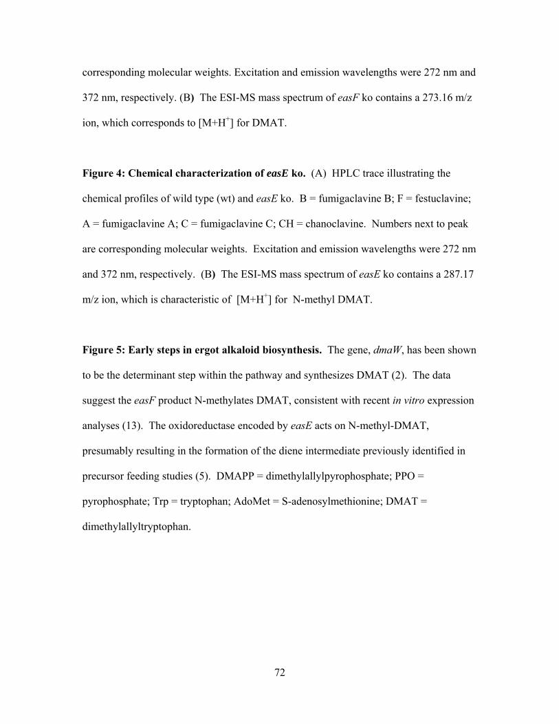
72
corresponding molecular weights. Excitation and emission wavelengths were 272 nm and
372 nm, respectively. (B) The ESI-MS mass spectrum of easF ko contains a 273.16 m/z
ion, which corresponds to [M+H+] for DMAT.
Figure 4: Chemical characterization of easE ko. (A) HPLC trace illustrating the
chemical profiles of wild type (wt) and easE ko. B = fumigaclavine B; F = festuclavine;
A = fumigaclavine A; C = fumigaclavine C; CH = chanoclavine. Numbers next to peak
are corresponding molecular weights. Excitation and emission wavelengths were 272 nm
and 372 nm, respectively. (B) The ESI-MS mass spectrum of easE ko contains a 287.17
m/z ion, which is characteristic of [M+H+] for N-methyl DMAT.
Figure 5: Early steps in ergot alkaloid biosynthesis. The gene, dmaW, has been shown
to be the determinant step within the pathway and synthesizes DMAT (2). The data
suggest the easF product N-methylates DMAT, consistent with recent in vitro expression
analyses (13). The oxidoreductase encoded by easE acts on N-methyl-DMAT,
presumably resulting in the formation of the diene intermediate previously identified in
precursor feeding studies (5). DMAPP = dimethylallylpyrophosphate; PPO =
pyrophosphate; Trp = tryptophan; AdoMet = S-adenosylmethionine; DMAT =
dimethylallyltryptophan.
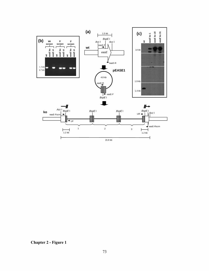
73
(a)BspE I
easE
easE-F
easE-R
1.5 kb
Acc IAcc I
wt
BspE I
4.8 kb
easE-R
easE-F
pEASE1
BspE I BspE IAcc IUR
easE-Rscrn2
BspE IAcc I
15.9 kb
1
ko BspE I
3
1.2 kb
UF
easE-Fscrn
1.2 kb
(b) Wt screen
3' screen
5' screen
wt
easE
koea
sE c
t
wt
easE
koea
sE c
tw
tea
sEko
easE
ct
1.3 kb0.7 kb
(c)
14 kb
1.4 kb
easE
ko
15
easE
ko
1
wt
easE
ko
31
1.9 kb
Chapter 2 - Figure 1
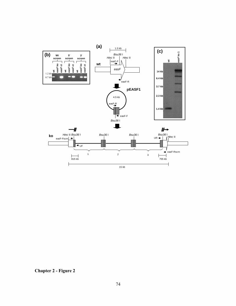
74
(a)Bsu36 I
easF
easF-F
easF-R
1.5 kb
Hinc IIHinc II
wt
Bsu36 I
4.5 kb
easF-R
easF-F
pEASF1
Bsu36 I Bsu36 IHinc IIUR
easF-Rscrn2
Bsu36 IHinc II
15 kb
1
ko Bsu36 I
3
755 kb
UF
easF-Fscrn
815 kb
(b) Wt screen
3' screen
5' screen
wt
easF
koea
sF c
t
wt
easF
koea
sF c
tw
tea
sFko
easF
ct
1.2 kb0.7 kb
14 kb
1.4 kb
(c)
easF
ko 1
wt
6.4 kb
3.7 kb
2.3 kb
Chapter 2 - Figure 2
75
Mass/charge
Rel
ativ
e in
tens
ity
B
A
wild typeeasF ko
272/372
C
F
BA
272.16
CH
Chapter 2 - Figure 3
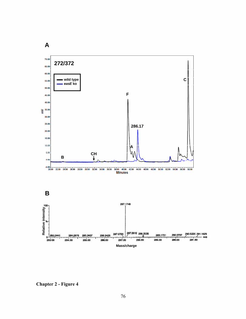
76
Mass/charge
Rel
ativ
e in
tens
ity
B
A
wild typeeasE ko
272/372
F
C
B
A
286.17
CH
Chapter 2 - Figure 4
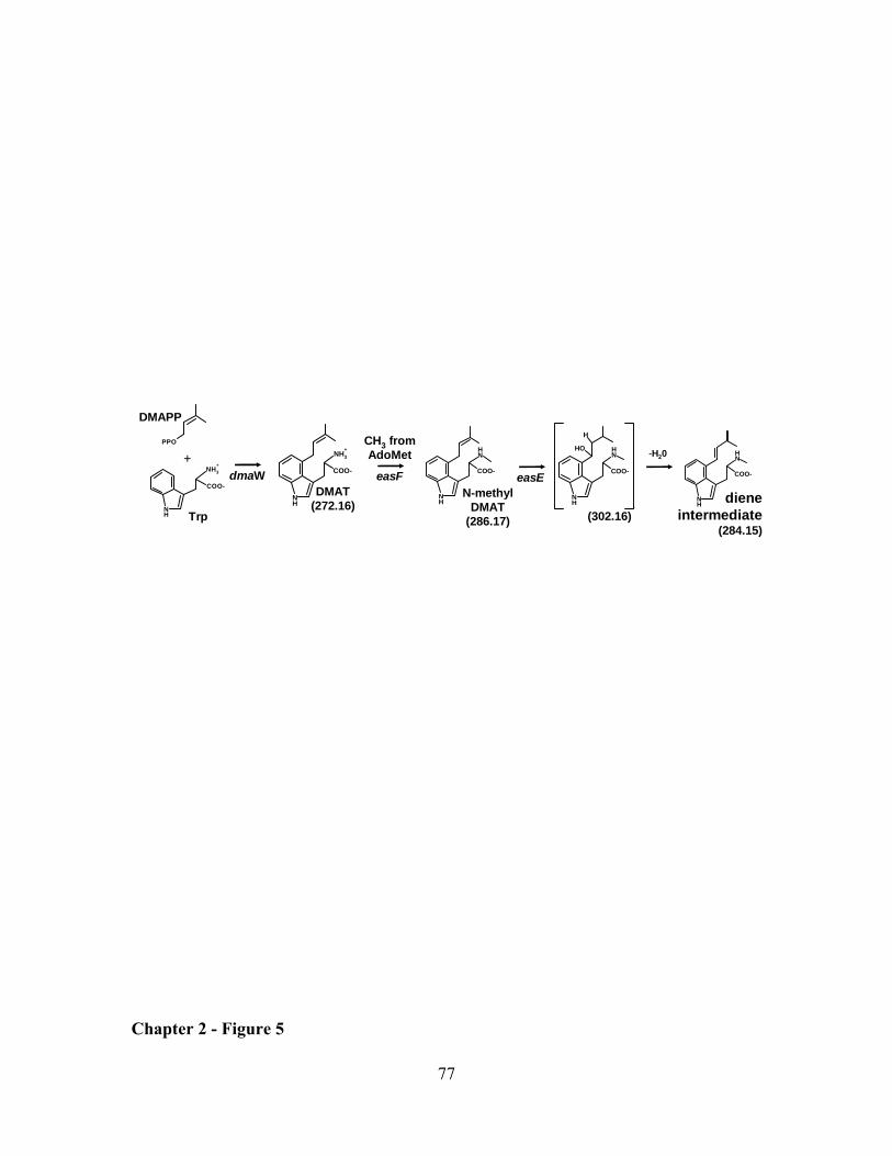
77
COO-
NH
NH
COO-
NH
NH
easEN-methyl
DMAT(286.17)
-H20
COO-
NH
NH3+
Trp
DMAPP
+
COO-
NH
NH3+
PPO
easF
CH3 fromAdoMet
DMAT(272.16)
dmaW
H
COO-
NH
NHOH
n
(302.16)diene
intermediate (284.15)
Chapter 2 - Figure 5
78
CHAPTER 3: An old yellow enzyme gene is required for ergot alkaloid biosynthesis and controls the branch point between Aspergillus fumigatus and Claviceps purpurea ergot alkaloid pathways
ABSTRACT
Ergot fungi in the genus Claviceps and several related grass endophytes in the genus
Neotyphodium produce toxic ergot alkaloids. Often these fungi produce a variety of ergot
alkaloids including clavines as well as lysergic acid derivatives. Ergot alkaloids also are
produced by the distantly related, opportunistic human pathogen, Aspergillus fumigatus.
However, this fungus produces festuclavine and fumigaclavines A, B, and C, which
collectively differ from clavines of clavicipitaceous fungi in saturation of the last
assembled of four rings in the tetracyclic ergoline structure. The two lineages are
hypothesized to share early steps of the ergot alkaloid pathway with divergence occurring
at some point after the synthesis of the tricyclic intermediate chanoclavine. Disruption of
easA, a gene predicted to encode a flavin-dependent oxidoreductase of the old yellow
enzyme class, in A. fumigatus led to accumulation of chanoclavine and an intermediate
believed to be chanoclavine aldehyde. Complementation of the A. fumigatus easA mutant
with a wild-type allele from the same fungus restored the wild-type profile of ergot
alkaloids. Complementation of the A. fumigatus easA knockout with a homologue of
easA from C. purpurea resulted in accumulation of alkaloids (agroclavine, setoclavine,
and its isomer isosetoclavine) typical of clavicipitaceous fungi. These data indicate that
the old yellow enzyme encoded by easA is required for ergot alkaloid biosynthesis and
functional differences in this enzyme in A. fumigatus and C. purpurea result in
79
divergence of their respective ergot alkaloid pathways. Sequence analysis of easA
homologues from several fungi producing different classes of ergot alkaloids indicates a
difference in a residue previously shown to be critical for double bond reduction in other
old yellow enzymes.
Keywords: Ergot alkaloid, Aspergillus fumigatus, easA, Claviceps spp., old yellow
enzyme (OYE)
INTRODUCTION
Different classes of ergot alkaloids are produced by members of two distinct fungal
lineages. Clavicipitaceous species, which includes Claviceps spp. and Neotyphodium
spp. (and its closely sexual relative, Epichloë spp.), are in the order Hypocreales and are
known to synthesize lysergic acid derivatives, which include simple amides of lysergic
acid and the more complex ergopeptines. These alkaloids have an 8,9 or 9,10 double
bond within the fourth ring of the tetracyclic ergoline structure. Ergot alkaloid
production also is associated with the distantly related opportunistic human pathogen,
Aspergillus fumigatus, in the order Eurotiales (4, 7). Aspergillus fumigatus ergot alkaloid
biosynthesis renders simple clavines and differs significantly from the more complex
profile of C. purpurea and Neotyphodium spp., with one important distinction being the
saturation of the fourth ring of the ergoline structure (Fig. 1).
Several genes involved in the ergot alkaloid pathways of A. fumigatus and
clavicipitaceous fungi are found clustered together within the genome of each species (2,
3, 5, 19). These distantly related fungi are hypothesized to share several early pathway
steps, after which the pathways diverge to yield distinct sets of ergot alkaloids (12, 15).
80
The gene, dmaW, which encodes for a dimethylallyltryptophan (DMAT) synthase in
Neotyphodium, functions similarly in A. fumigatus (2, 18, 20, 21). Annotation of the
regions surrounding this gene in A. fumigatus has identified potential homologues of
genes also found in N. coenophialum and C. purpurea ergot alkaloid gene clusters (2).
One of the shared genes, easA, is predicted to encode an old yellow enzyme (OYE). Old
yellow enzymes are flavin-containing oxidoreductases initially found in brewers’ bottom
yeast (22). Enzymes in this family use a reduced flavin cofactor and an active site
tyrosine residue to reduce an α/β-unsaturated aldehyde or ketone. Subsequently, the
enzymes use NADPH to restore the flavin cofactor to its reduced state. OYEs catalyze
multiple reactions useful for both biotechnological and pharmaceutical applications;
however physiological roles and natural substrates for many of these enzymes presently
are unknown. Based on the apparent need in the ergot alkaloid pathway of A. fumigatus
for reduction of a carbon-carbon double bond in the intermediate chanoclavine aldehyde,
we hypothesized that the OYE-encoding gene easA is required for ergot alkaloid
biosynthesis (2, 15). In this study, knockout and complementation analyses of easA in A.
fumigatus are performed to ascertain the role of this gene product in ergot alkaloid
biosynthesis.
MATERIALS AND METHODS
Fungi and culture conditions. Aspergillus fumigatus isolate FGSC A1141 (13) and its
mutant derivatives were cultured on potato dextrose agar (PDA; 20 g dehydrated potato,
20 g glucose, and 15 g agar per liter). Liquid cultures used for preparation of protoplasts
or DNA were grown at 37 ºC on an orbital shaker (150 rpm) overnight in a 250 mL
81
Erlenmeyer flask containing 100 mL of malt extract medium (50 g malt extract, 50 g
lactose, 5 g asparagine, and 0.4 g K2HPO4 per L).
Disruption and complementation of easA. A 595-bp fragment within the
coding sequence of easA was amplified by PCR, primed with oligonucleotides oyeF (5'-
ACCGAAGCAACAGACATCACC-3') and oyeR (5'-ATATGCGACATCCAATCGCC-
3'). The 50 µL PCR reaction contained 50 mM KCl, 10 mM Tris-HCl (pH 9.0), 0.1%
(v/v) Triton X-100, 1.5 mM MgCl2, 200 µM of each deoxyribonucleotide triphosphate, 1
µM of each primer, and 2.5 units of Taq DNA polymerase (Promega, Madison,
Wisconsin), which was added once the thermocycler reached 95 ºC in the initial
denaturing period. The reaction began with an initial denaturing step of 2 min 30 sec at
95 ºC, followed by 35 cycles of 1 min at 94 ºC, 1 min at 55 ºC, and 1 min at 72 ºC, with a
final extension step of 72 ºC for 7 min. A 4.5-kb disruption construct, pAFOYE1, was
generated by ligating the PCR product into the T/A overlap vector pCR2.1 (Invitrogen,
Carlsbad, California). Prior to transformation into the fungus, pAFOYE1 was linearized
by a restriction digest at the unique SmaI site within the easA region of the construct.
Protoplast preparation and transformation were preformed as described by Murray et al.
(10),with modifications previously noted by Coyle and Panaccione (2). Protoplasts were
separated from undigested hyphae by means of filtration through cheesecloth instead of
step-gradient centrifugation. Protoplasts in the filtrate were then pelleted by
centrifugation (5 min, 3436 x g) and washed twice with 0.7M NaCl and once in STC (10)
prior to a final resuspension in STC. Linearized disruption construct pAFOYE1 (Fig.
2A) and a hygromycin resistance plasmid, pMOcosX (11), linearized at a unique NotI site
(2) were co-transformed into the protoplasts. Transformants colonies were purified to a
82
homokaryotic state by culturing from single spores. These cultures then were screened in
three different PCR assays to identify strains in which the disruption construct inserted
within the native easA. With the exception of different primers and annealing
temperatures, the PCR reactions are as described above. The 5' border of the
homologous recombination event was confirmed by PCR (annealing temperature at
57 ºC) primed with oligonucleotides UR (5'- AGCTATGACCATGATTACGCCA -3',
which anneals to vector sequences near the universal primer annealing site in pCR2.1)
and oyeFscrn (5'-GGAAGATGTCATCTCCAACATAG-3', which is complementary to
easA sequences near the 5'-end of the easA gene and flanking the intended site of
integration) (Fig. 2A). The juxtaposition of sequences near the 3' border of the
integration event was verified by PCR (annealing temp 57 ºC) primed from
oligonucleotides UF (5'- GCCAGTGAATTGTAATACGACTC -3', complementary to
vector sequences near the reverse primer annealing site of pCR2.1), and oyeRscrn (5'-
AGCCTTCTCTTGATAGCGTGCT-3', which anneals to easA sequences near the 3'-end
of the gene and flanking the intended site of integration) (Fig. 2A). PCR across the entire
easA locus was conducted by priming with oligonucleotides oyeFscrn and oyeRscrn
(both described above) at an annealing temperature of 57 ºC.
Integration of pAFOYE1 into the native easA also was analyzed by Southern
hybridization as described previously (2). Exceptions were inclusion of an easA-specific
probe generated by amplifying wild-type A. fumigatus genomic DNA in a reaction
primed with oyeF and oyeR according to the PCR conditions described above, and
substitution of 1× DIG DNA Labeling Mix (Roche, Mannheim, Germany) for unlabeled
dNTPs. Genomic DNA was digested by SalI prior to Southern blot analysis.
83
The easA knockout (ko) strain was complemented by ectopic insertion of a
functional copy of A. fumigatus easA into the mutant genome (easA ct). This 3.3-kb
fragment contained the entire coding sequence along with approximately 1.2-kb of 5'-
flanking sequences and 1.0-kb of 3'-flanking sequences and was amplified by PCR
primed from WeasAF (5'-AGAATCGAGGTGAGCGTGTAGAATGC-3') and WeasAR
(5'-AAGCTGGAACACTACTGATCGAGTGC-3'). Reaction conditions were similar to
those previously described by Coyle et al. (1) except for an annealing temperature of
58 ºC and an extension period of 3 min 30 sec.
Additionally, the easA ko strain was complemented with the easA homologue
from Claviceps purpurea strain ATCC20102. Amplification of this gene was primed
from CpoyeF (5’-TGAGTTCGAACGATGATCTGTAGCG -3’) and CpoyeR (5’-
TGAGTTCGAAAGAGTCTTCTACGCC -3’), with PCR conditions as described above,
except for an annealing temperature of 54 ºC and an extension time of 2 min 30 sec. The
PCR product was co-transformed into an easA-disrupted mutant of A. fumigatus along
with a phleomycin resistance plasmid, pBC-phleo (Fungal Genetics Stock Center,
University of Missouri-Kansas City, Kansas City, MO) linearized at a unique NotI site.
Transformants were selected on complete regeneration medium (14) containing 100
µg/mL phleomycin and incubated at 37 ºC. Complemented transformants were cultured
from individual germinated conidia in order to achieve a homonucleate culture. To test
for the presence of the original disruption construct, PCR reactions were conducted as
described above, primed with oyeFscrn and UR (for the 5' flank of the integration) and
UF and oyeRscrn (for the 3' flank). An additional PCR screen was performed with either
primers WeasAF and WeasAR (described above) or CPoyeF and CpoyeR (described
84
above), to test for the presence of the introduced wild-type or C. purpurea allele in the
complemented transformants (Cp easA ct).
Sequencing of easA from Claviceps spp. Amplification of the C. purpurea
strain ATCC20102 homologue of easA is described above, except using A. fumigatus
easA complemented strain as template DNA. Internal portions (corresponding to coding
sequences for amino acids 19-341 based on the C. purpurea P1 (accession number
CAG28312 sequence) of the Claviceps gigantea and Claviceps africana easA
homologues were amplified in reactions primed with degenerative oligonucleotides
ClavoyeF (5’- CACAAARTYGTRCTYTCTCCYATG -3’) and ClavoyeR (5’-
YGTGTCTCGRTTGTACTTYTGCAG -3’). The reactions were described above, except
for an annealing temperature of 48 ºC and extension period of 2 min. PCR products were
ligated into the pCR2.1 vector. Sequencing was performed by Davis Sequencing
(www.davissequencing.com, Davis, CA). The initial round of sequencing for all three
Claviceps spp. was primed from universal primers M13F and M13R. To complete
double-stranded sequence from the longer C. purpurea easA clone, a second round of
sequencing was primed with custom primers CpoyeM13F2(5’-
GATCAAGCAGCGAAGTACCG -3’) and CpoyeM13R2 (5’-
TTGGAGGACTGGTCATGAGG -3’).
Sequences were aligned with additional available EasA sequences from ergot
alkaloid producing fungi (C. purpurea strain P1 (accession number CAG28312),
Neotyphodium lolii (accession number ABM91449), Claviceps fusiformis (accession
number ABV57819), and Epichloë festucae, contig 3372 nucleotides 256-1251 (reverse,
complement) within the available E. festucae genome
85
(http://www.genome.ou.edu/blast/ef_blastall.html), determined by tblastn with N. lolii
EasA sequence corresponding to amino acids 19-350, by application ClustalW at the
European Biotechnology Institute (EBI) web site (www.ebi.ac.uk/ClustalW/). The
alignment was analyzed in a distance analysis at the same EBI site (accepting defaults of
all parameters) to construct a Neighbor-Joining tree.
Analysis of ergot alkaloids. To analyze ergot alkaloid production, small cubes
(containing approximately 8 mm x 8 mm of colony surface area) were removed from
sporulating colonies that had been grown on PDA at 37 ºC for 5 days. Samples were
submerged in 0.4 mL of 80% methanol, vortexed briefly, rotated end-over-end (44 rpm)
for 2 hr, and centrifuged (10 min., 14,000 x g) to pellet fungal mass and agar.
Supernatants were analyzed by HPLC (C18 column) with acetonitrile-ammonium acetate
gradients and detected with dual fluorescence detectors set at excitation and emission
wavelengths of 272 nm / 372 nm and 310 nm / 410 nm (1, 13). The identities of peaks
corresponding to chanoclavine, festuclavine, and fumigaclavines A, B, and C were
previously established by mass spectral analyses of native and de-esterified fractions
(13). These peak identities also were confirmed by their elimination in extracts from
dmaW, a strain in which the determinant step of the ergot alkaloid pathway was
inactivated (2). Chanoclavine, agroclavine, setoclavine, and isosetoclavine were
identified by coelution with standards, and their masses were confirmed by LC-MS (see
below).
To quantify ergot alkaloids and conidiation from the wild type, easA ko and Cp
easA ct samples, cultures were grown on PDA for 5 days at 37 ºC. Samples,
86
containing 50.3 mm2 of colony surface area, were excised from cultures with the broader
end of a disposable 1-mL pipette tip (Fisher Scientific, catalog number 02-681-172),
submerged in 1 mL 80% methanol, agitated by vortexing, and rotated at 44 rpm for 2 hr.
Aliquots were removed for HPLC analyses and for quantification of conidia.
Concentrations of agroclavine, setoclavine, and isosetoclavine were based on peak area
relative to the external standards of agroclavine (Sigma, St. Louis, MO) (for agroclavine)
and ergotamine (for setoclavine and isosetoclavine) as previously described (Panaccione
and Coyle 2005). To determine spore counts, dilutions of the methanol extracts were
prepared and conidia were enumerated with a hemacytometer and light microscope. Five
samples were analyzed for each treatment. Data from easA ko and C. purpurea easA
complemented strains were compared by Wilcoxon/Krustal-Wallis tests (rank sums).
Extracts for LC-MS analyses were prepared from cultures inoculated on the
surface of liquid potato dextrose medium (PDA without agar, 50 mL in a 250-mL
Erlenmeyer flask) and grown at 37 ºC with no agitation. Ergot alkaloids were extracted
by agitation in 4 mL of 100% methanol. The methanol and dislodged conidia and hyphal
fragments were transferred to microcentrifuge tubes, vortexed, and rotated end-over-end
for a 2-hr period. Then the sample was centrifuged (10 min., 14,000 x g) to pellet fungal
material. Methanol extracts and individual peaks collected from HPLC analysis
performed as described above were concentrated under vacuum and sent to
Massachusetts Institute of Technology for liquid chromatography-mass spectrometry
(LC-MS) analyses. LC-MS samples were separated on an Acquity Ultra Performance
BEH C18 column with a 1.7 micrometer particle size, and 2.1 x 100 mm dimensions and
were eluted with a linear gradient (0.5 mL/min) of 20 parts acetonitrile + 80 parts 0.1%
87
formic acid in water, initially, to 80 parts acetonitrile + 20 parts 0.1% formic acid in
water at 5 min. Masses of eluates were detected by a Micromass LCT Premier TOF MS
with an electrospray ionization source.
RESULTS
Disruption and complementation of easA. Integration of the disruption construct
pAFOYE1 into the native easA gene of A. fumigatus via homologous recombination
occurred in two out of the 64 hygromycin-resistant transformants. Insertion of
pAFOYE1 within easA was verified by PCR, with screens of the 5’ and 3’ borders of the
integration resulting in 811-bp and 992-bp fragments, respectively (Fig 2B). In the
mutant, no 1-kb amplification product was detected when PCR was primed with
oligonucleotides with binding sites separated from each other by 1-kb in the wild type
due to the insertion of the disruption construct within the easA locus (Fig 2B). Southern
blot analysis showed multiple insertions of pAFOYE1 within easA by the shift in
mobility of the easA-specific region (Fig 2C).
The easA mutants did not produce festuclavine or fumigaclavines A, B, and C,
which are four ergot alkaloids typically detectable in A. fumigatus (13). However, an
important intermediate within the ergot alkaloid pathway, chanoclavine, which eluted
from the HPLC column at 32.8 min, was observed in the mutant’s chemical profile (Fig 3
& 4). In addition, a 255.05 m/z fragment was observed by mass spectrometry within the
easA ko extract, although elution time by HPLC was unknown (not shown). Trace
quantities of agroclavine, setoclavine, and isosetoclavine were identified in mutant
extracts by coelution with standards (Fig. 3). Co-transformation of a copy of A.
fumigatus easA restored normal ergot alkaloid production. This wild-type allele inserted
88
ectopically within the genome. The construct, pAFOYE1, was verified to be inserted
within the native easA locus by PCR screens of the recipient strain (Fig. 2B).
Complementation of easA mutant with easA homologue from C. purpurea.
Ectopic insertion of the easA homologue from C. purpurea resulted in accumulation of
larger quantities of agroclavine, setoclavine and its isomer isosetoclavine within the
complemented strain (Table 1, Fig. 3). The complemented strain was positive for the
presence of the knockout construct, pAFOYE1 (not shown).
Analyses of easA sequences among ergot alkaloid producers. The translated
sequence of A. fumigatus easA was 57% similar to EasA homologues of N. lolii, C.
fusiformis, and C. purpurea. A. fumigatus easA consists of 1.1 kb of coding sequence
uninterrupted by introns and has a tyrosine residue at the active site of the enzyme, as
defined by analyses of Kluyveromyces marxianus old yellow enzyme. The easA
homologues of C. gigantea and C. africana (fungi that, like A. fumigatus, produce
festuclavine and derivatives of festuclavine with a saturated “D” ring) also contain
tyrosine at the active site (Fig. 5). The easA homologues of N. lolii, E. festucae, C.
fusiformis, and two isolates of C. purpurea (all of which produce clavines with
unsaturated D rings) all have a phenylalanine substituted for the active site tyrosine (Fig
5). Based on overall amino acid sequence identity, the easA gene of A. fumigatus is most
distant from the easA genes of the other festuclavine-producing fungi (C. africana and C.
gigantea) (Fig. 6).
DISCUSSION
Disruption and complementation analyses of easA conducted in this study demonstrate
that this gene is required for ergot alkaloid biosynthesis in A. fumigatus. Disruption of
89
easA in A. fumigatus halted the conversion of chanoclavine into festuclavine. Besides
chanoclavine, this mutant also accumulated an intermediate with a molecular weight of
254.05. This intermediate is hypothesized to be chanoclavine aldehyde, an oxidized form
of chanoclavine likely to serve as the substrate for the easA product. However, the
product was not present in sufficient concentration to perform further structural analyses.
Chanoclavine aldehyde was hypothesized to be a transitional step between chanoclavine
and festuclavine in the A. fumigatus ergot alkaloid biosynthetic pathway, which also was
suggested by our knockout analysis and appears to be the last common intermediate
between the A. fumigatus and ergot pathways prior to their divergence (Fig 7).
The chemical profile of the easA knockout showed the presence of small
quantities of agroclavine and its isomeric oxidation products setoclavine and
isosetoclavine. In total, these alkaloids were present at a concentration less than 0.03
fg/conidium (Table 1). We hypothesize that chanoclavine aldehyde, which accumulates
due to the knock out of easA, tautomerizes to its enol form, thus allowing rotation of the
aldehyde group attached at carbon 8 into close proximity to the N at position 6 for ring
closure via a Schiff’s base reaction. Since the 8,9 double bond was not reduced (due to
the lack of EasA activity), agroclavine as opposed to festuclavine is the product.
Agroclavine produced in this way may then be acted on by endogenous peroxidases to
yield setoclavine and isosetoclavine. The oxidation of agroclavine to yield setoclavine
and its isomers by peroxidases from a variety of sources has been demonstrated
numerous times (16). Pseudomonas aeruginosa and peroxidases from homogenates of
plants, such as tomatoes, potato sprouts, and morning glory seedlings, have been shown
to conduct this bioconversion (8). Complementation of the A. fumigatus easA knockout
90
with C. purpurea easA resulted in a significant increase in agroclavine, setoclavine, and
isosetoclavine (Table 1).
While the A. fumigatus easA homologue may closely resemble the gene sequence
of Claviceps purpurea easA, they differ in their enzymatic capabilities. Both easA
homologues resembles OYE, the flavin-dependent oxidoreductase (2). Data from this
study suggest that the pathways diverge after chanoclavine aldehyde, due to the differing
enzymatic properties of EasA. Chanoclavine aldehyde is an example of an α,β-
unsaturated aldehyde, a suitable substrate for OYE, which would have the capability to
reduce the 8,9 double bond in chanoclavine aldehyde, allowing rotation of the aldehyde
group to form dihydrochanoclavine aldehyde, and positions the aldehyde group close to
the N-methyl group (22). Formation of the C-N bond and closure of the fourth ring
causes loss of H20, converting dihydrochanoclavine aldehyde into festuclavine, which is
observed in A. fumigatus (15) (Fig 7). Through complementation studies, our data
suggest that C. purpurea EasA only temporarily reduces the 8,9 double bond in
chanoclavine aldehyde so that agroclavine is synthesized instead of festuclavine.
Agroclavine can be synthesized into setoclavine and its isomer by numerous peroxidases
(16). Chemical analyses indicate that peroxidases endogenous to A. fumigatus are
capable of this conversion due to the accumulation of these two products (Fig. 3 & 4)
OYEs have been studied intensively in yeasts, and much is known about their
mechanism and important active site residues (6, 9, 22). OYEs reduce their substrates by
a two-step mechanism. Initially a hydride ion is transferred from the reduced flavin
cofactor to the β carbon (relative to the aldehyde or ketone). Subsequently, a proton is
donated by Y196 which sits at the active site of the enzyme (9). Yeast mutants with F
91
substituted for Y at that position transfer the hydride ion but never the second proton (6,
9, 22). Interestingly, C. purpurea EasA does not contain tyrosine at this position within
its sequence and the phenylalanine substitution may be responsible for the differing
enzymatic activity between the homologues. The phenylalanine is incapable of donating
the necessary hydrogen to the substrate. Therefore, the enzyme cannot complete the
reduction reaction even though the hydride ion was already transferred to the substrate,
allowing temporary rotation of the 8,9 double bond in chanoclavine aldehyde. Rotation
around this bond can occur spontaneously, but most likely occurs at a slow rate. This can
be observed in the dramatic difference in accumulation between easA ko and Cp easA ct
strains. The much smaller quantities of agroclavine and setoclavine and isosetoclavine
observed in the easA ko prior to complementation are hypothesized to arise after
noncatalyzed isomerization in the enol tautomer of chanoclavine aldehyde, more of
which must accumulate in the easA ko (Fig. 3 & 8).
Interestingly, N. lolii, E. festucae, and C. fusiformis, all agroclavine producers,
have phenylalanine at position 196, whereas those which synthesize festuclavine, such as
C. gigantea and C. africana, have a tyrosine at this specific residue. Notably, A.
fumigatus easA is most distant from the easA homologues of other festuclavine
producers, C. africana and C. gigantea. Therefore, even though the presence of the Y196
is common among all festuclavine producers, there are other differences among the easA
sequences affecting their phylogenetic relationships (Fig. 5 & 6).
The gene easA is believed to be the branch point between the two lineage-specific
pathways, making chanoclavine aldehyde the last common intermediate. Whether the

92
presence of tyrosine or phenylalanine at the amino acid residue 196 is the only basis for
production of festuclavine versus agroclavine still needs to be investigated further.
ACKNOWLEDGEMENTS
We thank Johnathan Cheng and Dr. Sarah O’Connor (Massachusetts Institute of
Technology) for performing mass spectrum analyses and for helpful suggestions on
pathway intermediates. We thank Dr. Paul Tooley, USDA ARS, Frederick, MD, for
generously providing DNA from C. africana and C. gigantea.
93
REFERENCES
1. Coyle, C. M., S. C. Kenaley, W. R. Rittenour, and D. G. Panaccione. 2007.
Association of ergot alkaloids with conidiation in Aspergillus fumigatus.
Mycologia 99:804–811.
2. Coyle, C. M., and D. G. Panaccione. 2005. An ergot alkaloid biosynthesis gene
and clustered hypothetical genes from Aspergillus fumigatus. Appl. Environ.
Microbiol. 71:3112–3118.
3. Fleetwood, D. J., B. Scott, G. A. Lane, A. Tanaka, and R. D. Johnson. 2007.
A complex ergovaline gene cluster in Epichloe endophytes of grasses. Appl.
Environ. Microbiol. 73:2571–2579.
4. Flieger, M., M. Wurst, and R. Shelby. 1997. Ergot alkaloids -- sources,
structures and analytical methods. Folia Microbiol. 42:3–30.
5. Haarmann, T., C. Machado, Y. Lübbe, T. Correia, C. L. Schardl, D. G.
Panaccione, and P. Tudzynski. 2005. The ergot alkaloid gene cluster in
Claviceps purpurea: extension of the cluster sequence and intra species evolution.
Phytochemistry 66:1312–1320.
6. Kohli, R. M., and V. Massey. 1998. The oxidative half-reaction of old yellow
enzyme. The role of tyrosine 196. J. Biol. Chem. 273:32763-32770.
7. Kozlovsky, A. G. 1999. Producers of ergot alkaloids out of Claviceps genus, p.
479–499. In V. Kren and L. Cvak (ed.), In Ergot: The Genus Claviceps. Harwood
Academic Publishers, The Netherlands.
94
8. Křen, V. 1999. Biotransformation of ergot alkaloids, p. 229-266. In V. Kren and
L. Cvak (ed.), In Ergot: The Genus Claviceps. Harwood Academic Publishers,
The Netherlands.
9. Meah, Y., and V. Massey. 2000. Old Yellow Enzyme: Stepwise reduction of
nitro-olefins and catalysis of aci-nitro tautomerization. Proc. Natl. Acad. Sci.
USA 97:10733–10738.
10. Murray, F. R., G. C. M. Latch, and D. B. Scott. 1992. Surrogate
transformation of perennial ryegrass, Lolium perenne, using genetically modified
Acremonium endophyte. Mol. Gen. Genet. 233:1–9.
11. Orbach, M. J. 1994. A cosmid with a HyR marker for fungal library
construction and screening. Gene 150:159–162.
12. Panaccione, D. G. 2005. Origins and significance of ergot alkaloid diversity in
fungi. FEMS Microbiol. Lett. 251:9–17.
13. Panaccione, D. G., and C. M. Coyle. 2005. Abundant respirable ergot alkaloids
from the common airborne fungus Aspergillus fumigatus. Appl. Environ.
Microbiol. 71:3106–3111.
14. Panaccione, D. G., R. D. Johnson, J. Wang, C. A. Young, P. Damrongkool, B.
Scott, and C. L. Schardl. 2001. Elimination of ergovaline from a grass-
Neotyphodium endophyte symbiosis by genetic modification of the endophyte.
Proc. Natl. Acad. Sci. USA 98:12820–12825.
15. Panaccione, D. G., C. Schardl, and C. M. Coyle. 2006. Pathways to diverse
ergot alkaloid profiles in fungi, p. 23–52 In J. T. Romeo (ed.), Recent Advances
in Phytochemistry, vol. 40. Elsevier, Amsterdam, The Netherlands.
95
16. Panaccione, D. G., B. A. Tapper, G. A. Lane, E. Davies, and K. Fraser. 2003.
Biochemical outcome of blocking the ergot alkaloid pathway of a grass
endophyte. J. Agric. Food Chem. 51:6429–6437.
17. Schardl, C., D. G. Panaccione, and P. Tudzynski. 2006. Ergot alkaloids--
biology and molecular biology, p. 45–86. In G. A. Cordell (ed.), Alkaloids Chem.
Biol., vol. 63. Academic Press, San Diego, CA.
18. Tsai, H.-F., H. Wang, J. C. Gebler, C. D. Poulter, and C. L. Schardl. 1995.
The Claviceps purpurea gene encoding dimethylallyltryptophan synthase, the
committed step for ergot alkaloid biosynthesis. Biochem. Bioph. Res. Co.
216:119–125.
19. Tudzynski, P., K. Holter, T. Correia, C. Arntz, N. Grammel, and U. Keller.
1999. Evidence for an ergot alkaloid gene cluster in Claviceps purpurea. Mol.
Gen. Genet. 261:133–141.
20. Unsöld, I. A., and S.-M. Li. 2005. Overproduction, purification and
characterization of FgaPT2, a dimethylallyltryptophan synthase from Aspergillus
fumigatus. Microbiology 151:1499–1505.
21. Wang, J., C. Machado, D. G. Panaccione, H.-F. Tsai, and C. L. Schardl.
2004. The determinant step in ergot alkaloid biosynthesis by an endophyte of
perennial ryegrass. Fungal Genet. Biol. 41:189–198.
22. Williams, R. E., and N. C. Bruce. 2002. 'New uses for an old enzyme' - the old
yellow enzyme family of flavoenzymes. Microbiology 148:1607–1614.
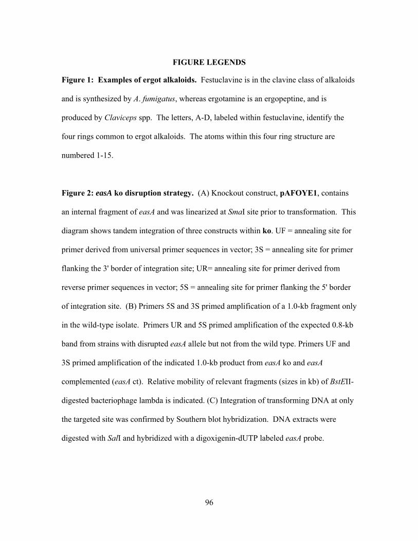
96
FIGURE LEGENDS
Figure 1: Examples of ergot alkaloids. Festuclavine is in the clavine class of alkaloids
and is synthesized by A. fumigatus, whereas ergotamine is an ergopeptine, and is
produced by Claviceps spp. The letters, A-D, labeled within festuclavine, identify the
four rings common to ergot alkaloids. The atoms within this four ring structure are
numbered 1-15.
Figure 2: easA ko disruption strategy. (A) Knockout construct, pAFOYE1, contains
an internal fragment of easA and was linearized at SmaI site prior to transformation. This
diagram shows tandem integration of three constructs within ko. UF = annealing site for
primer derived from universal primer sequences in vector; 3S = annealing site for primer
flanking the 3' border of integration site; UR= annealing site for primer derived from
reverse primer sequences in vector; 5S = annealing site for primer flanking the 5' border
of integration site. (B) Primers 5S and 3S primed amplification of a 1.0-kb fragment only
in the wild-type isolate. Primers UR and 5S primed amplification of the expected 0.8-kb
band from strains with disrupted easA allele but not from the wild type. Primers UF and
3S primed amplification of the indicated 1.0-kb product from easA ko and easA
complemented (easA ct). Relative mobility of relevant fragments (sizes in kb) of BstEII-
digested bacteriophage lambda is indicated. (C) Integration of transforming DNA at only
the targeted site was confirmed by Southern blot hybridization. DNA extracts were
digested with SalI and hybridized with a digoxigenin-dUTP labeled easA probe.
97
Figure 3: Chromatogram of wild-type A. fumigatus (wt), easA ko, and easA ko
complemented with a functional copy of C. purpurea easA (Cp easA ct). B =
fumigaclavine B; F = festuclavine; A = fumigaclavine A; C = fumigaclavine C; I =
isosetoclavine; S = setoclavine; AG = agroclavine; CH = chanoclavine. Excitation and
emission wavelengths are provided in the upper left corner of each chromatogram.
Figure 4: ESI-MS of A. fumigatus easA ko strain and its C. purpurea easA
complemented strain. (A) Mass spectrum of the major 32-min peak that accumulates in
easA ko and coelutes with chanoclavine during HPLC analysis. (B) Mass spectra of three
fractions collected from HPLC analysis of extract from Cp easA ct. The 44-min peak
coeluted with agroclavine; the 39-min peak and 42-min peak coeluted with setoclavine
and isosetoclavine, respectively.
Figure 5: Alignment of EasA sequences. Partial protein sequences, EasA, for A.
fumigatus, C. africana, C. fusiformis, C. gigantea, C. purpurea (P1 and 20102), E.
festucae, and N. lolii, were aligned by ClustalW. The residue of interest (highlighted in
yellow) corresponding to the active site tyrosine (Y) is as expected for A. fumigatus, C.
africana, and C. gigantea, whereas all remaining sequences have a phenylalanine (F) at
this position.
Figure 6: Distance analysis of EasA homologues among ergot alkaloid producers.
(A) Distance matrix based on alignment of EasA sequences. The furthest distance is
98
between A. fumigatus and C. gigantea. (B) Unrooted Neighbor-Joining tree derived
from the distance matrix calculated from aligned EasA sequences.
Figure 7: The branch point of the ergot alkaloid pathway. The pathways diverge after
chanoclavine aldehyde, due to the differing enzymatic properties of easA. The easA
product in A. fumigatus reduces the vinyl group in chanoclavine aldehyde, allowing
rotation of the aldehyde group into close proximity of the N-methyl group for ring
closure. The product of C. purpurea easA only temporarily reduces the double bond
resulting in formation of agroclavine instead of festuclavine. Setoclavine and its isomer
are readily produced from agroclavine by peroxidases from multiple sources, in this case
presumably endogenous A. fumigatus peroxidases (16). DMAPP =
dimethylallylpyrophosphate; PPO = pyrophosphate; Trp = tryptophan; AdoMet = S-
adenosylmethionine; DMAT = dimethylallyltryptophan.
Figure 8: Model for noncatalyzed closure of D ring via keto-enol tautomerism.
Chanoclavine aldehyde undergoes movement of electrons within its double bonds,
creating the intermediary enolate ion, which acquires hydrogen from solvent to form enol
tautomer. This change in double bond location allows rotation around the alpha carbon,
positioning the hydroxyl group within close proximity to the amine group. Loss of the
proton and reformation of the aldehyde creates isochanoclavine aldehyde. The carbonyl
group interacts with the amine group, and water is lost, resulting in closure of D ring as
an iminium ion. An additional NADPH-dependent reductase is required to reduce the
iminium ion to agroclavine (17).
99
Table 1. Accumulation of agroclavine, setoclavine, and isosetoclavine
in A. fumigatus easA ko and A. fumigatus easA ko complemented with
C. purpurea easA
Fg ergot alkaloid per conidium (mean ± standard error)
Source Agroclavine 1 Setoclavine / Isosetoclavine
wild type nd 2 nd
easA ko 0.0095±0.0027a 0.012± 0.0019a
Cp easA ct 1.1±0.082 b 1.4±0.090b
1Comparison of easA ko and Cp easA ct data within each column of table
by Wilcoxon/Kruskal-Wallis Tests; means with different letter indicate
significant difference (H = 6.8, d.f. = 1, P = 0.009).
2 nd, none detected
Chapter 3 - Table 1
100
OO
O NH
NNO
OH
NH
N
Ergotamine
NH
N
Festuclavine
A
B
C
D
1
4
3
6
7
5
2
8
10
912
11
15
13
14
Chapter 3 - Figure 1
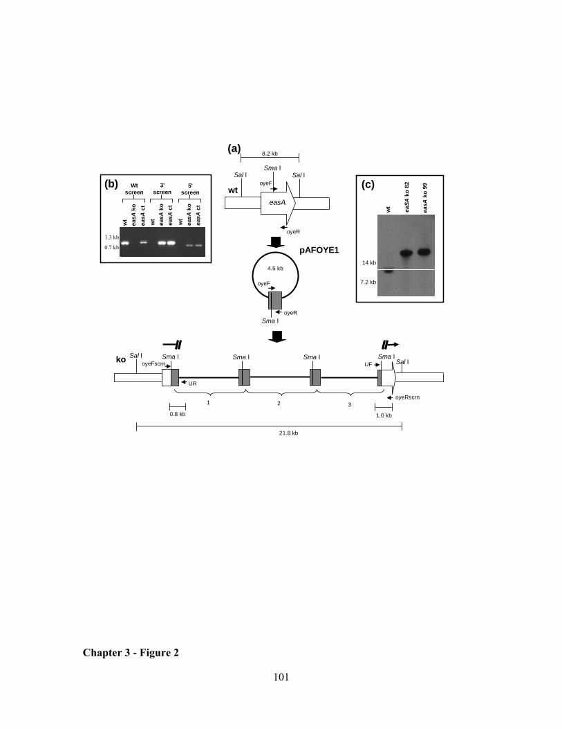
101
(a)Sma I
easA
oyeF
oyeR
8.2 kb
Sal ISal I
wt
Sma I
4.5 kb
oyeF
oyeR
pAFOYE1
Sma I Sma ISal IUF
oyeRscrn2
Sma ISal I
21.8 kb
1
ko Sma I
3
1.0 kb
UR
oyeFscrn
0.8 kb
(b) Wt screen
5' screen
3' screen
1.3 kb
0.7 kb
wt
easA
koea
sAct
wt
easA
koea
sAct
wt
easA
koea
sAct
14 kb
7.2 kb
(c)
easA
ko 9
9
eaSA
ko 8
2
wt
Chapter 3 - Figure 2
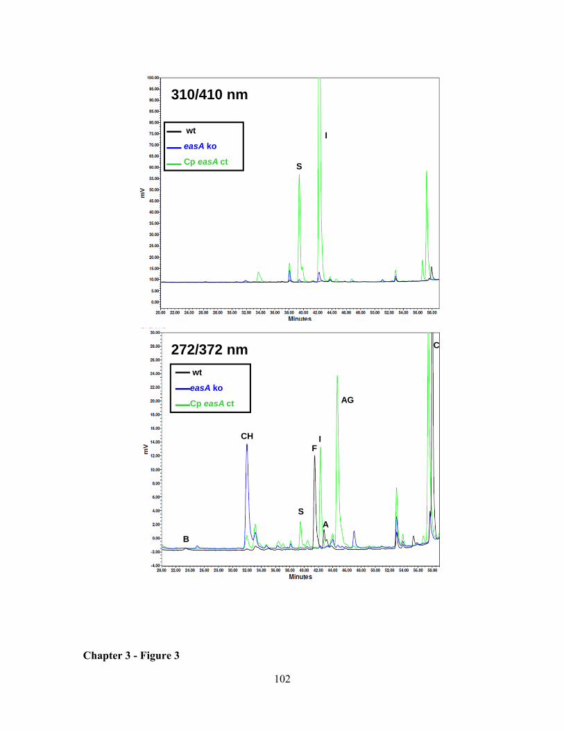
102
S
I
AG
CH
S
IF
C
AB
wt
easA ko
Cp easA ct
wt
easA ko
Cp easA ct
310/410 nm
272/372 nm
Chapter 3 - Figure 3
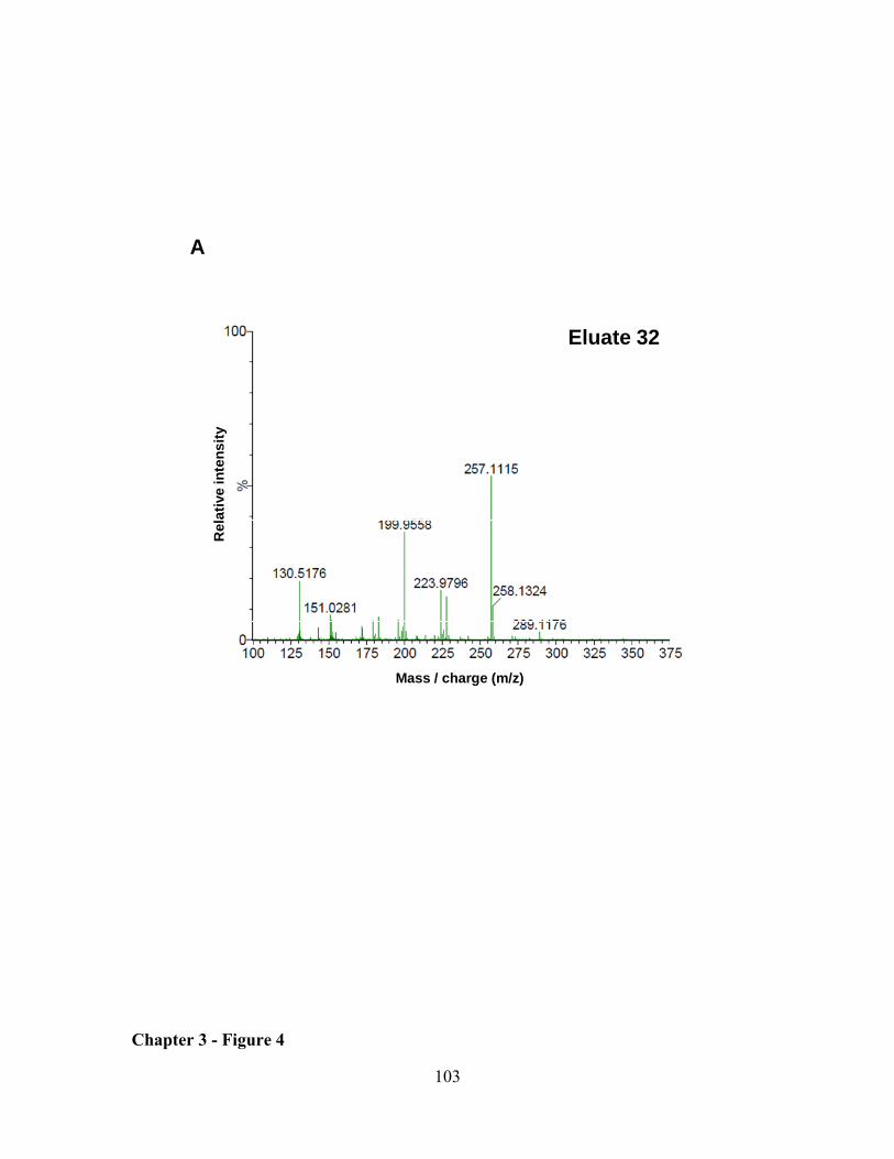
103
A
Mass / charge (m/z)
Rel
ativ
e in
tens
ity
Eluate 32
Chapter 3 - Figure 4
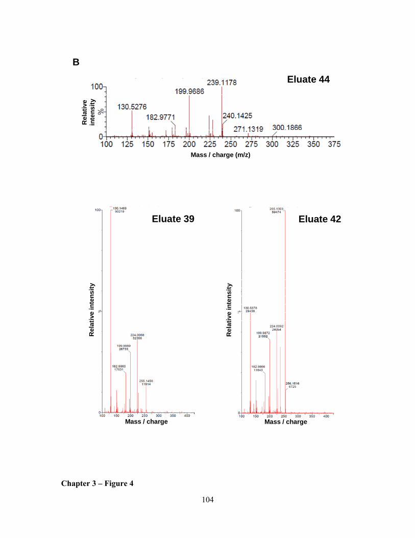
104
Eluate 39
B
Mass / charge Mass / charge
Rel
ativ
e in
tens
ity
Rel
ativ
e in
tens
ity
Eluate 42
Mass / charge (m/z)
Eluate 44R
elat
ive
inte
nsity
Chapter 3 – Figure 4
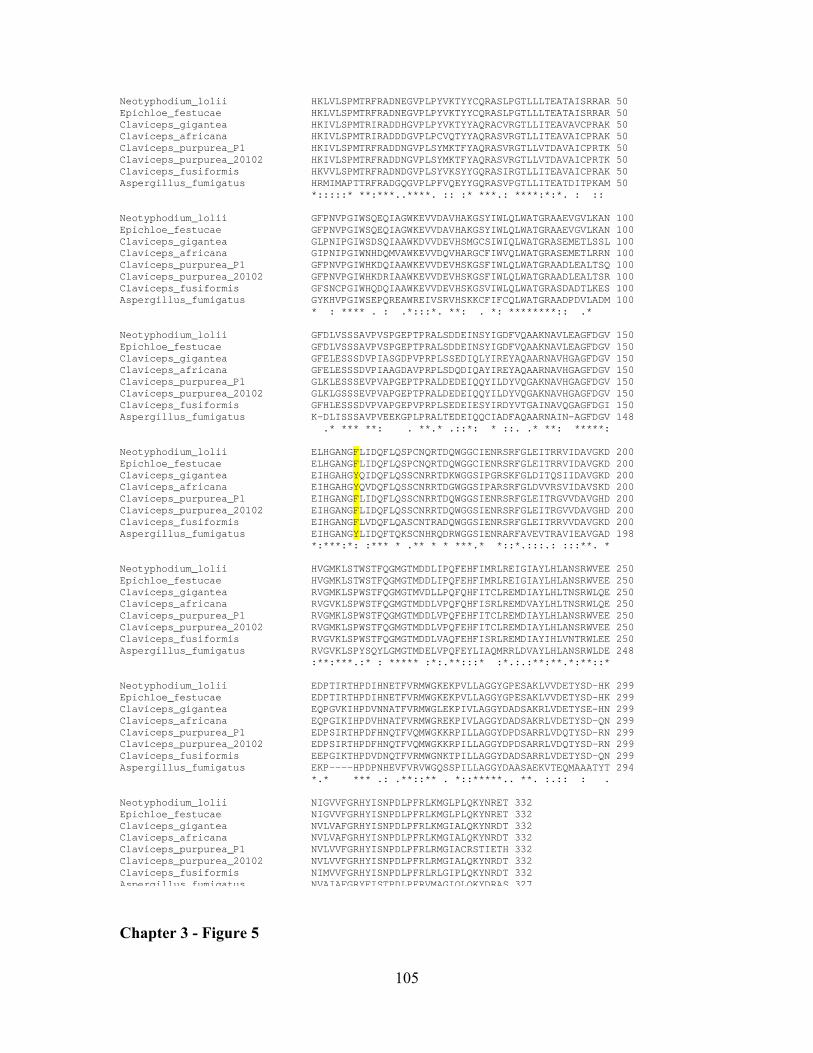
105
Neotyphodium_lolii HKLVLSPMTRFRADNEGVPLPYVKTYYCQRASLPGTLLLTEATAISRRAR 50 Epichloe_festucae HKLVLSPMTRFRADNEGVPLPYVKTYYCQRASLPGTLLLTEATAISRRAR 50 Claviceps_gigantea HKIVLSPMTRIRADDHGVPLPYVKTYYAQRACVRGTLLITEAVAVCPRAK 50 Claviceps_africana HKIVLSPMTRIRADDDGVPLPCVQTYYAQRASVRGTLLITEAVAICPRAK 50 Claviceps_purpurea_P1 HKIVLSPMTRFRADDNGVPLSYMKTFYAQRASVRGTLLVTDAVAICPRTK 50 Claviceps_purpurea_20102 HKIVLSPMTRFRADDNGVPLSYMKTFYAQRASVRGTLLVTDAVAICPRTK 50 Claviceps_fusiformis HKVVLSPMTRFRADNDGVPLSYVKSYYGQRASIRGTLLITEAVAICPRAK 50 Aspergillus_fumigatus HRMIMAPTTRFRADGQGVPLPFVQEYYGQRASVPGTLLITEATDITPKAM 50 *:::::* **:***..****. :: :* ***.: ****:*:*. : :: Neotyphodium_lolii GFPNVPGIWSQEQIAGWKEVVDAVHAKGSYIWLQLWATGRAAEVGVLKAN 100 Epichloe_festucae GFPNVPGIWSQEQIAGWKEVVDAVHAKGSYIWLQLWATGRAAEVGVLKAN 100 Claviceps_gigantea GLPNIPGIWSDSQIAAWKDVVDEVHSMGCSIWIQLWATGRASEMETLSSL 100 Claviceps_africana GIPNIPGIWNHDQMVAWKEVVDQVHARGCFIWVQLWATGRASEMETLRRN 100 Claviceps_purpurea_P1 GFPNVPGIWHKDQIAAWKEVVDEVHSKGSFIWLQLWATGRAADLEALTSQ 100 Claviceps_purpurea_20102 GFPNVPGIWHKDRIAAWKEVVDEVHSKGSFIWLQLWATGRAADLEALTSR 100 Claviceps_fusiformis GFSNCPGIWHQDQIAAWKEVVDEVHSKGSVIWLQLWATGRASDADTLKES 100 Aspergillus_fumigatus GYKHVPGIWSEPQREAWREIVSRVHSKKCFIFCQLWATGRAADPDVLADM 100 * : **** . : .*:::*. **: . *: ********:: .* Neotyphodium_lolii GFDLVSSSAVPVSPGEPTPRALSDDEINSYIGDFVQAAKNAVLEAGFDGV 150 Epichloe_festucae GFDLVSSSAVPVSPGEPTPRALSDDEINSYIGDFVQAAKNAVLEAGFDGV 150 Claviceps_gigantea GFELESSSDVPIASGDPVPRPLSSEDIQLYIREYAQAARNAVHGAGFDGV 150 Claviceps_africana GFELESSSDVPIAAGDAVPRPLSDQDIQAYIREYAQAARNAVHGAGFDGV 150 Claviceps_purpurea_P1 GLKLESSSEVPVAPGEPTPRALDEDEIQQYILDYVQGAKNAVHGAGFDGV 150 Claviceps_purpurea_20102 GLKLGSSSEVPVAPGEPTPRALDEDEIQQYILDYVQGAKNAVHGAGFDGV 150 Claviceps_fusiformis GFHLESSSDVPVAPGEPVPRPLSEDEIESYIRDYVTGAINAVQGAGFDGI 150 Aspergillus_fumigatus K-DLISSSAVPVEEKGPLPRALTEDEIQQCIADFAQAARNAIN-AGFDGV 148 .* *** **: . **.* .::*: * ::. .* **: *****: Neotyphodium_lolii ELHGANGFLIDQFLQSPCNQRTDQWGGCIENRSRFGLEITRRVIDAVGKD 200 Epichloe_festucae ELHGANGFLIDQFLQSPCNQRTDQWGGCIENRSRFGLEITRRVIDAVGKD 200 Claviceps_gigantea EIHGAHGYQIDQFLQSSCNRRTDKWGGSIPGRSKFGLDITQSIIDAVGKD 200 Claviceps_africana EIHGAHGYQVDQFLQSSCNRRTDGWGGSIPARSRFGLDVVRSVIDAVSKD 200 Claviceps_purpurea_P1 EIHGANGFLIDQFLQSSCNRRTDQWGGSIENRSRFGLEITRGVVDAVGHD 200 Claviceps_purpurea_20102 EIHGANGFLIDQFLQSSCNRRTDQWGGSIENRSRFGLEITRGVVDAVGHD 200 Claviceps_fusiformis EIHGANGFLVDQFLQASCNTRADQWGGSIENRSRFGLEITRRVVDAVGKD 200 Aspergillus_fumigatus EIHGANGYLIDQFTQKSCNHRQDRWGGSIENRARFAVEVTRAVIEAVGAD 198 *:***:*: :*** * .** * * ***.* *::*.:::.: :::**. * Neotyphodium_lolii HVGMKLSTWSTFQGMGTMDDLIPQFEHFIMRLREIGIAYLHLANSRWVEE 250 Epichloe_festucae HVGMKLSTWSTFQGMGTMDDLIPQFEHFIMRLREIGIAYLHLANSRWVEE 250 Claviceps_gigantea RVGMKLSPWSTFQGMGTMVDLLPQFQHFITCLREMDIAYLHLTNSRWLQE 250 Claviceps_africana RVGVKLSPWSTFQGMGTMDDLVPQFQHFISRLREMDVAYLHLTNSRWLQE 250 Claviceps_purpurea_P1 RVGMKLSPWSTFQGMGTMDDLVPQFEHFITCLREMDIAYLHLANSRWVEE 250 Claviceps_purpurea_20102 RVGMKLSPWSTFQGMGTMDDLVPQFEHFITCLREMDIAYLHLANSRWVEE 250 Claviceps_fusiformis RVGVKLSPWSTFQGMGTMDDLVAQFEHFISRLREMDIAYIHLVNTRWLEE 250 Aspergillus_fumigatus RVGVKLSPYSQYLGMGTMDELVPQFEYLIAQMRRLDVAYLHLANSRWLDE 248 :**:***.:* : ***** :*:.**:::* :*.:.:**:**.*:**::* Neotyphodium_lolii EDPTIRTHPDIHNETFVRMWGKEKPVLLAGGYGPESAKLVVDETYSD-HK 299 Epichloe_festucae EDPTIRTHPDIHNETFVRMWGKEKPVLLAGGYGPESAKLVVDETYSD-HK 299 Claviceps_gigantea EQPGVKIHPDVNNATFVRMWGLEKPIVLAGGYDADSAKRLVDETYSE-HN 299 Claviceps_africana EQPGIKIHPDVHNATFVRMWGREKPIVLAGGYDADSAKRLVDETYSD-QN 299 Claviceps_purpurea_P1 EDPSIRTHPDFHNQTFVQMWGKKRPILLAGGYDPDSARRLVDQTYSD-RN 299 Claviceps_purpurea_20102 EDPSIRTHPDFHNQTFVQMWGKKRPILLAGGYDPDSARRLVDQTYSD-RN 299 Claviceps_fusiformis EEPGIKTHPDVDNQTFVRMWGNKTPILLAGGYDADSARRLVDETYSD-QN 299 Aspergillus_fumigatus EKP----HPDPNHEVFVRVWGQSSPILLAGGYDAASAEKVTEQMAAATYT 294 *.* *** .: .**::** . *::*****.. **. :.:: : . Neotyphodium_lolii NIGVVFGRHYISNPDLPFRLKMGLPLQKYNRET 332 Epichloe_festucae NIGVVFGRHYISNPDLPFRLKMGLPLQKYNRET 332 Claviceps_gigantea NVLVAFGRHYISNPDLPFRLKMGIALQKYNRDT 332 Claviceps_africana NVLVAFGRHYISNPDLPFRLKMGIALQKYNRDT 332 Claviceps_purpurea_P1 NVLVVFGRHYISNPDLPFRLRMGIACRSTIETH 332 Claviceps_purpurea_20102 NVLVVFGRHYISNPDLPFRLRMGIALQKYNRDT 332 Claviceps_fusiformis NIMVVFGRHYISNPDLPFRLRLGIPLQKYNRDT 332 Aspergillus fumigatus NVAIAFGRYFISTPDLPFRVMAGIQLQKYDRAS 327 Chapter 3 - Figure 5
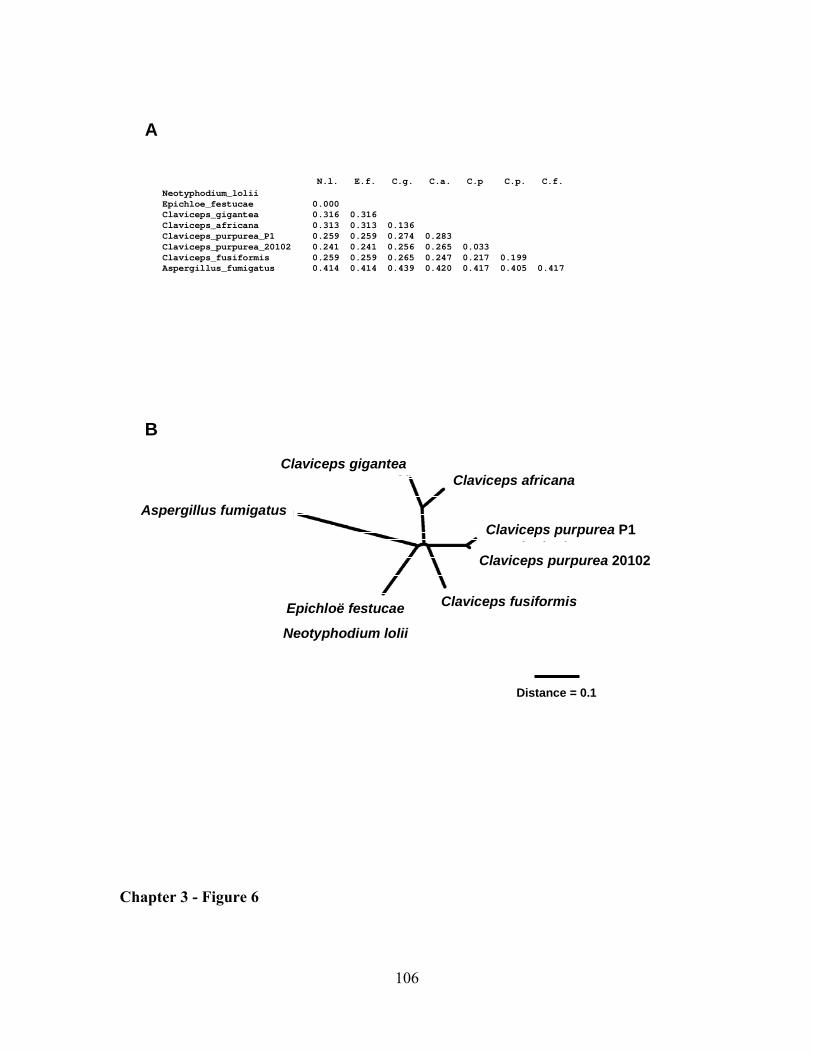
106
Epichloë festucae
Neotyphodium lolii
Claviceps fusiformis
Claviceps purpurea 20102
Aspergillus fumigatusClaviceps purpurea P1
Claviceps africanaClaviceps gigantea
Distance = 0.1
A
B
N.l. E.f. C.g. C.a. C.p C.p. C.f. Neotyphodium_loliiEpichloe_festucae 0.000 Claviceps_gigantea 0.316 0.316Claviceps_africana 0.313 0.313 0.136 Claviceps_purpurea_P1 0.259 0.259 0.274 0.283 Claviceps_purpurea_20102 0.241 0.241 0.256 0.265 0.033 Claviceps_fusiformis 0.259 0.259 0.265 0.247 0.217 0.199 Aspergillus_fumigatus 0.414 0.414 0.439 0.420 0.417 0.405 0.417
Chapter 3 - Figure 6
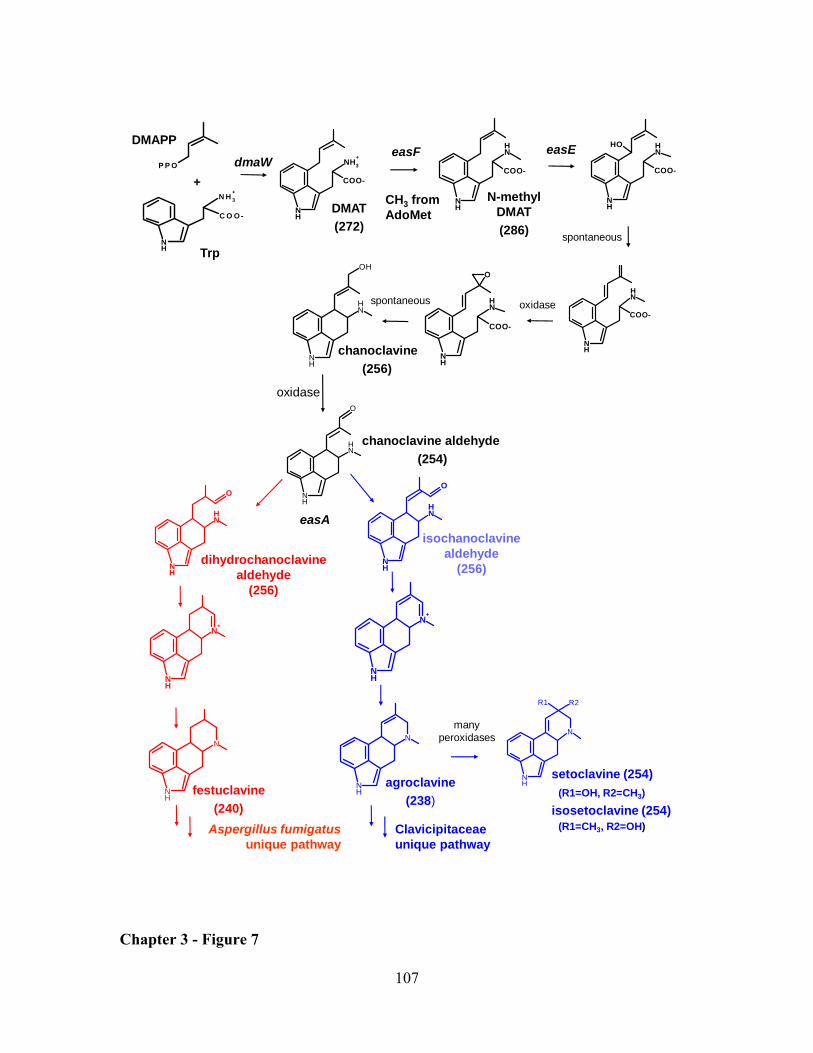
107
COO-
NH
NH3+
COO-
NH
NH
COO-
NH
NHOH
COO-
NH
NH
NH
NH
O
COO-
NH
NH
OHTrp
DMAPP
+
C O O -
NH
N H 3+
P P O
NH
N
NH
N
NH
NH
O
easF
CH3 fromAdoMet
oxidase
spontaneous
easA
Aspergillus fumigatusunique pathway
agroclavine(238)
festuclavine(240)
N-methylDMAT(286)
DMAT(272)
chanoclavine(256)
dmaW
oxidase
spontaneous
easE
Clavicipitaceaeunique pathway
chanoclavine aldehyde (254)
NH
R2
N
R1
manyperoxidases
setoclavine (254)(R1=OH, R2=CH3)
isosetoclavine (254)(R1=CH3, R2=OH)
NH
NH
O
dihydrochanoclavine aldehyde
(256)
NH
NH
O
isochanoclavine aldehyde
(256)
NH
N+
NH
N+
Chapter 3 - Figure 7
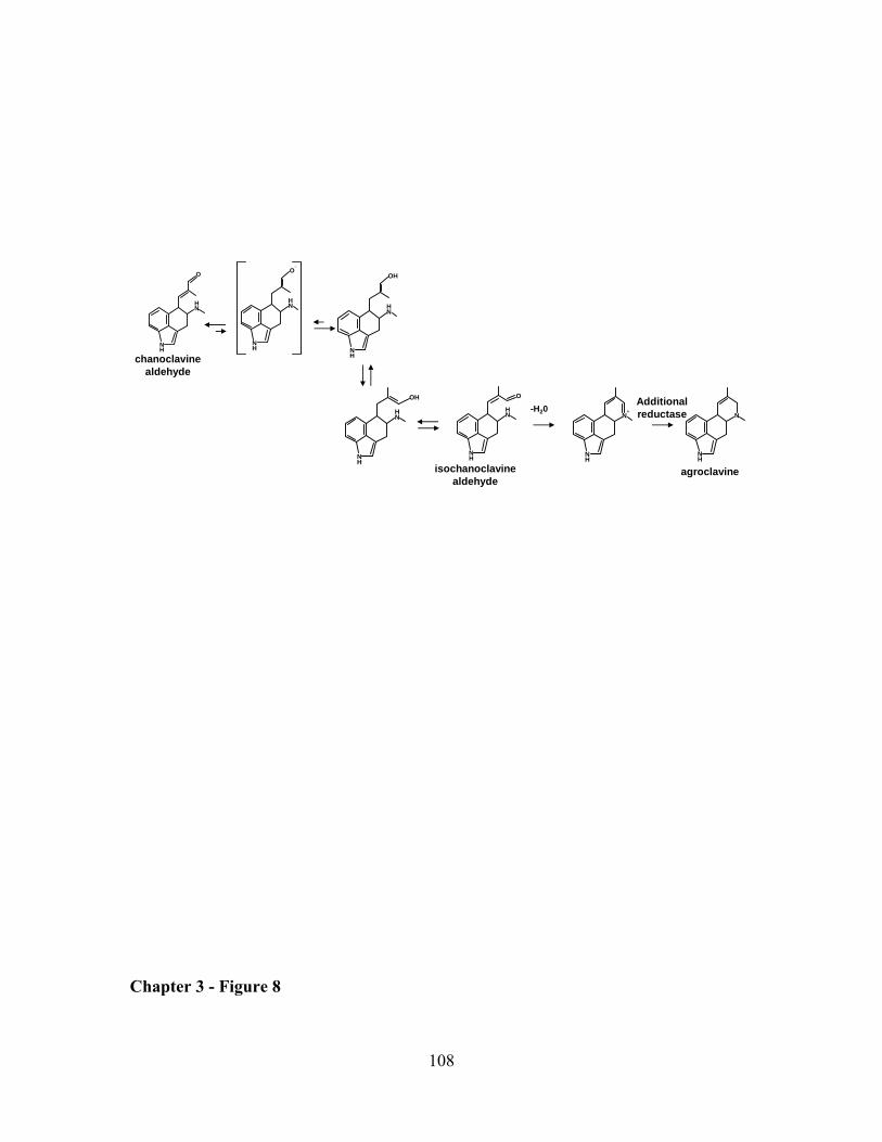
108
NH
NH
O
NH
N
NH
NH
O
agroclavineisochanoclavinealdehyde
chanoclavinealdehyde
NH
NH
OH
NH
NH
OH
NH
NH
O
n
NH
N+-H20
Additional reductase
Chapter 3 - Figure 8
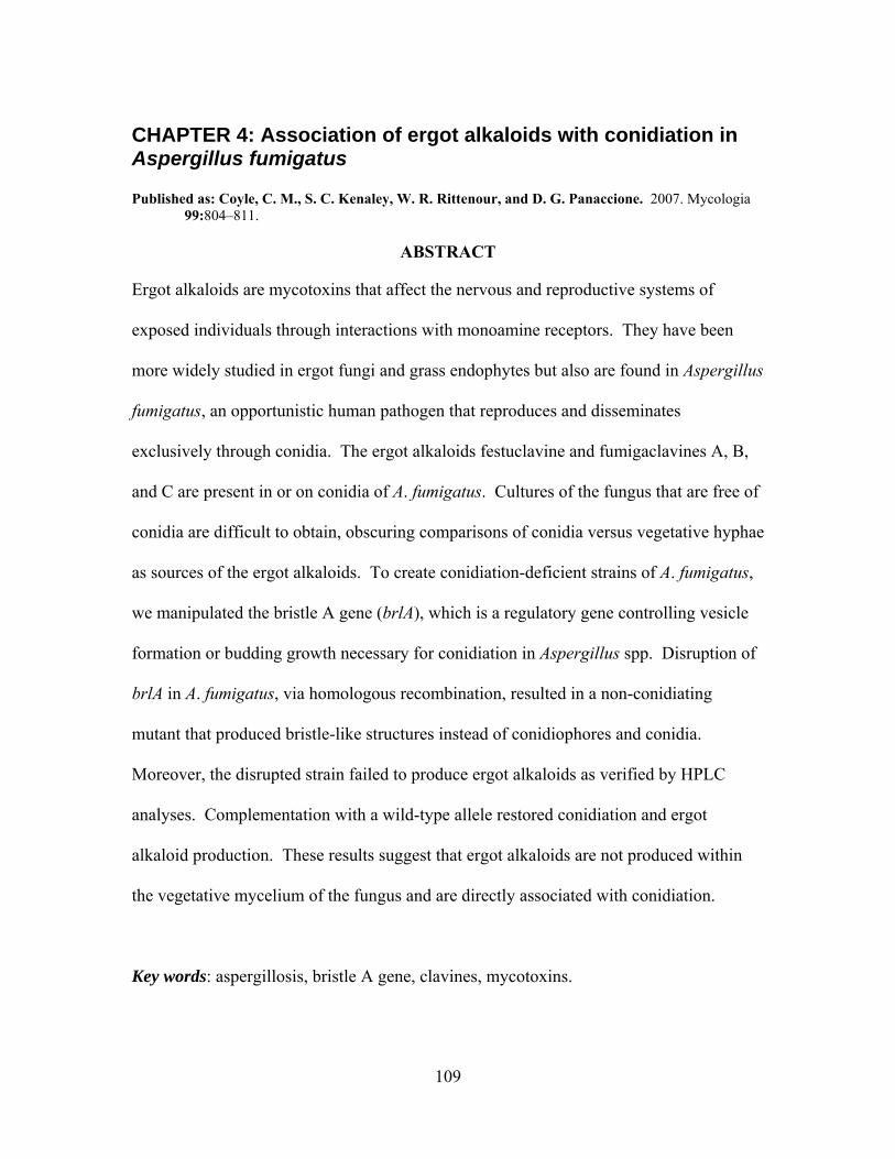
109
CHAPTER 4: Association of ergot alkaloids with conidiation in Aspergillus fumigatus Published as: Coyle, C. M., S. C. Kenaley, W. R. Rittenour, and D. G. Panaccione. 2007. Mycologia
99:804–811.
ABSTRACT
Ergot alkaloids are mycotoxins that affect the nervous and reproductive systems of
exposed individuals through interactions with monoamine receptors. They have been
more widely studied in ergot fungi and grass endophytes but also are found in Aspergillus
fumigatus, an opportunistic human pathogen that reproduces and disseminates
exclusively through conidia. The ergot alkaloids festuclavine and fumigaclavines A, B,
and C are present in or on conidia of A. fumigatus. Cultures of the fungus that are free of
conidia are difficult to obtain, obscuring comparisons of conidia versus vegetative hyphae
as sources of the ergot alkaloids. To create conidiation-deficient strains of A. fumigatus,
we manipulated the bristle A gene (brlA), which is a regulatory gene controlling vesicle
formation or budding growth necessary for conidiation in Aspergillus spp. Disruption of
brlA in A. fumigatus, via homologous recombination, resulted in a non-conidiating
mutant that produced bristle-like structures instead of conidiophores and conidia.
Moreover, the disrupted strain failed to produce ergot alkaloids as verified by HPLC
analyses. Complementation with a wild-type allele restored conidiation and ergot
alkaloid production. These results suggest that ergot alkaloids are not produced within
the vegetative mycelium of the fungus and are directly associated with conidiation.
Key words: aspergillosis, bristle A gene, clavines, mycotoxins.
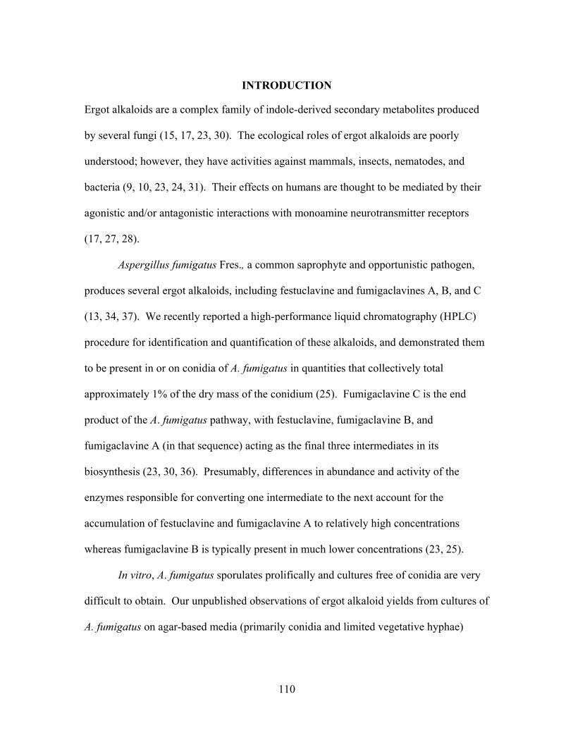
110
INTRODUCTION
Ergot alkaloids are a complex family of indole-derived secondary metabolites produced
by several fungi (15, 17, 23, 30). The ecological roles of ergot alkaloids are poorly
understood; however, they have activities against mammals, insects, nematodes, and
bacteria (9, 10, 23, 24, 31). Their effects on humans are thought to be mediated by their
agonistic and/or antagonistic interactions with monoamine neurotransmitter receptors
(17, 27, 28).
Aspergillus fumigatus Fres., a common saprophyte and opportunistic pathogen,
produces several ergot alkaloids, including festuclavine and fumigaclavines A, B, and C
(13, 34, 37). We recently reported a high-performance liquid chromatography (HPLC)
procedure for identification and quantification of these alkaloids, and demonstrated them
to be present in or on conidia of A. fumigatus in quantities that collectively total
approximately 1% of the dry mass of the conidium (25). Fumigaclavine C is the end
product of the A. fumigatus pathway, with festuclavine, fumigaclavine B, and
fumigaclavine A (in that sequence) acting as the final three intermediates in its
biosynthesis (23, 30, 36). Presumably, differences in abundance and activity of the
enzymes responsible for converting one intermediate to the next account for the
accumulation of festuclavine and fumigaclavine A to relatively high concentrations
whereas fumigaclavine B is typically present in much lower concentrations (23, 25).
In vitro, A. fumigatus sporulates prolifically and cultures free of conidia are very
difficult to obtain. Our unpublished observations of ergot alkaloid yields from cultures of
A. fumigatus on agar-based media (primarily conidia and limited vegetative hyphae)
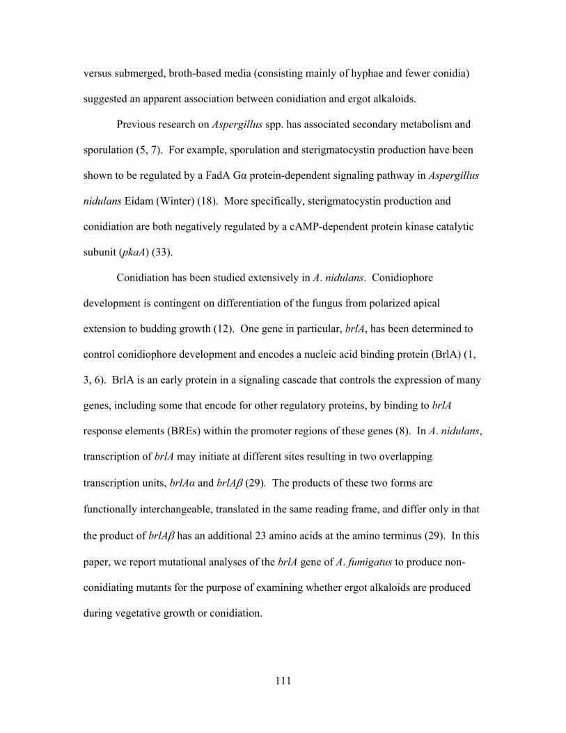
111
versus submerged, broth-based media (consisting mainly of hyphae and fewer conidia)
suggested an apparent association between conidiation and ergot alkaloids.
Previous research on Aspergillus spp. has associated secondary metabolism and
sporulation (5, 7). For example, sporulation and sterigmatocystin production have been
shown to be regulated by a FadA Gα protein-dependent signaling pathway in Aspergillus
nidulans Eidam (Winter) (18). More specifically, sterigmatocystin production and
conidiation are both negatively regulated by a cAMP-dependent protein kinase catalytic
subunit (pkaA) (33).
Conidiation has been studied extensively in A. nidulans. Conidiophore
development is contingent on differentiation of the fungus from polarized apical
extension to budding growth (12). One gene in particular, brlA, has been determined to
control conidiophore development and encodes a nucleic acid binding protein (BrlA) (1,
3, 6). BrlA is an early protein in a signaling cascade that controls the expression of many
genes, including some that encode for other regulatory proteins, by binding to brlA
response elements (BREs) within the promoter regions of these genes (8). In A. nidulans,
transcription of brlA may initiate at different sites resulting in two overlapping
transcription units, brlAα and brlAβ (29). The products of these two forms are
functionally interchangeable, translated in the same reading frame, and differ only in that
the product of brlAβ has an additional 23 amino acids at the amino terminus (29). In this
paper, we report mutational analyses of the brlA gene of A. fumigatus to produce non-
conidiating mutants for the purpose of examining whether ergot alkaloids are produced
during vegetative growth or conidiation.
112
MATERIALS AND METHODS
Fungi and culture conditions. Aspergillus fumigatus isolate FGSC A1141 (25) and
derivatives were routinely cultured on potato dextrose agar (PDA) prepared from
dehydrated potato flakes (Pillsbury, Minneapolis, MN)(20 g/L), D-glucose (20 g/L), and
agar (15 g/L). Cultures intended for preparation of protoplasts or DNA were grown
overnight in 15 mL of malt extract medium (50 g malt extract, 50 g lactose, 5 g
asparagine, and 0.4 g K2HPO4 per L) in a deep Petri dish (20-mm depth, 100-mm
diameter; Fisher Scientific, Pittsburgh, PA) on an orbital shaker set at 37 ºC and 150 rpm.
Non-conidiating mutants were cultured similarly except pieces of agar-based cultures
containing hyphal fragments were used in place of conidia as inoculum.
Disruption and complementation of brlA. The presence of a brlA orthologue in
the A. fumigatus genome (21) was determined by tblastn search with protein sequence
deduced from A. nidulans brlA (1) as a query sequence. A 640-bp fragment internal to
the coding sequence of the putative brlA orthologue was amplified by PCR primed with
oligonucleotides brlAF (5'-GCCACGCTTACGACAGAGGT-3') and brlAR (5'-
CGGTAGATAGGCGACTGGTG -3'). The 50 µL PCR reaction contained 50 mM KCl,
10 mM Tris-HCl (pH 9.0), 0.1% (v/v) Triton X-100, 1.5 mM MgCl2, 200 µM of each
deoxyribonucleotide triphosphate, 1 µM of each primer, and 2.5 units of Taq DNA
polymerase (Promega, Madison, Wisconsin), which was added once the thermocycler
reached 95 °C in the initial denaturing period. The reaction began with an initial
denaturing step of 2 min 30 sec at 95 °C, followed by 35 cycles of 1 min at 94 °C, 1 min
at 57 °C, and 1 min at 72 °C, with a final extension step of 72 °C for 7 min. A 4.6-kb
disruption construct, pBRLA1, was generated by ligating the PCR product into the T/A
113
overlap vector pCR2.1 (Invitrogen, Carlsbad, California). The construct was linearized at
the unique NdeI site located within the brlA fragment prior to transformation of the
fungus. Protoplasts were prepared and transformed according to the protocol described
by Murray et al. (20) and with modifications noted previously (14). Protoplasts were co-
transformed with the linearized disruption construct, pBRLA1, and a hygromycin
resistance plasmid, pMOcosX (22), linearized at a unique NotI site (14). Non-conidiating
transformants were identified by their altered colony morphology and color, and were
confirmed by light microscopy. Non-conidiating mutants were purified to nuclear
homogeneity through repetitive sub-culturing from hyphal tips (since nuclear purification
via single conidium isolation was not possible). Homologous recombination between
pBRLA1 and the native brlA locus within the transformants was verified by three PCR
assays similar to the PCR reaction described above but with different primers and
annealing temperatures. The 5' border of the recombination event was confirmed by PCR
(annealing temperature at 57 °C) primed with oligonucleotides UF (5'-
TGTAAAACGACGGCCAGTGAAT-3', which anneals to vector sequences near the
universal primer annealing site in pCR2.1) and brlAFscrn (5'-
CTCCAACGAATGTCCGTCTATG -3', complementary to brlA sequences near the 5'-
end of the brlA gene and flanking the intended site of integration) (Fig. 1A). The
juxtaposition of sequences near the 3' border of the integration event was verified by PCR
(annealing temp 55 °C) primed from oligonucleotides UR (5'-
AGCTATGACCATGATTACGCCA-3', complementary to vector sequences near the
reverse primer annealing site of pCR2.1), and brlARscrn (5'-
TAGTGACAAGCTCTGCTTGGA -3', which anneals to brlA sequences near the 3'-end
114
of the gene and flanking the intended site of integration) (Fig. 1A). PCR across the entire
brlA locus was conducted by priming with oligonucleotides brlAFscrn and brlARscrn
(both described above) at an annealing temperature of 55 °C.
Integration of the transformation construct also was analyzed by Southern
hybridization as described previously (14), except for substitution of a brlA-specific
probe. The brlA probe was made by amplifying wild-type A. fumigatus genomic DNA
via PCR from primers brlAF and brlAR, according to the PCR conditions described
above, substituting 1× DIG DNA Labeling Mix (Roche, Mannheim, Germany) for
unlabeled dNTPs, and increasing the annealing temperature to 57 °C.
The brlA ko strain was complemented with a 2.9-kb fragment containing a full-
length copy of the brlA coding sequences along with approximately 1 kb of 5'-flanking
sequences and 0.6 kb of 3'-flanking sequences. The complementing fragment was
amplified by PCR primed from WbrlAF (5'-
CTCTCCAGGAACCTTGAAACAGGTTCTC-3') and WbrlAR (5'-
CCACATAGGACACTCGAGGATGGAGACA-3') in a 50 µL PCR reaction containing
15 mM (NH4)2SO4, 50 mM Tris-HCl (pH 9.0), 0.1% (v/v) Triton X-100, 1.5 mM MgCl2,
200 µM of each dNTP, 1 µM of each primer, and 1 unit of DyNAzyme DNA polymerase
(New England Biolabs, Beverly, MA) added during the initial denaturing step. The
thermocycler program was as described above; however, the annealing temperature was
62 °C and the extension step was for 3 min 20 sec. The PCR product was cotransformed
into a brlA-disrupted mutant along with a phleomycin resistance plasmid, pBC-phleo
(Fungal Genetics Stock Center, University of Missouri-Kansas City, Kansas City, MO).
Transformants were selected on complete regeneration medium (26) containing 100
115
µg/mL phleomycin and incubated at 37 °C. Complemented transformants were cultured
from individual germinated conidia in order to achieve a homokaryotic culture. To test
for the presence of the original disruption construct, PCR reactions were initiated from
primers brlAFscrn and UF (for the 5' flank of the integration) and UR and brlARscrn (for
the 3' flank). An additional PCR screen was performed with primers WbrlAF and
WbrlaR (described above), to test for the presence of the introduced wild-type allele in
the complemented transformants. PCR conditions are provided above with descriptions
of the primers (above).
Analysis of ergot alkaloids and conidiation. For routine analysis of ergot
alkaloids, small cubes of cultures grown on PDA (containing approximately 8 mm x 8
mm of colony surface area) were submerged in 0.5 mL of 80% methanol in a 1.9-mL
microcentrifuge tube and pulverized with a plastic pestle. The samples were rotated, end
over end (44 rpm), for 2 hr, and spin-filtered through a 0.2-µM pore nylon filter. Ergot
alkaloids were detected and quantified from 20 µL of the prepared sample by reverse-
phase HPLC on a C18 column with fluorescence detection based on excitation and
emission wavelengths of 272 nm and 372 nm, respectively (25). The identities of peaks
corresponding to festuclavine, and fumigaclavines A, B, and C were previously
established by mass spectral analyses of native and de-esterified fractions (25) and
confirmed by elimination of these peaks from extracts of strains in which the first gene in
the ergot alkaloid pathway (dmaW) was inactivated by gene disruption (14).
To quantify ergot alkaloids and conidiation from the same samples, cultures were
gown on PDA for 5 days at 37 °C and then an additional 9 days at room temperature.
Samples containing 50.3 mm2 of colony surface area were extracted from cultures with
116
the broader end of a disposable 1-mL pipette tip (Fisher Scientific, catalog number 02-
681-172). Samples were submerged in 1 mL 80% methanol, agitated by vortexing, and
rotated at 44 rpm for 2 hr. Separate aliquots were removed for HPLC analyses and for
quantification of conidia. Concentrations of ergot alkaloids were based on peak area
relative to an external standard of agroclavine (Sigma, St. Louis, MO) as previously
described (25). Conidia were enumerated in dilutions of the methanol extracts with a
hemacytometer and light microscope. Six samples were analyzed for each treatment.
Data from wild type and a complemented brlA disruptant were compared by Student’s t-
test.
RESULTS
Disruption and complementation of brlA. The A. fumigatus genome contains an
orthologue of brlA (GenBank accession XM_747933). The sequence located on
chromosome 1 between nucleotides 2258000 and 2259280 was predicted to encode a
protein that is 68% identical to BrlAα from A. nidulans (19). Based on sequence
analysis, there is no evidence for capacity to encode the alternate BrlAβ product that has
been observed in A. nidulans (29). BrlAβ of A. nidulans is transcribed from the same
locus as BrlAα and is identical to BrlAα except for containing an additional 23 amino
acids at the amino terminus. In A. fumigatus only 2 of 19 amino acid residues preceding
the initiator methionine of BrlAα are identical to those in the analogous positions of
BrlAβ of A. nidulans. A fragment of A. fumigatus DNA corresponding to nucleotides
191 to 830 of the BrlAα coding sequence of A. nidulans was used to direct homologous
recombination of the gene disruption construct pBRLA1 at the brlA locus of A. fumigatus
117
(Fig. 1A). Homologous recombination of pBRLA1 at brlA would interrupt coding
sequences common to both brlAα and brlAβ (if it were to occur) in A. fumigatus.
Cotransformation of pBRLA1 and NotI-linearized pMOcosX into A. fumigatus
resulted in five non-sporulating colonies out of a total of 93 hygromycin-resistant
transformants. Agar-based cultures of the putative brlA disruption strains were white and
fluffy, unlike the dense, sooty, dark-green appearance of wild-type cultures (Fig. 2A).
Observed by light microscopy, the non-conidiating cultures had extensive, bristle-like,
aerial hyphae that lacked the swollen apical vesicles, phialides, and conidia of mature
conidiophores (Fig. 2B). Putative disruptants that had been sub-cultured from hyphal tips
six times in an effort to achieve a homokaryotic culture were analyzed by PCR and
Southern blot hybridization. Amplification of transformant DNA annealed to primer sets
UF and brlAFscrn resulted in an 899-bp product (Fig. 1). Similarly, PCR from UR and
brlARscrn produced an 863-bp product. Products of these sizes from transformants but
not from the wild type indicated integration of the disruption construct into the native
copy of brlA (Fig. 1). The lack of a PCR product from these transformants when
amplification was primed from brlAFscrn and brlARscrn indicated the absence of a wild-
type copy of brlA. Integration of one or more copies of pBRLA1 at brlA results in a large
distance between priming sites, whereas in the wild type the primer annealing sites are
separated by only 910 bp (Fig. 1). Moreover, the absence of a product in the brlA
disrupted strains (Fig. 1B) indicated nuclear homogeneity of these strains, since any wild-
type nuclei in the colony would provide template for amplification of the 910-bp product.
Southern blot analyses confirmed the presence of multiple disruption constructs within
brlA, which is evident by a large shift in the size of the AccI fragment containing the
118
targeted gene region (Fig. 1C). The lack of multiple bands hybridizing to transformant
brlA ko17 on the Southern blot verified that random integration of constructs elsewhere
in the genome had not occurred (Fig. 1C).
Disrupted strain brlA ko17 was cotransformed with a 2.9-kb PCR product
containing the wild-type brlA allele and NotI-linearized pBCphleo. Six conidiating,
phleomycin-resistant transformants were analyzed by PCR. The presence of a wild-type
copy of brlA in the conidiating transformants was demonstrated by the presence of a 2.9-
kb product in reactions primed with WbrlAF and WbrlAR (Fig. 3). Additional PCR
analyses of the brlA locus in the complemented strains indicated that pBRLA1 was still
present at the previously disrupted brlA locus, since products of the predicted size were
amplified from primer pairs flanking the site of integration of pBRLA1 (Fig. 3). Thus the
complementing wild-type brlA fragment had integrated at a locus other than the native
brlA, and the recipient strain was derived from a brlA disruptant. Similar quantities of
conidia were produced by a brlA-complemented disruptant and the wild type (Table 1).
Analysis of ergot alkaloid production in brlA-disrupted mutants and complemented
derivatives. Mycelia of brlA-disrupted mutants contained no detectable ergot alkaloids
(Fig. 4, Table 1). In contrast, conidiating colonies of wild-type and the brlA-
complemented strain accumulated festuclavine, and fumigaclavines A, B, and C in
quantities that were similar by strain (Fig. 4, Table 1). The relatively large peak eluting at
53 minutes in the brlA ko17 profile represents a metabolite associated with hyphae. It is
present in low levels in wild-type and complemented extracts (Fig. 4), which come from
colonies that contain abundant conidiophores and conidia but limited hyphae. Its
119
presence in ergot alkaloid-deficient strains derived by knocking out the gene dmaW (14)
indicates that it is unrelated to ergot alkaloids.
Examination of the promoter region of ergot alkaloid cluster genes for bristle
A response element (BRE) sequences. Since ergot alkaloids were only produced in
cultures expressing a functional brlA, the chromosomal region containing 11 genes from
easA to easF of the ergot alkaloid gene cluster (14, 30)was examined for the BRE
sequence, 5'-(C/A)(A/G)AGGG(G/A)-3' (8), and its 26 occurrences were mapped (Fig. 5).
Given the degenerate nature of the BRE sequence and its ability to function in both
orientations, it will occur by chance once in every 1024 bp of random sequence. BREs
occurred once every 1404 bp in the coding sequences, whereas in the 5' non-coding
regions these elements occurred once every 409 bp, making BREs about 3.4 times more
common in the 5' non-coding regions. As assessed in a paired t-test, BREs occurred at a
higher frequency per bp in 5' non-coding sequences than in the corresponding coding
sequences (P = 0.03). However, there were no occurrences of the BRE sequences
upstream of three of 11 genes, including dmaW, the gene controlling the first step in the
ergot alkaloid pathway (Fig. 5). Six other genes had less than three BREs within their
upstream non-coding sequences. One 5' non-coding region contained four BRE
sequences, and due to divergent orientation of genes, this non-coding sequence was part
of the promoter region of two ergot alkaloid cluster genes.
DISCUSSION
Disruption of brlA in A. fumigatus resulted in a non-conidiating mutant lacking any
recognizable conidiophores. A similar phenotype has been reported recently by Mah and
Yu (19) for brlA mutants of A. fumigatus constructed by a different knockout strategy.
120
The absence of ergot alkaloids in our non-conidiating mutants indicates that ergot
alkaloids are not produced during vegetative growth and are solely associated with
conidiation in A. fumigatus.
The phenotype of the A. fumigatus brlA mutants indicates that brlA functions
similarly in A. fumigatus as it does in A. nidulans. brlA was first discovered as the
Mendelian locus defective in mutants that failed to conidiate but instead grew bristles
reaching lengths 20-30 times the normal conidiophore length (11). The wild-type allele
was cloned by complementation of the brlA mutant via transformation and recovery of
the transforming DNA (35). Adams et al. (1) demonstrated that expression of brlA under
conditions that typically repress conidiation in A. nidulans (submerged broth culture)
resulted in production of structures that produced conidium-like cells. Two
developmentally regulated genes downstream of brlA, abaA and wetA, were not
expressed in the nonfunctional brlA mutant but were transcribed in the modified strains
expressing brlA. Furthermore, disruption of Cys2-His2-Zn(II) coordination sites resulted
in the inability of BrlA to induce the conidiation pathway, thus supporting the
functionality of BrlA as a transcription factor that requires two zinc fingers to bind DNA
(2).
Links between sporulation and secondary metabolite production in Aspergillus
spp. have been demonstrated by mutational analyses of other genes that regulate
sporulation (5, 7). Relevant studies in A. fumigatus include work done on the regulatory
genes stuA (32) and laeA (4). Strains mutated at stuA produced conidiophores that were
stunted, and appeared as vesicles that lacked phialides and arose directly from septate
hyphae (32). In microarray analysis, stuA mutants showed changes in expression levels
121
of genes in two separate gene clusters that are hypothesized to control production of
unidentified secondary metabolites. Numerous other genes were affected, including
those encoding and proteins involved in development and morphogenesis, such as the cell
wall protein, PhiA, and a sexual development repressor protein, Pro1. Mutation of laeA
impaired conidiophore (and conidium) development in broth cultures of A. fumigatus but
laeA mutants conidiated normally on agar-based media. LaeA regulates expression of
genes involved in conidiophore and rodlet formation as well as genes involved in
production of secondary metabolites such as gliotoxin (4).
Whether BrlA directly regulates the ergot cluster genes or whether those genes are
controlled by a factor or factors whose expression depends upon brlA has not been
established. Over all, BREs were more abundant in the 5′ non-coding sequences
compared to their corresponding coding sequence. However, the brlA response element
was absent from the 5' non-coding sequences of three ergot alkaloid cluster genes
(including dmaW) and was present in only one or two copies upstream of six other genes.
Chang and Timberlake (8) found that at least three BREs must be present in the upstream
region to increase transcript accumulation from that gene. The occasional absence and
relatively low copy number of the BRE associated with ergot cluster genes indicates that
factors controlled by or in addition to BrlA regulate the ergot cluster genes. Previous
work demonstrates that among the genes controlled by BrlA are genes encoding other
regulatory factors such as AbaA and WetA (1, 6). A global analysis of gene expression
in a brlA mutant compared to wild type, revealing the genes and secondary metabolite
gene clusters controlled by brlA, is already in progress (D.C. Sheppard, McGill
University, personal communication).
122
Association of ergot alkaloids with conidiation may provide insight into the
ecological significance and utility of these toxins to the fungus. When A. fumigatus
grows pathogenically, its hyphae colonize host tissue, and conidia are not produced (16).
Because ergot alkaloids are not produced in hyphae, it is unlikely that they contribute to
the invasive phase of aspergillosis. Conidia are produced during saprophytic growth and
serve primarily for dissemination. Due to their exceptionally small size and low specific
gravity (25), conidia can be inhaled deeply into the lungs of humans and animals. To
initiate infection, conidia must survive the initial host-defense response. The possibility
that ergot alkaloids contribute to increasing the survival of inhaled conidia remains to be
investigated.
ACKNOWLEDGEMENTS
This article is published with the approval of the Director of the WV Agricultural and
Forestry Experiment Station as Scientific Article no. 2990. This project was supported
by the National Research Initiative of the USDA Cooperative State Research, Education
and Extension Service, grant number 2005-35318-16184.
123
REFERENCES
1. Adams, T. H., M. T. Boylan, and W. E. Timberlake. 1988. brlA is necessary
and sufficient to direct conidiophore development in Aspergillus nidulans. Cell
54:353-362.
2. Adams, T. H., H. Deising, and W. E. Timberlake. 1990. brlA requires both
zinc fingers to induce development. Mol. Cell Biol. 10:1815-1817.
3. Adams, T. H., J. K. Wieser, and J.-H. Yu. 1998. Asexual Sporulation in
Aspergillus nidulans. Microbiol. Mol. Biol. Rev. 62:35–54.
4. Bok, J. W., S. A. Balajee, K. A. Marr, D. Andes, K. F. Nielsen, J. C. Frisvad,
and N. P. Keller. 2005. LaeA, a regulator of morphogenetic fungal virulence
factors. Eukaryot. Cell 4:1574-1582.
5. Bok, J. W., and N. P. Keller. 2004. LaeA, a regulator of secondary metabolism
in Aspergillus spp. Euk. Cell 3:527–535.
6. Boylan, M. T., P. M. Mirabito, C. E. Willett, C. R. Zimmerman, and W. T.
Timberlake. 1987. Isolation and physical characterization of three essential
conidiation genes from Aspergillus nidulans. Mol. Cell Biol. 7:3113-3118.
7. Calvo, A. M., R. A. Wilson, J. W. Bok, and N. P. Keller. 2002. Relationship
between secondary metabolism and fungal development. Microbiol. Mol. Biol.
Rev. 66:447–459.
8. Chang, Y. C., and W. E. Timberlake. 1992. Identification of Aspergillus brlA
response elements selection in yeast. Genetics 133:29-38.
124
9. Clay, K., and G. P. Cheplick. 1989. Effect of ergot alkaloids from fungal
endophyte-infected grasses on fall armyworm (Spodoptera frugiperda). J. Chem.
Ecol. 15:169–182.
10. Clay, K., and C. Schardl. 2002. Evolutionary origins and ecological
consequences of endophyte symbiosis with grasses. Am. Nat. 160:S199–S127.
11. Clutterbuck, A. J. 1969. A mutational analysis of conidial development in
Aspergillus nidulans. Genetics 63:317-327.
12. Cole, G. T. 1986. Models of cell differentiation in conidial fungi. Microbiol.
Rev. 96: 95-132.
13. Cole, R. J., J. W. Kirksey, J. W. Dorner, D. M. Wilson, J. C. Johnson, A. N.
Johnson, D. M. Bedell, J. P. Springer, and K. K. Chexal. 1977. Mycotoxins
produced by Aspergillus fumigatus species isolated from molded silage. J. Agric.
Food Chem. 25:826–830.
14. Coyle, C. M., and D. G. Panaccione. 2005. An ergot alkaloid biosynthesis gene
and clustered hypothetical genes from Aspergillus fumigatus. Appl. Environ.
Microbiol. 71:3112–3118.
15. Flieger, M., M. Wurst, and R. Shelby. 1997. Ergot alkaloids -- sources,
structures and analytical methods. Folia Microbiol. 42:3–30.
16. Fraser, R. S. 1993. Pulmonary aspergillosis: pathologic and pathogenetic
features. Pathol. Annu. 28:231-277.
17. Gröger, D., and H. G. Floss. 1998. Biochemistry of ergot alkaloids –
achievements and challenges. Alkaloids 50:171–218.
125
18. Hicks, J. K., J.-H. Yu, N. P. Keller, and T. H. Adams. 1997. Aspergillus
sporulation and mycotoxin production both require inactivation of the FadA G
protein-dependent signaling pathway. EMBO J. 16:4916–4923.
19. Mah, J.-H., and J.-H. Yu. 2006. Upstream and downstream regulation of
asexual development in Aspergillus fumigatus. Euk. Cell 5:1585-1595.
20. Murray, F. R., G. C. M. Latch, and D. B. Scott. 1992. Surrogate
transformation of perennial ryegrass, Lolium perenne, using genetically modified
Acremonium endophyte. Mol. Gen. Genet. 233:1–9.
21. Nierman, W. C., A. Pain, M. J. Anderson, and e. al. 2005. Genomic sequence
of the pathogenic and allergenic filamentous fungus Aspergillus fumigatus.
Nature 438:1151–1156.
22. Orbach, M. J. 1994. A cosmid with a HyR marker for fungal library
construction and screening. Gene 150:159–162.
23. Panaccione, D. G. 2005. Origins and significance of ergot alkaloid diversity in
fungi. FEMS Microbiol. Lett. 251:9–17.
24. Panaccione, D. G., J. R. Cipoletti, A. B. Sedlock, K. P. Blemings, C. L.
Schardl, C. Machado, and G. E. Seidel. 2006. Effects of ergot alkaloids on
food preference and satiety in rabbits, as assessed with gene-knockout endophytes
in perennial ryegrass (Lolium perenne). J. Agric.Food Chem. 54:4582–4587.
25. Panaccione, D. G., and C. M. Coyle. 2005. Abundant respirable ergot alkaloids
from the common airborne fungus Aspergillus fumigatus. Appl. Environ.
Microbiol. 71:3106–3111.
126
26. Panaccione, D. G., R. D. Johnson, J. Wang, C. A. Young, P. Damrongkool, B.
Scott, and C. L. Schardl. 2001. Elimination of ergovaline from a grass-
Neotyphodium endophyte symbiosis by genetic modification of the endophyte.
Proc. Natl. Acad. Sci. USA 98:12820–12825.
27. Pertz, H. 1996. Naturally occurring clavines: antagonism/partial agonism at 5-
HT2A receptors and antagonism at α1-adrenoceptors in blood vessels. Planta
Med. 62:387–392.
28. Pertz, H., and E. Eich. 1999. Ergot alkaloids and their derivatives as ligands for
serotoninergic, dopaminergic, and adrenergic receptors, p. 411–440. In V. Kren
and L. Cvak (ed.), Ergot: The Genus Claviceps. Harwood Academic Publishers,
The Netherlands.
29. Prade, R. A., and W. E. Timberlake. 1993. The Aspergillus nidulans brlA
regulatory locus consists of overlapping transcription units that are individually
required for conidiophore development. EMBO J. 12:2439-2447.
30. Schardl, C., D. G. Panaccione, and P. Tudzynski. 2006. Ergot alkaloids--
biology and molecular biology, p. 45–86. In G. A. Cordell (ed.), Alkaloids Chem.
Biol., vol. 63. Academic Press, San Diego, CA.
31. Schwarz, G., and E. Eich. 1983. Influence of ergot alkaloids on growth of
Streptomyces purpurascens and production of its secondary metabolites. Planta
Med. 47:212-214.
32. Sheppard, D. C., T. Doedt, L. Y. Chiang, H. S. Kim, D. Chen, W. C.
Nierman, and S. G. Filler. 2005. The Aspergillus fumigatus StuA protein
127
governs the up-regulation of a discrete transcriptional program during the
acquisition of developmental competence. Mol. Biol. Cell 16:5866–5879.
33. Shimizu, K., and N. P. Keller. 2001. Genetic involvement of a cAMP-
dependent protein kinase in a G protein signaling pathway regulating
morphological and chemical transitions in Aspergillus nidulans. Genetics
157:591–600.
34. Spilsbury, J. F., and S. Wilkinson. 1961. The isolation of festuclavine and two
new clavine alkaloids from Aspergillus fumigatus Fres. J. Chem. Soc. 5:2085–
2091.
35. Timberlake, W. E., M. T. Boylan, M. B. Cooley, P. M. Mirabito, E. B.
O’Hara, and C. E. Willett. 1985. Rapid identification of mutation-
complementing restriction fragments from Aspergillus nidulans cosmids. Exp.
Mycol. 9:351-355.
36. Unsöld, I. A., and S.-M. Li. 2006. Reverse prenyltransferase in the biosynthesis
of fumigaclavine C in Aspergillus fumigatus: gene expression, purification, and
characterization of fumigaclavine C synthase. ChemBioChem 7:158–164.
37. Yamano, T., K. Kishino, S. Yamantodani, and M. Abe. 1962. Investigation
on ergot alkaloids found in cultures of Aspergillus fumigatus. Takeda Kenkyusho
Nempo (Annual Report Takeda Research Laboratories) 21:95–101.
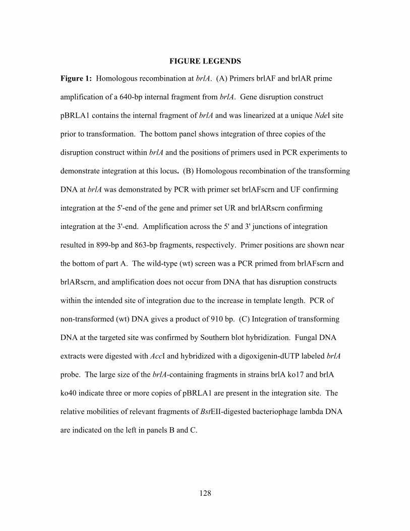
128
FIGURE LEGENDS
Figure 1: Homologous recombination at brlA. (A) Primers brlAF and brlAR prime
amplification of a 640-bp internal fragment from brlA. Gene disruption construct
pBRLA1 contains the internal fragment of brlA and was linearized at a unique NdeI site
prior to transformation. The bottom panel shows integration of three copies of the
disruption construct within brlA and the positions of primers used in PCR experiments to
demonstrate integration at this locus. (B) Homologous recombination of the transforming
DNA at brlA was demonstrated by PCR with primer set brlAFscrn and UF confirming
integration at the 5'-end of the gene and primer set UR and brlARscrn confirming
integration at the 3'-end. Amplification across the 5' and 3' junctions of integration
resulted in 899-bp and 863-bp fragments, respectively. Primer positions are shown near
the bottom of part A. The wild-type (wt) screen was a PCR primed from brlAFscrn and
brlARscrn, and amplification does not occur from DNA that has disruption constructs
within the intended site of integration due to the increase in template length. PCR of
non-transformed (wt) DNA gives a product of 910 bp. (C) Integration of transforming
DNA at the targeted site was confirmed by Southern blot hybridization. Fungal DNA
extracts were digested with AccI and hybridized with a digoxigenin-dUTP labeled brlA
probe. The large size of the brlA-containing fragments in strains brlA ko17 and brlA
ko40 indicate three or more copies of pBRLA1 are present in the integration site. The
relative mobilities of relevant fragments of BstEII-digested bacteriophage lambda DNA
are indicated on the left in panels B and C.
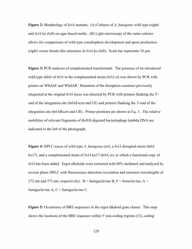
129
Figure 2: Morphology of brlA mutants. (A) Cultures of A. fumigatus wild type (right)
and brlA ko (left) on agar-based media. (B) Light microscopy of the same cultures
allows for comparisons of wild-type conidiophore development and spore production
(right) versus bristle-like structures in brlA ko (left). Scale bar represents 10 µm.
Figure 3: PCR analyses of complemented transformant. The presence of an introduced
wild-type allele of brlA in the complemented strain (brlA ct) was shown by PCR with
primer set WbrlAF and WbrlAR. Retention of the disruption construct previously
integrated at the original brlA locus was detected by PCR with primers flanking the 5'-
end of the integration site (brlAFscrn and UF) and primers flanking the 3'-end of the
integration site (brlARscrn and UR). Primer positions are shown in Fig. 1. The relative
mobilities of relevant fragments of BstEII-digested bacteriophage lambda DNA are
indicated to the left of the photograph.
Figure 4: HPLC traces of wild-type A. fumigatus (wt), a brlA disrupted strain (brlA
ko17), and a complemented strain of brlA ko17 (brlA ct), to which a functional copy of
brlA has been added. Ergot alkaloids were extracted with 80% methanol and analyzed by
reverse phase HPLC with fluorescence detection (excitation and emission wavelengths of
272 nm and 372 nm, respectively). B = fumigaclavine B; F = festuclavine; A =
fumigaclavine A; C = fumigaclavine C.
Figure 5: Occurrence of BRE sequences in the ergot alkaloid gene cluster. This map
shows the locations of the BRE sequence within 5′ non-coding regions (13), coding

130
regions (9), 3′ non-coding regions (3), and within an intron (1, marked with asterisk) of
these genes.
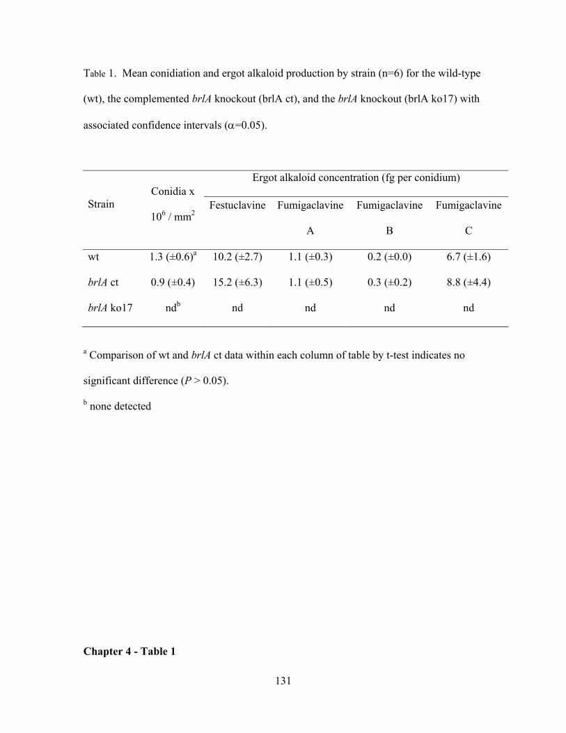
131
Table 1. Mean conidiation and ergot alkaloid production by strain (n=6) for the wild-type
(wt), the complemented brlA knockout (brlA ct), and the brlA knockout (brlA ko17) with
associated confidence intervals (α=0.05).
Strain Conidia x
106 / mm2
Ergot alkaloid concentration (fg per conidium)
Festuclavine
Fumigaclavine
A
Fumigaclavine
B
Fumigaclavine
C
wt 1.3 (±0.6)a 10.2 (±2.7) 1.1 (±0.3) 0.2 (±0.0) 6.7 (±1.6)
brlA ct 0.9 (±0.4) 15.2 (±6.3) 1.1 (±0.5) 0.3 (±0.2) 8.8 (±4.4)
brlA ko17 ndb nd nd nd nd
a Comparison of wt and brlA ct data within each column of table by t-test indicates no
significant difference (P > 0.05).
b none detected
Chapter 4 - Table 1
132
ANde I
brlA
brlAF
brlAR
1.4 kb
Acc IAcc Iwt
Nde I
4.6 kb
brlAR
brlAF
pBRLA11.3 kb
0.7 kb
brlA
ko17
brlA
ko17
wt
wt
brlA
ko17
wt
Wt screen
3' screen
5' screenB
Nde I Nde IAcc I
UR
brlARscrn2
Nde IAcc I
15 kb
1
Nde I
3
0.86 kb
UF
brlAFscrn
0.9 kb
C
brlA
ko17
brlA
ko40
wt
14 kb
1.4 kb
Coyle et al., Fig 1
WbrlAR
WbrlAF
Chapter 4 - Figure 1
133
Coyle et al., Fig 2
A
B
Chapter 4 - Figure 2
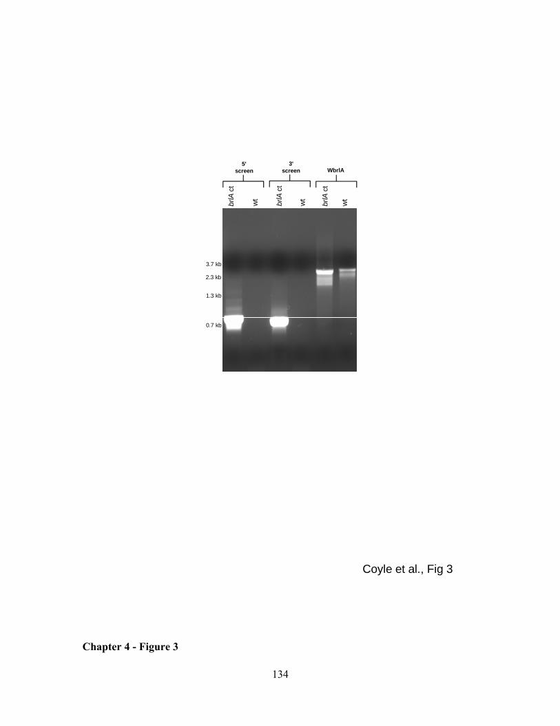
134
0.7 kb
1.3 kb
2.3 kb
3.7 kb
brlA
ct
brlA
ct
wt
wt
wt
brlA
ct
WbrlA3'
screen5'
screen
Coyle et al., Fig 3
Chapter 4 - Figure 3
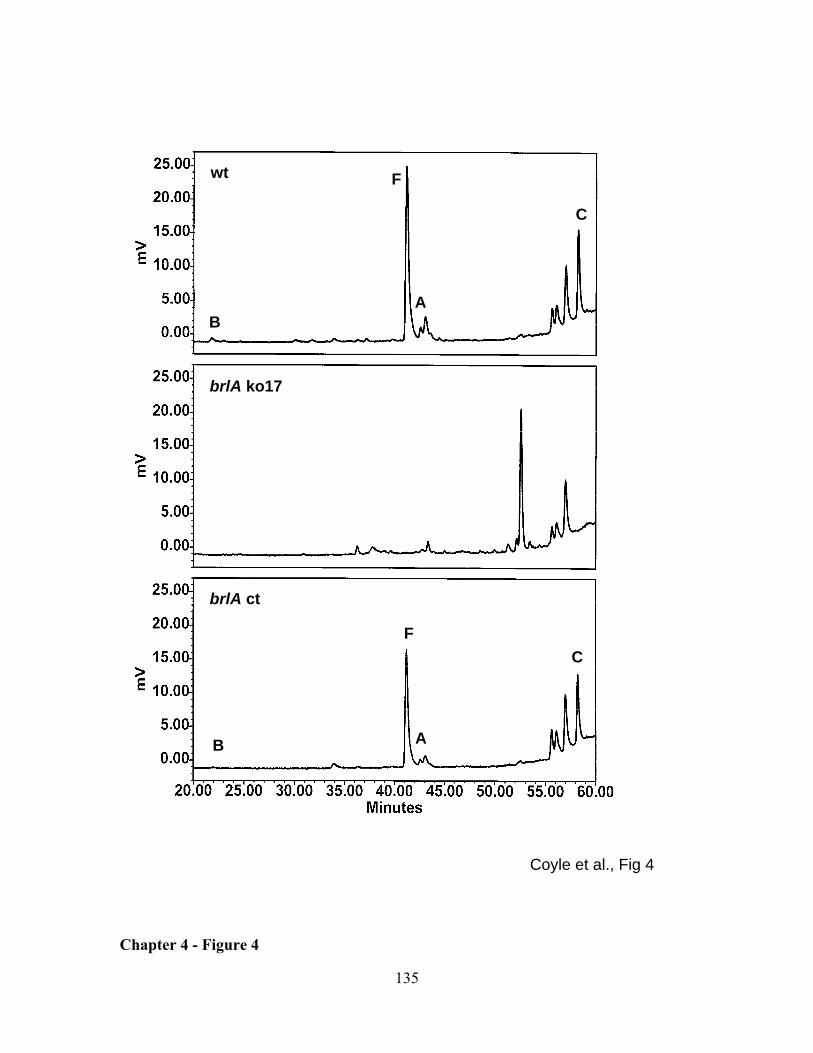
135
Chapter 4 - Figure 4
wt
brlA ko17
brlA ct
B
B
F
F
A
A
C
C
Coyle et al., Fig 4
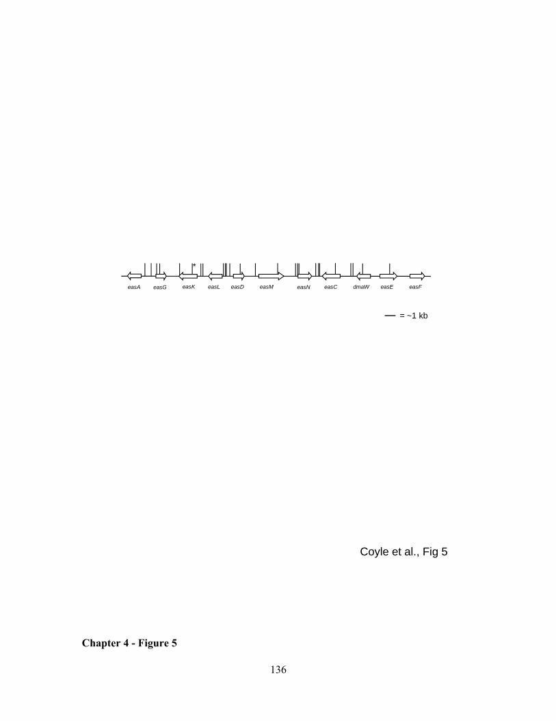
136
easM easCeasG dmaWeasLeasK easNeasA easFeasEeasD
= ~1 kb
*
Coyle et al., Fig 5
Chapter 4 - Figure 5
137
SUMMARY AND FUTURE DIRECTIONS
Ergot alkaloids are mycotoxins capable of interacting with neurotransmitter receptors and
causing toxicoses. However, they are also important metabolites that can be medicinally
advantageous. The ability to control the spectrum of alkaloids produced is contingent
upon understanding how they are synthesized. Aspergillus fumigatus may be a good
model system to study ergot alkaloid biosynthesis compared to other ergot alkaloid
producers. Therefore this study focuses on ergot alkaloid production in A. fumigatus,
examining specifically its association with sporulation and ergot alkaloid synthesis genes
common to both A. fumigatus and ergot fungi.
In previous work, Dr. Panaccione had determined that ergot alkaloids were
associated with the conidia of A. fumigatus. Results of analyses of ergot alkaloid content
of spores produced on different substrates indicated that these mycotoxins are present in
relatively abundant concentrations regardless of medium. Analysis of the physical
properties of conidia of A. fumigatus and several other fungi showed that the spores of A.
fumigatus are significantly smaller, lighter, and of lower specific gravity when compared
to those of Aspergillus niger, Aspergillus nidulans, and Stachybotrys chartarum, a fungus
of public concern [(5); Appendix 1]. Investigations involving alkaloid accumulation in
or on spores prompted a study of whether ergot alkaloids were produced only during
sporulation. Knockout of brlA, a gene known to be involved in sporulation in A.
nidulans, demonstrated this gene to be involved in conidiation in A. fumigatus [(1);
Chapter 1]. Lack of a functional BrlA rendered the fungus unable to form proper
conidiophores (asexual sporulation structures) and devoid of spores. This mutant also
138
lacked ergot alkaloids, thereby indicating that brlA does affect their production and that
the ergot alkaloid pathway and the sporulation pathway are linked, which has also been
recently demonstrated with some other secondary metabolites.
The second part of this research focused on understanding genes common to both
A. fumigatus and ergot fungi that act early in the ergot alkaloid biosynthesis pathway.
Annotation of the genomic region surrounding a homologue of dmaW, the gene
controlling the determinant step in the ergot alkaloid pathway as demonstrated previously
in studies with Neotyphodium sp. Lp1 (7), revealed the presence of a cluster of genes that
potentially could be involved in ergot alkaloid biosynthesis [(2); Chapter 2]. Individual
genes in the ergot alkaloid gene cluster and their potential involvement in ergot alkaloid
production were investigated in a series of gene knockout experiments. Knockout of the
dmaW homologue in A. fumigatus eliminated all ergot alkaloid production in the fungus.
This result is consistent with the role DMAT (dimethylallyltryptophan) synthase plays in
Neotyphodium sp. Lp1.
Knockout of other clustered genes, easA, easE, and easF, revealed their
involvement in synthesis of these toxins. This research suggested that easF and easE
encode products involved in the second and third steps of the pathway, respectively. The
ergot alkaloid profile of easF knockout showed an accumulation of DMAT, due to the
mutant’s inability to methylate it into N-methyl DMAT (Chapter 2), as well as small
accumulations of downstream intermediates, such as festuclavine. These observations
imply that the product of easF N-methylates DMAT but that ergot alkaloid synthesis can
occur very inefficiently even without EasF, given that DMAT synthase can prenylate
abrine to form N-methyl DMAT (6). The easE mutant accumulated an intermediate
139
characteristically similar to N-methyl DMAT and is presumed to act as an oxidase via
adding an hydroxyl group onto this substrate.
Lastly, easA was determined to encode a putative OYE oxidoreductase, and its
product was found to be involved in synthesis of festuclavine from chanoclavine
aldehyde in A. fumigatus. The Claviceps purpurea homologue of easA catalyzed
synthesis of agroclavine instead of festuclavine, which was evident from
complementation of the C. purpurea easA into A. fumigatus easA knockout (Chapter 3).
Protein sequence analysis revealed a key difference in a specific amino acid, which is
known to be important in other OYEs for reduction of double bonds (3, 4). Fungi that
produce festuclavine contain a critical active site tyrosine, whereas those that synthesize
agroclavine-derived alkaloids typical of ergot fungi (ergopeptines, etc) had phenylalanine
at the corresponding position. Therefore, differences in EasA function among
homologues have been demonstrated to be responsible for the divergence of the pathway.
This study has demonstrated the coupling of ergot alkaloid production with
sporulation. Future research should focus on understanding where expression of these
eas genes occurs. Such results may allow us to link intermediates with specific cell types
within A. fumigatus. Also future research could focus on analysis of ergot alkaloid
production in other sporulation mutants to further characterize the association between
ergot alkaloid production and conidiation.
This research also has demonstrated the role of multiple eas genes previously
hypothesized to be involved in ergot alkaloid biosynthesis. The genes, easF and easE,
control steps that occur early in the pathway and theoretically should function similarly in
ergot fungi. Future research should focus on delineating the roles of other clustered
140
genes, such as easD and easG, which are common between A. fumigatus and ergot fungi
and may share a similar function in both pathways. Additional, unassigned pathway
steps that these genes may control include formation of the epoxide following the diene
intermediate, chanoclavine aldehyde synthesis, or reduction of the imine, which results
from closure of the fourth ring. Further elucidation of the pathway will unveil the
involvement of these and other genes within the eas gene cluster.
Since easA is the point of divergence for the pathway, the discovery of functions
of later pathway genes from clavicipitaceous fungi might be discovered by adding
candidate genes to the Cp easA-complemented A. fumigatus easA knockout. Further
characterization of the EasA homologues via point mutation analyses are necessary to
understand the genetic basis for differences observed in their enzymatic functions.
141
REFERENCES
1. Coyle, C. M., S. C. Kenaley, W. R. Rittenour, and D. G. Panaccione. 2007.
Association of ergot alkaloids with conidiation in Aspergillus fumigatus.
Mycologia 99:804–811.
2. Coyle, C. M., and D. G. Panaccione. 2005. An ergot alkaloid biosynthesis gene
and clustered hypothetical genes from Aspergillus fumigatus. Appl. Environ.
Microbiol. 71:3112–3118.
3. Kohli, R. M., and V. Massey. 1998. The oxidative half-reaction of old yellow
enzyme. The role of tyrosine 196. J. Biol. Chem. 273:32763-32770.
4. Meah, Y., and V. Massey. 2000. Old Yellow Enzyme: Stepwise reduction of
nitro-olefins and catalysis of aci-nitro tautomerization. Proc. Natl. Acad. Sci.
USA 97:10733–10738.
5. Panaccione, D. G., and C. M. Coyle. 2005. Abundant respirable ergot alkaloids
from the common airborne fungus Aspergillus fumigatus. Appl. Environ.
Microbiol. 71:3106–3111.
6. Unsöld, I. A., and S.-M. Li. 2005. Overproduction, purification and
characterization of FgaPT2, a dimethylallyltryptophan synthase from Aspergillus
fumigatus. Microbiology 151:1499–1505.
7. Wang, J., C. Machado, D. G. Panaccione, H.-F. Tsai, and C. L. Schardl.
2004. The determinant step in ergot alkaloid biosynthesis by an endophyte of
perennial ryegrass. Fungal Genet. Biol. 41:189–198.
142
APPENDIX 1: Abundant respirable ergot alkaloids from the common airborne fungus Aspergillus fumigatus
Published by: Panaccione, D. G., and C. M. Coyle. 2005. Appl. Environ. Microbiol. 71:3106–3111.
ABSTRACT
Ergot alkaloids are mycotoxins that interact with several monoamine receptors,
negatively affecting cardiovascular, nervous, reproductive, and immune systems of
exposed humans and animals. Aspergillus fumigatus, a common airborne fungus and
opportunistic human pathogen, can produce ergot alkaloids in broth culture. The
objectives of this study were to determine if A. fumigatus accumulates ergot alkaloids in
respirable form in or on its conidia, to quantify ergot alkaloids associated with conidia
produced on several different substrates, and to measure relevant physical properties of
those conidia. We found at least four ergot alkaloids — fumigaclavine C, festuclavine,
fumigaclavine A, and fumigaclavine B, in order of abundance — associated with conidia
of A. fumigatus. Under environmentally relevant conditions, the total mass of ergot
alkaloids often constituted > 1% of the mass of the conidium. Ergot alkaloids were
extracted from conidia produced on all media tested, with the greatest quantities observed
when the fungus was cultured on latex paint or cultured maize seedlings. Physical
properties of conidia likely to affect their respirability, i.e. diameter, mass, and specific
gravity, were significantly lower in A. fumigatus than in Aspergillus nidulans, Aspergillus
niger, and Stachybotrys chartarum. Demonstration of relatively high concentrations of
ergot alkaloids associated with conidia of A. fumigatus presents opportunities for
investigations of potential contributions of the toxins to adverse health effects associated
with the fungus and to aspects of the biology of the fungus that contribute to its success.
143
INTRODUCTION
Ergot alkaloids are a complex family of indole-derived alkaloids that have a long history
of association with human suffering. The contamination of rye and other grain crops
with alkaloid-rich sclerotia of the ergot fungus Claviceps purpurea was responsible for
gangrenous and convulsive forms of ergotism known as St. Anthony’s fire or holy fire
(19). Other ergot alkaloid-producing fungi such as the closely related Neotyphodium spp.
endophytes of forage grasses negatively affect agriculture by reducing animal
productivity and health (2, 22). The ability of different ergot alkaloids to act as partial
agonists or antagonists of various serotonin, dopamine, and α-adrenaline receptors results
in effects on nervous, circulatory, reproductive, and immune systems leading to high or
low blood pressure, muscle contractions, reduced fertility, disturbances in sleep/wake
cycles, lowered immune response, and, at high doses, hallucinations and gangrene of the
extremities (14, 16, 22, 25, 31).
Screens of other fungi for ergot alkaloids have identified several distantly related
fungi as potential sources (13, 17, 29). Among these fungi, the best characterized is
Aspergillus fumigatus. This fungus was first noted to produce the ergot alkaloid
festuclavine and two novel derivatives of festuclavine named fumigaclavine A and
fumigaclavine B in semidefined broth culture (29). Later Cole et al. (3) described an
additional festuclavine derivative, fumigaclavine C, from broth cultures of A. fumigatus
originally isolated from moldy silage.
Aspergillus fumigatus is associated with several human health issues. It is the
most common airborne fungal pathogen of humans (6, 18). It can cause invasive
aspergillosis in immunocompromised individuals, with resulting mortality at > 50%. In
144
immunocompetent individuals, the fungus can colonize preexisting cavities in the lungs
or sinuses without penetrating into tissues, a condition known as aspergilloma or fungus
ball (6, 11, 18). The fungus also is associated with air quality issues in indoor
environments and near composting facilities (11, 12, 27). Presence of conidia of A.
fumigatus and of several other fungi in such environments has been loosely associated
with respiratory allergic symptoms and miscellaneous other ailments, but causal
associations have not been demonstrated.
Most mycotoxin-producing fungi produce their toxins in the substrate in which
they grow and ingestion of that contaminated substrate is required for intoxication.
Although less frequently documented, the presence of mycotoxins in or on conidia of the
producing fungus provides the potential for delivery of mycotoxins via the alternate and
less voluntary route of inhalation. For example, the black mold fungus Stachybotrys
chartarum contains trichothecenes associated with its spores (28).
Our objectives in this study were to determine if conidia of A. fumigatus contain
ergot alkaloids in respirable form, to quantify ergot alkaloids associated with conidia
produced on several environmentally relevant substrates, and to measure physical
properties of conidia likely to affect their respirability. Detection and quantification of
ergot alkaloids associated with conidia is prerequisite to investigations of potential
contributions of the alkaloids to adverse human health effects or to other aspects of the
biology of the fungus.
MATERIALS AND METHODS
Fungi and culture conditions. Aspergillus fumigatus isolate WVU1943 (FGSC) was
cultured from the lungs of a deceased parakeet in Charleston, West Virginia, during 1977.
145
Isolate WVU2026 (FGSC) was obtained by passive sampling of the air (by opening a
Petri dish of potato dextrose agar for 20 minutes under ambient conditions) in a house in
Monongalia County, West Virginia, during 2003. All viable cultures were manipulated
in a class II biosafety cabinet. Isolates were maintained on potato dextrose agar [PDA;
20 g/L dehydrated mashed potatoes (Idaho Spuds, Pillsbury, Minneapolis, MN), 20 g/L
glucose, 15 g/L agar (Bacto-Agar, Difco, Detroit, MI)] at room temperature or at 37 ºC,
and most ergot alkaloid analyses were conducted on cultures grown on this medium.
Additional alkaloid analyses were conducted with A. fumigatus isolates cultured on the
following substrates: 1.5% (w/v) water agar plates (prepared with Difco Bacto-Agar);
Pittsburgh Paint (Pittsburgh, PA) interior latex base 88-110 (low luster semigloss latex
100% acrylic white and pastel mixing base) that had been applied with a brush in two
thin layers on the inside of lids of Petri dishes and allowed to dry for 2 hr prior to
inoculation; maize (Zea mays DeKalb hybrid DK626RR, DeKalb, IL) seedlings four days
after germination of surface-sterilized kernels on water agar; and, water-rinsed and
autoclaved pistachio (Pistacia vera; Blue Diamond, Sacramento, CA) shells. Painted and
inoculated Petri dish lids were placed over 1.5% water agar plates to promote high
humidity for growth of the fungus. Pistachio shells were incubated in Petri dishes
containing 8 mL of sterile distilled water to promote high humidity. Cultures were
inoculated by lightly dusting the medium with conidia that had adhered to a Petri dish lid
formerly covering a mature culture. In mock inoculations, an average of 1.2 × 10⁷
conidia per Petri dish were deposited by this inoculation method.
Large numbers of conidia, free of culture medium and hyphae, were collected
from PDA cultures by tapping a culture dish such that conidia accumulated on the inside
146
of the lid. In this way, alkaloids could be extracted exclusively from conidia.
Alternatively, conidia could be made airborne from a Petri dish lid by agitating the lid by
hand. Airborne conidia were then collected on a 0.2-µm SFCA filter (Nalge Nunc
International, Rochester, NY) or a 0.45-µm nylon filter (Millipore, Bedford, MA) under
vacuum and extracted by washing the filter with 800 µl of 80% methanol. In other
experiments, conidia were collected from water agar and paint cultures by very briefly
washing small portions of the sporulating cultures with 800 µL of 80% methanol.
Conidia were harvested from corn or pistachio tissue by suspending pieces of the
colonized and spore-bearing tissue in 1 mL of 80% methanol. Microscopic examination
of conidial suspensions prepared from any of these alternate substrates revealed the lack
of hyphae. However, with the exception of the conidia collected from PDA cultures, the
extraction solvent had been briefly in contact with hyphae and the colonized substrate or
medium.
Stachybotrys chartarum and Aspergillus nidulans were isolated from
contaminated building materials; Aspergillus niger was isolated by passive air sampling
(as described above for A. fumigatus). Conidia of these fungi, and of A. fumigatus,
collected for measurement of their physical properties, were harvested with 80%
methanol from cultures grown on PDA. The number of conidia per mL of suspension
was determined by counting dilutions on a hemacytometer (Hausser Scientific, Horsham,
PA). Conidia (of defined number) were collected by filtration though tared 0.22-µm
nylon filters (Costar, Corning, NY) and retained for mass determination on a Mettler-
Toledo (Columbus, OH) AG104 balance after drying in a Labconco (Kansas City, MO)
147
centrivap concentrator. Conidial diameter was determined microscopically with the aid
of an ocular micrometer.
HPLC, chemical manipulations, and mass spectrometry. Aliquots of conidia
were suspended in 80% methanol, transferred to 1.5-ml microcentrifuge tubes, and
rotated end-over-end at 40 rpm for 1 hr on a Glas-Col (Terre Haute, IN) mini-rotator.
The number of conidia in the extraction tube was determined by counting dilutions on a
hemacytometer. Conidia from PDA-based cultures were separated from the alkaloid
extract by centrifugal filtration and weighed as described above. For conidia derived
from cultures on alternate substrates, conidial mass was calculated as the product of the
number of conidia counted in the sample and the value previously calculated for mass of
an individual conidium. These calculations are based on the assumption that the mass of
a conidium produced on these alternate substrates is the same as the mass of a conidium
produced on PDA.
Methanol extracts of conidia (with no further preparation beyond removal of
conidia by filtration) were analyzed by HPLC with conditions described previously (23)
except for changes in the equipment used. The HPLC apparatus consisted of a model 600
pump controller with in-line degasser, model 717plus autosampler, model 2487
absorbance detector or model 996 photodiode array detector (all from Waters Corp.,
Milford, MA), and a Rainin (Woburn, MA) Fl2 fluorescence detector set with excitation
and emission wavelengths of 272 nm and 372 nm, respectively. Alkaloid quantities were
determined based on comparison of peak areas relative to a standard curve derived from
four dilutions each of three parallel samples of the highly similar ergot alkaloid
148
agroclavine (Sigma, St. Louis, MO) and adjusted for relative molecular weights of
analytes and standard.
For PDA-based cultures, several dilutions of each A. fumigatus spore extract were
analyzed and the concentration of each alkaloid determined by linear regression analysis.
Analyses of PDA-based cultures (Table 2) were conducted in triplicate on each of two
separate occasions. Three additional preliminary analyses yielded similar results.
Extracts from A. fumigatus-colonized plant materials, water agar, or paint-based cultures
(n = 3 to 6 / medium / isolate), which were less dense and contained fewer conidia than
PDA-based cultures, were analyzed without dilution.
Agroclavine was validated as a standard for the A. fumigatus clavines by
comparing experimentally measured concentrations of it versus calculated concentrations
of four separate preparations of dihydroergotamine and dihydroergocristine (Sigma),
which carry the identical ergoline chromophore as the A. fumigatus ergot alkaloids.
Concentrations of dihydroergotamine measured based on fluorescence relative to an
agroclavine standard curve were 1.0 ± 0.04 (mean ± SE) times the calculated values,
whereas the measured concentrations of dihydroergocristine solutions were 0.98 ± 0.05
times the calculated values.
The relationships among compounds represented in different peaks were
investigated by deacetylation of alkaloid fractions with 1 M NaOH at room temperature
for 15 hr (29). Base-treated samples were neutralized with an equinormal solution of
acetic acid and analyzed by HPLC as described above. Masses of untreated and base-
treated compounds were obtained on a Finnegan Mass Spectrometer (model SSQ7000) at
the WVU Mass Spectrometry Center. Isolated peaks collected after HPLC were
149
concentrated 10-fold under vacuum, introduced into the ion source with an infusion
pump, and ionized in positive electrospray ionization mode.
Statistical Analyses. Data were analyzed by analysis of variance, and means
were compared by the Tukey-Kramer honestly significant difference test in the JMP
software package (SAS Institute, Cary, NC). Sets of values having unequal variances
were log-transformed prior to analysis.
RESULTS
Identification and quantification of ergot alkaloids associated with conidia of A.
fumigatus. Several compounds from simple 80% methanolic extracts of conidia from
PDA-cultured A. fumigatus produced large fluorescent peaks when analyzed by HPLC
with excitation and emission wavelengths of 272 nm and 372 nm, respectively (Fig. 1).
Electrospray ionization-mass spectrometry (ESI-MS) of the compound in peak C (Fig. 1),
which had the greatest area and was the most nonpolar of the alkaloids analyzed, revealed
an ion at m/z 367 corresponding in mass to [fumigaclavine C + H]+, which had previously
been described from broth cultures of A. fumigatus (3). Treatment of this compound with
1 M NaOH resulted in a more polar compound (peak dC, Fig. 1) that yielded an ion at
m/z 325, corresponding in mass to [deacetylated fumigaclavine C + H]+.
Similarly the compound represented by peak A (Fig. 1) yielded an ion of m/z 299
in its native form, consistent with a proposed identity as fumigaclavine A, and was
converted after treatment with NaOH to a more polar compound (peak dA, Fig. 1) with
an ion of m/z 257, consistent with [deacetylated fumigaclavine A + H]+. Peak B, which
cochromatographed with experimentally deacetylated fumigaclavine A (Fig. 1), is
proposed to be fumigaclavine B, which is identical to deacetylated fumigaclavine A (3,
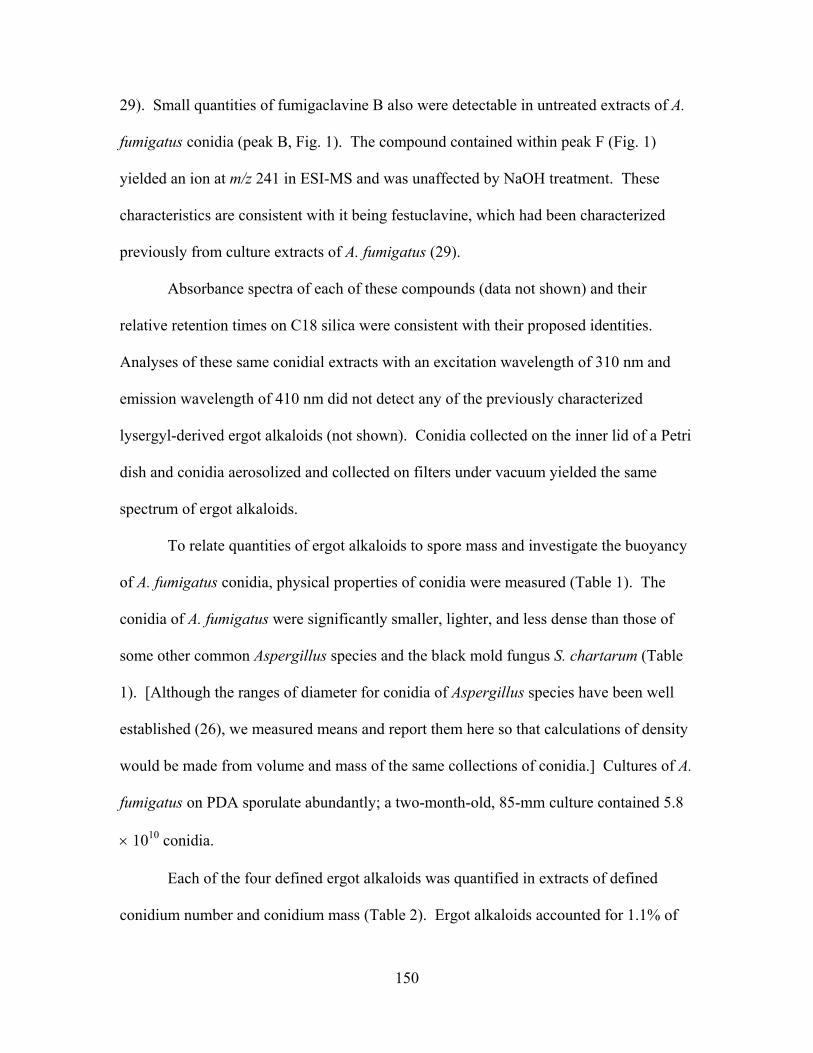
150
29). Small quantities of fumigaclavine B also were detectable in untreated extracts of A.
fumigatus conidia (peak B, Fig. 1). The compound contained within peak F (Fig. 1)
yielded an ion at m/z 241 in ESI-MS and was unaffected by NaOH treatment. These
characteristics are consistent with it being festuclavine, which had been characterized
previously from culture extracts of A. fumigatus (29).
Absorbance spectra of each of these compounds (data not shown) and their
relative retention times on C18 silica were consistent with their proposed identities.
Analyses of these same conidial extracts with an excitation wavelength of 310 nm and
emission wavelength of 410 nm did not detect any of the previously characterized
lysergyl-derived ergot alkaloids (not shown). Conidia collected on the inner lid of a Petri
dish and conidia aerosolized and collected on filters under vacuum yielded the same
spectrum of ergot alkaloids.
To relate quantities of ergot alkaloids to spore mass and investigate the buoyancy
of A. fumigatus conidia, physical properties of conidia were measured (Table 1). The
conidia of A. fumigatus were significantly smaller, lighter, and less dense than those of
some other common Aspergillus species and the black mold fungus S. chartarum (Table
1). [Although the ranges of diameter for conidia of Aspergillus species have been well
established (26), we measured means and report them here so that calculations of density
would be made from volume and mass of the same collections of conidia.] Cultures of A.
fumigatus on PDA sporulate abundantly; a two-month-old, 85-mm culture contained 5.8
× 1010 conidia.
Each of the four defined ergot alkaloids was quantified in extracts of defined
conidium number and conidium mass (Table 2). Ergot alkaloids accounted for 1.1% of
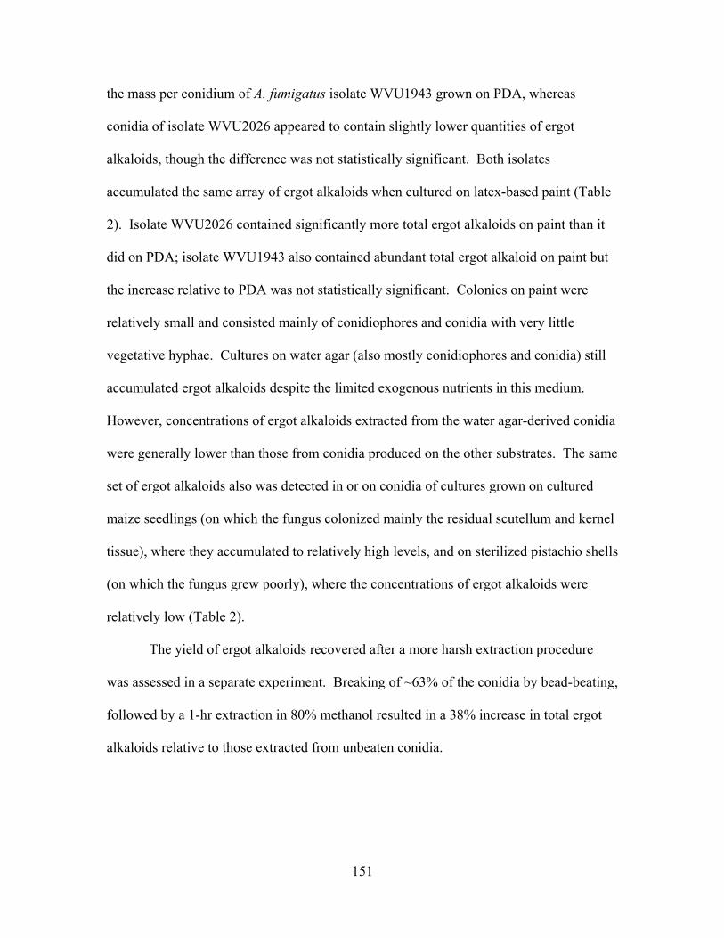
151
the mass per conidium of A. fumigatus isolate WVU1943 grown on PDA, whereas
conidia of isolate WVU2026 appeared to contain slightly lower quantities of ergot
alkaloids, though the difference was not statistically significant. Both isolates
accumulated the same array of ergot alkaloids when cultured on latex-based paint (Table
2). Isolate WVU2026 contained significantly more total ergot alkaloids on paint than it
did on PDA; isolate WVU1943 also contained abundant total ergot alkaloid on paint but
the increase relative to PDA was not statistically significant. Colonies on paint were
relatively small and consisted mainly of conidiophores and conidia with very little
vegetative hyphae. Cultures on water agar (also mostly conidiophores and conidia) still
accumulated ergot alkaloids despite the limited exogenous nutrients in this medium.
However, concentrations of ergot alkaloids extracted from the water agar-derived conidia
were generally lower than those from conidia produced on the other substrates. The same
set of ergot alkaloids also was detected in or on conidia of cultures grown on cultured
maize seedlings (on which the fungus colonized mainly the residual scutellum and kernel
tissue), where they accumulated to relatively high levels, and on sterilized pistachio shells
(on which the fungus grew poorly), where the concentrations of ergot alkaloids were
relatively low (Table 2).
The yield of ergot alkaloids recovered after a more harsh extraction procedure
was assessed in a separate experiment. Breaking of ~63% of the conidia by bead-beating,
followed by a 1-hr extraction in 80% methanol resulted in a 38% increase in total ergot
alkaloids relative to those extracted from unbeaten conidia.
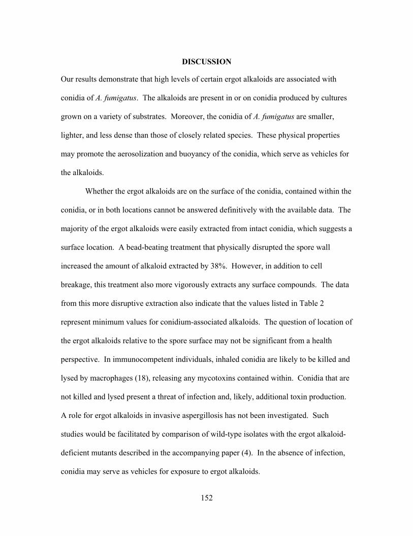
152
DISCUSSION
Our results demonstrate that high levels of certain ergot alkaloids are associated with
conidia of A. fumigatus. The alkaloids are present in or on conidia produced by cultures
grown on a variety of substrates. Moreover, the conidia of A. fumigatus are smaller,
lighter, and less dense than those of closely related species. These physical properties
may promote the aerosolization and buoyancy of the conidia, which serve as vehicles for
the alkaloids.
Whether the ergot alkaloids are on the surface of the conidia, contained within the
conidia, or in both locations cannot be answered definitively with the available data. The
majority of the ergot alkaloids were easily extracted from intact conidia, which suggests a
surface location. A bead-beating treatment that physically disrupted the spore wall
increased the amount of alkaloid extracted by 38%. However, in addition to cell
breakage, this treatment also more vigorously extracts any surface compounds. The data
from this more disruptive extraction also indicate that the values listed in Table 2
represent minimum values for conidium-associated alkaloids. The question of location of
the ergot alkaloids relative to the spore surface may not be significant from a health
perspective. In immunocompetent individuals, inhaled conidia are likely to be killed and
lysed by macrophages (18), releasing any mycotoxins contained within. Conidia that are
not killed and lysed present a threat of infection and, likely, additional toxin production.
A role for ergot alkaloids in invasive aspergillosis has not been investigated. Such
studies would be facilitated by comparison of wild-type isolates with the ergot alkaloid-
deficient mutants described in the accompanying paper (4). In the absence of infection,
conidia may serve as vehicles for exposure to ergot alkaloids.
153
The issue of health risks posed by inhalation of mycotoxin-containing conidia is
complex and affected by several factors including the physical nature of the conidia with
respect to their potential for dispersal and inhalation and the production of mycotoxins on
environmentally relevant substrates. The conidia of A. fumigatus have properties that
appear to facilitate their dispersal and inhalation. The 2.8-µm diameter of conidia is
small enough for them to penetrate deep into the alveoli of the lungs (18). The low
specific gravity of the conidia (0.24) probably promotes efficient air dispersal. For
comparison, the conidia of the trichothecene producer S. chartarum, which have
generated considerable health concerns (10, 28), have a mass that is 48 times greater and
a specific gravity that is 4 times greater than those of conidia of A. fumigatus.
Ergot alkaloids were detected on all tested media, including environmentally
relevant substrates such as latex paint, two different plant materials, and water agar
(representative of moist, nutritionally poor environments). The reported concentrations
of ergot alkaloids associated with conidia produced on substrates other than PDA are
based on the number of conidia extracted and a value for conidial mass measured from
conidia produced on PDA. Thus, these values are based on the assumption that conidia
produced on any of the alternate substrates have the same mass as conidia produced on
PDA. We contend that this is a reasonable assumption. However, if the mass per
conidium were, in fact, greater on a natural substrate than on PDA, then the calculated
values of ergot alkaloid per conidium would be lower. Conversely, if the conidial mass
were lower on a natural substrate than on PDA, then alkaloid concentration would be
greater than listed in Table 2.
154
A more complicated factor in determination of health risks associated with
conidial mycotoxins is the issue of whether the toxins can or will be encountered in
quantities sufficient for them to exert their effects. There are at least three components to
this issue, including (a) concentration of mycotoxin per conidium, (b) the number of
conidia encountered, and (c) the toxicity of the mycotoxins.
Concentrations of ergot alkaloids exceeding 1% by mass of A. fumigatus conidia
are relatively high for fungal natural products. There are few data available for direct
comparison, because most mycotoxin analyses are expressed as mass of toxin per unit
volume of culture. Data on respirable trichothecenes from S. chartarum (28) indicate a
mean of 17 ng of trichothecenes per mg of dust aerosolized from S. chartarum cultured
on rice. Assuming that conidia, which constituted 85% of the particles in the aerosolized
dust (28), contribute 85% of the mass of the dust sample, then 0.002% of the conidium by
mass was trichothecenes. With a different isolate of this same fungal species and
measuring only the most abundant trichothecene, Nikulin et al. (21) reported 0.1 pg of
satratoxin H per conidium, which would translate to 0.0007% satratoxin H per mass of
conidium. Another source of alkaloid data per unit fungal mass comes from the
poisonous mushroom literature. For example, basidiocarps of Amanita muscaria contain
ibotenic acid at 0.45% by mass dry weight and muscimol at 0.036% (30) [assuming 12.5
mg fresh weight basidiocarp yields 1 mg dry weight (9)]. Amatoxins have been
measured at 0.1% to 0.7% of the dry weight of Amanita phalloides basidiocarps (9). In a
more relevant example, ergot sclerotia of field-grown C. purpurea contain 1% to 2%
ergot alkaloids by mass (5, 20).
155
The number of A. fumigatus conidia available in the air depends on the substrate
and the environment in which the fungus is growing. Under favorable conditions the
fungus can sporulate prolifically. A typical culture of A. fumigatus on PDA can yield
~109 conidia per cm2 of culture surface area. Viable A. fumigatus conidia per m3 of air
range from 0 to 200 CFU in clean environments (27), to 107 CFU near composting
facilities (12), to 1011 CFU near moldy hay or other stored organic materials (27).
Considering an intake rate of 0.63 m3 of air per hr (32), a conidial ergot alkaloid content
of 1% by mass, and no further ergot alkaloid production after inhalation of the fungus,
ergot alkaloid dose would range from 3.7 pg per hr (at 200 CFU/m3), to 180 ng per hr (at
107 CFU per m3), to 1.8 mg per hr (at 1011 CFU per m3). An interesting point of
reference is that an ingested dose of the illicit and highly active ergot alkaloid LSD can
range from hundreds of ng to hundreds of µg (1), but based on US Drug Enforcement
Agency data is frequently in the 20 to 80 µg range
(http://www.nida.nih.gov/Infofax/lsd.html). To inhale a comparable mass of A.
fumigatus ergot alkaloids in one hour would require exposure to 107 to 1010 CFU per m3.
Such high concentrations of conidia will be encountered only rarely. A more practical
issue to consider is whether there are potential health effects of a less remarkable nature
(e.g., effects on depression, blood pressure, or sleep/wake cycles) associated with chronic
daily doses of ergot alkaloids in the ng to µg range. This issue has not been investigated.
Toxicity of the particular ergot alkaloids associated with the A. fumigatus conidia
has not been studied extensively, but the available data suggest that these mycotoxins
have considerable biological activity. Similar to other studied ergot alkaloids,
festuclavine interacts with receptors for serotonin, dopamine, and α-adrenaline (7, 14, 24,
156
25). Festuclavine and synthetic derivatives of festuclavine also are cytostatic in in vitro
assays with several bacteria and mouse lymphoma cell lines (7, 8). Moreover,
festuclavine is unique among the naturally occurring ergot alkaloids in that it is directly
mutagenic in the Ames test (15). In the sole published animal study of fumigaclavine C,
ingestion of relatively crude preparations of this ergot alkaloid greatly reduced feed
intake of treated calves, and caused hemorrhagic enteritis of small and large intestines as
well as patchy interstitial thickening of alveolar walls of the animals (3).
Demonstration of the presence of ergot alkaloids at relatively high concentrations
in or on conidia of A. fumigatus does not ipso facto indicate a role for the toxins in
pathogenesis, other health effects, or any specific aspect of the biology of the fungus.
However, it does raise interesting questions for further research. Studies with ergot
alkaloid-deficient mutants such as the dmaW-knockout strain described in the
accompanying paper (4) should be useful for assessing the contribution of ergot alkaloids
to virulence towards animals or potential contributions of the alkaloids to the ecological
success of A. fumigatus. Since minimization of the mass of a conidium appears to have
been selected for in this fungus, the inclusion of alkaloids in quantities that exceed 1% of
the mass of the conidium should have been selected against unless they provided some
advantage to the fungus.
ACKNOWLEDGEMENTS
This work was supported by USDA-NRI grant No. 2001-3531 9-10930 and Hatch funds,
and was published with the approval of the Director of the WV Agricultural and Forestry
Experiment Station as Scientific Article no. 2895. We thank Robert Smith of the WVU
Mass Spectrometry Center for assistance with mass spectrometry, Barton Baker and Jim

157
Kotcon for helpful discussions, and Brian Lewis and Jessica Cipoletti for technical
assistance.
158
REFERENCES
1. Baudot, P., and J. C. Andre. 1985. Identification and quantitative determination of
LSD by fluorescence: new data. Bull. Narc. 37:79–94.
2. Clay, K., and C. Schardl. 2002. Evolutionary origins and ecological consequences
of endophyte symbiosis with grasses. Am. Nat. 160:S199–S127.
3. Cole, R. J., J. W. Kirksey, J. W. Dorner, D. M. Wilson, J. C. Johnson Jr., A. N.
Johnson, D. M. Bedell, J. P. Springer, K. K. Chexal, J. C. Clardy, and R. H. Cox.
1977. Mycotoxins produced by Aspergillus fumigatus species isolated from molded
silage. J. Agric. Food Chem. 25:826–830.
4. Coyle, C. M., and D. G. Panaccione. 2005. An ergot alkaloid biosynthesis gene
and clustered hypothetical genes from Aspergillus fumigatus. Appl. Environ.
Microbiol. 71:3112–3118.
5. Cvak, L. 1999. Industrial production of ergot alkaloids, p. 373–409. In V. Kren,
and L. Cvak (ed.), Ergot: The Genus Claviceps. Harwood Academic Publishers, The
Netherlands.
6. Denning, D. W. 1998. Invasive aspergillosis. Clin. Infect. Dis. 26:781–805.
7. Eich, E., and H. Pertz. 1999. Antimicrobial and antitumor effects of ergot alkaloids
and their derivatives, p. 411–440. In V. Kren, and L. Cvak (ed.), Ergot: The Genus
Claviceps. Harwood Academic Publishers, The Netherlands.
8. Eich, E., C. Becker, and W. E. G. Müller. 1984. Clavines – new antibiotics with
cytostatic activity. Biochem. Pharmacol. 33:523–526.
159
9. Enjalbert, F., C. Gallion, F. Jehl, H. Monteil, and H. Faulstich. 1993. Amatoxins
and phallotoxins in Amanita species: high-performance liquid chromatography
determination. Mycologia 85:579–584.
10. Etzel, R. A., E. Montana, W. G. Sorenson, G. J. Kullman, T. M. Allan, and D. G.
Dearborn. 1998. Acute pulmonary hemorrhage in infants associated with exposure
to Stachybotrys atra and other fungi. Arch. Pediatr. Adolesc. Med. 152:757–762.
11. Fischer, G., and W. Dott. 2003. Relevance of airborne fungi and their secondary
metabolites for environmental, occupational and indoor hygiene. Arch. Microbiol.
179:75–82.
12. Fischer, G., T. Muller, R. Schwalbe, R. Ostrawski, and W. Dott. 2000. Species-
specific profiles of mycotoxins produced in cultures and associated with conidia of
airborne fungi derived from biowaste. Int. J. Hyg. Environ. Health 203:105–116.
13. Flieger, M., M. Wurst, and R. Shelby. 1997. Ergot alkaloids –– sources,
structures, and analytical methods. Folia Microbiol. 42:3–30.
14. Fuxe, K., B. B. Fredholm, L. F. Agnati, S.-O. Ögren, B. J. Everitt, G. Jonsson,
and J.-Ä. Gustafsson. 1978. Interactions of ergot drugs with central monoamine
systems. Pharmacology 16:99–134.
15. Glatt, H., H. Pertz, R. Kasper, and E. Eich. 1992. Clavine alkaloids and
derivatives as mutagens detected in the AMES test. Anti-Cancer Drugs 3:609–614.
16. Gröger, D., and H. G. Floss. 1998. Biochemistry of ergot alkaloids – achievements
and challenges. Alkaloids 50:171–218.
160
17. Kozlovsky, A. G. 1999. Producers of ergot alkaloids out of Claviceps genus, p.
479–499. In V. Kren, and L. Cvak (ed.), Ergot: The Genus Claviceps. Harwood
Academic Publishers, The Netherlands.
18. Latgé, J.-P. 1999. Aspergillus fumigatus and aspergillosis. Clin. Microbiol. Rev.
12:310–350.
19. Matossian, M. K. 1989. Poisons of the Past: Molds, Epidemics, and History. Yale
University Press, New Haven, CT. 190 p.
20. Németh, E. 1999. Parasitic production of ergot alkaloids, p. 303–319. In V. Kren,
and L. Cvak (ed.), Ergot: The Genus Claviceps. Harwood Academic Publishers, The
Netherlands.
21. Nikulin, M., K. Reijula, B. B. Jarvis, and E. L. Hintikka. 1996. Experimental
lung mycotoxicosis in mice induced by Stachybotrys atra. Intl. J. Exp. Path. 77:213–
218.
22. Panaccione, D. G., and C. L. Schardl. 2003. Molecular genetics of ergot alkaloid
biosynthesis, p. 399–424. In J. F. White Jr., C. W. Bacon, N. L. Hywel-Jones, and J.
W. Spatafora (ed.), The Clavicipitalean Fungi: Evolutionary Biology, Chemistry,
Biocontrol, and Cultural Impacts. Marcel-Dekker, New York.
23. Panaccione, D. G., B. A. Tapper, G. A. Lane, E. Davies, and K. Fraser. 2003.
Biochemical outcome of blocking the ergot alkaloid pathway of a grass endophyte. J.
Agric. Food Chem. 51:6429–6437.
24. Pertz, H. 1996. Naturally occurring clavines: antagonism/partial agonism at 5-HT2A
receptors and antagonism at α1-adrenoceptors in blood vessels. Planta Med. 62:387–
392.
161
25. Pertz, H., and E. Eich. 1999. Ergot alkaloids and their derivatives as ligands for
serotoninergic, dopaminergic, and adrenergic receptors, p. 411–440. In V. Kren, and
L. Cvak (ed.), Ergot: The Genus Claviceps. Harwood Academic Publishers, The
Netherlands.
26. Raper, K. B., and D. I. Fennell. 1965. The Genus Aspergillus. Williams &
Wilkins, Balitmore, MD. 686 p.
27. Solomon, W. R., and H. A. Burge. 1984. Allergens and pathogens in indoor air
quality, p. 173-191. In P. J. Walsh, C. S. Dudney, and E. D. Copenhaver (ed.), Indoor
Air Quality. CRC Press, Boca Raton, FL.
28. Sorenson, W. G., D. G. Fraser, B. B. Jarvis, J. Simpson, and V. A. Robinson.
1987. Trichothecene mycotoxins in aerosolized conidia of Stachybotrys atra. Appl.
Environ. Microbiol. 53:1370–1375.
29. Spilsbury, J. F., and S. Wilkinson. 1961. The isolation of festuclavine and two
new clavine alkaloids from Aspergillus fumigatus Fres. J. Chem. Soc. 5:2085–2091.
30. Tsunoda, K., N. Inoue, Y. Aoyagi, and T. Sugahara. 1993. Changes in
concentration of ibotenic acid and muscimol in the fruit body of Amanita muscaria
during the reproduction stage. J. Food Hyg. Soc. Japan 34:18–24
31. Tudzynski, P., T. Correia, and U. Keller. 2001. Biotechnology and genetics of
ergot alkaloids. Appl. Microbiol. Biotechnol. 57:593–605.
32. US Environmental Protection Agency. 1997. Volume I: General Factors.
Exposure Factors Handbook. EPA Office of Research and Development Publication
EPA/600/P-95/002Fa-c. Washington, DC.
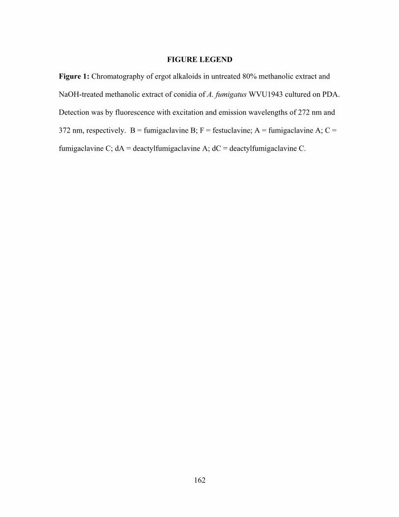
162
FIGURE LEGEND
Figure 1: Chromatography of ergot alkaloids in untreated 80% methanolic extract and
NaOH-treated methanolic extract of conidia of A. fumigatus WVU1943 cultured on PDA.
Detection was by fluorescence with excitation and emission wavelengths of 272 nm and
372 nm, respectively. B = fumigaclavine B; F = festuclavine; A = fumigaclavine A; C =
fumigaclavine C; dA = deactylfumigaclavine A; dC = deactylfumigaclavine C.
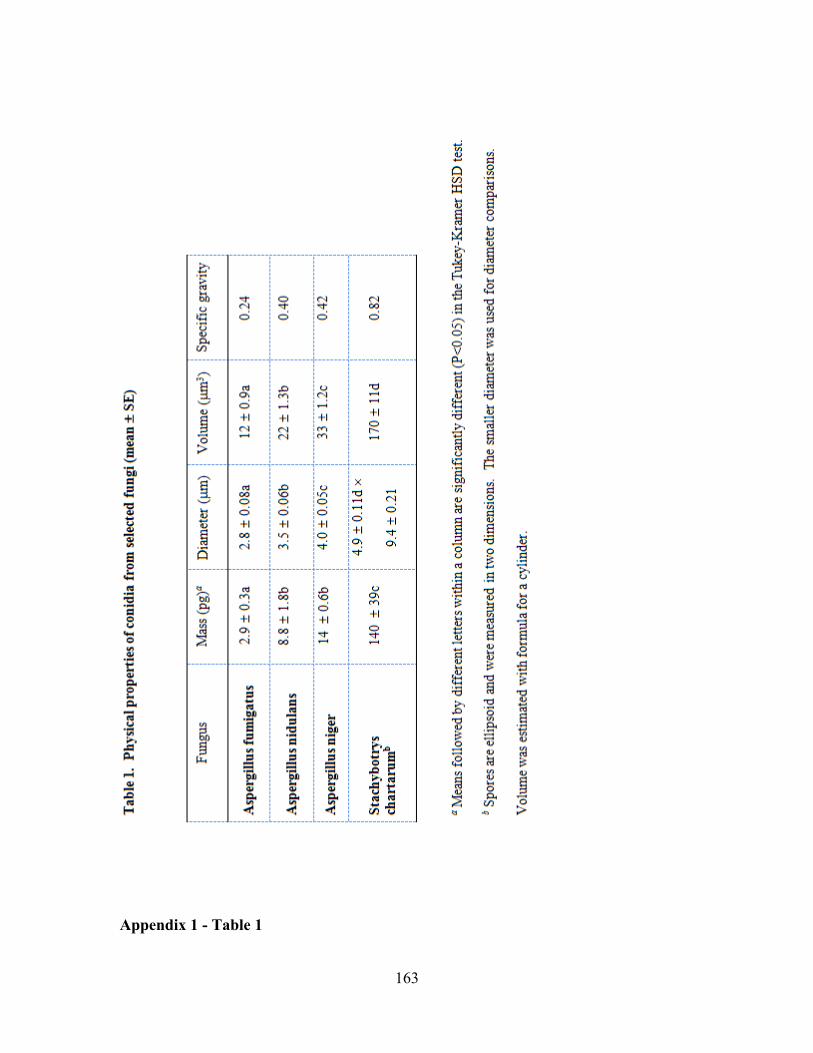
163
Appendix 1 - Table 1
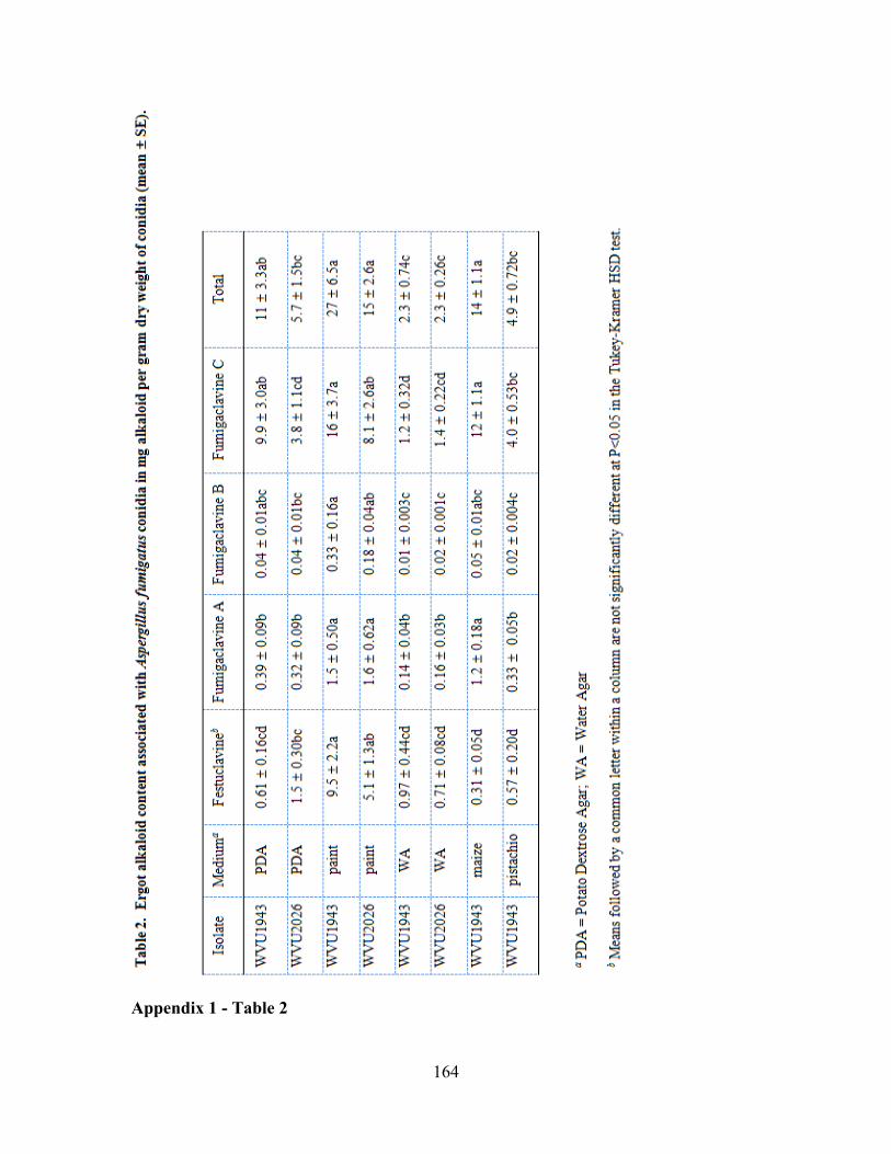
164
Appendix 1 - Table 2
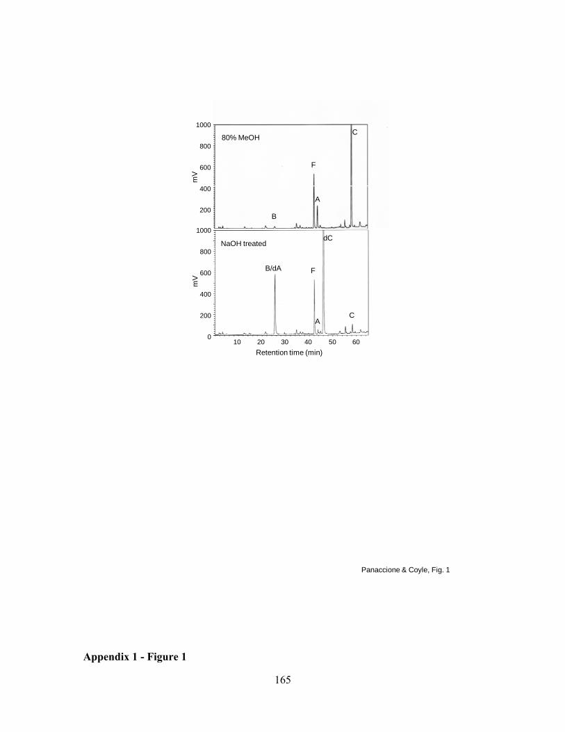
165
80% MeOH
NaOH treated
B
B/dA F
AC
F
A
C
dC
Panaccione & Coyle, Fig. 1
1000
800
600
400
200
0
1000
800
600
400
200
mV
mV
10 20 30 40 50 60Retention time (min)
Appendix 1 - Figure 1
166
APPENDIX 2: Analysis of ergot alkaloids in spore only samples versus entire fungal (plate) cultures in the fungus, Aspergillus fumigatus
INTRODUCTION
Cultures of Aspergillus fumigatus consist of spores, conidiophores (asexual sporulation
structures containing a stalked vesicle and a layer of phialides), and mycelia (vegetative
growth). Ergot alkaloids accumulate in high concentrations in or on spores of A.
fumigatus, and production of ergot alkaloids is restricted to sporulating cultures of the
fungus [(1, 3); Ch. 4, appendix 1]. Preliminary work indicated that extracts from spores
contained a different ratio of the four major A. fumigatus ergot alkaloids than extracts
made from plate cultures, containing all four cell types. This observation raises the
possibility that ergot alkaloids may be produced in cell types other than spores or in
addition to spores. The location of the production of alkaloids and the possibility of
accumulation in other cell types has not yet been determined. The purpose of this
experiment was to compare ergot alkaloid accumulation in spore only extracts to extracts
from plate cultures to gain insight into whether these alkaloids are present in other cell
types of sporulating cultures.
MATERIALS AND METHODS
Ten plates of A. fumigatus isolate WVU1943 were cultured on potato dextrose agar
(PDA) for two weeks at 37 ºC as previously described (3). All manipulations were
conducted in a Class II safety cabinet. Agar plugs containing 50.3 mm2 of colony
surface area from each of the ten A. fumigatus plate cultures were extracted in 1 mL of
80% methanol as described by Coyle et al. (1). To analyze spore only samples, plates
were inverted, gently agitated and the abscised conidia that had collected on the inner
167
surface of the lid of each plate (n=10) were suspended in 1 mL of 80% methanol and
extracted as above. Aliquots were removed for ergot alkaloid analysis and enumeration
of conidia for each sample.
Ergot alkaloids present in each sample were identified and quantified by reverse-
phase HPLC analysis with fluorescent detection. Peak identities were established based
on work done by Panaccione and Coyle (3) together with elimination of those peaks by
disruption of dmaW, the determinant step in the ergot alkaloid pathway [(2); Chapter 1].
Quantification of ergot alkaloids was based on comparison of peak areas relative to an
external standard as previously described (3). Conidia present in dilutions of the initial
methanol extracts were counted on a hemacytometer and visualized by light microscopy.
Proportions of each individual ergot alkaloid were determined relative to total alkaloids
in the sample. Comparison of conidia-only extracts versus entire culture extracts was
done by application of the Wilcoxon / Kruskal-Wallis (rank sum) test present in the JMP
software package (SAS, Cary, NC). This nonparametric test was chosen due to the
unequal variance within some data sets.
RESULTS
Samples from the entire culture (and thus containing all four cell types) produced on
average 3.26 fg of ergot alkaloids per conidium, whereas samples made up exclusively of
mature, abscised conidia contained an average of 1.21 fg of ergot alkaloids per conidium.
The difference in accumulation of total ergot alkaloids per spore is statistically significant
(p=0.002)
The proportion of festuclavine relative to total alkaloids extracted was
significantly greater in plate extracts versus mature spore extracts (p=0.0002) (Fig.1).
168
The converse was true for the other three alkaloids, fumigaclavines B (p=0.0002), A
(p=0.0025) and C (p=0.0002) (Fig.1).
DISCUSSION
It has been shown previously that ergot alkaloids accumulate in high concentrations in or
on spores of A. fumigatus (3) and that production of ergot alkaloids is restricted to
sporulating cultures (1). The increase in total clavines per spore within samples
comprised of all cell types versus only mature spores suggests that ergot alkaloids are
also associated with other cell types within conidiating cultures. The increase in
percentage of festuclavine and the decrease in proportion of downstream fumigaclavines
within these multicell-type culture extracts relative to mature spore extracts suggest that
festuclavine may be produced prior to spore formation and that the final stages in
fumigaclavine production occur preferentially in spores. Whether there is discrete
compartmentalization of these activities or simply an enrichment of enzymes for
particular steps in different cell types has not been determined.
169
REFERENCES
1. Coyle, C. M., S. C. Kenaley, W. R. Rittenour, and D. G. Panaccione. 2007.
Association of ergot alkaloids with conidiation in Aspergillus fumigatus.
Mycologia 99:804–811.
2. Coyle, C. M., and D. G. Panaccione. 2005. An ergot alkaloid biosynthesis gene
and clustered hypothetical genes from Aspergillus fumigatus. Appl. Environ.
Microbiol. 71:3112–3118.
3. Panaccione, D. G., and C. M. Coyle. 2005. Abundant respirable ergot alkaloids
from the common airborne fungus Aspergillus fumigatus. Appl. Environ.
Microbiol. 71:3106–3111.
170
FIGURE LEGEND
Figure 1: Proportion of each individual ergot alkaloid relative to total alkaloids
extracted. A significant difference was found for each alkaloid using the nonparametric
Wilcoxon / Kruskal-Wallis rank sum test. Error bars represent standard error. The insert
represents a magnified view of proportions of fumigaclavine A and B.
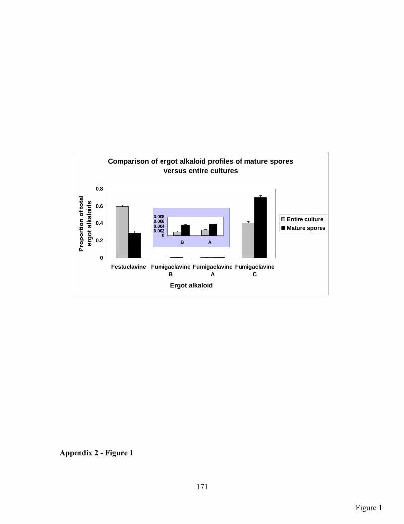
171
Comparison of ergot alkaloid profiles of mature spores versus entire cultures
0
0.2
0.4
0.6
0.8
Festuclavine FumigaclavineB
FumigaclavineA
FumigaclavineC
Ergot alkaloid
Prop
ortio
n of
tota
l er
got a
lkal
oids
Entire cultureMature spores
00.0020.0040.0060.008
B A
Appendix 2 - Figure 1
Figure 1
172
CURRICULUM VITAE
CHRISTINE M. COYLE
50 Fitzwilliams Road, Washington, PA 15301 | 724‐229‐5089 | [email protected]
EDUCATION
West Virginia University, Morgantown, WV Ph.D. in Genetics and Developmental Biology – Spring 2009 [Expected] Dissertation: “Characterization of the Ergot Alkaloid Biosynthesis Pathway in the
Pathogenic Fungus Aspergillus fumigatus”
University of Maryland Baltimore County, Baltimore, MD B.S. in Biochemistry and Molecular Biology ‐ 2003 Honors: Cum Laude
TEACHING EXPERIENCE
West Virginia University, Morgantown, WV Guest Lectures – “General Microbiology” [AEM 341] ‐2007, 2008 Topic: Microbial Genetics and Microbial Metabolism
Guest Lecture – “The Human Environment” [BIOL 115] ‐ 2007 Topic: Genetically Modified Foods
Teaching Assistant – “Physiology of Fungi” [PPTH 730] ‐ 2007 Prepared lab, instructed laboratory sessions, and met with students upon request Teaching Assistant – “Principles of Biology” [BIOL 115] ‐ 2003 Lectured/instructed laboratory sessions, met with students upon request, and graded all
written work Genetics Seminar – Prepared and presented 7 seminars – 2004‐2008 Journal Club – Discussion of refereed publications regarding current concepts in
environmental microbiology – 8 presentations – 2004‐2008
RESEARCH EXPERIENCE
West Virginia University, Morgantown, WV Research Assistant ‐ 2004 ‐ present Chemical analysis by high performance liquid chromatography (HPLC), multiple molecular
analyses (e.g., PCR, Southern Blot, and cloning) microbiological applications (e.g.,
173
bacterial and fungal transformations), and bioinformatics. Other responsibilities include ordering laboratory supplies, monitoring and directing undergraduate students in the laboratory.
University of Maryland Baltimore County, Baltimore, MD Laboratory Technician ‐ 2003 Prepared materials and solutions for biochemistry laboratory sessions University of Maryland Baltimore County, Baltimore, MD Undergraduate Research Assistant ‐ 2002 ‐ 2003 Focused on incorporating a modified nucleotide into an RNA product corresponding to the
third stem‐loop of HIV‐1 for analysis by mass spectrometry
PUBLICATIONS
REFEREED JOURNAL ARTICLES
Coyle, C. M., S. C. Kenaley, W. R. Rittenour, and D. G. Panaccione. 2007. Association of ergot alkaloids with conidiation in Aspergillus fumigatus. Mycologia 99(6):804‐11. Coyle, C. M. and D. G. Panaccione. 2005. An ergot alkaloid biosynthesis gene and clustered hypothetical genes from Aspergillus fumigatus. Appl. Environ. Microbiol. 71:3112‐3118. Panaccione, D. G. and C. M. Coyle. 2005. Abundant respirable ergot alkaloids from the common airborne fungus Aspergillus fumigatus. Appl. Environ. Microbiol. 71:3106–3111.
BOOK CHAPTERS
Panaccione, D. G., C. L.. Schardl, and C. M. Coyle. 2006. Pathways to diverse ergot alkaloid profiles in fungi, p. 23‐52. In, J. T. Romeo (ed.), Recent Advances in Phytochemistry, vol. 40, Elsevier, Amsterdam.
ABSTRACTS
Coyle, C. M., K. E. Goetz, and D. G. Panaccione. 2009. Clustered genes common to Aspergillus fumigatus and ergot fungi control early steps in ergot alkaloid biosynthesis. Fungal Genet. Reports 56(Suppl):205. (Abstract) Coyle, C. M., K. E. Goetz, and D. G. Panaccione. 2008. Clustered genes common to both Aspergillus fumigatus and ergot fungi control early steps within the ergot alkaloid pathway. Phytopathology 98:S214. (Abstract)
174
PRESENTATIONS
Poster Presentation ‐ Clustered genes common to both Aspergillus fumigatus and ergot fungi control early steps within the ergot alkaloid pathway Fungal Genetics Conference, Asilomar, CA (2009) Oral Presentation ‐ Clustered genes common to both Aspergillus fumigatus and ergot fungi control early steps within the ergot alkaloid pathway Davis College of Agriculture and Consumer Sciences, WVU, Morgantown, WV (2008) American Phytopathological Society, Potomac Division, Morgantown, WV (2008) Poster Session ‐ Association of Ergot Alkaloids with Sporulation in Aspergillus fumigatus American Phytopathological Society, Potomac Division, Morgantown, WV (2008) Sigma Xi, WVU, Morgantown, WV (2006) Davis College of Agriculture and Consumer Sciences, WVU, Morgantown, WV (2006)
Poster Session ‐ The Ergot Alkaloid Biosynthesis Gene Cluster from Aspergillus fumigatus Sigma Xi, WVU, Morgantown, WV (2005) Davis College of Agriculture and Consumer Sciences, WVU, Morgantown, WV (2005)
CONTRIBUTION TO OTHER PRESENTATIONS
Panaccione, D.G., C.M. Coyle, E. Konnova, D.A. Potter, J.T. Stokes, J.R. Cipoletti, J.B. Kotcon, K.E. Goetz, C. Machado, and C.L. Schardl. 2007. Toxin biosynthesis genes in ergot alkaloid‐producing fungi (poster presentation). USDA sponsored meeting “From Genes to Products,” Bethesda, MD (2007) American Phytopathological Society, Potomac Division, Morgantown, WV (2008)
FACILITATION OF STUDENT RESEARCH/LABORATORY EXPERIENCE
Graduate Students Shaun Stanley – M. S. Agriculture Kelly Mayhew – M. S. Applied Environmental Microbiology Undergraduate Students Dawn Chamberlain ‐ Biochemistry Michael Hamstead ‐ Biology Michael Chaisson ‐ Biology Kevin Junkins ‐ Biology Ashley Reynolds ‐ Psychology Brian Lewis ‐ Biochemistry
AWARDS
175
Honorable Mention: Student Paper Presentation, American Phytopathology Society, Potomac
Division, Morgantown, WV (2008)
SERVICE
Local Arrangements Committee for American Phytopathology Society, Potomac Division Meeting (2008)
Division of Plant and Soil Science (Plant Pathology and Environmental Microbiology) Building
Safety Committee (2005‐present)
MEMBERSHIPS
American Phytopathological Society, Potomac Division Gamma Sigma Delta