equine herpesvirus type 1 (ehv-1) utilizes microtubules, dynein, and rock1 to productively infect...
TRANSCRIPT
Equine herpesvirus type 1 (EHV-1) utilizes microtubules, dynein,and ROCK1 to productively infect cells.
Arthur R. Frampton Jr.1,*,†, Hiroaki Uchida1, Jens von Einem2, William F. Goins1, PaolaGrandi3, Justus B. Cohen1, Nikolaus Osterrieder2,4, and Joseph C. Glorioso11Department of Microbiology and Molecular Genetics, University of Pittsburgh, School of Medicine,Pittsburgh, PA 152612Department of Microbiology and Immunology, College of Veterinary Medicine, Cornell University,Ithaca, NY 148533Department of Neurological Surgery, University of Pittsburgh, School of Medicine, Pittsburgh, PA,152614Institut für Virologie, Freie Universität Berlin, 10115 Berlin Germany
AbstractTo initiate infection, equine herpesvirus type 1 (EHV-1) attaches to heparan sulfate on cell surfacesand then interacts with a putative glycoprotein D receptor(s). After attachment, virus entry occurseither by direct fusion of the virus envelope with the plasma membrane or via endocytosis followedby fusion between the virus envelope and an endosomal membrane. Upon fusion, de-enveloped virusparticles are deposited into the cytoplasm and travel to the nucleus for viral replication. In this report,we examined the mechanism of EHV-1 intracellular trafficking and investigated the ability of EHV-1to utilize specific cellular components to efficiently travel to the nucleus post-entry. Using a panelof microtubule depolymerizing drugs and inhibitors of microtubule motor proteins, we show thatEHV-1 infection is dependent on both the integrity of the microtubule network and the minus-endmicrotubule motor protein, dynein. In addition, we show that EHV-1 actively induces the acetylationof tubulin, a marker of microtubule stabilization, as early as 15 minutes post-infection. Finally, ourdata support a role for the cellular kinase, ROCK1, in virus trafficking to the nucleus.
KeywordsEHV-1; trafficking; microtubules; dynein; ROCK1
1. IntroductionEquine herpesvirus 1 (EHV-1) is a major pathogen of horses. Most horses are exposed to thevirus within 6 months to a year after birth and are frequently re-infected throughout theirlifetime (Allen, 1986). Clinical signs that appear early after infection include fever,
© 2009 Elsevier B.V. All rights reserved.*To whom correspondence should be addressed.†Current affiliation: Arthur R. Frampton Jr. Ph.D Assistant Professor University of North Carolina Wilmington Department of Biologyand Marine Biology Phone: (910) 962-2643 Fax: (910)962-4066 [email protected]'s Disclaimer: This is a PDF file of an unedited manuscript that has been accepted for publication. As a service to our customerswe are providing this early version of the manuscript. The manuscript will undergo copyediting, typesetting, and review of the resultingproof before it is published in its final citable form. Please note that during the production process errors may be discovered which couldaffect the content, and all legal disclaimers that apply to the journal pertain.
NIH Public AccessAuthor ManuscriptVet Microbiol. Author manuscript; available in PMC 2011 February 24.
Published in final edited form as:Vet Microbiol. 2010 February 24; 141(1-2): 12. doi:10.1016/j.vetmic.2009.07.035.
NIH
-PA Author Manuscript
NIH
-PA Author Manuscript
NIH
-PA Author Manuscript
inappattence, malaise, coughing, and mucopurulent discharge (O'Callaghan et al., 1983). Asthe infection progresses, horses may exhibit signs of serious neurological illness includingataxia, disorientation, and partial to full paralysis. In addition to the neurological sequelae,EHV-1 also causes abortigenic disease in pregnant mares.
At the cellular level, EHV-1 must engage host cellular proteins and induce specific signalingpathways very early in infection in order to successfully infect cells. EHV-1 initially binds totarget cells via an interaction between viral glycoproteins B and C and glycosaminoglycans onthe plasma membrane (Sugahara et al., 1997, Osterrieder, 1999). After attachment, a putativeentry receptor is recognized (Frampton et al., 2005) by glycoprotein D (Csellner et al., 2000).The next step in the EHV-1 entry pathway varies depending upon the cell type (Frampton etal., 2007). EHV-1 fuses with the plasma membrane and deposits de-enveloped particles intothe cytoplasm of equine dermal (ED) and rabbit kidney (RK-13) cells. On the contrary, fullyenveloped particles are endocytosed in CHO-K1 cells and naked capsids are then released intothe cytosol after the virus fuses with the endosomal membrane. Regardless of the initial modeof entry, we recently showed that the cellular kinase Rho associated coiled-coil kinase 1(ROCK1) must be induced for the virus to complete the infection process (Frampton et al.,2007).
Once inside the cells, EHV-1 must traverse the intricate meshwork of the intracellularcytoskeleton in order to reach the nucleus and deposit the viral genome. The mechanismsemployed by EHV-1 to traffic to the nucleus have yet to be defined, but certain clues areprovided by studies with other viruses that utilize microtubules and their associated machineryfor intracellular trafficking. Microtubules are composed of α and β dimers of tubulin andcontain a structural polarity with a positive end that is oriented toward the periphery of the celland a negative end that is anchored at the microtubule organizing center (MTOC) adjacent tothe nucleus (Nogales, 2000). To reach the nucleus, many viruses attach themselves to, and aredirectionally propelled by dynein, a minus-end directed microtubule motor, towards the MTOC(Kelkar et al., 2004, Alonso et al., 2001, Suikkanen et al., 2003, Petit et al., 2003, Ye et al.,2000, Douglas et al., 2004, Jacob et al., 2000, Raux et al., 2000, Florin et al., 2006). Sodeik etal. initially showed that the alphaherpesvirus herpes simplex virus type 1 (HSV-1) usesmicrotubules to traffic to the nucleus. Their data revealed that nuclear transport of HSV-1 wassignificantly inhibited in the presence of the microtubule-depolymerizing drugs, nocodazole,vinblastine, or colchicine (Sodeik et al., 1997). The movement of HSV-1 capsids alongmicrotubules was also shown to require dynein as expression of the dynein inhibitor, dynamitin(Dohner et al., 2002), and specific inhibitors of dynein ATPase (Lee et al., 2006, Kobayashiet al., 1978, Kristensson et al., 1986, Penningroth et al., 1982) decreased HSV-1 transport tothe nucleus. The movement of virus particles was also shown to be independent of microtubuleturnover or treadmilling (microtubule polymerization/depolymerization) as the microtubule-stabilizing drug paclitaxel had no effect on nucleocapsid transport (Sodeik et al., 1997).
In addition to utilizing microtubules for transport, viruses also generate an intracellularenvironment that is conducive for nuclear transport by triggering various signaling pathways.One common theme is the induction of signaling pathways that lead to the reorganization ofcytoskeletal components. Many viruses, including HSV-1, stimulate members of the RasGTPase superfamily and this induction can lead to a wide array of morphological changes ofinfected cells, including, but not limited to, stabilization of microtubules (Naranatt et al.,2005), formation of focal adhesions (Cheshenko et al., 2005) and endocytosis or phagocytosis(Clement et al., 2006).
In this study, we show that EHV-1 causes the stabilization of microtubules via the acetylationof tubulin and utilizes the minus-end directed microtubule motor dynein to traffic to the nucleus
Frampton et al. Page 2
Vet Microbiol. Author manuscript; available in PMC 2011 February 24.
NIH
-PA Author Manuscript
NIH
-PA Author Manuscript
NIH
-PA Author Manuscript
along stabilized microtubules. In addition, we show that nuclear accumulation of capsids isdecreased in the presence of the ROCK1 inhibitor, Y-27632.
2. Materials and methods2.1. Cells, viruses, and plasmids
RK-13 cells were grown in Dulbecco's modified Eagle medium (DMEM) supplemented with10% fetal bovine serum (FBS) (Invitrogen, Carlsbad, CA). Chinese hamster ovary (CHO-K1)cells were kindly provided by Dr. Patricia Spear (Northwestern University, Chicago, IL) andgrown in F12-K media (Invitrogen) supplemented with 10% FBS. Equine dermal cells (ED),a gift from Dr. Ron Montelaro (University of Pittsburgh, Pittsburgh, PA), were maintained inminimal essential medium (MEM) supplemented with 10% FBS. All cells were maintained at37°C in 5% CO2.
The EHV-1 recombinant virus L11ΔgIΔgE contains a lacZ reporter cassette in place of gI andgE (Frampton et al., 2002). The EHV-1 recombinant virus L11VP26mRed was describedpreviously (J. von Einem et al., 24th annual ASV meeting 2005). This recombinant viruscontains an mRFP1 fluorescent protein fused to the EHV-1 VP26 small capsid protein.
2.2. Drug inhibition assaysFor entry assays, 2.5 × 10⁴ ED or CHO-K1 cells were seeded in a 96-well plate. The next day,cells were incubated with increasing amounts of the microtubule depolymerizing drugs,nocodazole, vinblastine, colchicine, the microtubule stabilizing drug, paclitaxel (PTX), thedynein inhibitor, EHNA (erythro-9-(2-hydroxy-3-nonyl) adenine, or the kinesin inhibitorAMP-PNP (adenosine 5′-(β, γ-imido) triphosphate tetralithium salt) (Sigma, St. Louis, MO)at 37°C for 30 min. Cells were infected with EHV-1 (L11ΔgIΔgE) at an MOI of 5 and incubatedfor 5.5 h in the presence of the drugs at 37°C. Cells were washed once with PBS and thenONPG was added to the cells and the absorbance read at 405nm. Triplicate samples weremeasured for each concentration of drug. A 1mM stock of paclitaxel was prepared in distilledH2O. One-hundred mM stock solutions of EHNA and nocodazole were prepared in DMSO.A 100 mM stock solution of AMP-PNP was prepared in H2O. 10 mM stocks of the ROCK1inhibitor, Y-27632 (EMD Biosciences, La Jolla, CA) were made in H2O. 10mM stocks ofvinblastine, and colchicine were also made in H2O. Cell viability was measured in triplicateusing the CellTiter 96 aqueous non-radioactive cell proliferation assay (Promega, Madison,WI) following the manufacturer's protocol.
2.3. Western Blot AnalysesConfluent ED cells in a 12-well plate were serum starved for 1 h and cells were then mock-treated or treated with paclitaxel (10 μM) or Y-27632 (100 μM). Cells treated with paclitaxelwere washed with MEM at 15, 30, and 60 min and cell lysates were harvested with PARP lysisbuffer (6 M urea, 2% SDS, 10% glycerol, 62.5 mM Tris-HCl, pH 6.8, and 5% β-mercaptoethanol). Mock-treated cells or cells treated with Y-27632 were infected with EHV-1(L11VP26mRed) at an MOI of 10 in the presence or absence of Y-27632. At 15, 30, and 60min post-infection, cells were washed once with MEM and lysates were harvested with PARPlysis buffer. Samples were run on a 10% SDS-PAGE gel and transferred to an immobilon-Pmembrane (Millipore, Billerica, MA). The membranes were blocked for 1 h in 10% non-fatdry milk (NFDM) at room temperature (RT) before monoclonal anti-acetylated tubulinantibody 6-11B-1 (Sigma) was added at a concentration of 1:2000 in TBST with 5% NFDMfor 16 h at 4°C. The membranes were washed 3x for 10 min/wash and then anti-mouse-HRPantibody SC-2031 (Santa Cruz Biotechnology, Santa Cruz, CA) was added at a concentrationof 1:3,000 in TBST with 5% NFDM for 1 h at RT. Membranes were washed 3x for 10 min/wash then ECL detection reagent (Amersham Biosciences, Buckinghamshire, England) was
Frampton et al. Page 3
Vet Microbiol. Author manuscript; available in PMC 2011 February 24.
NIH
-PA Author Manuscript
NIH
-PA Author Manuscript
NIH
-PA Author Manuscript
added and the membranes exposed to film. After stripping the membrane with stripping buffer(10% SDS, 0.5M Tris pH 6.8, 0.5% β-mercaptoethanol) for 30 min at 50°C, the membraneswere washed 2x for 10 min/wash with TBST and then blocked for 1 h with 10% NFDM. Totaltubulin was detected by adding monoclonal anti-α-tubulin antibody B-5-1-2 (Sigma) at 1:2,000for 16 h at 4°C with the secondary antibody and detection steps as described earlier.
2.4. Infectious virus recovery assay4 × 105 cells in 24-well plates were incubated with 100 μM of Y-27632 in DMEM or DMEMalone for 30 min at 37°C. Plates were placed on ice for 5 min and then EHV-1 (MOI = 10) inDMEM or DMEM containing 100 μM Y-27632 was incubated on the cells at 4°C for 3 h. Cellswere washed once with cold DMEM and then warm media (37°C) was added to the cells. Ateach time-point, cells were washed with glycine (pH 3.0) for 30 s, washed once with DMEM,and harvested. Virus samples were freeze-thawed once then sonicated 3x for 15 s. Virusharvested from each time-point was titered on RK13 cells. Triplicate samples were measuredfor each time-point.
2.5. Confocal microscopy of EHV-1 infected cellsCHO-K1 cells were seeded to confluency in collagen-coated 35 mm dishes (MatTekCorporation, Ashland, MA). Cells were mock-treated or treated with 100 μM Y-27632 or 10μM paclitaxel for 30 min at 37°C. Cells were chilled on ice for 10 min and then L11VP26mRedwas added to the cells at an MOI of 10. Virus was incubated on the cells at 4°C for 2 h in thepresence or absence of drug. After 2 h, cells for the 0 time-point were fixed with 4%paraformaldehyde (PFA) at 4°C. Media pre-warmed to 37°C, with or without drug, was addedto the remaining cells and then the cells were fixed with 4% PFA at 15, 30, 60, and 120 minpost temperature shift. After 30 min of fixation with 4% PFA, cells were rehydrated with PBSfor 15 min at 4°C. Cells were washed once more with PBS for 15 min and then cells wereexamined using an inverted Olympus Fluo-View 1000 confocal microscope (OlympusAmerica Inc., Center Valley, PA). Prior to imaging, cells were stained with Hoechst dye (1μg/mL) and wheat germ agglutinin (5 μg/mL) to label the nucleus and plasma membrane,respectively. All images were captured at a 60x magnification.
3. Results3.1. EHV-1 utilizes microtubules for infection
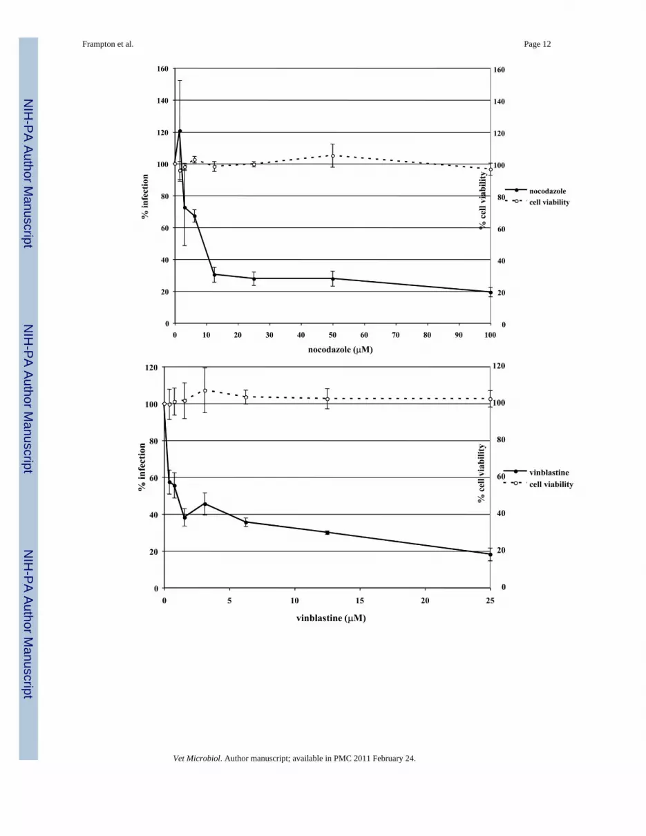
To determine if EHV-1 requires microtubules for infection, a virus entry assay was performedin the presence or absence of microtubule depolymerizing agents. Equine dermal (ED) cellswere untreated or treated with increasing concentrations of nocodazole, vinblastine, orcolchicine for 30 min prior to infection and the drugs were kept on the cells throughout theexperiment. Cells were infected with L11ΔgIΔgE, an EHV-1 recombinant virus containing alacZ reporter gene, at an MOI of 5 for 5.5 hours. EHV-1 infection, as measured by β-galactosidase expression, was significantly inhibited in a dose-dependent manner by all of thedrugs. Nocodazole inhibited infection by 80% at the highest concentration tested (Figure 1A)and vinblastine (Figure 1B) and colchicine (Figure 1C) inhibited infection by 82% and 56%,respectively. This inhibition of infection was not the result of drug-associated cell toxicity ascell viability was similar for all of the concentrations used. In addition to inhibiting infectionon ED cells, these drugs also inhibited infection on CHO-K1 cells (data not shown). Theseresults indicate that an intact microtubule network is required regardless of the mode of EHV-1entry.
To address the importance of microtubule dynamics or treadmilling (polymerization/depolymerization of microtubules) in virus transport, microtubules were first stabilized withpaclitaxel or left untreated before and during infection with EHV-1. Virus entry was assessed
Frampton et al. Page 4
Vet Microbiol. Author manuscript; available in PMC 2011 February 24.
NIH
-PA Author Manuscript
NIH
-PA Author Manuscript
NIH
-PA Author Manuscript
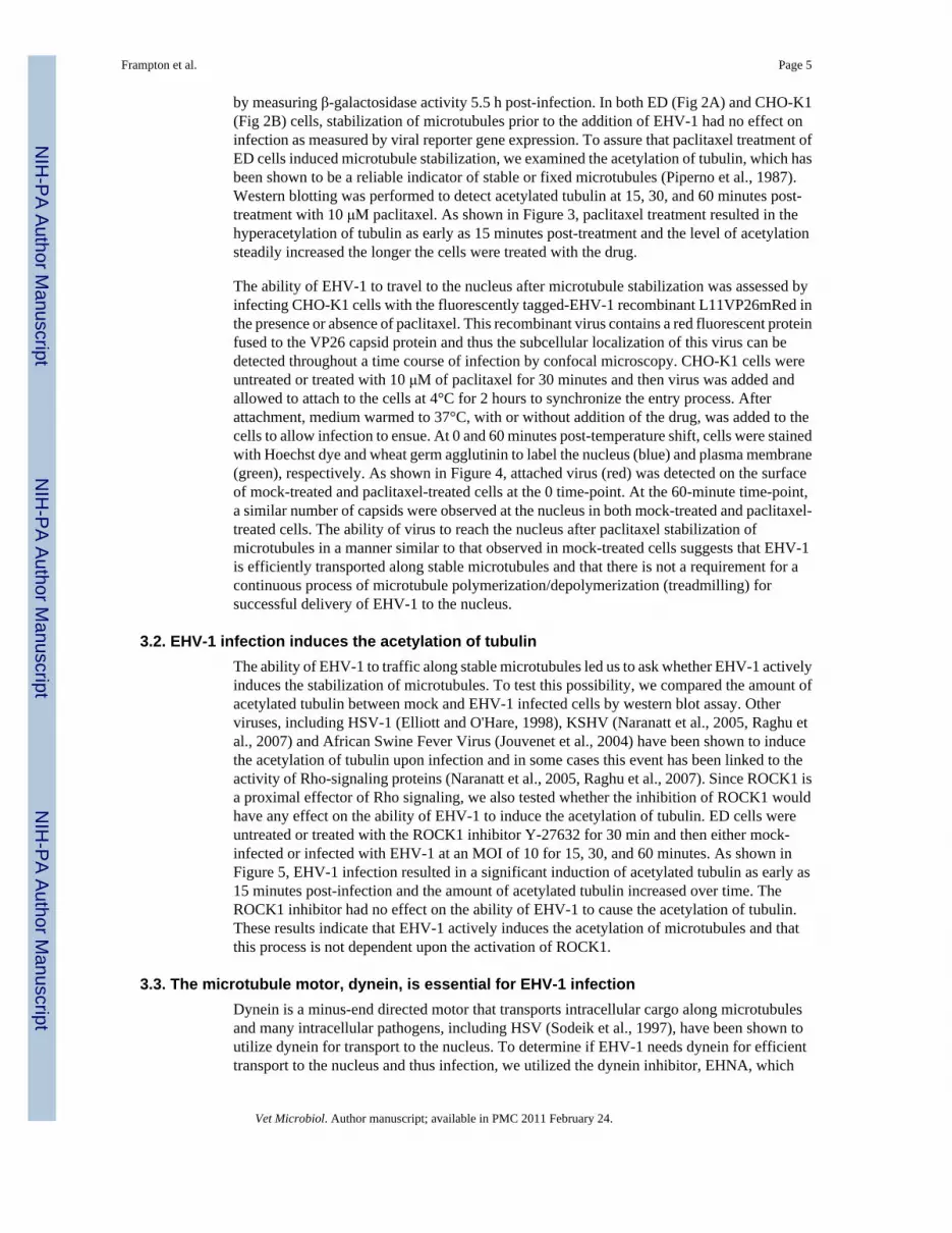
by measuring β-galactosidase activity 5.5 h post-infection. In both ED (Fig 2A) and CHO-K1(Fig 2B) cells, stabilization of microtubules prior to the addition of EHV-1 had no effect oninfection as measured by viral reporter gene expression. To assure that paclitaxel treatment ofED cells induced microtubule stabilization, we examined the acetylation of tubulin, which hasbeen shown to be a reliable indicator of stable or fixed microtubules (Piperno et al., 1987).Western blotting was performed to detect acetylated tubulin at 15, 30, and 60 minutes post-treatment with 10 μM paclitaxel. As shown in Figure 3, paclitaxel treatment resulted in thehyperacetylation of tubulin as early as 15 minutes post-treatment and the level of acetylationsteadily increased the longer the cells were treated with the drug.
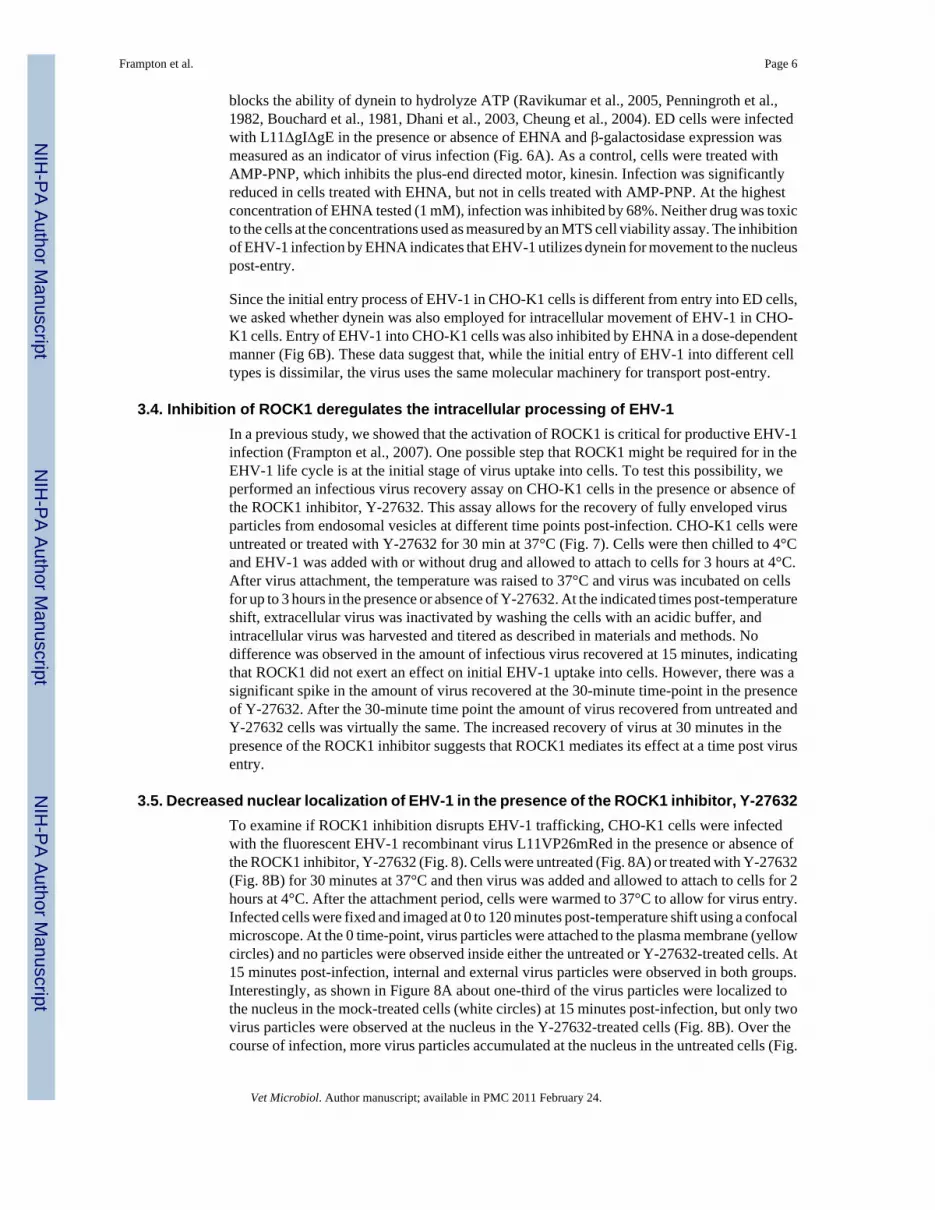
The ability of EHV-1 to travel to the nucleus after microtubule stabilization was assessed byinfecting CHO-K1 cells with the fluorescently tagged-EHV-1 recombinant L11VP26mRed inthe presence or absence of paclitaxel. This recombinant virus contains a red fluorescent proteinfused to the VP26 capsid protein and thus the subcellular localization of this virus can bedetected throughout a time course of infection by confocal microscopy. CHO-K1 cells wereuntreated or treated with 10 μM of paclitaxel for 30 minutes and then virus was added andallowed to attach to the cells at 4°C for 2 hours to synchronize the entry process. Afterattachment, medium warmed to 37°C, with or without addition of the drug, was added to thecells to allow infection to ensue. At 0 and 60 minutes post-temperature shift, cells were stainedwith Hoechst dye and wheat germ agglutinin to label the nucleus (blue) and plasma membrane(green), respectively. As shown in Figure 4, attached virus (red) was detected on the surfaceof mock-treated and paclitaxel-treated cells at the 0 time-point. At the 60-minute time-point,a similar number of capsids were observed at the nucleus in both mock-treated and paclitaxel-treated cells. The ability of virus to reach the nucleus after paclitaxel stabilization ofmicrotubules in a manner similar to that observed in mock-treated cells suggests that EHV-1is efficiently transported along stable microtubules and that there is not a requirement for acontinuous process of microtubule polymerization/depolymerization (treadmilling) forsuccessful delivery of EHV-1 to the nucleus.
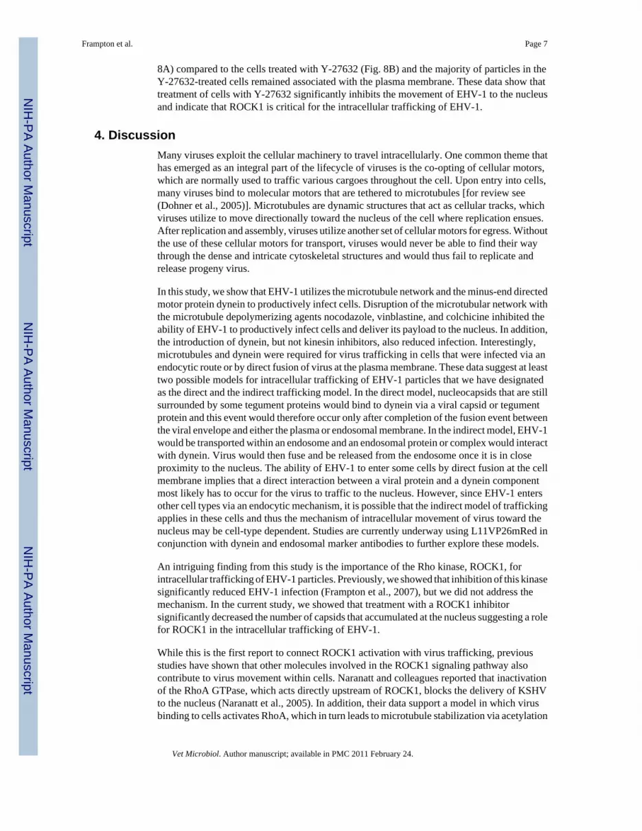
3.2. EHV-1 infection induces the acetylation of tubulinThe ability of EHV-1 to traffic along stable microtubules led us to ask whether EHV-1 activelyinduces the stabilization of microtubules. To test this possibility, we compared the amount ofacetylated tubulin between mock and EHV-1 infected cells by western blot assay. Otherviruses, including HSV-1 (Elliott and O'Hare, 1998), KSHV (Naranatt et al., 2005, Raghu etal., 2007) and African Swine Fever Virus (Jouvenet et al., 2004) have been shown to inducethe acetylation of tubulin upon infection and in some cases this event has been linked to theactivity of Rho-signaling proteins (Naranatt et al., 2005, Raghu et al., 2007). Since ROCK1 isa proximal effector of Rho signaling, we also tested whether the inhibition of ROCK1 wouldhave any effect on the ability of EHV-1 to induce the acetylation of tubulin. ED cells wereuntreated or treated with the ROCK1 inhibitor Y-27632 for 30 min and then either mock-infected or infected with EHV-1 at an MOI of 10 for 15, 30, and 60 minutes. As shown inFigure 5, EHV-1 infection resulted in a significant induction of acetylated tubulin as early as15 minutes post-infection and the amount of acetylated tubulin increased over time. TheROCK1 inhibitor had no effect on the ability of EHV-1 to cause the acetylation of tubulin.These results indicate that EHV-1 actively induces the acetylation of microtubules and thatthis process is not dependent upon the activation of ROCK1.
3.3. The microtubule motor, dynein, is essential for EHV-1 infectionDynein is a minus-end directed motor that transports intracellular cargo along microtubulesand many intracellular pathogens, including HSV (Sodeik et al., 1997), have been shown toutilize dynein for transport to the nucleus. To determine if EHV-1 needs dynein for efficienttransport to the nucleus and thus infection, we utilized the dynein inhibitor, EHNA, which
Frampton et al. Page 5
Vet Microbiol. Author manuscript; available in PMC 2011 February 24.
NIH
-PA Author Manuscript
NIH
-PA Author Manuscript
NIH
-PA Author Manuscript
blocks the ability of dynein to hydrolyze ATP (Ravikumar et al., 2005, Penningroth et al.,1982, Bouchard et al., 1981, Dhani et al., 2003, Cheung et al., 2004). ED cells were infectedwith L11ΔgIΔgE in the presence or absence of EHNA and β-galactosidase expression wasmeasured as an indicator of virus infection (Fig. 6A). As a control, cells were treated withAMP-PNP, which inhibits the plus-end directed motor, kinesin. Infection was significantlyreduced in cells treated with EHNA, but not in cells treated with AMP-PNP. At the highestconcentration of EHNA tested (1 mM), infection was inhibited by 68%. Neither drug was toxicto the cells at the concentrations used as measured by an MTS cell viability assay. The inhibitionof EHV-1 infection by EHNA indicates that EHV-1 utilizes dynein for movement to the nucleuspost-entry.
Since the initial entry process of EHV-1 in CHO-K1 cells is different from entry into ED cells,we asked whether dynein was also employed for intracellular movement of EHV-1 in CHO-K1 cells. Entry of EHV-1 into CHO-K1 cells was also inhibited by EHNA in a dose-dependentmanner (Fig 6B). These data suggest that, while the initial entry of EHV-1 into different celltypes is dissimilar, the virus uses the same molecular machinery for transport post-entry.
3.4. Inhibition of ROCK1 deregulates the intracellular processing of EHV-1In a previous study, we showed that the activation of ROCK1 is critical for productive EHV-1infection (Frampton et al., 2007). One possible step that ROCK1 might be required for in theEHV-1 life cycle is at the initial stage of virus uptake into cells. To test this possibility, weperformed an infectious virus recovery assay on CHO-K1 cells in the presence or absence ofthe ROCK1 inhibitor, Y-27632. This assay allows for the recovery of fully enveloped virusparticles from endosomal vesicles at different time points post-infection. CHO-K1 cells wereuntreated or treated with Y-27632 for 30 min at 37°C (Fig. 7). Cells were then chilled to 4°Cand EHV-1 was added with or without drug and allowed to attach to cells for 3 hours at 4°C.After virus attachment, the temperature was raised to 37°C and virus was incubated on cellsfor up to 3 hours in the presence or absence of Y-27632. At the indicated times post-temperatureshift, extracellular virus was inactivated by washing the cells with an acidic buffer, andintracellular virus was harvested and titered as described in materials and methods. Nodifference was observed in the amount of infectious virus recovered at 15 minutes, indicatingthat ROCK1 did not exert an effect on initial EHV-1 uptake into cells. However, there was asignificant spike in the amount of virus recovered at the 30-minute time-point in the presenceof Y-27632. After the 30-minute time point the amount of virus recovered from untreated andY-27632 cells was virtually the same. The increased recovery of virus at 30 minutes in thepresence of the ROCK1 inhibitor suggests that ROCK1 mediates its effect at a time post virusentry.
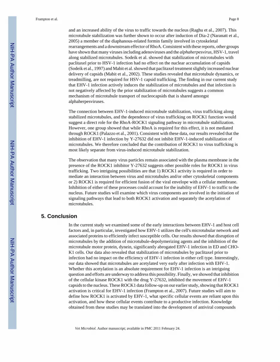
3.5. Decreased nuclear localization of EHV-1 in the presence of the ROCK1 inhibitor, Y-27632To examine if ROCK1 inhibition disrupts EHV-1 trafficking, CHO-K1 cells were infectedwith the fluorescent EHV-1 recombinant virus L11VP26mRed in the presence or absence ofthe ROCK1 inhibitor, Y-27632 (Fig. 8). Cells were untreated (Fig. 8A) or treated with Y-27632(Fig. 8B) for 30 minutes at 37°C and then virus was added and allowed to attach to cells for 2hours at 4°C. After the attachment period, cells were warmed to 37°C to allow for virus entry.Infected cells were fixed and imaged at 0 to 120 minutes post-temperature shift using a confocalmicroscope. At the 0 time-point, virus particles were attached to the plasma membrane (yellowcircles) and no particles were observed inside either the untreated or Y-27632-treated cells. At15 minutes post-infection, internal and external virus particles were observed in both groups.Interestingly, as shown in Figure 8A about one-third of the virus particles were localized tothe nucleus in the mock-treated cells (white circles) at 15 minutes post-infection, but only twovirus particles were observed at the nucleus in the Y-27632-treated cells (Fig. 8B). Over thecourse of infection, more virus particles accumulated at the nucleus in the untreated cells (Fig.
Frampton et al. Page 6
Vet Microbiol. Author manuscript; available in PMC 2011 February 24.
NIH
-PA Author Manuscript
NIH
-PA Author Manuscript
NIH
-PA Author Manuscript
8A) compared to the cells treated with Y-27632 (Fig. 8B) and the majority of particles in theY-27632-treated cells remained associated with the plasma membrane. These data show thattreatment of cells with Y-27632 significantly inhibits the movement of EHV-1 to the nucleusand indicate that ROCK1 is critical for the intracellular trafficking of EHV-1.
4. DiscussionMany viruses exploit the cellular machinery to travel intracellularly. One common theme thathas emerged as an integral part of the lifecycle of viruses is the co-opting of cellular motors,which are normally used to traffic various cargoes throughout the cell. Upon entry into cells,many viruses bind to molecular motors that are tethered to microtubules [for review see(Dohner et al., 2005)]. Microtubules are dynamic structures that act as cellular tracks, whichviruses utilize to move directionally toward the nucleus of the cell where replication ensues.After replication and assembly, viruses utilize another set of cellular motors for egress. Withoutthe use of these cellular motors for transport, viruses would never be able to find their waythrough the dense and intricate cytoskeletal structures and would thus fail to replicate andrelease progeny virus.
In this study, we show that EHV-1 utilizes the microtubule network and the minus-end directedmotor protein dynein to productively infect cells. Disruption of the microtubular network withthe microtubule depolymerizing agents nocodazole, vinblastine, and colchicine inhibited theability of EHV-1 to productively infect cells and deliver its payload to the nucleus. In addition,the introduction of dynein, but not kinesin inhibitors, also reduced infection. Interestingly,microtubules and dynein were required for virus trafficking in cells that were infected via anendocytic route or by direct fusion of virus at the plasma membrane. These data suggest at leasttwo possible models for intracellular trafficking of EHV-1 particles that we have designatedas the direct and the indirect trafficking model. In the direct model, nucleocapsids that are stillsurrounded by some tegument proteins would bind to dynein via a viral capsid or tegumentprotein and this event would therefore occur only after completion of the fusion event betweenthe viral envelope and either the plasma or endosomal membrane. In the indirect model, EHV-1would be transported within an endosome and an endosomal protein or complex would interactwith dynein. Virus would then fuse and be released from the endosome once it is in closeproximity to the nucleus. The ability of EHV-1 to enter some cells by direct fusion at the cellmembrane implies that a direct interaction between a viral protein and a dynein componentmost likely has to occur for the virus to traffic to the nucleus. However, since EHV-1 entersother cell types via an endocytic mechanism, it is possible that the indirect model of traffickingapplies in these cells and thus the mechanism of intracellular movement of virus toward thenucleus may be cell-type dependent. Studies are currently underway using L11VP26mRed inconjunction with dynein and endosomal marker antibodies to further explore these models.
An intriguing finding from this study is the importance of the Rho kinase, ROCK1, forintracellular trafficking of EHV-1 particles. Previously, we showed that inhibition of this kinasesignificantly reduced EHV-1 infection (Frampton et al., 2007), but we did not address themechanism. In the current study, we showed that treatment with a ROCK1 inhibitorsignificantly decreased the number of capsids that accumulated at the nucleus suggesting a rolefor ROCK1 in the intracellular trafficking of EHV-1.
While this is the first report to connect ROCK1 activation with virus trafficking, previousstudies have shown that other molecules involved in the ROCK1 signaling pathway alsocontribute to virus movement within cells. Naranatt and colleagues reported that inactivationof the RhoA GTPase, which acts directly upstream of ROCK1, blocks the delivery of KSHVto the nucleus (Naranatt et al., 2005). In addition, their data support a model in which virusbinding to cells activates RhoA, which in turn leads to microtubule stabilization via acetylation
Frampton et al. Page 7
Vet Microbiol. Author manuscript; available in PMC 2011 February 24.
NIH
-PA Author Manuscript
NIH
-PA Author Manuscript
NIH
-PA Author Manuscript
and an increased ability of the virus to traffic towards the nucleus (Raghu et al., 2007). Thismicrotubule stabilization was further shown to occur after induction of Dia-2 (Naranatt et al.,2005) a member of the diaphanous-related formin family involved in cytoskeletalrearrangements and a downstream effector of RhoA. Consistent with these reports, other groupshave shown that many viruses including adenoviruses and the alphaherpesvirus, HSV-1, travelalong stabilized microtubules. Sodeik et al. showed that stabilization of microtubules withpaclitaxel prior to HSV-1 infection had no effect on the nuclear accumulation of capsids(Sodeik et al., 1997) and Mabit et al. showed that paclitaxel treatment slightly increased nucleardelivery of capsids (Mabit et al., 2002). These studies revealed that microtubule dynamics, ortreadmilling, are not required for HSV-1 capsid trafficking. The finding in our current studythat EHV-1 infection actively induces the stabilization of microtubules and that infection isnot negatively affected by the prior stabilization of microtubules suggests a commonmechanism of microtubule transport of nucleocapsids that is shared amongstalphaherpesviruses.
The connection between EHV-1-induced microtubule stabilization, virus trafficking alongstabilized microtubules, and the dependence of virus trafficking on ROCK1 function wouldsuggest a direct role for the RhoA-ROCK1 signaling pathway in microtubule stabilization.However, one group showed that while RhoA is required for this effect, it is not mediatedthrough ROCK1 (Palazzo et al., 2001). Consistent with these data, our results revealed that theinhibition of EHV-1 infection by Y-27632 did not inhibit EHV-1-induced stabilization ofmicrotubules. We therefore concluded that the contribution of ROCK1 to virus trafficking ismost likely separate from virus-induced microtubule stabilization.
The observation that many virus particles remain associated with the plasma membrane in thepresence of the ROCK1 inhibitor Y-27632 suggests other possible roles for ROCK1 in virustrafficking. Two intriguing possibilities are that 1) ROCK1 activity is required in order tomediate an interaction between virus and microtubules and/or other cytoskeletal componentsor 2) ROCK1 is required for efficient fusion of the viral envelope with a cellular membrane.Inhibition of either of these processes could account for the inability of EHV-1 to traffic to thenucleus. Future studies will examine which virus components are involved in the initiation ofsignaling pathways that lead to both ROCK1 activation and separately the acetylation ofmicrotubules.
5. ConclusionIn the current study we examined some of the early interactions between EHV-1 and host cellfactors and, in particular, investigated how EHV-1 utilizes the cell's microtubular network andassociated proteins to efficiently infect susceptible cells. Our results showed that disruption ofmicrotubules by the addition of microtubule-depolymerizing agents and the inhibition of themicrotubule motor protein, dynein, significantly abrogated EHV-1 infection in ED and CHO-K1 cells. Our data also revealed that stabilization of microtubules by paclitaxel prior toinfection had no impact on the efficiency of EHV-1 infection in either cell type. Interestingly,our data showed that microtubules are acetylated very early after infection with EHV-1.Whether this acetylation is an absolute requirement for EHV-1 infection is an intriguingquestion and efforts are underway to address this possibility. Finally, we showed that inhibitionof the cellular kinase ROCK1 with the drug Y-27632, inhibited the movement of EHV-1capsids to the nucleus. These ROCK1 data follow-up on our earlier study, showing that ROCK1activation is critical for EHV-1 infection (Frampton et al., 2007). Future studies will aim todefine how ROCK1 is activated by EHV-1, what specific cellular events are reliant upon thisactivation, and how these cellular events contribute to a productive infection. Knowledgeobtained from these studies may be translated into the development of antiviral compounds
Frampton et al. Page 8
Vet Microbiol. Author manuscript; available in PMC 2011 February 24.
NIH
-PA Author Manuscript
NIH
-PA Author Manuscript
NIH
-PA Author Manuscript
that can be used to inhibit the identified critical processes that EHV-1 needs to complete itspathogenic program within the horse.
AcknowledgmentsWe thank Patricia Spear for providing the CHO cell lines and Ron Montelaro for the ED cells. We thank SimonWatkins for the use of the confocal microscope. This work was supported by NIH grants CA119298, NS44323,NS040923, and HL066949.
ReferencesALLEN GP, BRYANS JT. Molecular epizootiology, pathogenesis, and prophylaxis of equine
herpesvirus-1 infections. Prog. Vet. Microbiol. Immunol 1986;2:78–144. [PubMed: 2856183]ALONSO C, MISKIN J, HERNAEZ B, FERNANDEZ-ZAPATERO P, SOTO L, CANTO C,
RODRIGUEZ-CRESPO I, DIXON L, ESCRIBANO JM. African swine fever virus protein p54interacts with the microtubular motor complex through direct binding to light-chain dynein. J Virol2001;75:9819–27. [PubMed: 11559815]
BOUCHARD P, PENNINGROTH SM, CHEUNG A, GAGNON C, BARDIN CW. erythro-9-[3-(2-Hydroxynonyl)]adenine is an inhibitor of sperm motility that blocks dynein ATPase and proteincarboxylmethylase activities. Proc Natl Acad Sci U S A 1981;78:1033–6. [PubMed: 6453342]
CHESHENKO N, LIU W, SATLIN LM, HEROLD BC. Focal adhesion kinase plays a pivotal role inherpes simplex virus entry. J Biol Chem 2005;280:31116–25. [PubMed: 15994312]
CHEUNG PY, ZHANG Y, LONG J, LIN S, ZHANG M, JIANG Y, WU Z. p150(Glued), Dynein, andmicrotubules are specifically required for activation of MKK3/6 and p38 MAPKs. J Biol Chem2004;279:45308–11. [PubMed: 15375157]
CLEMENT C, TIWARI V, SCANLAN PM, VALYI-NAGY T, YUE BY, SHUKLA D. A novel role forphagocytosis-like uptake in herpes simplex virus entry. J Cell Biol 2006;174:1009–21. [PubMed:17000878]
CSELLNER H, WALKER C, WELLINGTON JE, MCLURE LE, LOVE DN, WHALLEY JM. EHV-1glycoprotein D (EHV-1 gD) is required for virus entry and cell-cell fusion, and an EHV-1 gD deletionmutant induces a protective immune response in mice. Arch Virol 2000;145:2371–85. [PubMed:11205124]
DHANI SU, MOHAMMAD-PANAH R, AHMED N, ACKERLEY C, RAMJEESINGH M, BEAR CE.Evidence for a functional interaction between the ClC-2 chloride channel and the retrograde motordynein complex. J Biol Chem 2003;278:16262–70. [PubMed: 12601004]
DOHNER K, NAGEL CH, SODEIK B. Viral stop-and-go along microtubules: taking a ride with dyneinand kinesins. Trends Microbiol 2005;13:320–7. [PubMed: 15950476]
DOHNER K, WOLFSTEIN A, PRANK U, ECHEVERRI C, DUJARDIN D, VALLEE R, SODEIK B.Function of dynein and dynactin in herpes simplex virus capsid transport. Mol Biol Cell2002;13:2795–809. [PubMed: 12181347]
DOUGLAS MW, DIEFENBACH RJ, HOMA FL, MIRANDA-SAKSENA M, RIXON FJ, VITTONEV, BYTH K, CUNNINGHAM AL. Herpes simplex virus type 1 capsid protein VP26 interacts withdynein light chains RP3 and Tctex1 and plays a role in retrograde cellular transport. J Biol Chem2004;279:28522–30. [PubMed: 15117959]
ELLIOTT G, O'HARE P. Herpes simplex virus type 1 tegument protein VP22 induces the stabilizationand hyperacetylation of microtubules. J Virol 1998;72:6448–55. [PubMed: 9658087]
FLORIN L, BECKER KA, LAMBERT C, NOWAK T, SAPP C, STRAND D, STREECK RE, SAPPM. Identification of a dynein interacting domain in the papillomavirus minor capsid protein l2. JVirol 2006;80:6691–6. [PubMed: 16775357]
FRAMPTON AR JR. GOINS WF, COHEN JB, VON EINEM J, OSTERRIEDER N, O'CALLAGHANDJ, GLORIOSO JC. Equine herpesvirus 1 utilizes a novel herpesvirus entry receptor. J Virol2005;79:3169–73. [PubMed: 15709036]
FRAMPTON AR JR. SMITH PM, ZHANG Y, MATSUMURA T, OSTERRIEDER N, O'CALLAGHANDJ. Contribution of gene products encoded within the unique short segment of equine herpesvirus 1to virulence in a murine model. Virus Res 2002;90:287–301. [PubMed: 12457983]
Frampton et al. Page 9
Vet Microbiol. Author manuscript; available in PMC 2011 February 24.
NIH
-PA Author Manuscript
NIH
-PA Author Manuscript
NIH
-PA Author Manuscript
FRAMPTON AR JR. STOLZ DB, UCHIDA H, GOINS WF, COHEN JB, GLORIOSO JC. EquineHerpesvirus 1 Enters Cells by Two Different Pathways, and Infection Requires the Activation of theCellular Kinase ROCK1. J Virol 2007;81:10879–89. [PubMed: 17670830]
JACOB Y, BADRANE H, CECCALDI PE, TORDO N. Cytoplasmic dynein LC8 interacts withlyssavirus phosphoprotein. J Virol 2000;74:10217–22. [PubMed: 11024152]
JOUVENET N, MONAGHAN P, WAY M, WILEMAN T. Transport of African swine fever virus fromassembly sites to the plasma membrane is dependent on microtubules and conventional kinesin. JVirol 2004;78:7990–8001. [PubMed: 15254171]
KELKAR SA, PFISTER KK, CRYSTAL RG, LEOPOLD PL. Cytoplasmic dynein mediates adenovirusbinding to microtubules. J Virol 2004;78:10122–32. [PubMed: 15331745]
KOBAYASHI T, MARTENSEN T, NATH J, FLAVIN M. Inhibition of dynein ATPase by vanadate,and its possible use as a probe for the role of dynein in cytoplasmic motility. Biochem Biophys ResCommun 1978;81:1313–8. [PubMed: 149544]
KRISTENSSON K, LYCKE E, ROYTTA M, SVENNERHOLM B, VAHLNE A. Neuritic transport ofherpes simplex virus in rat sensory neurons in vitro. Effects of substances interacting withmicrotubular function and axonal flow [nocodazole, taxol and erythro-9-3-(2-hydroxynonyl)adenine]. J Gen Virol 1986;67(Pt 9):2023–8. [PubMed: 2427647]
LEE GE, MURRAY JW, WOLKOFF AW, WILSON DW. Reconstitution of herpes simplex virusmicrotubule-dependent trafficking in vitro. J Virol 2006;80:4264–75. [PubMed: 16611885]
MABIT H, NAKANO MY, PRANK U, SAAM B, DOHNER K, SODEIK B, GREBER UF. Intactmicrotubules support adenovirus and herpes simplex virus infections. J Virol 2002;76:9962–71.[PubMed: 12208972]
NARANATT PP, KRISHNAN HH, SMITH MS, CHANDRAN B. Kaposi's sarcoma-associatedherpesvirus modulates microtubule dynamics via RhoA-GTP-diaphanous 2 signaling and utilizes thedynein motors to deliver its DNA to the nucleus. J Virol 2005;79:1191–206. [PubMed: 15613346]
NOGALES E. Structural insights into microtubule function. Annu Rev Biochem 2000;69:277–302.[PubMed: 10966460]
O'CALLAGHAN, DJ.; GENTRY, GA.; RANDALL, CC. The equine herpesviruses. In: ROIZMAN, B.,editor. The Herpesviruses, Comprehensive Virology. Plenum Press; New York: 1983.
OSTERRIEDER N. Construction and characterization of an equine herpesvirus 1 glycoprotein C negativemutant. Virus Res 1999;59:165–77. [PubMed: 10082388]
PALAZZO AF, COOK TA, ALBERTS AS, GUNDERSEN GG. mDia mediates Rho-regulated formationand orientation of stable microtubules. Nat Cell Biol 2001;3:723–9. [PubMed: 11483957]
PENNINGROTH SM, CHEUNG A, BOUCHARD P, GAGNON C, BARDIN CW. Dynein ATPase isinhibited selectively in vitro by erythro-9-[3-2-(hydroxynonyl)]adenine. Biochem Biophys ResCommun 1982;104:234–40. [PubMed: 6462140]
PETIT C, GIRON ML, TOBALY-TAPIERO J, BITTOUN P, REAL E, JACOB Y, TORDO N, DE THEH, SAIB A. Targeting of incoming retroviral Gag to the centrosome involves a direct interaction withthe dynein light chain 8. J Cell Sci 2003;116:3433–42. [PubMed: 12857789]
PIPERNO G, LEDIZET M, CHANG XJ. Microtubules containing acetylated alpha-tubulin inmammalian cells in culture. J Cell Biol 1987;104:289–302. [PubMed: 2879846]
RAGHU H, SHARMA-WALIA N, VEETTIL MV, SADAGOPAN S, CABALLERO A, SIVAKUMARR, VARGA L, BOTTERO V, CHANDRAN B. Lipid rafts of primary endothelial cells are essentialfor Kaposi's sarcoma-associated herpesvirus/human herpesvirus 8-induced phosphatidylinositol 3-kinase and RhoA-GTPases critical for microtubule dynamics and nuclear delivery of viral DNA butdispensable for binding and entry. J Virol 2007;81:7941–59. [PubMed: 17507466]
RAUX H, FLAMAND A, BLONDEL D. Interaction of the rabies virus P protein with the LC8 dyneinlight chain. J Virol 2000;74:10212–6. [PubMed: 11024151]
RAVIKUMAR B, ACEVEDO-AROZENA A, IMARISIO S, BERGER Z, VACHER C, O'KANE CJ,BROWN SD, RUBINSZTEIN DC. Dynein mutations impair autophagic clearance of aggregate-prone proteins. Nat Genet 2005;37:771–6. [PubMed: 15980862]
SODEIK B, EBERSOLD MW, HELENIUS A. Microtubule-mediated transport of incoming herpessimplex virus 1 capsids to the nucleus. J Cell Biol 1997;136:1007–21. [PubMed: 9060466]
Frampton et al. Page 10
Vet Microbiol. Author manuscript; available in PMC 2011 February 24.
NIH
-PA Author Manuscript
NIH
-PA Author Manuscript
NIH
-PA Author Manuscript
SUGAHARA Y, MATSUMURA T, KONO Y, HONDA E, KIDA H, OKAZAKI K. Adaptation of equineherpesvirus 1 to unnatural host led to mutation of the gC resulting in increased susceptibility of thevirus to heparin. Arch Virol 1997;142:1849–56. [PubMed: 9672642]
SUIKKANEN S, AALTONEN T, NEVALAINEN M, VALILEHTO O, LINDHOLM L, VUENTO M,VIHINEN-RANTA M. Exploitation of microtubule cytoskeleton and dynein during parvoviral traffictoward the nucleus. J Virol 2003;77:10270–9. [PubMed: 12970411]
YE GJ, VAUGHAN KT, VALLEE RB, ROIZMAN B. The herpes simplex virus 1 U(L)34 proteininteracts with a cytoplasmic dynein intermediate chain and targets nuclear membrane. J Virol2000;74:1355–63. [PubMed: 10627546]
Frampton et al. Page 11
Vet Microbiol. Author manuscript; available in PMC 2011 February 24.
NIH
-PA Author Manuscript
NIH
-PA Author Manuscript
NIH
-PA Author Manuscript
Frampton et al. Page 12
Vet Microbiol. Author manuscript; available in PMC 2011 February 24.
NIH
-PA Author Manuscript
NIH
-PA Author Manuscript
NIH
-PA Author Manuscript

Fig. 1.Depolymerization of microtubules inhibits EHV-1 infection. Triplicate cultures of ED cellswere mock-treated or treated with increasing amounts of the microtubule depolymerizing drugs(A) nocodazole, (B) vinblastine, or (C) colchicine for 30 min at 37°C and then infected withEHV-1 (L11ΔgIΔgE) at an MOI of 5 for 5.5 h in the presence of the drugs. β-galactosidaseexpression was measured by an ONPG assay (filled circles). Absorbance at 405nm wasmeasured for each sample and A405nm values from infected cells that were not treated withdrug were set as 100% infection. Cell viability at each concentration of drug was measured byan MTS assay (open circles) and viability of cells that were not treated with drug was set at100%.
Frampton et al. Page 13
Vet Microbiol. Author manuscript; available in PMC 2011 February 24.
NIH
-PA Author Manuscript
NIH
-PA Author Manuscript
NIH
-PA Author Manuscript
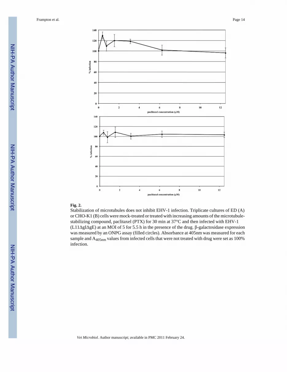
Fig. 2.Stabilization of microtubules does not inhibit EHV-1 infection. Triplicate cultures of ED (A)or CHO-K1 (B) cells were mock-treated or treated with increasing amounts of the microtubule-stabilizing compound, paclitaxel (PTX) for 30 min at 37°C and then infected with EHV-1(L11ΔgIΔgE) at an MOI of 5 for 5.5 h in the presence of the drug. β-galactosidase expressionwas measured by an ONPG assay (filled circles). Absorbance at 405nm was measured for eachsample and A405nm values from infected cells that were not treated with drug were set as 100%infection.
Frampton et al. Page 14
Vet Microbiol. Author manuscript; available in PMC 2011 February 24.
NIH
-PA Author Manuscript
NIH
-PA Author Manuscript
NIH
-PA Author Manuscript
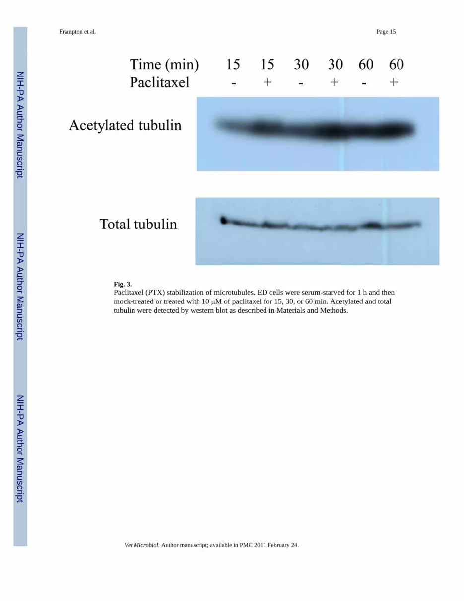
Fig. 3.Paclitaxel (PTX) stabilization of microtubules. ED cells were serum-starved for 1 h and thenmock-treated or treated with 10 μM of paclitaxel for 15, 30, or 60 min. Acetylated and totaltubulin were detected by western blot as described in Materials and Methods.
Frampton et al. Page 15
Vet Microbiol. Author manuscript; available in PMC 2011 February 24.
NIH
-PA Author Manuscript
NIH
-PA Author Manuscript
NIH
-PA Author Manuscript
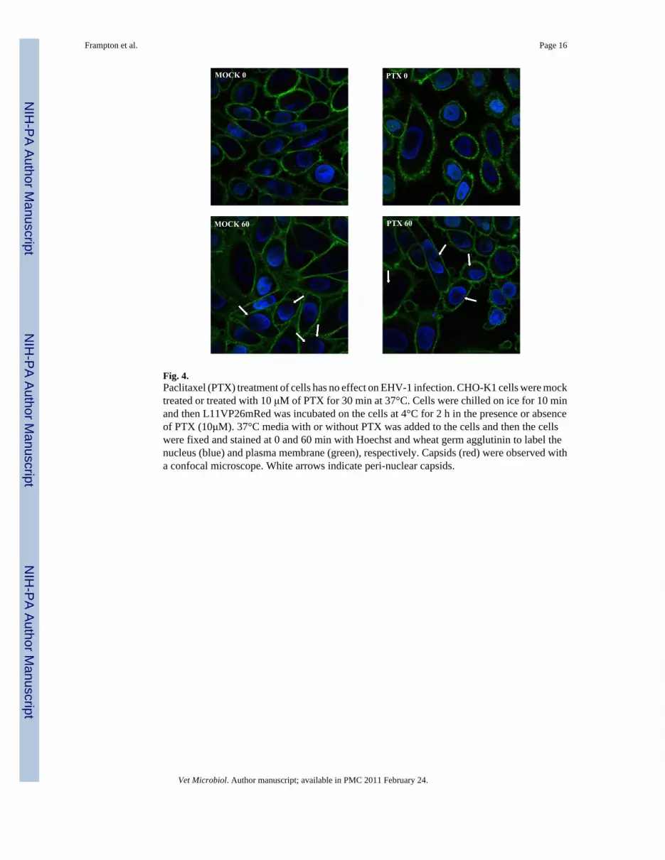
Fig. 4.Paclitaxel (PTX) treatment of cells has no effect on EHV-1 infection. CHO-K1 cells were mocktreated or treated with 10 μM of PTX for 30 min at 37°C. Cells were chilled on ice for 10 minand then L11VP26mRed was incubated on the cells at 4°C for 2 h in the presence or absenceof PTX (10μM). 37°C media with or without PTX was added to the cells and then the cellswere fixed and stained at 0 and 60 min with Hoechst and wheat germ agglutinin to label thenucleus (blue) and plasma membrane (green), respectively. Capsids (red) were observed witha confocal microscope. White arrows indicate peri-nuclear capsids.
Frampton et al. Page 16
Vet Microbiol. Author manuscript; available in PMC 2011 February 24.
NIH
-PA Author Manuscript
NIH
-PA Author Manuscript
NIH
-PA Author Manuscript

Fig. 5.EHV-1 induces the acetylation of tubulin. ED cells were serum starved for 1 h and then mock-treated or treated with Y-27632 for 30 min. Cells were mock-infected or infected with EHV-1(L11VP26mRed) at an MOI of 10 in the presence or absence of Y-27632. Acetylated and totaltubulin were detected by western blot at 15, 30, and 60 min post-infection as described inMaterials and Methods.
Frampton et al. Page 17
Vet Microbiol. Author manuscript; available in PMC 2011 February 24.
NIH
-PA Author Manuscript
NIH
-PA Author Manuscript
NIH
-PA Author Manuscript
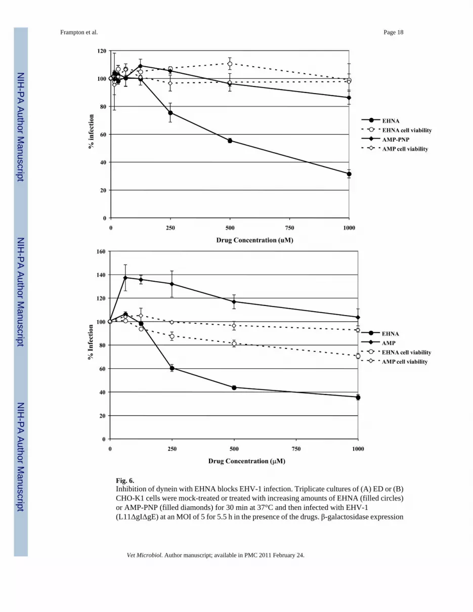
Fig. 6.Inhibition of dynein with EHNA blocks EHV-1 infection. Triplicate cultures of (A) ED or (B)CHO-K1 cells were mock-treated or treated with increasing amounts of EHNA (filled circles)or AMP-PNP (filled diamonds) for 30 min at 37°C and then infected with EHV-1(L11ΔgIΔgE) at an MOI of 5 for 5.5 h in the presence of the drugs. β-galactosidase expression
Frampton et al. Page 18
Vet Microbiol. Author manuscript; available in PMC 2011 February 24.
NIH
-PA Author Manuscript
NIH
-PA Author Manuscript
NIH
-PA Author Manuscript
was measured by ONPG assay and cell viability at each concentration of drug was measuredby MTS assay, EHNA (open circles), AMP-PNP (open diamonds).
Frampton et al. Page 19
Vet Microbiol. Author manuscript; available in PMC 2011 February 24.
NIH
-PA Author Manuscript
NIH
-PA Author Manuscript
NIH
-PA Author Manuscript
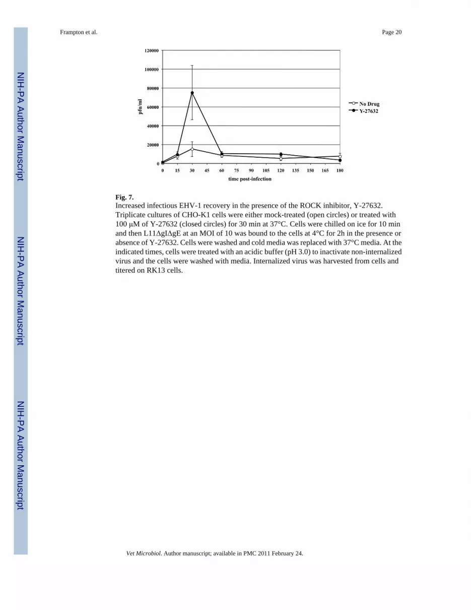
Fig. 7.Increased infectious EHV-1 recovery in the presence of the ROCK inhibitor, Y-27632.Triplicate cultures of CHO-K1 cells were either mock-treated (open circles) or treated with100 μM of Y-27632 (closed circles) for 30 min at 37°C. Cells were chilled on ice for 10 minand then L11ΔgIΔgE at an MOI of 10 was bound to the cells at 4°C for 2h in the presence orabsence of Y-27632. Cells were washed and cold media was replaced with 37°C media. At theindicated times, cells were treated with an acidic buffer (pH 3.0) to inactivate non-internalizedvirus and the cells were washed with media. Internalized virus was harvested from cells andtitered on RK13 cells.
Frampton et al. Page 20
Vet Microbiol. Author manuscript; available in PMC 2011 February 24.
NIH
-PA Author Manuscript
NIH
-PA Author Manuscript
NIH
-PA Author Manuscript
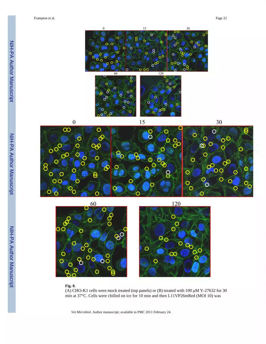
Fig. 8.(A) CHO-K1 cells were mock treated (top panels) or (B) treated with 100 μM Y-27632 for 30min at 37°C. Cells were chilled on ice for 10 min and then L11VP26mRed (MOI 10) was
Frampton et al. Page 21
Vet Microbiol. Author manuscript; available in PMC 2011 February 24.
NIH
-PA Author Manuscript
NIH
-PA Author Manuscript
NIH
-PA Author Manuscript
incubated on the cells at 4°C for 2 h in the absence (A) or presence (B) of Y-27632 (100 μM).37°C media with or without Y-27632 was added to the cells and then the cells were fixed andstained at 0, 15, 30, 60, and 120 min. with Hoechst and wheat germ agglutinin to label thenucleus (blue) and plasma membrane (green), respectively. Capsids (red) were observed witha confocal microscope. Nuclear-associated capsids are circled in white and non-nuclearassociated capsids are circled in yellow.
Frampton et al. Page 22
Vet Microbiol. Author manuscript; available in PMC 2011 February 24.
NIH
-PA Author Manuscript
NIH
-PA Author Manuscript
NIH
-PA Author Manuscript