enhancement of the photocatalytic performance of ag-modified tio2 photocatalyst under visible light
TRANSCRIPT
Enhancement of the photocatalytic performance of Ag-modified TiO2
photocatalyst under visible light
Cheewita Suwanchawalit a,*, Sumpun Wongnawa b, Pimpaporn Sriprang b, Pachara Meanha a
a Department of Chemistry, Faculty of Science, Silpakorn University, Sanam Chandra Palace Campus, Nakornpathom 73000, Thailandb Department of Chemistry, Faculty of Science, Prince of Songkla University, Hat Yai, Songkhla 90112, Thailand
Received 23 December 2011; received in revised form 24 February 2012; accepted 9 March 2012
Available online 19 March 2012
Abstract
A highly visible-light photocatalytic active Ag-modified TiO2 (Ag–TiO2) was prepared by a simple sol–gel process using TiOSO4 as the starting
material, AgNO3 as a silver doping source, and hydrazine as a reducing agent. The prepared Ag–TiO2 samples were characterized by several
techniques such as X-ray powder diffraction (XRD), BET surface area measurement, scanning electron microscopy (SEM), transmission electron
microscopy (TEM), inductively coupled plasma optical emission spectroscopy (ICP-OES), energy dispersive X-ray spectrometry (EDX), X-ray
absorption spectroscopy (XAS) and UV–vis diffuse reflectance spectroscopy (DRS). The Ag–TiO2 photocatalyst, a mixture of amorphous and
anatase phases, has a high surface area. The silver contents in the Ag–TiO2 samples were determined by ICP measurements. The diffused
reflectance UV–vis spectra indicated that the Ag–TiO2 samples exhibited higher red shifts compared with the undoped TiO2 sample. Indigo
carmine degradation under visible irradiation indicated that the Ag–TiO2 catalyst gave higher photocatalytic efficiency than those of commercial
P25-TiO2 and undoped-TiO2 samples. The Ag–TiO2 catalyst can be reused many times without any additional treatment.
# 2012 Elsevier Ltd and Techna Group S.r.l. All rights reserved.
Keywords: A. Sol–gel process; Titanium dioxide; Mesoporous titanium dioxide; Ag-modified TiO2; Indigo camine; Dye decolorization
www.elsevier.com/locate/ceramint
Available online at www.sciencedirect.com
Ceramics International 38 (2012) 5201–5207
1. Introduction
Heterogeneous semiconductor photocatalysis is an attractive
technology among the most promising technologies for solar
energy conversion and environmental applications. Of the
semiconducting materials employed, TiO2 is the most effective
because of its high photosensitivity, chemical stability, non-
toxicity, easy availability, environmental friendliness, and low
cost [1–4].
However, a major drawback of TiO2 is the large band gap of
3.2 eV which limits its activity when sunlight is used. To
overcome these limitations of TiO2, many studies have been
carried out to enhance the electron–hole separation and to
extend the absorption range of TiO2 into the visible region.
These studies involved incorporation of metal ions or nonmetal
ions into the TiO2 lattice [5–8], dye photosensitization onto the
* Corresponding author. Tel.: +66 3425 5797; fax: +66 3427 1356.
E-mail addresses: [email protected], [email protected]
(C. Suwanchawalit).
0272-8842/$36.00 # 2012 Elsevier Ltd and Techna Group S.r.l. All rights reserve
doi:10.1016/j.ceramint.2012.03.027
TiO2 surface [9–12], and deposition of noble metals onto the
TiO2 surface [13–15].
In particular, noble metal-modified TiO2 particles have
become the focus of many studies to maximize the efficiency of
photocatalytic reactions. The noble metals deposited or doped
on TiO2 have high Schottky barriers among the metals and act
as electron traps, facilitating the electron–hole separation and
promoting the interfacial electron transfer process [16]. These
noble metals may enhance the electron–hole separation, extend
the light absorption into the visible range and enhance the
surface electron excitation.
Among the noble metals used as electron traps, silver (Ag) is
extremely suitable for industrial application due to its low cost
and easy preparation. Ag-modified TiO2 powders have become
current interests due to its improvement of photocatalytic
reactions and anti-microbial activity. There are several techniques
for the preparation of Ag-modified TiO2 such as sol–gel [16,17],
photocatalytic deposition [13], and deposition precipitation [14].
In this work, undoped and Ag-modified TiO2 samples were
synthesized by the sol–gel process using TiOSO4 and AgNO3
as starting materials and hydrazine as a reducing agent. Herein,
the synthesized Ag-modified TiO2 catalyst was characterized
d.
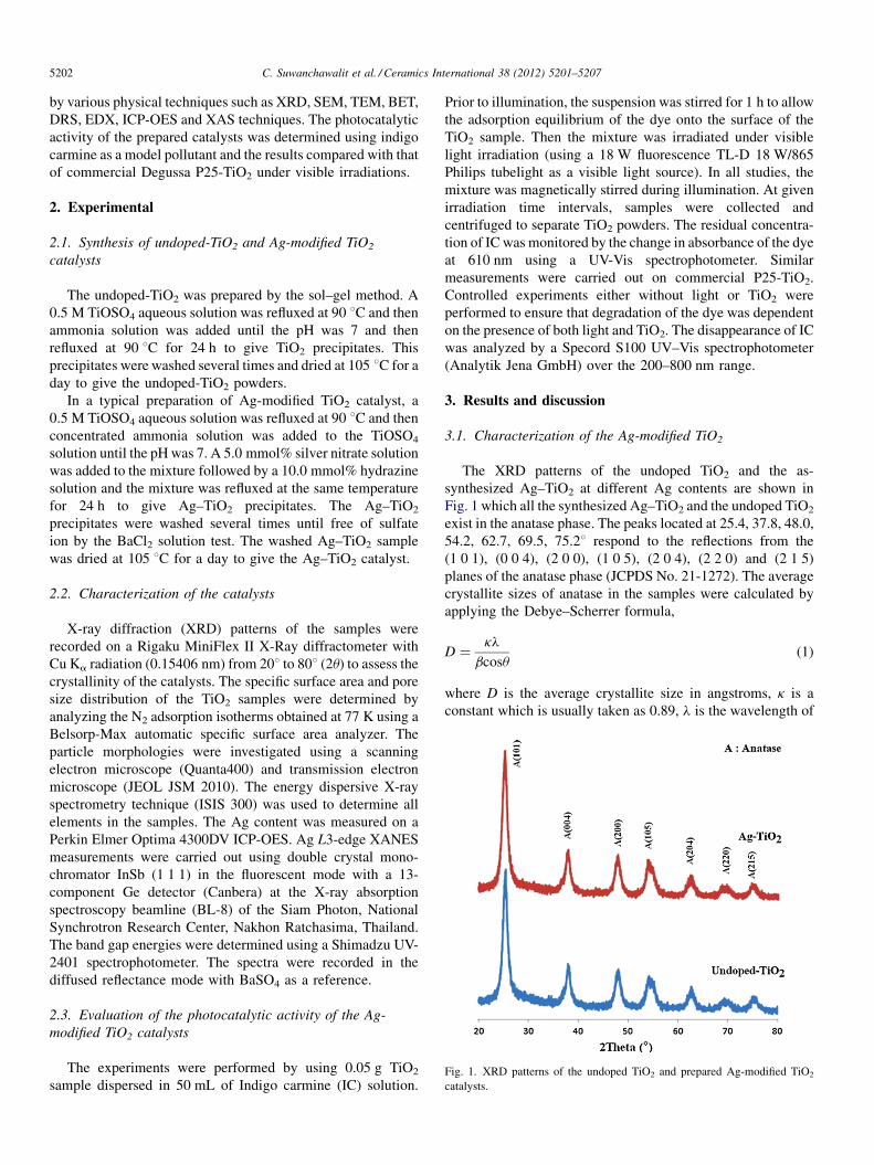
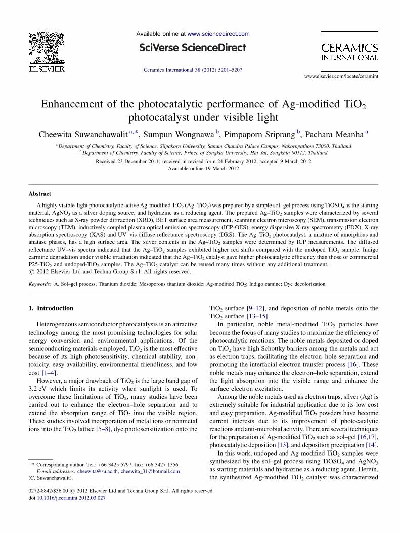
Fig. 1. XRD patterns of the undoped TiO2 and prepared Ag-modified TiO2
catalysts.
C. Suwanchawalit et al. / Ceramics International 38 (2012) 5201–52075202
by various physical techniques such as XRD, SEM, TEM, BET,
DRS, EDX, ICP-OES and XAS techniques. The photocatalytic
activity of the prepared catalysts was determined using indigo
carmine as a model pollutant and the results compared with that
of commercial Degussa P25-TiO2 under visible irradiations.
2. Experimental
2.1. Synthesis of undoped-TiO2 and Ag-modified TiO2
catalysts
The undoped-TiO2 was prepared by the sol–gel method. A
0.5 M TiOSO4 aqueous solution was refluxed at 90 8C and then
ammonia solution was added until the pH was 7 and then
refluxed at 90 8C for 24 h to give TiO2 precipitates. This
precipitates were washed several times and dried at 105 8C for a
day to give the undoped-TiO2 powders.
In a typical preparation of Ag-modified TiO2 catalyst, a
0.5 M TiOSO4 aqueous solution was refluxed at 90 8C and then
concentrated ammonia solution was added to the TiOSO4
solution until the pH was 7. A 5.0 mmol% silver nitrate solution
was added to the mixture followed by a 10.0 mmol% hydrazine
solution and the mixture was refluxed at the same temperature
for 24 h to give Ag–TiO2 precipitates. The Ag–TiO2
precipitates were washed several times until free of sulfate
ion by the BaCl2 solution test. The washed Ag–TiO2 sample
was dried at 105 8C for a day to give the Ag–TiO2 catalyst.
2.2. Characterization of the catalysts
X-ray diffraction (XRD) patterns of the samples were
recorded on a Rigaku MiniFlex II X-Ray diffractometer with
Cu Ka radiation (0.15406 nm) from 208 to 808 (2u) to assess the
crystallinity of the catalysts. The specific surface area and pore
size distribution of the TiO2 samples were determined by
analyzing the N2 adsorption isotherms obtained at 77 K using a
Belsorp-Max automatic specific surface area analyzer. The
particle morphologies were investigated using a scanning
electron microscope (Quanta400) and transmission electron
microscope (JEOL JSM 2010). The energy dispersive X-ray
spectrometry technique (ISIS 300) was used to determine all
elements in the samples. The Ag content was measured on a
Perkin Elmer Optima 4300DV ICP-OES. Ag L3-edge XANES
measurements were carried out using double crystal mono-
chromator InSb (1 1 1) in the fluorescent mode with a 13-
component Ge detector (Canbera) at the X-ray absorption
spectroscopy beamline (BL-8) of the Siam Photon, National
Synchrotron Research Center, Nakhon Ratchasima, Thailand.
The band gap energies were determined using a Shimadzu UV-
2401 spectrophotometer. The spectra were recorded in the
diffused reflectance mode with BaSO4 as a reference.
2.3. Evaluation of the photocatalytic activity of the Ag-
modified TiO2 catalysts
The experiments were performed by using 0.05 g TiO2
sample dispersed in 50 mL of Indigo carmine (IC) solution.
Prior to illumination, the suspension was stirred for 1 h to allow
the adsorption equilibrium of the dye onto the surface of the
TiO2 sample. Then the mixture was irradiated under visible
light irradiation (using a 18 W fluorescence TL-D 18 W/865
Philips tubelight as a visible light source). In all studies, the
mixture was magnetically stirred during illumination. At given
irradiation time intervals, samples were collected and
centrifuged to separate TiO2 powders. The residual concentra-
tion of IC was monitored by the change in absorbance of the dye
at 610 nm using a UV-Vis spectrophotometer. Similar
measurements were carried out on commercial P25-TiO2.
Controlled experiments either without light or TiO2 were
performed to ensure that degradation of the dye was dependent
on the presence of both light and TiO2. The disappearance of IC
was analyzed by a Specord S100 UV–Vis spectrophotometer
(Analytik Jena GmbH) over the 200–800 nm range.
3. Results and discussion
3.1. Characterization of the Ag-modified TiO2
The XRD patterns of the undoped TiO2 and the as-
synthesized Ag–TiO2 at different Ag contents are shown in
Fig. 1 which all the synthesized Ag–TiO2 and the undoped TiO2
exist in the anatase phase. The peaks located at 25.4, 37.8, 48.0,
54.2, 62.7, 69.5, 75.28 respond to the reflections from the
(1 0 1), (0 0 4), (2 0 0), (1 0 5), (2 0 4), (2 2 0) and (2 1 5)
planes of the anatase phase (JCPDS No. 21-1272). The average
crystallite sizes of anatase in the samples were calculated by
applying the Debye–Scherrer formula,
D ¼ kl
bcosu(1)
where D is the average crystallite size in angstroms, k is a
constant which is usually taken as 0.89, l is the wavelength of
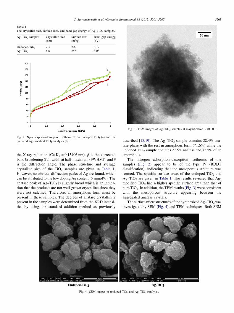
Table 1
The crystallite size, surface area, and band gap energy of Ag–TiO2 samples.
Ag–TiO2 samples Crystallite size Surface area Band gap energy
(nm) (m2/g) (eV)
Undoped-TiO2 7.3 200 3.19
Ag–TiO2 6.8 256 3.08
Fig. 2. N2-adsorption–desorption isotherm of the undoped TiO2 (a) and the
prepared Ag-modified TiO2 catalysts (b).
Fig. 3. TEM images of Ag–TiO2 samples at magnification �40,000.
C. Suwanchawalit et al. / Ceramics International 38 (2012) 5201–5207 5203
the X-ray radiation (Cu Ka = 0.15406 nm), b is the corrected
band broadening (full width at half-maximum (FWHM)), and u
is the diffraction angle. The phase structure and average
crystallite size of the TiO2 samples are given in Table 1.
However, no obvious diffraction peaks of Ag are found, which
can be attributed to the low doping Ag content (5 mmol%). The
anatase peak of Ag–TiO2 is slightly broad which is an indica-
tion that the products are not well-grown crystalline since they
were not calcined. Therefore, an amorphous form must be
present in these samples. The degrees of anatase crystallinity
present in the samples were determined from the XRD intensi-
ties by using the standard addition method as previously
Fig. 4. SEM images of undoped
described [18,19]. The Ag–TiO2 sample contains 28.4% ana-
tase phase with the rest in amorphous form (71.6%) while the
undoped TiO2 sample contains 27.5% anatase and 72.5% of an
amorphous.
The nitrogen adsorption–desorption isotherms of the
samples (Fig. 2) appear to be of the type IV (BDDT
classification), indicating that the mesoporous structure was
formed. The specific surface areas of the undoped TiO2 and
Ag–TiO2 are given in Table 1. The results revealed that Ag-
modified TiO2 had a higher specific surface area than that of
pure TiO2. In addition, the TEM results (Fig. 3) were consistent
with the mesoporous structure appearing between the
aggregated anatase crystals.
The surface microstructures of the synthesized Ag–TiO2 was
investigated by SEM (Fig. 4) and TEM techniques. Both SEM
TiO2 and Ag–TiO2 catalysts.
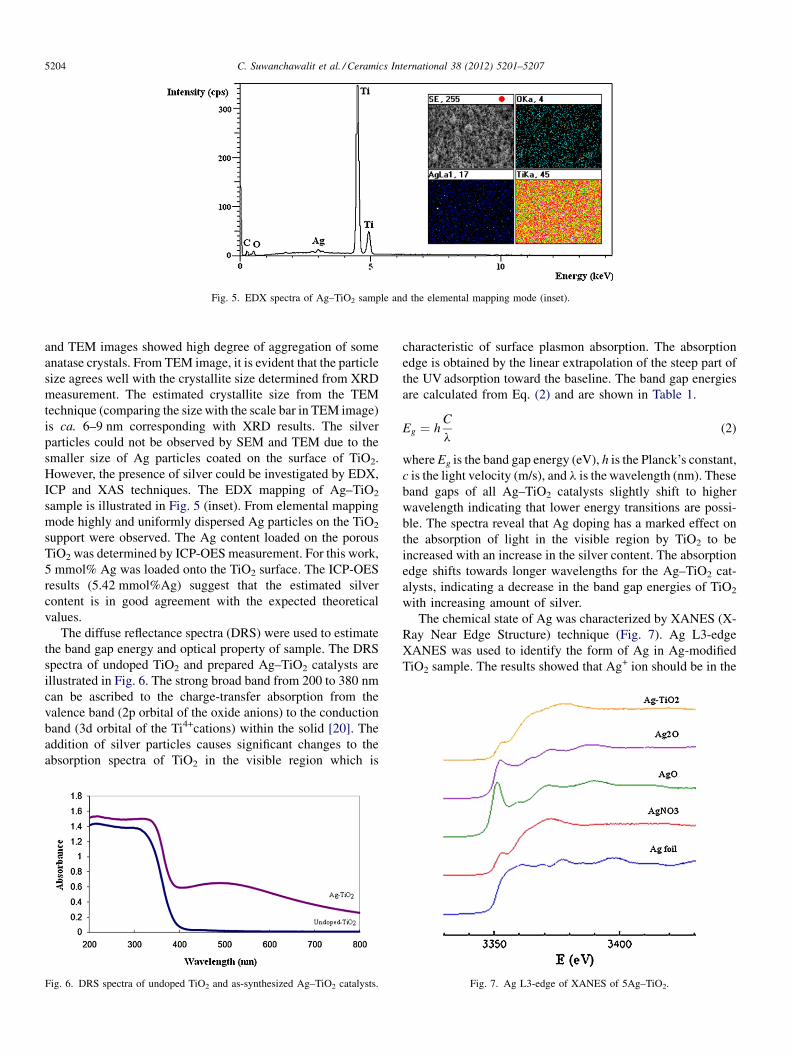
Fig. 5. EDX spectra of Ag–TiO2 sample and the elemental mapping mode (inset).
C. Suwanchawalit et al. / Ceramics International 38 (2012) 5201–52075204
and TEM images showed high degree of aggregation of some
anatase crystals. From TEM image, it is evident that the particle
size agrees well with the crystallite size determined from XRD
measurement. The estimated crystallite size from the TEM
technique (comparing the size with the scale bar in TEM image)
is ca. 6–9 nm corresponding with XRD results. The silver
particles could not be observed by SEM and TEM due to the
smaller size of Ag particles coated on the surface of TiO2.
However, the presence of silver could be investigated by EDX,
ICP and XAS techniques. The EDX mapping of Ag–TiO2
sample is illustrated in Fig. 5 (inset). From elemental mapping
mode highly and uniformly dispersed Ag particles on the TiO2
support were observed. The Ag content loaded on the porous
TiO2 was determined by ICP-OES measurement. For this work,
5 mmol% Ag was loaded onto the TiO2 surface. The ICP-OES
results (5.42 mmol%Ag) suggest that the estimated silver
content is in good agreement with the expected theoretical
values.
The diffuse reflectance spectra (DRS) were used to estimate
the band gap energy and optical property of sample. The DRS
spectra of undoped TiO2 and prepared Ag–TiO2 catalysts are
illustrated in Fig. 6. The strong broad band from 200 to 380 nm
can be ascribed to the charge-transfer absorption from the
valence band (2p orbital of the oxide anions) to the conduction
band (3d orbital of the Ti4+cations) within the solid [20]. The
addition of silver particles causes significant changes to the
absorption spectra of TiO2 in the visible region which is
Fig. 6. DRS spectra of undoped TiO2 and as-synthesized Ag–TiO2 catalysts.
characteristic of surface plasmon absorption. The absorption
edge is obtained by the linear extrapolation of the steep part of
the UV adsorption toward the baseline. The band gap energies
are calculated from Eq. (2) and are shown in Table 1.
Eg ¼ hC
l(2)
where Eg is the band gap energy (eV), h is the Planck’s constant,
c is the light velocity (m/s), and l is the wavelength (nm). These
band gaps of all Ag–TiO2 catalysts slightly shift to higher
wavelength indicating that lower energy transitions are possi-
ble. The spectra reveal that Ag doping has a marked effect on
the absorption of light in the visible region by TiO2 to be
increased with an increase in the silver content. The absorption
edge shifts towards longer wavelengths for the Ag–TiO2 cat-
alysts, indicating a decrease in the band gap energies of TiO2
with increasing amount of silver.
The chemical state of Ag was characterized by XANES (X-
Ray Near Edge Structure) technique (Fig. 7). Ag L3-edge
XANES was used to identify the form of Ag in Ag-modified
TiO2 sample. The results showed that Ag+ ion should be in the
Fig. 7. Ag L3-edge of XANES of 5Ag–TiO2.
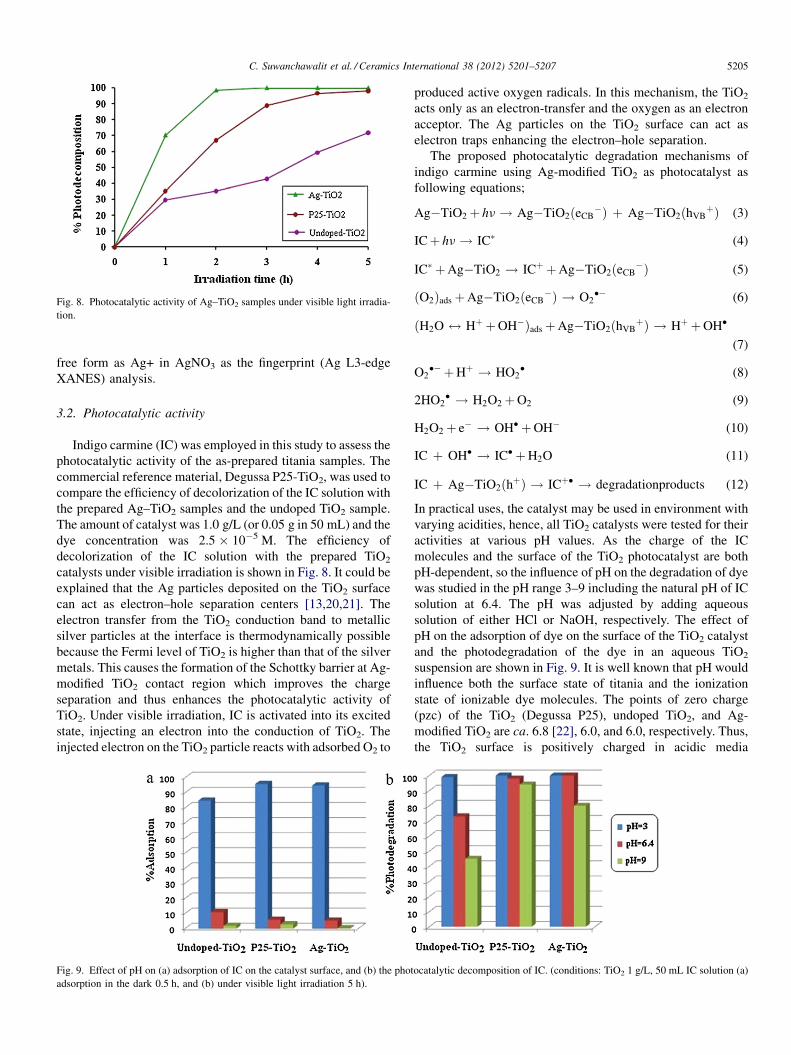
Fig. 8. Photocatalytic activity of Ag–TiO2 samples under visible light irradia-
tion.
C. Suwanchawalit et al. / Ceramics International 38 (2012) 5201–5207 5205
free form as Ag+ in AgNO3 as the fingerprint (Ag L3-edge
XANES) analysis.
3.2. Photocatalytic activity
Indigo carmine (IC) was employed in this study to assess the
photocatalytic activity of the as-prepared titania samples. The
commercial reference material, Degussa P25-TiO2, was used to
compare the efficiency of decolorization of the IC solution with
the prepared Ag–TiO2 samples and the undoped TiO2 sample.
The amount of catalyst was 1.0 g/L (or 0.05 g in 50 mL) and the
dye concentration was 2.5 � 10�5 M. The efficiency of
decolorization of the IC solution with the prepared TiO2
catalysts under visible irradiation is shown in Fig. 8. It could be
explained that the Ag particles deposited on the TiO2 surface
can act as electron–hole separation centers [13,20,21]. The
electron transfer from the TiO2 conduction band to metallic
silver particles at the interface is thermodynamically possible
because the Fermi level of TiO2 is higher than that of the silver
metals. This causes the formation of the Schottky barrier at Ag-
modified TiO2 contact region which improves the charge
separation and thus enhances the photocatalytic activity of
TiO2. Under visible irradiation, IC is activated into its excited
state, injecting an electron into the conduction of TiO2. The
injected electron on the TiO2 particle reacts with adsorbed O2 to
Fig. 9. Effect of pH on (a) adsorption of IC on the catalyst surface, and (b) the phot
adsorption in the dark 0.5 h, and (b) under visible light irradiation 5 h).
produced active oxygen radicals. In this mechanism, the TiO2
acts only as an electron-transfer and the oxygen as an electron
acceptor. The Ag particles on the TiO2 surface can act as
electron traps enhancing the electron–hole separation.
The proposed photocatalytic degradation mechanisms of
indigo carmine using Ag-modified TiO2 as photocatalyst as
following equations;
Ag�TiO2þ hn ! Ag�TiO2ðeCB�Þ þ Ag�TiO2ðhVB
þÞ (3)
IC þ hn ! IC� (4)
IC� þ Ag�TiO2 ! ICþ þ Ag�TiO2ðeCB�Þ (5)
ðO2Þadsþ Ag�TiO2ðeCB�Þ ! O2
�� (6)
ðH2O $ Hþ þ OH�Þadsþ Ag�TiO2ðhVBþÞ ! Hþ þ OH�
(7)
O2�� þ Hþ ! HO2
� (8)
2HO2� ! H2O2þ O2 (9)
H2O2þ e� ! OH� þ OH� (10)
IC þ OH� ! IC� þ H2O (11)
IC þ Ag�TiO2ðhþÞ ! ICþ� ! degradationproducts (12)
In practical uses, the catalyst may be used in environment with
varying acidities, hence, all TiO2 catalysts were tested for their
activities at various pH values. As the charge of the IC
molecules and the surface of the TiO2 photocatalyst are both
pH-dependent, so the influence of pH on the degradation of dye
was studied in the pH range 3–9 including the natural pH of IC
solution at 6.4. The pH was adjusted by adding aqueous
solution of either HCl or NaOH, respectively. The effect of
pH on the adsorption of dye on the surface of the TiO2 catalyst
and the photodegradation of the dye in an aqueous TiO2
suspension are shown in Fig. 9. It is well known that pH would
influence both the surface state of titania and the ionization
state of ionizable dye molecules. The points of zero charge
(pzc) of the TiO2 (Degussa P25), undoped TiO2, and Ag-
modified TiO2 are ca. 6.8 [22], 6.0, and 6.0, respectively. Thus,
the TiO2 surface is positively charged in acidic media
ocatalytic decomposition of IC. (conditions: TiO2 1 g/L, 50 mL IC solution (a)
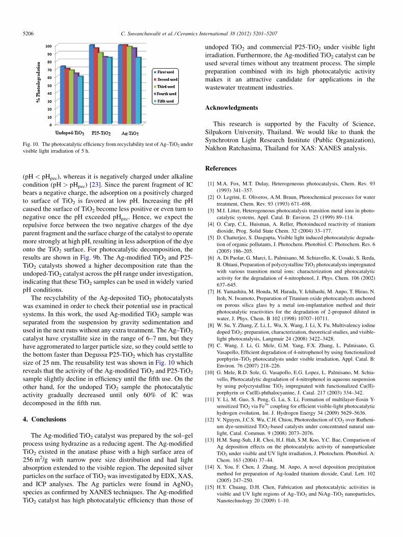
Fig. 10. The photocatalytic efficiency from recyclability test of Ag–TiO2 under
visible light irradiation of 5 h.
C. Suwanchawalit et al. / Ceramics International 38 (2012) 5201–52075206
(pH < pHpzc), whereas it is negatively charged under alkaline
condition (pH > pHpzc) [23]. Since the parent fragment of IC
bears a negative charge, the adsorption on a positively charged
to surface of TiO2 is favored at low pH. Increasing the pH
caused the surface of TiO2 become less positive or even turn to
negative once the pH exceeded pHpzc. Hence, we expect the
repulsive force between the two negative charges of the dye
parent fragment and the surface charge of the catalyst to operate
more strongly at high pH, resulting in less adsorption of the dye
onto the TiO2 surface. For photocatalytic decomposition, the
results are shown in Fig. 9b. The Ag-modified TiO2 and P25-
TiO2 catalysts showed a higher decomposition rate than the
undoped-TiO2 catalyst across the pH range under investigation,
indicating that these TiO2 samples can be used in widely varied
pH conditions.
The recyclability of the Ag-deposited TiO2 photocatalysts
was examined in order to check their potential use in practical
systems. In this work, the used Ag-modified TiO2 sample was
separated from the suspension by gravity sedimentation and
used in the next runs without any extra treatment. The Ag–TiO2
catalyst have crystallite size in the range of 6–7 nm, but they
have aggromerated to larger particle size, so they could settle to
the bottom faster than Degussa P25-TiO2 which has crystallite
size of 25 nm. The reusability test was shown in Fig. 10 which
reveals that the activity of the Ag-modified TiO2 and P25-TiO2
sample slightly decline in efficiency until the fifth use. On the
other hand, for the undoped TiO2 sample the photocatalytic
activity gradually decreased until only 60% of IC was
decomposed in the fifth run.
4. Conclusions
The Ag-modified TiO2 catalyst was prepared by the sol–gel
process using hydrazine as a reducing agent. The Ag-modified
TiO2 existed in the anatase phase with a high surface area of
256 m2/g with narrow pore size distribution and had light
absorption extended to the visible region. The deposited silver
particles on the surface of TiO2 was investigated by EDX, XAS,
and ICP analyses. The Ag particles were found in AgNO3
species as confirmed by XANES techniques. The Ag-modified
TiO2 catalyst has high photocatalytic efficiency than those of
undoped TiO2 and commercial P25-TiO2 under visible light
irradiation. Furthermore, the Ag-modified TiO2 catalyst can be
used several times without any treatment process. The simple
preparation combined with its high photocatalytic activity
makes it an attractive candidate for applications in the
wastewater treatment industries.
Acknowledgments
This research is supported by the Faculty of Science,
Silpakorn University, Thailand. We would like to thank the
Synchrotron Light Research Institute (Public Organization),
Nakhon Ratchasima, Thailand for XAS: XANES analysis.
References
[1] M.A. Fox, M.T. Dulay, Heterogeneous photocatalysis, Chem. Rev. 93
(1993) 341–357.
[2] O. Legrini, E. Oliveros, A.M. Braun, Photochemical processes for water
treatment, Chem. Rev. 93 (1993) 671–698.
[3] M.I. Litter, Heterogeneous photocatalysis transition metal ions in photo-
catalytic systems, Appl. Catal. B: Environ. 23 (1999) 89–114.
[4] O. Carp, C.L. Huisman, A. Reller, Photoinduced reactivity of titanium
dioxide, Prog. Solid State Chem. 32 (2004) 33–177.
[5] D. Chatterjee, S. Dasgupta, Visible light induced photocatalytic degrada-
tion of organic pollutants, J. Photochem. Photobiol. C: Photochem. Rev. 6
(2005) 186–205.
[6] A. Di Paolar, G. Marci, L. Palmisano, M. Schiavello, K. Uosaki, S. Ikeda,
B. Ohtani, Preparation of polycrystalline TiO2 photocatalysts impregnated
with various transition metal ions: characterization and photocatalytic
activity for the degradation of 4-nitrophenol, J. Phys. Chem. 106 (2002)
637–645.
[7] H. Yamashita, M. Honda, M. Harada, Y. Ichihashi, M. Anpo, T. Hirao, N.
Itoh, N. Iwamoto, Preparation of Titanium oxide photocatalysts anchored
on porous silica glass by a metal ion-implantation method and their
photocatalytic reactivities for the degradation of 2-propanol diluted in
water, J. Phys. Chem. B 102 (1998) 10707–10711.
[8] W. Su, Y. Zhang, Z. Li, L. Wu, X. Wang, J. Li, X. Fu, Multivalency iodine
doped TiO2: preparation, characterization, theoretical studies, and visible-
light photocatalysis, Langmuir 24 (2008) 3422–3428.
[9] C. Wang, J. Li, G. Mele, G.M. Yang, F.X. Zhang, L. Palmisano, G.
Vasapollo, Efficient degradation of 4-nitrophenol by using functionalized
porphyrin–TiO2 photocatalysts under visible irradiation, Appl. Catal. B:
Environ. 76 (2007) 218–226.
[10] G. Mele, R.D. Sole, G. Vasapollo, E.G. Lopez, L. Palmisano, M. Schia-
vello, Photocatalytic degradation of 4-nitrophenol in aqueous suspension
by using polycrystalline TiO2 impregnated with functionalized Cu(II)-
porphyrin or Cu(II)-phthalocyanine, J. Catal. 217 (2003) 334–342.
[11] Y. Li, M. Guo, S. Peng, G. Lu, S. Li, Formation of multilayer-Eosin Y-
sensitized TiO2 via Fe3+ coupling for efficient visible-light photocatalytic
hydrogen evolution, Int. J. Hydrogen Energy 34 (2009) 5629–5636.
[12] V. Nguyen, J.C.S. Wu, C.H. Chiou, Photoreduction of CO2 over Rutheni-
um dye-sensitized TiO2-based catalysts under concentrated natural sun-
light, Catal. Commun. 9 (2008) 2073–2076.
[13] H.M. Sung-Suh, J.R. Choi, H.J. Hah, S.M. Koo, Y.C. Bae, Comparison of
Ag deposition effects on the photocatalytic activity of nanoparticulate
TiO2 under visible and UV light irradiation, J. Photochem. Photobiol. A:
Chem. 163 (2004) 37–44.
[14] X. You, F. Chen, J. Zhang, M. Anpo, A novel deposition precipitation
method for preparation of Ag-loaded titanium dioxide, Catal. Lett. 102
(2005) 247–250.
[15] H.Y. Chuang, D.H. Chen, Fabrication and photocatalytic activities in
visible and UV light regions of Ag–TiO2 and NiAg–TiO2 nanoparticles,
Nanotechnology 20 (2009) 1–10.

C. Suwanchawalit et al. / Ceramics International 38 (2012) 5201–5207 5207
[16] M.S. Lee, S.S. Hong, M. Mohseni, Synthesis of photocatalytic nanosized
TiO2–Ag particles with sol–gel method using reduction agent, J. Mol.
Catal. A: Chem. 242 (2005) 135–140.
[17] H.E. Chao, Y.U. Yun, H.U. Xingfang, A. Larbot, Effect of silver doping on
the phase transformation and grain growth of sol–gel titania powder, J.
Eur. Ceram. Soc. 23 (2003) 1457–1464.
[18] M. Kanna, S. Wongnawa, Mixed amorphous and nanocrystalline TiO2
powders prepared by sol–gel method: characterization and photocatalytic
study, Mater. Chem. Phys. 110 (2008) 166–175.
[19] C. Suwanchawalit, S. Wongnawa, Triblock copolymer-templated synthe-
sis of porous TiO2 and its photocatalytic activity, J. Nanopart Res. 12
(2010) 2895–2906.
[20] N. Sobana, M. Muruganadham, M. Swaminathan, Nano-Ag particles
doped TiO2 for efficient photodegradation of direct azo dyes, J. Mol.
Catal. A: Chem. 258 (2006) 124–132.
[21] S. Anandan, P.S. Kumar, N. Pugazhenthiran, J. Madhavan, P. Maruthamuthu,
Effect of loaded silver nanoparticles on TiO2 for photocatalytic degradation
of Acid Red 88, Sol. Energy Mater. Sol. Cells 92 (2008) 929–937.
[22] I.K. Konstantinou, T.A. Albanis, TiO2-assisted photocatalytic degradation
of azo dyes in aqueous solution: kinetic and mechanistic investigations,
Appl. Catal. B: Environ. 49 (2004) 1–14.
[23] B. Wen, C. Liu, Y. Liu, optimization of the preparation methods synthesis
of meso structures TiO2 with high photocatalytic activities, J. Photochem.
Photobiol. A: Chem. 173 (2005) 7–12.