enhanced salt stress tolerance of rice plants expressing a vacuolar h+-atpase subunit c1 (savhac1)...
TRANSCRIPT

Enhanced salt stress tolerance of rice plants expressing avacuolar H+-ATPase subunit c1 (SaVHAc1) gene from thehalophyte grass Spartina alterniflora LoiselNiranjan Baisakh1,*, Mangu V. RamanaRao1, Kanniah Rajasekaran2, Prasanta Subudhi1, Jaroslav Janda3,David Galbraith3, Cheryl Vanier4 and Andy Pereira5
1School of Plant, Environmental, and Soil Sciences, Louisiana State University Agricultural Center, Baton Rouge, LA, USA2Southern Regional Research Center, Agricultural Research Service, United States Department of Agriculture, New Orleans, LA, USA3Department of Plant Sciences, University of Arizona, Tucson, AZ, USA4University of Nevada, Las Vegas, NV, USA5Department of Crop, Soil, and Environmental Sciences, University of Arkansas, Fayetteville, AK, USA
Received 22 October 2011;
revised 8 December 2011;
accepted 12 December 2011.
*Correspondence (fax +1 225 5781403;
email [email protected])
Keywords: gene expression, halo-
phyte, rice, salt tolerance, vacuolar
ATPase.
SummaryThe physiological role of a vacuolar ATPase subunit c1 (SaVHAc1) from a halophyte grass
Spartina alterniflora was studied through its expression in rice. The SaVHAc1-expressing plants
showed enhanced tolerance to salt stress than the wild-type plants, mainly through adjust-
ments in early stage and preparatory physiological responses. In addition to the increased
accumulation of its own transcript, SaVHAc1 expression led to increased accumulation of
messages of other native genes in rice, especially those involved in cation transport and ABA
signalling. The SaVHAc1-expressing plants maintained higher relative water content under salt
stress through early stage closure of the leaf stoma and reduced stomata density. The
increased K+ ⁄ Na+ ratio and other cations established an ion homoeostasis in SaVHAc1-
expressing plants to protect the cytosol from toxic Na+ and thereby maintained higher chloro-
phyll retention than the WT plants under salt stress. Besides, the role of SaVHAc1 in cell wall
expansion and maintenance of net photosynthesis was implicated by comparatively higher
root and leaf growth and yield of rice expressing SaVHAc1 over WT under salt stress. The
study indicated that the genes contributing toward natural variation in grass halophytes could
be effectively manipulated for improving salt tolerance of field crops within related taxa.
Introduction
Soil salinity is a major threat to agricultural productivity world-
wide. At high concentrations of salts in the soil, plants experi-
ence a physiological drought because of the inability of roots to
extract water, and high concentrations of salts within the plant
can be toxic (Munns and Tester, 2008). Because salt tolerance
is a quantitative trait governed by multiple genes, success with
classical breeding has been low in developing salt-tolerant crops
(Winicov, 1998). Genetic manipulation provides an alternative
yet sustainable approach to engineering rice plants for salinity
tolerance. Recently, however, four markers from candidate
genes including SKC1 within the ‘Saltol’ QTL have been identi-
fied in rice and are being used in marker-assisted selective
breeding of salt-tolerant rice (Thomson et al., 2010).
Halophyte models have been used as a source of genes for
engineering salt tolerance in heterologous systems. Considering
the differences in the anatomical features between dicots and
monocots and also in their adaptation regulation networking, it
is necessary to explore halophytic models among grass species
as a source for superior alleles ⁄ regulation machinery through
genetic engineering onto food crops, which are mostly grasses,
such as rice (Tester and Bacic, 2005). Earlier we reported that
Spartina alterniflora (smooth cordgrass), a monocot halophyte
with reportedly all possible mechanisms of salt tolerance, shares
80–90% similarity with rice with regard to their DNA and
protein sequences (Baisakh et al., 2008). Although there are a
handful of reports documenting transgenic overexpressers
showing some degree of salt tolerance, actual production of
transgenic plants with demonstrably improved salt stress toler-
ance is few and slow (Flowers, 2004).
Among various mechanisms, control of ion movement across
tonoplast (and plasma membrane) to maintain low Na+ concen-
tration in the cytoplasm is the key cellular factor in salinity toler-
ance. Salt-tolerant plants differ from salt-sensitive ones in
having a low rate of Na+ and Cl) transport to leaves and the
ability to compartmentalize these ions in vacuoles to avoid salt
toxicity by preventing their build-up in cytoplasm or cell walls.
In halophytes such as common ice plant (Mesembryathemum
crystallinum), vacuolar sodium sequestration under salinity is
mediated by an active Na+ ⁄ H+ antiporter energized by the pro-
ton motive force, which is generated and maintained by plant
V-ATPase (EC3.61.34), which is one of the several members of
the ATP-dependent protein pumps, through primary active H+
transport at the tonoplast (Barkla et al., 1995). The most abun-
dant subunit of the V0 complex of vacuolar ATPase is subunit c,
which is encoded by the largest multigene family. It is present
in six copies that form the part of the proton-conducting pore
responsible for proton translocation (Sze et al., 1999), although
the biological significance of this is unknown. Subunit c was
the first multigene family reported in eukaryote V-ATPases (Sze
et al., 1992). H+-ATPase acts as a primary transporter that
ª 2012 Louisiana State University Agricultural Center
Plant Biotechnology Journal ª 2012 Society for Experimental Biology, Association of Applied Biologists and Blackwell Publishing Ltd 453
Plant Biotechnology Journal (2012) 10, pp. 453–464 doi: 10.1111/j.1467-7652.2012.00678.x

pumps protons out of the cytoplasm, thus creating a pH and
electric potential gradient across the vacuole that activates
many secondary transporters involved in ion and metabolite
uptake (reviewed in Serrano, 1989; Sussman, 1994; Michelet
and Boutry, 1995; Palmgren, 1998). Subunit c is a highly hydro-
phobic protein in the V0 domain. It is essential for the produc-
tion of an active V-ATPase holoenzyme and is likely to be
directly involved in H+-transport. Transcriptional changes of
subunits of the vacuolar ATPase in response to salinity stress
have been reported from a number of plants. For example, salt-
induced transcriptional activation of V-ATPase subunit c has
been observed in common ice plant (Dietz and Arbinger, 1996;
Low et al., 1996; Tsiantis et al., 1996), which included a consid-
erable and fast increase in vacuolar-type H+-ATPase activity in
tonoplast vesicles, irrigated with high NaCl concentrations
(Ratajczak et al., 1994). This demonstrates the prime impor-
tance of V-ATPase in the adaptation of common ice plant to
high sodium concentrations. In the halotolerant sugar beet,
transcripts of the V-ATPase subunits A and c were found in root
and leaf tissue, and NaCl treatment caused an increase in the
transcript levels in leaves, but not in the roots (Kirsch et al.,
1996; Lehr et al., 1999). In Porteresia coarctata, roots showed
immediate pronounced (two-fold) increase in V-ATPase c tran-
script, whereas in the leaves, there was no significant increase
until or after 5 h and it achieved twofold and threefold accu-
mulation only after 24 and 48 h following salt stress (Senthilku-
mar et al., 2005). The subunit c transcript level declined in both
leaf and root after 10 h of salt withdrawal. Recently, we dem-
onstrated that an expressed sequence tag (EST) showing similar-
ity to vacuolar H+-ATPase subunit c1 (SaVHAc1) was highly
induced in both leaf and root tissues of smooth cordgrass (Spar-
tina alterniflora) when plants were grown under hypersaline
(500 mM NaCl) condition (Baisakh et al., 2008). These studies
indicate that coordinated enhanced steady-state transcript levels
of specific V-ATPase subunits in root and ⁄ or shoot are a charac-
teristic for halotolerant plants, whereas salinity-induced up-reg-
ulation of V-ATPase subunits has also been demonstrated in
non-halophytes (Tyagi et al., 2005) including resurrection plants
(Chen et al., 2002).
The phenotypes caused by ectopic expression of a vacuolar
pyrrophosphatase (AVP1) in Arabidopsis suggested that manipu-
lation of vacuolar proton pumps in economically important
crops holds promise for the reclamation of farmlands lost to
salinization and lack of rainfall (Gaxiola et al., 2002). Little,
however, is known about the effect and biological role of
orthologous expression of genes encoding V-ATPase subunits
on the plants’ ability to cope with salt stress, although its role
in salt stress response has been indirectly shown in RNAi
mutants (Padmanaban et al., 2004) or, as described earlier, by
its increased transcript accumulation under salt stress. The pres-
ent work was undertaken with an objective to understand the
role that SaVHAc1 plays in plant’s physiological response to
salinity through its functional expression in transgenic rice.
Results
Sequence analysis of SaVHAc1
A cDNA clone (897 bp) containing the entire coding sequence
of SaVHAc1 was isolated from the cDNA library of Spartina
alterniflora, which contained a 498-bp-long open reading
form (ORF). The SaVHAc1 was predicted to be a membrane
protein of 165 (16.62 kDa) deduced amino acid residues under
transport and binding function GO category with subcellular
localization in the inner tonoplast (Figure S1). The SaVHAc1
protein consisted of two hydrophilic and two hydrophobic
alpha-helices with an average hydrophobicity of 0.979. The pro-
tein consisted of three transmembrane helices (each of 23 aa
length) and a primary signal peptide (30 aa).
Comparison of the genomic sequences, isolated through
standard genome walking procedure using primer specific to
5¢- and 3¢- UTR and ACP primer (Seegene Inc.), and cDNA-
sequences of SaVHAc1 revealed five exons ranging from 28 to
289 nucleotides in length separated by four introns ranging
from 14 to 1034 nucleotides in length with GT-AG conforma-
tion expected for eukaryotic nuclear genome (Figure 1a). Com-
parative analysis of the V-ATPase subunit c sequences from
other plants showed that SaVHAc1 formed an independent
group within the cluster that comprised of Eleusine glauca, Ara-
bidopsis thaliana, Gossypium hirsutum and Mesembryathemum
crystallinum (Figure 1b). It shared 87% and 99% identity with
rice at the DNA and protein level, respectively. However, at the
protein level, it showed 100% identity with the V-type proton
ATPase (Asvat-P1) from Avena sativa. Restriction mapping (with
Figure 1 Characterization of SaVHAc1 from Spartina alterniflora. Geno-
mic organization (a) showing 5 exons (E1…E5). DNA sequence similarity
of SaVHAc1 with V-ATPase gene from other plants (b). Copy number
analysis of SaVHAc1 in Spartina alterniflora (c). Size fragments of k ⁄ Hind
III marker in kb are shown next to the horizontal bars. Sa, Spartina alter-
niflora; Eg, Eleucine glauca; At, Arabidopsis thaliana; Gh, Gossypium
herbaceum; Mc, Mesembryathemum crystallinum; Zm, Zea mays; As,
Avena sativa; Pc, Porteresia coarctata; Os, Oryza sativa; Te, Triticum
aestivum; Pg, Pennisetum galucum; HIII, HindIII; BHI, BamHI; EI, EcoRI;
Xh, XhoI; Xb, XbaI; SI, SacI.
ª 2012 Louisiana State University Agricultural Center
Plant Biotechnology Journal ª 2012 Society for Experimental Biology, Association of Applied Biologists and Blackwell Publishing Ltd, Plant Biotechnology Journal, 10, 453–464
Niranjan Baisakh et al.454

six different restriction endonucleases viz., HindIII, BamHI, EcoRI,
XhoI, XbaI, and SacI) showed the presence of more than one
copy of the SaVHAc1 in the genome of S. alterniflora
(Figure 1c). The complexity of the restriction patterns detected
by full length SaVHAc1 suggested that a multigene family
encodes the 16-kD proteolipid SaVHAc1 in S. alterniflora, which
may account for more than the fact that S. alterniflora in itself
is a allohexaploid (2n = 6x = 62).
Stable integration and inheritance of SaVHAc1in transgenic rice
The integration of SaVHAc1 expression cassette (Figure 2a) in
the rice genome was initially identified by positive PCR signals
using gene-specific primers and subsequently confirmed by
Southern blot analysis. The copy number and integration was
further validated by T-DNA-rice genomic DNA flanking
sequence analysis. In CLA9 and CLA20, SaVHAc1 was inte-
grated as a single copy (Figure 2b), whereas in CLA19, four
copies were integrated. SaVHAc1 was mapped in the long arm
of chromosome 7 of CLA20 (Figure 2c). The segregation of
SaVHAc1 in the first selfing generation (T1) progenies of CLA9
and CLA20 followed a single gene (3:1) Mendelian inheritance
(Figure 2d). The SaVHAc1 locus was fixed to homozygosity in T2
generation (Figure 2e).
SaVHAc1 expression conferred salt tolerance in riceplants
Upon imposition of salt stress at 100 and 200 mM NaCl, the
SaVHAc1 plants showed greater tolerance than the WT plants
with respect to leaf chlorophyll bleaching in leaf-floating assay,
leaf rolling, leaf withering, tip burning and other physiological
and agronomic traits (Figure 3). The SaVHAc1-rice plants
exposed to seedling stage salt stress grew normally after recov-
ery and set seeds in the greenhouse, whereas the WT plants
had very stunted growth, abnormal ⁄ incomplete panicle
exsertion and were very highly sterile (Figure 3c). Tobacco
transgenics expressing SaVHAc1 also showed enhanced toler-
ance to salinity as compared to the WT plants (Figure S2). No
apparent difference was observed between WT and SaVHAc1-
rice plants with regard to their growth and development under
normal (unstressed) conditions (data not shown).
After 72 h of salt stress, the SaVHAc1-rice plants maintained
shoot growth as was evident by 22% reduction of shoot length
compared to approximately 43% reduction of the WT plants
over their respective unstressed control (Figure 4a). Similarly,
the SaVHAc1 plants had a better root growth with a root
length reduction of 9% under salinity than the WT (22%;
Figure 4a). The reduction in the shoot dry weight of SaVHAc1-
rice associated with stress was substantially less (16%) than the
Figure 2 Generation and characterization of SaVHAc1 plants. Partial linear map of the plant transformation binary vector p35S:SaVHAc1 (a), Restric-
tion analysis showing copy number of three independent SaVHAc1-rice (CLA9, CLA20 and CLA19), WT, wild type; M, k ⁄ Hind III DNA size marker (b).
Genome mapping of SaVHAc1 showing its integration (arrow marked) in Chromosome seven of rice (c). Single gene Mendelian segregation of SaV-
HAc1 in T1 generation (d) and homozyosity of SaVHAc1-rice in T2 generation (e). PC, plasmid positive control.
ª 2012 Louisiana State University Agricultural Center
Plant Biotechnology Journal ª 2012 Society for Experimental Biology, Association of Applied Biologists and Blackwell Publishing Ltd, Plant Biotechnology Journal, 10, 453–464
Enhanced salt tolerance of rice and vacuolar ATPase 455

WT rice (34%; Figure 4b). The SaVHAc1 plants did not show
any significant reduction (0.8%) in root dry weight relative to
the unstressed control plants, which was much lower than the
8% reduction of WT plants under stress (Figure 4b).
SaVHAc1-rice retained high chlorophyll, relative watercontent, and maintained higher yield under salinity
Rice plants expressing SaVHAc1 maintained higher chlorophyll
concentration over WT under saline conditions (Figure 5a). The
loss of total chlorophyll because of salt stress (150 mM NaCl)
was less (32%) in SaVHAc1-rice compared to WT plants (48%)
when averaged over three time points (Figure 5a). The SaV-
HAc1-rice plants showed much less reduction (13%) in chloro-
phyll a after 36 h of salt stress compared to the WT plants
(35%). The loss of the chlorophyll was directly proportional to
the duration of salt stress (Figure 5a). Chlorophyll concentration
(a) (b) (c)
Figure 3 Salt tolerance of SaVHAc1-rice vis-a-vis WT rice under salt stress (S1 = 100 mM NaCl, S2 = 200 mM NaCl) in leaf disc assay (a), whole plant
assay under hydroponics at 150 mM NaCl (b). The SaVHAc1-rice plants showed better growth and yielded more compared to WT rice under salt stress
at reproductive stage (c).
Figure 4 Reduction in shoot and root length (a) and dry weight (b) of
SaVHAc1-rice vis-a-vis WT one week after salt stress (150 mM NaCl).
Error bars represent standard error of means.
Figure 5 Chlorophyll a (a), relative water content (b) and grain yield per
panicle of SAVHAC1- rice vis-a-vis WT plants under salt stress (150 mM
NaCl) (c). Note that their yield was comparable under no-stress control
condition. Error bars represent standard errors of means.
ª 2012 Louisiana State University Agricultural Center
Plant Biotechnology Journal ª 2012 Society for Experimental Biology, Association of Applied Biologists and Blackwell Publishing Ltd, Plant Biotechnology Journal, 10, 453–464
Niranjan Baisakh et al.456

derived from the SPAD reading and estimated by acetone
extraction method was comparable (data not shown).
Relative water content (RWC) is considered an appropriate
measure of plant water status as well as osmotic adjustment
under stress. RWC, which also reflects, in part, the transpira-
tional loss of water, was estimated from the leaves of the SaV-
HAc1- and WT rice plants subject to salt stress. After a week
under stress, the SaVHAc1-rice plants maintained higher RWC
(93%) as compared with the WT (77%; Figure 5b).
Both SaVHAc1- and WT rice plants showed a reduction in
grain yield per panicle upon salt stress during 2 weeks of the
critical reproductive stage (i.e. panicle initiation). However, the
SaVHAc1-rice produced nearly six times higher grain yield
(1.42 g) per panicle than WT plants (0.24 g) under reproductive
stage salt stress (Figure 5c).
SaVHAc1 expression led to accumulation of higherK+ under salt stress
Only one SaVHAc1-rice line (CLA20) along with WT was
included in the elemental analysis. The root Na+ concentration
of the SaVHAc1-rice was less than the WT plants, while the leaf
Na+ was high in both genotypes. In contrast, leaf K+ concentra-
tion in SaVHAc1-rice was much higher than in WT, which was
clear from the high K+ ⁄ Na+ values in the transgenic lines
(Figure 6a). Concomitantly, the SaVHAc1-rice lines also accumu-
lated higher levels of Ca2+ and Mg2+ in their leaf and root
tissues with or without stress (Figure 6b).
SaVHAc1 expression altered the expression of nativerice genes upon salt stress
From the microarray experiment, many of the 43 311 probes
were significantly different: 4287 (9.9%) between genotypes
(WT and SaVHAc1-rice), 6761 (15.6%) between environments
(saline or non-saline conditions) and 705 (1.6%) responded dif-
ferently to the environment depending upon the genotype
(Table S1). Although a large number of genes showed up-regu-
lation in SaVHAc1-rice relative to WT (Table S1; Gene Expres-
sion Omnibus Accession no. GSE34724), eighteen genes that
showed more than twofold increase or decrease in transcription
in SaVHAc1-rice were considered significantly up- or down-
regulated, respectively. Fourteen genes encoding proteins with
either ion transport or metal binding function were significantly
up-regulated (Table 1). The four genes that showed significant
down-regulation in SaVHAc1-rice did not have any functional
annotation (hypothetical or expressed proteins). Interestingly,
none of the other subunits of V-ATPase were observed as being
affected by SaVHAc1.
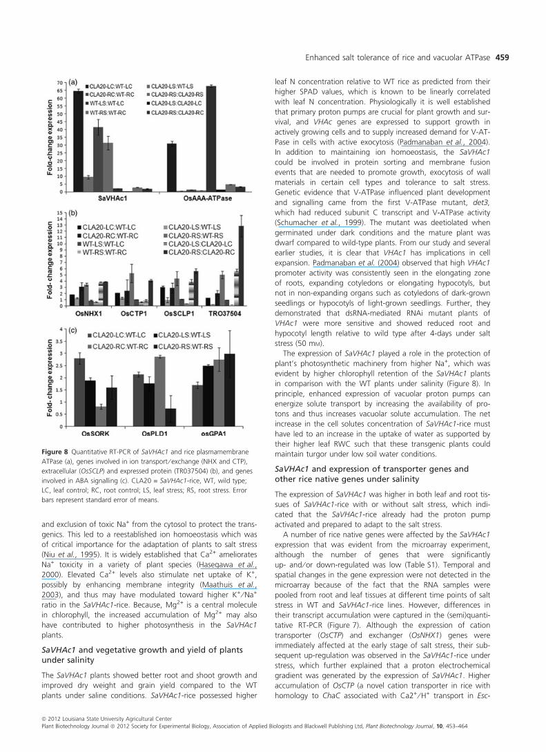
Semiquantitative expression analysis of genes (Figure 7)
showed that SaVHAc1 transcript was much higher in the SaV-
HAc1-rice than the WT without or with salt stress imposition at
all time points. Quantitatively, SaVHAc1 transcript accumulated
Table 1 Genes showing up-regulation (‡2-fold) by SAVHAC1-expression in rice under salt stress (analysed by microarray)
Probe ID Gene GO Slim ID Term Ontology Fold increase
TR038922 Pyrophosphate-energized vacuolar membrane proton pump GO:0005773 Membrane; vacuole, hydrolase activity Cellular component 2.5
TR055074 Glutamine synthetase expressed GO:0003824 Catalytic activity Molecular function 2.7
TR066667 Metallothionein-like protein 1 GO:0046872 Metal binding Molecular function 2.7
TR066578 Pathogenesis-related protein 10 GO:0035556 Intracellular signal transduction Biological process 2.6
TR057433 Hypothetical protein 2.6
TR051207 Germin-like protein subfamily 1 member 7 precursor GO:0005618 Cell wall Cellular component 2.9
TR037990 Cortical cell delineating protein precursor GO:0006810 Transport Biological process 2.9
TR068459 Plastocyanin-like domain containing protein GO:0005488 Binding, membrane Molecular function 3.1
TR066580 Pathogenesis-related protein Bet v I family protein GO:0035556 Intracellular signal transduction Biological process 4.0
TR066664 Metallothionein-like protein 1 GO:0046872 Metal binding Molecular function 3.8
TR067039 Cysteine synthase GO:0003824 Catalytic activity Molecular function 3.6
TR037504 Expressed protein 5.0
TR066668 Metallothionein-like protein 1 GO:0046872 Metal binding Molecular function 5.0
TR047684 SCP-like extracellular protein GO:0005576 Extracellular region Cellular component 6.1
Figure 6 Inductive coupled plasma (ICP) analysis showing higher K+:Na+
(a), and Ca2+ and Mg2+ concentration (b) in leaf and root tissues of SaV-
HAc1-rice (CLA20) in comparison with WT rice under control (C) and
salinity (S). LC, leaf control; RC, root control; LS, leaf stress; RS, root
stress. Error bars represent standard error of means.
ª 2012 Louisiana State University Agricultural Center
Plant Biotechnology Journal ª 2012 Society for Experimental Biology, Association of Applied Biologists and Blackwell Publishing Ltd, Plant Biotechnology Journal, 10, 453–464
Enhanced salt tolerance of rice and vacuolar ATPase 457

up to 25-fold higher in the SaVHAc1-rice under salt stress than
the SaVHAc1-rice without salt stress and approximately 40-fold
higher over the WT rice plants (Figure 8a). The overexpression
of SaVHAc1 in the transgenics also led to higher constitutive
transcript accumulation of rice plasma membrane AAA-ATPase
in the leaf tissues under no stress. Although there was no
apparent change in its transcript in leaf tissues under salt stress,
the root tissues showed comparable increase in its expression in
both WT and SaVHAc1-rice under stress.
Other genes that were tested for their transcript accumula-
tion showed up-regulation in SaVHAc1-rice as compared to WT
plants under salt stress in a tissue and time-dependent manner
(Figures 7 and 8b). One important function of the V-ATPase is
to provide the driving force for H+-coupled Na+ antiporters,
such as NHX1, which sequesters sodium into the vacuole (Apse
et al., 1999). Cation transporter genes such as OsNHX1 and
OsCTP showed little difference among SaVHAc1-rice and WT
rice in leaf tissues, but their transcript was highly accumulated
in the roots of SaVHAc1-rice compared to WT plants. OsSCPL1
showed a slight decline in its transcript accumulation until 24 h
of salt stress in their root, but overaccumulated after 48 h
through the recovery stage. Cysteine synthase (OsCS) behaved
similarly. Genes such as metallothionein (OsMT1) and PR protein
(BetV) showed only root-specific expression with subtle differ-
ence between SaVHAc1-rice and WT plants.
Quantitative expression analysis showed up-regulation of
OsPLDa1 and OsGPA1, the two positive regulators of abscisic
acid (ABA) signalling genes (Nilson and Assmann, 2010) in the
SaVHAc1-rice over WT (Figure 8c) without and with stress. The
leaves and roots of SaVHAc1-rice accumulated higher transcript
of these two genes even without stress. Similarly, the expres-
sion of OsSORK (rice homologue of Arabidopsis GORK) was
2.8- and 1.9-fold higher in SaVHAc1-rice than WT with and
without salt stress, respectively.
SaVHAc1 expression affected the density and openingof stomata
The SaVHAc1-rice had 39% fewer stomata per unit leaf area
than the WT rice (Figure 9a,c). Further, the stomata were
mostly closed in the SaVHAc1 lines under salt stress as
compared with the WT (Figure 9b). However, there was no sig-
nificant difference with respect to the stomata size as measured
by their dimension. Cross-sections of leaf and root tissue did
not show any apparent difference between the SaVHAc1- and
WT rice (data not shown).
Discussion
The present results identified a major role for SaVHAc1, a gene
similar to vacuolar H+-ATPase, from a halophyte grass in several
physiological processes particularly those related to salt stress.
The vacuolar H+-ATPase is the major proton pump that estab-
lishes and maintains an electrochemical proton gradient across
the tonoplast, thus providing the driving force for the secondary
active transport of ions and metabolites against their concentra-
tion gradient (Gaxiola et al., 2007). The functional significance
of V-ATPase in plant’s ability to adapt to unfavourable stress
was provided earlier (Dietz et al., 2001). Transcriptional activa-
tion of V-ATPase at an early stage of salt stress has been shown
in several instances (Baisakh et al., 2008; Golldack and Deitz,
2000). The ability of a plant to change its gene expression pro-
file to respond to salt stress, as we observed, might be a key
mechanism for salt tolerance.
SaVHAc1 and ion homoeostasis
The capacity of plants to maintain a high cytosolic K+ ⁄ Na+ ratio
is likely to be one of the key determinants of salt tolerance.
Halophytes utilize ion homoeostasis in the cytosol as one of the
most important strategies for their adaptation to salinity. The
SaVHAc1 plants accumulated high level of Na+ in roots and
leaves; growth, however, was little affected by the toxic Na+,
which could be due to the sequestration of Na+ at the tono-
plast by a secondary Na+ ⁄ H+ transporter that was energized by
a proton motive force created by the overexpression of SaV-
HAc1 (Apse et al., 1999). Further, the increase in the concentra-
tion of other cations (K+, Ca2+, and Mg2+) in the leaf and root
tissues in the SaVHAc1-rice plants (Figure 6) could be due to
the electrochemical gradient generated by the SaVHAc1 expres-
sion, which is used by proton-coupled antiporters to accumulate
more of Ca2+ and Mg2+ inside the lumen (Hirschi et al., 1996)
Figure 7 Semiquantitative RT-PCR analysis of rice native genes of rice in SaVHAc1-rice (CLA20) vis-a-vis WT under control (0 h) and at different time
points under salt stress (150 mM NaCl) and 4 days after recovery (R) in both leaf and root tissues.
ª 2012 Louisiana State University Agricultural Center
Plant Biotechnology Journal ª 2012 Society for Experimental Biology, Association of Applied Biologists and Blackwell Publishing Ltd, Plant Biotechnology Journal, 10, 453–464
Niranjan Baisakh et al.458
and exclusion of toxic Na+ from the cytosol to protect the trans-
genics. This led to a reestablished ion homoeostasis which was
of critical importance for the adaptation of plants to salt stress
(Niu et al., 1995). It is widely established that Ca2+ ameliorates
Na+ toxicity in a variety of plant species (Hasegawa et al.,
2000). Elevated Ca2+ levels also stimulate net uptake of K+,
possibly by enhancing membrane integrity (Maathuis et al.,
2003), and thus may have modulated toward higher K+ ⁄ Na+
ratio in the SaVHAc1-rice. Because, Mg2+ is a central molecule
in chlorophyll, the increased accumulation of Mg2+ may also
have contributed to higher photosynthesis in the SaVHAc1
plants.
SaVHAc1 and vegetative growth and yield of plantsunder salinity
The SaVHAc1 plants showed better root and shoot growth and
improved dry weight and grain yield compared to the WT
plants under saline conditions. SaVHAc1-rice possessed higher
leaf N concentration relative to WT rice as predicted from their
higher SPAD values, which is known to be linearly correlated
with leaf N concentration. Physiologically it is well established
that primary proton pumps are crucial for plant growth and sur-
vival, and VHAc genes are expressed to support growth in
actively growing cells and to supply increased demand for V-AT-
Pase in cells with active exocytosis (Padmanaban et al., 2004).
In addition to maintaining ion homoeostasis, the SaVHAc1
could be involved in protein sorting and membrane fusion
events that are needed to promote growth, exocytosis of wall
materials in certain cell types and tolerance to salt stress.
Genetic evidence that V-ATPase influenced plant development
and signalling came from the first V-ATPase mutant, det3,
which had reduced subunit C transcript and V-ATPase activity
(Schumacher et al., 1999). The mutant was deetiolated when
germinated under dark conditions and the mature plant was
dwarf compared to wild-type plants. From our study and several
earlier studies, it is clear that VHAc1 has implications in cell
expansion. Padmanaban et al. (2004) observed that high VHAc1
promoter activity was consistently seen in the elongating zone
of roots, expanding cotyledons or elongating hypocotyls, but
not in non-expanding organs such as cotyledons of dark-grown
seedlings or hypocotyls of light-grown seedlings. Further, they
demonstrated that dsRNA-mediated RNAi mutant plants of
VHAc1 were more sensitive and showed reduced root and
hypocotyl length relative to wild type after 4-days under salt
stress (50 mM).
The expression of SaVHAc1 played a role in the protection of
plant’s photosynthetic machinery from higher Na+, which was
evident by higher chlorophyll retention of the SaVHAc1 plants
in comparison with the WT plants under salinity (Figure 8). In
principle, enhanced expression of vacuolar proton pumps can
energize solute transport by increasing the availability of pro-
tons and thus increases vacuolar solute accumulation. The net
increase in the cell solutes concentration of SaVHAc1-rice must
have led to an increase in the uptake of water as supported by
their higher leaf RWC such that these transgenic plants could
maintain turgor under low soil water conditions.
SaVHAc1 and expression of transporter genes andother rice native genes under salinity
The expression of SaVHAc1 was higher in both leaf and root tis-
sues of SaVHAc1-rice with or without salt stress, which indi-
cated that the SaVHAc1-rice already had the proton pump
activated and prepared to adapt to the salt stress.
A number of rice native genes were affected by the SaVHAc1
expression that was evident from the microarray experiment,
although the number of genes that were significantly
up- and ⁄ or down-regulated was low (Table S1). Temporal and
spatial changes in the gene expression were not detected in the
microarray because of the fact that the RNA samples were
pooled from root and leaf tissues at different time points of salt
stress in WT and SaVHAc1-rice lines. However, differences in
their transcript accumulation were captured in the (semi)quanti-
tative RT-PCR (Figure 7). Although the expression of cation
transporter (OsCTP) and exchanger (OsNHX1) genes were
immediately affected at the early stage of salt stress, their sub-
sequent up-regulation was observed in the SaVHAc1-rice under
stress, which further explained that a proton electrochemical
gradient was generated by the expression of SaVHAc1. Higher
accumulation of OsCTP (a novel cation transporter in rice with
homology to ChaC associated with Ca2+ ⁄ H+ transport in Esc-
Figure 8 Quantitative RT-PCR of SaVHAc1 and rice plasmamembrane
ATPase (a), genes involved in ion transport ⁄ exchange (NHX and CTP),
extracellular (OsSCLP) and expressed protein (TR037504) (b), and genes
involved in ABA signalling (c). CLA20 = SaVHAc1-rice, WT, wild type;
LC, leaf control; RC, root control; LS, leaf stress; RS, root stress. Error
bars represent standard error of means.
ª 2012 Louisiana State University Agricultural Center
Plant Biotechnology Journal ª 2012 Society for Experimental Biology, Association of Applied Biologists and Blackwell Publishing Ltd, Plant Biotechnology Journal, 10, 453–464
Enhanced salt tolerance of rice and vacuolar ATPase 459

herichia coli) in the SaVHAc1-rice may have contributed to the
increased Ca2+ transport. The high expression of SaVHAc1,
even without salt stress and subsequent up-regulation of other
genes including cation transporter genes, which may have been
energized by the SaVHAc1 expression as a component of coor-
dinated regulation under salinity stress, led us to the presump-
tion that the SaVHAc1 lines maintained an anticipatory
preparedness to adapt to salt stress.
SaVHAc1 and stomata closure
As an adaptation ⁄ avoidance strategy, plants are known to close
the stomata under dehydration stress to save water and main-
tain turgor (Skirycz and Inze, 2010). The possible involvement
of ABA in the regulatory pathway leading to induction of
V-ATPase activity has been established (Tsiantis et al., 1996).
Overaccumulation of V-ATPase in SaVHAc1-rice might play an
important role in the coregulation of ABA signalling induced by
salt stress. ABA treatment mimics NaCl treatment of plants, and
only the c-subunit of the V-ATPase has been shown to be trans-
criptionally regulated by NaCl in plants (Tsiantis et al., 1996).
Stomata opening is induced by increased turgor pressure as a
result of K+ and anion influx energized by H+-ATPase. Osmotic
stress, at the early stage of salt stress first experienced by the
roots, releases ABA signalling molecules, which under the over-
expression of vacuolar ATPase could induce stomata closure
(Allen et al., 2000). The loss of turgidity as a result of K+ and
water efflux from the guard cell may have contributed to the
stomata closure in SaVHAc1-rice plants (Figure 9). H+ is known
as secondary messenger in hormone action of plants. The
increase in cytoplasmic pH (by the expression of vacuolar
ATPase) in the SaVHAc1 in coordination with ABA molecules
may have resulted in the opening of K+ (out) channel and clos-
ing of K+ (in) channel. Further, the increase in Ca2+Cyt in the
SaVHAc1-rice possibly brought about a reduction in the K+ (in)
activity of the plasma membrane, leading to the reduction in
guard cell turgidity and ultimately stomata closure. Although
the Ca2+- and H+-mediated K+ efflux are independent of each
other, it is possible both the mechanisms are in operation to
maintain a balance of the Ca2+Cyt and alkalinization (Swamy,
1999). Also, the kinetics of expression changes of the ABA sig-
nalling genes (OsPLDa1 and OsGPA1) in the SaVHAc1-rice vis-a-
vis WT plants provided clues to the closure of stomata in the
leaves of SaVHAc1-rice. The expression of these two positive
regulators of stomata ABA signalling was higher in SaVHAc1-
rice as compared with the WT (Figure 8) without and with salt
stress. Similarly, the expression of OsSORK (rice homologue of
Arabidopsis GORK) was 2.8- and 1.9-fold higher in SaVHAc1-
rice than WT with and without salt stress, respectively.
Although the mechanism of reduced stomata density in SaV-
HAc1-rice remains to be investigated, the earlier observation
that det3 mutants were defective in guard cell signalling or
movement could explain the apparent anatomical adjustments
of SaVHAc1-rice to adapt to the stress condition. Further, the
reduced stomata density in the SaVHAc1-rice could contribute
to the slow ⁄ lower rate of water loss from SaVHAc1-rice leaves
for which they maintained a higher RWC under salt stress as
compared to the WT leaves. Higher RWC is one of the impor-
tant factors contributing to tolerance to physiological drought
(a)
(b)
(c)
Figure 9 Scanning electron microscope picture of a leaf showing closed stomata (arrow marked and numbered) in SaVHAc1-rice (a) compared to
open-type stomata under salt stress in WT rice (b). The SaVHAc1-rice has less stomata ⁄ sqcm in leaf surface than the WT under salinity (c). A 50· mag-
nified picture of a representative stoma is shown as an inset on the top right corner of Panel a and b). Error bars represent standard error of means.
ª 2012 Louisiana State University Agricultural Center
Plant Biotechnology Journal ª 2012 Society for Experimental Biology, Association of Applied Biologists and Blackwell Publishing Ltd, Plant Biotechnology Journal, 10, 453–464
Niranjan Baisakh et al.460

that is experienced by the plant at the initial stage of salt stress
as a result of low osmotic potential caused by the accumulation
of soluble salts.
The time point analysis of SaVHAc1 as well as other related
genes further established that SaVHAc1-rice tolerated salt stress
by an early stage priming and preconditioning response (Harb
et al., 2010), thus maintaining a higher RWC and chlorophyll
(thereby photosynthesis), protecting cytosol from ion toxicity,
and early stomata closure. Present results suggested a clearly
defined functional role of SaVHAc1 with implications in the
modification of a signalling pathway in the salt stress response
by driving the expression of other genes involved in ion homo-
eostasis, solute accumulation, and ABA signalling. This study
including that of others as described earlier again substantiates
that grass halophytes, in addition to their usefulness to under-
stand the gene regulation mechanism of their natural salinity-
adaptability, could be effectively used as a source for mining
and bioprospecting superior alleles for the manipulation of salt
tolerance in monocot field crops such as rice.
Experimental procedures
Cloning of SaVHAc1, sequence analysis, andconstruction of binary vector
An expressed sequence tag (EST#617) of Spartina alterniflora
(GenBank Acc. No EH277293; Baisakh et al., 2008) showed
similarity to plant vacuolar H+-ATPase subunit c1 when its
nucleotide and deduced protein sequences were blasted against
the non-redundant nucleotide and protein database using
BLASTN and BLASTP interface, respectively. The sequence align-
ment of this EST (hereinafter referred to as SaVHAc1) was car-
ried out with orthologs from other plants (see Figure 1 for
details) using CLUSTALW.
The complete open reading frame of SaVHAc1 was amplified
from the S. alterniflora root cDNA library (Baisakh et al., 2008) by
PCR using primers SaVHAc1 Fwd: 5¢- ggaagatctatgtcgtcgacgttcag
-3¢ and SaVHAc1 Rev: 5¢- gggtwaccctaatctgcacggac -3¢ contain-
ing the BglII and BstEII restriction endonuclease (RE) recognition
sites (underlined), respectively. The PCR product and pCAM-
BIA1301 (CAMBIA, Australia) was digested using the same REs
and ligated to yield the binary plasmid p35S:SaVHAc1 (Figure 2a).
The identity and orientation of p35S:SaVHAc1 was confirmed by
restriction digestion and sequencing. The plasmid was mobilized
into Agrobacterium tumefaciens LBA4404 using the freeze-thaw
method (An et al., 1998).
Rice transformation
Agrobacterium tumefaciens transformation of rice cultivar
‘Cocodrie’ was performed following the method described earlier
(Rao et al., 2009). LBA4404 ⁄ p35S:SaVHAc1 was precultured
overnight at 28 �C in Luria-Bertani (LB) broth with rifampicin
(20 lg ⁄ mL), spectinomycin (100 lg ⁄ mL), streptomycin
(50 lg ⁄ mL) and kanamycin (50 lg ⁄ mL) under constant shaking
at 200 rpm. The precultured bacteria were subcultured in fresh
LB with the same antibiotics and grown for 24 h. Bacteria cells
were resuspended in the MS (Murashige and Skoog, 1962) liquid
medium supplemented with 2 mg ⁄ L 2,4-D and 100 lM acetosy-
ringone (MSco) to a final titre of A600 = 1.0 for transformation.
Three–four-week-old seed-derived rice embryogenic calli were
vacuum-infiltrated (0.4–0.6 atm) with the bacterial suspension
for 10 min and co-cultivated for 3 days in solid MSco medium
at 25 �C in the dark for 3 days. Embryogenic callus develop-
ment, and selection and regeneration of the putative transgenic
calli was performed following the method described earlier (Bai-
sakh et al., 2001).
Molecular analysis of plants expressing SaVHAc1
Polymerase chain reaction
Total genomic DNA was isolated from leaf tissues using a
modified CTAB method (Murray and Thompson, 1980). One
hundred ng of DNA was subject to PCR analysis for selectable
marker gene (hph) and target gene (SaVHAc1) using gene-spe-
cific primers (5¢–3¢) as follows: HPH F- tacttctacacagccatc, HPH
R- tatgtcctgcgggtaaat; SaVHAc1 F- aggagggtgtaccattcgtcaatg,
SaVHAc1 R- ccaggctcgtagagaataccattg.
Southern blot analysis
Southern blot analysis was performed following Baisakh et al.
(2006a). Ten micrograms of genomic DNA were digested with
a single cutter Sst I (for copy number analysis) and BglII and
BstEII (for releasing the full length SaVHAc1), electrophoresed
on a 1% (w ⁄ v) TAE agarose gel, and transferred under alkaline
denaturing conditions to Hybond N+ nylon membrane (GE
Healthcare, Piscataway, NJ). A PCR generated 200-bp fragment
of SaVHAc1, radiolabeled using (a-32P)dCTP and the Rediprime
labelling system (GE Healthcare), was used as the hybridization
probe. Hybridization and follow-up membrane washing, and
exposure to X-ray Hyperfilm� MP (GE Healthcare) was per-
formed as per Baisakh et al. (2006a).
Mapping the SaVHAc1 integration site in rice
The insertion site of SaVHAc1 was mapped by isolating the
T-DNA-rice genome flanking sequences using TAIL-PCR tech-
nique as described by Liu et al. (1995). The gene-specific primers
designed from 35S promoter and nosT sequences were used in
combination with AD1 and AD4, and the nested primers were
designed from the left and right border sequences of the T-DNA.
Three nested gene-specific reverse primers and one of the four
arbitrary degenerate (AD1-4) primers were used in successive
rounds of TAIL-PCR cycling. The primary PCR product was diluted
40-fold and used in the secondary reaction, while the latter was
diluted 5-fold for the tertiary reaction. The products of the pri-
mary, secondary and tertiary reactions were analysed on a 1.5%
agarose gel. Fragments exhibiting a difference in size consistent
with nested gene-specific primer positions were cloned using
pGEMT-easy vector (Promega, Madison, WI) and sequenced as
described earlier (Baisakh et al., 2006b). The genomic sequences
flanking SaVHAc1 were blasted against and mapped to the refer-
ence rice genome using megaBLASTN interface (http://blas-
t.ncbi.nlm.nih.gov/Blast.cgi).
Salt stress treatment
Initially, cut leaves of three independent PCR positive SaVHAc1-
rice plants were floated in 40 mL of water with 100 and
200 mM NaCl in plastic deep dishes and kept under continuous
light for 72 h before scoring for chlorophyll bleach (Sanan-Mish-
ra et al., 2005).
Four-week-old seedlings of homozygous SaVHAc1-rice (CLA9,
CLA19, and CLA20) and WT rice were subject to salt stress
(150 mM NaCl) under hydroponics in Yoshida’s nutrient solution
(Yoshida et al., 1976) following the method described earlier
(Batlang et al., 2012). Twenty plants were included in each of
ª 2012 Louisiana State University Agricultural Center
Plant Biotechnology Journal ª 2012 Society for Experimental Biology, Association of Applied Biologists and Blackwell Publishing Ltd, Plant Biotechnology Journal, 10, 453–464
Enhanced salt tolerance of rice and vacuolar ATPase 461

the four replications. After 1 week of stress, shoot and root
length, shoot and root fresh and dry weight, chlorophyll con-
centration and tissue ion concentrations were measured. After
a week of stress, the SaVHAc1-rice and a few surviving WT
plants (those not completely dead) were transferred to fresh
nutrient solution without salt, and a week-old recovered plants
were grown to maturity in the greenhouse maintained at
29 ⁄ 21 �C day ⁄ night temperature regime under natural day light
condition. At reproductive stage, salt stress was imposed as
described by Batlang et al. (2012). Ten SaVHAc1 and WT plants
during panicle initiation stage (90–95 days-old) were subject to
150 mM NaCl in the irrigation water continuously for 1 week
before the pots were submerged up to the soil level in a deep
plastic tray with salt-free water for 2 days and then irrigated
normally until maturity. Data were recorded for number of
seeds per plant and single panicle (primary tiller) grain yield.
Expression analysis
Microarray analysis
Total RNA was extracted from salt stressed and unstressed
WT and transgenic plants at different time points (24, 48,
72 h, and 4 days of recovery) using RNeasy plant minikit (Qia-
gen, Valencia, CA). For microarray experiment, total RNA
quality was checked in gel as well as through Agilent 2100
bioanalyzer. The RNA samples from different time points
except recovery point were pooled for WT and SaVHAc1
plants for each of the four biological replications. The four
samples were named WT-Control (WT-C), WT-Stress (WT-S),
SaVHAc1-rice-Control (SaVHAc1-rice-C) and SaVHAc1-
rice-stress (SaVHAc1-rice-S).
Target preparation, that is, first-strand synthesis, second-
strand synthesis, aRNA purification, dye labelling, and hybridiza-
tion and washing, was performed as per the protocol described
earlier (Edwards et al., 2008; Data S1). Sixty-mer oligonucleo-
tides designed from the rice unigenes and synthesized by
Operon technologies were printed on slides in Galbraith labora-
tory (http://ag.arizona.edu/~dgalbrai) for use on microarray
chips (45K). The GPR result file was analysed in R software
(http://www.R-project.org; Data S1).
Enrichment of functional Gene Ontology categories (GO;
http://www.geneontology.org) for genotype, environment and
their interaction was tested using the GOEAST package (Zheng
and Wang, 2008) with a Fisher’s exact test, which considered
topology of the GO relationships (Alexa et al., 2006), followed
by a Yekutieli adjustment for multiple comparisons (Yekutieli
and Benjamini, 1999). Results were considered significant at
0.05 after adjustment. The set of oligos that responded to the
environment were enriched for the biological processes related
to biotic stimuli and stress responses.
(Semi)quantitative reverse transcription polymerase chainreaction (Sq ⁄ qRT-PCR)
The total RNA was extracted from 100 mg of freshly collected
leaf and root tissues of SaVHAc1-rice and WT rice at 0 h (con-
trol), 2, 24, 48, 72 h and 4 days of recovery following salt
stress. Two micrograms of total RNA were subject to sqRT-PCR
of SaVHAc1 gene and other genes that were up ⁄ down-regu-
lated in the microarray data, employing a single-step RT-PCR kit
(Qiagen, Valencia, CA). The products were resolved in 1.5%
TAE agarose gel, visualized under UV transilluminator in a
Kodak 200 gel doc apparatus (Carestream Health, Inc., Roches-
ter, NY). Rice actin gene 1 (OsAct1) was used as an internal
control for the template validation. The primer sequences used
in the study are provided in Table S2.
The qRT-PCR was carried out using the same RNA samples
that were used for sqRT-PCR as per the method described (Bai-
sakh et al., 2008). Essentially, 1 lg total RNA was reverse tran-
scribed using iScript 1st strand cDNA synthesis kit (Bio-rad,
Carlsbad, CA). PCR was performed in triplicate (biological repli-
cate) with two independent cDNA preparations (technical repli-
cate) using SYBR green master mix (Bio-rad, Hercules, CA), 2 lL
cDNA and 3.25 pmol of each gene-specific primer in a MyiQ
Real-Time PCR detection system (Bio-rad, Hercules, CA). The rel-
ative expression ratio was calculated using the 2)DDCt method
(Baisakh et al., 2008) with rice elongation factor 1a gene
(OsEF1a) as the reference gene.
Estimation of chlorophyll concentration
Total chlorophyll was extracted from one fully expanded leaf
per plant (three plants from each of the SaVHAc1-rice and WT
plants following salt stress) with 80% acetone twice. The chlo-
rophyll a and b concentration was measured spectrophotometri-
cally following the method described by Lichtenthaler (1987) to
determine the extent of bleaching and chlorophyll loss. Total
chlorophyll was also estimated from the SPAD502 meter
(Konica Minolta Sensing, Inc., Ramsey, NJ) reading as described
earlier (Monje and Bugbee, 1992).
Estimation of tissue ion concentration
Leaf and root tissues were harvested from 1-month-old seed-
lings of unstressed (control) and salt-treated SaVHAc1-rice and
WT, and oven-dried at 80 �C for 48 h. Five hundred mg of
dried tissues was extracted with HNO3 digestion. The Na+, K+,
Ca2+ and Mg2+ concentrations were measured through induc-
tively coupled plasma-mass spectrometry (ICP-MS, Perkin-Elmer
Plasma 400 emission spectrometer) in an in-house plant and soil
testing laboratory.
Growth parameters study
Data were collected on different vegetative growth parame-
ters (root and shoot length, root and shoot dry weight) of
the SaVHAc1-rice lines vis-a-vis WT plants one week after salt
stress was imposed. The single panicle grain yield from the
primary tiller was recorded on the salt-stressed greenhouse-
grown plants at maturity. Data were analysed for ANOVA with
statistical analysis software SAS 9.1.3 (SAS Institute Inc,
2004).
Relative water content
The RWC of the leaves was determined following Slatyer
(1967). Middle sections of second-youngest fully expanded
leaves were collected and wrapped from three different WT
and SaVHAc1-rice plants after a week of salt stress and
weighed [fresh weight (FW)]. The leaf pieces were immersed in
dH2O placed in dark at 4 �C overnight and weighed after brief
blot-drying [turgid weight (TW)]. Then, the pieces were dried at
60 �C for 24 h and weighed [dry weight (DW)]. RWC was esti-
mated in percentage of the water content at a given time and
tissue as related to the water content at full turgor using the
formula:
RWCð%Þ ¼ ðFW� DWÞ=ðTW� DWÞ � 100
ª 2012 Louisiana State University Agricultural Center
Plant Biotechnology Journal ª 2012 Society for Experimental Biology, Association of Applied Biologists and Blackwell Publishing Ltd, Plant Biotechnology Journal, 10, 453–464
Niranjan Baisakh et al.462

Scanning electron microscopy (SEM)
Leaf samples were collected from SaVHAc1-rice and WT
before and 24 h after salt stress and were fixed with glutar-
aldehyde buffer followed by gradual alcohol dehydration. The
leaves were then critical point dried under liquid CO2 and
the probe surface sputter-coated with an electric-conducting
gold layer before imaging with SEM (Cambridge S-260) at
5 kV.
Acknowledgements
The technical assistance of Greg Ford, SRRC, USDA-ARS is duly
acknowledged. This work was financially supported by USDA-
CSREES. This manuscript is approved for publication by the
Director of Louisiana Agricultural Experiment Station as
MS#2011-306-6572.
References
Alexa, A., Rahnenfuhrer, J. and Lengauer, T. (2006) Improved scoring of
functional groups from gene expression data by decorrelating GO graph
structure. Bioinformat. 22, 1600–1607.
Allen, G.J., Chu, S.P., Schumacher, K., Shimazaki, C.T., Vafeados, D.,
Kemper, A., Hawke, S.D., Tallman, G., Tsien, R.Y., Harper, J.F., Chory, J.
and Schroeder, J.I. (2000) Alteration of Stimulus-specific guard cell calcium
oscillations and stomatal closing in Arabidopsis det3 mutant. Science, 289,
2338–2342.
An, G., Ebert, P.R, Mitra, A. and Ha, S.B. (1998) Binary vectors. In Plant
Molecular Biology (Gelvin, S.B. and Shilperoort, R.A., eds), pp. 1–19.
Dordrecht, the Netherlands: Kluwer Academic Publishers.
Apse, M.P., Aharon, G.S., Snedden, W.A. and Blumwald, E. (1999) Salt
tolerance conferred by over expression of a vacuolar Na+ ⁄ H+ antiporter in
Arabidopsis. Science, 285, 1256–1258.
Baisakh, N., Datta, K., Oliva, N., Ona, I., Rao, G.J.N., Mew, T.W. and Datta,
S.K. (2001) Rapid development of homozygous transgenic rice using anther
culture harboring rice chitinase gene for enhanced sheath blight resistance.
Plant Biotechnol. 18, 101–108.
Baisakh, N., Subudhi, P. and Parami, N. (2006a) cDNA-AFLP analysis reveals
differential gene expression in response to salt stress in a halophyte
Spartina alterniflora Loisel. Plant Sci. 170, 1141–1149.
Baisakh, N., Rehana, S., Rai, M., Oliva, N., Tan, J., Mackill, D., Khush, G.S., Datta,
K. and Datta, S.K. (2006b) Marker-free transgenic (MFT) near-isogenic
introgression lines (NILs) of ‘golden’ indica rice (cv IR64) with accumulation of
provitaminA in the endosperm tissue. Plant Biotechnol. J. 4, 467–475.
Baisakh, N., Subudhi, P. and Varadwaj, P. (2008) Primary responses to salt
stress in a halophyte Spartina alterniflora (Loisel). Funct. Integr. Genomics,
8, 287–300.
Barkla, B.J., Zingarelli, L., Blumwald, E. and Smith, J.A.C. (1995) Tonoplast
Na+ ⁄ H+ antiport activity and its energization by the vacuolar H+-ATPase in
the halophytic plant Mesembryanthemum crystallinum L. Plant Physiol.
109, 549–556.
Batlang, U., Baisakh, N., Ambavaram, M.M.R. and Pereira, A. (2012)
Phenotypic and physiological evaluation of rice drought and salinity stress
responses. In Methods Molecular Biology (Yang, Y., ed.), New York, NY:
Humana Press (in press).
Chen, X., Kanopkorn, T., Zeng, Q., Wilkins, T.A. and Wood, A.J. (2002)
Characterization of the V-type H+—ATPase in the resurrection plant
Tortula ruralis: accumulation and polysomal recruitment of the proteolipid
c subunit in response to salt stress. J. Exp. Bot. 53, 225–232.
Dietz, K.J. and Arbinger, B. (1996) cDNA sequence and expression of SuE of
the vacuolar H+-ATPase in the inducible crassulacean acid metabolism
plant Mesembryanthemum crystallinum. Biochim. et Biophys. Acta, 1281,
134–138.
Dietz, K.J., Tavakoli, N., Kluge, C., Mimura, T., Sharma, S.S., Harris, G.C.,
Chardonnens, A.N. and Golldack, D. (2001) Significance of the V-type
ATPase for the adaptation to stressful growth conditions and its regulation
on the molecular and biochemical level. J. Exp. Bot. 52, 1969–1980.
Edwards, J.D., Janda, J., Sweeney, M.T., Gaikwad, A.B., Liu, B., Leung, H.
and Galbraith, D.W. (2008) Development and evaluation of a high-
throughput, low-cost genotyping platform based on oligonucleotide
microarrays in rice. Plant Methods, 4, 13.
Flowers, T.J. (2004) Improving crop salt tolerance. J. Exp. Bot. 55, 307–319.
Gaxiola, R.A., Fink, G.R. and Hirschi, K.D. (2002) Genetic manipulation of
vacuolar proton pumps and transporters. Plant Physiol. 129, 967–973.
Gaxiola, R.A., Palmgren, M.G. and Schumacher, K. (2007) Plant proton
pumps. FEBS Lett. 581, 2204–2214.
Golldack, D. and Deitz, K.J. (2000) Salt-Induced expression of the vacuolar
H+-ATPase in the common ice plant is developmentally controlled and
tissue specific. Plant Physiol. 125, 1643–1654.
Harb, A., Krishnan, A., Ambavaram, M.M.R. and Pereira, A. (2010) Molecular
and physiological analysis of drought stress in Arabidopsis reveals early
responses leading to acclimation in plant growth. Plant Physiol. 154, 1254–
1271.
Hasegawa, P.M., Bressan, R.A., Zhu, J.K. and Bohnert, J.H. (2000) Plant
cellular and molecular responses to high salinity. Annu. Rev. Plant Physiol.
Plant Mol. Biol. 51, 463–499.
Hirschi, K.D., Zhen, R.G., Cunningham, K.W., Rea, P.A. and Fink, G.R. (1996)
CAX1, an H+ ⁄ Ca2+ antiporter from Arabidopsis. Proc. Natl. Acad. Sci. USA,
93, 8782–8786.
Kirsch, M., An, Z., Viereck, R., Low, R. and Rausch, T. (1996) Salt stress
induces an increased expression of V-type H(+)-ATPase in mature sugar
beet leaves. Plant Mol. Biol. 32, 543–547.
Lehr, A., Kirsch, M., Viereck, R., Schiemann, J. and Rausch, T. (1999) cDNA and
genomic cloning of sugar beet V-type H+-ATPase subunit A and c isoforms:
evidence for coordinate expression during plant development and coordinate
induction in response to high salinity. Plant Mol. Biol. 39, 463–475.
Lichtenthaler, H.K. (1987) Chlorophyll and carotenoids pigments of
photosynthetic biomembranes. Methods Enzymol. 148, 350–382.
Liu, Y.G., Mitsukawa, N., Oosumi, T. and Whittier, R.F. (1995) Efficient
isolation and mapping of Arabidopsis thaliana T-DNA insert junctions by
thermal asymmetric interlaced PCR. Plant J. 8, 457–463.
Low, R., Rockel, B., Kirsch, M., Ratajczak, R., Hortensteiner, S., Martinoia, E.,
Luttge, U. and Rausch, T. (1996) Early salt stress effects on the differential
expression of vacuolar H(+)-ATPase genes in roots and leaves of
Mesembryanthemum crystallinum. Plant Physiol. 110, 259–265.
Maathuis, F.J., Filatov, V., Herzyk, P., Krijger, G.C., Axelsen, K.B., Chen, S.,
Green, B.J., Li, Y., Madagan, K.L., Sanchez-Fernandez, R., Forde, B.G.,
Palmgren, M.G., Rea, P.A., Williams, L.E., Sanders, D. and Amtmann, A.
(2003) Transcriptome analysis of root transporters reveals participation of
multiple gene families in the response to cation stress. Plant J. 35, 675–692.
Michelet, B. and Boutry, M. (1995) The Plasma membrane H+-ATPase
A highly regulated enzyme with multiple physiological functions. Plant
Physiol. 108, 1–6.
Monje, O.A. and Bugbee, B. (1992) Inherent limitations of nondestructive
chlorophyll meters: a comparison of two types of meters. HortSci. 27, 69–71.
Munns, R. and Tester, M. (2008) Mechanisms of salinity tolerance. Annu.
Rev. Plant Biol. 59, 651–681.
Murashige, T. and Skoog, F. (1962) A revised medium for rapid growth and
bio-assays with tobacco tissue cultures. Physiol. Plant. 15, 473–497.
Murray, M.G. and Thompson, W.F. (1980) Rapid isolation of high molecular
weight plant DNA. Nucl. Acids Res. 8, 4321–4325.
Nilson, S.E. and Assmann, S.M. (2010) The alpha-subunit of the Arabidopsis
heterotrimeric G protein, GPA1, is a regulator of transpiration efficiency.
Plant Physiol. 152, 2067–2077.
Niu, X., Bressan, R.A., Hasegawa, P.M. and Pardo, J.M. (1995) Ion
homeostasis in NaCl stress environments. Plant Physiol. 109, 735–742.
Padmanaban, S., Lin, X., Perera, I., Kawamura, Y. and Sze, H. (2004)
Differential expression of vacuolar H+-ATPase subunit c genes in tissues
active in membrane trafficking and their roles in plant growth as revealed
by RNAi. Plant Physiol. 134, 1514–1626.
Palmgren, M.G. (1998) Proton gradients and plant growth: role of the
plasma membrane H+-ATPase. Adv. Bot. Res. 28, 1–70.
ª 2012 Louisiana State University Agricultural Center
Plant Biotechnology Journal ª 2012 Society for Experimental Biology, Association of Applied Biologists and Blackwell Publishing Ltd, Plant Biotechnology Journal, 10, 453–464
Enhanced salt tolerance of rice and vacuolar ATPase 463

Rao, M.V.R., Behera, K.S., Baisakh, N., Datta, S.K. and Rao, G.J.N. (2009)
Transgenic indica rice cultivar ‘Swarna’ expressing a potato chymotrypsin
inhibitor pin2 gene show enhanced levels of resistance to yellow stem
borer. Plant Cell Tiss. Organ Cult. 99, 277–285.
Ratajczak, R., Richter, J. and Luttge, U. (1994) Adaptation of the tonoplast
V-type H+-ATPase of Mesembryanthemum crystallinum to salt stress,
C3-CAM transition and plant age. Plant Cell Environ. 17, 1101–1112.
Sanan-Mishra, N., Pham, X.H., Sopory, S.K. and Tuteja, N. (2005) Pea DNA
helicase 45 overexpression in tobacco confers high salinity tolerance
without affecting yield. Proc. Natl Acad. Sci. USA, 102, 509–514.
SAS Institute Inc. (2004) SAS OnlineDoc� 9.1.3. Cary, NC: SAS Institute Inc.
Schumacher, K., Vafeados, D., McCarthy, M., Sze, H., Wilkins, T. and Chory,
J. (1999) The Arabidopsis det3 mutant reveals a central role for the
vacuolar H+-ATPase in plant growth and development. Genes Dev. 13,
3259–3270.
Senthilkumar, P., Jithesh, M.N., Parani, M., Rajalakshmi, S., Praseetha, K. and
Parida, A. (2005) Salt stress effects on the accumulation of vacuolar H+-
ATPase subunit c transcripts in wild rice, Porteresia coarctata (Roxb.)
Tateoka. Curr. Sci. 89, 1386–1394.
Serrano, R. (1989) Structure and function of plasma membrane ATPase.
Annu. Rev. Plant Physiol. Plant Mol. Biol. 40, 61–94.
Skirycz, A. and Inze, D. (2010) More from less: plant growth under limited
water. Curr. Opini. Biotechnol. 21, 1–7.
Slatyer, R.O. (1967) Plant–Water Relationships. London ⁄ New York: Academic
Press. pp. 121–126.
Sussman, M.R. (1994) Molecular analysis of proteins in the plasma
membrane. Annu. Rev. Plant Physiol. Plant Mol. Biol. 45, 211–234.
Swamy, S.K. (1999) Drought signaling in plants. Resonance, 4, 34–44.
Sze, H., Ward, J.M. and Lai, S. (1992) Vacuolar-H+-ATPases from plants.
J. Bioenerg. Biomemb. 24, 371–381.
Sze, H., Li, X. and Palmgren, M.G. (1999) Energization of plant cell
membranes by H+ pumping ATPases: regulation and biosynthesis. Plant
Cell, 11, 677–690.
Tester, M. and Bacic, A. (2005) Abiotic stress tolerance in grasses. From
model plants to crop plants. Plant Physiol. 137, 791–793.
Thomson, M., Ocampo, M., Egdane, J., Rahman, M.A., Sajise, A.G., Adorada,
D.L., Timimbang-Raiz, E., Blumwald, E., Seraj, Z.I., Singh, R.K., Gregorio,
G.B. and Ismail, A.M. (2010) Characterizing the Saltol quantitative trait
locus for salinity tolerance in rice. Rice, 3, 148–160.
Tsiantis, M.S., Bartholomew, D.M. and Smith, J.A. (1996) Salt regulation of
transcript levels for the c subunit of a leaf vacuolar H+-ATPase in the
halophyte Mesembryanthemum crystallinum. Plant J. 9, 729–736.
Tyagi, W., Rajagopal, D., Singla-pareek, S.L., Reddy, M.K. and Sopory, S.K.
(2005) Cloning and regulation of a stress-regulated Pennisetum glaucum
Vacuolar ATPase c gene and characterization of its promoter that is expressed
in shoot hairs and floral organs. Plant Cell Physiol. 46, 1411–1422.
Winicov, I. (1998) New molecular approaches to improving salt tolerance in
crop plants. Annals of Bot. 82, 703–710.
Yekutieli, D. and Benjamini, Y. (1999) Resampling-based false discovery rate
controlling multiple test procedures for correlated test statistics. J. Stat.
Plan. Inf. 82, 171–196.
Yoshida, S., Forno, D.A., Cock, J.H. and Gomez, K.A. (1976) Laboratory
manual for physiological studies of rice. Philippines: IRRI, 83.
Zheng, Q. and Wang, X.J. (2008) GOEAST: a web-based software toolkit for
Gene Ontology enrichment analysis. Nucl. Acids Res. 36, 358–363.
Supporting information
Additional Supporting information may be found in the online
version of this article:
Figure S1 Vacuolar expression of 35S:SaVHAc1-GFP fusion (B)
whereas the 35S:GFP (A) showed constitutive expression with
very minimal expression in the vacuole in onion epidermal
cell.
Figure S2 Enhanced tolerance of transgenic tobacco (SaVHAc1-
SR1) plants to salt stress (100 mM NaCl) in comparison to WT-
SR1 as evident from the low chlorophyll bleaching in leaf disc
float assay (A), less reduction in growth (B), and less reduction
in root and shoot length (C).
Table S1 Expression changes in SaVHAc1-rice versus WT
(genotype) under control vis-a-vis salt stress condition (environ-
ment).
Table S2 Materials and methods—Primer sequences used for
expression analysis (s ⁄ qRT-PCR) of rice native genes.
Data S1 Materials and methods—microarray protocol.
Please note: Wiley-Blackwell are not responsible for the content
or functionality of any supporting materials supplied by the
authors. Any queries (other than missing material) should be
directed to the corresponding author for the article.
ª 2012 Louisiana State University Agricultural Center
Plant Biotechnology Journal ª 2012 Society for Experimental Biology, Association of Applied Biologists and Blackwell Publishing Ltd, Plant Biotechnology Journal, 10, 453–464
Niranjan Baisakh et al.464