encoding of reach and grasp by single neurons in premotor cortex is independent of recording site
TRANSCRIPT
97:3351-3364, 2007. First published Mar 14, 2007; doi:10.1152/jn.01328.2006 J NeurophysiolEran Stark, Itay Asher and Moshe Abeles
You might find this additional information useful...
50 articles, 37 of which you can access free at: This article cites http://jn.physiology.org/cgi/content/full/97/5/3351#BIBL
2 other HighWire hosted articles: This article has been cited by
[PDF] [Full Text] [Abstract]
, August 1, 2007; 27 (31): 8387-8394. J. Neurosci.E. Stark and M. Abeles
Predicting Movement from Multiunit Activity
[PDF] [Full Text] [Abstract], October 15, 2008; 28 (42): 10618-10630. J. Neurosci.
E. Stark, A. Globerson, I. Asher and M. Abeles
Correlations between Groups of Premotor Neurons Carry Information about Prehension
including high-resolution figures, can be found at: Updated information and services http://jn.physiology.org/cgi/content/full/97/5/3351
can be found at: Journal of Neurophysiologyabout Additional material and information http://www.the-aps.org/publications/jn
This information is current as of February 20, 2009 .
http://www.the-aps.org/.American Physiological Society. ISSN: 0022-3077, ESSN: 1522-1598. Visit our website at (monthly) by the American Physiological Society, 9650 Rockville Pike, Bethesda MD 20814-3991. Copyright © 2005 by the
publishes original articles on the function of the nervous system. It is published 12 times a yearJournal of Neurophysiology
on February 20, 2009
jn.physiology.orgD
ownloaded from
Encoding of Reach and Grasp by Single Neurons in Premotor Cortex IsIndependent of Recording Site
Eran Stark,1 Itay Asher,1,2 and Moshe Abeles1,2,3
1Department of Physiology, Hadassah Medical School, Hebrew University, Jerusalem; 2The Interdisciplinary Center for NeuralComputation, Hebrew University, Jerusalem; and 3Gonda Brain Research Center, Bar-Ilan University, Ramat-Gan, Israel
Submitted 18 December 2006; accepted in final form 5 March 2007
Stark E, Asher I, Abeles M. Encoding of reach and grasp by singleneurons in premotor cortex is independent of recording site. J Neu-rophysiol 97: 3351–3364, 2007. First published March 14, 2007;doi:10.1152/jn.01328.2006. Neural activity has been studied duringreaching and grasping separately, yet little is known about theircombined representation. To study the functional organization ofreaching and grasping in the premotor cortex (PM), we trained twomonkeys to reach in one of six directions and grasp one of threeobjects. During prehensile movements, activity of proximal (shoulderand elbow) muscles was mainly modulated by reach direction,whereas distal (finger) muscles were also modulated by grasp type.Using intracortical microstimulation, we identified spatially distinctPM sites from which movements of proximal or distal joints wereevoked. In contrast to muscles, modulation of neural activity by reachdirection was similar for single units recorded in proximal and distalsites. Similarly, grasp type encoding was the same for units recordedin the different sites. This pattern of encoding reach and graspirrespective of recoding site was observed throughout the task: before,during, and after prehension movements. Despite the similaritiesbetween single units within different sites, we found differencesbetween pairs of units. Pairs of directionally selective units recordedby the same electrode in the same proximal site preferred similarreach directions but not grasp types, whereas pairs of object-selectiveunits recorded in the same distal site tended to prefer the same grasptype but not reach direction. We suggest that the unexpected “mixingneurons” encoding reach and grasp within distal and proximal sites,respectively, provide a neural substrate for coordination betweenreach and grasp during prehension.
I N T R O D U C T I O N
During prehension, reaching and grasping are combined intocoherent movement (Jeannerod 1984). Reaching mainly involvesmovements of proximal joints, the shoulder and elbow, whereasgrasping entails shaping the hand by manipulating distal joints, thefingers. Distinct brain regions have been implicated in the controlof reach and grasp. In the premotor cortex (PM), inactivation(Fogassi et al. 2001; Kurata and Hoffman 1994), anatomical (Heet al. 1993; Tanne-Gariepy et al. 2002), and neurophysiologicalstudies (Luppino and Rizzolatti 2000; Wise et al. 1997) haverelated the caudal part of dorsal PM (PMd) to proximal armmovements and the control of reaching, whereas the rostral part ofventral PM (PMv) has been implicated in distal hand movementsand the control of grasping.
Neurophysiological studies of PM have employed a combi-nation of intracortical microstimulation (ICMS) (Godschalk etal. 1995; Stoney et al. 1968), informal mapping techniques
(Gentilucci et al. 1988; Graziano et al. 1997), and single-unitrecordings (Kurata and Tanji 1986; Rizzolatti et al. 1988) tostudy the functional organization of PMd and PMv. Generally,a good match was observed between single neuron activity,ICMS, and other mapping results: neurons recorded at sitesmapped as proximal modulated their activity with reach direc-tion (Crammond and Kalaska 1996; Gentilucci et al. 1988), andneurons recorded at sites in which ICMS evoked finger move-ments modulated their activity with grasp type (Murata et al.1997; Rizzolatti et al. 1988).
Several issues suggest that the neural representations ofreaching and grasping in PM are not completely separate. First,recent studies have shown that within PMd, there are sitesrelated to distal movements (Dum and Strick 2005; He et al.1993; Raos et al. 2004) and that neural activity in the PMv maybe modulated by reach direction (Schwartz et al. 2004). Second,there are spatial differences between single neuron recordings andICMS: whereas the former are local, the latter affect larger areas(Tehovnik et al. 2006). Third, most experiments to date havestudied reach (Caminiti et al. 1991; Shen and Alexander 1997)and grasp (Hepp-Reymond et al. 1994; Murata et al. 1997) usingseparate experimental paradigms.
Here we studied the functional organization of PM in intactbehaving monkeys as both reach and grasp were varied sys-tematically. We found that during prehension, proximal mus-cles were modulated by reach direction more than distal mus-cles, and distal muscles were modulated by grasp type morethan proximal muscles. We expected that neurons recorded insites from which ICMS evoked proximal movements would bereach-related and neurons recorded in distal sites would begrasp-related. However, we found that the proportion andproperties of single-units related to reaching and to graspingwere the same regardless of the responses elicited by thresholdICMS through the same electrode at the recording point.
M E T H O D S
Animals and behavioral task
Two monkeys (Macaca fascicularis, females, D and J, 2.5 and 3.2kg, respectively) were trained to perform unconstrained prehensionmovements. All surgical and animal handling procedures were ac-cording to the National Institutes of Health Guide for the Care andUse of Laboratory Animals (1996), complied with Israeli law, ap-proved by the Ethics Committee of the Hebrew University, andsupervised by a veterinarian.
Address for reprint requests and other correspondence: E. Stark, Dept. ofPhysiology, Hadassah Medical School, Hebrew University, Jerusalem 91120,Israel (E-mail: [email protected]).
The costs of publication of this article were defrayed in part by the paymentof page charges. The article must therefore be hereby marked “advertisement”in accordance with 18 U.S.C. Section 1734 solely to indicate this fact.
J Neurophysiol 97: 3351–3364, 2007.First published March 14, 2007; doi:10.1152/jn.01328.2006.
33510022-3077/07 $8.00 Copyright © 2007 The American Physiological Societywww.jn.org
on February 20, 2009
jn.physiology.orgD
ownloaded from
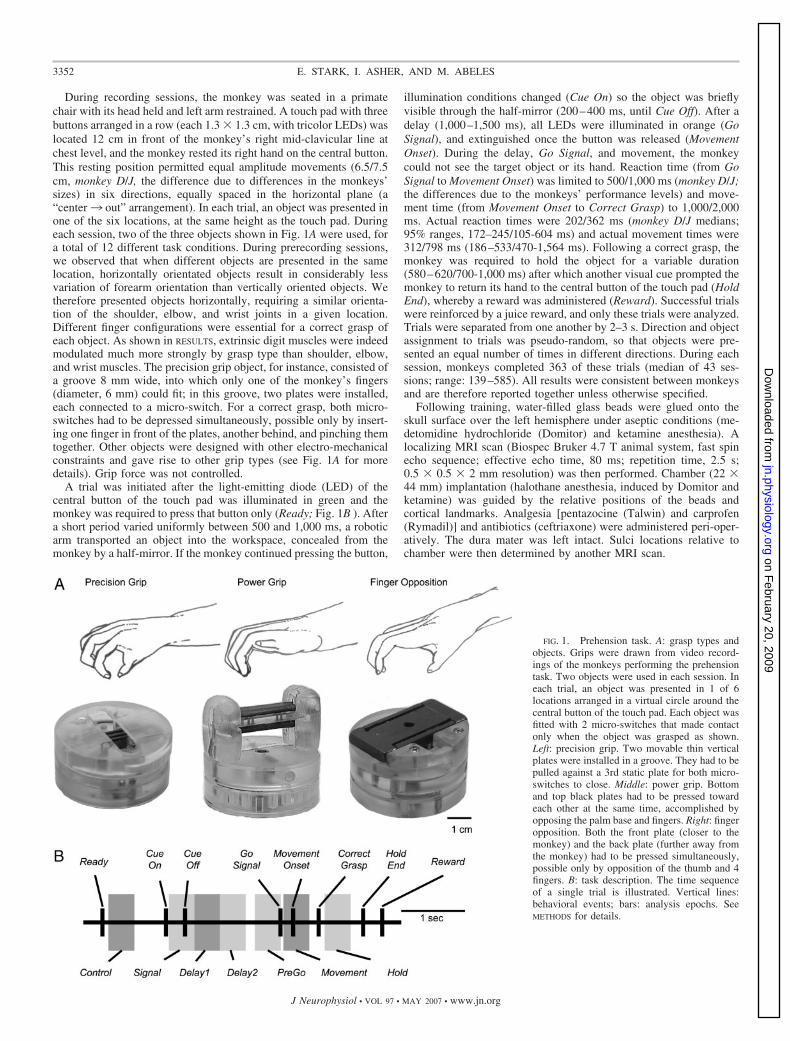
During recording sessions, the monkey was seated in a primatechair with its head held and left arm restrained. A touch pad with threebuttons arranged in a row (each 1.3 � 1.3 cm, with tricolor LEDs) waslocated 12 cm in front of the monkey’s right mid-clavicular line atchest level, and the monkey rested its right hand on the central button.This resting position permitted equal amplitude movements (6.5/7.5cm, monkey D/J, the difference due to differences in the monkeys’sizes) in six directions, equally spaced in the horizontal plane (a“center3 out” arrangement). In each trial, an object was presented inone of the six locations, at the same height as the touch pad. Duringeach session, two of the three objects shown in Fig. 1A were used, fora total of 12 different task conditions. During prerecording sessions,we observed that when different objects are presented in the samelocation, horizontally orientated objects result in considerably lessvariation of forearm orientation than vertically oriented objects. Wetherefore presented objects horizontally, requiring a similar orienta-tion of the shoulder, elbow, and wrist joints in a given location.Different finger configurations were essential for a correct grasp ofeach object. As shown in RESULTS, extrinsic digit muscles were indeedmodulated much more strongly by grasp type than shoulder, elbow,and wrist muscles. The precision grip object, for instance, consisted ofa groove 8 mm wide, into which only one of the monkey’s fingers(diameter, 6 mm) could fit; in this groove, two plates were installed,each connected to a micro-switch. For a correct grasp, both micro-switches had to be depressed simultaneously, possible only by insert-ing one finger in front of the plates, another behind, and pinching themtogether. Other objects were designed with other electro-mechanicalconstraints and gave rise to other grip types (see Fig. 1A for moredetails). Grip force was not controlled.
A trial was initiated after the light-emitting diode (LED) of thecentral button of the touch pad was illuminated in green and themonkey was required to press that button only (Ready; Fig. 1B ). Aftera short period varied uniformly between 500 and 1,000 ms, a roboticarm transported an object into the workspace, concealed from themonkey by a half-mirror. If the monkey continued pressing the button,
illumination conditions changed (Cue On) so the object was brieflyvisible through the half-mirror (200–400 ms, until Cue Off). After adelay (1,000–1,500 ms), all LEDs were illuminated in orange (GoSignal), and extinguished once the button was released (MovementOnset). During the delay, Go Signal, and movement, the monkeycould not see the target object or its hand. Reaction time (from GoSignal to Movement Onset) was limited to 500/1,000 ms (monkey D/J;the differences due to the monkeys’ performance levels) and move-ment time (from Movement Onset to Correct Grasp) to 1,000/2,000ms. Actual reaction times were 202/362 ms (monkey D/J medians;95% ranges, 172–245/105-604 ms) and actual movement times were312/798 ms (186–533/470-1,564 ms). Following a correct grasp, themonkey was required to hold the object for a variable duration(580–620/700-1,000 ms) after which another visual cue prompted themonkey to return its hand to the central button of the touch pad (HoldEnd), whereby a reward was administered (Reward). Successful trialswere reinforced by a juice reward, and only these trials were analyzed.Trials were separated from one another by 2–3 s. Direction and objectassignment to trials was pseudo-random, so that objects were pre-sented an equal number of times in different directions. During eachsession, monkeys completed 363 of these trials (median of 43 ses-sions; range: 139–585). All results were consistent between monkeysand are therefore reported together unless otherwise specified.
Following training, water-filled glass beads were glued onto theskull surface over the left hemisphere under aseptic conditions (me-detomidine hydrochloride (Domitor) and ketamine anesthesia). Alocalizing MRI scan (Biospec Bruker 4.7 T animal system, fast spinecho sequence; effective echo time, 80 ms; repetition time, 2.5 s;0.5 � 0.5 � 2 mm resolution) was then performed. Chamber (22 �44 mm) implantation (halothane anesthesia, induced by Domitor andketamine) was guided by the relative positions of the beads andcortical landmarks. Analgesia [pentazocine (Talwin) and carprofen(Rymadil)] and antibiotics (ceftriaxone) were administered peri-oper-atively. The dura mater was left intact. Sulci locations relative tochamber were then determined by another MRI scan.
FIG. 1. Prehension task. A: grasp types andobjects. Grips were drawn from video record-ings of the monkeys performing the prehensiontask. Two objects were used in each session. Ineach trial, an object was presented in 1 of 6locations arranged in a virtual circle around thecentral button of the touch pad. Each object wasfitted with 2 micro-switches that made contactonly when the object was grasped as shown.Left: precision grip. Two movable thin verticalplates were installed in a groove. They had to bepulled against a 3rd static plate for both micro-switches to close. Middle: power grip. Bottomand top black plates had to be pressed towardeach other at the same time, accomplished byopposing the palm base and fingers. Right: fingeropposition. Both the front plate (closer to themonkey) and the back plate (further away fromthe monkey) had to be pressed simultaneously,possible only by opposition of the thumb and 4fingers. B: task description. The time sequenceof a single trial is illustrated. Vertical lines:behavioral events; bars: analysis epochs. SeeMETHODS for details.
3352 E. STARK, I. ASHER, AND M. ABELES
J Neurophysiol • VOL 97 • MAY 2007 • www.jn.org
on February 20, 2009
jn.physiology.orgD
ownloaded from
Data acquisition and preprocessing
During each recording session, �16 glass-coated tungsten micro-electrodes were employed (impedance 0.2–2 M� at 1 kHz). Elec-trodes were arranged in two circular guide tubes that were lowereddown to �1 mm above the dura mater (8 electrodes in each guidetube; inter-electrode spacing within tube �300 �m; Double MT,Alpha-Omega Engineering, Nazareth, Israel). During each session,one guide tube was aimed toward PMd and another toward PMv. Theborder between primary motor cortex (M1) and PM was establishedbased on stimulations made during separate mapping sessions (Fig.4A): in M1 movements were typically evoked at low currents (�40�A) and visual responses during mapping were rare (for details ofstimulation parameters and mapping protocol, see Cortical mapping).The border between PMd and PMv was defined as the line extendingcaudally from the arcuate spur (dotted lines in Fig. 4, C and D). Eachelectrode was lowered through the dura mater independently (EPS1.31, Alpha-Omega Eng.) until spiking activity was encountered,inserted an additional distance into the cortex (median, 0.71 mm), andleft in the same site during the entire recording and mapping session.The signal from each electrode was amplified (10,000), band-passfiltered (1–10,000 Hz), and sampled at 25 kHz (Alpha-Map 5.4,Alpha-Omega Eng.).
An off-line procedure was applied to identify and sort spikewaveforms in the 25-kHz digitized traces. First, �50-Hz line influ-ences were removed by cycle-triggered averaging: the signal follow-ing AC polarity change in a main was averaged over many cycles andsubtracted from the original signal in an adaptive manner. Second,spikes were detected by computing a modified second derivative (7samples backward and 11 forward), accentuating “spiky” signal fea-tures (bimodal, skewed, and sharp). Segments that crossed a threshold(4.5 SDs from the mean derivative) were identified. Within eachsegment, the occurrence time of the putative spike was defined as thetime of the maximal derivative. If a sharper spike was not encounteredwithin 1.2 ms, then 64 samples, starting 10 before the peak, wereextracted. We also detected spikes based on amplitudes (�5 times theSD of the 300- to 6,000-Hz band-passed trace): spikes detected usingthe two methods were the same in 95% of the cases, and results wereindependent of the detection method. Third, extracted spike wave-forms were linearly de-trended and aligned so each started at the pointof maximal fit with two library principal components accounting, onaverage, for 93% of the waveform variance (Abeles and Goldstein1977). Finally, waveforms were projected onto the principal compo-nent basis to arrive at two coefficients. These coefficients weresubjected to manual spike-sorting in the environment of Alpha-Sort(4.0, Alpha-Omega Eng.).
Eye movements were recorded using an infra-red beam systemtracking movements of one eye (Oculometer, Dr. Bouis, Karlsruhe,Germany). The horizontal and vertical signals from this system weresampled at 400 Hz and low-pass filtered (40 Hz). Behavioral events(LEDs, switches, lights, and so on) were sampled at 6 kHz. Theworkspace and monkey’s movements were monitored using threeinfra-red cameras and recorded on VHS tapes.
During some sessions intra-muscular electromyograms (EMGs)were recorded in parallel to neurons. Wire pairs were inserted intoshoulder (acromion deltoid, latissimus dorsi, pectoralis major), elbow(biceps brachii, brachialis, triceps brachii), wrist (extensor carpiiradialis longus, extensor carpii ulnaris, palmaris longus), and extrinsicdigit (extensor digitorum communis, flexor digitorum profundus,flexor digitorum sublimis) muscles. Wire positions were verified bystimulation (90-ms trains of 0.2-ms biphasic pulses at 330 Hz) atcurrents of 50–500 �A. The signals from these wires were amplified(30,000), band-pass filtered (30–3,000 Hz), and sampled at 6.25 kHz.Root mean square (RMS) values of EMG were computed by raisingsignals to the second power, applying a low-pass filter (100 Hz), andtaking the square root. Each muscle was recorded during 2–10separate sessions for a total of 61 recordings; each recording was
184–527 (median, 414) trials long. Different recordings of the samemuscle were consistent (correlation-coefficient between mean activityin each of the 12 task conditions, 0.86 � 0.05, mean � SE; 164 pairsof same-muscle recordings).
Cortical mapping
During each session, immediately following neural recordings andwithout moving the electrodes, two standard methods were used tocharacterize each electrode’s recording site: threshold ICMS andsensory-motor mappings (SMM). In the SMM, we assessed recordingsite properties by listening to the modulation of the multi-unit rustle ofeach electrode during proprioceptive input (passive movements ofindividual joints, palpation of muscles), tactile input (stroking of skin,stroking of facial skin while eyes were covered), visual input (3dimensional objects, LEDs, hand movements of the experimentalist;at reaching distance and beyond), and active movements (see Genti-lucci et al. 1988; Graziano et al. 1997; Kakei et al. 2001; Schwartz etal. 2004 for similar SMM protocols). The latter were examined usinga Kluver board variant: a Perspex plate with nine holes in three rows,6.5 cm apart. Each hole contained a small food pellet and was coveredby an object requiring a different grasp (ball, cork, plate, string, andso on). The SMM in each site was summarized by two properties:response modality (motor, somatosensory, and/or visual) and organ(elbow, eye, face, finger, multi-joint, mouth, pinna, shoulder, andwrist were observed).
In the ICMS we noted, for each electrode’s recording site sepa-rately, the movement elicited by the lowest possible current during atleast half of the stimulation trials (0.2-ms biphasic pulses at 330 Hzfor 90 ms at currents of 5–90 �A) (Godschalk et al. 1995; seeGentilucci et al. 1988; Kakei et al. 2001 for similar ICMS protocolsemployed in PM). Movement was summarized by two properties:movement type (flexion, extension, adduction, abduction, opposition,and so on) and organ (same as SMM organs in the preceding text). Weclassified evoked forelimb movements as follows: proximal: move-ments around the shoulder and elbow flexion/extension; wrist: fore-arm supination/pronation and wrist movements; and distal: fingermovements. Only results from proximal and distal sites are reported indetail in this paper. Although at suprathreshold currents we sometimesobserved complex multi-joint movements (Godschalk et al. 1995;Graziano et al. 2002), lower currents always yielded single-jointmovements with the exception of distal movements that combinedseveral fingers in 2/3 of the cases. We did not observe transitions fromproximal to distal (or vice versa) movements as current was changed.
Neural database and data analyses
One baseline (control) and six task epochs were defined for anal-yses, all 400 ms long (Fig. 1B, bars). During the Control epoch,starting 100 ms after Ready, the monkey did not know in whichdirection it would have to reach and what type of grip it would haveto use. During the Signal epoch, starting 50 ms after Cue On, theidentity and location of the target objects was briefly visible yet nomovement was required. During Delay epochs, starting 450 ms afterCue On, there was no visual cue and no movement was required.These exact same conditions were maintained throughout the PreGoepoch (starting 400 ms prior to the Go Signal). The Movement epochstarted 150 ms before the hand left the touch pad, and the Hold epochstarted 100 ms after a Correct Grasp. Note that the Delay2 and PreGoepochs overlap on average by 100 ms, and the Movement epochincludes both reaction and movement time (as in Weinrich and Wise1982).
Units fulfilling the following criteria were analyzed. 1) Anatomy.Only units recorded at PM sites were considered. 2) Mapping. Onlyunits recorded at sites classified as proximal or distal by ICMS and/orSMM were included. 3) Isolation. Quality of single-unit isolation wasdetermined by the homogeneity of spike waveforms, separation of the
3353REACH AND GRASP IN PREMOTOR CORTEX
J Neurophysiol • VOL 97 • MAY 2007 • www.jn.org
on February 20, 2009
jn.physiology.orgD
ownloaded from
projections of spike waveforms onto principal components duringspike-sorting and clear refractory periods in ISI histograms. Onlywell-isolated units were considered. 4) Number of trials. Each unithad to be recorded for at least five trials per task condition and exhibitstationary activity. This was determined by visual inspection of meanfiring rates and raster plots of individual trials. 5) Firing rate. Onlyunits with mean firing rates �1 spike/s during at least one task epochwere used. 6) Task dependency. Spike counts for all task conditionstogether, during the Control and during each task epoch, were com-pared using a two-sample, two-tailed t-test. Only units with P values�0.01/6 during at least one of the six epochs were included.
We made a total of 617 PM penetrations, 531 during recordingsessions, of which 378 were in sites classified as proximal or distal. Atotal of 724 units, recorded from 326 of these sites, passed thepreceding criteria (Table 1, 1st 3 columns, lists the sites, units, andtheir classification). Although the number of units varied when chang-ing parameters used for inclusion criteria 4–6, results were notsensitive to specific parameter values. These units were recordedduring 70–582 (median, 257) trials.
For each unit a preferred direction (PD) and a preferred object wereestimated. PDs were estimated by summation of six vectors: thedirection of each vector was the instructed movement direction, andits amplitude was the mean spike count over all trials in that direction.The direction of the vector sum is the PD. Estimates of PDs made byfitting a cosine function were virtually identical (mean absolute PDdifference, 2.7 �0.28°, mean � SE; 724 units). A preferred objectwas defined as the object that elicited the maximal spike count.
We used a two-way ANOVA, with direction and object as factors, todetermine relation of neural activity to task parameters (effects wereconsidered significant at P � 0.01 levels). This analysis assumes thatspike counts distribute normally: because spike counts are nonnegativeand distribute as a Poisson counting process, for some units a normaldistribution was not a good approximation (Bera-Jarque test of normality:76/724 units with P � 0.01). We therefore repeated analyses using anonparametric ANOVA (2-way Kruskal-Wallis test); results were almostidentical for the two ANOVA tests. Because vector summation was usedto estimate PDs and ANOVA to determine significance of directionaltuning, we compared the vector summation PD estimate with a discreteestimate, the direction in which a unit fired maximally: the mean absolutedifference between the two estimates was 30 � 0.9° (SE) (724 units).Thus for the neural data used in this study, an ANOVA is a reasonablemethod for estimating the significance of tuning. The same results wereobtained when estimating significance using a resampling test (Cram-mond and Kalaska 1996).
To estimate effect sizes we computed eta-squared, defined as �2 ��effect
2 /�total2 (Fisher 1925). Effect variance is defined as the variance of
the mean discharge in each relevant task condition (e.g., �task2 is the
variance of 12 numbers, and �dir2 is the variance of 6 numbers). �2 is
a unit-less measure of the fraction of the total variance associated withan effect. If the signal-to-noise ratio (SNR) is defined as �effect
2 /(�total
2 – �effect2 ) then �2 equals SNR/(1 � SNR) and is thus a
monotonic function of the SNR. �2 is bounded between 0 and 1 andaccounts for linear and nonlinear effects. Because �2 is additive fordifferent effects, the total variance associated with the prehensiontask parameters is equal to the sum of direction, object, andinteraction effect sizes: �task
2 � �dir2 � �obj
2 � �int2 . To compare
variance associated with direction with that not associated withdirection alone, we employed a “reach-grasp index”: RGI � (�dir
2 –�obj
2 – �int2 )/�task
2 . This index equals 1 when there is only a directioneffect and �1 when there is no direction effect.
We used multiple linear regression, model Fu ,t � b0 � b1 �X t �b2 �Y t � � t, to test for relations between neural activity and eyepositions. In this model, F is the spike count of unit u during therelevant epoch of trial t, X and Y are the horizontal and vertical eyepositions during the same time, and � is an error term. All values werestandardized prior to regressing (the mean subtracted and divided bythe SD) for each of the 12 task conditions separately to removepossible task-related effects.
R E S U L T S
Proximal and distal muscles are active differentlyduring prehension
We measured the activity of shoulder, elbow, wrist, andextrinsic digit muscles during the prehension task (Fig. 1) asmonkeys reached in different directions and grasped variousobjects. Muscles were generally quiescent before the Go Signaland became active just before Movement Onset as the mon-key’s hand released the touch pad (Fig. 2). To quantify thetemporal relations between reach and grasp movement com-ponents and muscle activity, we identified, for each muscle, thetime of peak activity. Activity of all muscles peaked duringmovement, and activation of proximal (shoulder and elbow)and distal (extrinsic digit) muscles overlapped (Fig. 3A). How-ever, activity of proximal muscles peaked 125 ms after Move-ment Onset, whereas activity of distal muscles peaked 221 ms
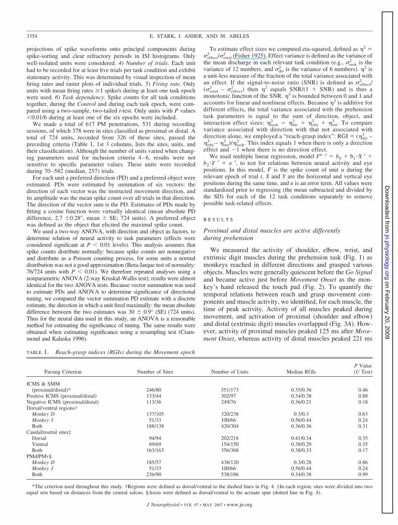
TABLE 1. Reach-grasp indices (RGIs) during the Movement epoch
Parsing Criterion Number of Sites Number of Units Median RGIsP Value(U Test)
ICMS & SMM(proximal/distal)* 246/80 551/173 0.35/0.36 0.46
Positive ICMS (proximal/distal) 133/44 302/97 0.34/0.38 0.88Negative ICMS (proximal/distal) 113/36 249/76 0.36/0.21 0.18Dorsal/ventral regions†
Monkey D 137/105 320/238 0.3/0.3 0.63Monkey J 51/33 100/66 0.56/0.44 0.24Both 188/138 420/304 0.36/0.36 0.31
Caudal/rostral sites‡Dorsal 94/94 202/218 0.41/0.34 0.35Ventral 69/69 154/150 0.38/0.29 0.35Both 163/163 356/368 0.38/0.33 0.17
PMd/PMv§Monkey D 185/57 438/120 0.3/0.28 0.86Monkey J 51/33 100/66 0.56/0.44 0.24Both 236/90 538/186 0.34/0.38 0.99
*The criterion used throughout this study. †Regions were defined as dorsal/ventral to the dashed lines in Fig. 4. ‡In each region, sites were divided into twoequal sets based on distances from the central sulcus. §Areas were defined as dorsal/ventral to the acruate spur (dotted line in Fig. 4).
3354 E. STARK, I. ASHER, AND M. ABELES
J Neurophysiol • VOL 97 • MAY 2007 • www.jn.org
on February 20, 2009
jn.physiology.orgD
ownloaded from
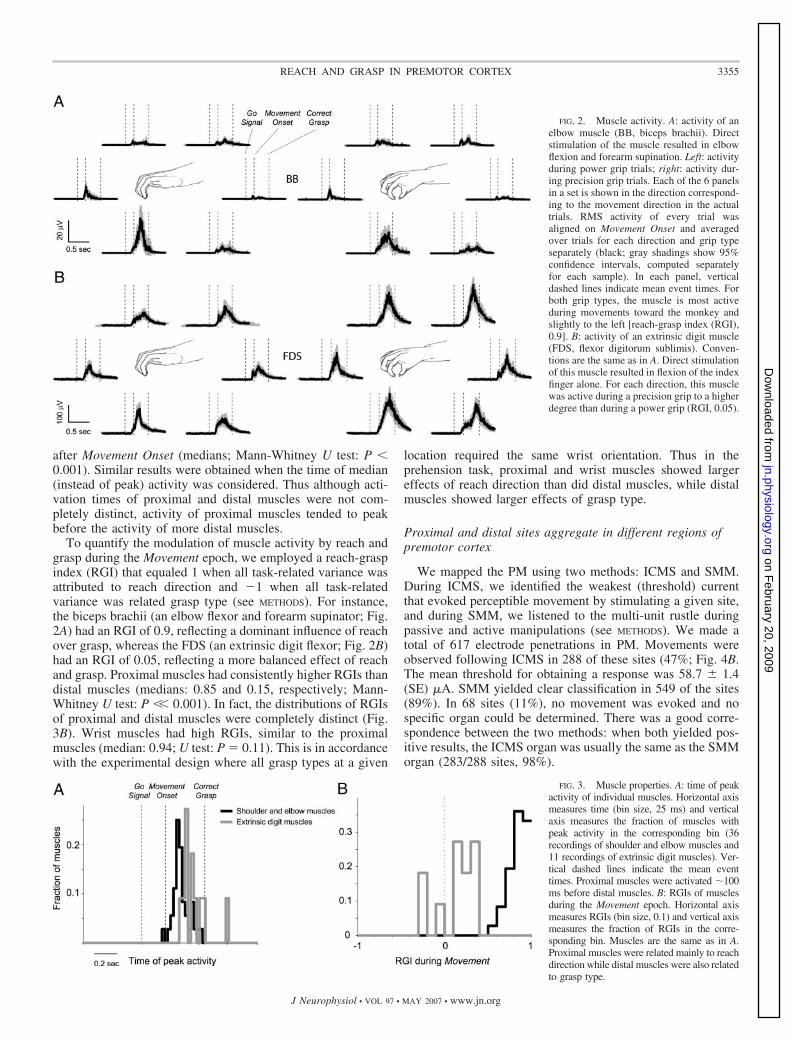
after Movement Onset (medians; Mann-Whitney U test: P �0.001). Similar results were obtained when the time of median(instead of peak) activity was considered. Thus although acti-vation times of proximal and distal muscles were not com-pletely distinct, activity of proximal muscles tended to peakbefore the activity of more distal muscles.
To quantify the modulation of muscle activity by reach andgrasp during the Movement epoch, we employed a reach-graspindex (RGI) that equaled 1 when all task-related variance wasattributed to reach direction and �1 when all task-relatedvariance was related grasp type (see METHODS). For instance,the biceps brachii (an elbow flexor and forearm supinator; Fig.2A) had an RGI of 0.9, reflecting a dominant influence of reachover grasp, whereas the FDS (an extrinsic digit flexor; Fig. 2B)had an RGI of 0.05, reflecting a more balanced effect of reachand grasp. Proximal muscles had consistently higher RGIs thandistal muscles (medians: 0.85 and 0.15, respectively; Mann-Whitney U test: P �� 0.001). In fact, the distributions of RGIsof proximal and distal muscles were completely distinct (Fig.3B). Wrist muscles had high RGIs, similar to the proximalmuscles (median: 0.94; U test: P � 0.11). This is in accordancewith the experimental design where all grasp types at a given
location required the same wrist orientation. Thus in theprehension task, proximal and wrist muscles showed largereffects of reach direction than did distal muscles, while distalmuscles showed larger effects of grasp type.
Proximal and distal sites aggregate in different regions ofpremotor cortex
We mapped the PM using two methods: ICMS and SMM.During ICMS, we identified the weakest (threshold) currentthat evoked perceptible movement by stimulating a given site,and during SMM, we listened to the multi-unit rustle duringpassive and active manipulations (see METHODS). We made atotal of 617 electrode penetrations in PM. Movements wereobserved following ICMS in 288 of these sites (47%; Fig. 4B.The mean threshold for obtaining a response was 58.7 � 1.4(SE) �A. SMM yielded clear classification in 549 of the sites(89%). In 68 sites (11%), no movement was evoked and nospecific organ could be determined. There was a good corre-spondence between the two methods: when both yielded pos-itive results, the ICMS organ was usually the same as the SMMorgan (283/288 sites, 98%).
FIG. 2. Muscle activity. A: activity of anelbow muscle (BB, biceps brachii). Directstimulation of the muscle resulted in elbowflexion and forearm supination. Left: activityduring power grip trials; right: activity dur-ing precision grip trials. Each of the 6 panelsin a set is shown in the direction correspond-ing to the movement direction in the actualtrials. RMS activity of every trial wasaligned on Movement Onset and averagedover trials for each direction and grip typeseparately (black; gray shadings show 95%confidence intervals, computed separatelyfor each sample). In each panel, verticaldashed lines indicate mean event times. Forboth grip types, the muscle is most activeduring movements toward the monkey andslightly to the left [reach-grasp index (RGI),0.9]. B: activity of an extrinsic digit muscle(FDS, flexor digitorum sublimis). Conven-tions are the same as in A. Direct stimulationof this muscle resulted in flexion of the indexfinger alone. For each direction, this musclewas active during a precision grip to a higherdegree than during a power grip (RGI, 0.05).
FIG. 3. Muscle properties. A: time of peakactivity of individual muscles. Horizontal axismeasures time (bin size, 25 ms) and verticalaxis measures the fraction of muscles withpeak activity in the corresponding bin (36recordings of shoulder and elbow muscles and11 recordings of extrinsic digit muscles). Ver-tical dashed lines indicate the mean eventtimes. Proximal muscles were activated �100ms before distal muscles. B: RGIs of musclesduring the Movement epoch. Horizontal axismeasures RGIs (bin size, 0.1) and vertical axismeasures the fraction of RGIs in the corre-sponding bin. Muscles are the same as in A.Proximal muscles were related mainly to reachdirection while distal muscles were also relatedto grasp type.
3355REACH AND GRASP IN PREMOTOR CORTEX
J Neurophysiol • VOL 97 • MAY 2007 • www.jn.org
on February 20, 2009
jn.physiology.orgD
ownloaded from
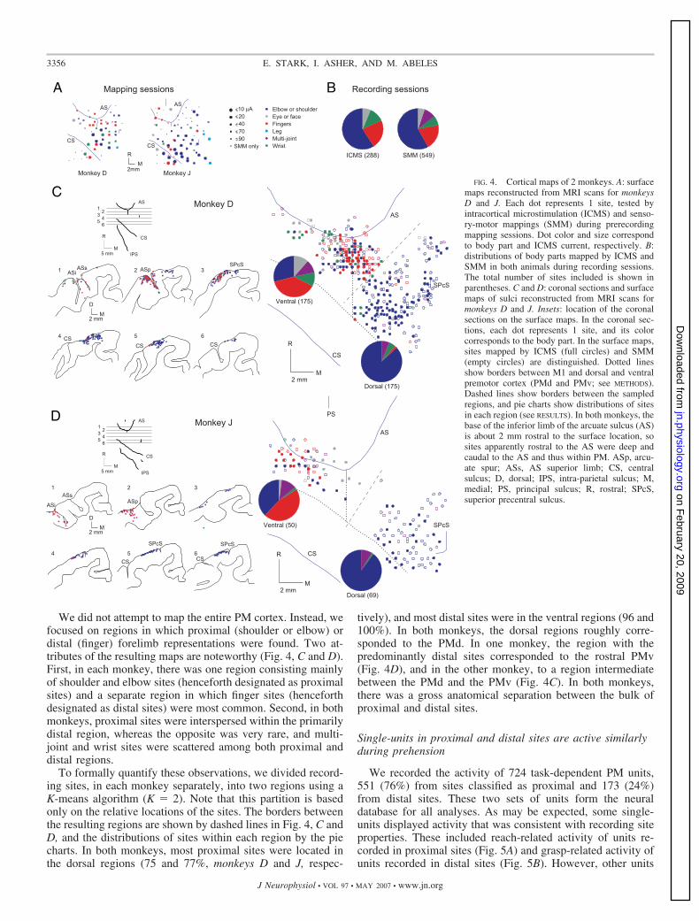
We did not attempt to map the entire PM cortex. Instead, wefocused on regions in which proximal (shoulder or elbow) ordistal (finger) forelimb representations were found. Two at-tributes of the resulting maps are noteworthy (Fig. 4, C and D).First, in each monkey, there was one region consisting mainlyof shoulder and elbow sites (henceforth designated as proximalsites) and a separate region in which finger sites (henceforthdesignated as distal sites) were most common. Second, in bothmonkeys, proximal sites were interspersed within the primarilydistal region, whereas the opposite was very rare, and multi-joint and wrist sites were scattered among both proximal anddistal regions.
To formally quantify these observations, we divided record-ing sites, in each monkey separately, into two regions using aK-means algorithm (K � 2). Note that this partition is basedonly on the relative locations of the sites. The borders betweenthe resulting regions are shown by dashed lines in Fig. 4, C andD, and the distributions of sites within each region by the piecharts. In both monkeys, most proximal sites were located inthe dorsal regions (75 and 77%, monkeys D and J, respec-
tively), and most distal sites were in the ventral regions (96 and100%). In both monkeys, the dorsal regions roughly corre-sponded to the PMd. In one monkey, the region with thepredominantly distal sites corresponded to the rostral PMv(Fig. 4D), and in the other monkey, to a region intermediatebetween the PMd and the PMv (Fig. 4C). In both monkeys,there was a gross anatomical separation between the bulk ofproximal and distal sites.
Single-units in proximal and distal sites are active similarlyduring prehension
We recorded the activity of 724 task-dependent PM units,551 (76%) from sites classified as proximal and 173 (24%)from distal sites. These two sets of units form the neuraldatabase for all analyses. As may be expected, some single-units displayed activity that was consistent with recording siteproperties. These included reach-related activity of units re-corded in proximal sites (Fig. 5A) and grasp-related activity ofunits recorded in distal sites (Fig. 5B). However, other units
FIG. 4. Cortical maps of 2 monkeys. A: surfacemaps reconstructed from MRI scans for monkeysD and J. Each dot represents 1 site, tested byintracortical microstimulation (ICMS) and senso-ry-motor mappings (SMM) during prerecordingmapping sessions. Dot color and size correspondto body part and ICMS current, respectively. B:distributions of body parts mapped by ICMS andSMM in both animals during recording sessions.The total number of sites included is shown inparentheses. C and D: coronal sections and surfacemaps of sulci reconstructed from MRI scans formonkeys D and J. Insets: location of the coronalsections on the surface maps. In the coronal sec-tions, each dot represents 1 site, and its colorcorresponds to the body part. In the surface maps,sites mapped by ICMS (full circles) and SMM(empty circles) are distinguished. Dotted linesshow borders between M1 and dorsal and ventralpremotor cortex (PMd and PMv; see METHODS).Dashed lines show borders between the sampledregions, and pie charts show distributions of sitesin each region (see RESULTS). In both monkeys, thebase of the inferior limb of the arcuate sulcus (AS)is about 2 mm rostral to the surface location, sosites apparently rostral to the AS were deep andcaudal to the AS and thus within PM. ASp, arcu-ate spur; ASs, AS superior limb; CS, centralsulcus; D, dorsal; IPS, intra-parietal sulcus; M,medial; PS, principal sulcus; R, rostral; SPcS,superior precentral sulcus.
3356 E. STARK, I. ASHER, AND M. ABELES
J Neurophysiol • VOL 97 • MAY 2007 • www.jn.org
on February 20, 2009
jn.physiology.orgD
ownloaded from
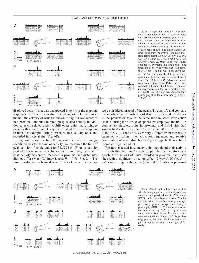
displayed activity that was unexpected in terms of the mappingresponses of the corresponding recording sites. For instance,the unit the activity of which is shown in Fig. 6A was recordedin a proximal site but exhibited grasp-related activity in addi-tion to reach-related activity. Still other units had dischargepatterns that were completely inconsistent with the mappingresults; for example, strictly reach-related activity of a unitrecorded in a distal site (Fig. 6B).
Single-units were active throughout the task. To assignspecific values to the time of activity, we measured the time ofpeak activity of single units: for 318/724 (44%) units, activitypeaked prior to movement. In contrast to muscles, the time ofpeak activity of neurons recorded in proximal and distal sitesdid not differ (Mann-Whitney U test: P � 0.76; Fig. 7A). Thesame results were obtained when times of median activation
were considered instead of the peaks. To quantify and comparethe involvement of units recorded in proximal and distal sitesin the prehension task at the same time muscles were active(that is, during the Movement epoch), we employed the RGI. Incontrast to muscles, units in proximal and distal sites hadsimilar RGI values (median RGIs: 0.35 and 0.36; U test: P �0.46; Fig. 7B). Thus units were very different from muscles interms of activation time, activation sequence, and relativecontribution of reach direction and grasp type to their activity(compare Figs. 3 and 7).
We further tested how many units modulated their activityby reach direction and/or grasp type. During the Movementepoch, the fractions of units recorded in proximal and distalsites with a significant direction effect (2-way ANOVA, P �0.01) were roughly the same (386 and 126 units in proximal
FIG. 5. Single-unit activity consistentwith the mapping results. A: raster displaysand peri-event time histograms (PETHs) of aunit recorded in a proximal site in PMdwhere ICMS resulted in shoulder extension.Panels are laid out as in Fig. 2A. Bottom partof each panel shows spike times (short blacklines) and behavioral events (long gray lines,from left to right: On, Cue On; Off, Cue Off;Go, Go Signal; M, Movement Onset; Gr,Correct Grasp; H, Hold End). Top: PETHobtained by averaging the single trial spiketimes and convolving with a Gaussian kernel(SD, 15 ms). The unit was most active dur-ing the Movement epoch of trials in whichmovement direction was left, regardless ofgrip type (RGI, 0.8). B: activity of a unitrecorded in a distal site in PMv, where ICMSresulted in flexion of all fingers. For eachand every direction, the unit’s discharge dur-ing the Movement epoch was stronger for apower grip than for a precision grip (RGI,�0.56).
FIG. 6. Single-unit activity inconsistentwith the mapping results. A: activity of a unitrecorded in a proximal site in PMd whereICMS resulted in elbow extension. Yet foreach direction, the unit’s discharge during aprecision grip was stronger than during apower grip (RGI, �0.65). Conventions arethe same as in Fig. 5. B: activity of a unitrecorded in a distal site in PMv where ICMSresulted in flexion of fingers 2–5. Regardlessof grip type, the unit’s discharge was stron-gest during movements to the right (RGI,0.65).
3357REACH AND GRASP IN PREMOTOR CORTEX
J Neurophysiol • VOL 97 • MAY 2007 • www.jn.org
on February 20, 2009
jn.physiology.orgD
ownloaded from
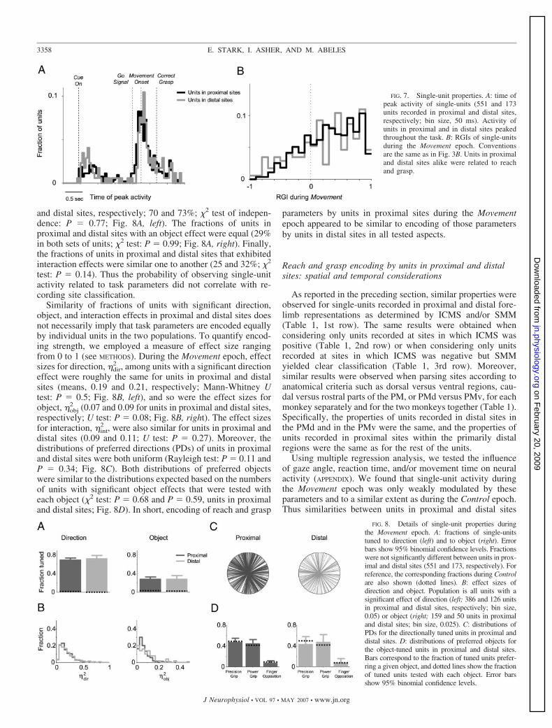
and distal sites, respectively; 70 and 73%; �2 test of indepen-dence: P � 0.77; Fig. 8A, left). The fractions of units inproximal and distal sites with an object effect were equal (29%in both sets of units; �2 test: P � 0.99; Fig. 8A, right). Finally,the fractions of units in proximal and distal sites that exhibitedinteraction effects were similar one to another (25 and 32%; �2
test: P � 0.14). Thus the probability of observing single-unitactivity related to task parameters did not correlate with re-cording site classification.
Similarity of fractions of units with significant direction,object, and interaction effects in proximal and distal sites doesnot necessarily imply that task parameters are encoded equallyby individual units in the two populations. To quantify encod-ing strength, we employed a measure of effect size rangingfrom 0 to 1 (see METHODS). During the Movement epoch, effectsizes for direction, �dir
2 , among units with a significant directioneffect were roughly the same for units in proximal and distalsites (means, 0.19 and 0.21, respectively; Mann-Whitney Utest: P � 0.5; Fig. 8B, left), and so were the effect sizes forobject, �obj
2 (0.07 and 0.09 for units in proximal and distal sites,respectively; U test: P � 0.08; Fig. 8B, right). The effect sizesfor interaction, �int
2 , were also similar for units in proximal anddistal sites (0.09 and 0.11; U test: P � 0.27). Moreover, thedistributions of preferred directions (PDs) of units in proximaland distal sites were both uniform (Rayleigh test: P � 0.11 andP � 0.34; Fig. 8C). Both distributions of preferred objectswere similar to the distributions expected based on the numbersof units with significant object effects that were tested witheach object (�2 test: P � 0.68 and P � 0.59, units in proximaland distal sites; Fig. 8D). In short, encoding of reach and grasp
parameters by units in proximal sites during the Movementepoch appeared to be similar to encoding of those parametersby units in distal sites in all tested aspects.
Reach and grasp encoding by units in proximal and distalsites: spatial and temporal considerations
As reported in the preceding section, similar properties wereobserved for single-units recorded in proximal and distal fore-limb representations as determined by ICMS and/or SMM(Table 1, 1st row). The same results were obtained whenconsidering only units recorded at sites in which ICMS waspositive (Table 1, 2nd row) or when considering only unitsrecorded at sites in which ICMS was negative but SMMyielded clear classification (Table 1, 3rd row). Moreover,similar results were observed when parsing sites according toanatomical criteria such as dorsal versus ventral regions, cau-dal versus rostral parts of the PM, or PMd versus PMv, for eachmonkey separately and for the two monkeys together (Table 1).Specifically, the properties of units recorded in distal sites inthe PMd and in the PMv were the same, and the properties ofunits recorded in proximal sites within the primarily distalregions were the same as for the rest of the units.
Using multiple regression analysis, we tested the influenceof gaze angle, reaction time, and/or movement time on neuralactivity (APPENDIX). We found that single-unit activity duringthe Movement epoch was only weakly modulated by theseparameters and to a similar extent as during the Control epoch.Thus similarities between units in proximal and distal sites
FIG. 7. Single-unit properties. A: time ofpeak activity of single-units (551 and 173units recorded in proximal and distal sites,respectively; bin size, 50 ms). Activity ofunits in proximal and in distal sites peakedthroughout the task. B: RGIs of single-unitsduring the Movement epoch. Conventionsare the same as in Fig. 3B. Units in proximaland distal sites alike were related to reachand grasp.
FIG. 8. Details of single-unit properties duringthe Movement epoch. A: fractions of single-unitstuned to direction (left) and to object (right). Errorbars show 95% binomial confidence levels. Fractionswere not significantly different between units in prox-imal and distal sites (551 and 173, respectively). Forreference, the corresponding fractions during Controlare also shown (dotted lines). B: effect sizes ofdirection and object. Population is all units with asignificant effect of direction (left; 386 and 126 unitsin proximal and distal sites, respectively; bin size,0.05) or object (right; 159 and 50 units in proximaland distal sites; bin size, 0.025). C: distributions ofPDs for the directionally tuned units in proximal anddistal sites. D: distributions of preferred objects forthe object-tuned units in proximal and distal sites.Bars correspond to the fraction of tuned units prefer-ring a given object, and dotted lines show the fractionof tuned units tested with each object. Error barsshow 95% binomial confidence levels.
3358 E. STARK, I. ASHER, AND M. ABELES
J Neurophysiol • VOL 97 • MAY 2007 • www.jn.org
on February 20, 2009
jn.physiology.orgD
ownloaded from
observed during the Movement epoch are unlikely to resultfrom modulations by the tested parameters.
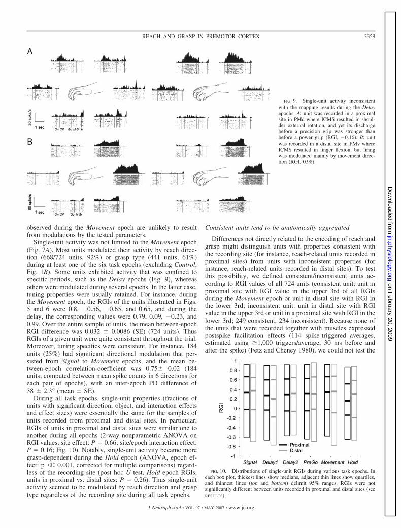
Single-unit activity was not limited to the Movement epoch(Fig. 7A). Most units modulated their activity by reach direc-tion (668/724 units, 92%) or grasp type (441 units, 61%)during at least one of the six task epochs (excluding Control,Fig. 1B). Some units exhibited activity that was confined tospecific periods, such as the Delay epochs (Fig. 9), whereasothers were modulated during several epochs. In the latter case,tuning properties were usually retained. For instance, duringthe Movement epoch, the RGIs of the units illustrated in Figs.5 and 6 were 0.8, �0.56, �0.65, and 0.65, and during thedelay, the corresponding values were 0.79, 0.09, �0.23, and0.99. Over the entire sample of units, the mean between-epochRGI difference was 0.032 � 0.0086 (SE) (724 units). ThusRGIs of a given unit were quite consistent throughout the trial.Moreover, tuning specifics were consistent. For instance, 184units (25%) had significant directional modulation that per-sisted from Signal to Movement epochs, and the mean be-tween-epoch correlation-coefficient was 0.75� 0.02 (184units; computed between mean spike counts in 6 directions foreach pair of epochs), with an inter-epoch PD difference of38 � 2.3° (mean � SE).
During all task epochs, single-unit properties (fractions ofunits with significant direction, object, and interaction effectsand effect sizes) were essentially the same for the samples ofunits recorded from proximal and distal sites. In particular,RGIs of units in proximal and distal sites were similar one toanother during all epochs (2-way nonparametric ANOVA onRGI values, site effect: P � 0.66; site/epoch interaction effect:P � 0.16; Fig. 10). Notably, single-unit activity became moregrasp-dependent during the Hold epoch (ANOVA, epoch ef-fect: p �� 0.001, corrected for multiple comparisons) regard-less of the recording site (post hoc U test, Hold epoch RGIs,units in proximal vs. distal sites: P � 0.26). Thus single-unitactivity seemed to be modulated by reach direction and grasptype regardless of the recording site during all task epochs.
Consistent units tend to be anatomically aggregated
Differences not directly related to the encoding of reach andgrasp might distinguish units with properties consistent withthe recording site (for instance, reach-related units recorded inproximal sites) from units with inconsistent properties (forinstance, reach-related units recorded in distal sites). To testthis possibility, we defined consistent/inconsistent units ac-cording to RGI values of all 724 units (consistent unit: unit inproximal site with RGI value in the upper 3rd of all RGIsduring the Movement epoch or unit in distal site with RGI inthe lower 3rd; inconsistent unit: unit in distal site with RGIvalue in the upper 3rd or unit in a proximal site with RGI in thelower 3rd; 249 consistent, 234 inconsistent). Because none ofthe units that were recorded together with muscles expressedpostspike facilitation effects (114 spike-triggered averages,estimated using �1,000 triggers/average, 30 ms before andafter the spike) (Fetz and Cheney 1980), we could not test the
FIG. 9. Single-unit activity inconsistentwith the mapping results during the Delayepochs. A: unit was recorded in a proximalsite in PMd where ICMS resulted in shoul-der external rotation, and yet its dischargebefore a precision grip was stronger thanbefore a power grip (RGI, �0.16). B: unitwas recorded in a distal site in PMv whereICMS resulted in finger flexion, but firingwas modulated mainly by movement direc-tion (RGI, 0.98).
FIG. 10. Distributions of single-unit RGIs during various task epochs. Ineach box plot, thickest lines show medians, adjacent thin lines show quartiles,and thinnest lines (top and bottom) delimit 95% ranges. RGIs were notsignificantly different between units recorded in proximal and distal sites (seeRESULTS).
3359REACH AND GRASP IN PREMOTOR CORTEX
J Neurophysiol • VOL 97 • MAY 2007 • www.jn.org
on February 20, 2009
jn.physiology.orgD
ownloaded from
tendency of consistent units to have direct output (corticomo-toneuronal) connections. We examined other features thatincluded firing rates during the Control epoch (spike/s), trial-to-trial variability (of firing rates), spike waveform peak-to-peak amplitude (�V), ICMS thresholds (�A), and recordingdepth (mm). However, no significant differences between con-sistent versus inconsistent units were observed for any of thetested features.
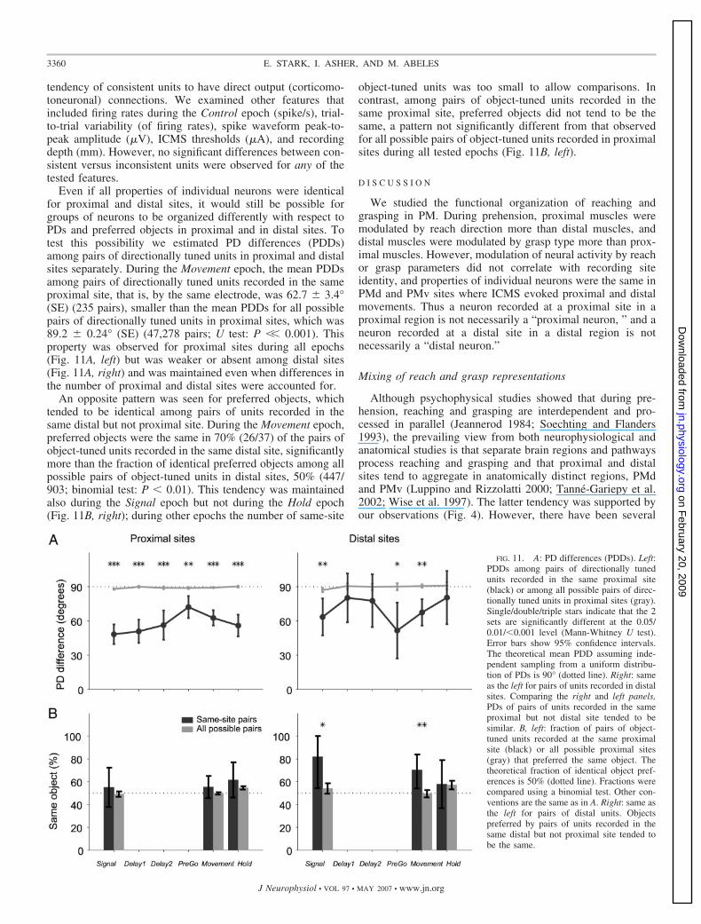
Even if all properties of individual neurons were identicalfor proximal and distal sites, it would still be possible forgroups of neurons to be organized differently with respect toPDs and preferred objects in proximal and in distal sites. Totest this possibility we estimated PD differences (PDDs)among pairs of directionally tuned units in proximal and distalsites separately. During the Movement epoch, the mean PDDsamong pairs of directionally tuned units recorded in the sameproximal site, that is, by the same electrode, was 62.7 � 3.4°(SE) (235 pairs), smaller than the mean PDDs for all possiblepairs of directionally tuned units in proximal sites, which was89.2 � 0.24° (SE) (47,278 pairs; U test: P �� 0.001). Thisproperty was observed for proximal sites during all epochs(Fig. 11A, left) but was weaker or absent among distal sites(Fig. 11A, right) and was maintained even when differences inthe number of proximal and distal sites were accounted for.
An opposite pattern was seen for preferred objects, whichtended to be identical among pairs of units recorded in thesame distal but not proximal site. During the Movement epoch,preferred objects were the same in 70% (26/37) of the pairs ofobject-tuned units recorded in the same distal site, significantlymore than the fraction of identical preferred objects among allpossible pairs of object-tuned units in distal sites, 50% (447/903; binomial test: P � 0.01). This tendency was maintainedalso during the Signal epoch but not during the Hold epoch(Fig. 11B, right); during other epochs the number of same-site
object-tuned units was too small to allow comparisons. Incontrast, among pairs of object-tuned units recorded in thesame proximal site, preferred objects did not tend to be thesame, a pattern not significantly different from that observedfor all possible pairs of object-tuned units recorded in proximalsites during all tested epochs (Fig. 11B, left).
D I S C U S S I O N
We studied the functional organization of reaching andgrasping in PM. During prehension, proximal muscles weremodulated by reach direction more than distal muscles, anddistal muscles were modulated by grasp type more than prox-imal muscles. However, modulation of neural activity by reachor grasp parameters did not correlate with recording siteidentity, and properties of individual neurons were the same inPMd and PMv sites where ICMS evoked proximal and distalmovements. Thus a neuron recorded at a proximal site in aproximal region is not necessarily a “proximal neuron, ” and aneuron recorded at a distal site in a distal region is notnecessarily a “distal neuron.”
Mixing of reach and grasp representations
Although psychophysical studies showed that during pre-hension, reaching and grasping are interdependent and pro-cessed in parallel (Jeannerod 1984; Soechting and Flanders1993), the prevailing view from both neurophysiological andanatomical studies is that separate brain regions and pathwaysprocess reaching and grasping and that proximal and distalsites tend to aggregate in anatomically distinct regions, PMdand PMv (Luppino and Rizzolatti 2000; Tanne-Gariepy et al.2002; Wise et al. 1997). The latter tendency was supported byour observations (Fig. 4). However, there have been several
FIG. 11. A: PD differences (PDDs). Left:PDDs among pairs of directionally tunedunits recorded in the same proximal site(black) or among all possible pairs of direc-tionally tuned units in proximal sites (gray).Single/double/triple stars indicate that the 2sets are significantly different at the 0.05/0.01/�0.001 level (Mann-Whitney U test).Error bars show 95% confidence intervals.The theoretical mean PDD assuming inde-pendent sampling from a uniform distribu-tion of PDs is 90° (dotted line). Right: sameas the left for pairs of units recorded in distalsites. Comparing the right and left panels,PDs of pairs of units recorded in the sameproximal but not distal site tended to besimilar. B, left: fraction of pairs of object-tuned units recorded at the same proximalsite (black) or all possible proximal sites(gray) that preferred the same object. Thetheoretical fraction of identical object pref-erences is 50% (dotted line). Fractions werecompared using a binomial test. Other con-ventions are the same as in A. Right: same asthe left for pairs of distal units. Objectspreferred by pairs of units recorded in thesame distal but not proximal site tended tobe the same.
3360 E. STARK, I. ASHER, AND M. ABELES
J Neurophysiol • VOL 97 • MAY 2007 • www.jn.org
on February 20, 2009
jn.physiology.orgD
ownloaded from
reports of anatomical mixing of muscle fields, both in M1(finger muscles; Rathelot and Strick 2006; Sato and Tanji1989) and in the PM (proximal and distal muscles; Dum andStrick 2005; Godschalk et al. 1995; Raos et al. 2004). This wasalso confirmed in the present study.
The main result of this study, mixing of single neuronsencoding reach and grasp within a given site, is novel. How isthis possible, given the large number of studies that tested PMactivity in relation to reach and grasp? One possibility isrelated to learning-induced changes in cortical circuitry duringtask performance. Alternatively, mixing may not have beendemonstrated because of the novel way reach and grasp werecombined in the current task. Previous experiments testingdirectional modulation of PM neurons have typically alteredreach direction in an orderly manner without manipulatinggrasp type (Caminiti et al. 1991; Shen and Alexander 1997),whereas experiments examining grasping used reach-to-graspmovements, varying the grasped object while keeping reachdirection fixed (Hepp-Reymond et al. 1994; Murata et al.1997). The current prehension task systematically varied bothreach direction and grasp type, opening the possibility toobserve neural activity related to either component.
We focused on PM areas to study the neural control ofprehension, but other brain regions are clearly involved. Cer-ebellar activity has been related to reaching and grasping (vanKan et al. 1994) but not at the same time (Mason et al. 2006).Here we showed that although activity of proximal musclestended to precede activity of distal muscles (Fig. 3A), theactivity of single PM neurons was related to reach and graspthroughout preparation and execution of prehension (Fig. 7A).We did, however, observe a general tendency of PM neurons tobecome more grasp-related during the Hold period (Fig. 10).Whether these differences are due to anatomical or method-ological differences is an issue for future studies.
Behavioral separation between reach and grasp
The current task aimed to differentiate between reach andgrasp by systematically varying reach direction and grasp type.As the analysis of muscle activity showed, this was largelysuccessful (Fig. 3B). There are other possible experimentalparadigms that may be used for the same purpose. One possi-bility is to impose time restrictions on the reach and graspphases, inducing artificial temporal separation. However, evenduring unconstrained reaching and grasping, the activity ofproximal and distal muscles does not peak simultaneously (Fig.3A). A second venue may be to vary reach alone, with nograsping whatsoever, and then vary grasp alone without reach-ing. This approach was partially adopted in the current studyby requiring reaching from one touch pad to another identicallyshaped pad (APPENDIX). The main advantage of the current taskwas to enable characterization of muscle and neural activityduring unconstrained reaching and grasping in a systematicmanner.
None of the measured muscles displayed activity relatedonly to reach or grasp. Although this may seem surprising,during natural movements reach and grasp are in fact biome-chanically dependent. For instance, activity of wrist musclesaccompanies digit movements (Schieber 1995), and shouldermovements are accompanied by stabilization of distal joints.Accordingly, activation patterns of proximal and distal muscles
are expected to overlap. There were, however, some cleardifferences between proximal and distal muscles: proximalmuscles were related mainly to reach direction while moredistal muscles were also related to grasp type (Fig. 3B).
Activity of intrinsic hand muscles (Lemon et al. 1986) wasnot monitored in this study due to technical constraints. Be-cause the activity of extrinsic digit muscles was modulated bygrasp type much more than the activity of proximal muscles,we expect that if monitored, intrinsic muscle activity would bemodulated mainly by grasp type.
The task did not call for any changes in wrist orientationwhen grasping different objects. Although we did not measurehand orientation directly, we videotaped movements fromthree directions. Examination of these records did not revealany gross changes in wrist orientation while moving to eitherobject at the same location. Wrist orientation is influenced byactivity of wrist muscles, which depends on the movementtask. In previous studies, wrist orientation and muscles weremodulated by grasp type when grasping different verticallyoriented objects at the same location (Brochier et al. 2004). Inother behavioral paradigms, the activity of wrist muscles wasrelated to movement direction (Kakei et al. 2001). In thepresent task, wrist muscles had RGI values close to one andwere therefore mainly related to reach direction.
Anatomical and functional considerations
For most analyses, we parsed the sample of neurons into twosets, recorded in proximal and distal sites. There were tworeasons for this “functional” rather than an “anatomical”choice. First, there were some differences between the mon-keys. Although in both monkeys, the majority of proximal siteswere aggregated in similar regions, the caudal PMd (Wise et al.1997), the predominantly distal regions differed between mon-keys, corresponding to the rostral PMv in one monkey and aregion between PMd and PMv in another (Luppino and Riz-zolatti 2000). This variability is not unique: functional corticalfields are known to vary with respect to sulcal landmarksbetween animals (Gabernet et al. 1999; Merzenich et al. 1975).Second, it was previously shown that proximal and distal sitesare mixed in PM (Dum and Strick 2005; Godschalk et al.1995). This was also observed in the current study. Thus it wasmore meaningful to parse sites according to the results ofICMS and/or SMM than by anatomy. Yet parsing sites accord-ing to anatomical criteria did not reveal any differences either(Table 1). Nevertheless, it is possible that in other PM sites, notexplored in the current study, neurons with different propertiesdo exist.
Possible explanations for the lack of correlation betweensingle-unit properties and recording site identity
Single-unit activity was measured during the prehension taskwhile recording site properties were tested using thresholdICMS and SMM. Whereas recording single-unit activity isequivalent to making an observation, presumably with minimaldisruption of spontaneous activity, ICMS involves interferencewith the ongoing neural activity in a nonbiological yet causalmanner. Whereas single-units are related to local circuit prop-erties, mapping results represent more distributed properties:SMM involves listening to multi-unit activity, and the number
3361REACH AND GRASP IN PREMOTOR CORTEX
J Neurophysiol • VOL 97 • MAY 2007 • www.jn.org
on February 20, 2009
jn.physiology.orgD
ownloaded from
of neurons activated by threshold ICMS depends on the excit-ability of individual neurons (Stoney et al. 1968). Moreover,ICMS may result in complex activation of large areas (Butovasand Schwarz 2003) and remote targets (Tokuno and Nambu2000). Despite these differences, a good match between single-unit properties, ICMS, and SMM has been reported repeatedly(Aflalo and Graziano 2006; Asanuma et al. 1968; Gentilucci etal. 1988; Murphy et al. 1978; Strick and Preston 1982). Howcan the discrepancy between single-unit and recording siteproperties observed in the current study (Figs. 7, 8, and 10) beaccounted for?
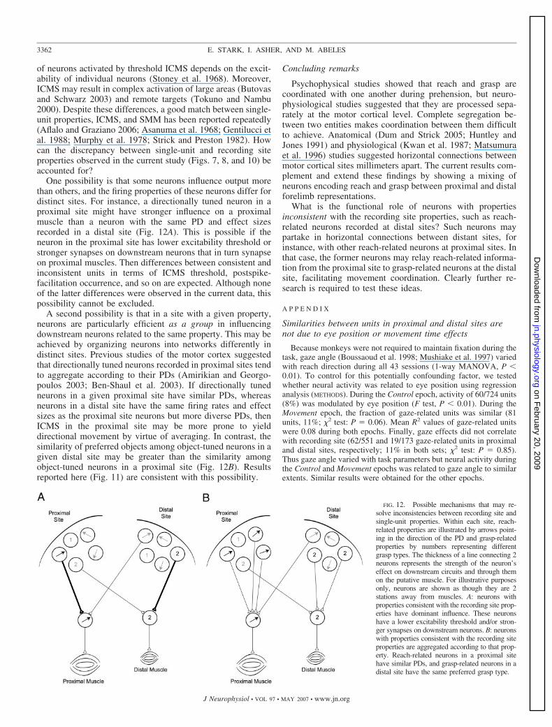
One possibility is that some neurons influence output morethan others, and the firing properties of these neurons differ fordistinct sites. For instance, a directionally tuned neuron in aproximal site might have stronger influence on a proximalmuscle than a neuron with the same PD and effect sizesrecorded in a distal site (Fig. 12A). This is possible if theneuron in the proximal site has lower excitability threshold orstronger synapses on downstream neurons that in turn synapseon proximal muscles. Then differences between consistent andinconsistent units in terms of ICMS threshold, postspike-facilitation occurrence, and so on are expected. Although noneof the latter differences were observed in the current data, thispossibility cannot be excluded.
A second possibility is that in a site with a given property,neurons are particularly efficient as a group in influencingdownstream neurons related to the same property. This may beachieved by organizing neurons into networks differently indistinct sites. Previous studies of the motor cortex suggestedthat directionally tuned neurons recorded in proximal sites tendto aggregate according to their PDs (Amirikian and Georgo-poulos 2003; Ben-Shaul et al. 2003). If directionally tunedneurons in a given proximal site have similar PDs, whereasneurons in a distal site have the same firing rates and effectsizes as the proximal site neurons but more diverse PDs, thenICMS in the proximal site may be more prone to yielddirectional movement by virtue of averaging. In contrast, thesimilarity of preferred objects among object-tuned neurons in agiven distal site may be greater than the similarity amongobject-tuned neurons in a proximal site (Fig. 12B). Resultsreported here (Fig. 11) are consistent with this possibility.
Concluding remarks
Psychophysical studies showed that reach and grasp arecoordinated with one another during prehension, but neuro-physiological studies suggested that they are processed sepa-rately at the motor cortical level. Complete segregation be-tween two entities makes coordination between them difficultto achieve. Anatomical (Dum and Strick 2005; Huntley andJones 1991) and physiological (Kwan et al. 1987; Matsumuraet al. 1996) studies suggested horizontal connections betweenmotor cortical sites millimeters apart. The current results com-plement and extend these findings by showing a mixing ofneurons encoding reach and grasp between proximal and distalforelimb representations.
What is the functional role of neurons with propertiesinconsistent with the recording site properties, such as reach-related neurons recorded at distal sites? Such neurons maypartake in horizontal connections between distant sites, forinstance, with other reach-related neurons at proximal sites. Inthat case, the former neurons may relay reach-related informa-tion from the proximal site to grasp-related neurons at the distalsite, facilitating movement coordination. Clearly further re-search is required to test these ideas.
A P P E N D I X
Similarities between units in proximal and distal sites arenot due to eye position or movement time effects
Because monkeys were not required to maintain fixation during thetask, gaze angle (Boussaoud et al. 1998; Mushiake et al. 1997) variedwith reach direction during all 43 sessions (1-way MANOVA, P �0.01). To control for this potentially confounding factor, we testedwhether neural activity was related to eye position using regressionanalysis (METHODS). During the Control epoch, activity of 60/724 units(8%) was modulated by eye position (F test, P � 0.01). During theMovement epoch, the fraction of gaze-related units was similar (81units, 11%; �2 test: P � 0.06). Mean R2 values of gaze-related unitswere 0.08 during both epochs. Finally, gaze effects did not correlatewith recording site (62/551 and 19/173 gaze-related units in proximaland distal sites, respectively; 11% in both sets; �2 test: P � 0.85).Thus gaze angle varied with task parameters but neural activity duringthe Control and Movement epochs was related to gaze angle to similarextents. Similar results were obtained for the other epochs.
FIG. 12. Possible mechanisms that may re-solve inconsistencies between recording site andsingle-unit properties. Within each site, reach-related properties are illustrated by arrows point-ing in the direction of the PD and grasp-relatedproperties by numbers representing differentgrasp types. The thickness of a line connecting 2neurons represents the strength of the neuron’seffect on downstream circuits and through themon the putative muscle. For illustrative purposesonly, neurons are shown as though they are 2stations away from muscles. A: neurons withproperties consistent with the recording site prop-erties have dominant influence. These neuronshave a lower excitability threshold and/or stron-ger synapses on downstream neurons. B: neuronswith properties consistent with the recording siteproperties are aggregated according to that prop-erty. Reach-related neurons in a proximal sitehave similar PDs, and grasp-related neurons in adistal site have the same preferred grasp type.
3362 E. STARK, I. ASHER, AND M. ABELES
J Neurophysiol • VOL 97 • MAY 2007 • www.jn.org
on February 20, 2009
jn.physiology.orgD
ownloaded from
Another potential confound stems from reaction and movementtimes (inversely related to speed) which varied with reach direction,grasp type, or both during all sessions (2-way ANOVA, P � 0.01,corrected for multiple comparisons). We tested whether neural activ-ity was modulated by reaction and/or movement time using regressionanalysis as for gaze angle. During the Movement epoch, the activity of77/724 units (11%) was modulated by reaction/movement times; themean R2 was 0.05. Modulation was similar for units in proximal anddistal sites (10 and 13%, respectively; �2 test: P � 0.36). During thePreGo epoch similar results were obtained (9 and 6% of units inproximal and distal sites, respectively; �2 test: P � 0.3), but duringother epochs, the number of units modulated by reaction/movementtimes was at chance level. Thus neural activity during the PreGo andMovement epochs was weakly modulated by reaction and movementtimes, regardless of the recording site.
Dominance of reach-related single-unit activity
Reach-related modulations were larger and more frequent thangrasp-related modulations (Figs. 8, A and B, and 10). This could resultfrom a statistical bias: for each unit, six directions but only two objectswere sampled. To control for this factor, we diluted reach directionsby keeping trials from two randomly selected directions, so diluteddata included an equal number of sampled directions and sampledobjects. In these data, the dominance of direction over object tuningwas smaller yet maintained (fractions of tuned units: 33 vs. 19%; �2
test: P �� 0.001; median RGI, �0.11). Thus the apparent dominanceof reaching reported in RESULTS resulted only in part from the exper-imental design.
Neural activity modulated solely according to whether grasping isor is not performed will not result in an object effect. Thereforeviewing object effects as reflecting grasp-related activity is a conser-vative estimate. To test this possibility, single-unit activity during theprehension task, requiring reaching and grasping, was compared withthe activity of the same units during a task that required only reaching,in which targets were touch pads identical to the resting position touchpad (a total of 367 units were recorded in monkey D during both tasks,uniformly from all sites). During the Movement epoch, 66/238 units(28%) that did not exhibit an object effect modulated their activityaccording to whether grasping was or was not instructed, yielding atotal of 195/367 units (53%) related to grasp performance. Thus thegrasp-related activity reported in this study may be regarded as alower bound estimate. Although we did not conduct a “grasp withoutreach” experiment, the same logic presumably applies to that case.
A C K N O W L E D G M E N T S
We thank Y. Ben-Shaul, R. Drori, and R. Paz for help in experiments, A.Globerson and A. Stark for critically reading the manuscript, G. Goelman forMRI scans, M. Nakar for help in constructing the experimental setup, V.Sharkansky for technical assistance, and E. Vaadia for helpful suggestions.
G R A N T S
This study was supported in part by a Center of Excellence grant (1564/04)administered by the Israel Science Foundation, the Yeshaya Horowitz Asso-ciation, the Rich Center, and the Deutsch-Israelische Projectkooperation.
R E F E R E N C E S
Abeles M, Goldstein MH. Multispike train analysis. Proc IEEE 65: 762–773,1977.
Aflalo TN, Graziano MS. Partial tuning of motor cortex neurons to finalposture in a free-moving paradigm. Proc Natl Acad Sci USA 103: 2909–2914, 2006.
Amirikian B, Georgopoulos AP. Modular organization of directionally tunedcells in the motor cortex: is there a short-range order? Proc Natl Acad SciUSA 100: 12474–12479, 2003.
Asanuma H, Stoney SD Jr, Abzug C. Relationship between afferent inputand motor outflow in cat motorsensory cortex. J Neurophysiol 31: 670–681,1968.
Ben-Shaul Y, Stark E, Asher I, Drori M, Nadasdy Z, Abeles M. Dynamicalorganization of directional tuning in the primate premotor and primarymotor cortex. J Neurophysiol 89: 1136–1142, 2003.
Boussaoud D, Jouffrais C, Bremmer F. Eye position effects on the neuronalactivity of dorsal premotor cortex in the macaque monkey. J Neurophysiol80: 1132–1150, 1998.
Brochier T, Spinks RL, Umilta MA, Lemon RN. Patterns of muscle activityunderlying object-specific grasp by the macaque monkey. J Neurophysiol92: 1770–1782, 2004.
Butovas S, Schwarz C. Spatiotemporal effects of microstimulation in ratneocortex: a parametric study using multielectrode recordings. J Neuro-physiol 90: 3024–3039, 2003.
Caminiti R, Johnson PB, Galli C, Ferraina S, Burnod Y. Making armmovements within different parts of space: the premotor and motor corticalrepresentation of a coordinate system for reaching to visual targets. J Neu-rosci 11: 1182–1197, 1991.
Crammond DF, Kalaska JF. Differential relation of discharge in primarymotor and premotor cortex to movement posture during reaching move-ments. Exp Brain Res 108: 45–61, 1996.
Dum RP, Strick PL. Frontal lobe inputs to the digit representations of themotor areas on the lateral surface of the hemisphere. J Neurosci 25:1375–1386, 2005.
Fetz EE, Cheney PD. Postspike facilitation of forelimb muscle activity byprimate corticomotoneuronal cells. J Neurophysiol 44: 751–772, 1980.
Fisher RA. Statistical Methods for Research Workers. London: Oliver andBoyd, 1925.
Fogassi L, Gallese V, Buccino G, Craighero L, Fadiga L, Rizzolatti G.Cortical mechanism for the visual guidance of hand grasping movements inthe monkey: a reversible inactivation study. Brain 124: 571–586, 2001.
Gabernet L, Meskenaite V, Hepp-Reymond MC. Parcellation of the lateralpremotor cortex of the macaque monkey based on staining with the neuro-filament antibody SMI-32. Exp Brain Res 128: 188–193, 1999.
Gentilucci M, Fogassi L, Luppino G, Matelli M, Camarda R, Rizzolatti G.Functional organization of inferior area 6 in the macaque monkey. I.Somatotopy and control of proximal movements. Exp Brain Res 71: 475–490, 1988.
Godschalk M, Mitz AR, van Duin B, van der Burg H. Somatotopy ofmonkey premotor cortex examined with microstimulation. Neurosci Res 23:269–279, 1995.
Graziano MS, Hu XT, Gross CG. Visuospatial properties of ventral premotorcortex. J Neurophysiol 77: 2268–2292, 1997.
Graziano MS, Taylor CS, Moore T. Complex movements evoked by micro-stimulation of precentral cortex. Neuron 34: 841–851, 2002.
He SQ, Dum RP, Strick PL. Topographic organization of corticospinalprojections from the frontal lobe: motor areas on the lateral side of thehemisphere. J Neurosci 13: 952–980, 1993.
Hepp-Reymond MC, Husler EJ, Maier MA, Ql HX. Force-related neuronalactivity in two regions of the primate ventral premotor cortex. Can J PhysiolPharmacol 72: 571–579, 1994.
Huntley GW, Jones EG. Relationship of intrinsic connections to forelimbmovement representations in monkey motor cortex: a correlative anatomicand physiological study. J Neurophysiol 66: 390–413, 1991.
Jeannerod M. The timing of natural prehension movements. J Mot Behav 16:235–254, 1984.
Kakei S, Hoffman DS, Strick PL. Direction of action is represented in theventral premotor cortex. Nat Neurosci 4: 1020–1025, 2001.
Kurata K, Hoffman DS. Differential effects of muscimol microinjection intodorsal and ventral aspects of the premotor cortex of monkeys. J Neuro-physiol 71: 1151–1164, 1994.
Kurata K, Tanji J. Premotor cortex neurons in macaques: activity beforedistal and proximal forelimb movements. J Neurosci 6: 403–411, 1986.
Kwan HC, Murphy JT, Wong YC. Interaction between neurons in precentralcortical zones controlling different joints. Brain Res 400: 259–269, 1987.
Lemon RN, Mantel GW, Muir RB. Corticospinal facilitation of handmuscles during voluntary movement in the conscious monkey. J Physiol381: 497–527, 1986.
Luppino G, Rizzolatti G. The organization of the frontal motor cortex. NewsPhysiol Sci 15: 219–224, 2000.
Mason CR, Hendrix CM, Ebner TJ. Purkinje cells signal hand shape andgrasp force during reach-to-grasp in the monkey. J Neurophysiol 95:144–158, 2006.
3363REACH AND GRASP IN PREMOTOR CORTEX
J Neurophysiol • VOL 97 • MAY 2007 • www.jn.org
on February 20, 2009
jn.physiology.orgD
ownloaded from
Matsumura M, Chen D, Sawaguchi T, Kubota K, Fetz EE. Synapticinteractions between primate precentral cortex neurons revealed by spike-triggered averaging of intracellular membrane potentials in vivo. J Neurosci16: 7757–7767, 1996.
Merzenich MM, Knight PL, Roth GL. Representation of cochlea in primaryauditory cortex in the cat. J Neurophysiol 38: 231–249, 1975.
Murata A, Fadiga L, Fogassi L, Gallese V, Raos V, Rizzolatti G. Objectrepresentation in the ventral premotor cortex (area F5) of the monkey.J Neurophysiol 78: 2226–2230, 1997.
Murphy JT, Kwan HC, MacKay WA, Wong YC. Spatial organization ofprecentral cortex in awake primates. III. Input-output coupling. J Neuro-physiol 41: 1132–1139, 1978.
Mushiake H, Tanatsugu Y, Tanji J. Neuronal activity in the ventral part ofpremotor cortex during target-reach movement is modulated by direction ofgaze. J Neurophysiol 78: 567–571, 1997.
Raos V, Umilta MA, Gallese V, Fogassi L. Functional properties of grasping-related neurons in the dorsal premotor area F2 of the macaque monkey.J Neurophysiol 92: 1990–2002, 2004.
Rathelot JA, Strick PL. Muscle representation in the macaque motor cortex:an anatomical perspective. Proc Natl Acad Sci USA 103: 8257–8262, 2006.
Rizzolatti G, Camarda R, Fogassi L, Gentilucci M, Luppino G, Matelli M.Functional organization of inferior area 6 in the macaque monkey. II. AreaF5 and the control of distal movements. Exp Brain Res 71: 491–507, 1988.
Sato KC, Tanji J. Digit-muscle responses evoked from multiple intracorticalfoci in monkey precentral motor cortex. J Neurophysiol 62: 959–970, 1989.
Schieber MH. Muscular production of individuated finger movements: theroles of extrinsic finger muscles. J Neurosci 15: 284–297, 1995.
Schwartz AB, Moran DW, Reina GA. Differential representation of percep-tion and action in the frontal cortex. Science 303: 380–383, 2004.
Shen L, Alexander GE. Preferential representation of instructed target loca-tion versus limb trajectory in dorsal premotor area. J Neurophysiol 77:1195–1212, 1997.
Soechting JF, Flanders M. Parallel, interdependent channels for location andorientation in sensorimotor transformations for reaching and grasping.J Neurophysiol 70: 1137–1150, 1993.
Stoney SD Jr, Thompson WD, Asanuma H. Excitation of pyramidal tractcells by intracortical microstimulation: effective extent of stimulating cur-rent. J Neurophysiol 31: 659–669, 1968.
Strick PL, Preston JB. Two representations of the hand in area 4 of a primate.II. Somatosensory input organization. J Neurophysiol 48: 150–159, 1982.
Tanne-Gariepy J, Rouiller EM, Boussaoud D. Parietal inputs to dorsalversus ventral premotor areas in the macaque monkey: evidence for largelysegregated visuomotor pathways. Exp Brain Res 145: 91–103, 2002.
Tehovnik EJ, Tolias AS, Sultan F, Slocum WM, Logothetis NK. Direct andindirect activation of cortical neurons by electrical microstimulation. J Neu-rophysiol 96: 512–521, 2006.
Tokuno H, Nambu A. Organization of nonprimary motor cortical inputs onpyramidal and nonpyramidal tract neurons of primary motor cortex: anelectrophysiological study in the macaque monkey. Cereb Cortex 10:58–68, 2000.
van Kan PL, Horn KM, Gibson AR. The importance of hand use todischarge of interpositus neurons of the monkey. J Physiol 480: 171–190,1994.
Weinrich M, Wise SP. The premotor cortex of the monkey. J Neurosci 2:1329–1345, 1982.
Wise SP, Boussaoud D, Johnson PB, Caminiti R. Premotor and parietalcortex: corticocortical connectivity and combinatorial computations. AnnuRev Neurosci 20: 25–42, 1997.
3364 E. STARK, I. ASHER, AND M. ABELES
J Neurophysiol • VOL 97 • MAY 2007 • www.jn.org
on February 20, 2009
jn.physiology.orgD
ownloaded from