effects of estrogen on beta-amyloid-induced cholinergic cell death in the nucleus basalis...
TRANSCRIPT

Fax +41 61 306 12 34E-Mail [email protected]
Original Paper
Neuroendocrinology DOI: 10.1159/000321119
Effects of Estrogen on Beta-Amyloid-Induced Cholinergic Cell Death in the Nucleus Basalis Magnocellularis
Éva M. Szegő a–c Attila Csorba b Tamás Janáky b Katalin A. Kékesi a István M. Ábrahám a Gábor M. Mórotz a Botond Penke b Miklós Palkovits d Ünige Murvai e Miklós S.Z. Kellermayer e József Kardos f Gábor D. Juhász a
a Laboratory of Proteomics, Eötvös Loránd University, Budapest , b Medical Chemistry Department,University of Szeged, Szeged , Hungary; c Department of Neurodegeneration and Restorative Research,Georg-August University, DFG Research Center Molecular Physiology of the Brain (CMPB), Göttingen , Germany; d Neuromorphological and Neuroendocrine Research Laboratory, Department of Anatomy, Histology and Endocrinology, Semmelweis University and the Hungarian Academy of Sciences, e Department of Biophysicsand Radiation Biology, Semmelweis University, and f Department of Biochemistry, Eötvös Loránd University, Budapest , Hungary
toxicity 24 h following injection. The change in expression of, e.g., DJ-1, NADH ubiquinone oxidoreductase, ATP syn-thase, phosphatidylethanolamine-binding protein 1, pro-tein phosphatase 2A and dimethylarginine dimethylamino-hydrolase 1 support our hypothesis that A � induces mi-tochondrial dysfunction, decreases MAPK signaling, and increases NOS activation in NBM. On the other hand, altered expression of, e.g., MAP kinase kinase 1 and 2, protein phos-phatase 1 and 2A by A � might increase MAPK suppression and NOS signaling in the cortical target area. Estrogen pre-treatment reversed most of the changes in the proteome in both areas. Our experiments suggest that regulation of the MAPK pathway, mitochondrial pH and NO production may all contribute to A � toxicity, and their regulation can be pre-vented partly by estrogen pretreatment.
Copyright © 2010 S. Karger AG, Basel
Introduction
Alzheimer disease (AD) is the most prevalent age-re-lated neurodegenerative disease, and the most common form of dementia [1] . Besides the accumulation of � -am-
Key Words
Alzheimer disease � Estrogen � Amyloid � Micropunch � Proteomics � MAPK pathway � Differential two-dimensional gel electrophoresis � Nucleus basalis magnocellularis
Abstract
Alzheimer disease is characterized by accumulation of � -am-yloid (A � ) and cognitive dysfunctions linked to early loss of cholinergic neurons. As estrogen-based hormone replace-ment therapy has beneficial effects on cognition of dement-ed patients, and it may prevent memory impairments, we investigated the effect of estrogen-pretreatment on A � -in-duced cholinergic neurodegeneration in the nucleus basalis magnocellularis (NBM). We tested which A � species induces the more pronounced cholinotoxic effect in vivo. We inject-ed different A � assemblies in the NBM of mice, and mea-sured cholinergic cell and cortical fiber loss. Spherical A � oligomers had the most toxic effect. Pretreatment of ovari-ectomized mice with estrogen before A � injection de-creased cholinergic neuron loss and partly prevented fiber degeneration. By using proteomics, we searched for pro-teins involved in estrogen-mediated protection and in A �
Received: August 18, 2010 Accepted after revision: September 8, 2010 Published online: October 9, 2010
Eva M. Szego Department of Neurodegeneration and Restorative Research Georg-August University, DFG Research Center Molecular Physiology of the Brain Waldweg 33, DE–37073 Göttingen (Germany) Tel. +49 551 391 3547, Fax +49 551 391 3541, E-Mail eszego @ uni-goettingen.de
© 2010 S. Karger AG, Basel0028–3835/10/0000–0000$26.00/0
Accessible online at:www.karger.com/nen
Pagination Instructions: This article should begin on a RIGHT page!

Szegő et al. Neuroendocrinology 2
yloid (A � ) peptide, neurofibrillary tangles and synaptic loss [2] , another characteristic of AD is the early degen-eration of the basal forebrain cholinergic system (BFC) [3, 4] . Neurons of BFC express the receptor � 7 nicotinic ace-tylcholine receptor, which binds A � selectively and with high affinity, making cholinergic neurons vulnerable in AD or protecting against A � [5–7] .
AD is known to be gender dependent and age-related loss of estrogen (17 � -estradiol, E2) probably increases the risk of AD [8] . E2-based hormone replacement therapy (ERT) decreased the risk of AD and cognitive dysfunc-tions in some clinical studies, suggesting a protective role of E2 [9–12] . Supporting these findings, E2 was found to be neuroprotective in in vitro and in vivo experimental AD models [13–16] . It is known that E2 destabilizes A � fibrils in vitro [17] and decreases A � load in transgenic mice [18] . In addition, E2 reduces A � -induced Ca 2+ over-load [19] and p38 mitogen-activated protein kinase (p38MAPK) activation [20] in vitro . E2 is able to optimize brain metabolism, achieving optimal energy consump-tion during different mental functions [21–23] . Moreover, E2 treatment improves cognitive performance partly via interaction with the basal forebrain cholinergic system [24, 25] . However, despite the beneficial effects of E2 found in animal studies or reported from human trials, some studies demonstrated that ERT has no effect on cognitive performance and on severity of dementia in fe-male patients [26, 27] , meaning that ERT is still contro-versial for the treatment of dementia.
Both A � and E2 generate widespread changes in the brain by controlling gene expression among others [22, 28–31] , inducing cell death or preparing cells for toxicity and altering cellular reactions to A � . In our present study, by searching for possible target proteins, we aimed to find the mechanisms and raise a hypothesis underlying A � toxicity on cholinergic neurons and the possible neuropro-tective effects of E2 in vivo. Hence, we first determined the neurotoxic potential of differently aggregated A � 1–42 so-lutions on the mouse nucleus basalis magnocellularis-sub-stantia innominata (NBM-SI) cholinergic neurons in vivo. We measured cholinergic cell loss in the NBM-SI, and cho-linergic fiber density in the projection area (somatosen-sory cortex, SSCTX, layer V). After selection of the most potent A � solution, in the second part of our experiments, we investigated the effect of E2 pretreatment on A � choli-notoxic effects. In addition, we performed a proteomic in-vestigation to find the proteins influenced by A � and A � after E2 pretreatment. In this study, we present the possi-ble mechanisms explaining A � toxicity and the protective effects of E2 against A � on NBM-SI cholinergic neurons.
Methods
Animals Animal breeding and experiments were performed based on
the rules of the Local Animal Care Committee at the Eötvös Loránd University, in accordance with the European Union con-forming to the Hungarian Act of Animal Care and Experimenta-tion. Female wild-type C57BL6/J mice were maintained under a 12-hour light/dark cycle at 20 ° C, and were supplied with water and food ad libitum.
In vitro Characterization of A � Solutions: Atomic Force Microscopy and Thioflavin T Measurements A � 1–42 (a gift from Márta Zarándi [32] ) peptide was dissolved
in 100% hexafluoro-isopropanol (HFIP) for 6 h to prepare a monomer solution [33] . The monomer solution was centrifuged at 15,000 g (10 min), and the supernatant was lyophilized and kept at –80 ° C until use. The morphology and size of the A � aggregates in vitro was determined using atomic force microscopy (AFM). A � samples at 600 � M concentration in glucose-free artificial ce-rebrospinal fluid (ACSF, in m M : 144 NaCl; 3 KCl; 1 MgCl 2 ; 2 CaCl 2 ; pH 7.3) were incubated for 0, 12, 24 and 48 h at room temperature. As the samples with 24 and 48 h incubation time did not adhere sufficiently to the mica surface (Electron Microscopy Sciences, Hatfield, Pa., USA), they were diluted in deionized water (15- to 30-fold) and the solutions were then incubated on the mica surface for 2 min and replaced with water. These solutions were measured in water instead of ACSF. Images were obtained under similar conditions with an MFP3D AFM instrument (Asylum Re-search, Santa Barbara, Calif., USA), operating in a noncontact mode, using the Olympus BioLever silicon nitride cantilever (Olympus Co., Japan); typical resonance frequency: � 9 kHz. Drive amplitude and contact force were kept to a minimum. Areas were scanned at 0.5–1 Hz rate. Images were evaluated by measur-ing the height of 100–200 individual aggregates above the mica surface using MFP-3D software (Asylum Research). Objects clearly associated from a large number of aggregates were taken as sample preparation artefacts and were excluded from the mea-surements. It is important to note that AFM measurements may underestimate the diameter of A � structures because of sample compression by the AFM probe [34] . However, the technique is excellent to follow the progress of aggregation and the change in morphology.
For thioflavin T (ThT) fluorescence measurements, 1- � l ali-quots were taken from the samples and mixed with 1.0 ml of 5 � M ThT (Sigma) in 50 m M glycine-NaOH buffer at pH 8.5 [35] . ThT fluorescence was monitored immediately at 485 nm with excita-tion at 445 nm in a cell thermostatted to 25 ° C using a SPEX Fluo-roMax instrument (Jobin-Yvon, Longjumeau Cedex, France). Ex-citation and emission bandwidths were set to 5 nm. Data are from 5 measurements/time point.
Preparation of Tissue Sections for Toxicity Measurements of A � Assemblies To prepare different A � aggregates suitable for in vivo experi-
ments, the lyophilized samples were dissolved in ACSF at a con-centration of 600 � M and kept at room temperature for 0, 12, 24 or 48 h prior to administration. The concentration of the A � solu-tion was determined in a preliminary experiment, and we select-ed the lowest concentration to achieve a minimum of 25% cholin-

E2 Reduces A � -Induced Neuron Loss in the NBM
Neuroendocrinology 3
ergic cell death (data not shown). In order to eliminate the pos-sible interference of endogenous estrogen with A � toxicity, adult female 45- to 60-day-old mice were bilaterally ovariectomized (OVX) under deep anesthesia using Avertin (2% 2,2,2-tribromo-ethanol, 1,2% amyl-hydrate; 8% ethanol in physiological saline; Sigma, USA). In the first set of experiments, we aimed to deter-mine the cholinotoxic effect of different A � assemblies. A � was incubated in ACSF for 0, 12, 24 or 48 h at room temperature prior to injecting. On post-OVX day 14, 2 ! 1 � l 600 � M A � solution or ACSF was injected slowly into the NBM-SI using fused silica capillary (coordinates: L –2/+2, AP –0,65, DV –4,1/–4,3) [36] . During stereotactic surgery, mice were in deep anesthesia (1.5% halothane in air, 1.8 liters/min flow rate). ACSF was injected ran-domly into one side, and A � solution into the other side of the brain; therefore, the same animal served as its own control (ACSF-injected site, n = 5/group). On post-OVX day 29, mice were anesthetized by a lethal dose of Avertin, and transcardially per-fused with 4% paraformaldehyde (Merck, Germany), pH 7.6 in phosphate-buffered saline solution. Brains were removed, post-fixed for 2 h at 4 ° C and cryoprotected in Tris-phosphate-buffered solution (TBS), pH 7.6, containing 30% sucrose overnight at 4 ° C. 30- � m coronal sections were cut on a freezing microtome and four sets of sections were collected in TBS.
Determination of the Effect of E2 on A � Toxicity in vivo To study the effects of E2 pretreatment on the A � -exerted cho-
linotoxicity, the following procedures were used. 45- to 60-day-old mice were bilaterally ovariectomized (see above section). On post-OVX day 14, animals received a subcutaneous injection of 1 � g of 17 � -estradiol (E2; in 0.1 ml ethyl-oleate vehicle, Sigma) or the same volume of vehicle (EO). Twenty-four hours after the EO/E2 treatment, the previously prepared A � solution (2 ! 1 � l, 600 � M in ACSF, 24-hour incubation at room temperature) or ACSF was injected slowly into the NBM-SI (see the previous section). Altogether, 12 mice were treated with EO and A � and 12 mice with E2 and A � . Six EO-A � -injected and 6 E2-A � -injected mice were used for differential two-dimensional gel electrophoresis (DIGE) experiments (n = 6). These mice were sacrificed 24 h after the A � -injection (post-OVX day 16) by cervical dislocation, brains were rapidly removed ( ! 40 s) and frozen in dry ice. Serial 100- � m coronal cryosections were cut at –10 ° C and tissue sam-ples corresponding to the SSCTX and the NBM-SI were dissected using the ‘punch technique’ [37] according to a microdissection map [38] . Samples were stored at –80 ° C until use. Six EO-A � -, and six E2-A � -injected mice were used for the regeneration study (n = 6). These mice were sacrificed on post-OVX day 30, perfused, and their brains treated as described above.
Protein Isolation, Differential Two-Dimensional Gel Electrophoresis and In-Gel Digestion Eight different sets of tissue samples were collected for pro-
teomic studies: four from EO-treated mice (SSCTX and NBM-SI from the ACSF-injected side and SSCTX and NBM-SI from the A � -injected site), pieces of tissue from 6 different animals, there-fore 6 pieces/area/treatment group, and the same areas from the E2-treated animals (6 mice). Tissue samples were homogenized as we reported earlier [23] . The pH of the supernatant corresponding to cytosolic and membrane fractions was adjusted to 8.0. Five mi-crogram of each protein sample was labeled with Cy5 saturation dye CyDye DIGE Fluor-Labeling Kit for Scarce Samples (4 nmol/
5 � g protein, GE Healthcare, Uppsala, Sweden) according to the manufacturer’s instructions. The reference sample (internal stan-dard, equal amounts: 5 � g of protein from all E2- and all EO-treated samples from the same region) was labeled with Cy3 and the two differently marked samples (5 � g of Cy5-labeled sample and 5 � g Cy3-labeled reference) were multiplexed to be resolved in the same gel. Labelled proteins were separated and visualized as described previously (DryStrip pH: 4–7, 10% acrylamide gel) [23] . Differential protein analysis was performed using DeCyder software package 6.0, DIA and BVA modules (GE Healthcare). For the identification of proteins in spots of interest, preparative 2D electrophoresis was performed separately using 800 � g of pro-teins per gel [23] .
LC-MS Analysis and Protein Identification and Extended Literature Search LC-MS analysis was performed by an Agilent HPLC-Chip/MS
system consisting of 1100 Series HPLC system and a 6330 LC/MSD XCT Plus ion trap mass spectrometer. The LC system was operated in a sample enrichment/desalting mode using a ProtID-Chip-43 (II Chip-Column) (column material: ZORBAX 300 SB-C18 5 � m, trap column volume: 40 nl, analytical column: 43 ! ID 0.075 mm). The HPLC solvents were the following: solvent A: 0.1% formic acid in water and solvent B: 0.1% formic acid in ace-tonitrile. Two microliter of sample was enriched with solvent A at a flow rate of 4 � l/min for 2 min using a capillary pump then fol-lowed by gradient elution of tryptic peptides at a flow rate of 300 nl/min using a nano pump. The gradient was 5% B/min increase of concentration B from 5 to 45% under 10 min followed by a gra-dient column-wash from 60 to 90% solvent B in 2 min. The mass spectrometer was operated in the autoMSMS mode, the resolu-tion was less than 0.5 u (FWHM) and the scan speed was 8,100 u/s. The survey scan range was 300–1,600 m/z and 4 ions were selected for CID. MSMS scan range was set to 100–1800 m/z at 26,000 u/s at a resolution of less than 0.6 u (FWHM). All acquired MSMS data were processed and subjected to database search by Agilent Spectrum Mill MS Proteomics Workbench Ver. A.03.02 against NCBInr (11/16/2009 database). Database search parame-ters were set to 1.5 Da for precursor ion mass tolerance, 0.6 Da for fragment ion mass tolerance. Constant modification of Cys was specified as carbamidomethylation, variable modifications were set to methionine oxidation, deaminated glutamine, acetylation of protein N-terminal and oxidation of tryptophan. For protein assignment the minimum score was set to 20 for proteins and peptide hits were accepted above score of 9, 10, 11 and 12 accord-ing to their charge state of 1+, 2+, 3+ and 4+, respectively [39] . To make a functional interpretation of our data, a systematic litera-ture survey was conducted [23] .
Immunohistochemistry and Acetylcholinesterase Histochemistry To detect cholinergic neurons, free-floating, peroxidase-based
immunohistochemistry was performed in the same manner we reported previously with only a slight modification [40] . We used anticholine acetyl-transferase (anti-ChAT antibody (1: 2000), Santa Cruz Biotechnology, Calif., USA) for 48 h at 4 ° C. For fluo-rescent labeling of calbindin (1: 3,000, Swant, Switzerland) and ChAT (1: 1,000), we used florescent dye-conjugated secondary an-tibodies (1: 1,000, AlexaFluor-488 for ChAT and AlexaFluor-555 for calbindin; Invitrogen, Calif., USA). Visualization of acetyl-

Szegő et al. Neuroendocrinology 4
cholinesterase fibers was done according to the method of He-dreen et al. [41] .
Data Analysis and Statistics Number of ChAT-ir cells were counted by an Olympus BX51
microscope (Olympus Optical, Hamburg, Germany) using ! 40 objectives. Every fourth section was stained and ChAT-ir neurons of the NBM-SI were counted from both sides (ACSF- or A � -in-jected sides from four sections) by an investigator blind to the experimental groupings; finally, the number of neurons/ � m 3 and the percentage change was determined. Data are expressed as mean 8 SEM. ANOVA was carried out to examine the effects of EO/E2 and ACSF/A � treatment (R software packages, version 2.8.0, R Development Core Team 2008, Vienna, Austria).
Acetylcholinesterase (AChE) fiber density was determined in the SSCTX, layer V. As the cholinergic neurons of the NBM proj-ect almost exclusively unilaterally to the SSCTX, it allowed us to determine the differences between ACSF- and A � -injected sides in the same animal. Every fourth section was stained and six sec-tions were analyzed per animal. Six images were taken of every slice using a ! 100 objective (Olympus BX51) and the area covered by AChE fibers was measured ten times from the same image fol-lowing background subtraction and binarization. Therefore, six sections/animal, six images/section and 10 measurements/image resulted in a hierarchical nested design. The effect of A � on fi-ber density was expressed as a percentage change relative to the ACSF-injected side. A generalized linear mixed model was ap-plied, the dependent variable was the fiber density, and the inde-pendent variables were the EO or E2 treatment and ACSF or A � injection. The identification of animals, sections and images were set as random factors (R software).
For the DIGE experiments, ANOVAs were calculated by the DeCyder software Biological Variance Analysis (BVA module). The internal standard was a pool of equal amounts of all samples (from all SSCTX or all NBM-SI within the experiment); it was representative of every protein present and was the same across all gels from the same region. The standard provided an average image against which all other gel images were normalized, remov-ing much of the experimental variation and reducing gel-to-gel variation.
Results
In vitro Characterization of A � (1–42) Aggregate Size and Morphology Samples with 0 h incubation time mainly contained
particles exhibiting a height not exceeding 1 nm (approx-imately 73%; fig. 1 a, b). This size range possibly corre-sponds to the monomer state of A � , while particles larger than 1 nm could be assigned to the aggregated forms of A � peptide [33] . The percentage of the small particles sig-nificantly decreased with incubation time to approxi-mately 50, 25 and 5% after 12, 24 and 48 h, respectively ( fig. 1 a, b). We observed a greater portion of large parti-cles after longer incubation times. After 12 h incubation,
the ratio of particles with 1–2 nm height increased, a form corresponding to small oligomers [33] . Aggregates formed in the 2–4 nm range appeared after longer incubation times (24 and 48 h) with a maximum occurrence at 2.3–2.5 nm ( fig. 1 a, b). We observed a change in the morphol-ogy of the aggregates paralleling their increased size. Spherical aggregates appeared in the 24-hour samples ( fig. 1 b), while after 48 h, besides spherical oligomers, short, fibrillar structures were formed with 50–200 nm length and average height of 2.5–3 nm ( fig. 1 b). The latter species of aggregates could not have been mature amyloid fibrils because of their shortness and small diameter. Rather, these were protofibrils formed from oligomers.
Thioflavin T is a fluorescent dye that binds to protein aggregates, especially to amyloid fibrils, while it does not bind to monomers. Upon binding, it shows high fluores-cence intensity. 0-hour samples exhibited low ThT fluo-rescence intensity reflecting the high monomer content in the solution ( fig. 1 c). 12-, 24- and 48-hour samples ex-hibited an increasing ThT fluorescence intensity with time indicating the progress of the aggregation process and an increase in the amyloid-like structure content ( fig. 1 c).
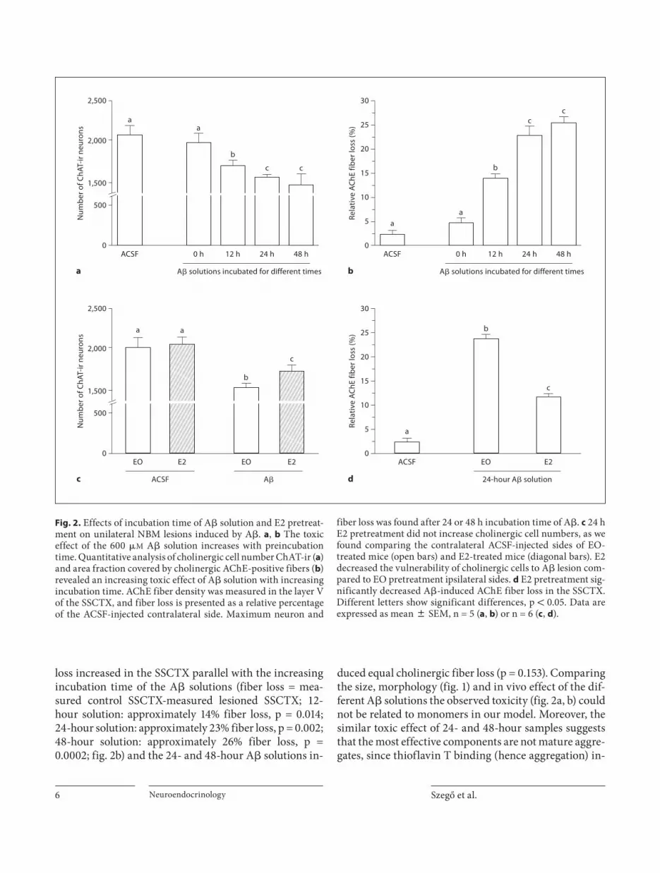
Determination of the Cholinotoxic Potential of Different A � Forms Injection of the 0-hour solution had no effect on the
number of cholinergic cells in the NBM-SI ( fig. 2 a) and failed to induce cholinergic fiber loss in the SSCTX ( fig. 2 b). In contrast, the cholinotoxic potential of the dif-ferent A � solutions increased with pre-incubation time (loss of neurons = counted control site-counted lesioned site; 12-hour solution: approximately 15% cell loss, p = 0.033; 24-hour solution: approximately 25% cell loss, p = 0.002; 48-hour solution: approximately 26% cell loss, p = 0.002) although the 48-hour sample had the same effect as the 24-hour sample (p = 0.679; fig. 2 a). Similarly, fiber
Fig. 1. In vitro characterization of size distribution and morphol-ogy of A � 1–42 aggregates. a Representative AFM images of A � samples after incubation for 0, 12, 24 and 48 h, respectively. Scale bars represent 200 nm. Color codes for height traces are present-ed. b Height distributions of A � samples (AFM measurements) after different incubation times at room temperature. Columns represent the percentage of individual aggregates of a certain height 0.2 nm interval. Large objects 1 6 nm were excluded from calculation. c Changes in the intensity of thioflavin T fluores-cence show increasing aggregation of A � 1–42 with time. ThT binds protein aggregates, but not monomers. Data are from 5 measurements/time point.

E2 Reduces A � -Induced Neuron Loss in the NBM
Neuroendocrinology 5
00
50
100
150
200
ThT
fluor
esce
nce
(AU
)
12 24
Incubation time (h)
c
a
b
48
00
10
20
0
10
20
0
10
20
0
10
20
30
0 h
12 h
24 h
48 h
1
0
–1
1
0
–1
2
0
1
–1
1 2 3 4 5
Abu
ndan
ce (%
)
Height (nm)
48 h
24 h
12 h
0 h
2
0
1
–1
Szegő et al. Neuroendocrinology 6
loss increased in the SSCTX parallel with the increasing incubation time of the A � solutions (fiber loss = mea-sured control SSCTX-measured lesioned SSCTX; 12-hour solution: approximately 14% fiber loss, p = 0.014; 24-hour solution: approximately 23% fiber loss, p = 0.002; 48-hour solution: approximately 26% fiber loss, p = 0.0002; fig. 2 b) and the 24- and 48-hour A � solutions in-
duced equal cholinergic fiber loss (p = 0.153). Comparing the size, morphology ( fig. 1 ) and in vivo effect of the dif-ferent A � solutions the observed toxicity ( fig. 2 a, b) could not be related to monomers in our model. Moreover, the similar toxic effect of 24- and 48-hour samples suggests that the most effective components are not mature aggre-gates, since thioflavin T binding (hence aggregation) in-
0ACSF
aa
b
c c
500
1,500
2,000
2,500
Num
ber o
f ChA
T-ir
neur
ons
0 h 12 h 24 h
A� solutions incubated for different times
48 h0
ACSF
aa
b
cc
5
10
20
15
25
30
Rela
tive
ACh
E fib
er lo
ss (%
)
0 h 12 h 24 h
A� solutions incubated for different times
48 h
0EO EO
a a
b
c
500
1,500
2,000
2,500
Num
ber o
f ChA
T-ir
neur
ons
E2 E2
ACSF A�
0ACSF
a
b
c
5
10
20
15
25
30
Rela
tive
ACh
E fib
er lo
ss (%
)
EO
24-hour A� solution
E2
a b
c d
Fig. 2. Effects of incubation time of A � solution and E2 pretreat-ment on unilateral NBM lesions induced by A � . a , b The toxic effect of the 600 � M A � solution increases with preincubation time. Quantitative analysis of cholinergic cell number ChAT-ir ( a ) and area fraction covered by cholinergic AChE-positive fibers ( b ) revealed an increasing toxic effect of A � solution with increasing incubation time. AChE fiber density was measured in the layer V of the SSCTX, and fiber loss is presented as a relative percentage of the ACSF-injected contralateral side. Maximum neuron and
fiber loss was found after 24 or 48 h incubation time of A � . c 24 h E2 pretreatment did not increase cholinergic cell numbers, as we found comparing the contralateral ACSF-injected sides of EO-treated mice (open bars) and E2-treated mice (diagonal bars). E2 decreased the vulnerability of cholinergic cells to A � lesion com-pared to EO pretreatment ipsilateral sides. d E2 pretreatment sig-nificantly decreased A � -induced AChE fiber loss in the SSCTX. Different letters show significant differences, p ! 0.05. Data are expressed as mean 8 SEM, n = 5 ( a , b ) or n = 6 ( c , d ).
E2 Reduces A � -Induced Neuron Loss in the NBM
Neuroendocrinology 7
creased with time ( fig. 1 c). Our data also indicate that toxic species are not protofibrils since these particles are missing from the 12- and 24-hour samples ( fig. 1 a, b). Based on our in vivo and in vitro experiments, we suggest that the most toxic species are not the protofibrils, but rather the spherical oligomer forms in the size range of 1–3 nm.
Pretreatment with 17 � -Estradiol Has a Protective Effect against A � We selected the 24-hour incubation time for the fur-
ther experiments, as this A � solution (and the 48-hour solution) had the more pronounced cholinotoxic effect. Pre-treatment with EO vehicle had no effect on the A � toxicity (cell loss after A � : approximately 23%, p = 0.001; fiber loss: approximately 25%, p = 0.0009, compared to the ACSF-injected site; fig. 2 c, d). E2 significantly de-creased the cytotoxic effect of A � compared to EO treat-ment (approximately 15 vs. 23% cell loss, p = 0.026), al-though it did not eliminate the toxic effect (p = 0.003, comparison of E2- and ACSF-treated sides). Parallel with decreased neuron loss, E2 pretreatment reduced A � -in-duced cholinergic fiber loss in the SSCTX (approximate-ly 12 vs. 25% fiber loss, p = 0.00006, fig. 2 d), but it could not eliminate the A � toxicity (p = 0.002, comparison of E2-ACSF and E2-A � ). The similar change in the A � -in-duced cell and fiber loss after E2 pretreatment suggests that E2 had no regenerative effect, rather a neuroprotec-tive capacity.
Identification of Differentially Expressed Proteins after A � or E2-A � Treatment In this experiment, we aimed to find proteins and pos-
sible early pathways influenced by A � or A � +E2 treat-ment, right after A � injection. Usually, changes in the expression of structural proteins occur in successive steps, but as a delayed response. Therefore, we analyzed protein expression pattern 24 h after A � injection using differential two-dimensional gel electrophoresis (DIGE) and mass spectrometry.
Altogether 1,440 spots were present on the master gel from samples of the SSCTX, and 1,723 from the NBM-SI as we determined using DeCyder software. Forty-eight hours after E2 and 24 h after A � treatment, the majority of protein spots showed only small changes between groups. However, a two-way ANOVA across all the gels showed a significant difference in the intensity of 127 spots in the NBM-SI and 95 in the SSCTX. We could identify 56 proteins from the 127 spots of the NBM-SI samples and 35 proteins from 95 spots of the SSCTX.
Representative 2-D gel maps and 3D reconstruction of protein spots are shown in figure 3 . Identified proteins, showing significant differences, are listed in tables 1 and 2 . We used the EO vs. E2 and ACSF vs. A � samples as in-dependent variables in the analysis and could thereby make four reasonable comparisons between groups: (1) EO-ACSF vs. EO-A � , (2) E2-ACSF vs. E2-A � , (3) ACSF vs. A � , and (4) EO-A � vs. E2-A � . Hereafter, we concen-trate on the comparison of animals treated with (1) EO-A � vs. EO-ACSF, as this comparison gives us informa-tion about the mechanism of A � toxicity (injected into the NBM-SI) and (4) EO-A � vs. E2-A � , as this later anal-ysis may provide evidence about the protective effects of E2 against A � . Regarding just these comparisons, we could identify 42 protein changes in the NBM-SI ( table 1 ), and 27 changes in the SSCTX ( table 2 ). We clustered the identified proteins into seven functional groups, namely ‘antioxidant defence’, ‘cytoskeleton’, ‘metabolism’, ‘pro-tein turnover and stability’, ‘signaling’, ‘synaptic process-es’ and ‘unknown’.
We could identify proteins from the NBM involved in the regulation of the redox homoeostasis ( table 1 ): expres-sion of protein disulfide isomerase associated 3 and di-methylarginine dimethylaminohydrolase 2 decreased, while level of DJ-1 protein increased after A � injection, suggesting a decreased antioxidant level. In contrast, 24-hour E2 pretreatment reversed these changes, and in-creased the expression of two further proteins (inner membrane protein and glutathione S-transferase). A � in-duced changes in the expression of cytoskeletal proteins (dihydropyrimidinase-like 2, fascin and � -actin), while E2 prevented or reversed these changes. Changes in the proteins involved in general cellular metabolism were also observed, and A � -induced changes were prevented or reversed by E2 pretreatment. Interestingly, A � de-creased expression of otubain-1 and protesome 26S sub-unit, proteins involved in protein degradation, while E2 pretreatment prevented these changes. Proteins involved in intracellular signaling (e.g. protein phosphatase 2A) or Ca 2+ buffering (calbindin) were also identified as targets of A � or E2.
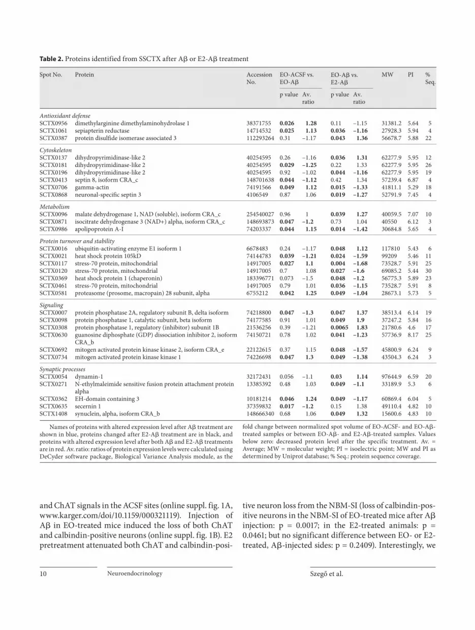
Proteins involved in antioxidant defense were identi-fied from SSCTX samples (table 2) after A � injection. Ex-pression of dimethylarginine dimethylaminohydrolase 1 and sepiapterin reductase increased, while protein disul-fide isomerase associated 3 decreased after A � injection. E2 pretreatement reversed these changes. Expression of some cytoskeletal proteins like dihydropyrimidinase-like 2 and septin 8 decreased, while � -actin increased af-ter A � -injection. E2 pretreatment before A � injection
Szegő et al. Neuroendocrinology 8
prevented these changes, and increased the expression of septin 3 protein. A relatively low number of proteins was identified from the ‘metabolism’ groups, meaning that A � had a lower effect on general cellular homeostasis out-side of the injection site. Interestingly, we found A � -in-duced upregulation of apolipoprotein A-1, while E2 pre-treatment decreased the expression of this protein. Pro-teins with chaperon activity (e.g. heat shock protein 105 kDa, stress protein 70) were also altered by A � or E2!A � treatment, moreover, expression of macropain subunit 28 increased after A � injection, but decreased with E2–A � treatment. In contrast to the findings from the NBM, we
observed increased MEKK1 and decreased protein phos-phatase 2A expression, suggesting the activation of ERK pathways following A � injection. Secernin 1, a protein involved in synaptic processes, decreased, while EH do-main containing 3 increased in expression following A � injection, and E2 treatment reversed these changes.
Decreased Calbindin Immunoreaction in the NBM-SI after A � Injection We selected calbindin from the protein list to check
the expression pattern after A � injection of EO- and E2-treated mice. We found no colocalization of calbindin
ba
c
c
d
f
e
ed f
Fig. 3. Representative 2D gel images from the NBM-SI ( a , c , d ) and SSCTX ( b , e , f ). a , b Pseudo-colored 2D maps of NMB-SI and SSCTX, respectively. The internal standard pooled from all of the samples from the same ana-tomical area was labeled with Cy3 green, samples from the treatment group A � -EO were labeled with Cy5 red. c – f 3D reconstruction of protein spots marked in ( a ) or ( b ) gel maps. Graphs represent the abundance of the spot relative to the internal standard.

E2 Reduces A � -Induced Neuron Loss in the NBM
Neuroendocrinology 9
Table 1. Proteins identified from NBM-SI after A� or E2-A� treatment
SpotNo.
Protein AccessionNo.
EO-ACSF vs.EO-A�
EO-A� vs.E2-A�
MW PI % Seq.
p value Av. ratio p value Av. ratio
Antioxidant defenseSI0210 inner membrane protein, mitochondrial 70608131 0.3 –1.12 0.044 1.19 86295.4 7.02 5SI0482 protein disulfide isomerase associated 3 112293264 0.042 1.1 0.03 –1.11 56678.7 5.88 5SI1474 N(G),N(G)-dimethylarginine dimethylaminohydrolase 2 45476968 0.048 1.07 0.037 –1.07 29646 5.66 6SI1633 DJ-1 protein 74212240 0.0013 –1.07 0.039 1.12 20025.4 6.32 12SI1669 glutathione S-transferase, alpha 4 160298217 0.37 –1.14 0.016 1.31 26890.5 6.02 13
CytoskeletonSI0254 dihydropyrimidinase-like 2 40254595 0.03 1.44 0.12 –1.27 62277.9 5.95 13SI0256 dihydropyrimidinase-like 2 40254595 0.038 1.4 0.23 –1.24 62277.9 5.95 29SI0381 dihydropyrimidinase-like 2 40254595 0.049 –1.12 0.44 –1.05 62277.9 5.95 5SI0633 fascin 497775 0.021 1.28 0.041 –1.33 54508.3 6.44 5SI0886 gamma-actin 74191566 0.036 –1.18 0.028 1.17 41811.1 5.29 15
MetabolismSI0212 NADH-ubiquinone oxidoreductase 75-kDa subunit, mitochondrial 47117271 0.017 1.29 0.67 –1.04 79777.3 5.51 4SI0433 creatine kinase, brain 74223625 0.016 –1.88 0.54 –1.07 42753.5 5.46 4SI0625 aldhehyde dehydrogenase family 5, subfamily A1 27369748 0.0082 1.17 0.37 –1.14 55968.5 8.53 4SI0660 aldehyde dehydrogenase family 7, member A1 188219757 0.82 –1.06 0.044 1.13 58861.8 7.16 10SI0665 enolase 2, gamma neuronal 70794816 0.025 1.12 0.002 –1.13 47141.1 6.37 7SI0685 ATP synthase, H+ transporting, mitochondrial F1 complex, alpha
subunit,isoform 1
74211977 0.037 –1.42 0.85 1.03 59782.9 9.22 5
SI0817 succinate-coenzyme A ligase, ADP-forming, beta subunit 74151797 0.05 1.18 0.45 –1.05 50797 7.7 11SI1191 lactate dehydrogenase B 6678674 0.0092 1.1 0.11 –1.04 36572.5 5.7 5SI1194 lactate dehydrogenase B 6678674 0.048 1.12 0.13 –1.06 36572.5 5.7 23SI1310 malate dehydrogenase 1, NAD (soluble) 254540027 0.19 1.07 0.036 –1.07 40059.5 7.07 7SI1414 tyrosine 3-monooxygenase/tryptophan 5-monooxygenase activation
protein, � polypeptide26344914 0.0016 –1.34 0.4 1.07 28212 4.81 30
SI1503 phosphoglycerate mutase 1 114326546 0.12 1.07 0.029 –1.11 28832.1 6.68 32SI1643 haloacid dehalogenase-like hydrolase domain containing 2 74190684 0.79 1.01 0.046 –1.2 28760.4 5.69 12SI1677 ATP synthase, H+ transporting, mitochondrial F1 complex, alpha
subunit, isoform 174211977 0.87 –1.02 0.029 1.41 59782.9 9.22 5
Protein turnover and stabilitySI0169 heat shock protein 1, alpha 74147335 0.037 –1.08 0.28 –1.04 84816.3 4.93 10SI0720 proteasome (prosome, macropain) 26S subunit, ATPase 2 33859604 0.0011 –1.45 0.03 1.38 52866.9 5.97 8SI0515 heat shock protein 1 (chaperonin) 148680184 0.042 1.13 0.47 –1.04 53095.7 5.3 12SI1350 otubain 1 19527388 0.043 –1.19 0.85 1.02 31270.2 4.85 16
SignalingSI0676 guanosine diphosphate (GDP) dissociation inhibitor 1 74150721 0.18 1.1 0.0062 –1.13 57736.9 8.17 7SI1313 protein phosphatase 2A, regulatory subunit B (PR 53), isoform CRA_b 26327445 0.024 1.1 0.0067 –1.11 38513.4 6.14 7SI1467 inositol (myo)-1(or 4)-monophosphatase 1, isoform CRA_b 148673218 0.0066 1.13 0.27 1.06 31025.9 4.79 4SI1538 calbindin 2 34098931 0.036 –1.3 0.34 1.09 31372.8 4.94 11SI1548 calbindin 2 34098931 0.0057 –1.17 0.96 1.01 31372.8 4.94 18SI1553 calbindin-28K, isoform CRA_a 6753242 0.013 1.34 0.54 –1.06 30093.4 4.71 24SI0564 protein phosphatase 3, catalytic subunit, alpha isoform, isoform CRA_d 148680184 0.038 –1.36 0.11 1.22 62277.9 5.95 6
Synaptic processesSI0122 Aldh1l1 protein 27532959 0.035 1.14 0.37 –1.07 98734.7 5.69 2SI0500 phosphatidylethanolamine binding protein 1 74222953 0.049 1.18 0.28 –1.11 20889.6 5.36 14SI0572 glutamate dehydrogenase 1 precursor 6680027 0.58 –1.07 0.0028 1.18 61337.1 8.05 10SI1307 phosphatidylethanolamine binding protein 1 74222953 0.031 1.16 0.13 –1.08 20889.6 5.36 25SI1711 phosphatidylethanolamine binding protein 1 74222953 0.71 1 0.0049 1.03 20889.6 5.36 51
UnknownSI1571 mCG9061, isoform CRA_c 148706375 0.049 1.14 0.31 –1.07 28401.8 8.17 21SI1716 RIKEN cDNA 1110067D22, isoform CRA_a 148675893 0.027 1.14 0.3 1.05 19556.5 5.48 6
Names of proteins with altered expression level after A� treatment are shown in blue, proteins changed after E2-A� treatment are in black and pro-teins with altered expression level after both A� and E2-A� treatments arein red. Av. ratio: ratios of protein expression levels were calculated using DeCyder software package, Biological Variance Analysis module, as the fold
change between normalized spot volume of EO-ACSF- and EO-A�-treated samples or between EO-A�- and E2-A�-treated samples. Values below zero: decreased protein level after the specific treatment. Av. = Average; MW = molecular weight; PI = isoelectric point; MW and PI as determined byUniprot database; % Seq.: protein sequence coverage.
Szegő et al. Neuroendocrinology 10
and ChAT signals in the ACSF sites (online suppl. fig. 1A, www.karger.com/doi/10.1159/000321119). Injection of A � in EO-treated mice induced the loss of both ChAT and calbindin-positive neurons (online suppl. fig. 1B). E2 pretreatment attenuated both ChAT and calbindin-posi-
tive neuron loss from the NBM-SI (loss of calbindin-pos-itive neurons in the NBM-SI of EO-treated mice after A � injection: p = 0.0017; in the E2-treated animals: p = 0.0461; but no significant difference between EO- or E2-treated, A � -injected sides: p = 0.2409). Interestingly, we
Table 2. Proteins identified from SSCTX after A� or E2-A� treatment
Spot No. Protein Accession No.
EO-ACSF vs. EO-A�
EO-A� vs.E2-A�
MW PI %Seq.
p value Av. ratio
p value Av. ratio
Antioxidant defenseSCTX0956 dimethylarginine dimethylaminohydrolase 1 38371755 0.026 1.28 0.11 –1.15 31381.2 5.64 5SCTX1061 sepiapterin reductase 14714532 0.025 1.13 0.036 –1.16 27928.3 5.94 4SCTX0387 protein disulfide isomerase associated 3 112293264 0.31 –1.17 0.043 1.36 56678.7 5.88 22
CytoskeletonSCTX0137 dihydropyrimidinase-like 2 40254595 0.26 –1.16 0.036 1.31 62277.9 5.95 12SCTX0181 dihydropyrimidinase-like 2 40254595 0.029 –1.25 0.22 1.33 62277.9 5.95 26SCTX0196 dihydropyrimidinase-like 2 40254595 0.92 –1.02 0.044 –1.16 62277.9 5.95 19SCTX0413 septin 8, isoform CRA_c 148701638 0.044 –1.12 0.42 1.34 57239.4 6.87 4SCTX0706 gamma-actin 74191566 0.049 1.12 0.015 –1.33 41811.1 5.29 18SCTX0868 neuronal-specific septin 3 4106549 0.87 1.06 0.019 –1.27 52791.9 7.45 4
MetabolismSCTX0096 malate dehydrogenase 1, NAD (soluble), isoform CRA_c 254540027 0.96 1 0.039 1.27 40059.5 7.07 10SCTX0871 isocitrate dehydrogenase 3 (NAD+) alpha, isoform CRA_c 148693873 0.047 –1.2 0.73 1.04 40550 6.12 3SCTX0986 apolipoprotein A-I 74203337 0.044 1.15 0.014 –1.42 30684.8 5.65 4
Protein turnover and stabilitySCTX0016 ubiquitin-activating enzyme E1 isoform 1 6678483 0.24 –1.17 0.048 1.12 117810 5.43 6SCTX0021 heat shock protein 105kD 74144783 0.039 –1.21 0.024 –1.59 99209 5.46 11SCTX0117 stress-70 protein, mitochondrial 14917005 0.027 1.1 0.004 –1.68 73528.7 5.91 25SCTX0120 stress-70 protein, mitochondrial 14917005 0.7 1.08 0.027 –1.6 69085.2 5.44 30SCTX0369 heat shock protein 1 (chaperonin) 183396771 0.073 –1.5 0.048 –1.2 56775.3 5.89 23SCTX0461 stress-70 protein, mitochondrial 14917005 0.79 1.01 0.036 –1.15 73528.7 5.91 8SCTX0581 proteasome (prosome, macropain) 28 subunit, alpha 6755212 0.042 1.25 0.049 –1.04 28673.1 5.73 5
SignalingSCTX0007 protein phosphatase 2A, regulatory subunit B, delta isoform 74218800 0.047 –1.3 0.047 1.37 38513.4 6.14 19SCTX0098 protein phosphatase 1, catalytic subunit, beta isoform 74177585 0.91 1.01 0.049 1.9 37247.2 5.84 16SCTX0308 protein phosphatase 1, regulatory (inhibitor) subunit 1B 21536256 0.39 –1.21 0.0065 1.83 21780.6 4.6 17SCTX0630 guanosine diphosphate (GDP) dissociation inhibitor 2, isoform
CRA_b74150721 0.78 1.02 0.041 –1.23 57736.9 8.17 25
SCTX0692 mitogen activated protein kinase kinase 2, isoform CRA_e 22122615 0.37 1.15 0.048 –1.57 45800.9 6.24 9SCTX0734 mitogen activated protein kinase kinase 1 74226698 0.047 1.3 0.049 –1.38 43504.3 6.24 3
Synaptic processesSCTX0054 dynamin-1 32172431 0.056 –1.1 0.03 1.14 97644.9 6.59 20SCTX0271 N-ethylmaleimide sensitive fusion protein attachment protein
alpha13385392 0.48 1.03 0.049 –1.1 33189.9 5.3 6
SCTX0362 EH-domain containing 3 10181214 0.046 1.24 0.049 –1.17 60869.4 6.04 5SCTX0635 secernin 1 37359832 0.017 –1.2 0.15 1.38 49110.4 4.82 10SCTX1408 synuclein, alpha, isoform CRA_b 148666340 0.68 1.06 0.049 1.32 15600.6 4.83 10
Names of proteins with altered expression level after A� treatment are shown in blue, proteins changed after E2-A� treatment are in black, and proteins with altered expression level after both A� and E2-A� treatments are in red. Av. ratio: ratios of protein expression levels were calculated using DeCyder software package, Biological Variance Analysis module, as the
fold change between normalized spot volume of EO-ACSF- and EO-A�-treated samples or between EO-A�- and E2-A�-treated samples. Values below zero: decreased protein level after the specific treatment. Av. =Average; MW = molecular weight; PI = isoelectric point; MW and PI as determined by Uniprot database; % Seq.: protein sequence coverage.

E2 Reduces A � -Induced Neuron Loss in the NBM
Neuroendocrinology 11
observed some calbindin-positive cholinergic neurons in the E2-A � -treated mice, but not in E2-ACSF- or EO-A � -treated animals (online suppl. fig. 1C).
Discussion
The present study demonstrates that (1) A � solutions of different composition have different toxic potential on NBM cholinergic neurons in vivo; (2) pretreatment with E2 protects cholinergic cells and fibers against A � toxic-ity, and (3) A � alone or in combination with E2 pretreat-ment induces specific changes in the brain proteome of mice.
Spherical A � 1–42 Oligomers Induce Cholinergic Cell Death, but E2 Pretreatment Is Protective against A � Toxicity There is no agreement in the literature as to which A �
form is the most toxic. Oligomers and fibrils are well known cytotoxins in vivo [42–45] , disrupting choliner-gic neurotransmission [46] ; however, the monomer was found to be neuroprotective in vitro [47] . In the present study, A � solution containing mainly monomers (0 h) had no toxic effect on cholinergic neurons ( fig. 1, 2 ). In contrast, we observed increasing cytotoxicity with in-creasing ratios of particles with 1–2/2–3 nm height. Spherical A � oligomers (observed at 24 and 48 h) induced the maximal neuron loss calculated as the difference be-tween the control and lesioned sites. On the other hand, as the protofibrils were present in the 48-hour solutions but not in the 24-hour ones, we exclude them as the most toxic species on cholinergic neurons. It is important to note that our data do not exclude the possibility that A � might affect other neurotransmitter systems. Indeed, in-jection of A � into the NBM induces hypofunction of GA-BAergic neurons [48] and disturbs the serotonergic in-nervation of the rat basal forebrain and cerebral cortex [49, 50] . Moreover, as we found loss of calbindin signal in the NBM-SI, but not in cholinergic neurons (online suppl. fig. 1), A � indeed regulated also other neurons, such as glutamatergic or GABAergic cells [51, 52] .
Although clinical evidence is still controversial, it is well accepted that protection by E2 may vary with dose and timing of the treatment in vivo and in vitro [53] . E2 provides protection even two hours after the insult by rapid activation of signaling pathways and antioxidant mechanisms [54, 55] . Furthermore, E2 pretreatment de-velops cellular tolerance by inducing gene transcription and protein synthesis [23, 56, 57] . This fine-tuning in-
cludes changes in metabolism and synthesis of anti-apo-ptotic and antioxidant proteins [58] . In the present study, although a single injection of E2 was not enough to in-crease the basal number of cholinergic neurons or corti-cal fiber density [59] , it was able to reduce the toxic effect of A � ( fig. 2 c, d). However, in contrast to in vitro experi-ments by others [60] , in our study E2 could not eliminate cellular A � toxicity or induce further regeneration of cor-tical projections ( fig. 2 d). Contrary to our data, in anoth-er study [59] a 2-week E2 treatment of rats enhanced cor-tical cholinergic projections but did not affect the excito-toxicity-induced relative lesion. However, in this study, prolonged E2 treatment more likely increases regenera-tion than a single injection 24 h before the A � exposure. Moreover, injection of N-methyl- D -aspartate (NMDA) in the NBM resulted in approximately 50% loss of choliner-gic neurons; while in our study, a more moderate cholin-ergic cell loss (approximately 25%) was induced using A � . In addition, the most likely different toxic mechanisms induced by A � and NMDA can also be responsible for the different results.
Putative Mechanisms of Neurotoxicity and Neuroprotection We hypothesized that A � and E2 induce changes in
the proteomes that mediate cell death or protect neurons. Comparison of proteomes of (1) EO-ACSF vs. EO-A � , and (2) EO-A � vs. E2-A � provides information about the neurotoxic effect of (1) A � and (2) protective mechanisms of E2 against A � toxicity. We analyzed protein changes in the NBM-SI (cell bodies, injection), and changes in the SSCTX (projection). Hereafter, the names of proteins with altered expression are in bold print; see tables 1 and 2. Hypothetical pathways suggested to be influenced by A � or E2-A � treatment are depicted in figure 4 .
A � induced upregulation of NADH ubiquinone oxi-doreductase, and downregulation of ATP synthase ( ta-ble 1 ; fig. 4 a) in the NBM-SI. Increased NADH oxidation and decreased ATP production utilizing pH gradient might make mitochondria acidic, deplete ATP and trig-ger apoptotic signaling [61] . Recently, Rhein et al. [62] have found decreased mitochondrial electron transport complex IV activity, a drop in ATP level and upregulation of complex I proteins in A � -transgenic mice. These data are in line with the detected changes in our experiment. On the other hand, E2 pretreatment prevented the A � -induced regulation of these proteins, preserving mito-chondrial integrity. Mitochondrial dysfunction is often associated with increased production of reactive oxygen species inducing inactivation of proteins of the respira-
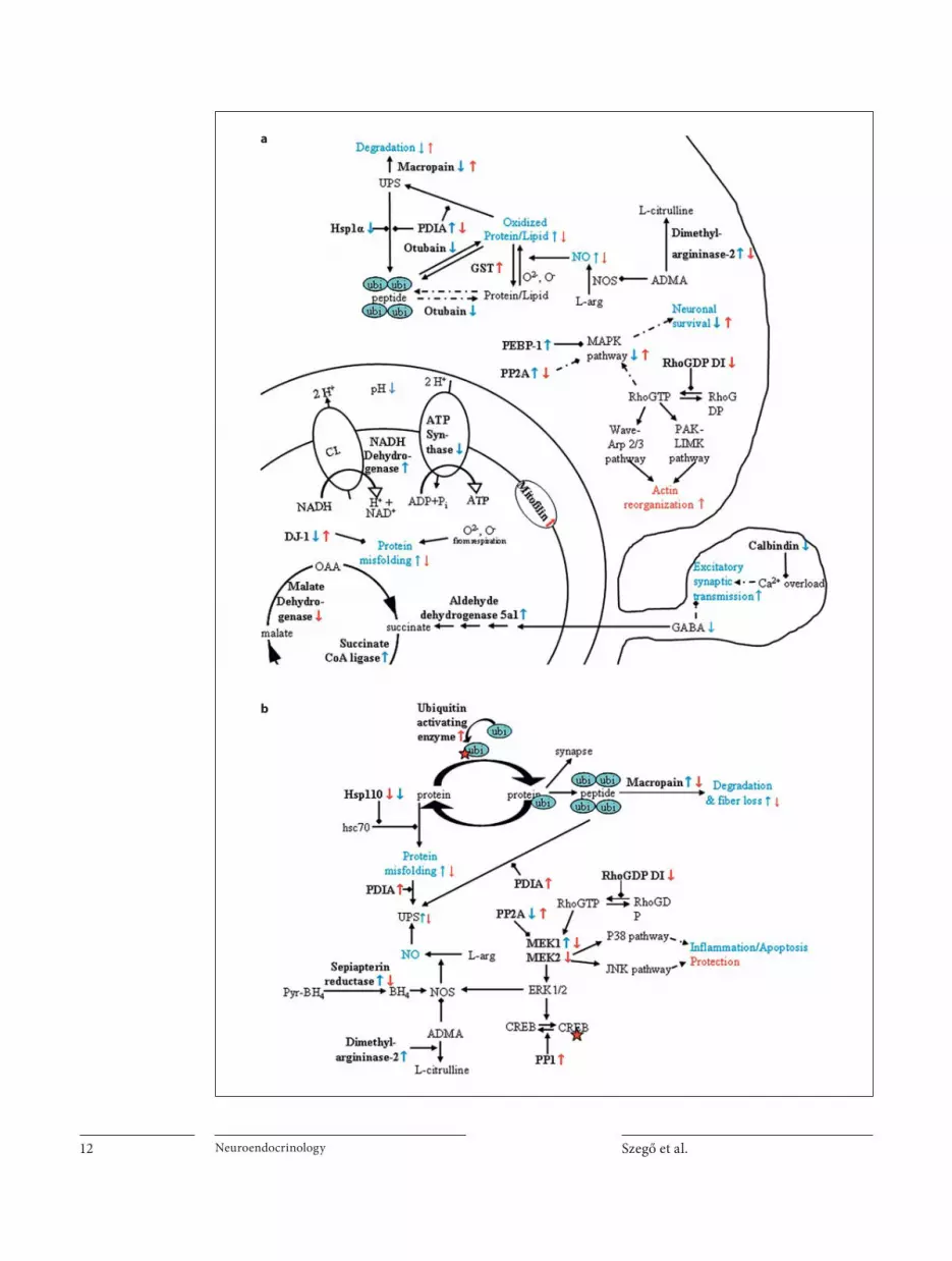
Szegő et al. Neuroendocrinology 12
a
b

E2 Reduces A � -Induced Neuron Loss in the NBM
Neuroendocrinology 13
tory chain, as it was shown in vivo [62] . Moreover, oxida-tive inactivation of neuronal proteins may lead to the de-velopment of AD [63] . In our experiments, A � decreased the level of DJ-1 protein, a chaperon which protects mito-chondrial complex I under oxidative stress [64] . More-over, A � increased the expression of dimethylargini-nase-2, an enzyme that hydrolyses asymmetric dimeth-ylarginine (ADMA) [65] . As ADMA is an endogenous inhibitor of nitrogen monoxide synthase [66] , A � could trigger NO production and induce further oxidative damage. E2, in turn, increased DJ-1, decreased dimethy-largininase-2 and increased glutathione S-transferase ex-pression ( fig. 3 a). Neurons are particularly susceptible to NO and peroxynitrite exposure, and the nitrosative stress may depend on the level of reduced glutathione [67] . Therefore, E2 pretreatment might reduce nitrosative stress in the NBM.
Signaling pathways associated with raf kinases plays a role in neuronal survival and death signaling. We found upregulation of two raf kinase inhibiting proteins follow-ing A � treatment, namely phosphatidylethanolamine-binding protein 1 or Raf kinase inhibitor protein (RKIP) and protein phosphatase 2A (PP2A) [68, 69] . The possible inactivation of ERK1/2, p38 or JNK pathways is in con-trast to some studies reporting A � -induced pathological activation of raf pathways [70, 71] . Contrary to our work, A � induced rapid ERK1/2 phosphorylation in vitro [71] , or a delayed (48 h) p38MAPK pathway activation in mi-
croglia cells in vivo [70] , suggesting that ERK pathways mediate rather death than survival signaling following A � exposure. The possible differences observed in our studies can be due to the different exposure time, timing of measurements after A � application, A � concentration, species and the different signals measured (phosphoryla-tion vs. inhibitor expression). Furthermore, we could not detect delayed, increased phosphorylation of p42/44 or p38MAPKs 2 weeks after A � injection in the NBM (data not shown). On the other hand, our results and findings from other groups might indicate that A � -induced sig-naling pathway activation can also be time dependent; probably A � induced a rapid or delayed activation of these signaling pathways in our study, before or beyond our DIGE experiment (less or more than 24 h). Support-ing this latter hypothesis, we found decreased calbindin (CB) expression after A � injection. By lowering intracel-lular Ca 2+ buffer capacity and increasing Ca 2+ concentra-tion, decreased calbindin expression could lead to in-creased MAPK activation in a later time point, and, ulti-mately, cell death [72, 73] . However, we found no colocalization of CB and ChAT signals in the NBM-SI; therefore, A � seems to regulate CB expression in other types of neurons or in glia cells. As significant propor-tions of the CB-positive cells are likely the cortically pro-jecting, possibly glutamatergic NBM neurons [52] , in-creased intracellular Ca 2+ concentration due to the loss of CB might induce excitotoxicity in the cortex, and con-tribute to cholinergic fiber loss and fiber-loss-induced dying back mechanism ( fig. 3 a). On the other hand, E2 prevented RKIP upregulation and calbindin downregu-lation and further decreased PP2A expression, decreas-ing MAPK pathway inhibition. E2 also attenuated calb-indin-positive cell loss from the NBM-SI (online suppl. fig. 1). Previously, we demonstrated that rapid action of E2 in vivo involves MAPK signaling in the NBM-SI in vivo [40] , and a link has been shown to exist between the neuroprotective effect of E2 and MAPK activation [57] . Altogether, A � alters respiration, metabolism and signal-ing systems of NBM-SI neurons, and these additive ef-fects might all converge on death pathways [74] , but can be reduced by E2 pretreatment.
Interestingly, injection of A � into the NBM-SI in-duced protein changes also in the cortical projection area. We found upregulation of sepiapterin reductase, an en-zyme essential for tetrahydrobiopterin (cofactor for all NOS isoforms), and dimethylarginine dimethylamino-hydrolase 1, a protein that increases NO production ( fig. 3 b). However, E2 prevented A � -dependent upregula-tion of both enzymes in the SSCTX.
Fig. 4. Schematic illustration of pathways supposed to be regu-lated by A � and/or E2 treatment. a We assume that A � injection in the NBM-SI increases mitochondrial acidity NADH-dehydro-genase, ATP synthase and decreases DJ-1 expression. A � might inhibit MAPK signaling PP2A, PDEBP-1. E2 prevented the regu-lation of these proteins. b A � injection in the NMB induced de-creased MAPK pathway inhibition PP2A, MEK1 and increased NOS activation sepiapterin reductase, dimethylargininase-2. However, 24 h E2 pretreatment rather led to decreased MAPK signaling PP2A, PP1, RhoGDI MEK1, MEK2 and inhibited NOS activation after A � treatment. Names of proteins with altered ex-pression are in bold, and protein regulation by A � is indicated with a blue arrow and by E2 with a red arrow. Continuous/dashed arrows indicate direct/indirect regulation, respectively. UPS = Ubiquitin-proteasome system; Hsp = heat shock protein; PDIA = protein disulfide isomerase associated; ubi = ubiquitin; GST = glutathione S-transferase; NOS = nitrogen oxide synthase; ADMA = asymmetric dimethylarginine; PEBP-1 = phosphatidyl-ethanolamine binding protein 1; PP = protein phosphatase; RhoGDP DI = Rho-GDP dissociation inhibitor; MAPK = mito-gen-activated protein kinase; BH4 = tetrahydrobiopterin; MEK = MAP kinase kinase 1.

Szegő et al. Neuroendocrinology 14
A � induced the upregulation of MAP kinase kinase 1 (MEK1) in the SSCTX and in this way – contrary to the findings in the NBM – A � may activate ERK1/2, p38 or JNK pathways. Although MEK-activated pathways are often associated with survival and neuroprotection even in in vivo models [75] , A � also activates the MAPK cas-cade [76] inducing pathologic phosphorylation of cyto-skeletal proteins. We injected the A � in the NBM, and we found ERK pathway upregulation just in the projection area, but not in the NBM. These results suggest that A � -induced signaling pathway activation is probably regu-lated with different timing in the two areas. A � might induce first a rapid activation in the site of injection (NBM, as suggested in Young et al. [71] ), then decrease signaling in the NMB but increase signaling in the pro-jection area, SSCTX (24 h). In addition, inhibition or per-manent activation of the same pathway might induce the same response [77, 78] . However, E2 prevented the change in MEK1 expression, and decreased MEK2 level. More-over, E2 almost doubled the expression of protein phos-phatase 1 (PP1) and PP2A, proteins responsible among others for inactivation of some MAPK targets. Similar to our data from the cortex, Valles et al. [20] found that A � induced upregulation of the p38MAPK pathway, whereas this activation was prevented by E2 pretreatment. In ad-dition, E2 was shown to induce phosphatases to exert a neuroprotective effect in vitro [79] . Therefore, under these conditions, E2 was able to reverse the proposed A � -induced kinase overactivation in the SSCTX ( fig. 3 b), and probably partly via prevention of cortical fibers and in-hibiting ‘dying back’ process, E2 protected cholinergic cell bodies in the NBM-SI.
It is well known that BFC neurons play an important role in learning and memory formation and that E2 de-pletion is associated with the cognitive decline observed in AD [24] . In the present study, we demonstrated that E2 is able to reduce A � -induced damage in the NBM-SI. We
found several cellular processes including regulation of mitochondrial enzymes and signaling pathways that could explain extracellular A � toxicity and E2 protec-tion. Collectively, our results demonstrate that in respect to A � , multiple factors converge upon pathways of both A � -mediated cholinergic neurodegeneration and E2-me-diated protection.
Acknowledgments
We are grateful to Péter Batáry for the assistance in statistical analysis. We thank Márta Zarándi (Department of Medical Chemistry, University of Szeged) for generously providing the amyloid- � peptide. This work was supported by the Regional Center of Excellence – Neurobiological Center of Excellence in Southern Hungary (RET-DNK to G.D.J., B.P. and T.J.), by OTKA (68464, 81950 to J.K.), by ETT (to I.M.A.), by the Hungarian Na-tional Office of Research and Technology (NANOAMI KFKT-1-2006-0021, OMFB-380/2006 to I.M.K.) and by the Deutsche Forschungsgemeinschaft through the DFG Research Center for Molecular Physiology of the Brain (CMPB, to E.M.S.). We thank András Czurkó and Zsolt Datki for the helpful discussion, and Cathy Ludwig for critical reading of the manuscript.
Author Contributions
E.M.S., G.D.J. and J.K. designed the study. E.M.S. performed the in vivo experiments, tissue staining, DIGE and data analysis. K.A.K. performed DIGE analysis. J.K. performed and analyzed AFM data, Ü.M. and M.S.Z.K. performed ThioflavinT and AFM measurements, B.P. provided the amyloid peptide, and A.C. and T.J. identified the proteins. M.P. prepared the micropunches and G.M. and I.M.A. prepared histochemical staining. E.M.S. and G.D.J. wrote the manuscript.
Disclosure Statement
The authors have no conflicts of interest to disclose.
References
1 Wimo A, Winblad B, Aguero-Torres H, von Strauss E: The magnitude of dementia oc-currence in the world. Alzheimer Dis Assoc Disord 2003; 17: 63–67.
2 Mattson MP: Pathways towards and away from Alzheimer’s disease. Nature 2004; 430: 631–639.
3 Schliebs R, Arendt T: The significance of the cholinergic system in the brain during aging and in Alzheimer’s disease. J Neural Transm 2006; 113: 1625–1644.
4 Mufson EJ, Counts SE, Perez SE, Ginsberg SD: Cholinergic system during the progres-sion of Alzheimer’s disease: therapeutic im-plications. Expert Rev Neurother 2008; 8: 1703–1718.
5 Wang HY, Lee DHS, Davis CB, Shank RP: Amyloid peptide a beta(1–42) binds selec-tively and with picomolar affinity to alpha 7 nicotinic acetylcholine receptors. J Neuro-chem 2000; 75: 1155–1161.
6 Hernandez CM, Kayed R, Zheng H, Sweatt JD, Dineley KT: Loss of alpha 7 nicotinic re-ceptors enhances beta-amyloid oligomer ac-cumulation, exacerbating early-stage cogni-tive decline and septohippocampal pathol-ogy in a mouse model of Alzheimer’s dis-ease. J Neurosci 2010; 30: 2442–2453.
7 Dineley KT: Beta-amyloid peptide – nicotin-ic acetylcholine receptor interaction: the two faces of health and disease. Front Biosci 2007; 12: 5030–5038.

E2 Reduces A � -Induced Neuron Loss in the NBM
Neuroendocrinology 15
8 Pike CJ, Carroll JC, Rosario ER, Barron AM: Protective actions of sex steroid hormones in Alzheimer’s disease. Front Neuroendocrinol 2009; 30: 239–258.
9 Waring SC, Rocca WA, Petersen RC, O’Brien PC, Tangalos EG, Kokmen E: Postmeno-pausal estrogen replacement therapy and risk of AD – a population-based study. Neu-rology 1999; 52: 965–970.
10 Hu L, Yue Y, Zuo P-p, Jin Z-y, Feng F, You H, li M-L, Ge Q-S: Evaluation of neuroprotec-tive effects of long-term low dose hormone replacement therapy on postmenopausal women brain hippocampus using magnetic resonance scanner. Chin Med Sci J 2006; 21: 214–218.
11 Asthana S, Craft S, Baker LD, Raskind MA, Birnbaum RS, Lofgreen CP, Veith RC, Plym-ate SR: Cognitive and neuroendocrine re-sponse to transdermal estrogen in post-menopausal women with Alzheimer’s dis-ease: results of a placebo-controlled, double- blind, pilot study. Psychoneuroendocrinol-ogy 1999; 24: 657–677.
12 Henderson VW, Watt L, Buckwalter JG: Cognitive skills associated with estrogen re-placement in women with Alzheimer’s dis-ease. Psychoneuroendocrinology 1996; 21: 421–430.
13 Simpkins JW, Wen Y, Perez E, Yang SH, Wang XF: Role of nonfeminizing estrogens in brain protection from cerebral ischemia – an animal model of Alzheimer’s disease neuropathology. Future of hormone therapy: what basic science and clinical studies teach us. Ann NY Acad Sci 2005; 1052: 233–242.
14 Singh M, Dykens JA, Simpkins JW: Novel mechanisms for estrogen-induced neuro-protection. Exp Biol Med 2006; 231: 514–521.
15 Simpkins JW, Singh M: More than a decade of estrogen neuroprotection. Alzheimers Dement 2008; 4:S131–S136.
16 Abraham IM, Koszegi Z, Tolod-Kemp E, Szego EM: Action of estrogen on survival of basal forebrain cholinergic neurons: pro-moting amelioration. Psychoneuroendocri-nology 2009; 34(suppl 1):S104–S112.
17 Morinaga A, Hirohata M, Ono K, Yamada M: Estrogen has anti-amyloidogenic effects on Alzheimer’s beta-amyloid fibrils in vitro. Biochem Biophys Res Commun 2007; 359: 697–702.
18 Levin-Allerhand JA, Lominska CE, Wang J, Smith JD: 17Alpha-estradiol and 17beta-es-tradiol treatments are effective in lowering cerebral amyloid-beta levels in abetaPPSWE transgenic mice. J Alzheimers Dis 2002; 4: 449–457.
19 Chen SH, Nilsen J, Brinton RD: Dose and temporal pattern of estrogen exposure deter-mines neuroprotective outcome in hippo-campal neurons: therapeutic implications. Endocrinology 2006; 147: 5303–5313.
20 Valles SL, Borras C, Gambini J, Furriol J, Ortega A, Sastre J, Pallardo FV, Vina J: Oes-tradiol or genistein rescues neurons from amyloid beta-induced cell death by inhibit-ing activation of p38. Aging Cell 2008; 7: 112–118.
21 Rasgon NL, Silverman D, Siddarth P, Miller K, Ercoli LM, Elman S, Lavretsky H, Huang SC, Phelps ME, Small GW: Estrogen use and brain metabolic change in postmenopausal women. Neurobiol Aging 2005; 26: 229–235.
22 Brinton RD: Estrogen regulation of glucose metabolism and mitochondrial function: Therapeutic implications for prevention of Alzheimer’s disease. Adv Drug Deliv Rev 2008; 60: 1504–1511.
23 Szego EM, Kekesi KA, Szabo Z, Janaky T, Ju-hasz GD: Estrogen regulates cytoskeletal f lexibility, cellular metabolism and synaptic proteins: a proteomic study. Psychoneuroen-docrinology 2010; 35: 807–819.
24 Gibbs RB: Does short-term estrogen therapy produce lasting benefits in brain? Endocri-nology 2010; 151: 843–845.
25 Leuner B, Mendolia-Loffredo S, Shors TJ: High levels of estrogen enhance associative memory formation of ovariectomized fe-males. Psychoneuroendocrinology 2004; 29: 883–890.
26 Henderson VW, Paganini-Hill A, Miller BL, Elble RJ, Reyes PF, Shoupe D, McCleary CA, Klein RA, Hake AM, Farlow MR: Estrogen for Alzheimer’s disease in women – random-ized, double-blind, placebo-controlled trial. Neurology 2000; 54: 295–301.
27 Wang PN, Liao SQ, Liu RS, Liu CY, Chao HT, Lu SR, Yu HY, Wang SJ, Liu HC: Effects of estrogen on cognition, mood, and cerebral blood flow in AD – a controlled study. Neu-rology 2000; 54: 2061–2066.
28 Manthey D, Behl C: From structural bio-chemistry to expression profiling: neuropro-tective activities of estrogen. Neuroscience 2006; 138: 845–850.
29 Joerchel S, Raap M, Bigl M, Eschrich K, Schliebs R: Oligomeric beta-amyloid(1–42) induces the expression of Alzheimer disease-relevant proteins in cholinergic sn56.B5.G4 cells as revealed by proteomic analysis. Int J Dev Neurosci 2008; 26: 301–308.
30 Prokai L, Stevens SM, Rauniyar N, Nguyen V: Rapid label-free identification of estro-gen-induced differential protein expression in vivo from mouse brain and uterine tissue. J Proteome Res 2009; 8: 3862–3871.
31 Aluise CD, Robinson RA, Beckett TL, Mur-phy MP, Cai J, Pierce WM, Markesbery WR, Butterfield DA: Preclinical Alzheimer dis-ease: brain oxidative stress, Abeta peptide and proteomics. Neurobiol Dis 2010;39:221–228.
32 Zarandi M, Soos K, Fulop L, Bozso Z, Datki Z, Toth GK, Penke B: Synthesis of a beta(1–42) and its derivatives with improved effi-ciency. J Pept Sci 2007; 13: 94–99.
33 Stine WB, Dahlgren KN, Krafft GA, LaDu MJ: In vitro characterization of conditions for amyloid-beta peptide oligomerization and fibrillogenesis. J Biol Chem 2003; 278: 11612–11622.
34 Harper JD, Wong SS, Lieber CM, Lansbury PT: Observation of metastable A beta amy-loid protofibrils by atomic force microscopy. Chem Biol 1997; 4: 119–125.
35 Naiki H, Gejyo F: Kinetic analysis of amyloid fibril formation: amyloid, prions, and other protein aggregates. Methods Enzymol 1999; 309: 305–318.
36 Paxinos G, Franklin KBJ: The Mouse Brain in Stereotaxic Coordinates. San Diego, Aca-demic Press, 2001.
37 Palkovits M: Isolated removal of hypotha-lamic or other brain nuclei of rat. Brain Res 1973; 59: 449–450.
38 Palkovits M, Brownstein MJ: Maps and Guide to Microdissection of the Rat Brain. New York, Elsevier, 1988.
39 Kapp EA, Schutz F, Reid GE, Eddes JS, Mori-tz RL, O’Hair RAJ, Speed TP, Simpson RJ: Mining a tandem mass spectrometry data-base to determine the trends and global fac-tors influencing peptide fragmentation. Anal Chem 2003; 75: 6251–6264.
40 Szego EM, Barabas K, Balog J, Szilagyi N, Korach KS, Juhasz G, Abraham IM: Estrogen induces estrogen receptor alpha-dependent camp response element-binding protein phosphorylation via mitogen-activated pro-tein kinase pathway in basal forebrain cho-linergic neurons in vivo. J Neurosci 2006; 26: 4104–4110.
41 Hedreen JC, Bacon SJ, Price DL: A modified histochemical technique to visualize acetyl-cholinesterase-containing axons. J Histo-chem Cytochem 1985; 33: 134–140.
42 Cleary JP, Walsh DM, Hofmeister JJ, Shan-kar GM, Kuskowski MA, Selkoe DJ, Ashe KH: Natural oligomers of the amyloid-pro-tein specifically disrupt cognitive function. Nat Neurosci 2005; 8: 79–84.
43 Shankar GM, Li SM, Mehta TH, Garcia-Mu-noz A, Shepardson NE, Smith I, Brett FM, Farrell MA, Rowan MJ, Lemere CA, Regan CM, Walsh DM, Sabatini BL, Selkoe DJ: Am-yloid-beta protein dimers isolated directly from Alzheimer’s brains impair synaptic plasticity and memory. Nat Med 2008; 14: 837–842.
44 Lesne S, Koh MT, Kotilinek L, Kayed R, Gla-be CG, Yang A, Gallagher M, Ashe KH: A specific amyloid-beta protein assembly in the brain impairs memory. Nature 2006; 440: 352–357.
45 Busciglio J, Lorenzo A, Yeh J, Yankner BA: Beta-amylid fibrils induce tau-phosphoryla-tion and loss of microtubule-binding. Neu-ron 1995; 14: 879–888.
Szegő et al. Neuroendocrinology 16
46 Machova E, Rudajev V, Smyckova H, Koivis-to H, Tanila H, Dolezal V: Functional cholin-ergic damage develops with amyloid accu-mulation in young adult appswe/ps1de9 transgenic mice. Neurobiol Dis 2010; 38: 27–35.
47 Giuffrida ML, Caraci F, Pignataro B, Cataldo S, De Bona P, Bruno V, Molinaro G, Pappa-lardo G, Messina A, Palmigiano A, Garozzo D, Nicoletti F, Rizzarelli E, Copani A: Beta-amyloid monomers are neuroprotective. J Neurosci 2009; 29: 10582–10587.
48 Scali C, Prosperi C, Giovannelli L, Bianchi L, Pepeu G, Casamenti F: Beta(1–40) amyloid peptide injection into the nucleus basalis of rats induces microglia reaction and enhanc-es cortical gamma-aminobutyric acid re-lease in vivo. Brain Res 1999; 831: 319–321.
49 Harkany T, O’Mahony S, Keijser J, Kelly JP, Konya C, Borostyankoi ZA, Gorcs TJ, Zaran-di M, Penke B, Leonard BE, Luiten PGM: Be-ta-amyloid((1–42))-induced cholinergic le-sions in rat nucleus basalis bidirectionally modulate serotonergic innervation of the basal forebrain and cerebral cortex. Neuro-biol Dis 2001; 8: 667–678.
50 Aguado-Llera D, Arilla-Ferreiro E, Chowen JA, Argente J, Puebla-Jimenez L, Frago LM, Barrios V: 17-Beta-estradiol protects deple-tion of rat temporal cortex somatostatinergic system by beta-amyloid. Neurobiol Aging 2007; 28: 1396–1409.
51 Gonzalez I, Arevalo-Serrano J, Perez JL, Gonzalo P, Gonzalo-Ruiz A: Effects of beta-amyloid peptide on the density of m2 musca-rinic acetylcholine receptor protein in the hippocampus of the rat: relationship with gaba-, calcium-binding protein and soma-tostatin-containing cells. Neuropathol Appl Neurobiol 2008; 34: 506–522.
52 Gritti I, Manns ID, Mainville L, Jones BE: Parvalbumin, calbindin, or calretinin in cortically projecting and gabaergic, cholin-ergic, or glutamatergic basal forebrain neu-rons of the rat. J Comp Neurol 2003; 458: 11–31.
53 Gibbs RB: Effects of estrogen on basal fore-brain cholinergic neurons vary as a function of dose and duration of treatment. Brain Res 1997; 757: 10–16.
54 Shughrue PJ, Merchenthaler I: Estrogen pre-vents the loss of ca1 hippocampal neurons in gerbils after ischemic injury. Neuroscience 2003; 116: 851–861.
55 Merchenthaler I, Shughrue PJ: Neuroprotec-tion by estrogen in animal models of isch-emia and Parkinson’s disease. Drug Dev Res 2005; 66: 172–181.
56 Nilsen J, Chen SH, Irwin RW, Iwamoto S, Brinton RD: Estrogen protects neuronal cells from amyloid beta-induced apoptosis via regulation of mitochondrial proteins and function. BMC Neurosci 2006; 7: 74.
57 Lebesgue D, Chevaleyre V, Zukin RS, Etgen AM: Estradiol rescues neurons from global ischemia-induced cell death: multiple cellu-lar pathways of neuroprotection. Steroids 2009; 74: 555–561.
58 Garcia-Segura LM, Azcoitia I, DonCarlos LL: Neuroprotection by estradiol. Prog Neu-robiol 2001; 63: 29–60.
59 Horvath KM, Hartig W, Van der Veen R, Keijser JN, Mulder J, Ziegert M, Van der Zee EA, Harkany T, Luiten PGM: 17-Beta-estra-diol enhances cortical cholinergic innerva-tion and preserves synaptic density follow-ing excitotoxic lesions to the rat nucleus basalis magnocellularis. Neuroscience 2002; 110: 489–504.
60 Guerra B, Diaz M, Alonso R, Marin R: Plas-ma membrane oestrogen receptor mediates neuroprotection against beta-amyloid toxic-ity through activation of raf-1/mek/erk cas-cade in septal-derived cholinergic sn56 cells. J Neurochem 2004; 91: 99–109.
61 Yang L, Mei Y, Xie Q, Han X, Zhang F, Gu L, Zhang Y, Chen Y, Li G, Gao Z: Acidification induces bax translocation to the mitochon-dria and promotes ultraviolet light-induced apoptosis. Cell Mol Biol Lett 2008; 13: 119–129.
62 Rhein V, Song XM, Wiesner A, Ittner LM, Baysang G, Meier F, Ozmen L, Bluethmann H, Drose S, Brandt U, Savaskan E, Czech C, Gotz J, Eckert A: Amyloid-beta and tau syn-ergistically impair the oxidative phosphory-lation system in triple transgenic Alzhei-mer’s disease mice. Proc Natl Acad Sci USA 2009; 106: 20057–20062.
63 Butterfield DA, Poon HF, St Clair D, Keller JN, Pierce WM, Klein JB, Markesbery WR: Redox proteomics identification of oxida-tively modified hippocampal proteins in mild cognitive impairment: Insights into the development of Alzheimer’s disease. Neuro-biol Dis 2006; 22: 223–232.
64 Hayashi T, Ishimori C, Takahashi-Niki K, Taira T, Kim YC, Maita H, Maita C, Ariga H, Iguchi-Ariga SMM: Dj-1 binds to mitochon-drial complex i and maintains its activity. Biochem Biophys Res Commun 2009; 390: 667–672.
65 Smith MA, Harris PLR, Sayre LM, Beckman JS, Perry G: Widespread peroxynitrite-medi-ated damage in Alzheimer’s disease. J Neu-rosci 1997; 17: 2653–2657.
66 Leiper J, Vallance P: Biological significance of endogenous methylarginines that inhibit nitric oxide synthases. Cardiovasc Res 1999; 43: 542–548.
67 Calabrese V, Boyd-Kimball D, Scapagnini G, Butterfield DA: Nitric oxide and cellular stress response in brain aging and neurode-generative disorders: the role of vitagenes. In Vivo 2004; 18: 245–267.
68 Granovsky AE, Rosner MR: Raf kinase in-hibitory protein: a signal transduction mod-ulator and metastasis suppressor. Cell Res 2008; 18: 452–457.
69 Zhao J, Wu HW, Chen YJ, Tian HP, Li LX, Han X, Guo J: Protein phosphatase 2a-nega-tive regulation of the protective signaling pathway of Ca 2+ /cam-dependent erk activa-tion in cerebral ischemia. J Neurosci Res 2008; 86: 2733–2745.
70 Giovannini MG, Scali C, Prosperi C, Bellu c-ci A, Vannucchi MG, Rosi S, Pepeu G, Casa-menti F: Beta-amyloid-induced inflamma-tion and cholinergic hypofunction in the rat brain in vivo: involvement of the p38mapk pathway. Neurobiol Dis 2002; 11: 257–274.
71 Young KF, Pasternak SH, Rylett RJ: Oligo-meric aggregates of amyloid beta peptide 1–42 activate erk/mapk in sh-sy5y cells via the alpha 7 nicotinic receptor. Neurochem Int 2009; 55: 796–801.
72 Christakos S, Liu Y: Biological actions and mechanism of action of calbindin in the pro-cess of apoptosis. J Steroid Biochem Mol Biol 2004; 89–90: 401–404.
73 Geula C, Bu J, Nagykery N, Scinto LFM, Chan J, Joseph J, Parker R, Wu CK: Loss of calbindin-d-28k from aging human cholin-ergic basal forebrain: relation to neuronal loss. J Comp Neurol 2003; 455: 249–259.
74 Ferrer I: Altered mitochondria, energy me-tabolism, voltage-dependent anion channel, and lipid rafts converge to exhaust neurons in Alzheimer’s disease. J Bioenerg Biomembr 2009; 41: 425–431.
75 Sawe N, Steinberg G, Zhao H: Dual roles of the mapk/erk1/2 cell signaling pathway after stroke. J Neurosci Res 2008; 86: 1659–1669.
76 Matos M, Augusto E, Oliveira CR, Agostin-ho P: Amyloid-beta peptide decreases gluta-mate uptake in cultured astrocytes: involve-ment of oxidative stress and mitogen-ac-tivated protein kinase cascades. Neurosci-ence 2008; 156: 898–910.
77 Calabrese EJ, Baldwin LA: Hormesis: U-shaped dose responses and their centrality in toxicology. Trends Pharmacol Sci 2001; 22: 285–291.
78 Marini AM, Jiang H, Pan H, Wu X, Lipsky RH: Hormesis: A promising strategy to sus-tain endogenous neuronal survival pathways against neurodegenerative disorders. Ageing Res Rev 2008; 7: 21–33.
79 Simpkins JW, Yi KD, Yang SH: Role of pro-tein phosphatases and mitochondria in the neuroprotective effects of estrogens. Front Neuroendocrinol 2009; 30: 93–105.