effects of drugs of abuse on putative rostromedial tegmental neurons, inhibitory afferents to...
TRANSCRIPT

Marco Pistis
1
Effects of drugs of abuse on putative rostromedial tegmental neurons, inhibitory afferents to midbrain dopamine cells.
Running title: Modulation of RMTg neurons by drugs of abuse.
Salvatore Lecca1, Miriam Melis1, Antonio Luchicchi1, Maria Grazia Ennas Ph.D.2, M. Paola
Castelli M.D.1, 3, Anna Lisa Muntoni M.D.3,4, and Marco Pistis M.D.1, 3*
1B.B. Brodie Department of Neuroscience, 2Department of Cytomorphology, 3Center of
Excellence for the Neurobiology of Addiction, 4C.N.R. Neuroscience Institute-Cagliari,
University of Cagliari, 09042, Monserrato, Italy
*Corresponding author: Marco Pistis, M.D.
B.B. Brodie Department of Neuroscience
Center of Excellence for the Neurobiology of Addiction
University of Cagliari, Cittadella Universitaria
09042 Monserrato (CA), Italy
Email: [email protected]
Phone:+390706754324
Fax: +390706754320
peer
-005
8996
6, v
ersi
on 1
- 3
May
201
1Author manuscript, published in "Neuropsychopharmacology (2010)"
DOI : 10.1038/npp.2010.190

Marco Pistis
2
Abstract
Recent findings have underlined the rostromedial tegmental nucleus (RMTg), a structure located
caudally to the ventral tegmental area, as an important site involved in the mechanisms of
aversion. RMTg contains γ-aminobutyric acid (GABA) neurons responding to noxious stimuli,
densely innervated by the lateral habenula and providing a major inhibitory projection to reward-
encoding midbrain dopamine (DA) neurons.
One of the key features of drug addiction is the perseverance of drug seeking in spite of negative
and unpleasant consequences, likely mediated by response suppression within neural pathways
mediating aversion. To investigate whether the RMTg plays a role in the mechanisms of
addicting drugs, we studied acute effects of morphine, cocaine, the cannabinoid agonist
WIN55212-2 (WIN) and nicotine on putative RMTg neurons. We utilized single unit
extracellular recordings in anesthetized rats and whole-cell patch clamp recordings in brain slices
to identify and characterize putative RMTg neurons and their responses to drugs of abuse.
Morphine and WIN inhibited both firing rate in vivo and excitatory postsynaptic currents
(EPSCs) evoked by stimulation of rostral afferents in vitro, whereas cocaine inhibited discharge
activity without affecting EPSC amplitude. Conversely, nicotine robustly excited putative RMTg
neurons and enhanced EPSCs, an effect mediated by α7-containing nicotinic acetylcholine
receptors.
Our results suggest that activity of RMTg neurons is profoundly influenced by drugs of abuse
and, as important inhibitory afferents to midbrain DA neurons, they might take place in the
complex interplay between the neural circuits mediating aversion and reward.
peer
-005
8996
6, v
ersi
on 1
- 3
May
201
1

Marco Pistis
3
Keywords
Lateral habenula, aversion, addiction, patch clamp, electrophysiology, Fos, rat.
peer
-005
8996
6, v
ersi
on 1
- 3
May
201
1

Marco Pistis
4
Introduction
Recent studies have provided information on how brain regions encoding aversion and reward
are integrated. Mesolimbic dopamine (DA) neurons encode rewarding and appetitive stimuli
with phasic excitation, whereas reward omission induces phasic inhibition (Schultz, 2007a, b).
Aversive stimuli have been reported to induce both excitation (Brischoux et al, 2009) and
inhibition (Ungless et al, 2004) of DA neurons, a heterogeneous response correlated to specific
subgroups of DA neurons (ventral and dorsal, respectively) within the ventral tegmental area
(VTA). On the other hand, glutamatergic neurons in the lateral habenula (LHb), an epithalamic
region involved in the mechanisms of fear, anxiety and stress, respond in a reverse fashion, being
inhibited by rewards and excited by aversive stimuli (Hikosaka et al, 2008; Matsumoto and
Hikosaka, 2007). Noteworthy, activity of DA and LHb neurons appears to be causally correlated,
since electrical stimulation of the LHb inhibits DA neurons (Christoph et al, 1986; Ji and
Shepard, 2007; Matsumoto and Hikosaka, 2007). However, the sparse innervation of DA
neurons by excitatory LHb afferents (Brinschwitz et al, 2010; Omelchenko et al, 2009) unlikely
explains this inhibition, and the presence of an area intermediate between the LHb and the VTA
was originally postulated. Elegant studies by Jhou and colleagues (Jhou et al, 2009a; Jhou et al,
2009b) revealed that γ-aminobutyric acid (GABA) neurons in the rostromedial tegmental nucleus
(RMTg), a region also denominated as "tail" of the VTA (Perrotti et al, 2005), integrate input
from the LHb and send output projection to midbrain DA neurons. As described, the missing link
between LHb and midbrain DA neurons is provided. Notably, both RMTg and LHb neurons are
phasically activated by aversive stimuli and inhibited by appetitive stimuli (Jhou et al, 2009a). In
turn, RMTg neurons form inhibitory synapses with DA neurons in the VTA and the substantia
peer
-005
8996
6, v
ersi
on 1
- 3
May
201
1

Marco Pistis
5
nigra (Balcita-Pedicino et al, 2009), and are now accounted among the major inhibitory afferents
to mesencephalic DA neurons (Jhou et al, 2009a). The electrical activity of DA neurons, and the
resulting DA output in terminal areas, crucially depends on the balance between excitatory and
inhibitory inputs (Marinelli et al, 2006). Thus, the characterization of this novel inhibitory
pathway to DA neurons is of interest to understand how DA neurons respond to behavioral and
pharmacological stimuli.
Understanding the relationships between brain areas encoding reward and aversion, such as the
VTA and the RMTg, is also relevant when it concerns the effects of addicting drugs. Since drug
abuse potential can be envisaged as a balance between rewarding and aversive effects (Hutchison
and Riley, 2008; Simpson and Riley, 2005; Wise et al, 1976), it can be hypothesized that the
components of the appetitive properties of addicting drugs might result from both direct
activation of neural reward pathways and suppression of responses in neural circuits mediating
aversion. Indeed, one of the hallmarks of drug addiction is the perseverance of drug taking
despite of negative and unpleasant consequences of drug use. Recent studies indicate that drugs
of abuse affect GABA neurons in the RMTg: these neurons, and notably those projecting to the
VTA, express ΔFosB (Kaufling et al, 2009; Perrotti et al, 2005) and c-Fos following repeated
cocaine exposure (Geisler et al, 2008). Additionally, RMTg neurons are immunoreactive against
μ-opioid receptors (Jhou et al, 2009b) and supposedly respond to opioid administration (Jhou et
al, 2009c).
To determine further whether RMTg neurons are a primary target of drugs of abuse, we took
advantage of extracellular single unit recordings in anesthetized rats together with whole-cell
patch clamp recordings in brain slices. We characterized the electrophysiological features of
putative neurons in the RMTg, identified by their excitatory responses to both a noxious stimulus
peer
-005
8996
6, v
ersi
on 1
- 3
May
201
1

Marco Pistis
6
(i.e. paw pinch) and LHb stimulation. We also studied the effects of the prototypical
psychostimulant and opioid (cocaine and morphine, respectively), the type 1-cannabinoid (CB1)
receptor agonist WIN55212,2 and nicotine on firing rate in vivo and on excitatory postsynaptic
currents (EPSCs) in vitro.
peer
-005
8996
6, v
ersi
on 1
- 3
May
201
1

Marco Pistis
7
Materials and Methods
In vivo electrophysiology.
Male Sprague Dawley albino rats (Harlan Nossan, San Pietro al Natisone, Italy) weighing 250 to
350g were used in all in vivo experiments. Experiments were performed in strict accordance with
the Guidelines for the Care and Use of Mammals in Neuroscience and Behavioral Research
(National Research Council 2004) and EEC Council Directive of 24 November 1986 (86/609).
All efforts were made to minimize pain and suffering and to reduce the number of animals used.
Animals were housed in groups of three to six in standard conditions of temperature and
humidity under a 12 h light/dark cycle (with lights on at 7:00 A.M.) with food and water
available ad libitum. We anesthetized rats with urethane (1300 mg/kg, i.p.), cannulated their
femoral vein for intravenous administration of pharmacological agents, and placed them in the
stereotaxic apparatus (Kopf, Tujunga, CA, USA) with their body temperature maintained at 37 ±
1°C by a heating pad.
Experiments in the RMTg. The skull surface was exposed and a burr hole was drilled over the
RMTg (7.0-7.4 mm posterior to bregma, 0.8-1.0 mm lateral to the midline, 6.5-7.5 mm ventral to
the cerebellar cortex) for the insertion of a recording electrode. For orthodromic stimulation
experiments, a Formvar-coated stainless steel bipolar electrode (250 μm tip diameter) was
inserted in the ipsilateral LHb (1.9 mm posterior to bregma, 0.7 mm lateral to the midline, 4.7
mm ventral to the cerebral cortex) with an inclination of 20° anteroposterior on the coronal
plane.
peer
-005
8996
6, v
ersi
on 1
- 3
May
201
1

Marco Pistis
8
Structures were localized according to the stereotaxic atlas of Paxinos and Watson (2007). Single
unit activity of RMTg cells was recorded extracellularly by glass micropipettes filled with 2%
Pontamine sky blue dye dissolved in 0.5 M sodium acetate (impedance 5-10 MΩ). Putative
GABA-containing neurons in the RMTg were first isolated and identified according to
previously described electrophysiological characteristics (Jhou et al, 2009a), including a
relatively high spontaneous firing rate (>10Hz) and a biphasic and short (<1.5 ms, see Results for
more details) action potential. Once a cell was selected, stimuli (~0.5 mA) were delivered to the
LHb at 1 Hz. Responses to electrical stimulation of the LHb were evaluated and a peri-stimulus
time histogram (PSTH) was generated on-line (Spike 2 software, CED, Cambridge, UK) for each
neuron. Only RMTg neurons with a robust excitatory response (latency range 2-8 ms) to LHb
stimulation were selected in the present study. We did not include cells whose onset latencies
were longer than 8 ms following LHb stimulation because they could exhibit a polysynaptic
response component.
The extracellular neuronal signal was filtered (bandpass 1-3 KHz) and amplified (Neurolog
System, Digitimer, Hertfordshire, UK), displayed on a digital storage oscilloscope (TDS 3012,
Tektronix, Marlow, UK) and digitally recorded. Experiments were sampled on- and off-line by a
computer connected to CED Power 1401 laboratory interface (Cambridge Electronic Design,
Cambridge, UK). The spontaneous firing rate was recorded for 2-3 min to establish a baseline
measure of firing rate. Interspike intervals and coefficient of variation (CV = standard deviation
of interspike intervals divided by the mean interspike interval; a measure of firing regularity)
were also determined.
Drugs were administered in bolus i.v. (1 ml/kg of body weight). Changes in firing rate and
regularity were calculated by averaging the effects of the drugs for the 2 min period following
peer
-005
8996
6, v
ersi
on 1
- 3
May
201
1

Marco Pistis
9
drug administration and comparing them to the mean of the pre-drug baseline. When drugs were
administered, only one cell was recorded per rat.
At the end of recording sessions, DC current (15 mA for 5 min) was passed through the
recording micropipette in order to eject Pontamine sky blue for marking the recording site.
Brains were then rapidly removed and fixed in 4% paraformaldehyde solution. The position of
the electrodes was microscopically identified on serial sections (60 μm) stained with Neutral
Red.
Experiments in the VTA. The scalp was retracted and one burr hole was drilled above the VTA
(6.0 mm posterior from bregma, 0.3–0.6 mm lateral from midline) for the placement of a
recording electrode. To evaluate the inhibitory input arising from the RMTg to the VTA, a
Formvar-coated stimulating stainless steel bipolar electrode (250 μm tip diameter) was inserted
in the ipsilateral RMTg (9.6 mm posterior from bregma, 0.8 mm lateral from the midline, 7.0
mm ventral from the cortical surface) with an inclination of 20° anteroposterior on the coronal
plane. Single unit activity of neurons located in the VTA (V 7.0–8.0 mm from the cortical
surface) was recorded extracellularly with the same instruments previously described for the
RMTg experiments. Single units were isolated and identified according to already published
criteria (Grace and Bunney, 1983, 1984; Ungless et al, 2004). We recorded VTA DA neurons
when all criteria for identification were fulfilled: firing rate ≤10 Hz, duration of action potential
≥2.5 ms, inhibitory responses to hindpaw pinching. Once a cell was selected, stimuli (~0.5 mA)
were delivered to the RMTg at 1 s intervals and stimulation currents were progressively adjusted
to induce an inhibitory response after each stimulus. The experimental protocol was essentially
that published by Ji and Shepard (2007). Responses to electrical stimulation of RMTg were
peer
-005
8996
6, v
ersi
on 1
- 3
May
201
1

Marco Pistis
10
evaluated and a peri-stimulus time histogram (PSTH) was generated on-line (Spike 2) for each
neuron. Histological verification was carried out as described above.
In vitro electrophysiology.
Experiments in the RMTg. Whole cell patch clamp recordings from Sprague Dawley rat cells
were as described previously (Melis et al, 2008). Briefly, male rats (14-31 d) were anesthetized
with halothane and killed. A block of tissue containing the midbrain was rapidly dissected and
sliced in the horizontal plane (300 µm) with a vibratome (VT1000S, Leica) in ice-cold low-Ca2+
solution containing (in mM): 126 NaCl, 1.6 KCl, 1.2 NaH2PO4, 1.2 MgCl2, 0.625 CaCl2, 18
NaHCO3, and 11 glucose. Slices were transferred to a holding chamber with artificial
cerebrospinal fluid (ACSF, 37° C) saturated with 95% O2 and 5% CO2 containing (in mM): 126
NaCl, 1.6 KCl, 1.2 NaH2PO4, 1.2 MgCl2, 2.4 CaCl2, 18 NaHCO3, and 11 glucose. Slices (two per
animal) were allowed to recover for at least 1 hr before being placed (as hemislices) in the
recording chamber and superfused with the ACSF (37° C) saturated with 95% O2 and 5% CO2.
Putative RMTg cells were visualized with an upright microscope with infrared illumination
(Axioskop FS 2 plus, Zeiss), and whole-cell current- and voltage-clamp recordings (one per
hemislice) were made by using an Axopatch 200B amplifier (Molecular Devices, CA). Current-
clamp experiments were made with electrodes filled with a solution containing the following (in
mM): 144 KCl, 10 HEPES, 3.45 BAPTA, 1 CaCl2, 2.5 Mg2ATP, and 0.25 Mg2GTP (pH 7.2-7.4,
275-285 mOsm). All voltage- clamp recordings were made with electrodes filled with a solution
containing the following (in mM): 117 Cs methansulfonic acid, 20 HEPES, 0.4 EGTA, 2.8 NaCl,
5 TEA-Cl, 2.5 Mg2ATP, and 0.25 Mg2GTP (pH 7.2-7.4, 275-285 mOsm). Experiments were
peer
-005
8996
6, v
ersi
on 1
- 3
May
201
1

Marco Pistis
11
begun only after series resistance had stabilized (typically 15-40 MΩ). Series and input
resistance were monitored continuously on-line with a 5 mV depolarizing step (25 ms).
Estimated resting potential was calculated by averaging Vm values measured 1 ms before the
initiation of each spontaneous action potential. Data were filtered at 2 kHz, digitized at 10 kHz,
and collected on-line with acquisition software (pClamp 8.2, Molecular Devices, CA). A bipolar
stainless steel stimulating electrode (FHC, USA) was placed 150 µm rostral to the recording
electrode to stimulate (duration 50 μs) the afferent fibers at a frequency of 0.1 Hz. Paired stimuli
were given with an interstimulus interval of 50 ms, and the ratio between the second and the first
EPSC was calculated and averaged for a 5 min baseline. Each slice received only a single drug
exposure.
Stimulation of RMTg FOS expression and immunofluorescent staining.
In a separate set of experiments, animals were first prepared for in vivo recordings as described
above. Intraperitoneal injection of (+) methamphetamine hydrochloride (10 mg/kg in 0.9%
saline; 1 ml/kg) given two hours prior sacrifice was used to produce Fos expression in the
RMTg, as previously described by Jhou et al. (2009b). See “Supplementary Materials and
Methods” for detailed information.
Drugs.
Morphine and cocaine hydrochloride were purchased from S.a.l.a.r.s (Como, Italy) and Akzo
Pharmadivision Diosynth (Oss, Netherlands), respectively. Nicotine ((-)-nicotine hydrogen
peer
-005
8996
6, v
ersi
on 1
- 3
May
201
1

Marco Pistis
12
tartrate), mecamylamine and (+) methamphetamine hydrochloride were purchased from Sigma
(Milano, Italy). WIN55212-2, methyllycaconitine and AM281 were purchased from Tocris
(Bristol, UK). Naloxone was purchased from RBI (Natick, MA, USA). SR141716A was a
generous gift of Sanofi-Aventis Recherche (Montpellier, France). For experiments in vivo
WIN55212-2 and SR141716A were emulsified in 1% Tween 80, then diluted in saline and
sonicated. All other drugs were diluted in saline. Nicotine solution was adjusted to pH=7.0 with
NaOH. For experiments in vitro, all drugs were applied in known concentrations to the
superfusion medium and, with the exception of WIN 55,212-2 and AM281, were dissolved in
saline. WIN 55,212-2 and AM281 were dissolved in DMSO as stock concentrations and the final
concentration of DMSO was < 0.01 %.
Statistical Analysis.
Drug-induced changes in rate and regularity of action potential activity were calculated by
averaging the effects after drug administration (2 min) and normalizing to the pre-drug baseline.
All the numerical data are given as mean ± SEM. Data were compared and analyzed by using
two-way ANOVA for repeated measures (treatment x time), or one-way ANOVA or Student's t
test for repeated measures, when appropriate. Post hoc multiple comparisons were made using
the Dunnett's or Bonferroni's tests. Statistical analysis was performed by means of the Graphpad
Prism software (La Jolla, CA). The significance level was established at p < 0.05.
peer
-005
8996
6, v
ersi
on 1
- 3
May
201
1

Marco Pistis
13
Results
Electrophysiological characterization of RMTg neurons
We recorded the spontaneous activity of single putative RMTg neurons (n=41, from 9 rats) in
urethane-anesthetized rats. Relatively high spontaneous firing rate (>10 Hz) and short (<1.5 ms),
biphasic action potential of RMTg cells recorded extracellularly were consistent with those
recently described in behaving rats (Jhou et al, 2009a). The action potential waveform of RMTg
neurons (n=41), filtered using standard bandpass settings of 1-3 KHz, were characterized by an
initial negative phase followed by a positive one (see Fig. 1a). The mean duration from the start
to the positive trough was 1.26±0.04 ms (range 0.90-1.49 ms, n=41). The width of the neuronal
action potentials was longer in our study when compared with that of Jhou and collaborators
(2009a) because of the difference in the experimental conditions (anesthetized versus behaving
rats, filtering parameters). In general, RMTg neurons fired regularly at an average basal rate of
15.5±1.0 Hz (n=41) (Fig. 1a). The regularity of firing was quantified by the CV of interspike
intervals (mean CV= 36.8±3.3%, n=41) (see Material and methods for CV detail). A typical
interspike interval histogram showing the relatively constant interspike intervals, and their
unimodal distribution peaking at around 50 ms, is shown in Figure 1a.
All in vitro data presented were obtained from cells (n= 57) located within the RMTg, identified
in the horizontal slice as the region posterior (~ 1 mm) to the VTA (Fig. 1b), which is in turn
placed medially to the medial terminal nucleus of the accessory optic tract (MT) and laterally to
the interpeduncular fossa (IPF) (Paxinos and Watson) (2007). Under current-clamp mode in a
whole-cell configuration, all of the neurons recorded (n= 22) were spontaneously active with a
peer
-005
8996
6, v
ersi
on 1
- 3
May
201
1

Marco Pistis
14
mean firing rate of 4.7 ± 0.3 Hz within a range of 3-9 Hz (Fig. 1c). The estimated resting
membrane potential was -44.5 ± 9.9 mV, and putative RMTg cells fired short-duration spikes
(mean width at threshold: 1.77 ± 0.01 ms), whose amplitude and input resistance were 65.0 ± 0.8
mV and 304 ± 37 GΩ, respectively. These electrophysiological features are similar to those
described for GABAergic neurons of the neighbor pars reticulata of the substantia nigra recorded
under the same conditions (Atherton and Bevan, 2005). EPSCs were evoked by repetitively
stimulating the afferents (duration 50 μs, frequency 0.1 Hz, intensity 0.01-0.30 mA) with a
stimulating electrode that was placed rostro-laterally (~ 150 μm) from the recording electrode
(Fig. 1b), and pharmacologically isolated by continuous perfusion of the GABAA antagonist
picrotoxin (100 μM). Bath application of 10 µM 6-cyano-7-nitroquinoxaline-2,3-dione (CNQX)
completely suppressed these currents (Fig. 1c) indicating that they are AMPA-mediated EPSCs.
EPSCs had a half rise time of 1.36 ± 0.20 ms (n=35) and a decay time constant of 4.71 ± 0.06 ms
(n = 35). Although we cannot identify definitively the sources of excitatory afferents in this slice
preparation, we can assume that most of the rostral inputs electrically stimulated in our
preparation are presumably originating from the LHb, given that it is one of the major excitatory
sources to the RMTg passing through the VTA (Jhou et al, 2009a).
Jhou and co-workers (2009a) recently showed that aversive stimuli induce Fos expression in
VTA-projecting RMTg neurons, and phasically stimulate their electrical activity in behaving
rats. Notably, a substantial number of VTA-projecting RMTg cells receives a major afferent
from the LHb (Jhou et al, 2009b), thus corroborating the idea that RMTg might signal aversive
events and inhibit midbrain DA neurons. Hence, in vivo we first tested the responses of putative
RMTg neurons to a standard aversive somatosensory stimulus (hindpaw mechanical
peer
-005
8996
6, v
ersi
on 1
- 3
May
201
1

Marco Pistis
15
stimulation), which is known to exceed the threshold of pain in freely moving animals (Cahusac
et al, 1990).
Following 2-3 minutes of stable baseline firing rate, a brief (approximately 2-4 s) pinch was
applied to the hindpaw ipsilateral to the recording site and the response analyzed during the 5 s
after stimulus onset (Fig. 2a). In agreement with recent data (Jhou et al, 2009a), we found that a
majority (73.2 %, 30 out of 41) of putative RMTg neurons were significantly stimulated by paw
pinch. The overall response of RMTg neuronal population to the pinch stimulus is illustrated in
Fig. 2a: phasic excitation peaked at around 2 s after the onset of the paw pinch (237.3±40.9% of
baseline firing rate; n=12; F13,43=6.7; p<0.0001; one way ANOVA and Dunnett’s test).
We next examined the response of putative RMTg neurons to electrical stimulation of the LHb.
Single-pulse stimulation (0.5 mA, 0.5ms, 1 Hz) of the LHb enhanced spiking activity in 46.3%
(19/41) of RMTg neurons sampled with a mean onset latency of 5.5 ± 0.3 ms after current
ejection (Fig. 2b). The vast majority (15 out of 19) of RMTg neurons responding to LHb
stimulation were also “pinch-excited”. The remaining (22 out of 41) cells tested failed to respond
to LHb stimulation.
Notably, Ji and Shepard (2007) reported that LHb stimulation transiently suppressed the activity
of 97% of midbrain DA neurons through a GABAA receptor-mediated mechanism. This
inhibition unlikely is monosynaptic, being relayed by GABA neurons in the RMTg, which
receive a major LHb input, and heavily project to midbrain DA neurons (see Jhou et al., 2009b).
To investigate this possibility, in a separate series of experiments, the effect of RMTg
stimulation on the spontaneous firing rate of DA neurons in the VTA was examined. As
expected, single-pulse electrical stimulation (0.5 mA, 0.5 ms, 1 Hz) of the RMTg resulted in a
short-term, though complete, suppression of VTA DA neuron activity (Fig. 2c). Peristimulus
peer
-005
8996
6, v
ersi
on 1
- 3
May
201
1

Marco Pistis
16
histograms and CUMSUM plots demonstrating the typical responses of these cells to a 0.5 mA
current pulse are illustrated in Figure 2c. The duration of inhibition averaged 124.8 ± 13.2 ms
(n=10) (see Fig. 2c). The stimulating and recording sites were verified to be within the RMTg
and the VTA, respectively (Paxinos and Watson, 2007).
Methamphetamine-induced Fos expression within RMTg recording sites
The RMTg is a newly characterized brain region and poorly defined anatomically. Although we
selected for our experiments only cells fulfilling precise electrophysiological criteria, the correct
localization of recording sites is crucial. Figure 3 shows that these sites were localized within the
RMTg region as recently defined (Jhou et al, 2009a; Jhou et al, 2009b; Kaufling et al, 2009)
(Fig. 3a and b). Additionally, the RMTg is reliably identified by accumulation of Fos-
immunoreactive (Fos-IR) neurons following pharmacological stimuli, such as an acute
methamphetamine administration (Jhou et al, 2009b). To verify that the recording sites (marked
with Pontamine sky blue) were located in the RMTg, we carried out electrophysiological
experiments as described above and thereafter induced Fos expression by injecting
methamphetamine (10 mg/kg, i.p., 2 hours before sacrifice). We used a specific antibody for c-
Fos raised against a synthetic peptide corresponding to amino acids 4-17 of human c-Fos, which
reacts with rat Fos proteins but not with Jun protein. As previously described (Jhou et al, 2009b;
Kaufling et al, 2009), two hours after methamphetamine administration Fos-IR cells were found
in the RMTg, which resides within and ventral to the superior cerebellar decussation (xscp) (Fig.
3). As shown in Figure 3c, clusters of Fos-IR neurons are observed in the RMTg, symmetrically
peer
-005
8996
6, v
ersi
on 1
- 3
May
201
1

Marco Pistis
17
to the electrophysiological recording site marked with Pontamine sky blue (which appears red at
wavelength 594-617).
Effects of drugs of abuse on RMTg neurons
The firing rate of GABA neurons within the RMTg is inhibited by natural rewards like food or
reward predictive stimuli, and, conversely, stimulated by aversive stimuli (Jhou et al, 2009a).
However, it is not known whether exposure to drugs of abuse influence RMTg neuronal activity.
Ergo we investigated the electrophysiological effects of four major addicting drugs (i.e.
morphine, cocaine, the cannabinoid receptor agonist WIN55212-2, and nicotine) on putative
RMTg neurons both in vivo and in vitro. In vivo all neurons were identified by their excitatory
responses to both electrical stimulation of the LHb and paw pinch. Only cells whose correct
localization was confirmed histologically were included in the study.
Morphine
GABA neurons projecting from the RMTg to the VTA are immunoreactive against μ-opioid
receptors (Jhou et al, 2009b). Accordingly, rats self-administer the μ-opioid receptor agonist
endomorphin-1 into the RMTg, but not into the regions dorsal, ventral, or lateral to it (Jhou et al,
2009c). Therefore, we first assessed if the μ-opioid receptor agonist morphine modulates the
neuronal activity of RMTg neurons. We selected 7 neurons that responded to both LHb
stimulation and paw pinch. Systemic administration of morphine (4 mg/kg, i.v.) strongly
inhibited the spontaneous firing rate of putative RMTg neurons (49.7±8.6% of baseline; n=7;
peer
-005
8996
6, v
ersi
on 1
- 3
May
201
1

Marco Pistis
18
F5,30=13.02, p<0.0001; one way ANOVA and Dunnett’s test) (Fig. 4a and c). This effect required
the activation of μ-opioid receptors, as pretreatment with the μ-opioid receptor antagonist
naloxone (0.1 mg/kg, i.v., 4 min before morphine) fully prevented morphine–induced
suppression of RMTg neuron activity (103.5±7.2% of baseline level; n=4; F1,45=14.55; p<0.01
vs. morphine; two way ANOVA and Bonferroni’s test) (Fig. 4b and c). Morphine did not
significantly affect firing regularity (basal CV: 45.1±9.8% ; morphine CV: 46.3±7.2%; p>0.05,
Student’s t-test).
To investigate whether or not μ-opioid receptor activation might affect excitatory synaptic
transmission onto putative RMTg cells in vitro, we measured EPSCs recorded from these
neurons while holding them in a voltage- clamp mode at -70 mV. Bath application of morphine
at a concentration of 1 μM (3 min) significantly reduced EPSCs (by 40.0 ± 15.0%, n=5; F18,72=
3.417; p=0.0001; one-way ANOVA for repeated measures) (Fig. 4d). Morphine did not
significantly affect either the mean value of the decay time constant (5.06 ± 0.10 ms and 5.14 ±
0.05 ms in the absence and presence of morphine, respectively) or the holding current (5.56 ±
3.01 pA). However, a decreased input resistance (from 0.42 ± 0.05 to 0.32 ± 0.06 GΩ; paired t-
test, p<0.05) was detected, which might contribute to the reduction in EPSC amplitude. The
effect of morphine was reversible on wash out, and it was fully abolished in the presence of
naloxone (0.1 μM; n= 5; F1,152= 23.77; p=0.0012; two-way ANOVA) (Fig. 4e). The reduction of
EPSC amplitude observed could be explained by either a presynaptic effect on glutamate release,
a postsynaptic change of the AMPA receptors, or a combination of these. We, therefore,
measured the paired-pulse ratio, whose modifications are considered to reflect changes in
transmitter release (Melis et al, 2002; Nie et al, 2004). Morphine did not modify the paired-pulse
peer
-005
8996
6, v
ersi
on 1
- 3
May
201
1

Marco Pistis
19
ratio (from EPSC2/EPSC1= 0.88 ± 0.10 to EPSC2/EPSC1=0.91± 0.10; n=5, paired t-test t=0.20;
p=0.849) (Fig. 4f), suggesting that it might reduce EPSC amplitude at a postsynaptic site.
Cocaine
GABA neurons in the RMTg have been demonstrated to display strong Fos, FosB and ΔFosB
activation after both acute and chronic exposure to psychostimulants (Colussi-Mas et al, 2007;
Geisler et al, 2008; Jhou et al, 2009b; Kaufling et al, 2009; Perrotti et al, 2005), thus suggesting
a potential role for this area in psychostimulant reward-related effects. Therefore, we
investigated the possibility that cocaine might influence RMTg neuron discharge activity. We
recorded 7 cells identified by both their excitatory response to LHb stimulation and paw pinch.
Acute injection of cocaine (1 mg/kg, i.v.) caused a transient depression of firing rate of putative
RMTg neurons (82.1 ± 6.0% of baseline level; n=7; F5,30=2.58; p<0.05; one way ANOVA and
Dunnett’s test) (Fig. 5a and b). The inhibitory effect of cocaine was less pronounced and shorter
than the one induced by morphine. There was also no significant change in CV after the
psychostimulant administration (basal CV: 30.9±4.6%; cocaine CV: 34.3±6.2%; p>0.05,
Student’s t-test).
Since cocaine-induced plasticity at excitatory synapses in the VTA plays a crucial role in those
adaptations contributing and promoting addictive behaviors (Chen et al, 2010), we decided to
examine whether it could trigger similar changes in the RMTg. Bath application of cocaine (1
μM, 5 min) caused no change in all measured parameters: in fact, neither EPSC amplitude (108.0
± 16.7% of baseline; n=5; F18,72= 0.80; p=0.68; one-way ANOVA) (Fig. 5c and d) nor paired-
pulse ratio (from EPSC2/EPSC1= 0.97 ± 0.10 to EPSC2/EPSC1= 1.03 ± 0.10; n=5, t=0.5583;
peer
-005
8996
6, v
ersi
on 1
- 3
May
201
1

Marco Pistis
20
p=0.6; paired t-test) were affected. Additionally, no changes were detected in either mean value
of the decay time constant (being 5.03 ± 0.08 ms and 4.87 ± 0.05 ms in the absence and presence
of cocaine, respectively) or the holding current (being the change of -0.76 ± 4.15 pA) or input
resistance (from 0.39 ± 0.05 to 0.48 ± 0.1 GΩ). This lack of effect could be ascribed to the
specificity of acute effect of cocaine on DA neurons, which appears to be merely on NMDA-
mediated EPSCs (Schilstrom et al, 2006).
WIN55212-2
Cannabinoid type 1 (CB1) receptors are widely distributed in the central nervous system, being
mostly located in presynaptic terminals (Freund et al, 2003) where they modulate both inhibitory
and excitatory neurotransmission in neuronal subpopulations relevant to responses to rewarding
and aversive stimuli (reviewed in Moreira and Lutz, 2008). Moreover, since DA neurons are
excited by cannabinoids (French et al, 1997; Gessa et al, 1998), we hypothesized that inhibition
of RMTg neurons could contribute to this effect. Hence, we thought to examine the effect of
acute exposure to the CB1 receptor agonist WIN55212-2 (WIN) on 7 putative RMTg neurons,
which were identified by their excitatory responses to both LHb stimulation and paw pinch.
Similarly to morphine, WIN (0.5 mg/kg, i.v.) produced a profound and long-lasting decrease in
discharge frequency (46.9 ± 6.7% of baseline level; n=7; F5,30=19.44; p<0.0001; one way
ANOVA and Dunnett’s test) (Fig. 6a and c), whereas no considerable changes were observed in
the regularity of firing (basal CV: 31.0±5.0%; WIN CV: 38.5±3.6%; p>0.05, Student’s t-test).
To clarify the involvement of CB1 receptors in this effect, we administered the CB1 receptor
antagonist SR141716A (SR, 0.5 mg/kg, i.v.) 4 minutes before WIN (Fig. 6b and c). SR, while
peer
-005
8996
6, v
ersi
on 1
- 3
May
201
1

Marco Pistis
21
per se ineffective, fully prevented WIN-induced depression of putative RMTg neurons (102.6 ±
3.1% of baseline level; n=4; F1,45=16.08; p<0.01; two way ANOVA and Bonferroni’s test) (Fig.
6b and c).
Since CB1 receptor activation by endogenous ligands has been shown to modulate synaptic
transmission in many brain regions (Kano et al, 2009), we examined whether WIN would affect
excitatory transmission onto putative RMTg cells. WIN (1 μM, 5 min) significantly and
irreversibly reduced EPSC amplitude (by 30.0 ± 4.0%, n= 5; F18,90= 3.62; p<0.0001; one-way
ANOVA) (Fig. 6d). Probably due to its high lipophilicity, WIN effect did not wash out.
However, when it was co-applied with the potent and selective CB1 receptor antagonist AM281
(0.5 μM), WIN-induced inhibition of EPSC amplitude was fully prevented (n=5; F1,171= 5.30;
p<0.05; two-way ANOVA) (Fig. 6e). WIN did not affect either the mean value of the decay time
constant (being 4.66 ± 0.2 ms and 4.86 ± 0.3 ms in the absence and presence of WIN,
respectively) or the holding current (being the change of 4.67 ± 2.60 pA) or the input resistance
(from 0.49 ± 0.05 to 0.4 ± 0.05 GΩ). Since probability of release is inversely related to paired-
pulse ratio, if WIN decreases EPSC amplitude through activation of presynaptic CB1 receptors,
one would expect an increased paired-pulse ratio in the presence of this drug. WIN-induced
decrease of EPSC amplitude was accompanied by an increased paired-pulse ratio (from
EPSC2/EPSC1= 0.95 ± 0.05 to EPSC2/EPSC1= 1.45 ± 0.20; n=5; t=3.039; p=0.019; paired t-
test) (Fig. 6f), thus suggesting that WIN reduced probability of glutamate release via activation
of presynaptic CB1 receptors.
Nicotine
peer
-005
8996
6, v
ersi
on 1
- 3
May
201
1
Marco Pistis
22
Acute injections of nicotine induce c-Fos expression specifically in non-DA neurons of the most
caudal tier of the rat VTA (Pang et al, 1993). Moreover, agonists at nicotinic acetylcholine
receptors (nAChRs) have been reported to be more readily self-administered into the posterior
than anterior VTA (Ikemoto et al, 2006). Thus, we next tested whether nicotine would affect the
spontaneous activity of RMTg cells. We recorded 7 putative RMTg neurons previously
identified by both LHb stimulation and paw pinch. After 2-3 minutes of stable baseline activity,
we administered nicotine (0.2 mg/kg, i.v.) (Fig. 7a and c). Nicotine caused a robust stimulation
of firing rate (198.8 ± 29.2% of baseline level; n=7; F5,30=6.63; p<0.001; one way ANOVA and
Dunnett’s test) (Fig. 7c), without significantly affecting spiking regularity (basal CV: 34.1 ±
3.8%; nicotine CV: 33.0 ± 3.8%; p>0.05, Student’s t-test). We then asked if the nAChR
antagonist mecamylamine (2 mg/kg, i.v.) would antagonize nicotine’s actions on putative RMTg
cells. Administration of mecamylamine 4 minutes before nicotine did not alter spontaneous
activity, but completely prevented nicotine-induced excitation (100.7 ± 10.7% of baseline level;
n=4; F1,45=18.98; p<0.01; two way ANOVA and Bonferroni’s test) (Fig. 7b and c), thus
supporting the role of nAChRs in the observed effects.
In vitro, nicotine (1 μM, 2 min) also produced a long-lasting enhancement of EPSC amplitude
(by 40.0 ± 7.0% in the presence of nicotine; n= 5; F18,72= 10.13; p<0.0001 one-way ANOVA)
(Fig. 7d). This effect was accompanied by an increased mean value of decay time constant (from
4.5 ± 0.1 ms to 5.8 ± 0.2 ms; p=0.005 paired t-test), which corresponds well with the increased
EPSC amplitude. We also observed that the decay time constant of EPSCs showed a positive
correlation with peak amplitude (Fig. 7d). This correlation was presumably not filtered by
properties of the cell membrane, since neither the holding current (being 44.2 ± 13.2 pA and 47.9
± 14.6 pA for baseline and nicotine, respectively) nor the input resistance (from 0.34 ± 0.1 to
peer
-005
8996
6, v
ersi
on 1
- 3
May
201
1

Marco Pistis
23
0.38±0.1 MΩ) changed during nicotine application. The positive relationship between the decay
time constant and the peak amplitude might suggest a dependency of amplitude on the rate of
glutamate diffusion from the terminal to the synaptic cleft (Glavinovic and Rabie, 1998;
Hirasawa et al, 2001). Notably, nicotine-induced potentiation of EPSC amplitude was sensitive
to methyllycaconitine (5 nM), an antagonist of α7-subtype nAChR subunit (n=5; F1,152= 17.87;
p=0.0029; two-way ANOVA) (Fig. 7e). Since nicotine-induced enhancement of EPSC amplitude
is associated with a decrease in the paired-pulse ratio (from EPSC2/EPSC1= 1.01 ± 0.07 to
EPSC2/EPSC1= 0.82 ± 0.05; n=5; t=2.45; p=0.03; paired t-test) (Fig. 7f), altogether these data
suggest that α7 subunits are the major components of nAChRs located on glutamatergic
presynaptic terminals onto RMTg neurons, and might mediate the long-lasting excitation of
putative RMTg neurons observed in vivo.
peer
-005
8996
6, v
ersi
on 1
- 3
May
201
1

Marco Pistis
24
Discussion
Here we studied putative RMTg neurons that were excited by LHb stimulation and aversive
stimuli (e.g. paw pinch). Conversely, we show that VTA DA neurons are inhibited by RMTg
stimulation. On these bases, we hypothesized that these anatomical and functional aspects of
RMTg neurons would be consistent with sensitivity to addicting drugs, which are known to
interfere with reward and aversion mechanisms. Indeed, here we found that these cells, selected
for their excitatory response to both LHb stimulation and a noxious stimulus, are inhibited by
morphine, cocaine and the cannabinoid agonist WIN. Additionally, patch-clamp experiments
demonstrate that EPSCs recorded from putative RMTg neurons and evoked by stimulation of
rostral fibres were unchanged by cocaine, whereas inhibited by both WIN and morphine, with
presynaptic and postsynaptic mechanisms, respectively. Notably, we also found that nicotine
exerts opposite effects to those of morphine and WIN, since it strongly enhances firing rate of
putative RMTg neurons and persistently increases the magnitude of EPSCs. Accordingly,
converging anatomical, physiological and behavioral studies show that the RMTg encodes
aversive stimuli similarly to the LHb and opposite to the VTA. RMTg neurons are considered as
an intermediate site between the LHb, which sends an extensive excitatory innervation, and VTA
DA neurons, which in response are uniformly inhibited.
The depressant effect of morphine on putative RMTg neurons is consistent with the finding that
these cells display immunoreactivity against μ-opioid receptors (Jhou et al, 2009b), and supports
the hypothesis that opioids excite VTA DA neurons also by inhibiting RMTg neurons. Notably,
RMTg cells exert a tonic inhibition on DA cells (Ikemoto, 2010). Consistently, rats will self
administer opioids into the RMTg but not in the adjoining regions (Jhou et al, 2009c). Morphine
peer
-005
8996
6, v
ersi
on 1
- 3
May
201
1

Marco Pistis
25
also depressed EPSCs without changing the paired–pulse ratio, although decreasing the input
resistance, thus suggesting that it does not affect the probability of glutamate release and that the
site of action is at postsynaptic μ-opioid receptors. However, the presence of Cesium in the
recording pipette argues against activation of G-protein coupled inward rectifier K+ (GIRK)
channels as contributing factors to the decreased input resistance (Davila et al, 2003; Lesage et
al, 1995).
Differently from μ-opioid receptors, CB1 receptors in the CNS are mostly localized
presynaptically on a large variety of axon terminals to suppress neurotransmitter release (Freund
et al, 2003). It is not currently known whether CB1 receptors are localized within the RMTg.
However, the LHb displays a relative abundance of CB1 mRNA, when compared with other
thalamic nuclei (Matsuda et al, 1993), suggesting that CB1 receptors may be localized
presynaptically at excitatory afferents in the RMTg to control glutamate release. Alternatively,
CB1 receptors might also be located on axon terminals of output neurons of the periaqueductal
gray, one of the major afferents to the RMTg (Jhou et al, 2009a). The observations that the CB1
agonist WIN depresses EPSC amplitude on putative RMTg neurons, induces a paired-pulse
facilitation, and inhibits firing rate in vivo are consistent with a presynaptic locus of CB1
receptors, whose activation leads to a decreased probability of glutamate release. Additionally,
one can speculate that inhibition of RMTg neurons by WIN might be one of the mechanisms
contributing to cannabinoid-induced excitation of DA neurons in vivo (French et al, 1997; Gessa
et al, 1998).
Cocaine was shown to evoke ΔFos B expression on GABA neurons in the VTA tail (Perrotti et
al, 2005), and this indicates that psychostimulants might affect DA neurons through the RMTg-
VTA projection. Here we confirmed the finding that methamphetamine induces Fos expression
peer
-005
8996
6, v
ersi
on 1
- 3
May
201
1

Marco Pistis
26
within RMTg. Prior studies show that psychostimulants activate RMTg Fos much more than in
adjacent regions, but in our study Fos levels detected in RMTg neurons do not appear to be
strikingly higher than surrounding areas. It must be pointed out that, differently from procedures
for methamphetamine-induced Fos activation described in previous papers, here rats were further
prepared for electrophysiological recordings with anesthesia, surgery, electrode insertion and dye
ejection. Those additional procedures might have interfered with Fos expression by increasing
aspecific expression within surrounding areas.
Cocaine was found to depress firing rate of putative RMTg neurons in vivo. The mechanism
underlying this effect remains to be established. Since cocaine does not affect the magnitude of
EPSC, and neither changes the paired-pulse ratio nor the membrane properties of these cells, we
can only hypothesize that effects in vivo result from indirect actions of cocaine on RMTg
neurons, possibly through a long synaptic loop, which does not directly involve effects on
excitatory afferents from the LHb.
Differently from the other drugs studied here, nicotine induces a robust and persistent excitation
on putative RMTg neuron firing rate and increases both EPSC amplitude and the mean value of
the decay time constant, while decreases the paired-pulse ratio. Taken together, our findings are
indicative of the enhancement of glutamate release from excitatory afferents likely induced by
α7-containing nAChRs located on presynaptic terminals. Habenular afferents are likely to
express α7-containing nAChRs, given that intense in situ hybridization for transcripts encoding
these receptors was detected in the habenula (Seguela et al, 1993). We cannot exclude, however,
that nicotine excites RMTg neurons also by acting at somatodendritic nAChRs. Hence,
subcellular localization of nAChRs is relevant for the functional impact of their activation:
nAChRs are expressed both on presynaptic terminals, where they directly modulate GABA
peer
-005
8996
6, v
ersi
on 1
- 3
May
201
1

Marco Pistis
27
release independent of action potential firing (Fisher et al, 1998; Lu et al, 1999; Radcliffe et al,
1999), and on cell bodies, where modulation of GABA release depends on discharge activity
(Alkondon et al, 1997; Alkondon et al, 1999; Frazier et al, 1998). Extensive evidence indicates
that nicotine stimulates discharge rate of VTA DA neurons (Mereu et al, 1987; Pidoplichko et al,
1997). In apparent contradiction with those findings, our results suggest that DA neurons might
be inhibited by nicotine-induced excitation of RMTg neurons. However, a transient inhibition of
DA neurons precedes long-lasting excitation in some instances (Erhardt et al, 2002) and the net
overall excitatory effect of nicotine on VTA DA neurons is considered to result from the
combination of fast sensitizing stimulation of GABA release and slowly sensitizing potentiation
of glutamate release within the VTA, as elegantly demonstrated by Mansvelder and colleagues
(2002). Thus, it is conceivable that stimulation of RMTg neurons by nicotine may contribute to
the elevation of GABA levels in the VTA, which is ultimately overcome by a larger and long-
lasting potentiation of glutamate release inducing DA neuron excitation (Mansvelder et al,
2002). Consistently, in the VTA, nAChR-induced modulation of GABA transmission was
completely tetrodotoxin-sensitive, implying that these receptors are expressed far away from
terminals (Mansvelder et al, 2002).
The inhibition evoked by addicting drugs, with the exception of nicotine, on discharge activity
and/or EPSCs on putative RMTg neurons is consistent with the notion that aversive stimuli
encoded by the LHb, relayed to the RMTg and finally conveyed to the VTA, might be attenuated
in their magnitude by concomitant administration of addicting drugs. Hence, RMTg lesions blunt
inhibitory behavioral responses, such as freezing elicited by aversive stimuli and open arm
avoidance in the elevated plus maze (Jhou et al, 2009a). Consistently, both opioids and
cannabinoids reduce the magnitude of aversion-elicited inhibitory behavioral responses (Haney
peer
-005
8996
6, v
ersi
on 1
- 3
May
201
1

Marco Pistis
28
and Miczek, 1994; Moreira et al, 2009; Vivian and Miczek, 1998). On the other hand, opposite
to morphine and cannabinoid agonists, nicotine enhances behavioral responses to aversive
stimuli, since it enhances contextual fear as evaluated by measuring freezing, a natural fear
response in rodent (Gould and Wehner, 1999). Data on the effects of cocaine are conflicting,
possibly because it is difficult to separate the locomotor activating effects of the drug: in one
study, impaired freezing was associated with locomotor sensitization (Morrow et al, 1995),
whereas in other studies enhanced conditioned startle, possibly related to decreased freezing
(Gordon and Rosen, 1999), or no effect on fear conditioning, were observed (Burke et al, 2006).
Our results demonstrate that putative RMTg neurons are a site of action of major addicting
drugs. Being an important relay inhibitory region to the VTA, the RMTg is involved in the
regulation of firing discharge of VTA DA neurons, and, therefore, might play an important role
in the mechanisms and functional aspects of drugs of abuse.
Supplementary information
Supplementary information is available at the Neuropsychopharmacology website.
Disclosure/Conflict of Interest
The authors declare that, except for income received from their primary employer, no financial
support or compensation has been received from any individual or corporate entity over the past
3 years for research or professional service and there are no personal financial holdings that
could be perceived as constituting a potential conflict of interest.
peer
-005
8996
6, v
ersi
on 1
- 3
May
201
1

Marco Pistis
29
Acknowledgements
We wish to thank Stefano Carta and Angelo Casu for their skillful technical assistance and the
Regione Autonoma della Sardegna, Assessorato alla Programmazione, for the support given to
SL and AL through the program "Bursaries for Young Researchers".
peer
-005
8996
6, v
ersi
on 1
- 3
May
201
1
Marco Pistis
30
References
Alkondon M, Pereira EF, Barbosa CT, Albuquerque EX (1997). Neuronal nicotinic acetylcholine
receptor activation modulates gamma-aminobutyric acid release from CA1 neurons of rat
hippocampal slices. J Pharmacol Exp Ther 283: 1396-1411.
Alkondon M, Pereira EF, Eisenberg HM, Albuquerque EX (1999). Choline and selective
antagonists identify two subtypes of nicotinic acetylcholine receptors that modulate GABA
release from CA1 interneurons in rat hippocampal slices. J Neurosci 19: 2693-2705.
Atherton JF, Bevan MD (2005). Ionic mechanisms underlying autonomous action potential
generation in the somata and dendrites of GABAergic substantia nigra pars reticulata neurons in
vitro. J Neurosci 25: 8272-8281.
Balcita-Pedicino J, Omelchenko N, Bell R, Sesack SR (2009). The rostromedial mesopontine
tegmentum as a relay between the lateral habenula and dopamine neurons in the ventral
tegmental area: Ultrastructural evidence in the rat Program No. 815.7/B91.2009 Neuroscience
Meeting Planner. Society for Neuroscience: Chicago, IL
Brinschwitz K, Dittgen A, Madai VI, Lommel R, Geisler S, Veh RW (2010). Glutamatergic
axons from the lateral habenula mainly terminate on GABAergic neurons of the ventral
midbrain. Neuroscience 168: 463-476.
peer
-005
8996
6, v
ersi
on 1
- 3
May
201
1

Marco Pistis
31
Brischoux F, Chakraborty S, Brierley DI, Ungless MA (2009). Phasic excitation of dopamine
neurons in ventral VTA by noxious stimuli. Proc Natl Acad Sci U S A 106: 4894-4899.
Burke KA, Franz TM, Gugsa N, Schoenbaum G (2006). Prior cocaine exposure disrupts
extinction of fear conditioning. Learn Mem 13: 416-421.
Cahusac PM, Morris R, Salt TE, Hill RG (1990). Sensory responses of caudal trigeminal neurons
to thermal and mechanical stimuli and their behavioural correlates in the rat. Neuroscience 36:
543-551.
Chen BT, Hopf FW, Bonci A (2010). Synaptic plasticity in the mesolimbic system: therapeutic
implications for substance abuse. Ann N Y Acad Sci 1187: 129-139.
Christoph GR, Leonzio RJ, Wilcox KS (1986). Stimulation of the lateral habenula inhibits
dopamine-containing neurons in the substantia nigra and ventral tegmental area of the rat. J
Neurosci 6: 613-619.
Colussi-Mas J, Geisler S, Zimmer L, Zahm DS, Berod A (2007). Activation of afferents to the
ventral tegmental area in response to acute amphetamine: a double-labelling study. Eur J
Neurosci 26: 1011-1025.
peer
-005
8996
6, v
ersi
on 1
- 3
May
201
1
Marco Pistis
32
Davila V, Yan Z, Craciun LC, Logothetis D, Sulzer D (2003). D3 dopamine autoreceptors do not
activate G-protein-gated inwardly rectifying potassium channel currents in substantia nigra
dopamine neurons. J Neurosci 23: 5693-5697.
Erhardt S, Schwieler L, Engberg G (2002). Excitatory and inhibitory responses of dopamine
neurons in the ventral tegmental area to nicotine. Synapse 43: 227-237.
Fisher JL, Pidoplichko VI, Dani JA (1998). Nicotine modifies the activity of ventral tegmental
area dopaminergic neurons and hippocampal GABAergic neurons. J Physiol Paris 92: 209-213.
Frazier CJ, Rollins YD, Breese CR, Leonard S, Freedman R, Dunwiddie TV (1998).
Acetylcholine activates an alpha-bungarotoxin-sensitive nicotinic current in rat hippocampal
interneurons, but not pyramidal cells. J Neurosci 18: 1187-1195.
French ED, Dillon K, Wu X (1997). Cannabinoids excite dopamine neurons in the ventral
tegmentum and substantia nigra. Neuroreport 8: 649-652.
Freund TF, Katona I, Piomelli D (2003). Role of endogenous cannabinoids in synaptic signaling.
Physiol Rev 83: 1017-1066.
Geisler S, Marinelli M, Degarmo B, Becker ML, Freiman AJ, Beales M, et al (2008). Prominent
activation of brainstem and pallidal afferents of the ventral tegmental area by cocaine.
Neuropsychopharmacology 33: 2688-2700.
peer
-005
8996
6, v
ersi
on 1
- 3
May
201
1

Marco Pistis
33
Gessa GL, Melis M, Muntoni AL, Diana M (1998). Cannabinoids activate mesolimbic dopamine
neurons by an action on cannabinoid CB1 receptors. Eur J Pharmacol 341: 39-44.
Glavinovic MI, Rabie HR (1998). Monte Carlo simulation of spontaneous miniature excitatory
postsynaptic currents in rat hippocampal synapse in the presence and absence of desensitization.
Pflugers Arch 435: 193-202.
Gordon MK, Rosen JB (1999). Lasting effect of repeated cocaine administration on acoustic and
fear-potentiated startle in rats. Psychopharmacology (Berl) 144: 1-7.
Gould TJ, Wehner JM (1999). Nicotine enhancement of contextual fear conditioning. Behav
Brain Res 102: 31-39.
Grace AA, Bunney BS (1983). Intracellular and extracellular electrophysiology of nigral
dopaminergic neurons--1. Identification and characterization. Neuroscience 10: 301-315.
Grace AA, Bunney BS (1984). The control of firing pattern in nigral dopamine neurons: burst
firing. J Neurosci 4: 2877-2890.
Haney M, Miczek KA (1994). Ultrasounds emitted by female rats during agonistic interactions:
effects of morphine and naltrexone. Psychopharmacology (Berl) 114: 441-448.
peer
-005
8996
6, v
ersi
on 1
- 3
May
201
1

Marco Pistis
34
Hikosaka O, Sesack SR, Lecourtier L, Shepard PD (2008). Habenula: crossroad between the
basal ganglia and the limbic system. J Neurosci 28: 11825-11829.
Hirasawa H, Shiells R, Yamada M (2001). Blocking AMPA receptor desensitization prolongs
spontaneous EPSC decay times and depolarizes H1 horizontal cells in carp retinal slices.
Neurosci Res 40: 217-225.
Hutchison MA, Riley AL (2008). Adolescent exposure to nicotine alters the aversive effects of
cocaine in adult rats. Neurotoxicol Teratol 30: 404-411.
Ikemoto S (2010). Brain reward circuitry beyond the mesolimbic dopamine system: A
neurobiological theory. Neurosci Biobehav Rev.
Ikemoto S, Qin M, Liu ZH (2006). Primary reinforcing effects of nicotine are triggered from
multiple regions both inside and outside the ventral tegmental area. J Neurosci 26: 723-730.
Jhou TC, Fields HL, Baxter MG, Saper CB, Holland PC (2009a). The rostromedial tegmental
nucleus (RMTg), a GABAergic afferent to midbrain dopamine neurons, encodes aversive stimuli
and inhibits motor responses. Neuron 61: 786-800.
Jhou TC, Geisler S, Marinelli M, Degarmo BA, Zahm DS (2009b). The mesopontine
rostromedial tegmental nucleus: A structure targeted by the lateral habenula that projects to the
ventral tegmental area of Tsai and substantia nigra compacta. J Comp Neurol 513: 566-596.
peer
-005
8996
6, v
ersi
on 1
- 3
May
201
1
Marco Pistis
35
Jhou TC, Xu S, Ikemoto S (2009c). Self-administration of a mu-opioid agonist into the
rostromedial tegmental nucleus (RMTg), a GABAergic afferent to midbrain dopamine neurons
and other arousal systems Program No. 683.1/GG66 . 2009 Neuroscience Meeting Planner.
Society for Neuroscience: Chicago, IL
Ji H, Shepard PD (2007). Lateral habenula stimulation inhibits rat midbrain dopamine neurons
through a GABA(A) receptor-mediated mechanism. J Neurosci 27: 6923-6930.
Kano M, Ohno-Shosaku T, Hashimotodani Y, Uchigashima M, Watanabe M (2009).
Endocannabinoid-mediated control of synaptic transmission. Physiol Rev 89: 309-380.
Kaufling J, Veinante P, Pawlowski SA, Freund-Mercier MJ, Barrot M (2009). Afferents to the
GABAergic tail of the ventral tegmental area in the rat. J Comp Neurol 513: 597-621.
Lesage F, Guillemare E, Fink M, Duprat F, Heurteaux C, Fosset M, et al (1995). Molecular
properties of neuronal G-protein-activated inwardly rectifying K+ channels. J Biol Chem 270:
28660-28667.
Lu Y, Marks MJ, Collins AC (1999). Desensitization of nicotinic agonist-induced [3H]gamma-
aminobutyric acid release from mouse brain synaptosomes is produced by subactivating
concentrations of agonists. J Pharmacol Exp Ther 291: 1127-1134.
peer
-005
8996
6, v
ersi
on 1
- 3
May
201
1

Marco Pistis
36
Mansvelder HD, Keath JR, McGehee DS (2002). Synaptic mechanisms underlie nicotine-
induced excitability of brain reward areas. Neuron 33: 905-919.
Marinelli M, Rudick CN, Hu XT, White FJ (2006). Excitability of dopamine neurons:
modulation and physiological consequences. CNS Neurol Disord Drug Targets 5: 79-97.
Matsuda LA, Bonner TI, Lolait SJ (1993). Localization of cannabinoid receptor mRNA in rat
brain. J Comp Neurol 327: 535-550.
Matsumoto M, Hikosaka O (2007). Lateral habenula as a source of negative reward signals in
dopamine neurons. Nature 447: 1111-1115.
Melis M, Camarini R, Ungless MA, Bonci A (2002). Long-lasting potentiation of GABAergic
synapses in dopamine neurons after a single in vivo ethanol exposure. J Neurosci 22: 2074-2082.
Melis M, Pillolla G, Luchicchi A, Muntoni AL, Yasar S, Goldberg SR, et al (2008). Endogenous
fatty acid ethanolamides suppress nicotine-induced activation of mesolimbic dopamine neurons
through nuclear receptors. J Neurosci 28: 13985-13994.
Mereu G, Yoon KW, Boi V, Gessa GL, Naes L, Westfall TC (1987). Preferential stimulation of
ventral tegmental area dopaminergic neurons by nicotine. Eur J Pharmacol 141: 395-399.
peer
-005
8996
6, v
ersi
on 1
- 3
May
201
1

Marco Pistis
37
Moreira FA, Aguiar DC, Campos AC, Lisboa SF, Terzian AL, Resstel LB, et al (2009).
Antiaversive effects of cannabinoids: is the periaqueductal gray involved? Neural Plast 2009:
625469.
Moreira FA, Lutz B (2008). The endocannabinoid system: emotion, learning and addiction.
Addict Biol 13: 196-212.
Morrow BA, Taylor JR, Roth RH (1995). Prior exposure to cocaine diminishes behavioral and
biochemical responses to aversive conditioning: reversal by glycine/N-methyl-D-aspartate
antagonist co-treatment. Neuroscience 69: 233-240.
Nie Z, Schweitzer P, Roberts AJ, Madamba SG, Moore SD, Siggins GR (2004). Ethanol
augments GABAergic transmission in the central amygdala via CRF1 receptors. Science 303:
1512-1514.
Omelchenko N, Bell R, Sesack SR (2009). Lateral habenula projections to dopamine and GABA
neurons in the rat ventral tegmental area. Eur J Neurosci 30: 1239-1250.
Pang Y, Kiba H, Jayaraman A (1993). Acute nicotine injections induce c-fos mostly in non-
dopaminergic neurons of the midbrain of the rat. Brain Res Mol Brain Res 20: 162-170.
Paxinos G, Watson C (2007). The rat brain in stereotaxic coordinates., 7th edn. Elsevier
Academic Press, : London.
peer
-005
8996
6, v
ersi
on 1
- 3
May
201
1

Marco Pistis
38
Perrotti LI, Bolanos CA, Choi KH, Russo SJ, Edwards S, Ulery PG, et al (2005). DeltaFosB
accumulates in a GABAergic cell population in the posterior tail of the ventral tegmental area
after psychostimulant treatment. Eur J Neurosci 21: 2817-2824.
Pidoplichko VI, DeBiasi M, Williams JT, Dani JA (1997). Nicotine activates and desensitizes
midbrain dopamine neurons. Nature 390: 401-404.
Radcliffe KA, Fisher JL, Gray R, Dani JA (1999). Nicotinic modulation of glutamate and GABA
synaptic transmission of hippocampal neurons. Ann N Y Acad Sci 868: 591-610.
Schilstrom B, Yaka R, Argilli E, Suvarna N, Schumann J, Chen BT, et al (2006). Cocaine
enhances NMDA receptor-mediated currents in ventral tegmental area cells via dopamine D5
receptor-dependent redistribution of NMDA receptors. J Neurosci 26: 8549-8558.
Schultz W (2007a). Behavioral dopamine signals. Trends Neurosci 30: 203-210.
Schultz W (2007b). Multiple dopamine functions at different time courses. Annu Rev Neurosci
30: 259-288.
Seguela P, Wadiche J, Dineley-Miller K, Dani JA, Patrick JW (1993). Molecular cloning,
functional properties, and distribution of rat brain alpha 7: a nicotinic cation channel highly
permeable to calcium. J Neurosci 13: 596-604.
peer
-005
8996
6, v
ersi
on 1
- 3
May
201
1

Marco Pistis
39
Simpson GR, Riley AL (2005). Morphine preexposure facilitates morphine place preference and
attenuates morphine taste aversion. Pharmacol Biochem Behav 80: 471-479.
Ungless MA, Magill PJ, Bolam JP (2004). Uniform inhibition of dopamine neurons in the
ventral tegmental area by aversive stimuli. Science 303: 2040-2042.
Vivian JA, Miczek KA (1998). Effects of mu and delta opioid agonists and antagonists on
affective vocal and reflexive pain responses during social stress in rats. Psychopharmacology
(Berl) 139: 364-375.
Wise RA, Yokel RA, DeWit H (1976). Both positive reinforcement and conditioned aversion
from amphetamine and from apomorphine in rats. Science 191: 1273-1275.
peer
-005
8996
6, v
ersi
on 1
- 3
May
201
1

Marco Pistis
40
Titles and legends to figures
Figure 1.
Characterization of neurons in the rostromedial tegmental nucleus (RMTg).
(a) Traces illustrating representative extracellular recordings of a putative GABA neuron in the
RMTg of an anesthetized rat. The left trace shows typical superimposed spike waveforms
characterized by an initial negative deflection followed by a positive phase. The right trace
shows the typical fast firing rate and regular pattern of a RMTg neuron. The representative
interspike interval histogram demonstrates the regularity of discharge pattern.
(b) Identification of RMTg in the horizontal section in the brain slice preparation. The RMTg
was identified as the region posterior to the ventral tegmental area (VTA), which is located
medially to the medial terminal nucleus of the accessory optic tract (MT) and laterally to the
interpeduncular fossa (IPF). The stimulating electrode is placed rostro-laterally to the recording
electrode (at a distance of ~150 μm), whose position is posterior to the VTA (at a distance of ~ 1
mm). A recorded putative RMTg neuron is visualized in the inset. SNC, substantia nigra pars
compacta, SNR, substantia nigra pars reticulata. The number indicates the ventral coordinate
with reference to the horizontal plane passing through the skull surface at bregma, as in Paxinos
and Watson (2007).(c) The electrophysiological properties exhibited by the neuron in (b) were
similar to all of the neurons recorded in the study. RMTg cell physiology includes short-duration
spikes (left panel, superimposed traces), regular spontaneous activity at rest (middle panel). The
right panel displays typical whole-cell patch clamp recordings and shows that bath application of
10 µM 6-cyano-7-nitroquinoxaline-2,3-dione (CNQX) completely suppressed evoked
peer
-005
8996
6, v
ersi
on 1
- 3
May
201
1
Marco Pistis
41
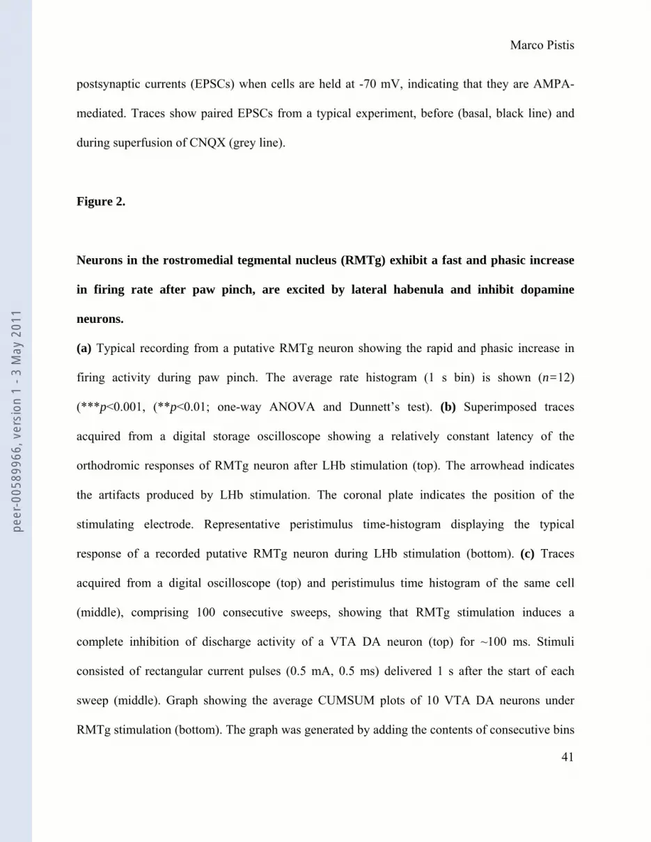
postsynaptic currents (EPSCs) when cells are held at -70 mV, indicating that they are AMPA-
mediated. Traces show paired EPSCs from a typical experiment, before (basal, black line) and
during superfusion of CNQX (grey line).
Figure 2.
Neurons in the rostromedial tegmental nucleus (RMTg) exhibit a fast and phasic increase
in firing rate after paw pinch, are excited by lateral habenula and inhibit dopamine
neurons.
(a) Typical recording from a putative RMTg neuron showing the rapid and phasic increase in
firing activity during paw pinch. The average rate histogram (1 s bin) is shown (n=12)
(***p<0.001, (**p<0.01; one-way ANOVA and Dunnett’s test). (b) Superimposed traces
acquired from a digital storage oscilloscope showing a relatively constant latency of the
orthodromic responses of RMTg neuron after LHb stimulation (top). The arrowhead indicates
the artifacts produced by LHb stimulation. The coronal plate indicates the position of the
stimulating electrode. Representative peristimulus time-histogram displaying the typical
response of a recorded putative RMTg neuron during LHb stimulation (bottom). (c) Traces
acquired from a digital oscilloscope (top) and peristimulus time histogram of the same cell
(middle), comprising 100 consecutive sweeps, showing that RMTg stimulation induces a
complete inhibition of discharge activity of a VTA DA neuron (top) for ~100 ms. Stimuli
consisted of rectangular current pulses (0.5 mA, 0.5 ms) delivered 1 s after the start of each
sweep (middle). Graph showing the average CUMSUM plots of 10 VTA DA neurons under
RMTg stimulation (bottom). The graph was generated by adding the contents of consecutive bins
peer
-005
8996
6, v
ersi
on 1
- 3
May
201
1

Marco Pistis
42
in the corresponding peristimulus time histograms to create a cumulative total average and SEM
of the number of spikes recorded across trials.
Figure 3
Methamphetamine-induced Fos immunoreactivity within the rostromedial tegmental
nucleus (RMTg).
The rat from which the brain sections were taken received an injection of methamphetamine (10
mg/kg, i.p.) 2 hours prior to sacrifice. (a) Schematic redrawing of coronal plates (based on
Paxinos and Watson, 2007) including the RMTg (bregma AP-7.2 and - 7.44) showing Pontamine
sky-blue labeling sites (black dots). (b) Microphotograph of a transmitted light image showing
the Pontamine sky blue dye (arrow). (c) This section shows Fos immunostaining (green). The
arrow indicates the dye, which appears red at wavelength 594-617. (d) Higher magnification of
the white boxed area in (c) revealing Fos-positive nuclei inside the RMTg (arrowheads). 4N,
trochlear nucleus; DR, dorshal raphe nuclei; lfp, longitudinal fasciculus of the pons; ml, medial
lemniscus; Pn, pontine nuclei; tth, trigeminothalamic tract; xscp, superior cerebellar peduncle
decussation; Scale bars: 1000 μm in (b-c); 100 μm in (d).
Figure 4.
Effects of morphine on rostromedial tegmental nucleus (RMTg) neurons.
(a) Representative firing rate histogram showing the inhibitory effects of intravenous morphine
(Mor, 4 mg/kg injected at arrow) on discharge activity of an individual putative RMTg neuron.
peer
-005
8996
6, v
ersi
on 1
- 3
May
201
1

Marco Pistis
43
(b) Representative firing rate histogram showing that naloxone (Nlx, 0.1 mg/kg, i.v. injected 4
min before morphine) fully prevents morphine-induced inhibition of firing rate of a RMTg
neuron. (c) Graph illustrating the time course of morphine’s effects (4 mg/kg, i.v.) on firing rate
of putative RMTg neurons with and without naloxone (0.1 mg/kg, i.v.) pretreatment. Naloxone
blocked the inhibition of RMTg neurons induced by morphine (arrow). Results are means with
vertical bars representing the SEM of firing rate expressed as a percentage of the baseline (BAS).
*** p<0.001 vs. baseline, one-way ANOVA and Dunnett’s test.
(d) Morphine induces inhibition of glutamatergic synaptic transmission in rat RMTg cells. A
typical whole-cell patch clamp recording showing that bath application of morphine (1 μM)
inhibits EPSC amplitude, when cells are held at -70 mV. The grey line represents mean EPSC
amplitude. The inset shows single EPSC from a typical experiment, before (black line) and
during (grey line) superfusion of morphine. (e) Morphine reduces EPSC amplitude through
activation of μ-opioid receptors. All data are normalized to the respective baseline (5 min of
baseline). Black bar shows time of superfusion of morphine in the presence (grey circles) and
absence (open circles) of the μ-opioid antagonist naloxone (100 nM). SEM bars are smaller than
symbols in some cases. The inset shows 12-trace averages of EPSCs in the absence (black line)
and presence (grey line) of naloxone. Black bars represent time of morphine application. (f)
Morphine does not change the paired-pulse ratio of EPSCs. The top graph plots the paired-pulse
ratio for each of the experiments in (e) before (open circles) and during (grey circles) the
application of morphine, while the bottom graph plots the averaged paired-pulse ratio in a bar
graph form. Representative traces are shown in the inset.
Figure 5.
peer
-005
8996
6, v
ersi
on 1
- 3
May
201
1

Marco Pistis
44
Cocaine’s effects on rostromedial tegmental nucleus (RMTg) neurons.
(a) Representative rate histogram illustrating the inhibition of firing rate of a putative RMTg
neuron after cocaine administration (1 mg/kg injected i.v. at arrow). (b) Graph showing the time
course of cocaine’s effects (1 mg/kg, i.v.) on firing rate of putative RMTg neurons. Results are
means with vertical bars representing the SEM of firing rate expressed as a percentage of
baseline (BAS). * p<0.05 vs. baseline, one-way ANOVA and Dunnett’s test.
(c) Cocaine does not affect glutamatergic synaptic transmission in rat putative RMTg cells. A
typical whole-cell patch clamp recording showing the effect of bath application of cocaine (1
µM) on EPSC amplitude, when cells are held at -70 mV. The grey line represents mean EPSC
amplitude. The inset shows single EPSC from a typical experiment, before (black line) and
during (grey line) application of cocaine. (d) Cocaine effect on EPSC amplitude. All data are
normalized to the respective baseline (5 min of baseline). Black bar shows time of superfusion of
cocaine. SEM bars are smaller than symbols in some cases. The inset shows 12-trace averages of
EPSCs in the absence (black line) and presence (grey line) of cocaine. Black bars represent time
of cocaine application. (e) Cocaine does not change the paired-pulse ratio of EPSCs. The top
graph plots the paired-pulse ratio for each of the experiments in (e) before (open circles) and
during (grey circles) the application of cocaine, while the bottom graph plots the averaged
paired-pulse ratio in a bar graph form. Representative traces are shown in the inset. Lines (black)
represent EPSC amplitude before and during (grey line) application of cocaine.
Figure 6.
peer
-005
8996
6, v
ersi
on 1
- 3
May
201
1

Marco Pistis
45
Responses of rostromedial tegmental nucleus (RMTg) neurons to the cannabinoid receptor
agonist WIN55212-2 (WIN).
(a) Typical firing rate histogram showing the inhibitory response of a putative RMTg neuron
after WIN (0.5 mg/kg, i.v.) administration (arrow). (b) Representative rate histogram showing
that the CB1 antagonist SR141716A (SR, 0.5 mg/kg, injected i.v. 4 min before WIN, at arrows)
blocks WIN-induced inhibition of firing rate. (c) Graph showing the time course of WIN-induced
inhibition of putative RMTg neurons, and that this inhibition by WIN was prevented by the
administration (arrow) of SR (0.5 mg/kg, i.v.). Results are means with vertical bars representing
the SEM of firing rate expressed as a percentage of baseline (BAS). *** p<0.0001 vs. baseline,
one-way ANOVA and Dunnett’s test.
(d) WIN inhibits glutamatergic synaptic transmission in rat putative RMTg cells. A typical
whole-cell patch clamp recording showing that bath application of WIN (1 μM) inhibits EPSC
amplitude, when cells are held at -70 mV. The grey line represents mean EPSC amplitude. The
inset shows single EPSC from a typical experiment, before (black line) and during (grey line)
perfusion of WIN. (e) WIN reduces EPSC amplitude through activation of CB1 receptors. All
data are normalized to the respective baseline (5 min of baseline). Black bar shows time of
superfusion of WIN in the presence (grey circles) and absence (open circles) of the CB1 receptor
antagonist AM281 (500 nM). SEM bars are smaller than symbols in some cases. The inset shows
12-trace averages of EPSCs in the absence (black line) and presence (grey line) of AM281.
Black bars represent time of WIN application. (f) WIN enhances the paired-pulse ratio of EPSCs,
producing paire-pulse facilitation. The top graph plots the paired-pulse ratio for each of the
experiments in (e) before (open circles) and during (grey circles) the application of WIN, while
the bottom graph plots the averaged paired-pulse ratio in a bar graph form. Representative traces
peer
-005
8996
6, v
ersi
on 1
- 3
May
201
1

Marco Pistis
46
are shown in the inset, where the EPSCs are represented before (black lines) and during WIN
(grey lines) application. * p<0.05, Student’s t-test.
Figure 7.
Rostromedial tegmental nucleus (RMTg) neurons are excited by nicotine.
(a) Representative rate histogram showing that nicotine (Nic, 0.2 mg/kg, i.v., arrow) strongly
excites firing activity of a putative RMTg neuron. (b) This example shows that the non-selective
nicotinic receptor antagonist mecamylamine (Mec, 2 mg/kg, i.v. 4 min before nicotine, arrow)
fully prevents the effects of nicotine. (c) Graph showing the time course of the stimulation of
firing rate induced by nicotine. Mecamylamine pretreatment fully prevents nicotine-induced
stimulation of firing rate. Results are means with vertical bars representing the SEM of firing rate
expressed as a percentage of the baseline (BAS). ** p<0.01, *** p<0.001 vs. baseline, one-way
ANOVA and Dunnett’s test.
(d) Nicotine enhances glutamatergic synaptic transmission in rat putative RMTg cells. A typical
whole–cell-patch clamp recording showing that bath application of nicotine (1 μM; Nic)
increases EPSC amplitude, when cells are held at -70 mV. The grey line represents mean EPSC
amplitude. The inset shows single EPSC from a typical experiment, before (black line) and
during (grey line) perfusion of nicotine. The graph shows that the decay time constant of EPSCs
is positively correlates with peak amplitude. (e) Nicotine enhances EPSC amplitude through
activation of α7-containing nicotinic receptors. All data are normalized to the respective baseline
(5 min of baseline). Black bar shows time of superfusion of nicotine in the presence (grey
circles) and absence (open circles) of the α7-subunit receptor antagonist methyllycaconitine (5
peer
-005
8996
6, v
ersi
on 1
- 3
May
201
1

Marco Pistis
47
nM; Mla). SEM bars are smaller than symbols in some cases. The inset shows 12-trace averages
of EPSCs in the absence (black line) and presence (grey line) of MLA. Black bars represent time
of nicotine application. (f) Nicotine reduces the paired-pulse ratio of EPSCs, producing paired-
pulse depression. The top graph plots the paired-pulse ratio for each of the experiments in E
before (open circles) and during (grey circles) the application of nicotine, while the bottom graph
plots the averaged paired-pulse ratio in a bar graph form. Representative traces are shown in the
inset, where the EPSCs are represented before (black lines) and during nicotine (grey lines)
application. * p<0.05, Student’s t-test.
peer
-005
8996
6, v
ersi
on 1
- 3
May
201
1

0.0 0.1 0.2 0.30
25
50
time (s)
even
ts (b
in 1
ms) 0.1 s
2 mV
1 ms
20 mV
10 ms 300 ms25 mV
a b
c
Lecca et al. Fig. 1
VTASNR
SNCMT
IPF
Depth: -8.42
RMTg
SNR
CNQX
basal 5 ms100 pA
MT VTA
500 μm
IPF
SNC
SNR
peer
-005
8996
6, v
ersi
on 1
- 3
May
201
1
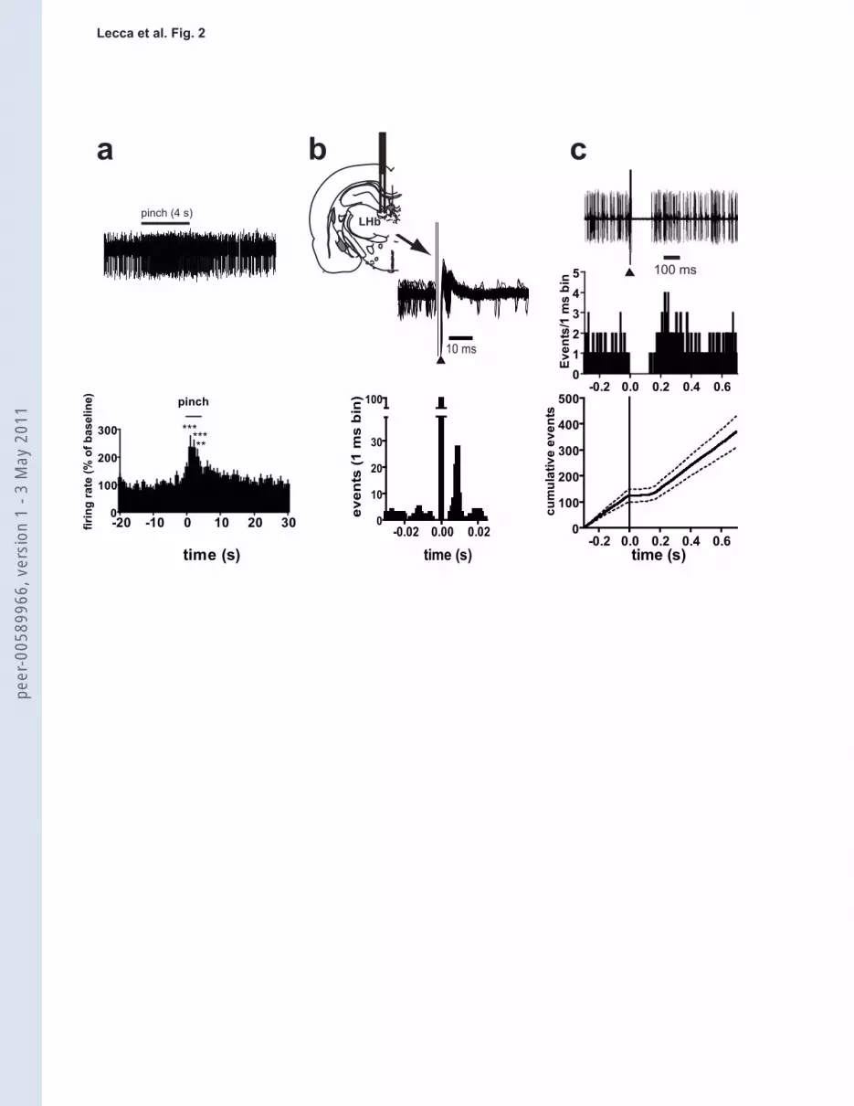
pinch (4 s)
-20 -10 0 10 20 300
100
200
300
pinch
********
time (s)
firin
g ra
te (%
of b
asel
ine)
-0.2 0.0 0.2 0.4 0.6012345
Even
ts/1
ms
bin
-0.2 0.0 0.2 0.4 0.60
100
200
300
400
500
time (s)cu
mul
ativ
e ev
ents
100 ms
a b
-0.02 0.00 0.020
10
20
30
100
time (s)
even
ts (1
ms
bin
)10 ms
LHb
c
Lecca et al. Fig. 2pe
er-0
0589
966,
ver
sion
1 -
3 M
ay 2
011

PN
tth
xscp
mllfp
4N
4N
xscp
ml
lfp tth
Pn
DR
a b
dc
peer
-005
8996
6, v
ersi
on 1
- 3
May
201
1

0 250 500 7500
50
100
150
200 MorSp
ikes
/10
s
0 200 400 600 800 10000
50
100
150
200 Nlx Mor
time (s)
Spik
es/1
0 s
Lecca et al. Fig. 4
0 10 200
50
100
EPSC
s (p
A)
time (min) time (min)10 200
-150
-100
-50
0
EPSC
2/EP
SC1
0
basal Mor
1
2
d f
100
pA
5 ms
Mor Mor
EPSC
s (%
)
EPSC
2/EP
SC1 2
0
1
150
control (n=5)Nlx (n=5)
5 ms
100
pA
e
a
b
c
2 4 6 8 10 12
25
50
75
100
125
150
Mor
Mor
BAS
Nlx + Mor
***
time (min)
Firin
g ra
te (%
of b
asel
ine)
control
Nlx
5 ms
100
pA
peer
-005
8996
6, v
ersi
on 1
- 3
May
201
1

0 250 500 7500
50
100
150
200 Cocaine
time (s)
Spik
es/1
0 s
EPSC
2/EP
SC1
0
1
2
EPSC
2/EP
SC1
0
basal Cocaine
1
2
c d
5 ms 100
pA
a b
Lecca et al. Fig. 5
0 10 200
100
time (min)
Cocaine
EPSC
s (%
)
200
2 4 6 8 10 12
25
50
75
100
125
150Cocaine
* *
BAStime (min)
Firin
g ra
te (%
of b
asel
ine)
100
pA
5 ms
EPSC
s (p
A)
time (min)10 200
0
Cocaine-800
-600
-400
-200
peer
-005
8996
6, v
ersi
on 1
- 3
May
201
1

Lecca et al. Fig. 6
0 250 500 7500
100
200
300WIN
Spik
es/1
0 s
0 200 400 600 800 10000
100
200
300SR WIN
time (s)
Spik
es/1
0 s
a
b
c
0 10 200
50
100
EPSC
s (p
A)
time (min) time (min)10 200
-400
-300
-200
-100
0
EPSC
2/EP
SC1
0
basal WIN
1
2
d f
100
pA
5 ms
*WINWIN
EPSC
s (%
)
EPSC
2/EP
SC1 2
0
1
control (n=5)AM281 (n=5)
e control
100
pA
5 msAM281
100
pA
5 ms
2 4 6 8 10 12
25
50
75
100
125
150
WIN
WINSR + WIN
BAS***
time (min)
Firin
g ra
te (%
of b
asel
ine)
peer
-005
8996
6, v
ersi
on 1
- 3
May
201
1

Lecca et al. Fig. 7
0 10 200
100
EPSC
s (p
A)
time (min) time (min)10 200
-200
-100
0
EPSC
2/EP
SC1
0
1
2
EPSC
2/EP
SC1
0
basal Nic
1
2
d f
Nic Nic
*
EPSC
s (%
)
200
control (n=6)Mla (n=5)
econtrol
Mla
5 ms
100
pA0 200 400 600 800 1000
0
100
200
300
400
Mec Nic
time (s)
Spik
es/1
0 s
0 250 500 7500
100
200
300
400 NicSp
ikes
/10
sa
b
c
2 4 6 8 10 12
50
100
150
200
250
Nic
Nic
** *****
Mec + Nic
BAS
*****
time (min)Fi
ring
rate
(% o
f bas
elin
e)
100
pA
5 ms
4567
50 100 150 200 250EPSCs (pA)de
cay
time
cos
tant
(ms)
R2=0.777
100
pA
5 ms
peer
-005
8996
6, v
ersi
on 1
- 3
May
201
1