e:\bik\2013-dipakai\september 2 - semantic scholar
TRANSCRIPT
101
Mardihusodo, The effect of active compound isolated from the leaves of kembang bulan[Tithonia diversifolia (Hemsley) A. Gray] on cell cycle and angiogenesis of WiDr cell line
J Med SciVolume 45, No. 3, September 2013: 101-111
* corresponding author: [email protected]
The effect of active compound isolatedfrom the leaves of kembang bulan[Tithonia diversifolia (Hemsley) A. Gray]on cell cycle and angiogenesis of WiDr cellline
Hajid Rahmadianto Mardihusodo1*, Mae Sri Hartati Wahyuningsih2, Indwiani Astuti21Postgraduate Program in Biomedical Sciences, 2Department of Pharmacology andTherapy, Faculty of Medicine, Universitas Gadjah Mada, Yogyakarta, Indonesia
ABSTRACTColorectal cancer is the tenth most common form of malignant tumor of hospital inpatients inIndonesia. Advance approaches in anticancer development is discovery molecular-targeted drugs.Molecular targets for anticancer drug have been identified including genes associated with cellcycle control and angiogenesis. Previously, an active and selective compound against WiDr fromTithonia diversifolia (Hemsley) A. has been isolated. The aim of this study was to evaluate theeffect of the isolated active compound from T. diversifolia on the WiDr cell cycle and angiogenesis.Isolation of the active compound was performed by preparative thin layer chromatography (TLC)method. WiDr cell cycle was analyzed by flowcytometry using propidium iodide (PI).Antiangiogenesis effect was evaluated by immunocytochemistry method using anti-human VEGFmonoclonal antibody. The results showed that the effect of the isolated active compound onthe WiDr cell cycle depended on the concentration and the incubation time periods. Atconcentration of 4 µg/mL, it inhibited the WiDr cell cycle SubG1 phase after 36 and 48 hoursincubation and G1 phase after 72 hours incubation. While at concentration of 8 µg/mL, it clearlyinhibited the WiDr cell cycle G1 phase after 36, 48 and 72 hours incubation. Furthermore, theisolated active compound at concentration of 4 µg/mL significantly inhibited the VEGF expressionuntil 47.38% compared to control. In conclusion, the isolated active compound from T. diversifoliainhibited cell cycle and angiogenesis of WiDr cell.
ABSTRAKKanker kolorektal merupakan tumor ganas paling umum ke 10 yang dijumpai pada pasien rawatinap di rumah sakit di Indonesia. Pendekatan terkini dalam pengembangan antikanker adalahpenemuan obat dengan target kerja di tingkat molekuler. Target molekuler untuk obat antikankertelah diidentifikasi di antaranya gen yang berperan dalam siklus sel dan angiogenesis. Penelitiansebelumnya menunjukkan sebuah senyawa aktif dan selektif terhadap sel WiDr dari Tithoniadiversifolia (Hemsley) A. telah berhasil diisolasi. Penelitian ini bertujuan untuk mengkaji pengaruhpemberian senyawa aktif hasil isolasi dari T. diversifolia terhadap siklus sel dan angiogenesis selWiDr. Isolasi senyawa aktif dilakukan dengan kromatografi lapis tipis (KLT) preparatif. Siklus selWiDr dianalisis dengan flowsitometri menggunakan propidium iodida (PI). Efek angiogenesisdikaji menggunakan metode imunositokimia menggunakan antibodi monoklonal anti-human VEGF.Hasil penelitian menunjukkan efek senyawa aktif hasil isolasi terhadap siklus sel WiDr tergantungpada konsentrasi dan masa inkubasi. Pada konsentrasi 4 µg/mL, senyawa ini menghambat siklussel WiDr fase SubG1 setelah inkubasi 36 dan 48 jam dan fase G1 setelah inkubasi 72 jam.
102
J Med Sci, Volume 45, No. 3, September 2013: 101-111
Sedangkan pada konsentrasi 8 µg/mL, senyawa ini dengan jelas menghambat siklus sel WiDrfase G1 setelah inkubasi 36, 48 dan 72 jam. Lebih lanjut senyawa aktif hasil isolasi pada konsentrasipada 4 µg/mL secara nyata menghambat ekspresi VEGF hingga 47.38% dibandingkan kontrol.Dapat disimpulkan senyawa aktif hasil isolasi dari T. diversifolia menghambat siklus sel danangiogenesis sel WiDr.
Keywords: isolated active compound – T. diversifolia - WiDr cells - cell cycle – antiangiogenesis
INTRODUCTION
Cancer has become a serious health problemin Indonesia. Cancer is the fifth leading causeof death in Indonesia. It was reported thatcervical, breast, skin, rectum, nasopharynx,ovary, lymphnode, colon, thyroid and soft tissuecancer are ten types of cancer that most oftenfound among cancer patients.1 Colorectal canceris the third most common form of cancer foundin men and the second in women in the world’s.The colorectal cancer causes 8% of cancerdeath with approximately 608.000 deathsannually. In Indonesia, colorectal cancer is thetenth most common form of malignant tumor ofhospital inpatients.3
New approaches to anticancer drugdevelopment involve the discovery ofmolecularly targeted anticancer agents havingselective of action to cancer cells without toxicto normal cells.4 Several molecular targets foranticancer drug discovery and developmenthave been identified including genes associatedwith cell cycle control and angiogenesis.5 Awide range of plants have been reported containcompound with cell cycle or angiogenesismodulating properties. Moreover, some plant-derived anticancer drugs including taxol,camptothecin and combretastatin areantiangiogenic.5,6
Tithonia diversifolia (Hemsley) A. Gray,locally known as kembang bulan, has beenreported to have anticancer activity by someauthors. Chloroformic extract of T. diversifolia,
chloroformic insoluble fraction, as well asbenzene-washed insoluble fraction III from thechloroformic extract have been proven to havecytotoxic effect on HeLa cells.7-9 Further study,an active compound was isolated and its thecytotoxic effect on HeLa cells was evaluatedwith an IC50 value of 5.86 µg/mL. Moreoverthis active compound caused apoptosis byincreasing p53 expression.10-12 Wahyuningsih andWijayanti13reported that the isolate of B2 is themost active and selective compound againstWiDr cell line with an IC50 value 0.59 ug/mLand selectivity index of 69.02.
An active compound known as tagitinin Chas been isolated from methanolic extract of T.diversifolia and the inhibitory activity againstmalignant glioblastoma has been reported.14
Furthermore, Garcia and Delgado15 isolatedtagitinin A and tagitinin C from T. diversifoliathat exhibited a cytotoxic effect on HCT-15 cells.While Gu et al.16 reported that tagitinin C showsantiproliferation activity on human colon cancer(Col2) cells.
In this study, we evaluated the activity ofan active compound isolated from T. diversifoliaagainst WiDr cancer cells. The effect of thisisolated active compound on WiDr cell cycleand its angiogenesis was also evaluated. 5-Fluorouracil was used as positive control inthis study due to this anticancer agent isfrequently used to treat several types of cancerincluding colorectal cancer.
103
Mardihusodo, The effect of active compound isolated from the leaves of kembang bulan[Tithonia diversifolia (Hemsley) A. Gray] on cell cycle and angiogenesis of WiDr cell line
MATERIALS AND METHODS
Isolation of the active compound from T.divesifolia
The active compound from T. diversifolialeaves was isolated from chloroformic extractin laboratory of Pharmacology and Therapy,Faculty of Medicine, Universitas Gadjah Mada,Yogyakarta. The isolation of the activecompound was performed by preparative thinlayer chromatography (TLC) method usingsilica gel GF254 as stationary phase and mixtureof benzene and ethyl acetate in the ratio of 2:1(v/v) as mobile phase. Visualisation of the bandsof the isolated active compound was performedusing UV light at 254 and 366 nm. The isolatedactive compound having similar band orretardation factor (Rf) with the standardcompound isolated by Soeprapto8 was subjectedto isolate and used for further investigation.
Cell culture and cytotoxicity assayWiDr cell lines were cultured in culture flask
containing containing complete Dulbecco’sModifiedEagle’s Medium(DMEM) supplement-edwith10%FBSand1%penicillin-streptomycin.Cultures were maintained in 5% CO2 incubator at37°Cand fedevery 3days withcomplete DMEM.Confluent cells were trypsinized, and harvestedcells were used for experiments.
Cytotoxicity of the isolated activecompound was evaluated on WiDr cells usingthe MTT [3-9,4,5-dimethylthiazole-2-yl-2,5-diphenyltetrazolium bromide assay]. Cells weredistributed in 96-wells microplates at 2 x 10⁴
cells per well in 100 mL and 100 mL ofcomplete DMEM were added. The cell cultureswere then incubated in 5% CO2 incubator at37oC for 24 hours.After incubation, the mediumwas removed and replaced with new completeDMEM containing various concentrations of theisolated active compound tested. The cellsculture and the isolated active compound were
incubated again in 5% CO2
incubator at 37oCfor 24 hours. After the incubation, the mediumwas removed and the cells were resuspendedwith DMEM. Ten 10 mL of 5 mg/mL MTT wasadded and then further incubated for 4 hours.The reaction was stopped by adding 100 mL of10% sodium dodecyl sulfate (SDS) in 0.01NHCl. Microculture plates were then shakengently for 5 minutes, covered with aluminiumfoil and incubated at room temperature over-night. Optical density (OD) of the microcultureplates was measured in an ELISA plate readerat lmax 595 nm. The OD values were directlyproportional to the number of viability cells.The OD values of plate in the presence ofisolated active compound tested were comparedwith that of control cultures without isolatedactive compound tested to obtain cells growthinhibition. Inhibitory Concentration 50% (IC50)values were then determined by probitregression analysis based on the relationshipbetween log concentrations versus thepercentage of cells growth inhibition. 5-Fluorouracil was used as positive control.
Cell cycle analysis
Cells cycle analysis was conducted byflowcytrometry. WiDr cell cultures weredistributed onto 24-well plates at density of 5 x10⁵ cells per well and incubated in 5% CO2
incubator at 37°C overnight. After overnightincubation, the WiDr cells culture were treatedwith 500 µL of the isolated active compoundtested at 2 different concentrations which wereequivalent to the value of IC50 and 2 IC50 or 5-fluorouracil for 24, 36, 48 and 72 hours.Following after incubation, the cells werecollected and harvested. After centrifugation,cell pellets were then washed twice with 500µL of cold PBS. Cells were then incubated with400 L of 50g/mL propidium iodide reagent
104
J Med Sci, Volume 45, No. 3, September 2013: 101-111
at 37°C for 10 minutes and transferred toflowcytotube. The cells were immediatelyanalyzed by FACS Calibur flowcytometer toevaluate cell cycle profile. Flowcytometric datawere analyzed using Cell Quest program toevaluate the cells distribution at each phase ofthe cell cycle namely the sub G1 (apoptosis), S,G2/M, and the cells undergoing polyploidy. Thecell cycle inhibition was observed bycomparing the cells distribution at G0/G1 andG2/M phases of treated and untreated cells.
Antiangiogenesis assayAntiangiogenesis activity was analyzed by
immunohistochemistry. WiDr cell cultures weredistributed onto 24-well plates at density of 5 x10⁴ cells per well and incubated in 5% CO2
incubator at 37°C overnight. After overnightincubation, the WiDr cells culture were treatedwith 500 µL of the isolated active compoundtested and 5-fluorouracil at concentrations of 4and 60 µg/mL, respectively and incubated for15 hours. Following incubation, the medium wasremoved and the cells were rinsed in 500 µL ofPBS. The cells were fixed with 300 µL methanoland incubated in freezer for 10 minutes. Thecells were rinsed three time in PBS and water,respectively. The cells were then blocked withserum blocking solution, incubated with aprimary anti-human VEGF monoclonal antibodyfor 10 minutes and rinsed in PBS. Followingincubation, the cells were incubated with abiotinylated universal secondary antibody for10 minutes and rinsed again in PBS. The cellswere incubated with a streptavidine peroxidasefor 10 minutes at room temperature, rinsed in
PBS. The cells were then incubated withdiamino-benzidine for 10 minutes and rinsedwith water. The cells were then dried andcoverslipped. All coverslip were examined andevaluated using light microscope withmagnification 400 times. The VEGF expressionwas identified as a browncolor of the cytoplasmiccell, while a blue color of cytoplasmic cellindicated no expression of the VEGF.
Stastitical analysisData were presented as the mean ± standard
error of the mean (SEM). Statisticalcomparisons were performed using analysis ofvariance (ANOVA) continued by Tukey post-hoc test. The differences between groups wereconsidered significant at a value of <0.05.Protocol of the study was approved by the theMedical and Health Research Ethic Committee,Faculty of Medicine, Universitas Gadjah Mada,Yogyakarta.
RESULTS
Isolation active compound from the leavesof T. diversifolia
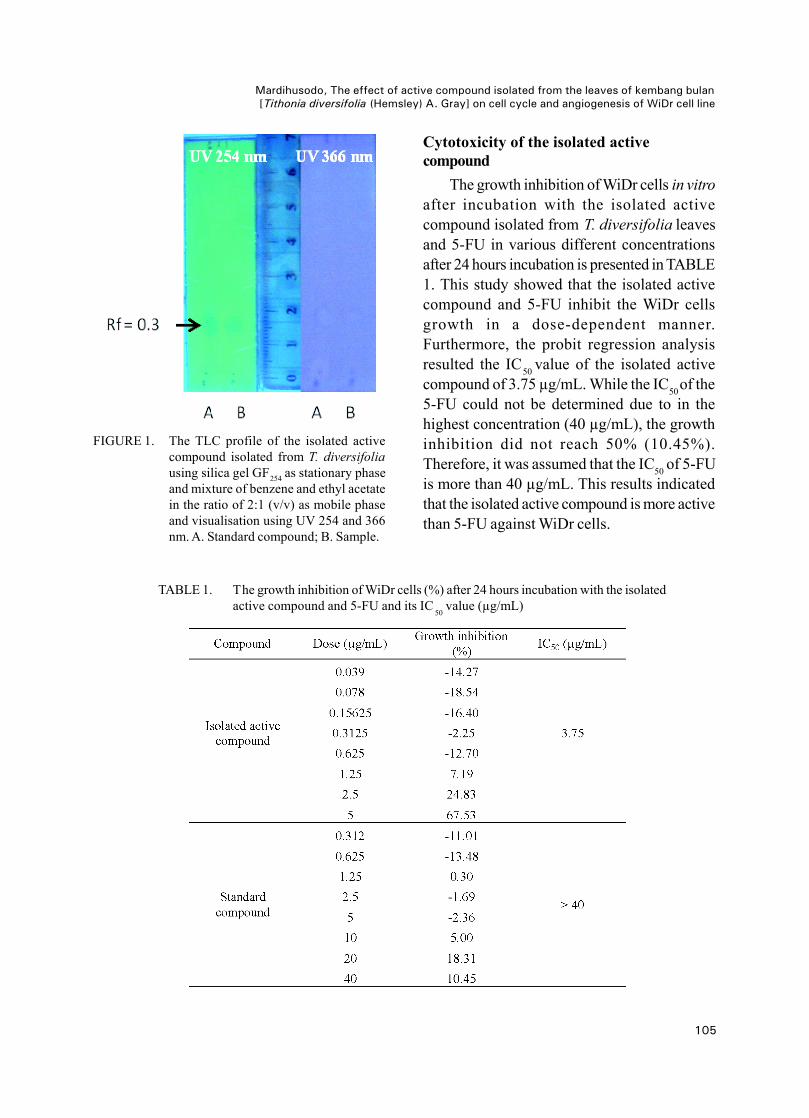
The TLC profile of the active compundisolated from the leaves of T. diversifolia ispresented in FIGURE 1. The isolated activecompound shows a single band with purplecolor on silica gel GF254 using visualisationat UV 254 nm. This isolated active compoundhas a similar band to isolated standardcompound with Rf value of 0.3. Moreover, bothof the isolated active compound and isolatedstandard compound are not observed usingvisualisation at UV 366 nm.
105
Mardihusodo, The effect of active compound isolated from the leaves of kembang bulan[Tithonia diversifolia (Hemsley) A. Gray] on cell cycle and angiogenesis of WiDr cell line
FIGURE 1. The TLC profile of the isolated activecompound isolated from T. diversifoliausing silica gel GF254 as stationary phaseand mixture of benzene and ethyl acetatein the ratio of 2:1 (v/v) as mobile phaseand visualisation using UV 254 and 366nm. A. Standard compound; B. Sample.
Cytotoxicity of the isolated activecompound
The growth inhibition of WiDr cells in vitroafter incubation with the isolated activecompound isolated from T. diversifolia leavesand 5-FU in various different concentrationsafter 24 hours incubation is presented in TABLE1. This study showed that the isolated activecompound and 5-FU inhibit the WiDr cellsgrowth in a dose-dependent manner.Furthermore, the probit regression analysisresulted the IC50 value of the isolated activecompound of 3.75 µg/mL. While the IC50of the5-FU could not be determined due to in thehighest concentration (40 µg/mL), the growthinhibition did not reach 50% (10.45%).Therefore, it was assumed that the IC50 of 5-FUis more than 40 µg/mL. This results indicatedthat the isolated active compound is more activethan 5-FU against WiDr cells.
TABLE 1. The growth inhibition of WiDr cells (%) after 24 hours incubation with the isolatedactive compound and 5-FU and its IC
50value (µg/mL)
106
J Med Sci, Volume 45, No. 3, September 2013: 101-111
The effect of isolated active compound oncell cycle
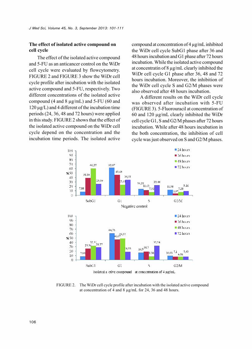
The effect of the isolated active compoundand 5-FU as an anticancer control on the WiDrcell cycle were evaluated by flowcytometry.FIGURE 2 and FIGURE 3 show the WiDr cellcycle profile after incubation with the isolatedactive compound and 5-FU, respectively. Twodifferent concentrations of the isolated activecompound (4 and 8 µg/mL) and 5-FU (60 and120 µg/L) and 4 different of the incubation timeperiods (24, 36, 48 and 72 hours) were appliedin this study. FIGURE 2 shows that the effect ofthe isolated active compound on the WiDr cellcycle depend on the concentration and theincubation time periods. The isolated active
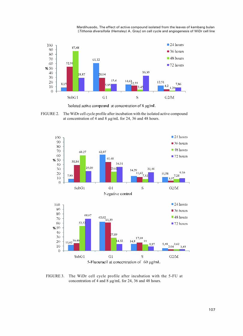
compound at concentration of 4 µg/mL inhibitedthe WiDr cell cycle SubG1 phase after 36 and48 hours incubation and G1 phase after 72 hoursincubation. While the isolated active compoundat concentratin of 8 µg/mL clearly inhibited theWiDr cell cycle G1 phase after 36, 48 and 72hours incubation. Moreover, the inhibition ofthe WiDr cell cycle S and G2/M phases werealso observed after 48 hours incubation.
A different results on the WiDr cell cyclewas observed after incubation with 5-FU(FIGURE 3). 5-Fluorouracil at concentration of60 and 120 µg/mL clearly inhibited the WiDrcell cycle G1, S and G2/M phases after 72 hoursincubation. While after 48 hours incubation inthe both concentration, the inhibition of cellcycle was just observed on S and G2/M phases.
FIGURE 2. The WiDr cell cycle profile after incubation with the isolated active compoundat concentration of 4 and 8 µg/mL for 24, 36 and 48 hours.
107
Mardihusodo, The effect of active compound isolated from the leaves of kembang bulan[Tithonia diversifolia (Hemsley) A. Gray] on cell cycle and angiogenesis of WiDr cell line
FIGURE 2. The WiDr cell cycle profile after incubation with the isolated active compoundat concentration of 4 and 8 µg/mL for 24, 36 and 48 hours.
FIGURE 3. The WiDr cell cycle profile after incubation with the 5-FU atconcentration of 4 and 8 µg/mL for 24, 36 and 48 hours.
108
J Med Sci, Volume 45, No. 3, September 2013: 101-111
FIGURE 3. The WiDr cell cycle profile after incubation with the 5-FU atconcentration of 4 and 8 µg/mL for 24, 36 and 48 hours.
The antiangiogenesis effect of the isolatedactive compound
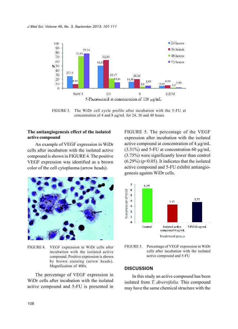
An example of VEGF expression in WiDrcells after incubation with the isolated activecompound is shown in FIGURE 4. The positiveVEGF expression was identified as a browncolor of the cell cytoplasma (arrow heads).
FIGURE 4. VEGF expression in WiDr cells afterincubation with the isolated activecompound. Positive expression is shownby brown staining (arrow heads).Magnification of 400x.
The percentage of VEGF expression inWiDr cells after incubation with the isolatedactive compound and 5-FU is presented in
FIGURE 5. The percentage of the VEGFexpression after incubation with the isolatedactive compound at concentration of 4 µg/mL(3.31%) and 5-FU at concentration 60 µg/mL(3.75%) were significantly lower than control(6.29%) (p<0.05). It indicates that the isolatedactive compound and 5-FU exhibit antiangio-genesis againts WiDr cells.
FIGURE 5. Percentage of VEGF expression in WiDrcells after incubation with the isolatedactive compound and 5-FU
DISCUSSION
In this study an active compound has beenisolated from T. diversifolia. This compoundmay have the same chemical structure with the
109
Mardihusodo, The effect of active compound isolated from the leaves of kembang bulan[Tithonia diversifolia (Hemsley) A. Gray] on cell cycle and angiogenesis of WiDr cell line
active compound previously isolated bySoeparto10 as indicated by the same Rf value(0.3) and the same band using visualisation atUV 254 nm. Furthermore, this active compoundis suggested as tagitinin C (1, 2-epoxytagininC) that previously isolated by Liao et al.14
The cytotoxicity testing showed that theisolated active compound has potential cytotoxicactivity against WiDr cell line with an IC50 valueof 3.75 µg/mL. This cytotoxic activity of thisisolated active compound is higher than 5-FU(>40 µg/mL), as positive control, indicating thatthe isolated active compound is more activeagainst WiDr cell line (TABLE 1).
The effect of the isolated active compoundon cell cycle of WiDr cell indicated that thiscompound inhibited non specifically on the cellcycle (FIGURE 2). Some of anticancer agentsinclude alkylating agent, cisplatin, procarbazineand nitrosourea are reported to inhibit nonspecifically on the cell cycle.17 These agentsnormally have a linear dose-response curve.18
In contrast, the anticancer agents that specificallyinbibit on cell cycle does not show a linier dose-response curve.
The inhibition effect of the isolated activecompound on cell cycles of WiDr cell may becaused its sesquiterpene lactones skeleton. Itwas resported that the sesquiterpene lactonesisolated from T. diversifolia can inhibit NF-kBactivity that have an important role in the cellcycle.19 The sesquiterpene lactones isconsidered as an unsaturated carbonyl structure,which has electrophilic properties and canrapidly interact with protein or DNA molecules.This interaction can cause DNA damage andactivate checkpoint of the cell cycle which isan important mechanism of the inhibition oftumor growth.20,21
This study also showed that the inhibitionprofile of WiDr cell cycle of the isolated activecompound was different compared to 5-FU. Theisolated active compound inhibited non
specifically on the cell cycle include all of theWiDr cell cycle phases, while 5-FU clearlyinhibited the G1, S and G2/M phases especiallyafter 72 hours incubation (FIGURE 3). Theeffect of 5-FU on the WiDr cell cycle has beenreported by some authors. It is well acceptedthat 5-FU inhibits the cancer cell cycle Sphase.18,22 However, Septisetyani23 andIkawati24 showed that 5-FU inhibits WiDr cellcycle in time-dependent manner. The 5-FUinhibits WiDr cell cycle G1 phase after 24 and26 hours incubation, however after 48 and 72hours incubation the inhibition was observedon cell cycle subG1 phase.
Some factors can influence the cell cycleprofile using flowcytometry analysis include:1) characteristic of the cell cycle; 2) incubationtime period; 3) preparation time of cell; 4)method of cell viability evaluation; 5) qualityand concentration of reagents used in staining;and 6) skills for staining techniques.
The VEGF expression on WiDr cell afterincubation with the isolated active compoundand 5-FU were significantly lower comparedto control (p<0.05) showing that the isolatedactive compound and 5-FU exhibit antiangio-genesis. Moreover, the effect of the isolatedactive compound at concentration of 4 µg/mLwas comparable to the effect of 5-FU atconcentration of 60 µg/mL (FIGURE 5).
The mechanism of action of the isolatedactive compound is suggested similar to threesesquiterpene lactones i.e. as diversifolin,diversifolin and methyl eter and tirotundinisolated from T. diversifolia by Rungeler et al.20
These sesquiterpene lactones have beenreported to inhibit NF-B activity inhibition hasbeen proven. NF-B is a protein composingsubunit p50 and p65 that play critical roles ininflammation, immunity, cell proliferation,differentiation, and survival.21 Sesquiterpenelactoones group are reported to inhibit NF-kBtrascription by alkylation on cysteine residuep65 (Cys 38).25
110
J Med Sci, Volume 45, No. 3, September 2013: 101-111
CONCLUSION
The active compound isolated from theleaves of kembang bulan (T. diversifolia)inhibits nonspecifically of WiDr cell cycle. Inaddition, the isolated active compound inhibitsthe VEGF expression of the WiDr cell.
ACKNOWLEDGEMENTS
This study was funded by Community Fundsof Faculty of Medicine, Universitas GadjahMada,Yogyakarta through Junior Lecture Grant.
REFERENCES1. Aziz MF. Gynecological cancer in Indonesia. J
Gynecol Oncol. 2009; 20(1):8-10.2. Anonim. Colorectal cancer incidence and
mortality worldwide in 2008 summary. 2008.[Accessed Jan 8, 2011] Available from: http://globocan.iarc.fr/factsheets/cancers/colorectal.asp
3. Anonim. Profil Kesehatan Indonesia 2008.Jakarta: Departemen Kesehatan RI. 2009.
4. Gali-Muhtasib H. Cyclin–dependent kinaseinhibitors from natural sources: recent advancesand future prospect for cancer treatment. In: KhanMTH, Ather A. (Eds.): Lead molecules fromnatural products. Amsterdam: Elsevier B.V. 2006;155-67.
5. Suggit M and Bibby MC. 50 years of preclinicalanticancer drug screening: empirical to target-driven approaches. Clin Cancer Res. 2005;11:971-81.
6. Fan TP, Yeh JC, Leung KW, Yue PYK, Wong RNS.Angiogenesis: from plants to blood vessels. TrendsPharmcol Sci. 2006; 27(6): 298-309.
7. Wicaksono AS. Efek sitotoksik ekstrak metanoldan ektrak kloroform daun kembang bulan (T.diversifolia) terhadap sel hela in vitro [Skripsi].Yogyakarta: Fakultas Kedokteran UniversitasGadjah Mada. 2007.
8. Saputra F. Uji sitotoksik senyawa hasil partisiekstrak kloroform daun kembang bulan (T.diversifolia) terhadap kultur sel hela secara invitro [Skripsi]. Yogyakarta: Fakultas KedokteranUniversitas Gadjah Mada. 2008.
9. Duana Y. Efek sitotoksik in vitro fraksi tidak larutwash benzene dari ekstrak kloroform daun
kembang bulan (T. diversifolia) pada sel hela[Skripsi]. Yogyakarta: Fakultas KedokteranUniversitas Gadjah Mada. 2008.
10. Soeparto A. Pemurnian isolat aktif (T.diversifolia) dan uji sitotoksiknya terhadap selhela in vitro [Skripsi]. Yogyakarta: FakultasKedokteran Universitas Gadjah Mada. 2010.
11. Rossila AB. Pengaruh senyawa isolat aktif daunkembang bulan (T. diversifolia) terhadapapoptosis sel hela dengan pengecetan HOECHST33342 [Skripsi]. Yogyakarta: Fakultas KedokteranUniversitas Gadjah Mada. 2010.
12. Mandela W. Pengaruh senyawa isolat aktif daunkembang bulan (T. diversifolia) terhadap ekspresiprotein p53 pada sel hela dengan metodeimmunohistokimia [Skripsi]. Yogyakarta: FakultasKedokteran Universitas Gadjah Mada. 2010.
13. Wahyuningsih MSH, Wijayanti MA. Isolasi,identifikasi senyawa antikanker dari fraksi aktifTithonia diversifolia (Hemsley) A. Gray,selektivitas dan mekanisme apoptosis secara invitro. Laporan Akhir Penelitian Riset PembinaanIptek Kedokteran. Yogyakarta: FakultasKedokteran Universitas Gadjah Mada. 2009.
14. Liao MH, Lin WC, Wen HC, Pu HF. Tithoniadiversifolia and its main active componenttagitinin C induce survivin inhibition and G2/Marrest in human malignant glioblastoma cells.Fitoterapia. 2011; 82(3):331-41.
15. Garcia A, Delgado G. Constituent from Tithoniadiversifolia stereochemical revision of 2a-hydroxytirorundin. J Mex Chem Soc. 2006;50(4):180-83.
16. Gu JQ, Gills JJ, Park EJ, Mata-Greenwood E,Hawthorne ME, Axelrod F, et al . Sesquiterpenesfrom Tithonia diversifolia with potential cancerchemopreventive activity. J Nat Prod. 2002;65:532-6.
17. Brunton LL, Parker K. Chemotherapy ofneoplastic disease. In: Brunton LL, Parker K.(Eds.). Goodman & Gilman’s Manual ofPharmacology and Therapeutics. New York: Mc-Graw Hill. 2008.
18. Page R, Takimoto C. Principles of chemotherapy.In: Pazdur R, Coia LR, Hoskins WJ, Wagman LD.(Eds.): Cancer Management: A MultidisciplinaryApproach. New York: The Oncology Group. 2003.
19. Rungeler P, Lyss G, Castro V, Mora G, Pahl HL,Merfort I. Study of three sesquiterpene lactonesfrom Thitonia diversifolia on their anti-inflammatory activity using the transcription
111
Mardihusodo, The effect of active compound isolated from the leaves of kembang bulan[Tithonia diversifolia (Hemsley) A. Gray] on cell cycle and angiogenesis of WiDr cell line
factor NF-Kappa B and enzymes of thearachidonic acid pathway as a target. Planta Med.1998; 64(7):588-93.
20. Rungeler P, Castro V, Mora G, Goren N,Vichnewski W, Pahl HL, et al. Inhibition oftranscription factor of NF-kappaB by sesquiter-pene lactones: a proposed molecular mechanismof action. Bioorg Med Chem. 1999; 7(11):2343-52.
21. Motoyama N, Naka K. DNA damage tumorsupressor genes and genomic instability. CurrOpin Genet Dev. 2004; 14(1):11-6.
22. Airley R. Cancer chemoterapy basic science tothe clinic. West Sussex: John Wiley & Sons Ltd.2009.
23. Septisetyani EP. Aktivitas sitotoksik pentagama-vunon-1 tunggal dan kombinasi dengan 5-fluorouracil pada sel kanker kolon WiDr [Tesis].Yogyakarta: Program Pascasarjana UGM. 2008.
24. Ikawati M. Modulasi daur sel dan pemacuanapoptosis pada sel kanker kolon WiDr oleh per-lakuan tunggal pentagamavunon-0 dan kombinasi-nya dengan 5-Fluorouracil [Tesis]. Yogyakarta:Program Pascasarjana Universitas Gadjah Mada.2008.
25. Garcia-Pineres AJ, Castro V, Mora G, Schmidt TJ,Strunck E, Pahl HL, et al. Cysteine 38 in p65/NF-kappaB plays a crucial role in DNA bindinginhibition by sesquiterpene lactones. J Biol Chem.2001; 276(43):39713-20.