biomolecules - semantic scholar
TRANSCRIPT
biomolecules
Review
Trehalose for Ocular Surface Health
Jarmo Laihia 1,* and Kai Kaarniranta 2,3
1 Finnsusp Ltd., Pääskykalliontie 5, FI-21420 Lieto, Finland2 Department of Ophthalmology, Institute of Clinical Medicine, University of Eastern Finland,
FI-70210 Kuopio, Finland; [email protected] Department of Ophthalmology, Kuopio University Hospital, FI-70210 Kuopio, Finland* Correspondence: [email protected]
Received: 3 April 2020; Accepted: 21 May 2020; Published: 25 May 2020�����������������
Abstract: Trehalose is a natural disaccharide synthesized in various life forms, but not found invertebrates. An increasing body of evidence demonstrates exceptional bioprotective characteristicsof trehalose. This review discusses the scientific findings on potential functions of trehalose in oxidativestress, protein clearance, and inflammation, with an emphasis on animal models and clinical trials inophthalmology. The main objective is to help understand the beneficial effects of trehalose in clinical trialsand practice, especially in patients suffering from ocular surface disease. The discussion is supplementedwith an overview of patents for the use of trehalose in dry eye and with prospects for the 2020s.
Keywords: trehalose; dry eye syndromes; oxidative stress; autophagy; inflammation; cytoprotection;randomized controlled trials; animal models; molecular chaperones; patents
1. Introduction
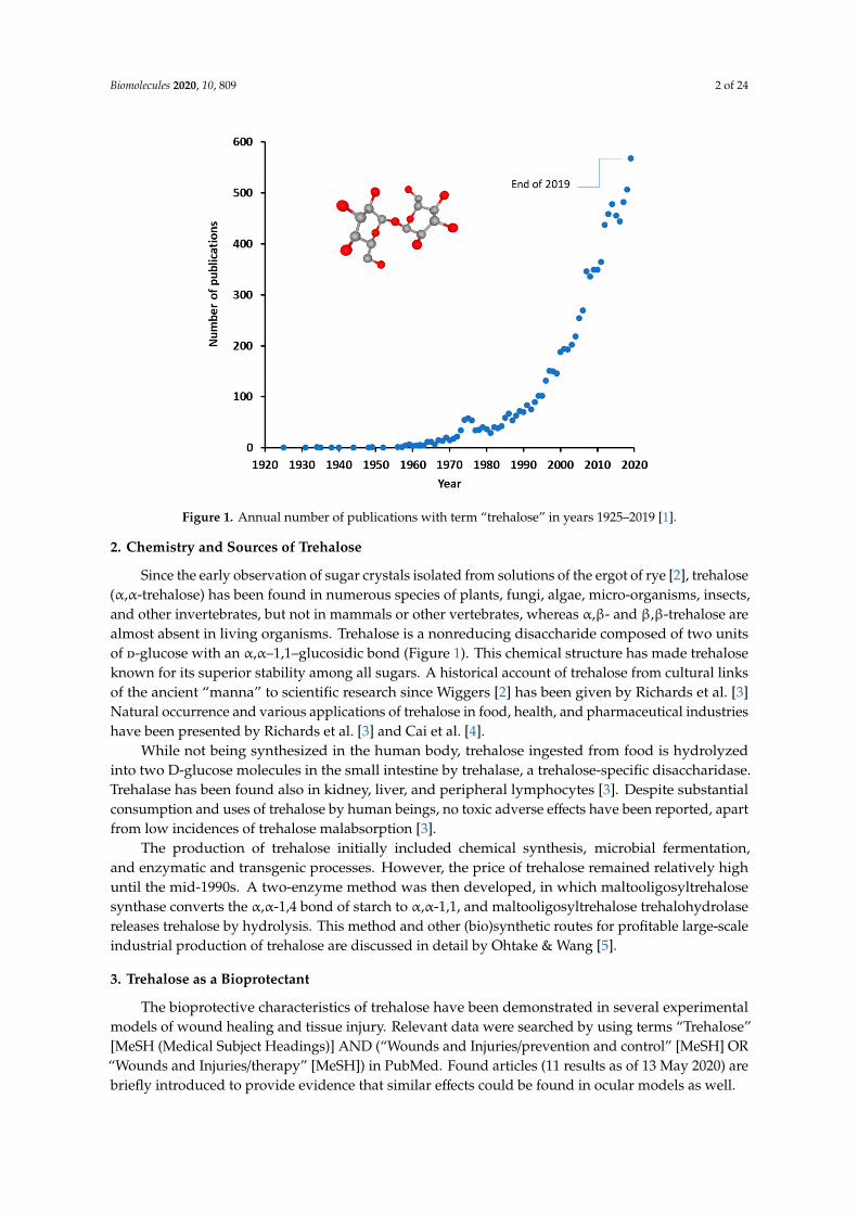
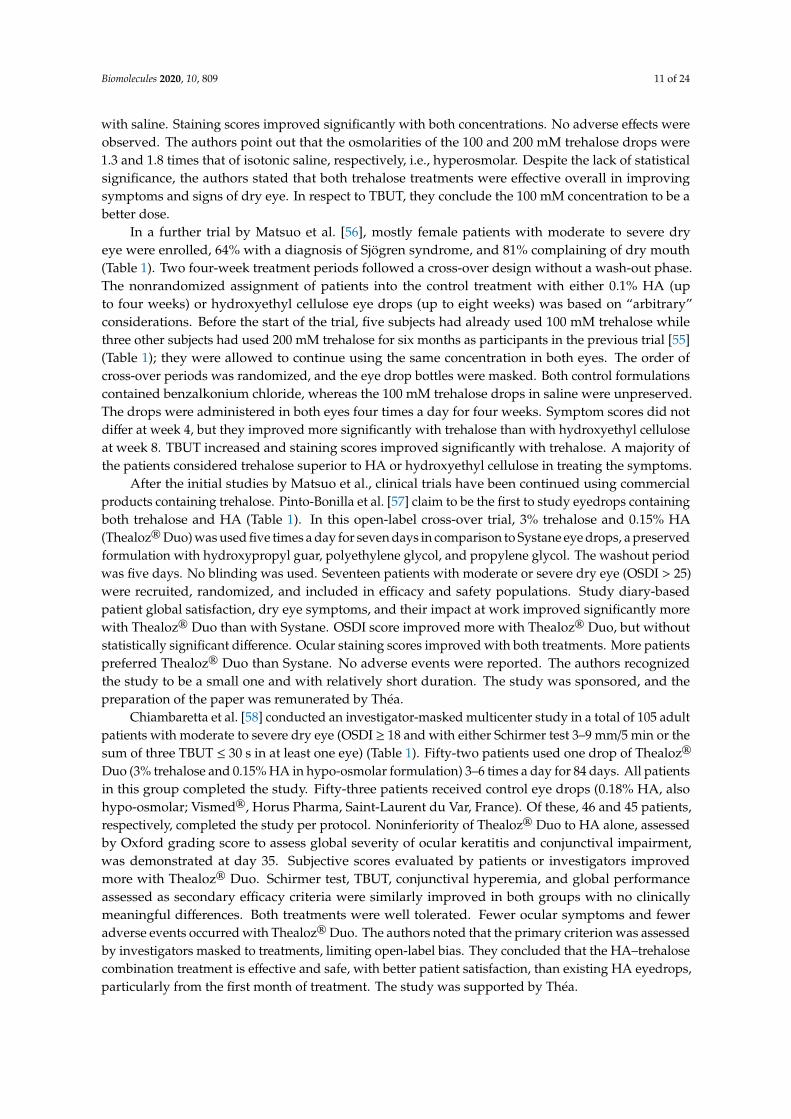
Trehalose is a disaccharide occurring naturally in various life forms, but not found in vertebrates.Trehalose is raising increasing interest for the development of various applications in food, cosmetic,and pharmaceutical industries, as reflected by almost an exponential growth in the accumulation ofscientific publications (Figure 1). This interest may be explained by the proliferating body of evidenceon the bioprotective characteristics of trehalose, its low toxicity, and proceedings in the development ofmore affordable production technologies.
Epithelial surfaces such as the cornea and the conjunctiva of the eye protect the underlying tissuesagainst exogenous threats. Various environmental, intrinsic, or iatrogenic stressors may induce celldamage or cell death, leading to clinical diseases such as dry eye disease, also known as ocular surfacedisease (OSD). The detrimental processes are counteracted by several endogenous defense mechanisms.
The focus of the present review is to discuss the scientific evidence on the functions of trehalosein oxidative stress, protein clearance, and inflammation, and in animal models related to dry eye.The main objective is to help understand why trehalose is continuously showing beneficial effects inclinical trials in ophthalmology, especially in patients with OSD. The discussion is supplemented withpatent data for the use of trehalose in dry eye and with some future prospects.
Biomolecules 2020, 10, 809; doi:10.3390/biom10050809 www.mdpi.com/journal/biomolecules
Biomolecules 2020, 10, 809 2 of 24
Figure 1. Annual number of publications with term “trehalose” in years 1925–2019 [1].
2. Chemistry and Sources of Trehalose
Since the early observation of sugar crystals isolated from solutions of the ergot of rye [2], trehalose(α,α-trehalose) has been found in numerous species of plants, fungi, algae, micro-organisms, insects,and other invertebrates, but not in mammals or other vertebrates, whereas α,β- and β,β-trehalose arealmost absent in living organisms. Trehalose is a nonreducing disaccharide composed of two unitsof d-glucose with an α,α–1,1–glucosidic bond (Figure 1). This chemical structure has made trehaloseknown for its superior stability among all sugars. A historical account of trehalose from cultural linksof the ancient “manna” to scientific research since Wiggers [2] has been given by Richards et al. [3]Natural occurrence and various applications of trehalose in food, health, and pharmaceutical industrieshave been presented by Richards et al. [3] and Cai et al. [4].
While not being synthesized in the human body, trehalose ingested from food is hydrolyzedinto two D-glucose molecules in the small intestine by trehalase, a trehalose-specific disaccharidase.Trehalase has been found also in kidney, liver, and peripheral lymphocytes [3]. Despite substantialconsumption and uses of trehalose by human beings, no toxic adverse effects have been reported, apartfrom low incidences of trehalose malabsorption [3].
The production of trehalose initially included chemical synthesis, microbial fermentation,and enzymatic and transgenic processes. However, the price of trehalose remained relatively highuntil the mid-1990s. A two-enzyme method was then developed, in which maltooligosyltrehalosesynthase converts the α,α-1,4 bond of starch to α,α-1,1, and maltooligosyltrehalose trehalohydrolasereleases trehalose by hydrolysis. This method and other (bio)synthetic routes for profitable large-scaleindustrial production of trehalose are discussed in detail by Ohtake & Wang [5].
3. Trehalose as a Bioprotectant
The bioprotective characteristics of trehalose have been demonstrated in several experimentalmodels of wound healing and tissue injury. Relevant data were searched by using terms “Trehalose”[MeSH (Medical Subject Headings)] AND (“Wounds and Injuries/prevention and control” [MeSH] OR“Wounds and Injuries/therapy” [MeSH]) in PubMed. Found articles (11 results as of 13 May 2020) arebriefly introduced to provide evidence that similar effects could be found in ocular models as well.
Biomolecules 2020, 10, 809 3 of 24
Animal studies by Cejková et al. [6–8] demonstrated the protecting effect of trehalose on theultraviolet-B (UVB)-irradiated cornea (see Section 5.1). The reports conclude that trehalose protectsocular tissues from photodamage by supporting the viability and healing of the irradiated cornea andby suppressing hypoxic, oxidative, inflammatory, and apoptotic pathways of tissue damage.
Takahashi et al. [9] demonstrated that trehalose reduces neuronal damage in a spinal cordischemia model in rabbits. Functional scores with 5% trehalose given as infusion or intravenouslywere significantly higher than in controls. Motor neurons were histologically normal with minimalinflammatory cell infiltration. Neither toxicity nor hemodynamic effects were observed. The authorspostulate the protective effect to be related to preservation of cell membranes, mitochondria, and othercytoplasmic structures.
Traumatic brain injury and neurodegenerative disorders involve changes in brain concentrations oftransition metals. Oral administration of 2% trehalose improved several cognitive outcomes impactedby brain injury [10] and induced a significant increase in brain zinc levels while not affecting iron andcopper in aged mice [11], but not in young mice [10]. The authors also found that the expression ofproteins involved in synaptic activity, neurogenesis, and neuroprotection was increased significantlyin the brain after trehalose treatment, while maltose (another disaccharide) was inactive. Theseobservations may have direct implications in ophthalmology because of the role of zinc in retina andcornea [12].
Lee et al. [13] reported a method to thermostabilize an enzyme with an activity for scarproteoglycans. When the trehalose-stabilized enzyme was administered by sustained topical release torats with a spinal cord hemisection injury, digestion of astroglial scar proteoglycans, growth axons, andrecovery of locomotor function were enhanced. The ability of trehalose to protect protein structures wasalso demonstrated in a study using trehalose in antibody-loaded nanoparticle emulsion [14]. Trehalosesignificantly improved the antagonist bioactivity of the antibody and protected it from denaturationduring processing steps, notably during lyophilization, whereas its release kinetics were not affected,likely due to the small molecular size of trehalose. Protection of nanoparticles from freeze-drying(lyophilization) was reported also by Ruozi et al. [15]. Perfusion of partial-thickness wounds with1% trehalose promoted healing and supported re-epithelialization and organizing of the tissue in awound dressing model in pigs [16]. These findings imply that trehalose may be used as an excipientprotectant in medicinal preparations comprising biological macromolecules or emulsion structures.
4. Inflammatory and Oxidative Stress Signaling
As a disaccharide, trehalose is usually considered impermeable to cell membranes, apparentlyleaving no chance to affect intracellular signaling. However, several studies have shown trehalose topass to the cytosol of mammalian cells spontaneously in millimolar concentrations (reviewed in [17]).The suggested mechanism for this behavior is fluid-phase endocytosis that involves internalization ofcell environment by vesicular “drinking” [18]. Moreover, trehalose traverses the plasma membranevia Solute Carrier Family 2 Member 8 (SLC2A8), a homolog of the trehalose transporter-1 (Tret1) [19].By the simple presence of a high extracellular concentration of trehalose, the cell would take uptrehalose that is conveyed to the lysosomal pathway. Due to its high chemical stability, trehalose is notdegraded in lysosomes; therefore, it may leak from lysosomes into the cytoplasm [20]. There is evidencethat the pH gradient changes the permeability of phospholipid bilayers of the lysosomal membraneand allows the leakage of trehalose and other low-molecular-weight molecules into the cytosol [20].It is therefore reasonable to infer that the effects of trehalose on intracellular processes presented inthis chapter are a result of trehalose acting directly in the cytosol. Representative and recent paperswere obtained using PubMed search terms “Trehalose”[MeSH] AND “Anterior Eye Segment”[MeSH](19 Dec 2019; Section 4.1), “trehalose” AND “oxidative stress” (16 Dec 2019; Section 4.2), “trehalose”AND “autophagy”, “trehalose” AND “proteasomes”, “trehalose” AND “inflammation” with term“eye” (16 Dec 2019; Section 4.3).
Biomolecules 2020, 10, 809 4 of 24
4.1. Trehalose in Ocular Surface Physiology
The ocular surface is constantly exposed to atmospheric oxygen and other environmental stressors,including various types of radiation, particulate or gaseous air pollutants, microbes, and desiccation.These stressors generate free radicals such as reactive oxygen species (ROS) that may damage cornealand conjunctival epithelia or disrupt corneal physiology and function. When cultured human cornealepithelial cells (hCEC) were subjected to desiccation stress for 5 to 45 min, trehalose-containing eye drops,unlike many other dry eye drops, showed high efficiency in maintaining normal cellular morphology,cell membrane function, and cell proliferative activity, and in preventing desiccation-induced celldeath [21]. Combining trehalose with sodium hyaluronate in eye drops has been shown to elicit cornealre-epithelialization in response to corneal cross-linking compared with sodium hyaluronate alone [22].Pretreatment with 3% trehalose alone preserved morphological and morphometric features of corneain the laser subepithelial keratomileusis (LASEK) [23]. Trehalose has been observed to improve drugpenetration from nanoparticles across the cornea, postulated to depend on pore formation duringfreeze-drying processing of the nanoparticles or the flexibility of hydrogen bonding between trehaloseand the particles [24].
Ocular surface cells respond to environmental stress by increasing antioxidant and molecularchaperone production [6,25]. Moreover, the ubiquitin proteasome system (UPS) and lysosomalautophagic clearance are activated to clean damaged proteins [26]. Molecular chaperones (heat shockproteins, HSPs) are the only sensor system in cells with the ability to restore proteins to their originalfolding state, maintaining their function and preventing detrimental protein aggregation. HSPs aredivided into different families according to their molecular weight and specialized cellular location.Interestingly, trehalose also functions as a molecular chaperone [27,28]. Similar to cytoplasmic HSPs,trehalose mediates its effects mainly via cytoplasmic connections. It is weakly known whether trehalosepenetrates other organelles than lysosomes in cells. Lysosomal accumulation seems to be involved inendocytic cellular intake [17]. Once the molecular chaperone capacity responding to oxidative stress isexceeded, denatured proteins are degraded by the UPS. Usually, large protein aggregates and damagedcellular organelles undergo autophagic clearance [29]. Inflammatory signaling is activated as a hostdefense mechanism in acute stress, but the response is shifted to detrimental chronic inflammation in aprolonged or excessive stress conditions that may finally lead to cellular morphological and functionalchanges and to cell death.
4.2. Trehalose in Oxidative Stress Signaling
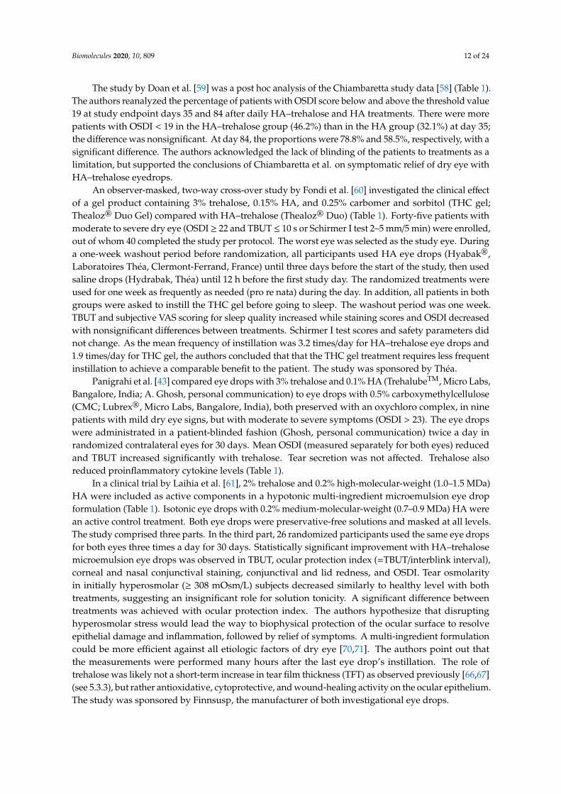
Oxidative stress is a consequence of the use of oxygen in aerobic respiration by living organisms,and it is denoted as a persistent condition of imbalance between the generation of ROS and theability of the endogenous antioxidant system to detoxify them. Nuclear factor erythroid 2-relatedfactor 2 (NFE2L2) is the key transcription factor in the sensing of oxidative stress. NFE2L2 was foundto associate with the outer mitochondrial membrane and to protect mitochondria from oxidativedamage, likely through direct interaction with mitochondria [30]. Upon activation, NFE2L2 is releasedfrom NFE2L2-KEAP1 (kelch-like ECH-associated protein 1) and the mitochondrial outer membraneserine/threonine protein phosphatase 5 complex, allowing for translocation of NFE2L2 from the cytosolinto the nucleus and binding to the antioxidant response element (ARE); this promotes the transcriptionof more than 200 genes, including detoxification and antioxidant enzymes [31,32]. Trehalose has beenshown to increase p62/SQSTM1 protein expression, activate NFE2L2, enhance the expression of itsdownstream antioxidant factors, and reduce the amount of ROS [33] (Figure 2). p62/SQSTM1 has amultifactorial role in acting as a regulatory protein between UPS and autophagy clearance as well as inthe antioxidant response during oxidative stress [32,34]. Ischemic insult-induced protein aggregationwas prevented by trehalose treatment via preservation of proteasome activity [35]. Benaroudj et al. [36]first documented that trehalose decreased the initial appearance of misfolded proteins presumably byscavenging free radicals. This observation was recently corroborated by Cejka et al. [8] who showed

Biomolecules 2020, 10, 809 5 of 24
that trehalose decreases the amount of ROS, lipid peroxidation end-products, and DNA damage inUVB irradiation-exposed rabbit cornea.
Figure 2. Schematic presentation of intracellular cytoprotective effects of trehalose. Trehalose intaketo cells is via endocytosis and under the control of Solute Carrier Family 2 Member 8 (SLC2A8).Ocular surface cells are constantly exposed to environmental oxidative stress, which may evokemitochondrial damage, protein misfolding, aggregation, and inflammation. Trehalose helps proteinrefolding together with heat shock proteins (HSPs) and enhances ubiquitin (Ub)-mediated proteasomaland microtubule-associated protein 1A/1B light chain 3 (LC3)-controlled autophagic clearance. LC3 andp62/SQSTM1 have binding sites to Ub that direct the sealed material to autophagic degradation.Moreover, p62/SQSTM1 regulates antioxidant production via transcription factor NFE2L2 released fromNFE2L2-KEAP1 (kelch-like ECH-associated protein 1) and p62/SQSTM1 complex, allowing its bindingto the antioxidant response element (ARE) protein and promote the transcription of antioxidativeproteins. All beneficial functions of trehalose (TREHALOSE (+)) prevent oxidative-stress-inducedinflammation (TREHALOSE (-)).
4.3. Trehalose in Autophagy and Inflammation Signaling
Autophagy is an essential catabolic lysosomal mechanism that enables cells to clean damagedintracellular components [37]. It is a conserved process involving double-membrane autophagosomesto seal the cytoplasmic contents. Once autophagosomes fuse with lysosomes, lysosomal enzymes arereleased into the lumen of the autophagosome, leading to protein degradation. Autophagy enhancesthe clearance of toxic, cytoplasmic, aggregate-prone proteins and infectious agents. The beneficial rolesof autophagy are linked to improved cell survival and to prevention of inflammation [38,39]. There isstrong evidence that trehalose stimulates autophagy through the adenosine monophosphate-activatedprotein kinase (AMPK) independent of mechanistic target of rapamycin (mTOR) [40–42]. In additionto activating autophagy, trehalose preserves protein structural integrity and reduces aggregationof pathologically misfolded proteins (Figure 2). Impaired autophagy may evoke inflammation,which is associated with OSD [43]. Trehalose induces autophagy and reduces secretion of cytokines
Biomolecules 2020, 10, 809 6 of 24
IL(interleukin)-6, IL-8, and monocyte chemoattractant protein-1 (MCP-1) in corneal cells under tumornecrosis factor-α (TNF-α) and desiccation stress. Lipopolysaccharide-induced secretion of IL-1β, IL-6,TNF-α, and nitric oxide are decreased in trehalose-treated cell cultures [44]. In addition, trehalosesuppresses microglial activation via transcription factors nuclear factor-κB (NF-κB) and activatingprotein-1 (AP-1). When lysosomal enzyme activity is disturbed, trehalose may prevent cellular damageand suppress neuroinflammation [45]. Local inflammation linked to peripheral nerve injury is animportant feature in dry eye [46].
5. Trehalose in Ophthalmic Applications
5.1. Animal Models
Publications describing the effects of trehalose in various ocular animal models were searched inPubMed by using terms “Trehalose” [MeSH] AND “Eye” [MeSH] AND “animals” [MeSH Terms:noexp](13 May 2020). Some of the found papers concern the use of trehalose as an adjuvant for cryoprotectionand in other cellular systems [24,47–50], or the administration by oral route [45]. These reports are notdirectly within the scope of this chapter, but they corroborate the findings by others in that trehalose issafe to various cell types and supports the biological and morphological properties of tissues in high(up to 35%) [47] concentrations.
Chen et al. [51] used an experimental model of dry eye in BALB/c mice. After being housedin a controlled environment with low humidity and constant airflow and temperature for 21 days,the animals were randomized for treatment with phosphate-buffered saline (PBS) control, 3% (87.6 mM)anhydrous trehalose, or pooled mouse serum eye drops (10 µL) administered every 6 h in the sameenvironment for the next 14 days. The housing conditions significantly reduced tear production from2.3 to 1.7 mm by phenol thread wetting test. Trehalose and serum eye drops increased tear secretionsignificantly and similarly to 1.9 and 2.0 mm, respectively, at 14 days, but not yet at seven days, whilecontrol treatment showed no improvement. In an analogous fashion, the desiccating environmentinduced corneal fluorescein staining from 1.6 to 9.8 score units (on scale 0–15) at day 21; trehaloseand serum treatments decreased staining scores to 6.6 and 4.5 units, respectively, at 14 days thatwas not seen in PBS controls. Trehalose also increased the thickness of corneal epithelium and thenumber of goblet cells, and, as expected, decreased the number of ruffling, desquamating, and activecaspase-3-positive apoptotic cells on the ocular surface epithelium; all changes were improvementsfrom the adverse effects induced by the dry environment. The authors concluded that the data wasconsistent with previous reports providing evidence on the role of apoptosis in dry eye pathogenesis.
Li et al. [52] used the controlled environment system described by Chen et al. [51] for a differentmouse strain. After being housed in the controlled dry environment for 21 days, C57BL/6 mice wererandomized for groups of untreated controls, treatment with PBS, or with 3% anhydrous trehaloseeye drops (10 µL) every 6 h in the same environment for another three weeks. Trehalose significantlyrestored corneal epithelial integrity that was impaired in the controlled environment, as graded bytwo masked observers. Corneal epithelial occludin staining, indicating integrity of corneal epithelialbarrier and tight junctions, was more homogenous in the trehalose group. Cell desquamating wasabsent, and expression of involucrin and small proline-rich protein 2 was at normal level with trehalose.Similarly, corneal epithelial expression levels of HSP70 and matrix metalloproteinase (MMP)-9increased markedly in response to desiccation but returned to normal levels in trehalose-treated eyes.Conjunctival IL-1β, IL-2, IL-6, IL-17, TNF-α, and MMP-9 mRNA expression was also lower withtrehalose than in control groups. The authors concluded that trehalose restored ocular surface integrity,suppressed the expression of inflammatory and proteolytic factors and keratinization in eyes exposedto dry environment.
Four studies on the protecting effect of trehalose in a UVB-irradiation-induced ocular damagemodel have been published by Cejková and coauthors. In the first study [25], the eyes of anesthetizedadult New Zealand white rabbits were exposed to a daily dose of 0.5 J/cm2 UVB irradiation (spectral
Biomolecules 2020, 10, 809 7 of 24
intensity peaking at 312 nm) for four days. During irradiation and at three other times during the day,3% (87.6 mM) anhydrous trehalose eye drops were instilled on one eye and saline control on the othereye. Central corneal thickness, used as a measure of corneal hydration in live animals by ultrasonicpachymeter, showed less increase with trehalose than with control drops on day 4. Trehalose treatmentreduced vascularisation and the number of inflammatory cells in excised corneas obtained fromsacrificed animals on day 5, compared to saline. Corneal transmittance in UV and visible wavelengthregions (about 290–570 nm measured) was greatly impaired after UVB treatments and partiallyprevented by trehalose in comparison to control. UVB-induced apoptosis, immunohistochemicallydetected as active caspase-3 staining of the corneal stroma, was significantly reduced or even absent ineyes treated with trehalose. Trehalose also decreased the corneal expression of nitric oxide synthaseand nitrotyrosine, a toxic reaction product of nitric oxide. The authors concluded that trehalose reducedboth UVB-induced damage caused by reactive oxygen and nitrogen species and their adverse effectson corneal optics.
In a test protocol similar to the previous study, Cejková et al. [6] analyzed the excised rabbitcorneas immunohistochemically. The NFE2L2-regulated expression of the antioxidant enzymescatalase, glutathione peroxidase, and superoxide dismutase decreased during UVB irradiation withsaline eye drop treatment, whereas their expression intensities were maintained close to the initiallevels with 3% trehalose. The expression of pro-oxidant enzyme xanthine oxidase increased duringUVB irradiation with saline and was maintained at normal level with trehalose. The expression ofproinflammatory cytokines IL-6 and IL-8 were almost absent at baseline, increased by UVB, and reducedby trehalose during irradiation. HSP70 and MMP-9 expression followed a similar pattern. The authorsconcluded that trehalose strongly protected the UVB-irradiated cornea against the development ofantioxidant/pro-oxidant imbalance, a condition where oxidative damage to the cornea takes place dueto insufficient depletion of reactive oxygen species.
Cejková et al. [7] further demonstrated that, in addition to corneal oxidative damage,the UVB-irradiated cornea also suffers from hypoxia due to the inability of the damaged corneal cellsto utilize oxygen normally. Hypoxic conditions delay re-epithelialization, the ingrowth of vesselsinto the cornea, and apoptotic cell death. In this study, trehalose, applied on the surface of corneasduring a daily UVB irradiation dose of 0.5 J/cm2 at 312 nm for two weeks, improved corneal healingand transparency and suppressed corneal neovascularization. Suppression of apoptotic cell death,as detected by expression of active caspase-3, was evident after one week, and the expression ofnitrotyrosine, malondialdehyde, and urokinase-type plasminogen activator returned to normal levelsduring two weeks of trehalose treatment. The authors’ conclusion was that trehalose accelerated thehealing of the UVB-irradiated cornea very probably via suppression of hypoxic injury.
In the fourth study [8], the authors believe to be the first to demonstrate that trehalose reducesexcessive ROS in the UVB-irradiated cornea. In one set of experiments, New Zealand white rabbitswere exposed to a daily dose of 0.5 J/cm2 UVB irradiation and simultaneously treated six times dailywith 3% or 6% trehalose eye drops for four days. ROS production, oxidative stress, and DNA damagewere reduced in corneas treated with 3% trehalose and more efficiently with 6% trehalose. In anotherset of experiments, the animals were irradiated for four days and thereafter treated with trehalosefor 14 days. Both eye drops healed oxidative injuries, restored corneal transparency, suppressedcorneal neovascularization, and decreased corneal thickness. The higher concentration was again moreeffective in these processes.
5.2. Clinical Pilot Studies in Human Subjects
In a study aiming to evaluate the effect of trehalose on the corneal epithelium in LASEK procedure,Aragona et al. [23] recruited 12 patients (five females, seven males) undergoing photorefractivekeratectomy to a controlled nonrandomized study. The patients received 3% trehalose (Thealoz®,Laboratoires Théa, Clermont-Ferrand, France) in their right eye, followed by 0.4% oxybuprocainehydrochloride in both eyes 5 min later. The vitality of epithelial flaps was significantly increased in

Biomolecules 2020, 10, 809 8 of 24
samples taken from trehalose-treated eyes. No apoptotic cells were observed in either eye. Statisticallysignificant differences were found for a number of morphometric parameters, including cornealepithelial thickness, basal cell area, optical cytoplasmic density, and the distribution of desmosomesand hemidesmosomes. Trehalose was concluded to be able to preserve the morphological andmorphometric features of the cornea during alcohol delamination used to expose the stroma forkeratectomy. Ethanol is known to replace intracellular water, and the authors deduce trehalose tosubstitute water molecules and form hydrogen bonds that stabilize protein structure and preserve thephysiological morphology of epithelial cells.
Fariselli et al. [53] reported the results of an open-label and uncontrolled pilot study in 15 patients(including 14 females) diagnosed with evaporative dry eye (Ocular Surface Disease Index (OSDI) > 18,Schirmer test > 10 mm/5 min, tear film break-up time (TBUT) < 10 s, National Eye Institute/Industrygrading (NEI) score > 3). For an initial wash-out period of two days, sterile saline solution wasinstilled in both eyes. Baseline results obtained before and after wash-out were combined. Patientsadministered one drop of Thealoz® Duo (Laboratoires Théa, Clermont-Ferrand, France) in both eyesthree times a day for two months. All enrolled participants completed the study. For OSDI, 14 out ofthe 15 patients reached a reduction of at least 20% set as a target outcome; a significant reduction ofthe mean OSDI score from about 39 to 28 units in a month and to about 23 units in two months wasobserved. Symptom intensity measured by Visual Analogue Scale (VAS) scoring reduced accordingly.Statistically significant reductions in vision-related functions and environmental triggers subscalesrather than in ocular symptoms subscales of OSDI were observed. Significant improvement wasachieved in TBUT, corneal and conjunctival damage scores, and goblet cell density at the two-monthendpoint, whereas the increase in MUC4 mucin expression did not reach significance. CytokinesIL-1β, IL-6, and IL-8 in tear fluid correlated to surface damage parameters at baseline and showed asignificant decrease at endpoint of two months.
The combination of 3% trehalose and 0.15% hyaluronic acid (HA) eye drops (Thealoz® Duo) werecompared with 0.15% HA (Eye Still®; Teka, Istanbul, Turkey) for treatment of corneal cross-linking andepithelial healing [22]. Both eyes of 23 patients underwent epithelium-off corneal cross-linking in twoseparate sessions. The first operated eye of each patient was treated with HA eye drops alone six timesa day until complete re-epithelialization. After about two weeks, the second eye was operated andtreated similarly with trehalose-HA eye drops until re-epithelialization. The observed mean cornealepithelial healing time was 2.3 days for trehalose-HA and 3.8 days for HA eye drops with significantdifference, suggesting faster corneal re-epithelialization with trehalose.
5.3. Randomized Controlled Trials
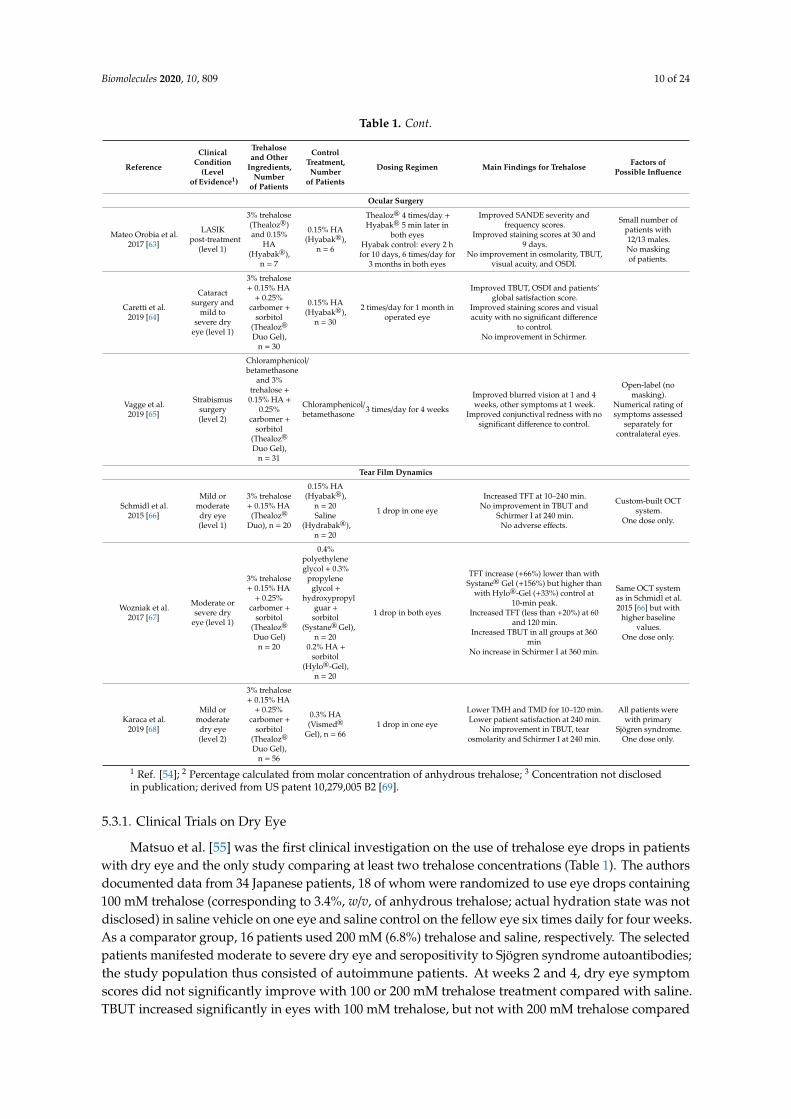
Evidence for the benefits and risks of trehalose in ophthalmologic applications including treatmentof dry eye should be presented in randomized controlled trials. The best approach for collectingall relevant data for this chapter was found using search terms “trehalose” and “eye” in PubMed.From among the 52 publications (27 Mar 2020), less than 20 articles fulfilling the requirement for arandomized controlled trial were selected and allocated into subsections for dry eye, ocular surgery,and tear film dynamics. The level of evidence addressed to each dataset was adapted from the modifiedAmerican Academy of Ophthalmology Preferred Practices guidelines [54]. The main details of eachstudy are summarized in Table 1.
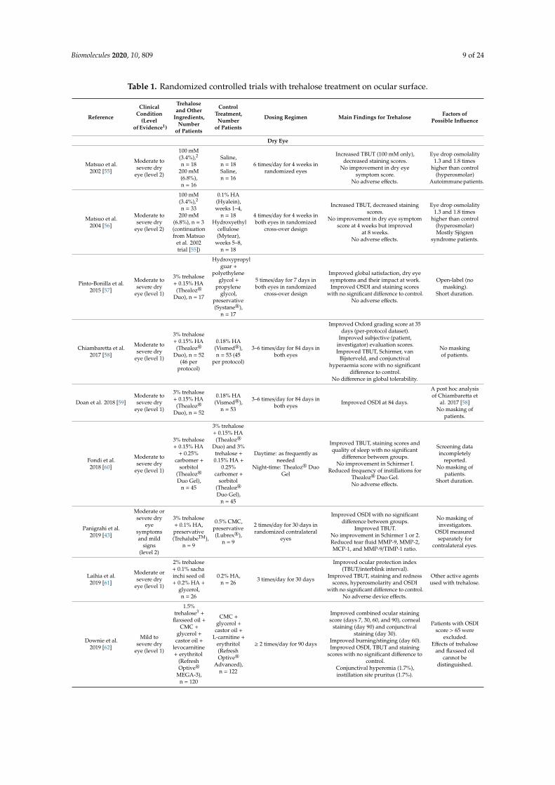
Biomolecules 2020, 10, 809 9 of 24
Table 1. Randomized controlled trials with trehalose treatment on ocular surface.
Reference
ClinicalCondition
(Levelof Evidence1)
Trehaloseand Other
Ingredients,Number
of Patients
ControlTreatment,Number
of Patients
Dosing Regimen Main Findings for Trehalose Factors ofPossible Influence
Dry Eye
Matsuo et al.2002 [55]
Moderate tosevere dry
eye (level 2)
100 mM(3.4%),2
n = 18200 mM(6.8%),n = 16
Saline,n = 18Saline,n = 16
6 times/day for 4 weeks inrandomized eyes
Increased TBUT (100 mM only),decreased staining scores.
No improvement in dry eyesymptom score.
No adverse effects.
Eye drop osmolality1.3 and 1.8 times
higher than control(hyperosmolar)
Autoimmune patients.
Matsuo et al.2004 [56]
Moderate tosevere dry
eye (level 2)
100 mM(3.4%),2
n = 33200 mM
(6.8%), n = 3(continuationfrom Matsuo
et al. 2002trial [55])
0.1% HA(Hyalein),weeks 1–4,
n = 18Hydroxyethyl
cellulose(Mytear),
weeks 5–8,n = 18
4 times/day for 4 weeks inboth eyes in randomized
cross-over design
Increased TBUT, decreased stainingscores.
No improvement in dry eye symptomscore at 4 weeks but improved
at 8 weeks.No adverse effects.
Eye drop osmolality1.3 and 1.8 times
higher than control(hyperosmolar)Mostly Sjögren
syndrome patients.
Pinto-Bonilla et al.2015 [57]
Moderate tosevere dry
eye (level 1)
3% trehalose+ 0.15% HA(Thealoz®
Duo), n = 17
Hydroxypropylguar +
polyethyleneglycol +
propyleneglycol,
preservative(Systane®),
n = 17
5 times/day for 7 days inboth eyes in randomized
cross-over design
Improved global satisfaction, dry eyesymptoms and their impact at work.Improved OSDI and staining scores
with no significant difference to control.No adverse effects.
Open-label (nomasking).
Short duration.
Chiambaretta et al.2017 [58]
Moderate tosevere dry
eye (level 1)
3% trehalose+ 0.15% HA(Thealoz®
Duo), n = 52(46 per
protocol)
0.18% HA(Vismed®),n = 53 (45
per protocol)
3–6 times/day for 84 days inboth eyes
Improved Oxford grading score at 35days (per-protocol dataset).
Improved subjective (patient,investigator) evaluation scores.Improved TBUT, Schirmer, van
Bijsterveld, and conjunctivalhyperaemia score with no significant
difference to control.No difference in global tolerability.
No maskingof patients.
Doan et al. 2018 [59]Moderate tosevere dry
eye (level 1)
3% trehalose+ 0.15% HA(Thealoz®
Duo), n = 52
0.18% HA(Vismed®),
n = 53
3–6 times/day for 84 days inboth eyes Improved OSDI at 84 days.
A post hoc analysisof Chiambaretta et
al. 2017 [58]No masking of
patients.
Fondi et al.2018 [60]
Moderate tosevere dry
eye (level 1)
3% trehalose+ 0.15% HA
+ 0.25%carbomer +
sorbitol(Thealoz®
Duo Gel),n = 45
3% trehalose+ 0.15% HA(Thealoz®
Duo) and 3%trehalose +0.15% HA +
0.25%carbomer +
sorbitol(Thealoz®
Duo Gel),n = 45
Daytime: as frequently asneeded
Night-time: Thealoz® DuoGel
Improved TBUT, staining scores andquality of sleep with no significant
difference between groups.No improvement in Schirmer I.
Reduced frequency of instillations forThealoz® Duo Gel.No adverse effects.
Screening dataincompletely
reported.No masking of
patients.Short duration.
Panigrahi et al.2019 [43]
Moderate orsevere dry
eyesymptomsand mild
signs(level 2)
3% trehalose+ 0.1% HA,preservative(TrehalubeTM),
n = 9
0.5% CMC,preservative(Lubrex®),
n = 9
2 times/day for 30 days inrandomized contralateral
eyes
Improved OSDI with no significantdifference between groups.
Improved TBUT.No improvement in Schirmer 1 or 2.Reduced tear fluid MMP-9, MMP-2,MCP-1, and MMP-9/TIMP-1 ratio.
No masking ofinvestigators.
OSDI measuredseparately for
contralateral eyes.
Laihia et al.2019 [61]
Moderate orsevere dry
eye (level 1)
2% trehalose+ 0.1% sachainchi seed oil+ 0.2% HA +
glycerol,n = 26
0.2% HA,n = 26 3 times/day for 30 days
Improved ocular protection index(TBUT/interblink interval).
Improved TBUT, staining and rednessscores, hyperosmolarity and OSDI
with no significant difference to control.No adverse device effects.
Other active agentsused with trehalose.
Downie et al.2019 [62]
Mild tosevere dry
eye (level 1)
1.5%trehalose3 +
flaxseed oil +CMC +
glycerol +castor oil +
levocarnitine+ erythritol
(RefreshOptive®
MEGA-3),n = 120
CMC +glycerol +castor oil +
L-carnitine +erythritol(RefreshOptive®
Advanced),n = 122
≥ 2 times/day for 90 days
Improved combined ocular stainingscore (days 7, 30, 60, and 90), corneal
staining (day 90) and conjunctivalstaining (day 30).
Improved burning/stinging (day 60).Improved OSDI, TBUT and staining
scores with no significant difference tocontrol.
Conjunctival hyperemia (1.7%),instillation site pruritus (1.7%).
Patients with OSDIscore > 65 were
excluded.Effects of trehalose
and flaxseed oilcannot be
distinguished.
Biomolecules 2020, 10, 809 10 of 24
Table 1. Cont.
Reference
ClinicalCondition
(Levelof Evidence1)
Trehaloseand Other
Ingredients,Number
of Patients
ControlTreatment,Number
of Patients
Dosing Regimen Main Findings for Trehalose Factors ofPossible Influence
Ocular Surgery
Mateo Orobia et al.2017 [63]
LASIKpost-treatment
(level 1)
3% trehalose(Thealoz®)and 0.15%
HA(Hyabak®),
n = 7
0.15% HA(Hyabak®),
n = 6
Thealoz® 4 times/day +Hyabak® 5 min later in
both eyesHyabak control: every 2 hfor 10 days, 6 times/day for
3 months in both eyes
Improved SANDE severity andfrequency scores.
Improved staining scores at 30 and9 days.
No improvement in osmolarity, TBUT,visual acuity, and OSDI.
Small number ofpatients with12/13 males.No maskingof patients.
Caretti et al.2019 [64]
Cataractsurgery and
mild tosevere dry
eye (level 1)
3% trehalose+ 0.15% HA
+ 0.25%carbomer +
sorbitol(Thealoz®
Duo Gel),n = 30
0.15% HA(Hyabak®),
n = 30
2 times/day for 1 month inoperated eye
Improved TBUT, OSDI and patients’global satisfaction score.
Improved staining scores and visualacuity with no significant difference
to control.No improvement in Schirmer.
Vagge et al.2019 [65]
Strabismussurgery(level 2)
Chloramphenicol/betamethasone
and 3%trehalose +0.15% HA +
0.25%carbomer +
sorbitol(Thealoz®
Duo Gel),n = 31
Chloramphenicol/betamethasone 3 times/day for 4 weeks
Improved blurred vision at 1 and 4weeks, other symptoms at 1 week.
Improved conjunctival redness with nosignificant difference to control.
Open-label (nomasking).
Numerical rating ofsymptoms assessed
separately forcontralateral eyes.
Tear Film Dynamics
Schmidl et al.2015 [66]
Mild ormoderatedry eye(level 1)
3% trehalose+ 0.15% HA(Thealoz®
Duo), n = 20
0.15% HA(Hyabak®),
n = 20Saline
(Hydrabak®),n = 20
1 drop in one eye
Increased TFT at 10–240 min.No improvement in TBUT and
Schirmer I at 240 min.No adverse effects.
Custom-built OCTsystem.
One dose only.
Wozniak et al.2017 [67]
Moderate orsevere dry
eye (level 1)
3% trehalose+ 0.15% HA
+ 0.25%carbomer +
sorbitol(Thealoz®
Duo Gel)n = 20
0.4%polyethyleneglycol + 0.3%
propyleneglycol +
hydroxypropylguar +
sorbitol(Systane®Gel),
n = 200.2% HA +
sorbitol(Hylo®-Gel),
n = 20
1 drop in both eyes
TFT increase (+66%) lower than withSystane® Gel (+156%) but higher than
with Hylo®-Gel (+33%) control at10-min peak.
Increased TFT (less than +20%) at 60and 120 min.
Increased TBUT in all groups at 360min
No increase in Schirmer I at 360 min.
Same OCT systemas in Schmidl et al.2015 [66] but with
higher baselinevalues.
One dose only.
Karaca et al.2019 [68]
Mild ormoderatedry eye(level 2)
3% trehalose+ 0.15% HA
+ 0.25%carbomer +
sorbitol(Thealoz®
Duo Gel),n = 56
0.3% HA(Vismed®
Gel), n = 661 drop in one eye
Lower TMH and TMD for 10–120 min.Lower patient satisfaction at 240 min.
No improvement in TBUT, tearosmolarity and Schirmer I at 240 min.
All patients werewith primary
Sjögren syndrome.One dose only.
1 Ref. [54]; 2 Percentage calculated from molar concentration of anhydrous trehalose; 3 Concentration not disclosedin publication; derived from US patent 10,279,005 B2 [69].
5.3.1. Clinical Trials on Dry Eye
Matsuo et al. [55] was the first clinical investigation on the use of trehalose eye drops in patientswith dry eye and the only study comparing at least two trehalose concentrations (Table 1). The authorsdocumented data from 34 Japanese patients, 18 of whom were randomized to use eye drops containing100 mM trehalose (corresponding to 3.4%, w/v, of anhydrous trehalose; actual hydration state was notdisclosed) in saline vehicle on one eye and saline control on the fellow eye six times daily for four weeks.As a comparator group, 16 patients used 200 mM (6.8%) trehalose and saline, respectively. The selectedpatients manifested moderate to severe dry eye and seropositivity to Sjögren syndrome autoantibodies;the study population thus consisted of autoimmune patients. At weeks 2 and 4, dry eye symptomscores did not significantly improve with 100 or 200 mM trehalose treatment compared with saline.TBUT increased significantly in eyes with 100 mM trehalose, but not with 200 mM trehalose compared
Biomolecules 2020, 10, 809 11 of 24
with saline. Staining scores improved significantly with both concentrations. No adverse effects wereobserved. The authors point out that the osmolarities of the 100 and 200 mM trehalose drops were1.3 and 1.8 times that of isotonic saline, respectively, i.e., hyperosmolar. Despite the lack of statisticalsignificance, the authors stated that both trehalose treatments were effective overall in improvingsymptoms and signs of dry eye. In respect to TBUT, they conclude the 100 mM concentration to be abetter dose.
In a further trial by Matsuo et al. [56], mostly female patients with moderate to severe dryeye were enrolled, 64% with a diagnosis of Sjögren syndrome, and 81% complaining of dry mouth(Table 1). Two four-week treatment periods followed a cross-over design without a wash-out phase.The nonrandomized assignment of patients into the control treatment with either 0.1% HA (upto four weeks) or hydroxyethyl cellulose eye drops (up to eight weeks) was based on “arbitrary”considerations. Before the start of the trial, five subjects had already used 100 mM trehalose whilethree other subjects had used 200 mM trehalose for six months as participants in the previous trial [55](Table 1); they were allowed to continue using the same concentration in both eyes. The order ofcross-over periods was randomized, and the eye drop bottles were masked. Both control formulationscontained benzalkonium chloride, whereas the 100 mM trehalose drops in saline were unpreserved.The drops were administered in both eyes four times a day for four weeks. Symptom scores did notdiffer at week 4, but they improved more significantly with trehalose than with hydroxyethyl celluloseat week 8. TBUT increased and staining scores improved significantly with trehalose. A majority ofthe patients considered trehalose superior to HA or hydroxyethyl cellulose in treating the symptoms.
After the initial studies by Matsuo et al., clinical trials have been continued using commercialproducts containing trehalose. Pinto-Bonilla et al. [57] claim to be the first to study eyedrops containingboth trehalose and HA (Table 1). In this open-label cross-over trial, 3% trehalose and 0.15% HA(Thealoz®Duo) was used five times a day for seven days in comparison to Systane eye drops, a preservedformulation with hydroxypropyl guar, polyethylene glycol, and propylene glycol. The washout periodwas five days. No blinding was used. Seventeen patients with moderate or severe dry eye (OSDI > 25)were recruited, randomized, and included in efficacy and safety populations. Study diary-basedpatient global satisfaction, dry eye symptoms, and their impact at work improved significantly morewith Thealoz® Duo than with Systane. OSDI score improved more with Thealoz® Duo, but withoutstatistically significant difference. Ocular staining scores improved with both treatments. More patientspreferred Thealoz® Duo than Systane. No adverse events were reported. The authors recognizedthe study to be a small one and with relatively short duration. The study was sponsored, and thepreparation of the paper was remunerated by Théa.
Chiambaretta et al. [58] conducted an investigator-masked multicenter study in a total of 105 adultpatients with moderate to severe dry eye (OSDI ≥ 18 and with either Schirmer test 3–9 mm/5 min or thesum of three TBUT ≤ 30 s in at least one eye) (Table 1). Fifty-two patients used one drop of Thealoz®
Duo (3% trehalose and 0.15% HA in hypo-osmolar formulation) 3–6 times a day for 84 days. All patientsin this group completed the study. Fifty-three patients received control eye drops (0.18% HA, alsohypo-osmolar; Vismed®, Horus Pharma, Saint-Laurent du Var, France). Of these, 46 and 45 patients,respectively, completed the study per protocol. Noninferiority of Thealoz® Duo to HA alone, assessedby Oxford grading score to assess global severity of ocular keratitis and conjunctival impairment,was demonstrated at day 35. Subjective scores evaluated by patients or investigators improvedmore with Thealoz® Duo. Schirmer test, TBUT, conjunctival hyperemia, and global performanceassessed as secondary efficacy criteria were similarly improved in both groups with no clinicallymeaningful differences. Both treatments were well tolerated. Fewer ocular symptoms and feweradverse events occurred with Thealoz® Duo. The authors noted that the primary criterion was assessedby investigators masked to treatments, limiting open-label bias. They concluded that the HA–trehalosecombination treatment is effective and safe, with better patient satisfaction, than existing HA eyedrops,particularly from the first month of treatment. The study was supported by Théa.
Biomolecules 2020, 10, 809 12 of 24
The study by Doan et al. [59] was a post hoc analysis of the Chiambaretta study data [58] (Table 1).The authors reanalyzed the percentage of patients with OSDI score below and above the threshold value19 at study endpoint days 35 and 84 after daily HA–trehalose and HA treatments. There were morepatients with OSDI < 19 in the HA–trehalose group (46.2%) than in the HA group (32.1%) at day 35;the difference was nonsignificant. At day 84, the proportions were 78.8% and 58.5%, respectively, with asignificant difference. The authors acknowledged the lack of blinding of the patients to treatments as alimitation, but supported the conclusions of Chiambaretta et al. on symptomatic relief of dry eye withHA–trehalose eyedrops.
An observer-masked, two-way cross-over study by Fondi et al. [60] investigated the clinical effectof a gel product containing 3% trehalose, 0.15% HA, and 0.25% carbomer and sorbitol (THC gel;Thealoz® Duo Gel) compared with HA–trehalose (Thealoz® Duo) (Table 1). Forty-five patients withmoderate to severe dry eye (OSDI ≥ 22 and TBUT ≤ 10 s or Schirmer I test 2–5 mm/5 min) were enrolled,out of whom 40 completed the study per protocol. The worst eye was selected as the study eye. Duringa one-week washout period before randomization, all participants used HA eye drops (Hyabak®,Laboratoires Théa, Clermont-Ferrand, France) until three days before the start of the study, then usedsaline drops (Hydrabak, Théa) until 12 h before the first study day. The randomized treatments wereused for one week as frequently as needed (pro re nata) during the day. In addition, all patients in bothgroups were asked to instill the THC gel before going to sleep. The washout period was one week.TBUT and subjective VAS scoring for sleep quality increased while staining scores and OSDI decreasedwith nonsignificant differences between treatments. Schirmer I test scores and safety parameters didnot change. As the mean frequency of instillation was 3.2 times/day for HA–trehalose eye drops and1.9 times/day for THC gel, the authors concluded that that the THC gel treatment requires less frequentinstillation to achieve a comparable benefit to the patient. The study was sponsored by Théa.
Panigrahi et al. [43] compared eye drops with 3% trehalose and 0.1% HA (TrehalubeTM, Micro Labs,Bangalore, India; A. Ghosh, personal communication) to eye drops with 0.5% carboxymethylcellulose(CMC; Lubrex®, Micro Labs, Bangalore, India), both preserved with an oxychloro complex, in ninepatients with mild dry eye signs, but with moderate to severe symptoms (OSDI > 23). The eye dropswere administrated in a patient-blinded fashion (Ghosh, personal communication) twice a day inrandomized contralateral eyes for 30 days. Mean OSDI (measured separately for both eyes) reducedand TBUT increased significantly with trehalose. Tear secretion was not affected. Trehalose alsoreduced proinflammatory cytokine levels (Table 1).
In a clinical trial by Laihia et al. [61], 2% trehalose and 0.2% high-molecular-weight (1.0–1.5 MDa)HA were included as active components in a hypotonic multi-ingredient microemulsion eye dropformulation (Table 1). Isotonic eye drops with 0.2% medium-molecular-weight (0.7–0.9 MDa) HA werean active control treatment. Both eye drops were preservative-free solutions and masked at all levels.The study comprised three parts. In the third part, 26 randomized participants used the same eye dropsfor both eyes three times a day for 30 days. Statistically significant improvement with HA–trehalosemicroemulsion eye drops was observed in TBUT, ocular protection index (=TBUT/interblink interval),corneal and nasal conjunctival staining, conjunctival and lid redness, and OSDI. Tear osmolarityin initially hyperosmolar (≥ 308 mOsm/L) subjects decreased similarly to healthy level with bothtreatments, suggesting an insignificant role for solution tonicity. A significant difference betweentreatments was achieved with ocular protection index. The authors hypothesize that disruptinghyperosmolar stress would lead the way to biophysical protection of the ocular surface to resolveepithelial damage and inflammation, followed by relief of symptoms. A multi-ingredient formulationcould be more efficient against all etiologic factors of dry eye [70,71]. The authors point out thatthe measurements were performed many hours after the last eye drop’s instillation. The role oftrehalose was likely not a short-term increase in tear film thickness (TFT) as observed previously [66,67](see 5.3.3), but rather antioxidative, cytoprotective, and wound-healing activity on the ocular epithelium.The study was sponsored by Finnsusp, the manufacturer of both investigational eye drops.
Biomolecules 2020, 10, 809 13 of 24
Downie et al. [62] compared nanoemulsion eye drops (OM3) containing CMC, glycerol, flaxseed(linseed) oil, castor oil, levocarnitine, erythritol, and trehalose (Refresh Optive® MEGA-3, Allergan,Dublin, Ireland) to a control vehicle without trehalose and flaxseed oil (Table 1) labelled as inactiveingredients in the product. Patients with stratified dry eye severities not exceeding an OSDI scoreof 65 were randomized to use OM3 (n = 120) or control eye drops (n = 122) in preservative-free anddouble-masked single-unit dose vials at least twice a day for 90 days. Significant improvement frombaseline was achieved in OSDI, TBUT, and corneal and conjunctival staining at each visit (days 7, 30,60, and 90) with OM3. OM3 was found to be noninferior to control eye drops in reducing symptomseverity at 90 days. Both treatments were well tolerated. The authors interpret trehalose and flaxseedoil to offer additional protection to the ocular surface, although distinguishing the relative benefit ofthe two ingredients was not possible. The study was funded by Allergan, the manufacturer of bothinvestigational eye drops; all authors reported a prior or current financial or personal relationship withthe company.
5.3.2. Clinical Trials on Ocular Surgery
A controlled investigator-blinded study evaluated the usability of trehalose for laser-assistedin situ keratomileusis (LASIK) post-treatment [63] (Table 1). Thirteen enrolled patients (12 males)undergoing sequential bilateral LASIK surgery were randomized into two treatment groups. In group 1(n = 6), 0.15% HA eye drops (Hyabak, Théa) were taken every two hours during the first 10 days and sixtimes a day thereafter until three months after surgery. In group 2 (n = 7), eye drops with 3% trehalose(Thealoz®, Théa; no HA) were administered four times a day, each time followed by HA (Hyabak) eyedrops five minutes later and starting three days before surgery. Ocular assessments were performedon days 1, 7, 30, and 90. In addition, frequent and gradually reducing doses of dexamethasone andtobramycin were administered for 10 days after surgery. Data corrected for presurgery values showedno significant differences between treatments in OSDI, tear osmolarity, and TBUT, while the SymptomAssessment in Dry Eye (SANDE) scoring on an analogue scale showed significant improvement insymptom severity with trehalose at all study visits after surgery. Vital staining data (Oxford andNEI scales) revealed superiority of trehalose treatment with significant difference to HA on days 30and 90. The authors postulate that the interaction of trehalose with ocular membrane lipids couldprovide additional protection against surgical trauma and increased osmolarity, resulting in reducedinflammation and cell death. The authors conclude that the recovery of tear and cell homeostasis afterLASIK was superior for 3% trehalose compared to HA. The study was partially supported by Théa.
Caretti et al. [64] investigated the treatment of eyes after cataract surgery with THC gel (Thealoz®
Duo Gel) or with 0.15% HA (Hyabak®, Théa; La Gloria Valerio, personal communication) eye drops in arandomized case-control study (Table 1). Sixty eyes of 60 patients with mild to severe dry eye symptomsand scheduled for unilateral cataract surgery were randomized into two groups. After surgery, 30 eyeswere treated with THC and 30 eyes with HA in a double-masked manner twice a day for one month.Steroid antibiotics and nonsteroidal anti-inflammatory drugs were given as well. All parameters wereassessed at baseline and at 7 and 30 days after surgery. In the trehalose group, mean TBUT improvedsignificantly from about 3.5 s to 6.3 s at day 7 and to 6.7 s at day 30; the corresponding values for HAwere 4.1, 4.8, and 5.1 s, with significant difference between treatments. The mean OSDI score decreasedwith THC treatment from a preoperative level of 31 to 5 units and with HA from 21 to 11 units onday 30, showing a significant difference between treatments. Fluorescein staining (Oxford scale) andpostoperative visual acuity improved with both treatments without statistically significant differencesbetween treatments. Tear production did not show significant changes. Patients’ global satisfactionscore was significantly greater for THC gel. It was concluded that THC was effective and well toleratedin reducing dry eye symptoms and in improving the clinical outcome after cataract surgery.
The efficacy of topical chloramphenicol 0.5%–betamethasone 0.2% (CB; Betabioptal®, LaboratoiresThéa, Clermont-Ferrand, France) with and without THC gel (Thealoz® Duo Gel) treatment followingstrabismus surgery was compared by Vagge et al. [65] (Table 1). This single-arm study involved 31
Biomolecules 2020, 10, 809 14 of 24
patients undergoing bilateral strabismus surgery. After surgery, the contralateral eyes were randomizedto receive either topical CB alone or in combination with THC gel, instilled three times a day for fourweeks. All patients received both treatments without masking. Conjunctival redness (Efron scale)decreased, but did not differ between eyes at 1 and 4 weeks after surgery. Subjective numerical ratingsfor foreign body sensation, burning or stinging, and stick feeling were significantly lower in theTHC-treated eyes at week 1, but not at week 4, whereas blurred vision was rated significantly higherfor THC at both time points. The authors interpreted that CB with THC gel was overall more effectivethan CB alone in reducing subjective symptoms of ocular discomfort after strabismus surgery, whereasthe treatments are equally effective in reducing conjunctival inflammation.
5.3.3. Clinical Trials on Tear Film Dynamics
The effect of trehalose on tear film dynamics was investigated in two studies utilizing a custom-builtoptical coherence tomography (OCT) system to measure TFT. The study by Schmidl et al. [66] involved60 completing patients with mild or moderate dry eye (OSDI 13–32, TBUT ≤10 s and Schirmer I test2–5 mm, 43 females and 17 males) (Table 1). After measurement of the baseline TFT, the study subjectsreceived randomized and double-masked instillations of single doses of 3% trehalose and 0.15% HA(Thealoz® Duo) or 0.15% HA (Hyabak®) or NaCl (Hydrabak®; all from Théa) in a parallel-groupdesign in the worst eye only. TFT was measured at 10, 20, 40, 60, 120, and 240 min after instillation.TFT increased significantly from a baseline of 2.4 ± 0.4 (mean, SD) to a peak of 3.1 ± 0.9 µm at 10 minwith HA–trehalose and from 2.4 ± 0.3 to 2.9 ± 0.5 µm with HA, followed by a gradual decrease upto 240 min. NaCl showed a negligible effect. A statistically significant difference in the time coursebetween treatments was obtained (repeated-measures analysis of variance model). The trehalose–HAcombination showed a significantly higher TFT at all time points, although the difference at 240 minwas only a few per cent from baseline. All treatments slightly increased TBUT and Schirmer I scoremeasured at 240 min only (p > 0.05), attributed by the authors to single dosing. Tolerability was similarwith all eye drops. The authors conclude that trehalose increases ocular residence time of HA eyedrops by an unknown mechanism that requires further studies to resolve. The study received anunrestricted grant from Théa and was used for a joint patent application (see Section 5.5).
The same research group conducted a similar OCT investigation of TFT using gel-basedlubricants [67] (Table 1). In this study, sixty completing patients with moderate or severe dryeye (OSDI ≥ 22, TBUT ≤ 10 s and Schirmer I test 2–5 mm, 23 females and 37 males) received randomizedand observer-masked instillations of single doses in both eyes in a parallel-group design. TFT wasmeasured at 10, 30, 60, 120, 240, and 360 min after instillation. Baseline TFT was 3.53 ± 0.73 µm, a valueremarkably higher than those reported [66] using the same OCT system. Peaking again at 10 min,Systane Gel (0.4% polyethylene glycol, 0.3% propylene glycol, hydroxypropyl guar and sorbitol; AlconPharma, Fort Worth, TX, USA) showed the greatest TFT increase to a mean of about 9 µm (+156% frombaseline), followed by THC gel (Thealoz® Duo Gel) and Hylo-Gel (0.2% HA and sorbitol; Ursapharm,Saarbrücken, Germany) with mean peak values of about 6 µm (+66%) and 5 µm (+33%), respectively.A relative increase of less than 20% from baseline at 60 and 120 min was significant for THC gelonly. No significant differences were observed at 30 min after instillations. Overall, TBUT increasedsignificantly and similarly at 6 h in all treatment groups. Schirmer I score did not change. The authorspostulate that the polymeric meshwork created by cross-linked hydroxypropyl guar of Systane Gelmay stabilize the ocular tear film and thus partially explain the increase in TFT compared to the othertwo formulations. In spite of this difference, they conclude that THC gel offers a longer residence timeon the cornea. This study was sponsored by Théa.
In a more recent study, Karaca et al. [68] enrolled 122 patients with mild to moderate dry eyeto investigate tear meniscus height (TMH) and depth (TMD), tear osmolarity, ocular residence time,and subjective comfort after single-drop administration of eye drops with THC gel (Thealoz® Duo Gel)or 0.3% HA (Vismed® Gel, TRB Chemedica, UK) (Table 1). For this randomized and observer-blindedstudy, only patients diagnosed with primary Sjögren syndrome and with OSDI score 13–32 units
Biomolecules 2020, 10, 809 15 of 24
were included. The study eye was the one with the lower TBUT. After instillation of one drop of therandomized eye drops by one ophthalmologist, another ophthalmologist, blinded to study treatments(E. Karaca, personal communication), measured TMH and TMD using swept-source OCT for up to240 min, interpreted to represent the corneal residence time of the eye drops. A significant increasewas observed in TMH at 10 min and in TMD at 10 and 60 min with both treatments. At each time pointfor up to 120 min, TMH and TMD were significantly higher for 0.3% HA than for THC gel. TBUT, tearosmolarity, and Schirmer I test measured at 240 min did not differ from baseline, interpreted by theauthors to reflect the single-dose treatment. The 0.3% HA eye drops were concluded to be superiorto THC gel drops in terms of corneal residence time and patient comfort duration, although longerobservation periods would be required to confirm this.
For understanding ocular residence time, it might be useful to note two nonclinical reports thatdiscuss the importance of physical and rheological properties of HA and trehalose in eye drops.White et al. [72] developed a “comfort agent index” to facilitate direct comparison between differentpolymer agents used in eye drops and to provide experimental validation and explanation for generaltrends suggested by the available clinical data. Firstly, the main finding was that the comfort-promotingproperties relate strongly to both the concentration and the molecular weight (i.e., chain length)of each agent. Secondly, polysaccharides as a group showed the best comfort agent index values,followed by acrylic agents. The greatest comfort property contributions, independent of specificmolecular weight and concentration considerations, were from HA, hydroxypropyl methylcellulose,and CMC. In accordance with the main findings, not only the concentration, but especially the molecularweight of HA strongly contributed to the comfort property, as suggested by several previous studiesin vitro [73–76]. This finding leads to the second paper by Salzillo et al. [77] who analyzed the molecularweights of HA and the viscosity of some eye drop formulations on the market. One of the analyzedbrands was Hyabak® (Théa), containing 0.15% HA and used in the TFT study of Schmidl et al. [66].The HA molecular weight in this product was found to be 0.36 MDa, while the viscosity of the solutionwas 2.3 mPa s. As compared to another product containing 0.28% of 1.1 MDa HA and with a zero-shearviscosity of 24 mPa s, the remarkably low viscosity of Hyabak® is obviously attributable to the lowmolecular weight of HA. Out of six products analyzed by Salzillo et al., Hyabak® showed both thelowest HA molecular weight and the lowest viscosity. The HA concentration on the product labelis the same for Hyabak® and Thealoz® Duo, and both product versions also utilize the same ABAKeye drop dispenser technology with a sterile filter membrane, suggesting that the HA species used inboth Hyabak® and Thealoz® Duo is probably of the same low molecular weight. In a more recentanalysis, the HA molecular weight in Thealoz® Duo was confirmed to be 0.22 MDa and the viscosity2.8 mPa s [78]. In an early study [79] cited both by Schmidl et al. [66] and Wozniak et al. [67], ocularresidence time was 11.1 min for 0.2% HA and 23.5 min for 0.3% HA; thus, increasing HA concentrationby 50% resulted in more than 100% longer residence time in real-life conditions. The results of thesestudies suggest that the observed ocular residence times [66,67] would be enhanced efficiently byincreasing the concentration and/or the molecular weight of HA, as demonstrated by Karaca et al. [68].
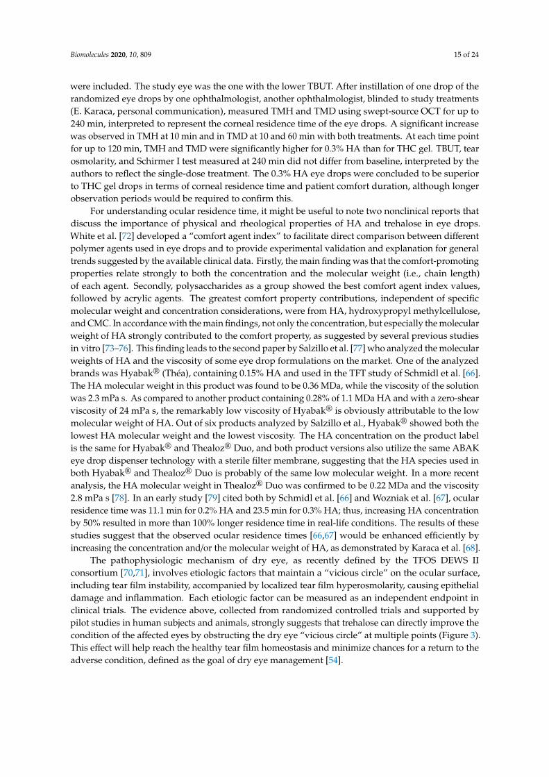
The pathophysiologic mechanism of dry eye, as recently defined by the TFOS DEWS IIconsortium [70,71], involves etiologic factors that maintain a “vicious circle” on the ocular surface,including tear film instability, accompanied by localized tear film hyperosmolarity, causing epithelialdamage and inflammation. Each etiologic factor can be measured as an independent endpoint inclinical trials. The evidence above, collected from randomized controlled trials and supported bypilot studies in human subjects and animals, strongly suggests that trehalose can directly improve thecondition of the affected eyes by obstructing the dry eye “vicious circle” at multiple points (Figure 3).This effect will help reach the healthy tear film homeostasis and minimize chances for a return to theadverse condition, defined as the goal of dry eye management [54].
Biomolecules 2020, 10, 809 16 of 24
Figure 3. Etiologic factors of dry eye affected by trehalose as demonstrated in clinical data. Redrawnfrom ref. [61], Figure S1, with permission from the authors.
5.4. Do We Know the Optimum Concentration?
The efficacy and safety of trehalose in adverse ocular conditions have been well established,but how was a profitable trehalose dose determined and who made that decision? Reviewing thescientific literature reveals that the “knowledge” of the “right” concentration obviously comes fromone source. Matsuo et al. [55] is the first clinical investigation and to date the only report comparing atleast two trehalose concentration levels in human subjects. The authors explain the concentrations100 mM (3.4%) and 200 mM (6.8%; calculated as anhydrous trehalose) were based on their earlierpreclinical work [80] in which hCEC cultures were shortly pretreated with trehalose and let dry at lowhumidity. Trehalose preincubation was found to protect cell viability significantly in concentrations50 mM (1.7%), 100 mM (3.4%), and 200 mM (6.8%), compared with concentrations 20 mM or lower,maltose, buffer vehicle, hydroxyethyl cellulose eye drops, or 0.1% HA. The average results for 50and 100 mM concentrations were practically identical (20.1% and 19.1% dead cells, respectively, withlarge deviations), while 200 mM trehalose was clearly more efficient (11.9% dead cells). Interestingly,however, Matsuo et al. [55] then interpreted those results to show that the 50 mM concentration was“less effective” and acknowledged the two highest concentrations as applicable for their clinical study.Considering the close relationship of the main author with Hayashibara Biochemical Laboratories,a company holding manufacturing and other patents for trehalose (see disclosure [55] and Section 5.5),there may be a commercial bias in the selection of concentrations. To our knowledge, scientific papershave remained silent about trehalose concentrations in eye drops since.
Consequently, almost all clinical trials with trehalose in dry eye patients since Matsuo’s reportshave been conducted at the 3% concentration level (Sections 5.2 and 5.3). Laboratoires Théa adoptedthis concentration for Thealoz®, Thealoz® Duo, and Thealoz® Duo Gel. Most preclinical studieswith ocular cells or dry eye animal models (Section 5.1) have used the finished eye drop productsinstead of pure trehalose in a control vehicle; no attempts to adjust the concentration has beenexpressed in these studies either. An unstated consensus thus prevails on the fact that the 3% trehaloseconcentration, originally determined in one cell culture study [80] and followed by one clinicalstudy with hyperosmolar trehalose eye drops and involving one ethnic patient population withautoimmune diseases [55] (Table 1), is sufficiently established. In another study [43], 1.2% trehalose wasfound to protect human corneal cells against desiccation-induced cell death, morphological changes,
Biomolecules 2020, 10, 809 17 of 24
and nduction of proinflammatory signaling, whereas 2.4% trehalose caused a significant loss of cellviability within the same time frame. Further, a more recent study showed that 6% trehalose was moreefficient than 3% trehalose against UVB irradiation-induced oxidative damage in the rabbit cornea [8],thus approaching the concentrations originally experimented by Matsuo et al. [55,80].
It should be kept in mind that an error of about 10% in the percentage concentration is producedin the absence of information of whether trehalose dihydrate (378 g/mol) or anhydrous trehalose(342 g/mol) is concerned. Currently at least one clinical trial exists in which 2% trehalose dihydrate(ca. 58 tmM) was included in combination with other active agents for dry eye [61], and another trialwith possibly 1.5% trehalose [62] (as explained in Table 1).
Studies in other medical fields reveal a wider range of usable concentrations of trehalose. A recentreview [81] lists several published studies on the effect of trehalose in cellular models of autophagy; theequally preferred concentrations were 50 (1.7%) or 100 mM (3.4%), and even 10–20 mM (0.34%–0.68%)trehalose induced autophagy activators [33]. Another review [82] of Parkinson’s disease models showsthat the effective concentrations of trehalose in direct contact with the target cells are in the range10–100 mM (0.34%–3.4%), in some cases even lower. There seems to be still a limited body of data toconclude that the trehalose concentration in topically administered eye drops should be much higher,although a higher concentration would still be perfectly safe. Eventually, it is the overall compositiondetermining other properties, such as formulation viscosity and retention on the ocular surface, thatwould bring about the clinical outcome.
5.5. Trehalose Patents in Dry Eye
The intention of this chapter is to give a brief overview of patents or patent applications for theuse of trehalose in ophthalmologic applications related to dry eye. What aspects are covered by patentsand in which countries? Manufacturing patents and other applications of trehalose are beyond thescope of the present review and were excluded. Patent information was collected by using the free andpublicly accessible database [69] provided by The European Patent Organisation (EPO).
The patent application WO97/24129 (“Tanaka 1995”; applicant: Rohto Pharmaceutical,Osaka, Japan) appears to be the first to disclose an ophthalmic application for trehalose. The medicaltreatment “relates to a pharmaceutical composition containing trehalose, which shows protecting effecton cornea and is used safely as an intraocular irrigating solution, eye drops, or eye ointment”. Thisapplication was granted in Japan (JP4033510 B2) and lapsed/expired in 2016.
Hayashibara Biochemical Laboratories (Okayama, Japan) was the applicant for an initial Europeanpatent EP1192947 (“Matsuo 2000”) claiming an ophthalmic pharmaceutical composition containingtrehalose. As the correspondence between the patent office and an applicant can be followed via theGlobal Dossier feature of the patent database, it appears that the original intention of the applicant toobtain wide protection for treatment of several ophthalmologic conditions was hindered by both theTanaka 1995 application and a scientific publication of the inventor [80]. These two prior publicationsthus became obstacles for the novelty of the invention. In the final version EP1192947 (B1) grantedin 2006, the patent claims were consequently narrowed to “use of trehalose free of pyrogen in themanufacture of an ophthalmic pharmaceutical composition for the treatment and/or prevention of anophthalmologic clinical symptom of Sjögren syndrome or an ophthalmologic clinical sign of Sjögrensyndrome”. A divisional application EP1649860 for the original Matsuo 2000 patent was submittedto EPO in 2006. Thus, EP1649860 and EP1192947 belong to the same patent family with the samepriority date. Again, Tanaka 1995 forced to restrict the divisional application to a somewhat crypticwording, claiming “use of α,α-trehalose as an effective ingredient for the preparation of an ophthalmicpharmaceutical composition for treating dry eye as an ophthalmologic clinical symptom of Sjögrensyndrome”. Both patents are in force in three European countries (Table 2).
Further non-European patents within the Matsuo/Hayashibara patent family share the samepriority application with EP1192947 and EP1649860. The claims in each patent granted in variouscountries are uniform in that the use of trehalose is indicated for treating or preventing symptoms and
Biomolecules 2020, 10, 809 18 of 24
signs of Sjögren syndrome alone (Table 2). This restriction may not be readily apparent in all cases.In US7732425 (B2) for example, “a method for treating a patient suffering from dry eye” is claimed,by the use of trehalose in an eyewash formulation. Although not explicitly worded in the claims,the patent description itself clarifies that the invention “exerts an outstanding improvement in theophthalmologic clinical symptoms and signs in Sjögren syndrome, and thus it can be advantageouslyused in the treatment and/or the prevention of the syndrome.” Obviously, the scope would be restrictedto Sjögren syndrome in this case as well. In all cases, the claimed effective trehalose concentrationrange is from at least 0.01% up to 10% (EP1649860) or to about 30% (US7732425).
Table 2. Summary of granted patents on trehalose for dry eye.
Patent General Field of Protection 1 DesignatedCountry
JP4033510 (B2) Trehalose for protecting cornea and used as eye drops JP
EP1192947 (B1) Trehalose in treatment and/or prevention of symptom or signof Sjögren syndrome DE FR GB
EP1649860 (B1) Trehalose in treatment of dry eye as a clinical symptom ofSjögren syndrome DE FR GB
US6555526 (B2)Ophthalmic pharmaceutical composition with trehalose for
treatment and/or prevention of symptom and sign in dry eyein Sjögren syndrome
US
US7732425 (B2) Method for treating dry eye (Sjögren syndrome) with trehalosein the form of eyewash solution US
CA2355814 (C)Ophthalmic pharmaceutical composition with trehalose for
treatment and/or prevention of symptom or sign ofSjögren syndrome
CA
AU781975 (2001065453) (B2)Ophthalmic pharmaceutical composition with trehalose for
treatment or prevention of symptom or sign ofSjögren syndrome
AU
KR100776124 (B1) Trehalose in treatment and/or prevention of symptom and signin dry eye in Sjögren syndrome KR
TWI291350 (B) Trehalose in treatment or prevention of symptom and sign ofSjögren syndrome TW
JP4982643 (B2) Trehalose in treatment and/or prevention of symptom or signof Sjögren syndrome JP
EP3110425 (B1) Trehalose (as a diholoside) with 100–800 kDa HA for treatingophthalmic diseases such as dry eye Most EPO countries
1 Descriptive presentation of patent claim(s).
It may be concluded that all patents within the Matsuo/Hayashibara patent family protect theuse of trehalose for treatment of dry eye related to Sjögren syndrome. These patents share the samepriority term, the priority date being Sept. 14, 2000.
Laboratoires Théa and the Medical University of Vienna were granted a European patentEP3110425 in 2019 for an ophthalmic formulation comprising HA and a diholoside, corresponding tothe composition of Thealoz® Duo with 0.15% HA and 3% trehalose (a diholoside). The incorporatedclaims for the molecular weight of HA (100–800 kDa) and viscosity (2–15 mPa s) match those determinedfor the product by others (see Section 5.3.3). The invention is for use in ophthalmic diseases such asdry eye, and it is based on experimental data showing increased TFT after a single drop applicationpublished by Schmidl et al. [66] (Table 1). The priority date of the application is Feb. 28, 2014, and it isin force in most European countries (Table 2).
6. Conclusions and an Outlook for Tomorrow
Considering the proceedings reviewed in the preceding chapters, all indicators suggest thattrehalose will continue to be a topic of intensive research in ophthalmology and other fields of science,maintaining the exponential trend in the number of medical publications (Figure 1) for the 2020s.These indicators include advancements in manufacturing technologies (Section 2) and consistent effectsof trehalose in signaling models for oxidative stress, protein clearance, and inflammation, and in animalmodels related to dry eye (Sections 4 and 5.1). To date, trehalose lacks known toxicity to cells and israther characterized by bioprotection of cells and macromolecules against adverse threats (Sections 3–5).The safety and protective effects of trehalose have been a common finding in several clinical trials with
Biomolecules 2020, 10, 809 19 of 24
topical application in the eyes (Section 5). Active research on the effects of trehalose in neuroprotectionand cryopreservation (Section 3) will likely lead to clinical applications in these fields. As trehaloseis being considered for the management of neurodegenerative diseases (reviewed in [82]), it is notdifficult to imagine that this field is likely to inseminate research in ophthalmic applications as well,such as for treatment of age-related macular degeneration and glaucoma, due to partially sharedpathogenetic features [83,84].
Trehalose can be chemically or enzymatically modified to obtain synthetic trehalose analogues tobe utilized as nondegradable bioprotectants, therapeutic inhibitors, or bioanalytical probes (reviewedin [85]). For therapeutic activity in a specific application, trehalose or its analogues need to be includedin an appropriate formulation and in a sufficient concentration, combined with a suitable deliveryoption. Development and optimization of these characteristics will facilitate the use of trehalose in amultitude of clinical applications in the field of ophthalmology and beyond.
Conflicts of Interest: This work was partially supported by Finnsusp Ltd., Lieto, Finland. The companyparticipated in the writing of the manuscript and the decision to submit the article for publication. JL isan employee of Finnsusp Ltd. KK declares no conflict of interest.
Abbreviations
AMPK monophosphate-activated protein kinaseAP-1 activating protein-1ARE antioxidant response elementCB combination preparation containing chloramphenicol and betamethasoneCMC carboxymethylcelluloseEPO European Patent OrganisationHA hyaluronic acidhCEC human corneal epithelial cellsHSP heat shock proteinKEAP1 kelch-like ECH-associated protein 1LASEK laser subepithelial keratomileusisLASIK laser-assisted in situ keratomileusisMeSH Medical Subject HeadingsMMP matrix metalloproteinasemTOR mechanistic target of rapamycinNEI National Eye Institute/Industry grading scaleNFE2L2 nuclear factor erythroid 2-related factor 2NF-κB nuclear factor-κBOCT optical coherence tomographyOSD ocular surface diseaseOSDI Ocular Surface Disease IndexPBS phosphate-buffered salineROS reactive oxygen speciesSANDE Symptom Assessment in Dry EyeSLC2A8 Solute Carrier Family 2 Member 8TBUT tear film break-up timeTFT tear film thicknessTHC combination preparation containing trehalose: hyaluronic acid: carbomer and sorbitolTMD tear meniscus depthTMH tear meniscus heightTNF-α tumor necrosis factor-αTret1 trehalose transporter-1UPS ubiquitin proteasome systemUVB ultraviolet-B
Biomolecules 2020, 10, 809 20 of 24
References
1. National Center for Biotechnology Information. Available online: https://pubmed.ncbi.nlm.nih.gov/?term=
trehalose (accessed on 2 April 2020).2. Wiggers, H. Untersuchung über das Mutterkorn, Secale cornutum. Annalen der Pharmacie 1832, 1, 129–182.
[CrossRef]3. Richards, A.B.; Krakowka, S.; Dexter, L.B.; Schmid, H.; Wolterbeek, A.P.M.; Waalkens-Berendsen, D.H.;
Shigoyuki, A.; Kurimoto, M. Trehalose: A review of properties, history of use and human tolerance,and results of multiple safety studies. Food Chem. Toxicol. 2002, 40, 871–898. [CrossRef]
4. Cai, X.; Seitl, I.; Mu, W.; Zhang, T.; Stressler, T.; Fischer, L.; Jiang, B. Biotechnical production of trehalosethrough the trehalose synthase pathway: Current status and future prospects. Appl. Microbiol. Biotechnol.2018, 102, 2965–2976. [CrossRef] [PubMed]
5. Ohtake, S.; Wang, Y.J. Trehalose: Current Use and Future Applications. J. Pharm. Sci. 2011, 100, 2020–2053.[CrossRef] [PubMed]
6. Cejková, J.; Ardan, T.; Cejka, C.; Luyckx, J. Favorable effects of trehalose on the development of UVB-mediatedantioxidant/pro-oxidant imbalance in the corneal epithelium, proinflammatory cytokine and matrixmetalloproteinase induction, and heat shock protein 70 expression. Graefe’s Arch. Clin. Exp. Ophthalmol.2011, 249, 1185–1194. [CrossRef]
7. Cejková, J.; Cejka, C.; Luyckx, J. Trehalose treatment accelerates the healing of UVB-irradiated corneas.Comparative immunohistochemical studies on corneal cryostat sections and corneal impression cytology.Histol. Histopathol. 2012, 27, 1029–1040.
8. Cejka, C.; Kossl, J.; Hermankova, B.; Holan, V.; Kubinova, S.; Olmiere, C.; Cejkova, J. The Healing of OxidativeInjuries with Trehalose in UVB-Irradiated Rabbit Corneas. Oxidative Med. Cell. Longev. 2019, 2019, 1857086-10.[CrossRef]
9. Takahashi, S.; Isaka, M.; Hamaishi, M.; Imai, K.; Orihashi, K.; Sueda, T. Trehalose protects against spinal cordischemia in rabbits. J. Vasc. Surg. 2014, 60, 490–496. [CrossRef]
10. Portbury, S.D.; Hare, D.J.; Finkelstein, D.I.; Adlard, P.A. Trehalose improves traumatic brain injury-inducedcognitive impairment. PLoS ONE 2017, 12, e0183683. [CrossRef]
11. Portbury, S.D.; Hare, D.J.; Bishop, D.P.; Doble, P.A.; Adlard, P.A.; Finkelstein, D.I. Trehalose elevates brainzinc levels following controlled cortical impact in a mouse model of traumatic brain injury. Metallomics 2018,10, 846–853. [CrossRef]
12. Ugarte, M.; Osborne, N.N. Recent advances in the understanding of the role of zinc in ocular tissues.Metallomics 2014, 6, 189–200. [CrossRef] [PubMed]
13. Lee, H.; McKeon, R.J.; Bellamkonda, R.V. Sustained delivery of thermostabilized chABC enhances axonalsprouting and functional recovery after spinal cord injury. In Proceedings of the National Academy of Sciences,Massachusetts Institute of Technology; Cambridge, MA, USA, 16 September 2009, National Academy of Sciences:Cambridge, MA, USA, 2010; Volume 107, pp. 3340–3345.
14. Stanwick, J.C.; Baumann, M.D.; Shoichet, M.S. In vitro sustained release of bioactive anti-NogoA, a moleculein clinical development for treatment of spinal cord injury. Int. J. Pharm. 2012, 426, 284–290. [CrossRef][PubMed]
15. Ruozi, B.; Belletti, D.; Sharma, H.S.; Sharma, A.; Muresanu, D.F.; Mössler, H.; Forni, F.; Vandelli, M.A.; Tosi, G.PLGA Nanoparticles Loaded Cerebrolysin: Studies on Their Preparation and Investigation of the Effect ofStorage and Serum Stability with Reference to Traumatic Brain Injury. Mol. Neurobiol. 2015, 52, 899–912.[CrossRef] [PubMed]
16. Plettig, J.; Johnen, C.M.; Bräutigam, K.; Knöspel, F.; Wönne, E.C.; Schubert, F.; Plöger, F.; Unger, J.K.;Reutzel-Selke, A.; Bornemann, R.; et al. Feasibility study of an active wound dressing based on hollow fibermembranes in a porcine wound model. Burns 2015, 41, 778–788. [CrossRef]
17. Stewart, S.; He, X. Intracellular Delivery of Trehalose for Cell Banking. Langmuir 2018, 35, 7414–7422.[CrossRef]
18. Zhang, M.; Oldenhof, H.; Sieme, H.; Wolkers, W.F. Combining endocytic and freezing-induced trehaloseuptake for cryopreservation of mammalian cells. Biotechnol. Prog. 2017, 33, 229–235. [CrossRef]
Biomolecules 2020, 10, 809 21 of 24
19. Mayer, A.L.; Higgins, C.B.; Heitmeier, M.R.; Kraft, T.E.; Qian, X.; Crowley, J.R.; Hyrc, K.L.; Beatty, W.L.;Yarasheski, K.; Hruz, P.W.; et al. SLC2A8 (GLUT8) is a mammalian trehalose transporter required fortrehalose-induced autophagy. Sci. Rep. 2016, 6, 1–15. [CrossRef]
20. Crowe, J.H.; Wolkers, W.F.; Oliver, A.E.; Ma, X.; Auh, J.-H.; Tang, M.; Zhu, S.; Norris, J.; Tablin, F. Stabilizationof Dry Mammalian Cells: Lessons from Nature. Integr. Comp. Boil. 2005, 45, 810–820. [CrossRef]
21. Hill-Bator, A.; Misiuk-Hojło, M.; Marycz, K.; Grzesiak, J. Trehalose-Based Eye Drops Preserve Viability andFunctionality of Cultured Human Corneal Epithelial Cells during Desiccation. BioMed Res. Int. 2014, 2014,1–8. [CrossRef]
22. Ozek, D.; Kemer, O.E.; Belfort, R.N.; Müller, M.S.; Isenberg, J.; Ferraz, P.; Szejnfeld, D. Effect of the bioprotectantagent trehalose on corneal epithelial healing after corneal cross-linking for keratoconus. Arq. Bras. de Oftalmol.2018, 81, 505–509. [CrossRef]
23. Aragona, P.; Colosi, P.; Rania, L.; Colosi, F.; Pisani, A.; Puzzolo, D.; Micali, A. Protective Effects of Trehaloseon the Corneal Epithelial Cells. Sci. World J. 2014, 2014, 1–9. [CrossRef] [PubMed]
24. Yacasi, G.R.R.; Lopéz, M.L.G.; García, M.E.; Coca, A.P.; Campmany, A.C.C. Influence of freeze-dryingand γ-irradiation in preclinical studies of flurbiprofen polymeric nanoparticles for ocular delivery usingd-(+)-trehalose and polyethylene glycol. Int. J. Nanomed. 2016, 11, 4093–4106. [CrossRef] [PubMed]
25. Cejková, J.; Cejka, C.; Ardan, T.; Sirc, J.; Michálek, J.; Luyckx, J. Reduced UVB-induced corneal damagecaused by reactive oxygen and nitrogen species and decreased changes in corneal optics after trehalosetreatment. Histol. Histopathol. 2010, 25, 1403–1416.
26. Kaarniranta, K.; Szalai, E.; Smedowski, A.; Hegyi, Z.; Kivinen, N.; Viiri, J.; Wowra, B.; Dobrowolski, D.;Módis, L.; Berta, A.; et al. A novel proteotoxic stress associated mechanism for macular corneal dystrophy.Histol. Histopathol. 2015, 30, 921–930. [CrossRef]
27. Sethi, R.; Iyer, S.S.; Das, E.; Roy, I. Discrete roles of trehalose and Hsp104 in inhibition of protein aggregationin yeast cells. FEMS Yeast Res. 2018, 18, 18–29. [CrossRef]
28. Surgucheva, I.; Ninkina, N.; Buchman, V.; Grasing, K.W.; Surguchov, A. Protein Aggregation in Retinal Cellsand Approaches to Cell Protection. Cell. Mol. Neurobiol. 2005, 25, 1051–1066. [CrossRef]
29. Blasiak, J.; Pawlowska, E.; Szczepanska, J.; Kaarniranta, K. Interplay between Autophagy and theUbiquitin-Proteasome System and Its Role in the Pathogenesis of Age-Related Macular Degeneration.Int. J. Mol. Sci. 2019, 20, 210. [CrossRef]
30. Strom, J.; Xu, B.; Tian, X.; Chen, Q.M. Nrf2 protects mitochondrial decay by oxidative stress. FASEB J. 2015,30, 66–80. [CrossRef]
31. Silva-Palacios, A.; Ostolga-Chavarría, M.; Zazueta, C.; Konigsberg, M. Nrf2: Molecular and epigeneticregulation during aging. Ageing Res. Rev. 2018, 47, 31–40. [CrossRef]
32. Felszeghy, S.; Viiri, J.; Paterno, J.J.; Hyttinen, J.M.; Koskela, A.; Chen, M.; Leinonen, H.; Tanila, H.; Kivinen, N.;Koistinen, A.; et al. Loss of NRF-2 and PGC-1α genes leads to retinal pigment epithelium damage resemblingdry age-related macular degeneration. Redox Boil. 2019, 20, 1–12. [CrossRef]
33. Mizunoe, Y.; Kobayashi, M.; Sudo, Y.; Watanabe, S.; Yasukawa, H.; Natori, D.; Hoshino, A.; Negishi, A.;Okita, N.; Komatsu, M.; et al. Trehalose protects against oxidative stress by regulating the Keap1–Nrf2 andautophagy pathways. Redox Boil. 2018, 15, 115–124. [CrossRef] [PubMed]
34. Johansson, I.; Monsen, V.T.; Pettersen, K.; Mildenberger, J.; Misund, K.; Kaarniranta, K.; Schønberg, S.;Bjørkøy, G. The marine n-3 PUFA DHA evokes cytoprotection against oxidative stress and protein misfoldingby inducing autophagy and NFE2L2 in human retinal pigment epithelial cells. Autophagy 2015, 11, 1636–1651.[CrossRef] [PubMed]
35. Li, Y.; Luo, Y.; Luo, T.; Lu, B.; Wang, C.; Zhang, Y.; Piao, M.; Feng, C.; Ge, P. Trehalose Inhibits ProteinAggregation Caused by Transient Ischemic Insults Through Preservation of Proteasome Activity, Not viaInduction of Autophagy. Mol. Neurobiol. 2016, 54, 6857–6869. [CrossRef] [PubMed]
36. Benaroudj, N.; Lee, D.H.; Goldberg, A.L. Trehalose Accumulation during Cellular Stress Protects Cells andCellular Proteins from Damage by Oxygen Radicals. J. Biol. 2001, 276, 24261–24267. [CrossRef]
37. Martin, L.M.; Jeyabalan, N.; Tripathi, R.; Panigrahi, T.; Johnson, P.J.; Ghosh, A.; Mohan, R. Autophagy incorneal health and disease: A concise review. Ocul. Surf. 2019, 17, 186–197. [CrossRef]
38. Rubinsztein, D.C.; Bento, C.F.; Deretic, V. Therapeutic targeting of autophagy in neurodegenerative andinfectious diseases. J. Exp. Med. 2015, 212, 979–990. [CrossRef]
Biomolecules 2020, 10, 809 22 of 24
39. Byun, S.; Lee, E.; Lee, Y. Therapeutic Implications of Autophagy Inducers in Immunological Disorders,Infection, and Cancer. Int. J. Mol. Sci. 2017, 18, 1959. [CrossRef]
40. Emanuele, E. Can trehalose prevent neurodegeneration? Insights from experimental studies.Curr. Drug Targets 2014, 15, 551–557. [CrossRef]
41. Mardones, P.; Rubinsztein, D.C.; Hetz, C. Mystery solved: Trehalose kickstarts autophagy by blockingglucose transport. Sci. Signal. 2016, 9, fs2. [CrossRef]
42. Huynh, K. Atherosclerosis: Trehalose induces macrophage autophagy-lysosomal biogenesis. Nat. Rev. Cardiol.2017, 14, 444. [CrossRef]
43. Panigrahi, T.; Shivakumar, S.; Shetty, R.; D’Souza, S.; Nelson, E.J.R.; Sethu, S.; Jeyabalan, N.; Ghosh, A.;Jacob, E. Trehalose augments autophagy to mitigate stress induced inflammation in human corneal cells.Ocul. Surf. 2019, 17, 699–713. [CrossRef] [PubMed]
44. He, Q.; Wang, Y.; Lin, W.; Zhang, Q.; Zhao, J.; Liu, F.-T.; Tang, Y.-L.; Xiao, B.-G.; Wang, J. TrehaloseAlleviates PC12 Neuronal Death Mediated by Lipopolysaccharide-Stimulated BV-2 Cells via InhibitingNuclear Transcription Factor NF-κB and AP-1 Activation. Neurotox. Res. 2014, 26, 430–439. [CrossRef][PubMed]
45. Lotfi, P.; Tse, D.Y.; Di Ronza, A.; Seymour, M.L.; Martano, G.; Cooper, J.D.; Pereira, F.A.; Passafaro, M.;Wu, S.M.; Sardiello, M. Trehalose reduces retinal degeneration, neuroinflammation and storage burdencaused by a lysosomal hydrolase deficiency. Autophagy 2018, 14, 1419–1434. [CrossRef] [PubMed]
46. Belmonte, C.; Nichols, J.J.; Cox, S.M.; Brock, J.A.; Begley, C.G.; Bereiter, D.A.; Dartt, D.A.; Galor, A.; Hamrah, P.;Ivanusic, J.J.; et al. TFOS DEWS II pain and sensation report. Ocul. Surf. 2017, 15, 404–437. [CrossRef][PubMed]
47. Fan, W.-X.; Ma, X.; Liu, T.-Q.; Cui, Z. Vitrification of Corneal Endothelial Cells in a Monolayer. J. Biosci. Bioeng.2008, 106, 610–613. [CrossRef]
48. Nakamura, T.; Sekiyama, E.; Takaoka, M.; Bentley, A.J.; Yokoi, N.; Fullwood, N.J.; Kinoshita, S. The use oftrehalose-treated freeze-dried amniotic membrane for ocular surface reconstruction. Biomaterials 2008, 29,3729–3737. [CrossRef]
49. Wusteman, M.C.; Simmonds, J.; Vaughan, D.; Pegg, D.E. Vitrification of rabbit tissues with propylene glycoland trehalose. Cryobiology 2008, 56, 62–71. [CrossRef]
50. Giesbers, M.E.; Bosman, G.J.C.G.M.; Bovee-Geurts, P.H.; De Grip, W.J. Introduction of a rod pigment aromaticcluster does not improve the structural stability of the human green cone pigment. J. Struct. Boil. 2007, 159,222–227. [CrossRef]
51. Chen, W.; Zhang, X.; Liu, M.; Zhang, J.; Ye, Y.; Lin, Y.; Luyckx, J.; Qu, J. Trehalose protects against ocularsurface disorders in experimental murine dry eye through suppression of apoptosis. Exp. Eye Res. 2009, 89,311–318. [CrossRef]
52. Li, J.; Roubeix, C.; Wang, Y.; Shi, S.; Liu, G.; Baudouin, C.; Chen, W. Therapeutic efficacy of trehalose eyedrops for treatment of murine dry eye induced by an intelligently controlled environmental system. Mol. Vis.2012, 18, 317–329.
53. Fariselli, C.; Versura, P.; Versura, P.; Versura, P. Trehalose/hyaluronate eyedrop effects on ocular surfaceinflammatory markers and mucin expression in dry eye patients. Clin. Ophthalmol. 2018, 12, 1293–1300.[CrossRef] [PubMed]
54. Jones, L.; Downie, L.E.; Korb, D.; Benitez-Del-Castillo, J.M.; Dana, R.; Deng, S.X.; Dong, P.N.; Geerling, G.;Hida, R.Y.; Liu, Y.; et al. TFOS DEWS II Management and Therapy Report. Ocul. Surf. 2017, 15, 575–628.[CrossRef] [PubMed]
55. Matsuo, T.; Tsuchida, Y.; Morimoto, N. Trehalose eye drops in the treatment of dry eye syndrome.Ophthalmology 2002, 109, 2024–2029. [CrossRef]
56. Matsuo, T. Trehalose Versus Hyaluronan or Cellulose in Eyedrops for the Treatment of Dry Eye.Jpn. J. Ophthalmol. 2004, 48, 321–327. [CrossRef] [PubMed]
57. Stolz, J.; Pinto-Bonilla, J.C.; Del Olmo-Jimeno, A.; Osuna, F.L.; Galilea, E.H. A randomized crossover studycomparing trehalose/hyaluronate eyedrops and standard treatment: Patient satisfaction in the treatment ofdry eye syndrome. Ther. Clin. Risk Manag. 2015, 11, 595–603. [CrossRef] [PubMed]
Biomolecules 2020, 10, 809 23 of 24
58. Chiambaretta, F.; Doan, S.; Labetoulle, M.; Rocher, N.; El Fekih, L.; Messaoud, R.; Khairallah, M.; Baudouin, C.;HA-trehalose Study Group. A Randomized, Controlled Study of the Efficacy and Safety of a New EyedropFormulation for Moderate to Severe Dry Eye Syndrome. Eur. J. Ophthalmol. 2016, 27, 1–9. [CrossRef][PubMed]
59. Doan, S.; Bremond-Gignac, D.; Chiambaretta, F. Comparison of the effect of a hyaluronate–trehalose solutionto hyaluronate alone on Ocular Surface Disease Index in patients with moderate to severe dry eye disease.Curr. Med. Res. Opin. 2018, 34, 1373–1376. [CrossRef]
60. Fondi, K.; Wozniak, P.A.; Schmidl, D.; Bata, A.M.; Witkowska, K.J.; Popa-Cherecheanu, A.; Schmetterer, L.;Garhöfer, G. Effect of Hyaluronic Acid/Trehalose in Two Different Formulations on Signs and Symptoms inPatients with Moderate to Severe Dry Eye Disease. J. Ophthalmol. 2018, 2018, 4691417-7. [CrossRef]
61. Laihia, J.; Järvinen, R.; Wylegała, E.; Kaarniranta, K. Disease aetiology-based design of multifunctionalmicroemulsion eye drops for moderate or severe dry eye: A randomized, quadruple-masked andactive-controlled clinical trial. Acta Ophthalmol. 2019, 98, 244–254. [CrossRef]
62. Downie, L.E.; Hom, M.M.; Berdy, G.J.; El-Harazi, S.; Verachtert, A.; Tan, J.; Liu, H.; Carlisle-Wilcox, C.;Simmons, P.; Vehige, J. An artificial tear containing flaxseed oil for treating dry eye disease: A randomizedcontrolled trial. Ocul. Surf. 2020, 18, 148–157. [CrossRef]
63. Mateo, A.; Casas, P.; Cristobal, J.A.; Pérez, D.; Peiro, C.; Del Buey, M.; Korobko, V.; Lafuente, N.; Orobia, A.J.M.;Pascual, P.C.; et al. Effects of 3% trehalose as an adjuvant treatment after LASIK. Clin. Ophthalmol. 2017, 11,347–353. [CrossRef] [PubMed]
64. Caretti, L.; Valerio, A.L.G.; Piermarocchi, R.; Badin, G.; Verzola, G.; Masarà, F.; Scalora, T.; Monterosso, C.Efficacy of carbomer sodium hyaluronate trehalose vs hyaluronic acid to improve tear film instability andocular surface discomfort after cataract surgery. Clin. Ophthalmol. 2019, 13, 1157–1163. [CrossRef] [PubMed]
65. Vagge, A.; Giannaccare, G.; Traverso, C.E. Comparison of the Efficacy of Topical Chloramphenicol0.5%-Betamethasone 0.2% (CB) and CB Associated with Sodium Hyaluronate/Trehalose/Carbomer GelFollowing Strabismus Surgery. J. Ocul. Pharmacol. Ther. 2019, 35, 403–406. [CrossRef] [PubMed]
66. Schmidl, D.; Schmetterer, L.; Witkowska, K.J.; Unterhuber, A.; Dos Santos, V.A.; Kaya, S.; Nepp, J.; Baar, C.;Rosner, P.; Werkmeister, R.M.; et al. Tear Film Thickness After Treatment With Artificial Tears in PatientsWith Moderate Dry Eye Disease. Cornea 2015, 34, 421–426. [CrossRef] [PubMed]
67. Wozniak, P.A.; Schmidl, D.; Bata, A.M.; Fondi, K.; Witkowska, K.J.; Dos Santos, V.A.; Baar, C.; Room, K.I.;Nepp, J.; Baumgartner, I.; et al. Effect of different lubricant eye gels on tear film thickness as measuredwith ultrahigh-resolution optical coherence tomography. Acta Ophthalmol. 2016, 95, e307–e313. [CrossRef][PubMed]
68. Karaca, E.E.; Özek, D.; Kemer, Ö.E. Comparison study of two different topical lubricants on tear meniscusand tear osmolarity in dry eye. Contact Lens Anterior Eye 2019, 1. [CrossRef]
69. European Patent Office Espacenet Patent Search. Available online: https://worldwide.espacenet.com(accessed on 2 April 2020).
70. Bron, A.J.; De Paiva, C.S.; Chauhan, S.K.; Bonini, S.; Gabison, E.E.; Jain, S.; Knop, E.; Markoulli, M.; Ogawa, Y.;Perez, V.; et al. TFOS DEWS II pathophysiology report. Ocul. Surf. 2017, 15, 438–510. [CrossRef]
71. Nelson, J.; Craig, J.P.; Akpek, E.K.; Azar, D.T.; Belmonte, C.; Bron, A.J.; Clayton, J.A.; Dogru, M.; Dua, H.S.;Foulks, G.N.; et al. TFOS DEWS II Introduction. Ocul. Surf. 2017, 15, 269–275. [CrossRef]
72. White, C.J.; Thomas, C.R.; Byrne, M.E. Bringing comfort to the masses: A novel evaluation of comfort agentsolution properties. Contact Lens Anterior Eye 2014, 37, 81–91. [CrossRef]
73. Pauloin, T.; Dutot, M.; Warnet, J.-M.; Rat, P. In vitro modulation of preservative toxicity: High molecularweight hyaluronan decreases apoptosis and oxidative stress induced by benzalkonium chloride. Eur. J.Pharm. Sci. 2008, 34, 263–273. [CrossRef]
74. Pauloin, T.; Dutot, M.; Joly, F.; Warnet, J.-M.; Rat, P. High molecular weight hyaluronan decreases UVB-inducedapoptosis and inflammation in human epithelial corneal cells. Mol. Vis. 2009, 15, 577–583. [PubMed]
75. Li, J.-M.; Chou, H.-C.; Wang, S.-H.; Wu, C.-L.; Chen, Y.-W.; Lin, S.-T.; Chen, Y.-H.; Chan, H.-L. Hyaluronicacid-dependent protection against UVB-damaged human corneal cells. Environ. Mol. Mutagen. 2013, 54,429–449. [CrossRef] [PubMed]
Biomolecules 2020, 10, 809 24 of 24
76. Albano, G.D.; Bonanno, A.; Cavalieri, L.; Ingrassia, E.; Di Sano, C.; Siena, L.; Riccobono, L.; Gagliardo, R.;Profita, M. Effect of High, Medium, and Low Molecular Weight Hyaluronan on Inflammation and OxidativeStress in an In Vitro Model of Human Nasal Epithelial Cells. Mediat. Inflamm. 2016, 2016, 1–13. [CrossRef][PubMed]
77. Salzillo, R.; Schiraldi, C.; Corsuto, L.; D’Agostino, A.; Filosa, R.; De Rosa, M.; La Gatta, A. Optimization ofhyaluronan-based eye drop formulations. Carbohydr. Polym. 2016, 153, 275–283. [CrossRef]
78. Aragona, P.; Simmons, P.A.; Wang, H.; Wang, T. Physicochemical Properties of Hyaluronic Acid–BasedLubricant Eye Drops. Transl. Vis. Sci. Technol. 2019, 8, 2–12. [CrossRef] [PubMed]
79. Snibson, G.R.; Greaves, J.L.; Soper, N.D.W.; Prydal, J.I.; Wilson, C.G.; Bron, A.J. Precorneal residence times ofsodium hyaluronate solutions studied by quantitative gamma scintigraphy. Eye 1990, 4, 594–602. [CrossRef]
80. Matsuo, T. Trehalose protects corneal epithelial cells from death by drying. Br. J. Ophthalmol. 2001, 85,610–612. [CrossRef]
81. Hosseinpour-Moghaddam, K.; Caraglia, M.; Sahebkar, A. Autophagy induction by trehalose: Molecularmechanisms and therapeutic impacts. J. Cell. Physiol. 2018, 233, 6524–6543. [CrossRef]
82. Khalifeh, M.; Barreto, G.E.; Sahebkar, A. Trehalose as a promising therapeutic candidate for the treatment ofParkinson’s disease. Br. J. Pharmacol. 2019, 176, 1173–1189. [CrossRef]
83. Kaarniranta, K.; Salminen, A.; Haapasalo, A.; Soininen, H.; Hiltunen, M. Age-Related Macular Degeneration(AMD): Alzheimer’s Disease in the Eye? J. Alzheimer’s Dis. 2011, 24, 615–631. [CrossRef]
84. Xu, Q.A.; Cao, S.; Rajapakse, S.; Matsubara, J.A. Understanding AMD by analogy: Systematic review oflipid-related common pathogenic mechanisms in AMD, AD, AS and GN. Lipids Heal. Dis. 2018, 17, 3–16.[CrossRef] [PubMed]
85. O’Neill, M.K.; Piligian, B.F.; Olson, C.D.; Woodruff, P.J.; Swarts, B.M. Tailoring Trehalose for Biomedical andBiotechnological Applications. Pure Appl. Chem. 2017, 89, 1223–1249. [CrossRef] [PubMed]
© 2020 by the authors. Licensee MDPI, Basel, Switzerland. This article is an open accessarticle distributed under the terms and conditions of the Creative Commons Attribution(CC BY) license (http://creativecommons.org/licenses/by/4.0/).