cancers - semantic scholar
TRANSCRIPT

cancers
Review
Locoregional Therapy Approaches for HepatocellularCarcinoma: Recent Advances andManagement Strategies
Mina S. Makary 1,*, Umang Khandpur 1 , Jordan M. Cloyd 2, Khalid Mumtaz 3 andJoshua D. Dowell 4
1 Division of Vascular and Interventional Radiology, Department of Radiology, The Ohio State UniversityWexner Medical Center, Columbus, OH 43210, USA; [email protected]
2 Division of Surgical Oncology, Department of Surgery, The Ohio State University Wexner Medical Center,Columbus, OH 43210, USA; [email protected]
3 Division of Hepatology and Gastroenterology, Department of Internal Medicine, The Ohio State UniversityWexner Medical Center, Columbus, OH 43210, USA; [email protected]
4 Northwest Radiology, Indianapolis, IN 46290, USA; [email protected]* Correspondence: [email protected]; Tel.: +1-614-293-8000
Received: 22 June 2020; Accepted: 14 July 2020; Published: 15 July 2020�����������������
Abstract: Hepatocellular carcinoma (HCC) is the most common primary liver malignancy and thirdleading cause of cancer-related mortality worldwide. While surgical resection and transplantationare the standard first-line treatments for early-stage HCC, most patients do not fulfill criteria forsurgery. Fortunately, catheter-directed and percutaneous locoregional approaches have evolved asmajor treatment modalities for unresectable HCC. Improved outcomes have been achieved withnovel techniques which can be employed for diverse applications ranging from curative-intentfor small localized tumors, to downstaging or bridging to resection and transplantation for earlyand intermediate disease, and locoregional control and palliation for advanced disease. Thisreview explores recent advances in liver-directed techniques for HCC including bland transarterialembolization, chemoembolization, radioembolization, and ablative therapies, with a focus on patientselection, procedural technique, periprocedural management, and outcomes.
Keywords: hepatocellular carcinoma; transarterial embolization; chemoembolization;radioembolization; ablation; immunotherapy; TAE; TACE; TARE; SIRT
1. Introduction
Hepatocellular carcinoma (HCC) is the most common primary liver malignancy [1]. The prognosisdepends on a multitude of clinical, laboratory, and radiologic parameters, but the overall 5-yearsurvival rate for liver cancer remains below 20% [2–4]. Traditional management options for patientswith HCC include surgical resection and orthotopic liver transplantation (OLT) [4]. However, ina recent comparative study of more than 8000 HCC cases worldwide, less than 10% of patientsfulfilled preoperative criteria for resection [5]. For patients who are not ideal surgical candidates,novel liver-directed strategies are being utilized to treat appropriately selected patients, and in somecases achieve curative effect. Other goals of locoregional approaches include tumor cytoreduction fordownstaging or “bridging” to maintain eligibility for transplantation, hypertrophy of hepatic tissueto increase liver function for future major resection, and palliation [6]. Over the past two decades,management approaches that increase overall survival and reduce adverse effects for a wide range ofpatients have increased with the incorporation of new image-guided techniques and enhanced targetedpharmaco- and radiotherapeutics [7,8]. This review discusses the recent advances in locoregional
Cancers 2020, 12, 1914; doi:10.3390/cancers12071914 www.mdpi.com/journal/cancers
Cancers 2020, 12, 1914 2 of 19
therapy for HCC including transarterial embolization (TAE), transarterial chemoembolization (TACE),transarterial radioembolization (TARE), and ablative therapies (Table 1).
Table 1. Summary of Locoregional Therapy Options for Hepatocellular Carcinoma.
Modality Techniques Clinical Utility Risks Benefits
TAEParticulate orliquid embolic
agents
Disease control (BCLC B)and
bridging/downstaging totransplant (BCLC A, B).
PES, liver failure,liver
abscess/biloma
Improves OS vs. bestsupportive care. Avoidschemotherapy toxicity.
Less expensive thanTACE.
TACE
Conventionalemulsified
chemotherapeuticagent (c-TACE) ordrug-eluting beads
(DEB-TACE)
Same as TAE. Cancombine with portal vein
embolization beforeresection.
PES, liver failure,liver
abscess/biloma
Improves OS vs. bestsupportive care.
Simultaneous embolicand chemotherapeutic
effects.
TARE
Yttrium-90radioisotopeloaded onto
microspheres
Same as TAE/TACE. RSfor nonsurgical early
stage patients (BCLC 0,A). Can also be used inportal vein thrombosis.
RILD,radiation-inducedpneumonitis, PES,liver failure, liver
abscess/biloma
Higher quality oflife/TTP vs. TACE. RS
outcomes comparable tocurative-intent
treatments (e.g., resectionand ablation) at 5 years
Ablation
Microwaves,radiofrequency
alternating current,laser, cooling
Early stage HCC < 2–3cm in non-surgical
candidates (BCLC 0, A).Improved outcomes for
tumors 3–5 cm whencombined with TACE.
PAS, bleeding,adjacent organ
injury
Similar outcomes asresection for tumors
< 3 cm.
PES—postembolization syndrome. PAS—postablation syndrome. OS—overall survival. RILD—radiation-inducedliver disease. CP—Childs-Pugh class. RS—radiation segmentectomy. TTP—time to progression.
2. Transarterial Embolization
2.1. Procedure
Intraarterial therapies are centered on the principle that hepatocellular tumors mainly recruithepatic artery branches for growth whereas normal liver parenchyma receives dual blood flow primarilyvia the portal vein [7,9,10]. The goal of transarterial embolization (TAE) is to restrict hepatic arteryblood flow to vessels supplying a tumor. This causes ischemia-induced cellular membrane disruptionand oncosis resulting in ischemic cell death [11]. Both particulate and liquid materials can be used asembolic agents [12]. TAE is also known as “bland” embolization because the particles themselves arenot equipped with additional functions such as chemotherapy or radiation.
During the procedure, identification of key hepatic artery branches supplying the tumor is crucialto maximize treatment effectiveness and avoid collateral ischemia to adjacent liver parenchyma.The targeted tumoral arterial supply is then treated with an embolic agent, most commonlymicroparticles ranging from 40 to 120 µm in size [13]. Depending on the disease distribution withinthe liver, the treatment approach can vary including lobar treatment for multifocal disease or targetedsegmental treatment for unifocal disease [14]. The procedure is commonly performed in the inpatientsetting under moderate sedation, although some patients may require general anesthesia [15–17].
The most common associated risk is that of postembolization syndrome (PES), the severity andduration of which might be correlated with the degree of healthy tissue ischemia and underlying liverfunction [18,19]. Additional risks include hepatic decompensation, renal injury, biliary injury, infection(abscess), and rarely pulmonary embolization via undetected arteriovenous shunts within the tumorand/or small embolic particle size [20–22]. Lastly, non-target embolization of extrahepatic vascularsupply such as the cystic artery to the gallbladder is another risk [23]. Meticulous technique including
Cancers 2020, 12, 1914 3 of 19
the use of novel intraprocedural technologies such as cone-beam CT is utilized to ensure completetumoral coverage while avoiding non-target embolization [24].
2.2. Periprocedural Management
Prior to the procedure, patients may be administered prophylactic antibiotics for coverage ofgram-negative enteric microbes. Watchmaker et al. evaluated the need for infection prophylaxis beforehepatic embolization and found that sterile technique of the procedure itself is enough to performthe procedure safely in patients with an intact sphincter of Oddi [25]; however, in patients withprevious biliary or bowel interventions leading to altered sphincter function, the risk of postproceduralinfection is significantly higher [26]. Thus, antibiotic administration and bowel preparation maybe beneficial in these patients. For example, Khan et al. found that 400 mg of oral moxifloxacingiven 3 days before and 17 days after hepatic transarterial therapy in patients at high risk for hepaticabscess formation was effective in preventing this complication [27]. Another protocol involveslevofloxacin and metronidazole daily for 2 days preceding the procedure and continued for 2 weeksafter combined with a bowel regimen of neomycin plus erythromycin at 1, 2 and 11 pm of the daybefore embolization [28]. Other preprocedural considerations include hydration status, antiemesis,antihistamines, and steroids. Some institutions use dexamethasone and hydrocortisone.
After the procedure, adequate hydration, pain and nausea control, and stable hepatic function testsare key criteria for discharge. PES is the most common complication of embolotherapy and presents withright upper quadrant pain, nausea, fatigue, fever, hypertransaminasemia, and hyperbilirubinemia [18,19,29]. It usually occurs within 72 h of the procedure but is self-limiting in most cases, and completelyresolves in 7 to 10 days [29]. Debate exists regarding routine administration of postproceduralantibiotics, and this should be case-dependent until more robust data are available [30]. Specifically,patients with any history of biliary abnormality, bilio-enteric intervention, or dysfunctional sphincter ofOddi should likely continue antibiotics for 2 weeks [27]. Appropriate periprocedural anticoagulationmanagement guidelines should be observed [31,32]. Follow-up imaging and laboratory investigationsare conducted 4–6 weeks later and then every 3–6 months thereafter to evaluate treatment success aswell as monitor disease progression [10,33]. CT or MRI can be used to confirm tumor necrosis [14].
2.3. Patient Selection
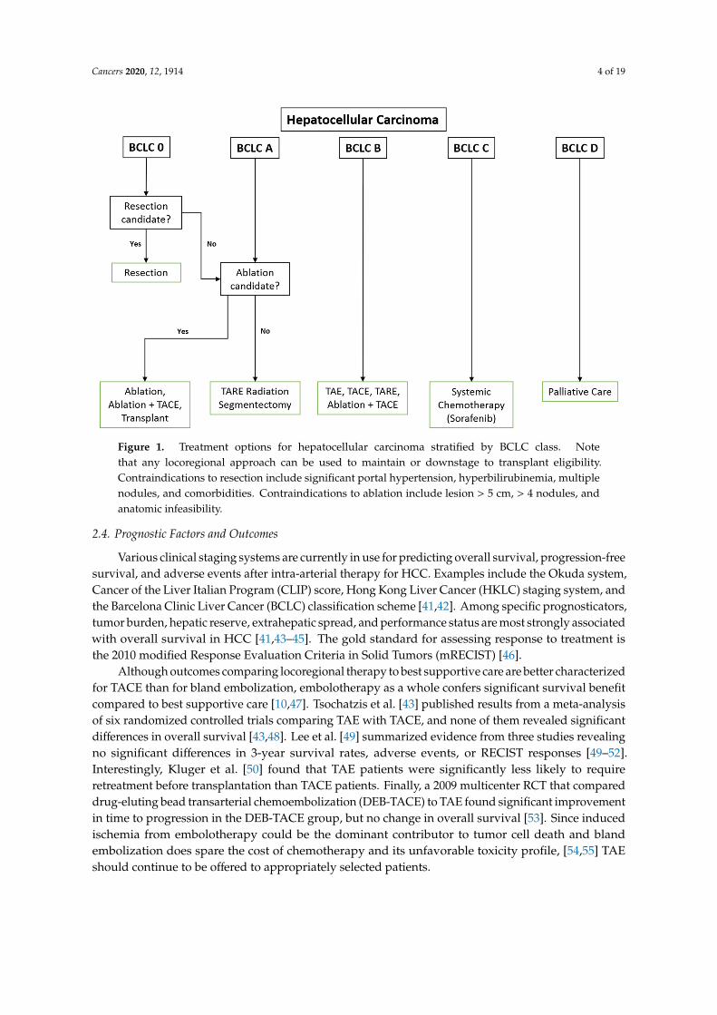
Generally, TAE is reserved for non-surgical candidates with liver-dominant disease. Patientselection for all locoregional therapies including TAE involves clinical and serologic evaluation ofthe patient including functional status, liver function tests, and clinical indices such as the ALBI(Albumin-Bilirubin), CP (Child-Pugh), MELD (Model for End-stage Liver Disease), and ECOG(Eastern Cooperative Oncology Group) performance status scores for patient stratification andassessment [34–36]. In addition to its role in the diagnosis of HCC, preprocedural imaging is paramountfor evaluation of the vascular anatomy, access site patency, and ensuring patency of the portal vein [37].Studies have demonstrated that patients in class B of the Barcelona Clinic Liver Cancer stagingclassification system (BCLC) derive the most benefit from this procedure followed by BCLC classC [10,38]. Patients in BCLC class A may undergo TAE to maintain eligibility for transplantation perthe Milan and UCSF criteria [38,39]. The contraindications for TAE include decompensated cirrhosis(Child-Pugh B8 or higher), significantly reduced portal venous flow, creatinine clearance < 30 mL/min,high tumor burden, severe comorbidities, untreated esophageal varices, and elevated liver functionmarkers [40]. Figure 1 shows possible treatment strategies stratified by BCLC class.
Cancers 2020, 12, 1914 4 of 19
Cancers 2020, 12, x 4 of 18
Figure 1. Treatment options for hepatocellular carcinoma stratified by BCLC class. Note that any
locoregional approach can be used to maintain or downstage to transplant eligibility.
Contraindications to resection include significant portal hypertension, hyperbilirubinemia, multiple
nodules, and comorbidities. Contraindications to ablation include lesion > 5 cm, > 4 nodules, and
anatomic infeasibility.
2.4. Prognostic Factors and Outcomes
Various clinical staging systems are currently in use for predicting overall survival, progression-
free survival, and adverse events after intra-arterial therapy for HCC. Examples include the Okuda
system, Cancer of the Liver Italian Program (CLIP) score, Hong Kong Liver Cancer (HKLC) staging
system, and the Barcelona Clinic Liver Cancer (BCLC) classification scheme [41,42]. Among specific
prognosticators, tumor burden, hepatic reserve, extrahepatic spread, and performance status are
most strongly associated with overall survival in HCC [41,43–45]. The gold standard for assessing
response to treatment is the 2010 modified Response Evaluation Criteria in Solid Tumors (mRECIST)
[46].
Although outcomes comparing locoregional therapy to best supportive care are better
characterized for TACE than for bland embolization, embolotherapy as a whole confers significant
survival benefit compared to best supportive care [10,47]. Tsochatzis et al. [43] published results from
a meta-analysis of six randomized controlled trials comparing TAE with TACE, and none of them
revealed significant differences in overall survival [43,48]. Lee et al. [49] summarized evidence from
three studies revealing no significant differences in 3-year survival rates, adverse events, or RECIST
responses [49–52]. Interestingly, Kluger et al. [50] found that TAE patients were significantly less
likely to require retreatment before transplantation than TACE patients. Finally, a 2009 multicenter
RCT that compared drug-eluting bead transarterial chemoembolization (DEB-TACE) to TAE found
significant improvement in time to progression in the DEB-TACE group, but no change in overall
survival [53]. Since induced ischemia from embolotherapy could be the dominant contributor to
tumor cell death and bland embolization does spare the cost of chemotherapy and its unfavorable
toxicity profile, [54,55] TAE should continue to be offered to appropriately selected patients.
Figure 1. Treatment options for hepatocellular carcinoma stratified by BCLC class. Notethat any locoregional approach can be used to maintain or downstage to transplant eligibility.Contraindications to resection include significant portal hypertension, hyperbilirubinemia, multiplenodules, and comorbidities. Contraindications to ablation include lesion > 5 cm, > 4 nodules, andanatomic infeasibility.
2.4. Prognostic Factors and Outcomes
Various clinical staging systems are currently in use for predicting overall survival, progression-freesurvival, and adverse events after intra-arterial therapy for HCC. Examples include the Okuda system,Cancer of the Liver Italian Program (CLIP) score, Hong Kong Liver Cancer (HKLC) staging system, andthe Barcelona Clinic Liver Cancer (BCLC) classification scheme [41,42]. Among specific prognosticators,tumor burden, hepatic reserve, extrahepatic spread, and performance status are most strongly associatedwith overall survival in HCC [41,43–45]. The gold standard for assessing response to treatment isthe 2010 modified Response Evaluation Criteria in Solid Tumors (mRECIST) [46].
Although outcomes comparing locoregional therapy to best supportive care are better characterizedfor TACE than for bland embolization, embolotherapy as a whole confers significant survival benefitcompared to best supportive care [10,47]. Tsochatzis et al. [43] published results from a meta-analysisof six randomized controlled trials comparing TAE with TACE, and none of them revealed significantdifferences in overall survival [43,48]. Lee et al. [49] summarized evidence from three studies revealingno significant differences in 3-year survival rates, adverse events, or RECIST responses [49–52].Interestingly, Kluger et al. [50] found that TAE patients were significantly less likely to requireretreatment before transplantation than TACE patients. Finally, a 2009 multicenter RCT that compareddrug-eluting bead transarterial chemoembolization (DEB-TACE) to TAE found significant improvementin time to progression in the DEB-TACE group, but no change in overall survival [53]. Since inducedischemia from embolotherapy could be the dominant contributor to tumor cell death and blandembolization does spare the cost of chemotherapy and its unfavorable toxicity profile, [54,55] TAEshould continue to be offered to appropriately selected patients.
Cancers 2020, 12, 1914 5 of 19
3. Transarterial Chemoembolization
3.1. Procedure and Periprocedural Management
Similar to bland embolization, transarterial chemoembolization (TACE) involves occlusion oftumor feeding vessels. In contrast to TAE, TACE permits the delivery of targeted chemotherapywith the embolic therapy. In the conventional approach (c-TACE), a lipiodolized chemotherapeuticagent is administered into a feeding artery followed by administration of an embolic agent. Thistheoretically allows for (1) increased pharmacologic concentration and (2) increased effect duration dueto decreased washout [7,10]. However, there is considerable variation in technique and drug mixtureacross institutions, and pharmacokinetic analysis revealed that plasma concentration after c-TACE mayapproximate systemic chemotherapy drug levels [56]. A newer approach using drug-eluting beads(DEB-TACE) provides better standardization and arguably less hepatotoxicity [57,58]. In DEB-TACE,drug-infused microspheres release chemotherapy in a sustained fashion and serve an embolic roleare injected. Doxorubicin is the most widely cited chemotherapeutic agent, but others use a solutionadding mitomycin C and cisplatin in c-TACE [59].
Periprocedural evaluation and management is identical to that for TAE (see Section 2.2.).Antimicrobial prophylaxis is recommended as is periprocedural clinical stabilization and laboratorymonitoring. PES is common after TACE occurring in up to 80% of patients [60]. Pharmacologicaltreatments include intra-arterial lidocaine, steroids, 5-HT3R antagonists, and antibiotics. Antibioticsseem to be of little clinical utility in managing fever [61], but intra-arterial lidocaine and/ordexamethasone have improved analgesic requirements and hospital length of stay [62,63].
3.2. Patient Selection
The most appropriate candidate for TACE is one with intermediate-stage HCC (BCLC class B,Child-Pugh B or better) without portal vein thrombosis or extrahepatic spread who is ineligible forsurgical resection or transplantation [10,64,65]. Numerous studies confirm that TACE can significantlyimpact survival if patients are selected on the aforementioned factors. For example, Burrel et al. [66]demonstrated a median survival up to 47.7 months after TACE for BCLC B patients with preserved liverfunction (no higher than Child-Pugh B7), no vascular invasion, extrahepatic spread, nor significantfunctional impairment [7,66].
BCLC A (early-stage) or BCLC 0 (very early-stage) patients with a solitary nodule and minimal tono underlying liver disease should undergo surgical resection which boasts a favorable prognosis [7].However, TACE may be indicated in these patients especially if they are ineligible for surgery/ablationor require a “bridge” to maintain transplant eligibility per the Milan/UCSF criteria [38]. TACE can alsobe combined with unilateral portal vein embolization (PVE) to induce hypertrophy of the contralateralfuture liver remnant before hepatectomy of the diseased liver [67]. In patients who have portalvein invasion, chemoembolization plus radiotherapy may be beneficial if hepatic compensation isadequate [68,69]. For advanced-stage patients (BCLC C), TACE may still be useful in combination withthe systemic drug sorafenib, but definitive evidence is lacking [70,71]. Thus, TACE provides a versatiletool in the arsenal of treatment options for HCC patients.
Absolute contraindications for TACE include decompensated cirrhosis (Child-Pugh B8 orhigher), severely reduced portal vein flow, creatinine clearance <30 mL/min, extensive bilobartumor involvement, and technical infeasibility [40]. Relative contraindications include high tumorburden, severe comorbidities, untreated esophageal varices, and elevated liver function markers [64].Generally, lobar and selective/segmental TACE may still be performed with a total bilirubin levelup to 3 and 4 mg/dL, respectively [72]. In Child-Pugh class C patients, the American Associationfor the Study of Liver Disease (AASLD) guidelines recommend against TACE if serum bilirubin isabove 3 mg/dL or there is main portal vein thrombosis unless segmental treatment is possible [73,74].However, Luo et al. noted significant survival improvement in patients with either segmental-branchor first-border branch portal vein thrombosis [75].
Cancers 2020, 12, 1914 6 of 19
3.3. Prognostic Factors and Outcomes
As mentioned previously, there are several prognostic models for predicting survival in HCC.The Child-Pugh score may be the most accurate for patients treated with TACE [76]. To determineprognosis for patients undergoing retreatment with TACE (and to decide whether there would beadditional benefit after two TACE treatments), the Assessment for Retreatment with TACE (ART)scoring system was developed [8,77].
While the outcomes for both c-TACE and DEB-TACE are more favorable than best supportivecare or other conservative management in appropriately selected patients [59,78–83], the superiorityof c-TACE versus DEB-TACE remains somewhat controversial. At least 12 studies have investigatedsuperiority between the two techniques, but a significant difference in overall survival remainsunconfirmed [6,84]. However, the well-known PRECISION V study did demonstrate significantincrease in tumor response, reduction in severe hepatotoxicity, and lower doxorubicin-related adverseevents in the DEB-TACE group compared with c-TACE for certain patient populations (Child-Pugh B,ECOG 1, bilobar disease, recurrent disease) [81].
The idea of combining locoregional therapy with systemic chemotherapy has been explored.Sorafenib, the first-line treatment for advanced stage (BCLC class C) HCC patients as establishedby the SHARP trial [85], is both an inhibitor of the growth and proliferation Raf pathway in tumorcells as well as an inhibitor of the angiogenic VEGFR/PDGFR pathway in endothelial cells [86].The compensatory angiogenesis from TACE-induced hypoxia could theoretically be attenuated fromthe antiangiogenic functions of sorafenib. Unfortunately, studies like the SPACE trial that randomizedpatients into DEB-TACE with sorafenib or DEB-TACE with placebo have not shown significantimprovements in time to progression [70].
4. Transarterial Radioembolization
4.1. Procedure and Periprocedural Management
Selective internal radiotherapy (SIRT) for HCC can be performed with transarterialradioembolization (TARE) [10]. This procedure primarily provides its therapeutic effect via radiationinstead of embolization [87]. Currently, a radioisotope of yttrium, 90Y, is either loaded onto orembedded within microspheres that are injected into a hepatic artery branch feeding tumor cells [6].90Y undergoes beta decay and irradiates surrounding tumor, ultimately damaging repair mechanismsand facilitating cell death [88].
Preprocedural angiographic mapping and evaluation are usually conducted 1–2 weeks prior sothat variant anatomy and intrahepatic portosystemic shunts can be identified. Technetium-99m labeledmacroaggregated albumin (99mTc-MAA) is used with single-photon emission computed tomography(SPECT) to determine the hepatopulmonary fraction that, if high, may increase the likelihood ofradiation pneumonitis after TARE [10,88]. Some patients with advanced hepatobiliary malignancies areprescribed gemcitabine. This chemotherapeutic agent should be held for at least 4 weeks prior to TAREand 2–4 weeks afterwards due to its radiosensitizing effects which increase the risk of radiation-inducedliver disease (RILD) [89–91]. The actual TARE procedure is performed in similar fashion to otherlocoregional endovascular approaches with targeting of the tumoral disease in a lobar or segmentalfashion [92,93]. Treatment effect is observed slightly later than with TACE or TAE, so follow-up imagingand labs usually take place 12 weeks after TARE [94].
4.2. Patient Selection
The indications and contraindications for TARE are generally similar to those for the otherembolotherapies. A total bilirubin up to 2 mg/dL is acceptable while encephalopathy and prior radiationto the liver are not [93]. A notable contraindication to TARE is significant (>20%) hepatopulmonary orhepato-enteric shunting as unintended radiation to the lungs or gastrointestinal tract may be serious [6].However, TARE offers a unique application in patients with portal vein thrombosis given the reduced
Cancers 2020, 12, 1914 7 of 19
embolic effect [94,95]. Several series have demonstrated the safety of 90Y-SIRT in cases where tumorinfiltrated either a main or lobar portal vein branch [96,97]. Although the BCLC guidelines recommendchemoembolization as first-line therapy for class B patients, expert recommendations from AASLD andNCCN do not posit radioembolization’s inferiority in the list of suitable treatments for unresectableintermediate-stage HCC patients [98,99]. For BCLC 0 and A patients, radiation segmentectomy withintraarterial 90Y-SIRT is safe and effective [100,101]. Neoadjuvant radiation lobectomy is also a safeand effective option to increase the function of the contralateral future liver remnant in patients whoplan to undergo resection and avoids the alternative risks of portal vein embolization [102,103]. Finally,just like the other embolotherapies, TARE can be used to maintain or encourage transplant/resectioneligibility through bridging as well as enhance overall survival in BCLC C patients [104,105].
4.3. Prognostic Factors and Outcomes
Prognosis after TARE is most associated with baseline patient stage (BCLC, Child-Pugh),performance status (ECOG), tumor burden, and extrahepatic disease [10]. According to a 2016meta-analysis by Lobo et al. [106], overall survival and complication rates for TARE are similar tothose of TACE, but the prospective trial PREMIERE demonstrated longer time to progression (TTP)for TARE [107]. Another randomized trial showed higher quality of life scores for TARE patients vs.TACE [108]. In a prospective study, Salem et al. reported excellent outcomes including an overallsurvival of 47.3 months for Child-Pugh A patients and 27 months in Child-Pugh B patients [109]. Withmore contemporary approaches such as radiation segmentectomy, response rates, tumor control, andsurvival outcomes have been comparable to curative-intent treatments (e.g., resection, transplantation,ablation) at 5 years [109,110].
When compared to sorafenib among advanced-stage patients, Hilgard et al. [111] actuallydemonstrated a survival benefit for TARE (10.7 vs. 16.4 months, respectively). Two randomizedcontrolled trials revealed higher tumor response rates and fewer adverse events with TARE vs.sorafenib for unresectable, treatment-naïve Child-Pugh A patients, although overall survival wassimilar between the two [112,113]. Considering the side effects of systemic sorafenib therapy, TAREmay be an attractive option for these patients [113].
5. Ablation
5.1. Procedure and Periprocedural Management
Ablative techniques for HCC include radiofrequency ablation (RFA), microwave ablation (MWA),cryoablation (CA), irreversible electroporation (IRE), laser-induced interstitial thermotherapy (LITT),and high-intensity focused ultrasound (HIFU). The choice of technique is often based on individualand institutional expertise, but historically the most commonly utilized technology was RFA. In recentyears, MWA has gained traction as an alternative modality [114]. CA has also been used regularly inthe past, although its use has diminished due to serious complications including cryogenic shock, acuterenal failure secondary to myohemoglobinuria, coagulopathy, and cardiac dysrhythmias [115,116].
In RFA, the tumor is heated to high temperatures via frictional heat in water molecules producedby an electrode, which can be with or without hooks, to increase and maximize heat productionin the tissue [117]. The ablation zone consists of the original space occupied by the tumor plusa 5-10 mm boundary of ablated adjacent liver parenchyma [118]. Over time, fibrosis causes retractionof the necrotic tissue. A homogenous, non-enhancing, well-circumscribed area is consistent withsuccessful radiographic response [118,119]. Conversely, MWA uses an electrode to deliver thermalenergy-induced cellular destruction. MWA may be suitable for larger tumors than RFA is indicated forand highly perfused regions [120]. Moreover, MWA can target multiple tumor sites simultaneously,yields shorter time to threshold temperature, achieves larger ablation treatment zones, results inbetter delineated ablation zone borders, and is less prone to heat-sink effects from adjacent vascularstructures [114,120,121]. While RFA yields smaller ablation zones, less uniform borders, and is more
Cancers 2020, 12, 1914 8 of 19
prone to heat-sink, it does offer the advantage of avoiding energy delivery to tracking structures suchas bile ducts or large vessels [122].
All ablative modalities utilize imaging guidance/monitoring during the procedures such ascomputed tomography (CT) or ultrasound (US), or a combination. New modalities such ascontrast-enhanced US (CEUS) have also been utilized as well as novel fused imaging technologies topredict treatment zones [123,124]. Follow-up imaging with CT or MRI every 3–6 months for the firsttwo years with serum alpha fetoprotein (AFP) monitoring is recommended by the NCCN [99]. Seriouscomplications include injury to adjacent organs e.g., diaphragm, gastrointestinal tract, gallbladder [125].Similar to PES, a postablation syndrome (PAS) may occur with analogous clinical presentation andself-limiting nature [126].
5.2. Patient Selection
Surgical resection or orthotopic liver transplantation is the mainstay of treatment for very earlyand early-stage (BCLC 0, A) HCC patients [127]. However, most patients are disqualified fromsurgical intervention due to significant comorbidities, portal hypertension, poor hepatic function,cardiovascular comorbidities, inability to tolerate general anesthesia, or lesion location [5,128]. Assuch, ablation offers a potentially curative option for these patients with early HCC with somestudies demonstrating equivalent survival outcomes to resection even with poorer baseline liverfunction [129–131]. Ablation with curative intent is an effective alternative to resection, particularlyfor tumors smaller than 3 cm [114]. For tumors 3–5 cm in diameter, the combination of TACE andablation demonstrate good outcomes albeit not curative [132–135]. Caution must be exercised whenlesions are close to major vessels, biliary structures, diaphragm, and other intra-abdominal organs.Hydrodissection, or artificial ascites, wherein 5% dextrose in water (D5W) fluid is injected betweenthe tumor area and adjacent extrahepatic organ to prevent transmission of thermal energy, helps toseparate the tumor and protects against unintentional organ injury [122,136,137]. RFA and MWA canalso be considered in advanced stage patients (BCLC C) for downstaging as a bridge to transplantationand in intermediate stage patients (BCLC B) when combined with TACE [98].
5.3. Prognostic Factors and Outcomes
The independent predictors of survival after RFA/MWA from several multivariate analyses wereChild-Pugh class, tumor size, and tumor number [138–142]. The overall survival outcomes betweenRFA and resection are not significantly different at 1 and 3 years [143–145], but MWA may see lowerlocal tumor progression rates than RFA for tumors > 5 cm or > 3 HCC nodules [120]. Complicationrates are similar between MWA and RFA [146]. Although there is a relative paucity of high-impactstudies pertaining to cryoablation, an RCT by Wang et al. found higher 3-year survival rates (butsimilar 5-year survival rates) and lower local progression rates (for tumors > 3 cm) in the cryoablationgroup vs. RFA group [147].
Combination therapy of RFA with chemoembolization has been investigated and has shownimproved locoregional control compared to either RFA or TACE alone for BCLC A and B patients [148].RFA helps to decrease total cellular resistance so that chemotherapy in TACE can yield relativelyhigher concentrations proximal to the tumor vascular bed at the periphery of the ablated tissue [149].If TACE is conducted first, peripherally situated tumor cells are preferentially destroyed so that laterRFA treatment, typically performed within 4 weeks, undergoes less vascular heat sinking and yieldsmore complete central necrosis particularly for lesions > 3 cm [150]. The optimum parameters for bothmechanisms require further exploration. MWA plus TACE has also shown to be effective for lesionsbetween 3–5 cm [133–135]. It should be noted that adjuvant sorafenib therapy following ablation orresection is not effective per results from the STORM trial [151].
Cancers 2020, 12, 1914 9 of 19
6. Future Directions
New developments in drug-eluting bead technology have allowed the loading of tyrosine kinaseinhibitors (e.g., sunitinib, vandetanib) and anti-VEGF antibodies (e.g., bevacizumab) in preclinicalstages with promising results in halting tumor growth [152–155]. Experiments in immunotherapysuch as oncolytic viruses, dendritic cells, and immune checkpoint inhibitors (against CTLA-4, PD-1,PD-L1) are also underway [156,157]. In fact, nivolumab, a PD-1 inhibitor (already FDA approved forsorafenib-refractory HCC) is being directly compared to sorafenib for advanced stage HCC patients [158,159]. Several combination strategies of innate and adaptive immunotherapies with RFA, MWA, andcryoablation are also be investigated in vitro and in animal models of HCC [160]. Lastly, personalizedtherapies and prognosticators are being appraised in human subjects research. Several putativehistological, epigenetic, and metabolomic biomarkers are being studied to individualize treatments forHCC patients [161]. For example, micro-RNA-122 (miRNA-122) is a tumor suppressor molecule that isoften severely reduced in hepatocytes linked to hepatocellular oncogenesis. Therapies that involvereintroduction of miRNA-122 to stabilize cell cycle regulation are being scrutinized for effectivenessand safety [162,163]. Advances in artificial intelligence (AI) are also being applied to HCC management.In addition to improved intra-procedural imaging guidance, AI has been used to construct predictionmodels for response to locoregional treatment [164,165]. While the role of interventional-basedliver-directed techniques continues to expand, additional research is needed regarding the applicationof these therapies in a neoadjuvant or adjuvant setting to improve the multidisciplinary care of HCCand reduce recurrence rates [166].
7. Conclusions
HCC is the most common primary liver malignancy and the third leading cause of cancer-relatedmortality worldwide [1]. Although overall survival for this complex disease has improved in recentyears, prognosis is still poor particularly for advanced and terminal-stage patients [1]. Surgicalextirpation and transplantation remain the curative standard of care for early-stage patients, but thereis an expanding role of locoregional therapies in the management of HCC including curative-intent,disease control, bridging to transplant and resection, downstaging patients, and palliation. Withthe addition of targeted chemotherapy and radiotherapy delivery, the inventory of transarterial hepaticembolization techniques offers major benefit in appropriately selected candidates. Ablative proceduresusing high frequency alternating currents or microwaves have also developed as excellent therapiesfor nonsurgical patients, achieving curative results in early-stage patients. Although advanced-stagepatients are currently limited to systemic therapy, novel advances in immunotherapy and personalizedbiomolecular signatures of HCC are paving the way for more robust strategies to tackle this disease.
Author Contributions: All authors have contributed to the writing of this review article, and have read andagreed to the published version of the manuscript.
Funding: This research received no external funding.
Conflicts of Interest: The authors declare no conflict of interest.
References
1. Kim, H.S.; El-serag, H.B. The epidemiology of hepatocellular Carcinoma in the USA. Curr. Gastroenterol. Rep.2019, 21, 17. [CrossRef] [PubMed]
2. Howlader, N.; Noone, A.M.; Krapcho, M.; Miller, D.; Brest, A.; Yu, M.; Ruhl, J.; Tatalovich, Z.; Mariotto, A.;Lewis, D.R.; et al. (Eds.) EEER Cancer Statistics Review, 1975–2016; National Cancer Institute: Bethesda, MD,USA, 2018. Available online: https://seer.cancer.gov/csr/1975_2016/ (accessed on 1 July 2020).
3. Onaca, N.; Davis, G.L.; Jennings, L.W.; Goldstein, R.M.; Klintmalm, G.B. Improved results of transplantationfor hepatocellular carcinoma: A report from the International registry of hepatic tumors in livertransplantation. Liver Transpl. 2009, 15, 574–580. [CrossRef] [PubMed]

Cancers 2020, 12, 1914 10 of 19
4. Byam, J.; Renz, J.; Millis, J.M. Liver transplantation for hepatocellular carcinoma. Hepatobiliary Surg. Nutr.2013, 2, 22–30. [PubMed]
5. Roayaie, S.; Jibara, G.; Tabrizian, P.; Park, J.; Yang, J.; Yan, L.; Schwartz, M.; Han, G.; Izzo, F.; Chen, M.;et al. The role of hepatic resection in the treatment of hepatocellular cancer. Hepatology 2015, 62, 440–451.[CrossRef]
6. Inchingolo, R.; Posa, A.; Mariappan, M.; Spiliopoulos, S. Locoregional treatments for hepatocellular carcinoma:Current evidence and future directions. World J. Gastroenterol. 2019, 25, 4614–4628. [CrossRef]
7. Kis, B.; El-Haddad, G.; Sheth, R.A.; Parikh, N.S.; Ganguli, S.; Shyn, P.B.; Choi, J.; Brown, K.T. Liver-directedtherapies for Hepatocellular Carcinoma and Intrahepatic Cholangiocarcinoma. Cancer Control 2017, 24.[CrossRef]
8. Mokdad, A.A.; Singal, A.G.; Yopp, A.C. Advances in local and systemic therapies for Hepatocellular cancer.Curr. Oncol. Rep. 2016, 18, 9. [CrossRef]
9. Breedis, C.; Young, G. The blood supply of neoplasms in the liver. Am. J. Pathol. 1954, 30, 969–977.10. Gbolahan, O.B.; Schacht, M.A.; Beckley, E.W.; Laroche, T.P.; O’neil, B.H.; Pyko, M. Locoregional and systemic
therapy for hepatocellular carcinoma. J. Gastrointest. Oncol. 2017, 8, 215–228. [CrossRef]11. Brown, K.T.; Nevins, A.B.; Getrajdman, G.I.; Brody, L.A.; Kurtz, R.C.; Fong, Y.; Blumgart, L.H. Particle
embolization for hepatocellular Carcinoma. J. Vasc. Interv. Radiol. 1998, 9, 822–828. [CrossRef]12. Vaidya, S.; Tozer, K.R.; Chen, J. An overview of embolic agents. Semin. Intervent. Radiol. 2008, 25, 204–215.
[CrossRef] [PubMed]13. Rand, T.; Loewe, C.; Schoder, M.; Schmook, M.T.; Peck-Radosavljevic, M.; Kettenbach, J.; Wolf, F.; Schneider, B.;
Lammer, J. Arterial embolization of unresectable hepatocellular carcinoma with use of microspheres, lipiodol,and cyanoacrylate. Cardiovasc. Intervent. Radiol. 2005, 28, 313–318. [CrossRef] [PubMed]
14. Gaba, R.C.; Lokken, R.P.; Hickey, R.M.; Lipnik, A.J.; Lewandowski, R.J.; Salem, R.; Brown, D.B.; Walker, T.G.;Silberzweig, J.E.; Baerlocher, M.O.; et al. Quality improvement guidelines for transarterial chemoembolizationand embolization of hepatic malignancy. J. Vasc. Interv. Radiol. 2017, 28, 1210–1223.e3. [CrossRef]
15. Zheng, N.; Wei, X.; Zhang, D.; Chai, W.; Che, M.; Wang, J.; Du, B. Hepatic resection or transarterialchemoembolization for hepatocellular carcinoma with portal vein tumor thrombus. Medicine 2016, 95, e3959.[CrossRef] [PubMed]
16. Coldwell, D.M.; Loper, K.A. Regional anesthesia for hepatic arterial embolization. Radiology 1989, 172,1039–1040. [CrossRef] [PubMed]
17. Makary, M.S.; Kapke, J.; Yildiz, V.; Pan, X.; Dowell, J.D. Conventional versus drug-eluting bead transarterialchemoembolization for neuroendocrine tumor liver metastases. J. Vasc. Interv. Radiol. 2016, 27, 1298–1304.[CrossRef]
18. Wigmore, S.J.; Redhead, D.N.; Thomson, B.N.J.; Currie, E.J.; Parks, R.W.; Madhavan, K.K.; Garden, O.J.Postchemoembolisation syndrome—Tumour necrosis or hepatocyte injury? Br. J. Cancer 2003, 89, 1423–1427.[CrossRef]
19. Paye, F.; Farges, O.; Dahmane, M.; Vilgrain, V.; Flejou, J.F.; Belghiti, J. Cytolysis following chemoembolizationfor hepatocellular carcinoma. Br. J. Surg. 1999, 86, 176–180. [CrossRef]
20. Chan, A.O.; Yuen, M.F.; Hui, C.K.; Tso, W.K.; Lai, C.L. A prospective study regarding the complications oftranscatheter intraarterial lipiodol chemoembolization in patients with hepatocellular carcinoma. Cancer2002, 94, 1747–1752. [CrossRef]
21. Garwood, E.R.; Fidelman, N.; Hoch, S.E.; Kerlan, R.K.; Yao, F.Y. Morbidity and mortality followingtransarterial liver chemoembolization in patients with hepatocellular carcinoma and synthetic hepaticdysfunction. Liver Transpl. 2013, 19, 164–173. [CrossRef]
22. Wu, G.C.; Perng, W.C.; Chen, C.W.; Chian, C.F.; Peng, C.K.; Su, W.L. Acute respiratory distress syndromeafter transcatheter arterial chemoembolization of hepatocellular carcinomas. Am. J. Med. Sci. 2009, 338,357–360. [CrossRef] [PubMed]
23. Shah, R.P.; Brown, K.T. Hepatic arterial embolization complicated by acute cholecystitis. Semin. Intervent.Radiol. 2011, 28, 252–257. [CrossRef] [PubMed]
24. Cornelis, F.H.; Borgheresi, A.; Petre, E.N.; Santos, E.; Solomon, S.B.; Brown, K. Hepatic arterial embolizationusing cone beam CT with tumor feeding vessel detection software: Impact on hepatocellular Carcinomaresponse. Cardiovasc. Intervent. Radiol. 2018, 41, 104–111. [CrossRef] [PubMed]
Cancers 2020, 12, 1914 11 of 19
25. Watchmaker, J.M.; Lipnik, A.J.; Fritsche, M.R.; Baker, J.C.; Mouli, S.K.; Geevarghese, S.; Banovac, F.;Omary, R.A.; Brown, D.B. Are prophylactic antibiotics necessary prior to transarterial chemoembolizationfor hepatocellular carcinoma in patients with native biliary anatomy? J. Surg. Oncol. 2018, 117, 1312–1317.[CrossRef]
26. Song, S.-Y.; Chung, J.W.; Han, J.K.; Lim, H.G.; Koh, Y.H.; Park, J.H.; Lee, H.-S.; Kim, C.Y. Liver abscessafter transcatheter oily chemoembolization for hepatic tumors: Incidence, predisposing factors, and clinicaloutcome. J. Vasc. Interv. Radiol. 2001, 12, 313–320. [CrossRef]
27. Khan, W.; Sullivan, K.L.; McCann, J.W.; Gonsalves, C.F.; Sato, T.; Eschelman, D.J.; Brown, D.B. Moxifloxacinprophylaxis for chemoembolization or embolization in patients with previous biliary interventions: A pilotstudy. AJR Am. J. Roentgenol. 2011, 197, W343–W345. [CrossRef]
28. Patel, S.; Tuite, C.M.; Mondschein, J.I.; Soulen, M.C. Effectiveness of an aggressive antibiotic regimen forchemoembolization in patients with previous biliary intervention. J. Vasc. Interv. Radiol. 2006, 17, 1931–1934.[CrossRef]
29. Castells, A.; Bruix, J.; Ayuso, C.; Bru, C.; Montayà, X.; Boix, L.; Rodés, J. Transarterial embolization forhepatocellular carcinoma. Antibiotic prophylaxis and clinical meaning of postembolization fever. J. Hepatol.1995, 22, 410–415. [CrossRef]
30. Brown, D.B.; Geschwind, J.F.; Soulen, M.C.; Millward, S.F.; Sacks, D. Society of Interventional Radiologyposition statement on chemoembolization of hepatic malignancies. J. Vasc. Interv. Radiol. 2006, 17, 217–223.[CrossRef]
31. Pudusseri, A.; Spyropoulos, A.C. Management of anticoagulants in the periprocedural period for patientswith cancer. J. Natl. Compr. Cancer Netw. 2014, 12, 1713–1720. [CrossRef]
32. Patel, I.J.; Davidson, J.C.; Nikolic, B.; Salazar, G.M.M.; Schwartzberg, M.S.; Walker, T.G.; Saad, W.A. Consensusguidelines for periprocedural management of coagulation status and hemostasis risk in percutaneousimage-guided interventions. J. Vasc. Interv. Radiol. 2012, 23, 727–736. [CrossRef]
33. Haste, P.; Johnson, M.S. Transarterial Chemoembolization. In IR Playbook; Keefe, N., Haskal, Z., Park, A.,Angle, J., Eds.; Springer: Cham, Switzerland, 2018.
34. Johnson, P.J.; Berhane, S.; Kagebayashi, C.; Satomura, S.; Teng, M.; Reeves, H.L.; O’Beirne, J.; Fox, R.;Skowronska, A.; Palmer, D.; et al. Assessment of liver function in patients with hepatocellular carcinoma:A new evidence-based approach-the ALBI grade. J. Clin. Oncol. 2015, 33, 550–558. [CrossRef] [PubMed]
35. Levy, I.; Sherman, M. Staging of hepatocellular Carcinoma: Assessment of the CLIP, Okuda, and Child-Pughstaging systems in a cohort of 257 patients in Toronto. Gut 2002, 50, 881–885. [CrossRef] [PubMed]
36. Guerrini, G.P.; Pinelli, D.; Marini, E.; Corno, V.; Guizzetti, M.; Zambelli, M.; Aluffi, A.; Lincini, L.; Fagiuoli, S.;Lucianetti, A.; et al. Value of HCC-MELD Score in patients with Hepatocellular Carcinoma undergoing livertransplantation. Prog. Transpl. 2018, 28, 63–69. [CrossRef] [PubMed]
37. Sieghart, W.; Hucke, F.; Peck-Radosavljevic, M. Transarterial chemoembolization: Modalities, indication, andpatient selection. J. Hepatol. 2015, 62, 1187–1195. [CrossRef]
38. Kishore, S.; Friedman, T.; Madoff, D.C. Update on embolization therapies for hepatocellular Carcinoma. Curr.Oncol. Rep. 2017, 19, 40. [CrossRef]
39. Hodavance, M.S.; Vikingstad, E.M.; Griffin, A.; Pabon-Ramos, W.M.; Berg, C.L.; Suhocki, P.V.; Kim, C.Y.Effectiveness of transarterial embolization of Hepatocellular Carcinoma as a bridge to transplantation. J.Vasc. Interv. Radiol. 2016, 27, 39–45. [CrossRef]
40. Raoul, J.-L.; Sangro, B.; Forner, A.; Mazzaferro, V.; Piscaglia, F.; Bolondi, L.; Lencioni, R. Evolving strategiesfor the management of intermediate-stage hepatocellular carcinoma: Available evidence and expert opinionon the use of transarterial chemoembolization. Cancer Treat. Rev. 2011, 37, 212–220. [CrossRef]
41. Grieco, A.; Pompili, M.; Caminiti, G.; Miele, L.; Covino, M.; Alfei, B.; Rapaccini, G.L.; Gasbarrini, G. Prognosticfactors for survival in patients with early-intermediate hepatocellular carcinoma undergoing non-surgicaltherapy: Comparison of Okuda, CLIP, and BCLC staging systems in a single Italian centre. Gut 2005, 54,411–418. [CrossRef]
42. Yau, T.; Tang, V.Y.; Yao, T.J.; Fan, S.T.; Lo, C.M.; Poon, R.T. Development of Hong Kong liver cancer stagingsystem with treatment stratification for patients with hepatocellular carcinoma. Gastroenterology 2014, 146,1691–1700.e3. [CrossRef]
Cancers 2020, 12, 1914 12 of 19
43. Tsochatzis, E.A.; Fatourou, E.; O’beirne, J.; Meyer, T.; Burroughs, A.K. Transarterial chemoembolization andbland embolization for hepatocellular carcinoma. World J. Gastroenterol. 2014, 20, 3069–3077. [CrossRef][PubMed]
44. Llovet, J.M.; Brú, C.; Bruix, J. Prognosis of hepatocellular carcinoma: The BCLC staging classification. SeminLiver Dis 1999, 19, 329–338. [CrossRef] [PubMed]
45. Ni, J.-Y.; Kong, J.; Sun, H.-L.; Chen, Y.-T.; Luo, J.-H.; Wang, W.-D.; Chen, N.; Jiang, X.-Y.; Xu, L. Prognosticfactors for survival after transarterial chemoembolization combined with sorafenib in the treatment of BCLCStage B and C hepatocellular Carcinomas. Acad. Radiol. 2018, 25, 423–429. [CrossRef] [PubMed]
46. Kim, M.N.; Kim, B.K.; Han, K.H.; Kim, S.U. Evolution from WHO to EASL and mRECIST for hepatocellularcarcinoma: Considerations for tumor response assessment. Expert Rev. Gastroenterol. Hepatol. 2015, 9,335–348. [CrossRef]
47. Llovet, J.M.; Real, M.I.; Montaña, X.; Planas, R.; Coll, S.; Aponte, J.; Ayuso, C.; Sala, M.; Muchart, J.; Sola, R.;et al. Arterial embolisation or chemoembolisation versus symptomatic treatment in patients with unresectablehepatocellular carcinoma: A randomised controlled trial. Lancet 2002, 359, 1734–1739. [CrossRef]
48. Meyer, T.; Kirkwood, A.; Roughton, M.; Beare, S.; Tsochatzis, E.A.; Yu, D.; Davies, N.; Williams, E.; Pereira, S.P.;Hochhauser, D.; et al. A randomised phase II/III trial of 3-weekly cisplatin-based sequential transarterialchemoembolisation vs. embolisation alone for hepatocellular carcinoma. Br. J. Cancer 2013, 108, 1252–1259.[CrossRef]
49. Lee, E.W.; Khan, S. Recent advances in transarterial embolotherapies in the treatment of hepatocellularcarcinoma. Clin. Mol. Hepatol. 2017, 23, 265–272. [CrossRef]
50. Kluger, M.D.; Halazun, K.J.; Barroso, R.T.; Fox, A.N.; Olsen, S.K.; Madoff, D.C.; Siegel, A.B.; Weintraub, J.L.;Sussman, J.; Jr, R.S.B.; et al. Bland embolization versus chemoembolization of hepatocellular carcinomabefore transplantation. Liver Transpl. 2014, 20, 536–543. [CrossRef]
51. Massarweh, N.N.; Davila, J.A.; El-Serag, H.B.; Duan, Z.; Temple, S.; May, S.B.; Sada, Y.H.; Anaya, D.A.Transarterial bland versus chemoembolization for hepatocellular carcinoma: Rethinking a gold standard. J.Surg. Res. 2016, 200, 552–559. [CrossRef]
52. Brown, K.T.; Do, R.K.; Gonen, M.; Covey, A.M.; Getrajdman, G.I.; Sofocleous, C.T.; Jarnagin, W.R.;D’Angelica, M.I.; Allen, P.J.; Erinjeri, J.P.; et al. Randomized trial of hepatic artery embolization forhepatocellular Carcinoma using doxorubicin-eluting microspheres compared with embolization withmicrospheres alone. J. Clin. Oncol. 2016, 34, 2046–2053. [CrossRef]
53. Malagari, K.; Pomoni, M.; Kelekis, A.; Pomoni, A.; Dourakis, S.; Spyridopoulos, T.; Moschouris, H.;Emmanouil, E.; Rizos, S.; Kelekis, D. Prospective randomized comparison of chemoembolization withdoxorubicin-eluting beads and bland embolization with BeadBlock for hepatocellular carcinoma. Cardiovasc.Intervent. Radiol. 2010, 33, 541–551. [CrossRef]
54. Chuang, V.P.; Wallace, S. Hepatic artery embolization in the treatment of hepatic neoplasms. Radiology 1981,140, 51–58. [CrossRef] [PubMed]
55. Tsochatzis, E.A.; Fatourou, E.M.; Triantos, C.K.; Burroughs, A.K. Transarterial therapies for hepatocellularcarcinoma. Recent Results Cancer Res. 2013, 190, 195–206. [PubMed]
56. Varela, M.; Real, M.I.; Burrel, M.; Forner, A.; Sala, M.; Brunet, M.; Ayuso, C.; Castells, L.; Montaña, X.;Llovet, J.M.; et al. Chemoembolization of hepatocellular carcinoma with drug eluting beads: Efficacy anddoxorubicin pharmacokinetics. J. Hepatol. 2007, 46, 474–481. [CrossRef] [PubMed]
57. Melchiorre, F.; Patella, F.; Pescatori, L.C.; Pesapane, F.; Fumarola, E.; Biondetti, P.; Brambillasca, P.; Monaco, C.;Carrafiello, G.; Franceschelli, G.; et al. DEB-TACE: A standard review. Future Oncol. 2018, 14, 2969–2984.[CrossRef] [PubMed]
58. Vogl, T.J.; Lammer, J.; Lencioni, R.; Malagari, K.; Watkinson, A.; Pilleul, F.; Denys, A.; Lee, C. Liver,gastrointestinal, and cardiac toxicity in intermediate hepatocellular carcinoma treated with PRECISIONTACE with drug-eluting beads: Results from the PRECISION V randomized trial. AJR Am. J. Roentgenol.2011, 197, W562–W570. [CrossRef]
59. Solomon, B.; Soulen, M.C.; Baum, R.A.; Haskal, Z.J.; Shlansky-goldberg, R.D.; Cope, C. Chemoembolizationof hepatocellular carcinoma with cisplatin, doxorubicin, mitomycin-C, ethiodol, and polyvinyl alcohol:Prospective evaluation of response and survival in a U.S. population. J. Vasc. Interv. Radiol. 1999, 10, 793–798.[CrossRef]

Cancers 2020, 12, 1914 13 of 19
60. Blackburn, H.; West, S. Management of postembolization syndrome following hepatic transarterialchemoembolization for primary or metastatic liver cancer. Cancer Nurs. 2016, 39, E1–E18. [CrossRef]
61. Jun, C.H.; Ki, H.S.; Lee, H.K.; Park, K.J.; Park, S.Y.; Cho, S.B.; Park, C.H.; Joo, Y.E.; Kim, H.S.; Choi, S.K.; et al.Clinical significance and risk factors of postembolization fever in patients with hepatocellular carcinoma.World J Gastroenterol. 2013, 19, 284–289. [CrossRef]
62. Hartnell, G.G.; Gates, J.; Stuart, K.; Underhill, J.; Brophy, D.P. Hepatic chemoembolization: Effect ofintraarterial lidocaine on pain and postprocedure recovery. Cardiovasc Intervent. Radiol. 1999, 22, 293–297.[CrossRef]
63. Molgaard, C.P.; Teitelbaum, G.P.; Pentecost, M.J.; Finck, E.J.; Davis, S.H.; Dziubinski, J.E.; Daniels, J.R.Intraarterial administration of lidocaine for analgesia in hepatic chemoembolization. J. Vasc. Interv. Radiol.1990, 1, 81–85. [CrossRef]
64. Benson, A.B.; Abrams, T.A.; Ben-Josef, E.; Bloomston, P.M.; Botha, J.F.; Clary, B.M.; Covey, A.; Curley, S.A.;D’Angelica, M.I.; Davila, R.; et al. Hepatobiliary Cancers. J. Natl. Compr. Cancer Netw. 2009, 7, 350–391.[CrossRef] [PubMed]
65. Villanueva, A.; Hernandez-gea, V.; Llovet, J.M. Medical therapies for hepatocellular carcinoma: A criticalview of the evidence. Nat. Rev. Gastroenterol. Hepatol. 2013, 10, 34–42. [CrossRef] [PubMed]
66. Burrel, M.; Reig, M.; Forner, A.; Barrufet, M.; Rodriguez-Lope, C.; Tremosini, S.; Ayuso, C.; Llovet, J.M.;Real, M.I.; Bruix, J. Survival of patients with hepatocellular carcinoma treated by transarterialchemoembolisation (TACE) using drug eluting beads. Implications for clinical practice and trial design. J.Hepatol. 2012, 56, 1330–1335. [CrossRef] [PubMed]
67. May, B.J.; Madoff, D.C. Portal vein embolization: Rationale, technique, and current application. SeminIntervent. Radiol. 2012, 29, 81–89. [CrossRef]
68. Tazawa, J.; Maeda, M.; Sakai, Y.; Yamane, M.; Ohbayashi, H.; Kakinuma, S.; Miyasaka, Y.; Nagayama, K.;Enomoto, N.; Sato, C. Radiation therapy in combination with transcatheter arterial chemoembolization forhepatocellular carcinoma with extensive portal vein involvement. J. Gastroenterol. Hepatol. 2001, 16, 660–665.[CrossRef]
69. Yoon, S.M.; Lim, Y.-S.; Won, H.J.; Kim, J.H.; Kim, K.M.; Lee, H.C.; Chung, Y.; Lee, Y.S.; Lee, S.G.; Park, J.-H.;et al. Radiotherapy plus transarterial chemoembolization for hepatocellular carcinoma invading the portalvein: Long-term patient outcomes. Int. J. Radiat. Oncol. Biol. Phys. 2012, 82, 2004–2011. [CrossRef]
70. Lencioni, R.; Llovet, J.M.; Han, G.; Tak, W.Y.; Yang, J.; Guglielmi, A.; Paik, S.W.; Reig, M.; Kim, Y.; Chau, G.-Y.;et al. Sorafenib or placebo plus TACE with doxorubicin-eluting beads for intermediate stage HCC: The SPACEtrial. J. Hepatol. 2016, 64, 1090–1098. [CrossRef]
71. Hu, H.; Duan, Z.; Long, X.; Hertzanu, Y.; Shi, H.; Liu, S.; Yang, Z. Sorafenib combined with transarterialchemoembolization versus transarterial chemoembolization alone for advanced-stage hepatocellularcarcinoma: A propensity score matching study. PLoS ONE 2014, 9, e96620. [CrossRef]
72. Golfieri, R.; Cappelli, A.; Cucchetti, A.; Piscaglia, F.; Carpenzano, M.; Peri, E.; Ravaioli, M.;D’Errico-Grigioni, A.; Pinna, A.D.; Bolondi, L. Efficacy of selective transarterial chemoembolization ininducing tumor necrosis in small (<5 cm) hepatocellular carcinomas. Hepatology 2011, 53, 1580–1589.[CrossRef]
73. Dorn, D.P.; Bryant, M.K.; Zarzour, J.; Smith, J.K.; Redden, D.T.; Saddekni, S.; Aal, A.K.A.; Gray, S.; White, J.;Eckhoff, D.E.; et al. Chemoembolization outcomes for hepatocellular carcinoma in cirrhotic patients withcompromised liver function. HPB 2014, 16, 648–655. [CrossRef] [PubMed]
74. Marrero, J.A.; Kulik, L.M.; Sirlin, C.B.; Zhu, A.X.; Finn, R.S.; Abecassis, M.; Roberts, L.R.; Heimbach, J.K.Diagnosis, Staging, and Management of Hepatocellular Carcinoma: 2018 Practice Guidance by the AmericanAssociation for the Study of Liver Diseases. Hepatology 2018, 68, 723–750. [CrossRef]
75. Luo, J.; Guo, R.; Lai, E.C.H.; Zhang, Y.; Lau, W.Y.; Chen, M.-S.; Shi, M. Transarterial chemoembolization forunresectable hepatocellular carcinoma with portal vein tumor thrombosis: A prospective comparative study.Ann. Surg. Oncol. 2011, 18, 413–420. [CrossRef] [PubMed]
76. Georgiades, C.; Liapi, E.; Frangakis, C.; Park, J.-U.; Kim, H.W.; Hong, K.; Geschwind, J.-F. Prognostic accuracyof 12 liver staging systems in patients with unresectable hepatocellular carcinoma treated with transarterialchemoembolization. J. Vasc. Interv. Radiol. 2006, 17, 1619–1624. [CrossRef]
Cancers 2020, 12, 1914 14 of 19
77. Hucke, F.; Sieghart, W.; Pinter, M.; Graziadei, I.W.; Vogel, W.; Müller, C.; Heinzl, H.; Waneck, F.; Trauner, M.;Peck-Radosavljevic, M. The ART-strategy: Sequential assessment of the ART score predicts outcome ofpatients with hepatocellular carcinoma re-treated with TACE. J. Hepatol. 2014, 60, 118–126. [CrossRef]
78. Lo, C.; Ngan, H.; Tso, W.; Liu, C.-L.; Lam, C.; Poon, R.T.P.; Fan, S.T.; Wong, J. Randomized controlled trial oftransarterial lipiodol chemoembolization for unresectable hepatocellular carcinoma. Hepatology 2002, 35,1164–1171. [CrossRef]
79. Marelli, L.; Stigliano, R.; Triantos, C.; Senzolo, M.; Cholongitas, E.; Davies, N.; Tibballs, J.; Meyer, T.;Patch, D.W.; Burroughs, A.K. Transarterial therapy for hepatocellular carcinoma: Which technique is moreeffective? A systematic review of cohort and randomized studies. Cardiovasc. Intervent. Radiol. 2007, 30,6–25. [CrossRef] [PubMed]
80. Llovet, J.M.; Bruix, J. Systematic review of randomized trials for unresectable hepatocellular carcinoma:Chemoembolization improves survival. Hepatology 2003, 37, 429–442. [CrossRef] [PubMed]
81. Lammer, J.; Malagari, K.; Vogl, T.; Pilleul, F.; Denys, A.; Watkinson, A.; Pitton, M.; Sergent, G.; Pfammatter, T.;Terraz, S.; et al. Prospective randomized study of doxorubicin-eluting-bead embolization in the treatment ofhepatocellular carcinoma: Results of the PRECISION V study. Cardiovasc. Intervent. Radiol. 2010, 33, 41–52.[CrossRef]
82. Golfieri, R.; Giampalma, E.; Renzulli, M.; Cioni, R.; Bargellini, I.; Bartolozzi, C.; Breatta, A.D.; Gandini, G.;Nani, R.; Gasparini, D.; et al. Randomised controlled trial of doxorubicin-eluting beads vs. conventionalchemoembolisation for hepatocellular carcinoma. Br. J. Cancer 2014, 111, 255–264. [CrossRef]
83. Facciorusso, A.; Mariani, L.; Sposito, C.; Spreafico, C.; Bongini, M.; Morosi, C.; Cascella, T.; Marchianò, A.;Camerini, T.; Bhoori, S.; et al. Drug-eluting beads versus conventional chemoembolization for the treatmentof unresectable hepatocellular carcinoma. J. Gastroenterol. Hepatol. 2016, 31, 645–653. [CrossRef] [PubMed]
84. Facciorusso, A.; Licinio, R.; Muscatiello, N.; Di leo, A.; Barone, M. Transarterial chemoembolization: Evidencesfrom the literature and applications in hepatocellular carcinoma patients. World J. Hepatol. 2015, 7, 2009–2019.[CrossRef] [PubMed]
85. Llovet, J.M.; Ricci, S.; Mazzaferro, V.M.; Hilgard, P.; Gane, E.; Blanc, J.-F.; De Oliveira, A.C.; Santoro, A.;Raoul, J.-L.; Forner, A.; et al. Sorafenib in advanced hepatocellular carcinoma. N. Engl. J. Med. 2008, 359,378–390. [CrossRef] [PubMed]
86. Cervello, M.; Bachvarov, D.; Lampiasi, N.; Cusimano, A.; Azzolina, A.; McCubrey, J.A.; Montalto, G.Molecular mechanisms of sorafenib action in liver cancer cells. Cell Cycle 2012, 11, 2843–2855. [CrossRef][PubMed]
87. Sato, K.; Lewandowski, R.J.; Bui, J.T.; Omary, R.; Hunter, R.D.; Kulik, L.; Mulcahy, M.; Liu, D.; Chrisman, H.;Resnick, S.; et al. Treatment of unresectable primary and metastatic liver cancer with yttrium-90 microspheres(TheraSphere): Assessment of hepatic arterial embolization. Cardiovasc. Intervent. Radiol. 2006, 29, 522–529.[CrossRef]
88. Bhangoo, M.S.; Karnani, D.R.; Hein, P.N.; Giap, H.; Knowles, H.; Issa, C.; Steuterman, S.; Pockros, P.;Frenette, C. Radioembolization with Yttrium-90 microspheres for patients with unresectable hepatocellularcarcinoma. J. Gastrointest. Oncol. 2015, 6, 469–478.
89. Lischalk, J.W.; Repka, M.C.; Unger, K. Radiation therapy for hepatobiliary malignancies. J. Gastrointest.Oncol. 2017, 8, 279–292. [CrossRef]
90. Murphy, J.D.; Lucas, D.R.; Somnay, Y.R.; Hamstra, D.A.; Ray, M.E. Gemcitabine-mediated radiosensitizationof human soft tissue sarcoma. Transl. Oncol. 2008, 1, 50–56. [CrossRef]
91. Kennedy, A.S.; Brown, D.B.; Feilchenfeldt, J.; Marshall, J.; Wasan, H.; Fakih, M.; Gibbs, P.; Knuth, A.; Sangro, B.;Soulen, M.C.; et al. Safety of selective internal radiation therapy (SIRT) with yttrium-90 microspherescombined with systemic anticancer agents: Expert consensus. J. Gastrointest. Oncol. 2017, 8, 1079–1099.[CrossRef]
92. Hickey, R.; Lewandowski, R.J.; Salem, R. Transarterial Radioembolization (TARE). In IR Playbook; Keefe, N.,Haskal, Z., Park, A., Angle, J., Eds.; Springer: Cham, Switzerland, 2018.
93. Makary, M.S.; Krishner, L.S.; Wuthrick, E.J.; Bloomston, M.P.; Dowell, J.D. Yttrium-90 microsphere selectiveinternal radiation therapy for liver metastases following systemic chemotherapy and surgical resection formetastatic adrenocortical carcinoma. World J. Clin. Oncol. 2018, 9, 20–25. [CrossRef]
Cancers 2020, 12, 1914 15 of 19
94. Salem, R.; Lewandowski, R.J.; Mulcahy, M.F.; Riaz, A.; Ryu, R.K.; Ibrahim, S.; Atassi, B.; Baker, T.;Gates, V.; Miller, F.H.; et al. Radioembolization for hepatocellular carcinoma using Yttrium-90 microspheres:A comprehensive report of long-term outcomes. Gastroenterology 2010, 138, 52–64. [CrossRef] [PubMed]
95. Kokabi, N.; Camacho, J.; Xing, M.; El-Rayes, B.F.; Spivey, J.R.; Knechtle, S.; Kim, H.S. Open-label prospectivestudy of the safety and efficacy of glass-based yttrium 90 radioembolization for infiltrative hepatocellularcarcinoma with portal vein thrombosis. Cancer 2015, 121, 2164–2174. [CrossRef] [PubMed]
96. Jia, Z.; Jiang, G.; Tian, F.; Zhu, C.; Qin, X. A systematic review on the safety and effectiveness of yttrium-90radioembolization for hepatocellular carcinoma with portal vein tumor thrombosis. Saudi J. Gastroenterol.2016, 22, 353–359. [PubMed]
97. Cho, Y.Y.; Lee, M.; Kim, H.-C.; Chung, J.W.; Kim, Y.H.; Gwak, G.-Y.; Bae, S.H.; Kim, Y.; Heo, J.; Kim, Y.J.Radioembolization Is a Safe and Effective Treatment for Hepatocellular Carcinoma with Portal VeinThrombosis: A Propensity Score Analysis. PLoS ONE 2016, 11, e0154986. [CrossRef]
98. Heimbach, J.K.; Kulik, L.M.; Finn, R.S.; Sirlin, C.B.; Abecassis, M.M.; Roberts, L.R.; Zhu, A.X.; Murad, M.H.;Marrero, J.A. AASLD guidelines for the treatment of hepatocellular carcinoma. Hepatology 2018, 67, 358–380.[CrossRef]
99. Benson, A.B.; D’Angelica, M.I.; Abbott, D.E.; Abrams, T.A.; Alberts, S.R.; Anaya, D.A.; Anders, R.; Are, C.;Brown, D.; Chang, D.T.; et al. Guidelines insights: Hepatobiliary cancers, version 2.2019. J. Natl. Compr.Cancer Netw. 2019, 17, 302–310. [CrossRef]
100. Riaz, A.; Gates, V.; Atassi, B.; Lewandowski, R.J.; Mulcahy, M.F.; Ryu, R.K.; Sato, K.T.; Baker, T.;Kulik, L.; Gupta, R.; et al. Radiation segmentectomy: A novel approach to increase safety and efficacy ofradioembolization. Int. J. Radiat. Oncol. Biol. Phys. 2011, 79, 163–171. [CrossRef]
101. Vouche, M.; Habib, A.; Ward, T.J.; Kim, E.; Kulik, L.; Ganger, D.; Mulcahy, M.; Baker, T.; Abecassis, M.;Sato, K.T.; et al. Unresectable solitary hepatocellular carcinoma not amenable to radiofrequency ablation:Multicenter radiology-pathology correlation and survival of radiation segmentectomy. Hepatology 2014, 60,192–201. [CrossRef]
102. Gabr, A.; Polineni, P.; Mouli, S.K.; Riaz, A.; Lewandowski, R.J.; Salem, R. Neoadjuvant radiation lobectomy asan alternative to portal vein embolization in hepatocellular Carcinoma. Semin. Nucl. Med. 2019, 49, 197–203.[CrossRef]
103. Gabr, A.; Abouchaleh, N.; Ali, R.; Baker, T.; Caicedo, J.; Katariya, N.; Abecassis, M.; Riaz, A.; Lewandowski, R.J.;Salem, R. Outcomes of Surgical Resection after Radioembolization for Hepatocellular Carcinoma. J. Vasc.Interv. Radiol. 2018, 29, 1502–1510.e1. [CrossRef]
104. Kulik, L.; Atassi, B.; Van Holsbeeck, L.; Souman, T.; Lewandowski, R.J.; Mulcahy, M.F.; Hunter, R.D.;Nemcek, A.A.; Abecassis, M.; Haines, K.G.; et al. Yttrium-90 microspheres (TheraSphere) treatment ofunresectable hepatocellular carcinoma: Downstaging to resection, RFA and bridge to transplantation. J. Surg.Oncol. 2006, 94, 572–586. [CrossRef]
105. De La Torre, M.A.; Buades-Mateu, J.; De La Rosa, P.A.; Lue, A.; Bustamante, F.J.; Serrano, M.T.; Testillano, M.;Lorente, S.; Arenas, J.I.; Gil, C.; et al. A comparison of survival in patients with hepatocellular carcinoma andportal vein invasion treated by radioembolization or sorafenib. Liver Int. 2016, 36, 1206–1212. [CrossRef][PubMed]
106. Lobo, L.; Yakoub, D.; Picado, O.; Ripat, C.; Pendola, F.; Sharma, R.; Eltawil, R.; Kwon, D.; Venkat, S.;Portelance, L.; et al. Unresectable Hepatocellular Carcinoma: Radioembolization Versus Chemoembolization:A Systematic Review and Meta-analysis. Cardiovasc. Intervent. Radiol. 2016, 39, 1580–1588. [CrossRef]
107. Salem, R.; Gordon, A.C.; Mouli, S.; Hickey, R.; Kallini, J.; Gabr, A.; Mulcahy, M.F.; Baker, T.; Abecassis, M.;Miller, F.H.; et al. Y90 Radioembolization Significantly Prolongs Time to Progression Compared WithChemoembolization in Patients With Hepatocellular Carcinoma. Gastroenterology 2016, 151, 1155–1163.e2.[CrossRef] [PubMed]
108. Salem, R.; Gilbertsen, M.; Butt, Z.; Memon, K.; Vouche, M.; Hickey, R.; Baker, T.; Abecassis, M.;Atassi, R.; Riaz, A.; et al. Increased quality of life among hepatocellular carcinoma patients treated withradioembolization, compared with chemoembolization. Clin. Gastroenterol. Hepatol. 2013, 11, 1358–1365.e1.[CrossRef] [PubMed]
109. Salem, R.; Gabr, A.; Riaz, A.; Mora, R.; Ali, R.; Abecassis, M.; Hickey, R.; Kulik, L.; Ganger, D.; Flamm, S.;et al. Institutional decision to adopt Y90 as primary treatment for hepatocellular carcinoma informed bya 1,000-patient 15-year experience. Hepatology 2018, 68, 1429–1440. [CrossRef] [PubMed]
Cancers 2020, 12, 1914 16 of 19
110. Lewandowski, R.J.; Gabr, A.; Abouchaleh, N.; Ali, R.; Al Asadi, A.; Mora, R.; Kulik, L.; Ganger, D.; Desai, K.;Thornburg, B.; et al. Radiation segmentectomy: Potential curative therapy for early hepatocellular Carcinoma.Radiology 2018, 287, 1050–1058. [CrossRef]
111. Hilgard, P.; Hamami, M.; El Fouly, A.; Scherag, A.; Müller, S.; Ertle, J.; Heusner, T.; Cicinnati, V.R.; Paul, A.;Bockisch, A.; et al. Radioembolization with yttrium-90 glass microspheres in hepatocellular carcinoma:European experience on safety and long-term survival. Hepatology 2010, 52, 1741–1749. [CrossRef] [PubMed]
112. Chow, P.K.; Gandhi, M.; Tan, S.-B.; Khin, M.W.; Khasbazar, A.; Ong, J.; Choo, S.P.; Cheow, P.C.; Chotipanich, C.;Lim, K.; et al. SIRveNIB: Selective Internal Radiation Therapy Versus Sorafenib in Asia-Pacific Patients WithHepatocellular Carcinoma. J. Clin. Oncol. 2018, 36, 1913–1921. [CrossRef]
113. Vilgrain, V.; Pereira, H.; Assenat, E.; Guiu, B.; Ilonca, A.D.; Pageaux, G.-P.; Sibert, A.; Bouattour, M.;Lebtahi, R.; Allaham, W.; et al. Efficacy and safety of selective internal radiotherapy with yttrium-90resin microspheres compared with sorafenib in locally advanced and inoperable hepatocellular carcinoma(SARAH): An open-label randomised controlled phase 3 trial. Lancet Oncol. 2017, 18, 1624–1636. [CrossRef]
114. Poulou, L.S.; Botsa, E.; Thanou, I.; Ziakas, P.D.; Thanos, L. Percutaneous microwave ablation vs.radiofrequency ablation in the treatment of hepatocellular carcinoma. World J. Hepatol. 2015, 7, 1054–1063.[CrossRef] [PubMed]
115. Huang, Y.Z.; Zhou, S.C.; Zhou, H.; Tong, M. Radiofrequency ablation versus cryosurgery ablation forhepatocellular carcinoma: A meta-analysis. Hepatogastroenterology 2013, 60, 1131–1135. [PubMed]
116. Seifert, J.K.; Junginger, T.; Morris, D.L. A collective review of the world literature on hepatic cryotherapy. J.R. Coll. Surg. Edinb. 1998, 43, 141–154. [PubMed]
117. Mcgahan, J.P.; Brock, J.M.; Tesluk, H.; Gu, W.Z.; Schneider, P.; Browning, P.D. Hepatic ablation with use ofradio-frequency electrocautery in the animal model. J. Vasc. Interv. Radiol. 1992, 3, 291–297. [CrossRef]
118. Sainani, N.I.; Gervais, D.A.; Mueller, P.R.; Arellano, R.S. Imaging after percutaneous radiofrequency ablationof hepatic tumors: Part 1, Normal findings. AJR Am. J. Roentgenol. 2013, 200, 184–193. [CrossRef] [PubMed]
119. Sainani, N.I.; Gervais, D.A.; Mueller, P.R.; Arellano, R.S. Imaging after percutaneous radiofrequency ablationof hepatic tumors: Part 2, Abnormal findings. AJR Am. J. Roentgenol. 2013, 200, 194–204. [CrossRef]
120. Facciorusso, A.; Di maso, M.; Muscatiello, N. Microwave ablation versus radiofrequency ablation forthe treatment of hepatocellular carcinoma: A systematic review and meta-analysis. Int. J. Hyperthermia 2016,32, 339–344. [CrossRef]
121. Yi, Y.; Zhang, Y.; Wei, Q.; Zhao, L.; Han, J.; Song, Y.; Ding, Y.; Lu, G.; Liu, J.; Ding, H.; et al. Radiofrequencyablation or microwave ablation combined with transcatheter arterial chemoembolization in treatment ofhepatocellular carcinoma by comparing with radiofrequency ablation alone. Chin. J. Cancer Res. 2014, 26,112–118.
122. Yang, W.; Yan, K.; Wu, G.-X.; Wu, W.; Fu, Y.; Lee, J.-C.; Zhang, Z.-Y.; Wang, S.; Chen, M.-H. Radiofrequencyablation of hepatocellular carcinoma in difficult locations: Strategies and long-term outcomes. World J.Gastroenterol. 2015, 21, 1554–1566. [CrossRef]
123. Song, K.D.; Lee, M.W.; Rhim, H.; Cha, D.I.; Chong, Y.; Lim, H.K. Fusion imaging-guided radiofrequencyablation for hepatocellular carcinomas not visible on conventional ultrasound. AJR Am. J. Roentgenol. 2013,201, 1141–1147. [CrossRef]
124. Lee, M.W.; Rhim, H.; Cha, D.I.; Kim, Y.J.; Choi, N.; Kim, Y.-S.; Lim, H.K. Percutaneous radiofrequency ablationof hepatocellular carcinoma: Fusion imaging guidance for management of lesions with poor conspicuity atconventional sonography. AJR Am. J. Roentgenol. 2012, 198, 1438–1444. [CrossRef] [PubMed]
125. Kasugai, H.; Osaki, Y.; Oka, H.; Kudo, M.; Seki, T. Severe complications of radiofrequency ablation therapyfor hepatocellular carcinoma: An analysis of 3,891 ablations in 2,614 patients. Oncology 2007, 72 (Suppl. 1),72–75. [CrossRef] [PubMed]
126. Dodd, G.D.; Napier, D.; Schoolfield, J.D.; Hubbard, L. Percutaneous radiofrequency ablation of hepatictumors: Postablation syndrome. AJR Am. J. Roentgenol. 2005, 185, 51–57. [CrossRef]
127. Balogh, J.; Victor, D.; Asham, E.H.; Burroughs, S.G.; Boktour, M.; Saharia, A.; Li, X.; Ghobrial, R.M.;Monsour, H.P. Hepatocellular carcinoma: A review. J. Hepatocell. Carcinoma 2016, 3, 41–53. [CrossRef]
128. Bruix, J.; Sherman, M. Management of hepatocellular carcinoma: An update. Hepatology 2011, 53, 1020–1022.[CrossRef] [PubMed]
Cancers 2020, 12, 1914 17 of 19
129. Fang, Y.; Chen, W.; Liang, X.; Li, D.; Lou, H.; Chen, R.; Wang, K.; Pan, H. Comparison of long-termeffectiveness and complications of radiofrequency ablation with hepatectomy for small hepatocellularcarcinoma. J. Gastroenterol. Hepatol. 2014, 29, 193–200. [CrossRef]
130. Lü, M.-D.; Kuang, M.; Liang, L.-J.; Xie, X.-Y.; Peng, B.-G.; Liu, G.-J.; Li, D.-M.; Lai, J.-M.; Li, S.-Q. Surgicalresection versus percutaneous thermal ablation for early-stage hepatocellular carcinoma: A randomizedclinical trial. Zhonghua Yi Xue Za Zhi 2006, 86, 801–805.
131. Huang, J.; Yan, L.; Cheng, Z.; Wu, H.; Du, L.; Wang, J.; Xu, Y.; Zeng, Y. A randomized trial comparingradiofrequency ablation and surgical resection for HCC conforming to the Milan criteria. Ann. Surg. 2010,252, 903–912. [CrossRef]
132. Xu, Z.; Xie, H.; Zhou, L.; Chen, X.; Zheng, S. The Combination strategy of transarterial Chemoembolizationand radiofrequency ablation or microwave ablation against hepatocellular Carcinoma. Anal Cell Pathol. 2019,2019. [CrossRef]
133. Sheta, E.; El-Kalla, F.; El-Gharib, M.; Kobtan, A.; Elhendawy, M.E.; Abd-Elsalam, S.; Mansour, L.; Amer, I.Comparison of single-session transarterial chemoembolization combined with microwave ablation orradiofrequency ablation in the treatment of hepatocellular carcinoma: A randomized-controlled study. Eur.J. Gastroenterol. Hepatol. 2016, 28, 1198–1203. [CrossRef]
134. Ginsburg, M.; Zivin, S.P.; Wroblewski, K.; Doshi, T.; Vasnani, R.J.; Van ha, T.G. Comparison ofcombination therapies in the management of hepatocellular carcinoma: Transarterial chemoembolizationwith radiofrequency ablation versus microwave ablation. J. Vasc. Interv. Radiol. 2015, 26, 330–341. [CrossRef][PubMed]
135. Ni, J.-Y.; Sun, H.-L.; Chen, Y.-T.; Luo, J.-H.; Chen, N.; Jiang, X.-Y.; Xu, L.-F. Prognostic factors for survival aftertransarterial chemoembolization combined with microwave ablation for hepatocellular carcinoma. World J.Gastroenterol. 2014, 20, 17483–17490. [CrossRef]
136. Kondo, Y.; Yoshida, H.; Shiina, S.; Tateishi, R.; Teratani, T.; Omata, M. Artificial ascites technique forpercutaneous radiofrequency ablation of liver cancer adjacent to the gastrointestinal tract. Br. J. Surg. 2006,93, 1277–1282. [CrossRef]
137. Ton, J.; Kuoy, E.; Abi-Jaoudeh, N. Liver Ablation. In IR Playbook; Keefe, N., Haskal, Z., Park, A., Angle, J.,Eds.; Springer: Cham, Switzerland, 2018.
138. Yan, K.; Chen, M.-H.; Yang, W.; Bin Wang, Y.; Gao, W.; Hao, C.Y.; Xing, B.C.; Huang, X.F. Radiofrequencyablation of hepatocellular carcinoma: Long-term outcome and prognostic factors. Eur. J. Radiol. 2008, 67,336–347. [CrossRef] [PubMed]
139. Shiina, S.; Tateishi, R.; Arano, T.; Uchino, K.; Enooku, K.; Nakagawa, H.; Asaoka, Y.; Sato, T.; Masuzaki, R.;Kondo, Y.; et al. Radiofrequency ablation for hepatocellular carcinoma: 10-year outcome and prognosticfactors. Am. J. Gastroenterol. 2012, 107, 569–577. [CrossRef] [PubMed]
140. Brunello, F.; Cantamessa, A.; Gaia, S.; Carucci, P.; Rolle, E.; Castiglione, A.; Ciccone, G.; Rizzetto, M.Radiofrequency ablation: Technical and clinical long-term outcomes for single hepatocellular carcinoma upto 30 mm. Eur. J. Gastroenterol. Hepatol. 2013, 25, 842–849. [CrossRef] [PubMed]
141. Francica, G.; Saviano, A.; De Sio, I.; De Matthaeis, N.; Brunello, F.; Cantamessa, A.; Giorgio, A.;Scognamiglio, U.; Fornari, F.; Giangregorio, F.; et al. Long-term effectiveness of radiofrequency ablation forsolitary small hepatocellular carcinoma: A retrospective analysis of 363 patients. Dig. Liver Dis. 2013, 45,336–341. [CrossRef]
142. Liang, P.; Dong, B.; Yu, X.; Yu, D.; Wang, Y.; Feng, L.; Xiao, Q. Prognostic factors for survival in patients withhepatocellular carcinoma after percutaneous microwave ablation. Radiology 2005, 235, 299–307. [CrossRef]
143. Weis, S.; Franke, A.; Mössner, J.; Jakobsen, J.C.; Schoppmeyer, K. Radiofrequency (thermal) ablation versusno intervention or other interventions for hepatocellular carcinoma. Cochrane Database Syst. Rev. 2013, 19,CD003046. [CrossRef]
144. Pompili, M.; Saviano, A.; De Matthaeis, N.; Cucchetti, A.; Ardito, F.; Federico, B.; Brunello, F.; Pinna, A.D.;Giorgio, A.; Giulini, S.M.; et al. Long-term effectiveness of resection and radiofrequency ablation for singlehepatocellular carcinoma≤3 cm. Results of a multicenter Italian survey. J. Hepatol. 2013, 59, 89–97. [CrossRef]
145. Ng, K.K.; Chok, K.S.-H.; Chan, A.C.Y.; Cheung, T.T.; Wong, T.C.L.; Fung, J.Y.Y.; Yuen, J.; Poon, R.T.P.; Fan, S.T.;Lo, C.M. Randomized clinical trial of hepatic resection versus radiofrequency ablation for early-stagehepatocellular carcinoma. Br. J. Surg. 2017, 104, 1775–1784. [CrossRef] [PubMed]

Cancers 2020, 12, 1914 18 of 19
146. Ding, J.; Jing, X.; Liu, J.; Wang, F.; Wang, Y.; Du, Z. Complications of thermal ablation of hepatic tumours:Comparison of radiofrequency and microwave ablative techniques. Clin. Radiol. 2013, 68, 608–615. [CrossRef][PubMed]
147. Wang, C.; Wang, H.; Yang, W.; Hu, K.; Xie, H.; Hu, K.-Q.; Bai, W.; Dong, Z.; Lu, Y.; Zeng, Z.; et al. Multicenterrandomized controlled trial of percutaneous cryoablation versus radiofrequency ablation in hepatocellularcarcinoma. Hepatology 2015, 61, 1579–1590. [CrossRef]
148. Kim, J.W.; Kim, J.-H.; Won, H.J.; Shin, Y.M.; Yoon, H.-K.; Sung, K.-B.; Kim, P.-N. Hepatocellular carcinomas2–3 cm in diameter: Transarterial chemoembolization plus radiofrequency ablation vs. radiofrequencyablation alone. Eur. J. Radiol. 2012, 81, e189–e193. [CrossRef] [PubMed]
149. Iezzi, R.; Pompili, M.; Posa, A.; Coppola, G.; Gasbarrini, A.; Bonomo, L. Combined locoregional treatment ofpatients with hepatocellular carcinoma: State of the art. World J. Gastroenterol. 2016, 22, 1935–1942. [CrossRef]
150. Lu, Z.; Wen, F.; Guo, Q.; Liang, H.; Mao, X.; Sun, H. Radiofrequency ablation plus chemoembolization versusradiofrequency ablation alone for hepatocellular carcinoma: A meta-analysis of randomized-controlled trials.Eur. J. Gastroenterol. Hepatol. 2013, 25, 187–194. [CrossRef]
151. Bruix, J.; Takayama, T.; Mazzaferro, V.M.; Chau, G.-Y.; Yang, J.; Kudo, M.; Cai, J.; Poon, R.T.; Han, K.-H.;Tak, W.Y.; et al. Adjuvant sorafenib for hepatocellular carcinoma after resection or ablation (STORM): Aphase 3, randomised, double-blind, placebo-controlled trial. Lancet Oncol. 2015, 16, 1344–1354. [CrossRef]
152. Fuchs, K.; Bize, P.-E.; Faes, O.D.S.; Denys, A.; Doelker, E.; Borchard, G.; Jordan, O. Drug-eluting beadsloaded with antiangiogenic agents for chemoembolization: In vitro sunitinib loading and release and in vivopharmacokinetics in an animal model. J. Vasc. Interv. Radiol. 2014, 25, 379–387.e3872. [CrossRef]
153. Bize, P.-E.; Duran, R.; Fuchs, K.; Faes, O.D.S.; Namur, J.; Decosterd, L.A.; Jordan, O.; Doelker, E.; Denys, A.Antitumoral Effect of Sunitinib-eluting beads in the rabbit VX2 tumor model. Radiology 2016, 280, 425–435.[CrossRef]
154. Hagan, A.; Phillips, G.J.; Macfarlane, W.M.; Lloyd, A.W.; Czuczman, P.; Lewis, A.L. Preparation andcharacterisation of vandetanib-eluting radiopaque beads for locoregional treatment of hepatic malignancies.Eur. J. Pharm. Sci. 2017, 101, 22–30. [CrossRef]
155. Sakr, O.S.; Berndt, S.; Carpentier, G.; Cuendet, M.; Jordan, O.; Borchard, G. Arming embolic beads withanti-VEGF antibodies and controlling their release using LbL technology. J. Control. Release 2016, 224, 199–207.[CrossRef] [PubMed]
156. Park, B.-H.; Hwang, T.; Liu, T.-C.; Sze, D.Y.; Kim, J.-S.; Kwon, H.-C.; Oh, S.Y.; Han, S.-Y.; Yoon, J.-H.;Hong, S.-H.; et al. Use of a targeted oncolytic poxvirus, JX-594, in patients with refractory primary ormetastatic liver cancer: A phase I trial. Lancet Oncol. 2008, 9, 533–542. [CrossRef]
157. Obeid, J.M.; Kunk, P.R.; Zaydfudim, V.M.; Bullock, T.N.; Slingluff, C.L.; Rahma, O.E. Immunotherapy forhepatocellular carcinoma patients: Is it ready for prime time? Cancer Immunol. Immunother. 2018, 67, 161–174.[CrossRef] [PubMed]
158. El-Khoueiry, A.B.; Sangro, B.; Yau, T.C.C.; Crocenzi, T.S.; Kudo, M.; Hsu, C.; Kim, T.-Y.; Choo, S.-P.; Trojan, J.;Welling, T.H.; et al. Nivolumab in patients with advanced hepatocellular carcinoma (CheckMate 040): Anopen-label, non-comparative, phase 1/2 dose escalation and expansion trial. Lancet 2017, 389, 2492–2502.[CrossRef]
159. Bteich, F.; Di bisceglie, A.M. Current and future systemic therapies for hepatocellular carcinoma. Gastroenterol.Hepatol. 2019, 15, 266–272.
160. Slovak, R.; Ludwig, J.M.; Gettinger, S.N.; Herbst, R.S.; Kim, H.S. Immuno-thermal ablations—Boostingthe anticancer immune response. J. Immunother. Cancer 2017, 5, 78. [CrossRef]
161. Burkhart, R.A.; Ronnekleiv-kelly, S.M.; Pawlik, T.M. Personalized therapy in hepatocellular carcinoma:Molecular markers of prognosis and therapeutic response. Surg. Oncol. 2017, 26, 138–145. [CrossRef]
162. Couri, T.; Pillai, A. Goals and targets for personalized therapy for HCC. Hepatol. Int. 2019, 13, 125–137.[CrossRef]
163. Ma, L.; Chua, M.S.; Andrisani, O.; So, S. Epigenetics in hepatocellular carcinoma: An update and futuretherapy perspectives. World J. Gastroenterol. 2014, 20, 333–345. [CrossRef]
164. Kim, H.S.; Chapiro, J.; Geschwind, J.-F.H. Interventional oncology: The fourth pillar of oncology. Cancer J.2016, 22, 363–364. [CrossRef]

Cancers 2020, 12, 1914 19 of 19
165. Abajian, A.; Murali, N.; Savic, L.J.; Laage-Gaupp, F.M.; Nezami, N.; Duncan, J.S.; Schlachter, T.; Lin, M.;Geschwind, J.-F.; Chapiro, J. Predicting treatment response to intra-arterial therapies for hepatocellularcarcinoma with the use of supervised machine learning-an artificial intelligence concept. J. Vasc. Interven.Radiol. 2018, 29, 850–857. [CrossRef] [PubMed]
166. Akateh, C.; Black, S.M.; Conteh, L.; Miller, E.D.; Noonan, A.; Elliott, E.; Pawlik, T.M.; Tsung, A.; Cloyd, J.M.Neoadjuvant and adjuvant treatment strategies for hepatocellular carcinoma. World J. Gastroenterol. 2019, 25,3704–3721. [CrossRef] [PubMed]
© 2020 by the authors. Licensee MDPI, Basel, Switzerland. This article is an open accessarticle distributed under the terms and conditions of the Creative Commons Attribution(CC BY) license (http://creativecommons.org/licenses/by/4.0/).