early morphological variation and induction of phenotypic plasticity in patagonian pejerrey
TRANSCRIPT
Neotropical Ichthyology, 10(2): 341-348, 2012Copyright © 2012 Sociedade Brasileira de Ictiologia
341
Early morphological variation and induction of phenotypic plasticity inPatagonian pejerrey
Sonia A. Crichigno, Miguel A. Battini and Víctor E. Cussac
The aim of this work was to study two aspects of phenotypic plasticity in the Patagonian pejerrey Odontesthes hatcheri(Teleostei: Atherinopsidae) the dependence of the early morphology on developmental time and temperature, and the inductionof morphological changes by controlled feeding in juveniles. Newly hatched free embryos, incubated at two different temperatures(13 and 18ºC), and juveniles were used for the study and induction of phenotypic plasticity. Body and head shapes were analyzedwith geometric morphometrics and linear measurements. Our results showed that shape variation at hatching was related to thebending of the embryo head on the yolk sac, increasing the head-trunk angle due to progressive straightening of the embryo. Thehead-trunk angle was related with temperature at incubation, with embryos incubated at higher temperature being more bent.Embryos that hatched earlier had bigger yolk sacs than those that hatched later. In juveniles, controlled feeding experimentsadded new morphological variation to that of wild juveniles. In all comparisons, the slenderness of the head, the size of premaxillaand jaw, and the position of the eye showed an enlarged variation due to controlled feeding. These results will contribute tocomprehending the complexity of the morphological variation of O. hatcheri.
O objetivo deste trabalho foi estudar a variação morfológica e plasticidade fenotípica do peixe-rei da Patagônia Odontestheshatcheri (Teleostei: Atherinopsidae), a dependência da morfologia inicial no tempo de desenvolvimento e temperatura, e aindução de alterações morfológicas pela alimentação controlada em juvenis. Embriões recém-nascidos, incubados a duastemperaturas diferentes (13 e 18ºC) e juvenis foram utilizados para o estudo de indução de plasticidade fenotípica. Formas docorpo e cabeça foram analisadas com técnicas de morfometria geométrica e medições lineares. Os nossos resultados mostraramque a variação da forma no nascimento foi relacionada com a curvatura da cabeça do embrião no saco vitelino, aumentando oângulo de cabeça-tronco devido ao endireitamento progressivo do embrião. O ângulo da cabeça-tronco relacionou-se com atemperatura de incubação, com os embriões incubados na temperatura elevada sendo mais curvados. Os embriões que eclodirammais cedo tinham sacos vitelinos maiores do que aqueles que eclodiram tardiamente. Em juvenis, os experimentos de alimentaçãocontrolada adicionaram nova variação morfológica àquela dos juvenis selvagens. Em todas as comparações, a espessura dacabeça, o tamanho da pré-maxila e mandíbula, e a posição do olho mostraram uma maior variação devido à alimentação controlada.Estes resultados irão contribuir para a compreensão da complexidade da variação morfológica de O. hatcheri.
Key words: Atherinopsidae, Development, Feeding, Morphometry, Odontesthes hatcheri.
Universidad Nacional del Comahue, Instituto de Investigaciones en Biodiversidad y Medioambiente (INIBIOMA), ConsejoNacional de Investigaciones Científicas y Técnicas. Quintral 1250, Bariloche, 8400 Río Negro, [email protected] (SAC)
Introduction
There is abundant evidence of phenotypic plasticity in fishes(Balon, 2004). It is considered as the ability of an organism toreact to environmental input with a change in form, state,movement, or rate of activity (West-Eberhard, 2003), or as theproperty of individual genotypes to produce differentphenotypes when exposed to different environmental conditions(Pigliucci, 2001; Pigliucci et al., 2006; Pfennig et al., 2010).
Induced phenotypic plasticity has been studied extensively(Grünbaum et al., 2007). During embryonic development, watertemperature is the most important environmental factor thatinfluences fish (Chambers & Leggett, 1987; Blaxter, 1992).
Temperature modulates the amount of time required to completeembryonic development, within a specific range for eachspecies (Kunz, 2004; Kamler, 2008), and has effects on themorphology, physiology, and behavior of fish duringdevelopment (Martell et al., 2005). Environmental effects otherthan temperature can also act along the ontogeny, inducing acertain morphology. For example, morphological reversion inthe head of Micropterus salmoides floridanus (= Micropterusfloridanus) could has been experimentally related to foodquality (Wintzer & Motta, 2005). Also, diet induced body andhead shape variation has been observed in Cichlasomamanaguense (= Parachromis managuensis) and Lepomishumilis (Meyer, 1987; Hegrenes, 2001).

Early morphological variation and induction of phenotypic plasticity in Patagonian pejerrey342
The distribution area of the Patagonian pejerrey Odontestheshatcheri (Eigenmann), Patagonia, was signed by old and newprocesses that shaped the landscape and fauna: a Gondwananheritage, the Andes uplifting, Pleistocene ice, volcanic activity,introduction of exotic fishes, and climate change (Pascual et al.,2007). Larvae and juveniles of O. hatcheri perform ontogenetichabitat and diet shifts in Patagonian lakes. After hatching in thelittoral zone, free embryos (sensu Balon, 1999) migrate to thelimnetic zone where the exogenous feeding begins. Later, theselarvae return to the littoral zone (Cussac et al., 1992). They feedmainly on both nauplii of Cyclopoida and the rotifer Pompholixsulcata in the limnetic and in the littoral zones, up to their juvenileperiod, when their diet changes (Cervellini et al., 1993). LarvalPatagonian pejerrey showed marked shape changes after fewdays of fasting (Battini et al., 1995). In adults, relationshipsbetween body shape and environmental factors such as totalphosphorus, coastline development, and altitude (Conte-Grand,2012), as well as between cephalic shape and mean depth,content of Chlorophyll-a, and mean summer air temperaturewere found (Crichigno, 2012).
The aim of this work was to study two aspects ofphenotypic plasticity in the Patagonian pejerrey Odontestheshatcheri; the dependence of the early morphology ondevelopmental time and temperature, and the induction ofmorphological changes by controlled feeding in juveniles.
Material and Methods
Morphology of newly hatched sibling free embryos. AdultOdontesthes hatcheri were captured using gillnets (15, 30, and40 mm bar mesh) in a shallow lake in the Patagonian plateau,Carrilafquen Chica (41º12’S 69º25’W, see Reissig et al., 2006for details). Individuals were anaesthetized with benzocainesolution (0.05 g . L-1). Ovocytes and sperm of four parentalcouples (PC) were obtained by stripping, and dry fertilization(Barnabé, 1990) was then performed. At the laboratory (Centrode Salmonicultura Bariloche, Universidad Nacional delComahue), the eggs corresponding to two PC were incubatedinto small baskets into a 200 L aquarium with aeration and adaily 20% water exchange, at 18ºC, and those corresponding tothe other two at 13ºC, always maintaining a 0.5% NaCl level.Both temperatures are included within the summer (breedingseason) range of surface water temperature in lakes andreservoirs where the species is present. Water supply camefrom Gutierrez River, 4 km downstream an oligotrophic lake(Gutierrez Lake, 41º09’59”S 71º24’35”W, Quirós, 1988).
When the eggs began to hatch, and during threeconsecutive days, newly hatched free embryos wereanaesthetized with benzocaine (0.05 g . L-1) and photographed(NIKON D70) under stereomicroscope (Leica Wild M3C). Twoimages of each individual were recorded, left side and cephalicdorsal view, taking care to minimize parallax error. In this way,newly hatched free embryos of different ages (AH = 1, 2, and 3,Table 1), in terms of days after fertilization (DAF), were obtained.
Ten landmarks were digitized on images, on the fish’s leftside: (1) anterior tip of the premaxilla, (2) anterior tip of dentary,
(3) anus, (4) dorsal profile of the body at the posterior end ofthe operculum, (5) angle of the first gill arch, between epihyaland ceratobranchial, (6, 7, 8, and 9) dorsal, ventral, anterior,and posterior edge of the eye, and (10) limit betweendiencephalon and telencephalon (Fig. 1).
Body shape was quantified using the GeometricMorphometric Analysis (GMA) approach of thin-plate splines(TPS; Bookstein, 1991; Rohlf & Marcus, 1993; Parsons et al.,2003; Adams et al., 2004). Images were first scaled and rotatedto a common size and orientation using a generalizedProcrustes superimposition approach (Bookstein, 1991;Adams et al., 2004). The mean (or consensus) body shape foreach population was estimated and quantified as partial warpscores using tpsRelw (Morphometrics at Sunny Stony Brook,2012). The description of shape variation was performedvisualizing the deformation grids and direction vectors, atthe extreme of each Relative Warp (RW) axis from left to rightand from bottom to top. Discriminant Analysis (DA, SPSSInc.) was performed employing the Partial Warps (PW),including uniform and non-uniform coordinates (weightmatrix), in order to discriminate free embryos according toAH, PC and incubation temperature (Table 1).
Linear measures of the dorsal view images were takenwith Image Pro-plus. These measures were (Fig. 2): mouthwidth (MW), head length (HL), yolk sac width (YW), yolk saclength (YL), eye diameter (ED), and total length (TL). Theresiduals of the double logarithmic regression of linearmeasurements on TL were obtained in order to avoid sizedependence and used to perform DA among ages.
Cephalic morphological variation of juveniles. Juveniles werecaptured with seine net in the same lake (Carrilafquen Chica)and transported to the laboratory in 1% NaCl. One subset(N= 22) was anaesthetized with benzocaine (0.05 g. L-1) andphotographed (left side). Captured fish were separated intotwo groups and put in 150 L aquaria, at room temperature(mean 12.4ºC, ranging from 10 to 15ºC). One group was fedwild zooplankton (coming from Lake Los Juncos, 41º03’S71º00’W) and the other was fed Tubifex sp. Both groups werefed ad libitum twice a day. After 60 days, individuals werephotographed again. Individuals fed with Tubifex sp. werephotographed also at day 240.
In all cases, 11 landmarks were digitized with TpsDig v2.10software (Morphometrics at Sunny Stony Brook, 2012): (1)anterior dorsal tip of premaxilla, (2) anterior ventral tip ofpremaxilla, (3) anterior tip of dentary, (4) posterior ventral tipof premaxilla, (5) posterior ventral tip of maxillary, (6) ventralcontact point between symplectic and preopercle, (7) uppertip of pelvic fin base, and (8, 9, 10, and 11) upper, lower, anteriorand posterior edge of the eye. Identity of landmarks wasconfirmed using an X-ray image (Fig. 3).
The following analyses and comparisons were performed:a) variation of head shape among just captured individuals(N = 22); b) comparison of head shape among individuals fedwith zooplankton for 60 days, individuals fed with Tubifex sp.for 60 days, and just captured individuals of similar size (N =

S. A. Crichigno, M. A. Battini & V. E. Cussac 343
64); and c) comparison of head shape between individualsfed with Tubifex sp., at 60 and 240 days, and just capturedindividuals of similar size (N = 71). The size ranges of thecompared groups showed overlap (Table 2).
Results
Newly hatched free embryos, lateral view. The first two RWsexplained 75.29% of variation (N = 128, RW1 = 61.07%, andRW2= 14.22%) and deformation grids showed the bending ofthe embryo over the yolk sac.
DA among AH (at 18ºC and within the same PC, Table 1)showed one significant Discriminant Function (DF) that correctlyclassified 88.7% of cases and explained 82.4% of variation (DF1,N = 71, Wilks´ lambda = 0.285, P< 0.001). Deformation gridsshowed how the head of the embryo, initially curved over theyolk sac, lifts dorsalwards and straightens itself out (Fig. 4).
Temperature (ºC) Parental couples DAF AH Lateral view
(N) Dorsal view
(N) Comparisons
13
1
- 1 - - - - - - 22 2 33 - - PCAH - TPCDAF - 3 - - - - - -
2
22 1 16 - - PCAH TPCDAF - - 2 - - - - - - - 3 - - - - - -
18
3
15 1 24 12 AH - TPCDAF - 16 2 31 28 AH PCDAF TPCDAF 17 3 16 13 AH - - -
4
- 1 - - - - - - 18 2 8 - - PCDAF - - - 3 - - - - - -
Table 1. Data (lateral and dorsal views) obtained for newly hatched Odontesthes hatcheri embryos of Lake Carrilafquen.Temperature (T = 13 and 18ºC), parental couples (PC 1 to 4), days after fertilization (DAF), and age of newly hatched embryos(AH 1 to 3) are indicated. Comparisons include the effect of AH, DAF, PC, the combined effect of temperature and PC (TPC)at AH 1 and AH 2, and the combined effect of PC and DAF (PCDAF) at AH 2. N = number of individuals.
Fig. 1. Homologous landmarks on the left lateral view of anewly hatched free embryo: (1) anterior tip of premaxilla, (2)anterior tip of dentary, (3) anus, (4) dorsal profile of the bodyat the posterior end of operculum, (5) angle of the first gillarch, between epihyal and ceratobranchial, (6, 7, 8, and 9)dorsal, ventral, anterior and posterior edge of eye, and (10)limit between diencephalon and telencephalon.
Fig. 2. Dorsal view of newly hatched free embryo. Mouthwidth (MW), head length (HL), yolk sac width (YW), yolk saclength (YL), eye diameter (ED).
DA between PC with different AH and the same DAF andtemperature (PCAH, Table 1) showed a significant DF (DF1,N = 49, Wilks´ lambda = 0.239, P< 0.001) that correctlyclassified 100% of cases and explained 100% of variation.

Early morphological variation and induction of phenotypic plasticity in Patagonian pejerrey344
DA between PC with different DAF and the same temperatureand AH (PCDAF, Table 1) failed to showed a significant DF(DF1, N = 39, Wilks´ lambda = 0.405, P = 0.051). However,considering the combined effect of temperature, PC, and DAF atAH 1 (comparison TPCDAF in Table 1), DA showed onesignificant DF that correctly classified 97.5% of cases andexplained 100% of variation (DF1, N = 40, Wilks´ lambda = 0.249,P< 0.001). In the same way, combined effects of temperature, PC,and DAF at AH 2 (comparison TPCDAF in Table 1), showed onesignificant DF that correctly classified 92.2 % of cases andexplained 100% of variation (DF1, N = 64, Wilks´ lambda = 0.344,P< 0.001). Deformation grids from both comparisons showedthat the individuals incubated at 18ºC had their heads curvedover the yolk sac, unlike individuals incubated at 13ºC (Fig. 5). Newly hatched free embryos, linear measures in dorsal view.
DA among AH (at 18ºC and within the same PC, Table 1)showed one significant DF that correctly classified 54.7%and explained 98.4% of variation (DF1, Wilks´ lambda = 0.598,P< 0.001). The two main measurements included in the DFwere the length and width of the yolk sac. AH 1 individualshad the widest and shortest yolk sacs (Fig. 6).
Juvenile cephalic morphological variation and feedingexperiment. Recently captured individuals: the first two RWsexplained 52.21% of variation (N = 22; RW1 = 32.63%; and RW2= 19.58%). Deformation grids in RW1 show individuals withelongated head, longer premaxilla, longer lower jaw, and slightlylower eyes in the negative extreme. The opposite morphology
Fig. 3. Homologous landmarks on the left lateral view of thehead of a just captured juvenile (a) and (b) X-ray image ofskeletal structures: (1) anterior dorsal tip of premaxilla, (2) anteriorventral tip of premaxilla, (3) anterior tip of dentary, (4) posteriorventral tip of premaxilla, (5) posterior ventral tip of maxillary, (6)ventral contact point between symplectic and preoperculum,(7) upper tip of pelvic fin base, and (8, 9, 10, and 11) upper,lower, anterior and posterior edge of eye. Bar = 10 mm.
Table 2. Feeding, size, and time of captivity of juvenileOdontesthes hatcheri. Performed analyses (Relative warps,RW), comparisons (Discriminant analysis, DA), and numberof individuals (N) are indicated.
Feeding Size mean and range (mm)
Time of captivity (days)
Analyses and Comparisons (N)
RW (22) DA (64) DA (71) No feed 49 (32-75) 0 (22) (22) (22)
Zooplankton 47 (40-63) 60 (13) Tubifex sp. 46 (32-66) 60 (29) (31) Tubifex sp. 56 (36-84) 240 (18)
Fig. 4. Deformations grids for newly hatched embryos, withdifferent DAF (15, 16, and 17), incubated at the sametemperature (18ºC): a) AH 1, b) AH 2, and c) AH 3 (seecomparison AH in Table 1).

S. A. Crichigno, M. A. Battini & V. E. Cussac 345
was observed in the positive extreme. In RW2, individuals withlonger head, longer premaxilla, longer lower jaw, and biggereyes, shifted forward and downward, were observed in thenegative extreme. The opposite morphology was observed inthe positive extreme.
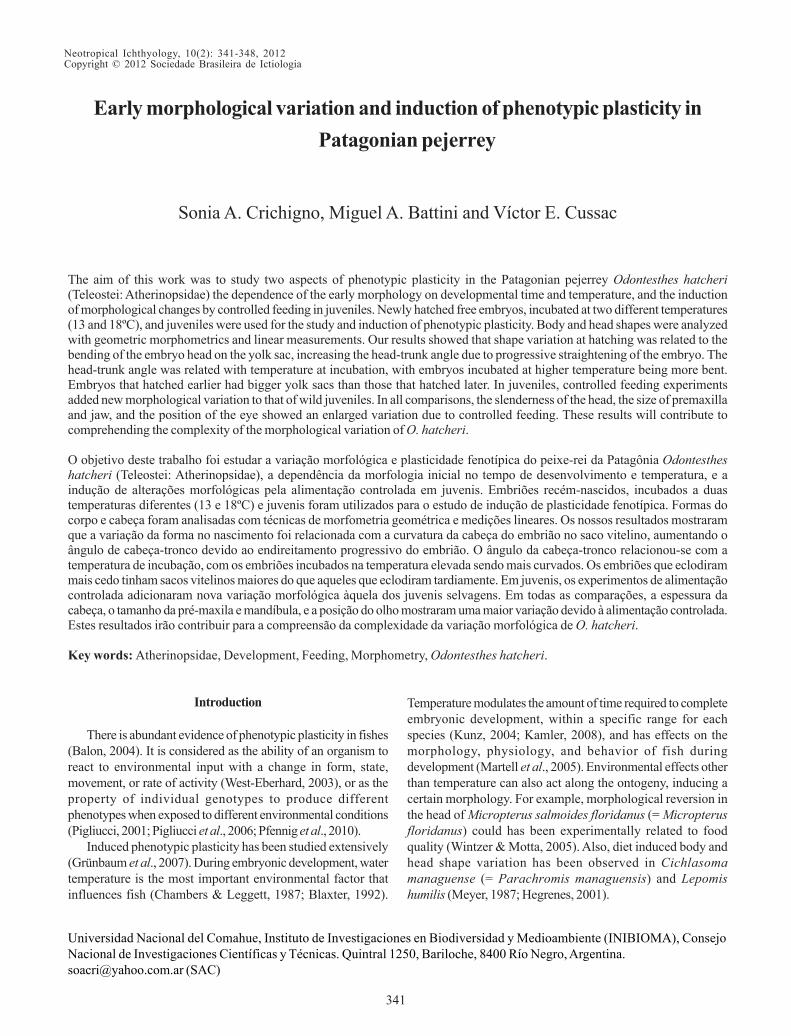
Feeding with zooplankton and Tubifex sp. for 60 days: DA(N = 64) among just caught individuals, individuals fed withzooplankton and individuals fed with Tubifex sp. showed twosignificant DFs, correctly classifying 98.4% of cases andexplaining 100% of variation (DF1, Wilks´ lambda = 0.062, P<0.001, and DF2, Wilks´ lambda = 0.339, P< 0.001, Fig. 7).Deformation grids showed, for individuals fed with zooplanktoncompared to just caught individuals, higher mouth (anteriortips of premaxilla and dentary), and more anterior isthmus.Individuals fed with Tubifex sp., when compared to just caughtindividuals, had bigger eyes in a higher and more posteriorposition, wider anterior portion of premaxilla, lower mouth(anterior tips of premaxilla and dentary), more anterior isthmus,and more posterior pectoral fin base.
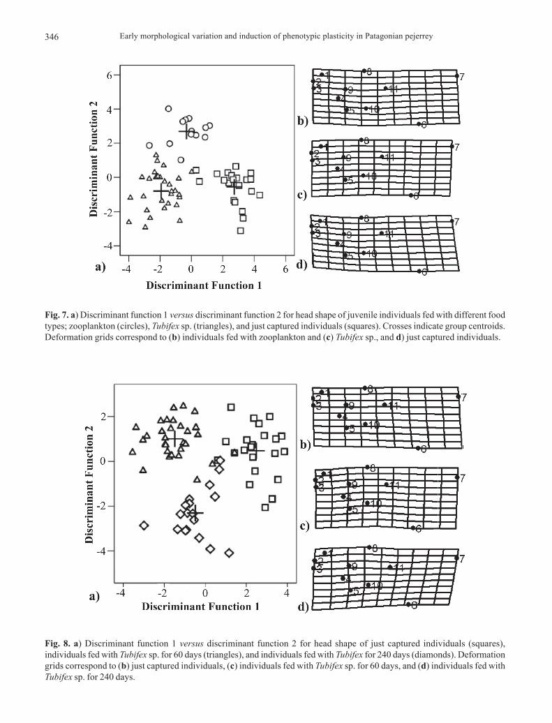
Feeding with Tubifex sp.: DA (N = 71) among times (0,60, and 240 days) correctly classified 94.4% of cases andexplained 100% of variation (DF1, Wilks´ lambda = 0.083, P<0.001, and DF2, Wilks´ lambda = 0.339, P< 0.001). DF1separated just captured individuals from treated individualsand DF2 separated 60 days of feeding from 240 days offeeding groups (Fig. 8). The morphological variations alongtime showed an increase of eye size, an elongation ofpremaxilla and lower jaw, and an upward and forwardmovement of the isthmus.
Discussion
Hatching is not a fixed threshold, but is triggered byenvironmental cues at different times during the embryonicperiod, thus even sibling individuals can hatch at very different
Fig. 5. Deformation grids for newly hatched individuals (AH1), coming from two PC and incubated at a) 13ºC and b) 18ºC(see Table 1).
Fig. 6. Box plots for linear measurements of newly hatchedfree embryos incubated at 18ºC: a) first discriminant function(Function 1), b) residuals yolk sac width, and c) residualsyolk sac length, all versus age at hatching (AH).
stages of development (Yamagami, 1988; Balon, 1999). Ourresults showed that morphological variation at hatching in O.hatcheri is related to the bending of the embryo over the yolksac, increasing the head-trunk angle due to the straighteningof the embryo along time, as was described by Kimmel et al.
Early morphological variation and induction of phenotypic plasticity in Patagonian pejerrey346
Fig. 7. a) Discriminant function 1 versus discriminant function 2 for head shape of juvenile individuals fed with different foodtypes; zooplankton (circles), Tubifex sp. (triangles), and just captured individuals (squares). Crosses indicate group centroids.Deformation grids correspond to (b) individuals fed with zooplankton and (c) Tubifex sp., and d) just captured individuals.
Fig. 8. a) Discriminant function 1 versus discriminant function 2 for head shape of just captured individuals (squares),individuals fed with Tubifex sp. for 60 days (triangles), and individuals fed with Tubifex for 240 days (diamonds). Deformationgrids correspond to (b) just captured individuals, (c) individuals fed with Tubifex sp. for 60 days, and (d) individuals fed withTubifex sp. for 240 days.

S. A. Crichigno, M. A. Battini & V. E. Cussac 347
(1995) in zebrafish. In the fish’s dorsal view, it became apparentthat earlier hatched O. hatcheri embryos (AH 1) had biggeryolk sacs than individuals that hatched later.
The effect of parental couples seems to be less conspicuousthan AH. Although DA between PC with different AH and thesame DAF and temperature (PCAH, Table 1) brought a significantDF, the DA between PC with different DAF and the sametemperature and AH (PCDAF, Table 1) failed. Our data(comparison TPCDAF in Table 1) suggest that, at hatching, O.hatcheri embryos incubated at higher temperature were morebent. Probably, the size of the yolk sac and the bending of theembryo after hatching will be major factors operating on thebeginning of exogenous feeding (Battini et al., 1995). This isconsistent with the differential consumption of yolk observed inAnarhichas minor (Sund & Falk-Petersen, 2005), wheredecreased consumption occurred at higher temperature and thusthe embryos had more yolk at hatching and were more bent overthe yolk sac. Sund & Falk-Petersen (2005) and Peterson et al.(2004) also observed in Anarhichas minor and Gadus morhuathat embryos incubated at lower temperature hatched with largerlength. Furthermore, water temperature directs the sexualdifferentiation process in the pejerrey Odontesthes bonariensis(Valenciennes) and affects larval condition and growth rate(Strüssmann & Patiño, 1995; Ito et al., 2005; Chalde et al., 2011).
Regarding juvenile feeding, morphological variationincreased when, in addition to just captured wild individuals,those subjected to controlled feeding were considered. In allcomparisons, the slenderness of the head, size of premaxillaand jaw, and position of the eyes showed appreciable variationin the RW1. Both individuals fed only with zooplankton andthose fed only with Tubifex sp. had slenderer heads than justcaptured wild individuals. Furthermore, shapes obtainedthroughout feeding with Tubifex sp. during increasing timelapses included bigger eyes and slenderer heads. In the sameway, Grünbaum et al. (2007) found different levels ofdevelopmental plasticity in critical ontogenetic periods ofSalvelinus alpinus that are most responsive to environmentalconstraints. Shape variation has been also induced by controlleddiet in other fishes. Cichlasoma managuense (= Parachromismanaguensis) (Meyer, 1987) and Lepomis humilis (Hegrenes,2001) fed with Artemia nauplii or Tenebrio larvae, showed thatindividuals fed with large prey (Tenebrio larvae) developed anelongated, fusiform shape with a sharply angled snout, aftereight months of treatment. In the same way, another nativePatagonian fish, Percichthys trucha (Valenciennes), showedjuveniles fed with zooplankton as having slenderer head andbody, and longer jaw than individuals fed with Tubifex sp. (largeprey) after 70 days of treatment (Crichigno, 2012).
Head shape, size and position of the eyes, and size ofpremaxilla and jaws are likely to have performanceconsequences for prey capture and consumption (Carson &Wainwright, 2010). Patagonian lakes show a wide range ofphotic habitats (Lattuca et al., 2007) and, particularly, theabundance of zooplanktophagous fish like O. hatcheri canmodify planktonic food webs and water transparency (Reissiget al., 2006). Although an overview of the diet of O. hatcheri
indicates an omnivorous diet, a more detailed observationshows that diet changes greatly between lakes, with most ofthe intralacustrine variation ascribed to the ontogenetic shift(Ferriz, 1987; Grosman & Rudzik, 1990; Cervellini et al., 1993;Macchi et al., 1999). This succession of more or lessstenophagous ontogenetic periods could be imposing preciserequirements for the oropharyngeal apparatus of the fish.
In conclusion, temperature and hatching timing could acton the head shape during the early life of O. hatcheri. Later on,juvenile feeding adds another variable to head shape variation.
Plastic induction of head shape of O. hatcheri wouldprovide, by mechanistic, epigenetic processes, additionalmorphological variation and in consequence a menu ofcapabilities for prey catching under changing selectionselective pressures. In this context, our results couldcontribute to the comprehension of the plastic component ofthe morphological variation of the species.
Acknowledgements
We would like to acknowledge the following institutionsfor granting the present project: Universidad Nacional delComahue (04B147), CONICET (PIP 112-200801-00282), andFONCYT (PICT2005-35241) of Argentina. This work wouldnot have been possible without the cooperation of Centro deSalmonicultura Bariloche, Universidad Nacional del Comahue.
Literature Cited
Adams, D. C., F. J. Rohlf & D. E. Slice. 2004. Geometricmorphometrics: ten years of progress following the ‘revolution’.Italian Journal of Zoology, 71: 5-16.
Balon, E. K. 1999. Alternative ways to become a juvenile or adefinitive phenotype (and on some persisting linguistic offenses).Environmental Biology of Fishes, 56: 17-38.
Balon, E. K. 2004. Evolution by epigenesis: farewell to Darwinism,neo- and otherwise. Rivista di Biologia / Biology Forum, 97:269-312.
Barnabé, G. 1990. Aquaculture (Volume 1 and 2). England, EllisHorwood Limited, 1104p.
Battini, M. A., M. F. Alonso & V. E. Cussac. 1995. Growth andnutritional condition of the larvae of Odontesthes microlepidotus(Atherinidae): An experimental approach. EnvironmentalBiology of Fishes, 42: 391-399.
Blaxter, J. H. S. 1992. The effect of temperature on larval fishes.Netherlands Journal of Zoology, 42: 336-357.
Bookstein, F. L. 1991. Morphometric tools for landmark data.Cambridge, Cambridge University Press, 436p.
Carlson, R. L. & P. C. Wainwright. 2010. The ecological morphologyof darter fishes (Percidae: Etheostomatinae). Biological Journalof the Linnean Society, 100: 30-45.
Cervellini, P. M., M. A. Battini & V. E. Cussac. 1993. Ontogeneticshifts in the feeding of Galaxias maculatus (Galaxiidae) andOdontesthes microlepidotus (Atherinidae). EnvironmentalBiology of Fishes, 36: 283-290.
Chalde, T., D. A. Fernández, V. E. Cussac & G. M. Somoza. 2011.The effect of rearing temperature in larval development ofpejerrey, Odontesthes bonariensis. Morphological indicatorsof development. Neotropical Ichthyology, 9: 747-756.

Early morphological variation and induction of phenotypic plasticity in Patagonian pejerrey348
Chambers, R. C. & W. C. Leggett. 1987. Size and age atmetamorphosis in marine fishes: an analysis of laboratory-rearedwinter flounder (Pseudopleuronectes americanus) with a reviewof variation in other species. Canadian Journal of Fisheries andAquatic Sciences, 44: 1936-1947.
Conte-Grand, C. 2012. El pejerrey patagónico, Odontesthes hatcheri:biología y potencialidades para su cultivo. Unpublished Ph.D.Dissertation, Universidad Nacional del Sur, Bahía Blanca. 174p.
Crichigno, S. 2012. Variación morfológica y plasticidad fenotípicadel aparato bucofaríngeo de Odontesthes hatcheri (Eigenmann,1909) y Percichthys trucha (Cuvier & Valenciennes, 1840).Unpublished Ph.D. Dissertation, Universidad Nacional delComahue, Bariloche. 95p.
Cussac, V. E., P. M. Cervellini & M. A. Battini. 1992. Intralacustrinemovements of Galaxias maculatus (Galaxiidae) and Odontesthesmicrolepidotus (Atherinidae) during their early life history.Environmental Biology of Fishes, 35: 141-148.
Ferriz, R. A. 1987. Alimentación del pejerrey patagónico Patagoninahatcheri (Eigenmann, 1909) en el embalse Ramos Mexia,Neuquén, Argentina. Hydrobiologia, 6: 61-66.
Grosman, F. & G. Rudzik. 1990. Análisis de la dieta del pejerreypatagónico Patagonina hatcheri Eigenmann, 1909, de la LagunaTerraplén, Chubut, Argentina. Biota, 6: 71-88.
Grünbaum T., R. Cloutier, P. M. Mabee & N. R. Le François. 2007.Early developmental plasticity and integrative responses in arcticcharr (Salvelinus alpinus): effects of water velocity on bodysize and shape. Journal of Experimental Zoology (Molecularand Developmental Evolution), 308B: 396-408.
Hegrenes, S. 2001. Diet-induced phenotypic plasticity of feedingmorphology in the orangespotted sunfish, Lepomis humilis.Ecology of Freshwater Fish, 10: 35-42.
Ito, L. S., M. Yamashita, F. Takashima & C. A. Strüssmann. 2005.Dynamics and histological characteristics of gonadal sexdifferentiation in pejerrey (Odontesthes bonariensis) atfeminizing and masculinizing temperatures. Journal of Experi-mental Zoology 303A: 504-514.
Kamler, E. 2008. Resource allocation in yolk-feeding fish. Reviewsin Fish Biology and Fisheries, 18: 143-200.
Kimmel, C. B., W. W. Ballard, S. R. Kimmel, B. Ullmann & T. F.Schilling. 1995. Stages of embryonic development of thezebrafish. Developmental Dynamics, 203: 255-310.
Kunz, Y. W. 2004. Developmental Biology of Teleost Fishes.Netherlands, Springer, 636p.
Lattuca, M. E., S. Ortubay, M. A. Battini, J. P. Barriga & V. E.Cussac. 2007. Presumptive environmental effects on body shapeof Aplochiton zebra (Pisces, Galaxiidae) in Northern Patagonianlakes. Journal of Applied Ichthyology, 23: 25-33.
Macchi P. J., V. E. Cussac, M. F. Alonso & M. A. Denegri. 1999.Predation relationships between introduced salmonids and thenative fish fauna in lakes and reservoirs in Northern Patagonia.Ecology of Freshwater Fish, 8: 227-236.
Martell, D. J., J. D. Kieffer & E. A. Trippel. 2005. Effects of temperatureduring early life history on embryonic and larval development andgrowth in haddock. Journal of Fish Biology, 66: 1558-1575.
Meyer, A. 1987. Phenotypic plasticity and heterochrony inCichlasoma managuense (Pisces, Chichlidae) and theirimplications for speciation in Cichlid fishes. Evolution, 6:1357-1369.
Morphometrics at Sunny Stony Brook. 2012. Available from: http://life.bio.sunysb.edu/morph/index.html (Date of access: Jan 30, 2012).
Parsons, K. J., B. R. Robinson & T. Herbert. 2003. Getting intoshape: an empirical comparison of truss-based morphometric
methods with a newer geometric method applied to new worldcichlids. Environmental Biology of Fishes, 67: 417-431.
Pascual, M. A., V. Cussac, B. Dyer, D. Soto, P. Vigliano, S. Ortubay& P. Macchi. 2007. Freshwater fishes of Patagonia in the 21st
century after a hundred years of human settlement, speciesintroductions, and environmental change. Aquatic EcosystemHealth and Management, 10: 212-227.
Peterson, R. H., D. J. Martin-Robichaud & P. Harmon. 2004.Influence of incubation temperature on body movements ofAtlantic cod (Gadus morhua L.) embryos and on size at hatch.Aquaculture Research, 35: 453-458.
Pfennig, D. W., A. W. Matthew, E. C. Snell-Rood, T. Cruickshank,C. D. Schlichting & A. P. Moczek. 2010. Phenotypic plasticity’simpacts on diversification and speciation. Trends in Ecologyand Evolution, 25: 459-467.
Pigliucci, M. 2001. Phenotypic Plasticity: Beyond Nature andNurture. Baltimore, Johns Hopkins University Press, 333p.
Pigliucci, M., C. J. Murren & C. D. Schlichting. 2006. Phenotypicplasticity and evolution by genetic assimilation. Journal of Ex-perimental Biology, 209: 2362-2367.
Quirós, R. 1988. Relationships between air temperature, depth,nutrients and chlorophyll in 103 Argentinian lakes. Verhandlungendes Internationalen Verein Limnologie, 23: 647-658.
Reissig, M., C. Trochine, C. Queimaliños, E. Balseiro & B.Modenutti. 2006. Impact of fish introduction on planktonicfood webs in lakes of the Patagonian Plateau. BiologicalConservation, 132: 437-447.
Rohlf, F. J. & L. F. Marcus. 1993. A revolution in morphometrics.Trends in Ecology and Evolution, 8: 129-132.
Sund, T. & I. Falk-Petersen. 2005. Effects of incubation temperatureon development and yolk sac conversion efciencies of spottedwolfsh (Anarhichas minor Olafsen) embryos until hatch.Aquaculture Research, 36: 1133-1143.
Strüssmann, C. A. & R. Patiño 1995. Temperature manipulation ofsex differentiation in fish. Pp. 153-157. In: F. Goetz & P. Thomas(Eds.) Proceedings of the Fifth International Symposium onthe Reproductive Physiology of Fish. The University of Texasat Austin, Texas, 389p.
Strüssmann, C. A., T. Akaba, K. Ijima, K. Yamaguchi, G. Yoshizaki& F. Takashima. 1997. Spontaneous hybridization in thelaboratory and genetic markers for the identification of hybridsbetween two atherinid species, Odontesthes bonariensis(Valenciennes, 1835) and Patagonina hatcheri (Eigenmann,1909). Aquaculture Research, 28: 291-300.
West-Eberhard, M. J. 2003. Developmental Plasticity and Evolution,Oxford University Press, New York, 618p.
Wintzer, A. P. & P. J. Motta. 2005. Diet-induced phenotypicplasticity in the skull morphology of hatchery-reared Floridalargemouth bass, Micropterus salmoides floridanus. Ecologyof Freshwater Fish, 14: 311-318.
Yamagami, K. 1988. Mechanisms of hatching in fish. Pp. 447-500.In: Hoar, W. S. & D. J. Randall (Eds.) Fish physiology. VolumeXIA. The physiology of the developing fish. New York,Academic Press Inc., 546p.
Submitted August 17, 2011Accepted May 8, 2012
Published June 29, 2012