drug-like leads for steric discrimination between substrate and inhibitors of human...
TRANSCRIPT

Drug-like Leads for Steric Discrimination betweenSubstrate and Inhibitors of HumanAcetylcholinesterase
Scott A. Wildman1,†, Xiange Zheng1,†,David Sept2, Jeffrey T. Auletta3, Terrone L.Rosenberry3 and Garland R. Marshall1,*
1Department of Biochemistry and Molecular Biophysics, WashingtonUniversity, St. Louis, MO 63110, USA2Department of Biomedical Engineering, University of Michigan, Ann
Arbor, MI 48109, USA3Department of Neuroscience, Mayo Clinic, Jacksonville, FL 32224,
USA
*Corresponding author: Garland R. Marshall,
[email protected]!Authors contributed equally to this work.
Protection of the enzyme acetylcholinesterase(AChE) from the toxic effects of organophosphateinsecticides and chemical warfare agents (OPs)may be provided by inhibitors that bind at theperipheral binding site (P-site) near the mouth ofthe active-site gorge. Compounds that bind to thissite may selectively block access to the acylationsite (A-site) catalytic serine for OPs, but not ace-tylcholine itself. To identify such compounds, weemployed a virtual screening approach using AUTO-
DOCK 4.2 and AutoDock Vina, confirmed usingcompounds experimentally known to bind specifi-cally to either the A-site or P-site. Both programsdemonstrated the ability to correctly predict thebinding site. Virtual screening of the NCI DiversitySet II was conducted using the apo form of theenzyme, and with acetylcholine bound at the crys-tallographic locations in the A-site only and inboth and A- and P-sites. The docking identified 32compounds that were obtained for testing, andone was demonstrated to bind specifically to theP-site in an inhibitor competition assay.
Key words: acetylcholine, acetylcholinesterase, AChE, docking, vir-tual screening
Abbreviations: AChE, acetylcholine esterase; OP, organophos-phate; DMSO, dimethyl sulfoxide; NCI, National Cancer Institute.
Received 10 December 2010, revised 14 March 2011 and accepted forpublication 28 May 2011
Acetylcholinesterase (AChE) terminates synaptic neuromusculartransmission by hydrolyzing the neurotransmitter acetylcholine (1).
One focus of AChE research lies in the development of new drugsthat could prevent and ⁄ or treat poisoning from organophosphates(OPs), toxic agents commonly used in insecticides as well as inchemical warfare agents (2). OP poisoning causes the inactivationof AChE that prevents synaptic transmission, leading to muscleparalysis, hypertension, and malfunction of various organ systems,ultimately leading to death.
Two ligand-binding sites in AChE have been identified, the acylationsite (A-site) at the base of a deep active-site gorge and the periph-eral site (P-site) near the gorge entrance through which ligands mustpass on their way to the A-site (3–5). Figure 1 shows the bindinggorge of human AChE with acetylcholine modeled into both the A-site (green) and P-site (pink) as described in Methods. OPs are toxicbecause they covalently react with S203 in the A-site. Wilson andGinsburg (6,7) conceived complementary oxime compounds thatcould re-activate OP-poisoned AChE. These drugs serve as defensesagainst OP toxicity, but they work only on OP-inactivated AChE. Itwould be beneficial to develop prophylactic drugs that prevent AChEfrom interacting with OPs in the first place, and our objective is todesign drug-like compounds to serve this function. One strategy is toobtain small molecules that bind at or near the AChE P-site. Suchmolecules could be modified, in principle, to sterically discriminateaccess to the A-site in a way that still allows entry of acetylcholinebut limits the entrance of the larger OPs. This is a challenging task.While a competitive inhibitor that binds to the A-site would reducethe irreversible OP inactivation rate, it would equally reduce the rateof acetylcholine hydrolysis and provide no benefit. The binding of aligand X to the AChE P-site is known to decrease the associationand dissociation rate constants for the binding of a ligand Y to theA-site by factors of up to 400 (5,8), and it is this feature that wepropose to exploit in designing a P-site ligand that would selectivelyblock OP association with the A-site. Such a ligand would need veryhigh affinity (pM) for the P-site to effectively block OP inactivation,and a strategy designed to evolve a series of lead compounds withprogressively higher affinity is necessary. Here we report on our firststeps in such an evolution.
The development of structure–activity relationships for ligands thatbind specifically to the AChE P-site is in its infancy. Because of apossible AChE P-site role in promoting amyloid-b aggregation in Alz-heimer's disease (9), in silico screening of a limited 270-memberlibrary of compounds was initiated to identify new ligands with P-site specificity (10). Several identified compounds as well as tetrahy-drocannabinol (9) were shown experimentally to bind to the P-site.
C B D D 1 1 5 7 B Dispatch: 24.6.11 Journal: CBDD CE: Manonmani
Journal Name Manuscript No. Author Received: No. of pages: 10 PE: Gayathri
1
Chem Biol Drug Des 2011
Research Article
ª 2011 John Wiley & Sons A/S
doi: 10.1111/j.1747-0285.2011.01157.x12345678910111213141516171819202122232425262728293031323334353637383940414243444546474849505152535455565758

We used a virtual screening approach to identify P-site ligands froma much larger library of compounds. To establish the ligand dockingprocedures used to screen the library, we selected several com-pounds known to bind to either the A-site or the P-site by X-raycrystallography. These were first redocked back into their originalcrystal structures with two docking programs, AutoDock Vina (12)and AUTODOCK version 4.2 (13). These compounds were then dockedinto human AChE to confirm that the docking programs could iden-tify a preference for the correct site. Finally, virtual screening wasused to identify compounds that bind preferentially to the P-site,and still allow acetylcholine to bind to the A-site of AChE. The NCIDiversity Set II (14) was first docked using Vina in combination witha consensus scoring approach, and after an initial round of testing,was docked using AUTODOCK 4.2. Candidate compounds predicted toshow selective P-site binding were obtained from the NCI andtested experimentally with an AChE inhibitor-competition assay thatcan distinguish between inhibitor binding to the A-site or P-site.
Methods
Preparation of crystal structures
The structure of human acetylcholinesterase (AChE) was derivedfrom the crystal structure of the AChE complexed with fasciculin-IIdeposited in the RCSB Protein Databank (15) (PDB id:1B41) by Kry-ger and co-workers;(16) only the AChE was retained for our model-ing studies. The locations of the A- and P-sites were defined fromthe crystal structure of the S203A mutant of mouse AChE is com-plexed with acetylcholine (17) (PDB id: 2HA4), which shows onemolecule of acetylcholine in each site. The 1B41 and 2HA4 struc-tures were superimposed, and the acetylcholine molecules from2HA4 were merged with AChE from 1B41. This formed our modelof human AChE complexed with acetylcholine used in subsequentstudies.
Docking controls
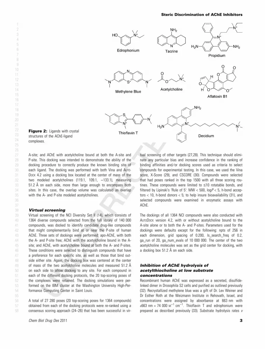
Crystal structures of several additional AChE-ligand complexes havebeen deposited in the PDB, including complexes at the A-site withedrophonium (18) (2ACK) or tacrine (19) (1ACJ) and complexes atthe P-site with thioflavin T (20) (2J3Q), decidium (21) (1J07), propidi-um (21) (1N5R), methylene blue (22) (2W9I), or aflatoxin B1(23)(2XI4). Crystal structures of AChE complexes involving ligands thatspan both sites also have been obtained, but these ligands are notof interest in this study and were not examined. These structuresall were obtained as complexes with Torpedo californica AChE(TcAChE) or Mus musculus (MmAChE). The complexes were super-imposed with our human model of AChE, and therefore, all coordi-nates are in relation to 1B41, with the A-site and P-site defined bythe acetylcholines originally from 2HA4. These ligands, shown inFigure 2, were used to test our docking procedures. In each case,the ligand was removed from the original structure and redockedinto that structure with a docking box centered on the originalligand position and measuring 25.2 ! on each side, sufficientlylarge to encompass the ligands in this study. For the redocking con-trols, all crystal waters were retained.
Ability to reproduce crystal structure binding mode was assessed bymolecular volume overlap, presented as a percentage of the volumeof the X-ray ligand. For these calculations, we used ROCS (OpenEyeScientific Software, Sante Fe, NM, USA) to determine the absolutevolume and divided the volume overlap of a ligand with itself todetermine percentages. Overlap close to 100% can be interpretedas correct prediction of binding mode, while mid-range overlaps typ-ically bind in the correct site with an incorrect conformation, andlow values indicate binding in a different site.
Additionally, each of these ligands was docked into our model ofhuman AChE under three different conditions: apo-AChE, with boththe A- and P-sites free; AChE with the acetylcholine bound in the
A B
Figure 1: 6The AChE catalytic site is a narrow active-site gorge some 20 ! deep (yellow) that penetrates nearly to the center of the!65 kDa catalytic subunits. Near the base of the gorge is the A-site where H447, E334, and S203 (red) participate in a triad that catalyzesthe transient acylation and deacylation of S203 during each substrate turnover. The P-site is spanned by residues W286 near the mouth ofthe gorge and D74 near a constriction at the boundary between the P-site and the A-site. (B) Acetylcholine modeled into the A-site (green)and P-site (pink) as described in Methods.
LOW
RESOLUTIO
NCOLOR
FIG
Wildman et al.
2 Chem Biol Drug Des 2011
12345678910111213141516171819202122232425262728293031323334353637383940414243444546474849505152535455565758
A-site; and AChE with acetylcholine bound at both the A-site andP-site. This docking was intended to demonstrate the ability of thedocking procedure to correctly produce the known binding site ofeach ligand. The docking was performed with both Vina and AUTO-DOCK 4.2 using a docking box located at the center of mass of thetwo modeled acetylcholines (119.1, 109.1, )133.1), measuring51.2 ! on each side, more than large enough to encompass bothsites. In this case, the overlap volume was calculated as overlapwith the A- and P-site modeled acetylcholines.
Virtual screening
Virtual screening of the NCI Diversity Set II (14), which consists of1364 diverse compounds selected from the full library of 140 000compounds, was docked to identify candidate drug-like compoundsthat might complementarily bind at or near the P-site of humanAChE. Three sets of dockings were performed: apo-AChE, with boththe A- and P-site free; AChE with the acetylcholine bound in the A-site; and AChE, with acetylcholine bound at both the A- and P-sites.These conditions were selected to distinguish compounds that havea preference for each specific site, as well as those that bind out-side either site. Again, the docking box was centered at the centerof mass of the two acetylcholine molecules and measured 51.2 !on each side to allow docking to any site. For each compound ineach of the different docking protocols, the 20 top-scoring poses ofthe complexes were retained. The docking simulations were per-formed on the IBM cluster at the Washington University High-Per-formance Computing Center in Saint Louis.
A total of 27 280 poses (20 top-scoring poses for 1364 compounds)obtained from each of the docking protocols were re-ranked using aconsensus scoring approach (24–26) that has been successful in vir-
tual screening of other targets (27,28). This technique should elimi-nate any particular bias and increase confidence in the ranking ofbinding affinities and ⁄ or docking scores used as criteria to selectcompounds for experimental testing. In this case, we used the Vinascore, X-Score (29), and CSCORE (30). Compounds were selectedthat had poses ranked in the top 1500 with all three scoring rou-tines. These compounds were limited to £10 rotatable bonds, andfiltered by Lipinski's 'Rule of 5': MW < 500, logP < 5, h-bond accep-tors < 10, h-bond donors < 5; to help insure bioavailability (31), andselected compounds were examined in enzymatic assays withAChE.
The dockings of all 1364 NCI compounds were also conducted withAUTODOCK version 4.2, with or without acetylcholine bound to theA-site alone or to both the A- and P-sites. Parameters used for thedockings were defaults except for the following: npts of 256 ineach dimension, grid spacing of 0.200, ls_search_freq of 0.2,ga_run of 20, ga_num_evals of 10 000 000. The center of the twoacetylcholine molecules was set as the grid center for docking, witha docking box 51.2 ! on each side.
Inhibition of AChE hydrolysis of
acetylthiocholine at low substrate
concentrations
Recombinant human AChE was expressed as a secreted, disulfide-linked dimer in Drosophila S2 cells and purified as outlined previously(32). Recrystallized methylene blue was a gift of Dr. Lev Weiner andDr Esther Roth at the Weizmann Institute in Rehovath, Israel, andconcentrations were assigned by absorbance at 663 nm withe663 nm = 74 000 M)1 cm)1. Thioflavin T and edrophonium wereprepared as described previously (33). Substrate hydrolysis rates v
Figure 2: Ligands with crystalstructures of the AChE-ligandcomplexes.
Steric Discrimination of AChE Inhibitors
Chem Biol Drug Des 2011 3
12345678910111213141516171819202122232425262728293031323334353637383940414243444546474849505152535455565758

were measured with acetylthiocholine and 5,5¢-dithiobis-(2-nitroben-zoic acid) (DTNB, 0.33 mM) in a coupled Ellman reaction in assaybuffer (20 mM sodium phosphate buffer (pH 7.0) and 0.02% TritonX-100 unless otherwise noted) at 25!C (34).
The assay used here to assess inhibition of AChE activity measuresthe second-order hydrolysis rate constant kE obtained when thesubstrate concentration [S] is much less than the Michaelis con-stant Kapp (34). Under these conditions, Scheme 1 includes all rele-vant enzyme species:
This scheme assumes that substrate binds initially to the P-site(ESP) before progressing to the A-site (ES) where it is hydrolyzed toproducts P, and it can be applied both to inhibitors that bind at theA-site (LP = I, and 1 ⁄ i = 0) and to inhibitors that bind at the P-site(LP = IP) with dissociation constants KI. Under these second-orderconditions, substrate hydrolysis rates v = kE[E]tot[S], where [E]tot isthe1 total enzyme concentration, and kE is measured from eqn 1(35).
"S# $ "S#0e%zt &1'
In eqn 1, z = kE[E]tot and t is the time after mixing E and S.Because acetylthiocholine is hydrolyzed by AChE at nearly diffusion-controlled rates, inhibitor binding to either the A- or P-sites resultsin an expression for the ratio of kE in the absence of inhibitor(kE[I] = 0) to kE in the presence of inhibitor (kE + I) given by eqn 2(34).
kE"I#$0
kE(I$ 1(
"I#KI
! "
&2'
If a series of assays are conducted with a fixed concentration ofone inhibitor I1 together with varying concentrations of a secondinhibitor I2, kE in the presence of both inhibitors (here denotedkE + I2) relative to kE when only I1 is present (here denotedkE[I2] = 0) is given by eqn 3 (33).
kE(I2
kE"I2#$0$
K2 1( "I1#K1
# $
K2 1( "I1#K1
# $
( "I2# 1( "I1#K12
# $ &3'
In this equation, K1, K2, and K12 are the equilibrium dissociationconstants for I1 with E, I2 with E, and I1 with the EÆI2 complex,respectively. The value of [I1] ⁄ K12 will be significantly >0 only if the
ternary complex EÆI1ÆI2 can form, and this analysis, thus, can beused to detect ternary complex formation.
Assay of NCI diversity set compounds
Compounds were dissolved in dimethyl sulfoxide (DMSO) at 10–50 mM and diluted (1:100) into assay buffer. If the compound wasinsoluble after dilution, the DMSO stock was diluted (1:10) intomethanol and then diluted further (1:10) into assay buffer. Theassay solvent for the control without a compound was matched tothat with compound. AChE activity was essentially unaffected by10% methanol but was inhibited about 2-fold in the presence of1% DMSO. However, inclusion of 10% methanol decreased thereproducibility of kE.
Results and Discussion
Docking controls
The AChE binding sites of the compounds presented in Figure 2 areknown from X-ray crystallography, and for each, the compound wasremoved from the structure and redocked with Vina or AUTODOCK4.2. The ability to reproduce the X-ray binding mode is presentedas the percent overlap of the docked ligand with the X-ray bindingmode in Table 1. Docking with Vina was more successful at locat-ing the ligand within the original binding site than AUTODOCK 4.2, aseach docked ligand had a higher overlap with the X-ray structure.However, lack of complete overlap for most ligands describes atleast a shift in position of the docked compounds, and some mole-cules, such as tacrine, are flipped within their correct site. Theligand ranking by docking energy agrees for the two programsexcept for propidium and decidium, likely due to the difficulty indocking these flexible molecules. For AUTODOCK 4.2, docking acetyl-choline to 2HA4 with both copies of the crystallographic acetylcho-line ligand removed results in consistent docking to only the A-site,even though both the A- and P-sites were available. Docking toAChE with the A-site occupied by the crystallographic acetylcholineresults the second acetylcholine docking reliably to the P-site. Using
S + E
+
I
S + EIP
ES
+
I
ESIP
E + P
EIP + P
iKI
k2
ak2
ESP
kS
k-S
k1
k-1
kS2
k-S2
Scheme 1: XXXXXXXXXXXX 5.
Table 1: Redocking of compounds with known crystal structures
PDB ID
AutoDock Vina AUTODOCK 4.2
X-rayoverlap (%)
Dockingenergy
X-rayOverlap (%)
Dockingenergy
A-site ligandsTacrine 1ACJ 98.9 )9.5 61.3 )9.0Edrophonium 2ACK 94.6 )6.2 96.5 )6.6
P-site ligandsThioflavin T 2J3Q 81.8 )7.9 27.8 )7.2Propidium 1N5R 91.4 )6.8 13.1 )8.6Decidium 1JO7 55.0 )6.2 42.9 )10.7Methylene Blue 2W9I 55.9 )8.5 42.5 )7.7Aflatoxin B1 2XI4 98.6 )9.6 97.0 )9.1Acetylcholine A-site 2HA4 83.7 )4.8 48.2 )5.5Acetylcholine P-site 2HA4 65.3 )4.3 68.9 )3.8
Wildman et al.
4 Chem Biol Drug Des 2011
12345678910111213141516171819202122232425262728293031323334353637383940414243444546474849505152535455565758

Vina, acetylcholine is docked into both the A-site and P-site if theyare unoccupied.
Crystal water molecules play an important role in accurately repro-ducing crystal structures in docking. This was noted specifically forAChE by Dickerson and co-workers (10), who observed that crystalwaters greatly improved docking results for propidium and decidium.We observe this same behavior with several ligands, including ace-tylcholine and edrophonium shown in Figure 3. Without crystalwaters, (Figure 3A) edrophonium docks to the A-site, but only par-tially overlaps with the crystal structure binding mode (28.8% vol-ume overlap). The inclusion of only four of the crystal structurewaters (Figure 3B) in the A- and P-sites results in a docked struc-ture that much more closely matches the crystal structure ligand(86.7% overlap).
As A-site waters in AChE are well-known, inclusion of explicitwaters in docking would be preferred. However, given the diversechemotypes in our compound database, it is not possible to knowprior to docking which compounds may interact with or displace thewater molecules. Therefore, as an additional control calculation, wedocked these same known ligands to human AChE with all watersremoved to assess the ability of Vina and AUTODOCK 4.2 to correctly
predict the preferred binding site for each ligand under virtualscreening conditions.
Docking with human AChE
The three-dimensional structures of TcAChE, MmAChE, and humanAChE are very similar, and most features of the active-site gorgeare conserved between the species. Nevertheless, to confirm thatthe results obtained with TcAChE and MmAChE also apply tohuman AChE, docking with Vina and AUTODOCK 4.2 to human AChEwas repeated for all the ligands in Figure 2. Docking trials wereperformed with three different conditions: apo-AChE, with both theA- and P-site free; AChE with the acetylcholine bound in the A-site;and AChE with acetylcholine bound at both the A- and P-site.
Docking energies for both Vina and AUTODOCK 4.2 are presented inTable 2 for the apo-AChE docking (no bound acetylcholine). Bindingin the correct site is described by the percent overlap with acetylc-holines in the A-site and P-site modeled into 1B41 through superpo-sition of 2HA4. A measurable percent overlap indicates it is unlikelythat the modeled acetylcholine and the docked ligand could bindsimultaneously in the given binding mode. When only a partial over-lap is observed, typically the ligand is docked immediately adjacentto the modeled acetylcholine.
All ligands are predicted to bind to the known preferred site exceptfor tacrine docking by Vina, which was docked to the P-site. Decidi-um docked by Vina spans both sites, but is predominantly locatedin the P-site. Acetylcholine was docked by both programs into boththe A- and P-sites, with a slight preference in predicted energy forthe A-site, although none exactly reproduces the crystal structurebinding mode. As with the redocking control, the ranking by dockingenergy agrees between Vina and AUTODOCK 4.2, with the exceptionof propidium and decidium.
The docking to human AChE with acetylcholine modeled into eitherthe A-site or both the A- and P-sites produces similar results (datanot shown). As each site is occupied, the docking algorithms willidentify another binding location, but with less favorable energy. It
A B
Figure 3: Docking of edrophonium into Torpedo apo-AChE usingAUTODOCK 4.2 (A) with crystal waters removed; (B) with crystalwaters retained. Crystal structure ligand and water in thin lines anddocked results in thick lines.
Table 2: Docking control ligands into human apo-AChE
AutoDock Vina AUTODOCK 4.2
A-siteoverlap (%)
P-siteoverlap (%)
Dockingenergy
A-siteoverlap (%)
P-siteoverlap (%)
Dockingenergy
A-site ligandsTacrine 0 84.6 )8.5 65.8 0 )8.7Edrophonium 65.8 0 )6.3 62.2 0 )6.6
P-site ligandsThioflavin T 0 73.3 )8.0 4.6 25.2 )7.6Propidium 0 15.9 )7.2 0 65.8 )10.2Decidium 46.2 72.7 )9.1 0 82.9 )10.4Methylene blue 0 85.7 )7.9 4.0 44.9 )7.2Aflatoxin B1 0 84.0 )9.0 0 100 )8.9Acetylcholine A-site 59.7 0 )4.4 43.5 0 )4.9Acetylcholine P-site 0 29.2 )3.9 0 33.7 )4.8
COLOR
Steric Discrimination of AChE Inhibitors
Chem Biol Drug Des 2011 5
12345678910111213141516171819202122232425262728293031323334353637383940414243444546474849505152535455565758

should be noted that the percent overlaps in Table 1 are with thecrystallographic ligands themselves but in Table 2 are with modeledacetylcholine originally from the 2HA4 crystal structure. Therefore,these values are not directly comparable except for acetylcholineitself. We do observe a decrease in percent overlap for acetylcho-line between the redocking (Table 1) and human docking (Table 2),but this is expected because of the removal of crystal structurewater from human AChE.
Overall, these docking controls suggest that both the Vina andAUTODOCK 4.2 programs may be useful for virtual screening for P-site binders of AChE. Vina more accurately predicted crystal struc-ture binding modes, but AUTODOCK 4.2 was better at predicting thecorrect binding site. The exact orientations of some docked poseswere not always reproduced, but this is not surprising consideringthe limitations in the scoring functions and search algorithms usedin docking. To minimize some of these limitations, our Vina virtualscreening protocol employed an established consensus scoringapproach (27,28) that incorporates X-Score and CSCORE with thepredicted binding energy from docking.
Identifying the binding site
The inhibition of second-order, substrate-hydrolysis rates kE as mea-sured with eqns 2 and 3 provides a convenient framework forexperimentally examining the interaction of an inhibitor with freeAChE. The assays are designed to assign the binding location of anunknown inhibitor by examining its competition with inhibitorswhose binding sites are known from X-ray crystallography. Theapproach is illustrated in Figure 4, where the A-site inhibitor edro-phonium and the P-site inhibitor thioflavin T are employed as theknown inhibitors and methylene blue is analyzed as the unknowninhibitor. The data in Figure 4A show that kE[I] = 0 ⁄ kE + I with allthree inhibitors has a linear dependence on the inhibitor concentra-tion [I], in accord with eqn 2. In this case, the simplified inhibitor-
competition analysis in eqn 3 can be applied (33,34,36) to deter-mine whether the unknown inhibitor binds to the A- and ⁄ or P-sites.The analysis is designed to detect any ternary complex formedwhen the unknown inhibitor and the inhibitor with a known bindingsite are added to AChE simultaneously. The data with methyleneblue alone are plotted in the reciprocal form kE + I ⁄ kE[I] = 0 in Fig-ure 4B,C to emphasize KI as the IC50, or inhibitor concentration atwhich kE + I ⁄ kE[I] = 0 was decreased by 50%. When a fixed concen-tration of edrophonium was included in the assays, the IC50 formethylene blue was shifted slightly to the right, indicating somecompetition between the two inhibitors. The affinity of methyleneblue or edrophonium in the ternary complex decreased by a factorof 3.3 relative to their affinities in their respective binary complexeswith the free enzyme, a small change that indicated slight stericoverlap between the two binding sites. For comparison, the relativeaffinities of edrophonium and thioflavin T in the ternary complexwith AChE were virtually identical to those in their respective bin-ary complexes (34), and thus, these two known standards show nooverlap between their binding sites. In contrast, the IC50 for methy-lene blue in Figure 4C was shifted well to the right by the additionof thioflavin T. One can calculate the extent of the shift that wouldresult from complete competition and no ternary complex formationfrom the KI values of the inhibitors individually, and these calcu-lated shifts are shown as the dotted lines in Figure 4B,C. Methy-lene blue and thioflavin T essentially showed complete competitionin Figure 4C. These data indicate that methylene blue binds to theAChE P-site in a fashion that is competitive with thioflavin T, butslightly overlaps edrophonium bound at the A-site. This interpreta-tion is confirmed by the P-site binding of methylene blue complexedwith AChE in the crystal structure 2W9I (22).
Virtual screening with Vina
Virtual screening of 1364 compounds that comprise the NCI DiversitySet II was initially performed with AutoDock Vina in the presence of
A B C
Figure 4: 7(A) Inhibition of substrate hydrolysis by the A-site inhibitor edrophonium and the P-site inhibitors thioflavin T and methyleneblue. The substrate S was acetylthiocholine ([S]0 = 10 lM), and [E]tot was 0.31 nM (for [I] £ 5 lM) or 1.6 nM (for [I] ‡ 5 lM, not shown), andthe inhibitor I is indicated. Second-order rate constants kE were obtained with eqn 1 and the ratios kE[I] = 0 ⁄ kE + I were fitted to eqn 2. TheKI values obtained were 110 " 4 nM for methylene blue ("), 0.43 " 0.02 lM for edrophonium (D), and 1.24 " 0.04 lM for thioflavin T (O). B,C) Inhibitor competition assay for assignment of AChE binding-site specificity. Values of kE, with inhibitor I2 in the presence (D) or absence(O) of inhibitor I1 as indicated, were obtained as described in Panel A, and the ratios kE + I2 ⁄ kE[I2] = 0 were fitted to eqn 3. K1 and K2 valueswere set as the KIs from Panel A, and K12 was fitted. For edrophonium and methylene blue (panel B), the affinity in the binary complexes rel-ative to that in the ternary complex was given by K12 ⁄ K1 = 3.3 " 0.3. For thioflavin T and methylene blue (panel C), K12 ⁄ K1 = 100 " 35. Dot-ted lines were calculated with the same values of K1 and K2 but with [I1] ⁄ K12 fixed at 0.
LOW
RESOLUTIO
NCOLOR
FIG
Wildman et al.
6 Chem Biol Drug Des 2011
12345678910111213141516171819202122232425262728293031323334353637383940414243444546474849505152535455565758

acetylcholine molecules at both the A- and P-sites, in an effort toidentify compounds that bound adjacent to the P-site. From consen-sus scoring using Vina binding energies, X-Score and CSCORE fol-lowed by Lipinski's-rule filtering, sixteen compounds with the highestscores were identified. These compounds were then docked to thehuman apo-AChE (with no acetylcholine bound to either the A- or P-sites) to determine if P-site docking was maintained. Most of thecompounds shifted to bind predominantly at the P-site, while someshifted sufficiently to span both the A- and P- sites.
All 16 compounds were obtained from the NCI for testing. Threecompounds were insoluble in the assay conditions, and eleven oth-ers were inactive (<10% inhibition at 10 lM). Two compoundsshown in Figure 5 showed inhibition and were tested in the edro-phonium competition assay to assess the extent to which theyblock the A-site. The affinity of both the compounds in the ternarycomplex with edrophonium and AChE decreased by a factor of >10relative to their affinity in their respective binary complexes (Fig-ure 5). This result indicated that the compounds overlapped stronglywith edrophonium bound at the A-site, in contrast to the bindingposes predicted by Vina. As the goal of this screening strategy wasto identify inhibitors that did not overlap with the A-site, thesecompounds were not considered further.
A second round of virtual screening with Vina was also performedwith acetylcholine bound only at the A-site, in an attempt to iden-
tify compounds that bound specifically at the P-site. Six compoundswere predicted to bind at the P-site and were obtained for experi-mental assay. All six were poorly soluble and were not consideredfurther.
Virtual screening with AUTODOCK 4.2
The docking-based virtual screening of NCI Diversity Set II wasrepeated using AUTODOCK 4.2, again using apo-AChE and AChE withacetylcholine in either the A-site alone or in both the A- and P-sites. This protocol was developed to indicate which compoundbound preferentially at the A-site, P-site, or outside either site.Twenty docking poses were generated and clustered, and com-pounds with predicted binding affinities better than )7.0 kcal ⁄molin each of the three docking runs were selected for further analysisand visual inspection. Thirty-eight compounds met these criteria,and sixteen were obtained from the NCI for experimental assay. Ofthese, four were insoluble and the inhibition from ten others wastoo low to be of interest (<20% inhibition at 50 lM compound).Two compounds gave greater inhibition of AChE, and one(NSC_358311) had sufficient solubility for analysis with the inhibitorcompetition assay (Figure 6). Although the affinity of NSC_358311for AChE was low (K2 = !100 lM), this assay indicated little over-lap with the A-site, as NSC_358311 affinity in the ternary complexwith edrophonium and AChE decreased by a factor of about 2 rela-tive to its affinity in the binary complex with AChE (Figure 6A).
A B
Figure 5: 8Inhibitor competition assay for assignment of AChE binding-site specificity for compounds selected from NCI Diversity Set II. Val-ues of kE, with inhibitors I1 and I2 as indicated, were obtained as described in the Figure. To maintain I2 solubility, assay buffer contained1% dimethyl sulfoxide (DMSO) with 63 680 and 1% DMSO, 10% methanol with 146769. The ratios kE + I2 ⁄ kE[I2] = 0 were fitted to eqn 3, K1was set from the Figure, and K2 values in the absence of edrophonium (O) were determined from eqn 3 for 63680 (280 " 13 nM) and146769 (16 " 2 lM). K12 was then fitted in the presence of edrophonium (D). The affinity in the binary complexes relative to that in the ter-nary complex with edrophonium (K12 ⁄ K1) was 19 " 2 for 63680 and 10 " 5 for 146769. Dotted lines were calculated with the same values ofK1 and K2 but with [I1] ⁄ K12 fixed at 0.
LOW
RESOLUTIO
NCOLOR
FIG
Steric Discrimination of AChE Inhibitors
Chem Biol Drug Des 2011 7
12345678910111213141516171819202122232425262728293031323334353637383940414243444546474849505152535455565758

However, no ternary complex involving NSC_358311, thioflavin T,and AChE could be detected (1 ⁄ K12 = 0), which shows thatNSC_358311 binds only at the P-site. This is consistent with thedocking result in Figure 7, which shows complete overlap with theP-site acetylcholine. This predicted binding mode would also allowa ternary complex with NSC_358311 bound at the P-site and acetyl-choline at the A-site. Figure 7 also shows that NSC_358311 dockswith the same binding mode both to apo-AChE (panel A) and AChEwith modeled acetylcholine occupying the A-site (panel B). In thisdocked conformation, NSC_358311 has 65% volume overlap withthe P-site (as defined previously) and zero overlap with the A-site.
Conclusion
Virtual screening of compounds with specificity for the AChE P-sitewith AutoDock Vina and AUTODOCK 4.2 was tested with compoundswhose specificity for the P-site was known from X-ray crystallogra-phy. The Vina and AUTODOCK 4.2 docking programs were then used forvirtual screening of compounds from the NCI Diversity database, andthey identified two different sets of candidates with potential P-sitespecificity. However, testing of two candidates predicted by Vina withan experimental inhibitor competition assay did not support this spec-ificity. Virtual screening with AUTODOCK 4.2 identified NSC_358311,
A B
Figure 6: 9Inhibitor competition assay for assignment of AChE binding-site specificity for NSC_358311. Values of kE, with inhibitors I1 andI2 as indicated, were obtained as described in Figure 5. To maintain I2 solubility, assay buffer contained 1% dimethyl sulfoxide and 10%methanol. The ratios kE + I2 ⁄ kE[I2] = 0 were fitted to eqn 3, K1 values were set from Figure. 5, and a K2 value in the absence of I1 (O) wasdetermined from eqn 3 as 111 " 25 lM. K12 was then fitted in the presence of I1 (D). The affinity in the binary complexes relative to that inthe ternary complex (K12 ⁄ K1) was 2.4 " 1.1 with edrophonium, but was infinite with thioflavin T (i.e., the fitted line superimposed on the dot-ted line calculated with the same values of K1 and K2 but with [I1] ⁄ K12 fixed at 0).
A B
Figure 7: NSC_358311 (colored by element) docked to A) apo-AChE, B) AChE with acetylcholine modeled at the A site. Acetylcholine mol-ecules from crystal structure 2HA4 are superimposed at the P–site (pink) and A-site (green).
LOW
RESOLUTIO
NCOLOR
FIG
COLOR
Wildman et al.
8 Chem Biol Drug Des 2011
12345678910111213141516171819202122232425262728293031323334353637383940414243444546474849505152535455565758

which the experimental binding competition assay showed to bindspecifically to the P-site of AChE, while allowing a ligand to bindsimultaneously at the A-site. This molecule may provide a seed foradditional virtual screening, and as a good starting point for syntheticmodifications that limit access to the A-site in a way that still allowsentry of acetylcholine but limits the entrance of the larger OPs.
Acknowledgments
This work was supported by contract HDTRA1-08-C-0015 from theDepartment of Defense. The authors acknowledge the assistance ofDr. Malcolm Tobias with systems administration.
References
1. Rosenberry T.L. (1975) Acetylcholinesterase. In: Meister A., edi-tor. Advances in Enzymology, Vol. 43. New York, USA: John Wi-ley & Sons, p. 103–218.
2. Millard C.B., Broomfield C.A. (1995) Anticholinesterases: medicalapplications of neurochemical principles. J Neurochem;64:1909–1918.
3. Taylor P., Lappi S. (1975) Interaction of fluorescence probes withacetylcholinesterase. The site and specificity of propidium bind-ing. Biochemistry;14:1989–1997.
4. Sussman J.L., Harel M., Frolow F., Oefner C., Goldman A., TokerL., Silman I. (1991) Atomic structure of acetylcholinesterase fromTorpedo californica: a prototypic acetylcholine-binding protein.Science;253:872–879.
5. Szegletes T., Mallender W.D., Rosenberry T.L. (1998) Nonequilib-rium analysis alters the mechanistic interpretation of inhibitionof acetylcholinesterase by peripheral site ligands. Biochemis-try;37:4206–4216.
6. Wilson I.B., Ginsburg S. (1955) A powerful reactivator of alkyl-phosphate-inhibited acetylcholinesterase. Biochim BiophysActa;18:168–170.
7. Kitz R.J., Ginsburg S., Wilson I.B. (1965) Activity-structure rela-tionships in the reactivation of diethylphosphoryl acetylcholines-terase by phenyl-1-methyl pyridinium ketoximes. BiochemPharmacol;14:1471–1477.
8. Szegletes T., Mallender W.D., Thomas P.J., Rosenberry T.L.(1999) Substrate binding to the peripheral site of acetylcholines-terase initiates enzymatic catalysis. Substrate inhibition arisesas a secondary effect. Biochemistry;38:122–133.
9. Inestrosa N.C., Alvarez A., P#rez C.A., Moreno R.D., Vicente M.,Linker C., Casanueva O.I., Soto C., Garrido J. (1996) Acetylcholin-esterase accelerates assembly of amyloid-b-peptides into Alzhei-mer's fibrils: possible role of the peripheral site of the enzyme.Neuron;16:881–891.
10. Dickerson T.J., Beuscher A.E.I.V., Rogers C.J., Hixon M.S.,Yamamoto N., Xu Y., Olson A.J., Janda K.D. (2005) Discovery ofacetylcholinesterase peripheral anionic site ligands through com-putational refinement of a directed library. Biochemis-try;44:14845–14853.
11. Eubanks L.M., Rogers C.J., Beuscher A.E.I.V., Koob G.F., OlsonA.J., Dickerson T.J., Janda K.D. (2006) A molecular link between
the active component of marijuana and Alzheimer's diseasepathology. Mol Pharmacol;3:773–777. 2
12. Trott O., Olson A.J. (2010) AutoDock Vina: improving the speedand accuracy of docking with a new scoring function, efficientoptimization, and multithreading. J Comput Chem;31:455–461.
13. Morris G.M., Huey R., Lindstrom W., Sanner M.F., Belew R.K.,Goodsell D.S., Olson A.J. (2009) AutoDock4 and AutoDockTools4:automated docking with selective receptor flexibility. J ComputChem;30:2785–2791.
14. Holbeck S.L. (2004) Update on NCI in vitro drug screen utilities.Eur J Cancer;40:785–793.
15. Berman H.M., Westbrook J., Feng Z., Gilliland G., Bhat T.N.,Weissig H., Shindyalov I.N., Bourne P.E. (2000) The protein databank. Nucleic Acids Res;28:235–242.
16. Kryger G., Harel M., Giles K., Toker L., Velan B., Lazar A., Kron-man C., Barak D., Ariel N., Shafferman A., Silman I., SussmanJ.L. (2000) Structure of recombinant native and E202Q mutanthuman acetylcholinesterase complexed with the snake-venomtoxin fasciculin-II. Acta Crystallogr D56:1385–1394. 3
17. Bourne Y., Radic Z., Sulzenbacher G., Kim E., Taylor P., MarchotP. (2006) Substrate and product trafficking through the activecenter gorge of acetylcholinesterase analyzed by crystallographyand equilibrium binding. J Biol Chem;281:29256–29267.
18. Ravelli R.B., Raves M.L., Ren Z., Bourgeois D., Roth M., KroonJ., Silman I., Sussman J.L. (1998) Static Laue diffraction studieson acetylcholinesterase. Acta Crystallogr D Biol Crystal-logr;54:1359–1366.
19. Harel M., Schalk I., Ehret-Sabatier L., Bouet F., Goeldner M., Hir-th C., Axelsen P.H., Silman I., Sussman J.L. (1993) Quaternaryligand binding to aromatic residues in the active-site gorge ofacetylcholinesterase. Proc Natl Acad Sci U S A;90:9031–9035.
20. Harel M., Sonoda L.K., Silman I., Sussman J.L., Rosenberry T.L.(2008) Crystal structure of thioflavin T bound to the peripheralsite of Torpedo californica acetylcholinesterase reveals how thio-flavin T acts as a sensitive fluorescent reporter of ligand bindingto the acylation site. J Am Chem Soc;130:7856–7861.
21. Bourne Y., Taylor P., Radic Z., Marchot P. (2003) Structuralinsights into ligand interactions at the acetylcholinesteraseperipheral anionic site. EMBO J;22:1–12.
22. Weiner L., Roth E., Silman I. (2011) Targeted oxidation of Tor-pedo californica acetylcholinesterase by singlet oxygen. Photo-chem Photobiol;87:308–316.
23. Sanson B., Colletier J.P., Xu Y., Lang P.T., Jiang H., Silman I.,Sussman J.L., Weik M. (2011) Backdoor opening mechanism inacetylcholinesterase based on X-ray crystallography and MDsimulations. Protein Sci;????:????–????. doi:10.1002/pro.661.[Epub ahead of print] 4.
24. Wang R., Lu Y., Fang X., Wang S. (2004) An extensive test of14 scoring functions using the PDBbind refined set of 800 pro-tein-ligand complexes. J Chem Inf Comput Sci;44:2114–2125.
25. Wang R., Lu Y., Wang S. (2003) Comparative evaluation of 11scoring functions for molecular docking. J Med Chem;46:2287–2303.
26. Oda A., Tsuchida K., Takakura T., Yamaotsu N., Hirono S. (2006)Comparison of consensus scoring strategies for evaluating com-putational models of protein-ligand complexes. J Chem InfModel;46:380–391.
Steric Discrimination of AChE Inhibitors
Chem Biol Drug Des 2011 9
12345678910111213141516171819202122232425262728293031323334353637383940414243444546474849505152535455565758

27. Taylor C.M., Barda Y., Kisselev O.G., Marshall G.R. (2008) Modu-lating G-protein coupled receptor ⁄G-protein signal transductionby small molecules suggested by virtual screening. J MedChem;51:5297–5303.
28. Yang R.Y.C., Yang K.S., Pike L.J., Marshall G.R. (2010) Targetingthe dimerization of epidermal growth factors with small-mole-cule inhibitors. Chem Biol Drug Des;76:1–9.
29. Wang R., Lai L., Wang S. (2002) Further development and vali-dation of empirical scoring functions for structure-based bindingaffinity prediction. J Comput Aided Mol Des;16:11–26.
30. Clark R.D., Strizhev A., Leonard J.M., Blake J.F., Matthew J.B.(2002) Consensus scoring for ligand ⁄ protein interactions. J MolGraph Model;20:281–295.
31. Lipinski C.A. (2000) Drug-like properties and the causes of poorsolubility and poor permeability. J Pharmacol Toxicol Meth-ods;44:235–249.
32. Mallender W.D., Szegletes T., Rosenberry T.L. (1999) Organ-ophosphorylation of acetylcholinesterase in the presence ofperipheral site ligands: distinct effects of propidium and fascicu-lin. J Biol Chem;274:8491–8499.
33. De Ferrari G.V., Mallender W.D., Inestrosa N.C., Rosenberry T.L.(2001) Thioflavin T is a fluorescent probe of the acetylcholines-terase peripheral site that reveals conformational interactionsbetween the peripheral and acylation sites. J BiolChem;276:23282–23287.
34. Auletta J.T., Johnson J.L., Rosenberry T.L. (2010) Molecularbasis of inhibition of substrate hydrolysis by a ligand bound tothe peripheral site of acetylcholinesterase. Chem Biol Inter-act;187:135–141.
35. Eastman J., Wilson E.J., Cervenansky C., Rosenberry T.L. (1995)Fasciculin 2 binds to a peripheral site on acetylcholinesteraseand inhibits substrate hydrolysis by slowing a step involving pro-ton transfer during enzyme acylation. J Biol Chem;270:19694–19701.
36. Camps P., Cusack B., Mallender W.D., El Achab R., Morral J.,MuÇoz-Torrero D., Rosenberry T.L. (2000) Huprine X is a novelhigh affinity inhibitor of acetylcholinesterase that is of interestfor the treatment of Alzheimer's disease. Mol Pharma-col;57:409–417.
Wildman et al.
10 Chem Biol Drug Des 2011
12345678910111213141516171819202122232425262728293031323334353637383940414243444546474849505152535455565758

Author Query Form
Journal: CBDD
Article: 1157
Dear Author,During the copy-editing of your paper, the following queries arose. Please respond to these by marking up your proofs withthe necessary changes/additions. Please write your answers on the query sheet if there is insufficient space on the pageproofs. Please write clearly and follow the conventions shown on the attached corrections sheet. If returning the proof by faxdo not write too close to the paper's edge. Please remember that illegible mark-ups may delay publication.
Many thanks for your assistance.
Query reference Query Remarks
Q1 AUTHOR: Please check all the display equations.
Q2 AUTHOR: Reference [11] has not been cited in the text. Please indi-
cate where it should be cited; or delete from the Reference List
and renumber the References in the text and Reference List.
Q3 AUTHOR: Please check the journal title for the ref. 16.
Q4 AUTHOR: Please provide the volume number, page range for refer-
ence [23].
Q5 AUTHOR: Please provide a suitable legend for Scheme 1.
Q6 AUTHOR: Figure 1 has been saved at a low resolution of 173 dpi.
Please resupply at 600 dpi. Check required artwork specifications
at http://authorservices.wiley.com/submit_illust.asp?site=1
Q7 AUTHOR: Figure 4 has been saved at a low resolution of 134 dpi.
Please resupply at 600 dpi. Check required artwork specifications
at http://authorservices.wiley.com/submit_illust.asp?site=1
Q8 AUTHOR: Figure 5 has been saved at a low resolution of 96 dpi.
Please resupply at 600 dpi. Check required artwork specifications
at http://authorservices.wiley.com/submit_illust.asp?site=1
Q9 AUTHOR: Figure 6 has been saved at a low resolution of 95 dpi.
Please resupply at 600 dpi. Check required artwork specifications
at http://authorservices.wiley.com/submit_illust.asp?site=1
Page 1 of 3
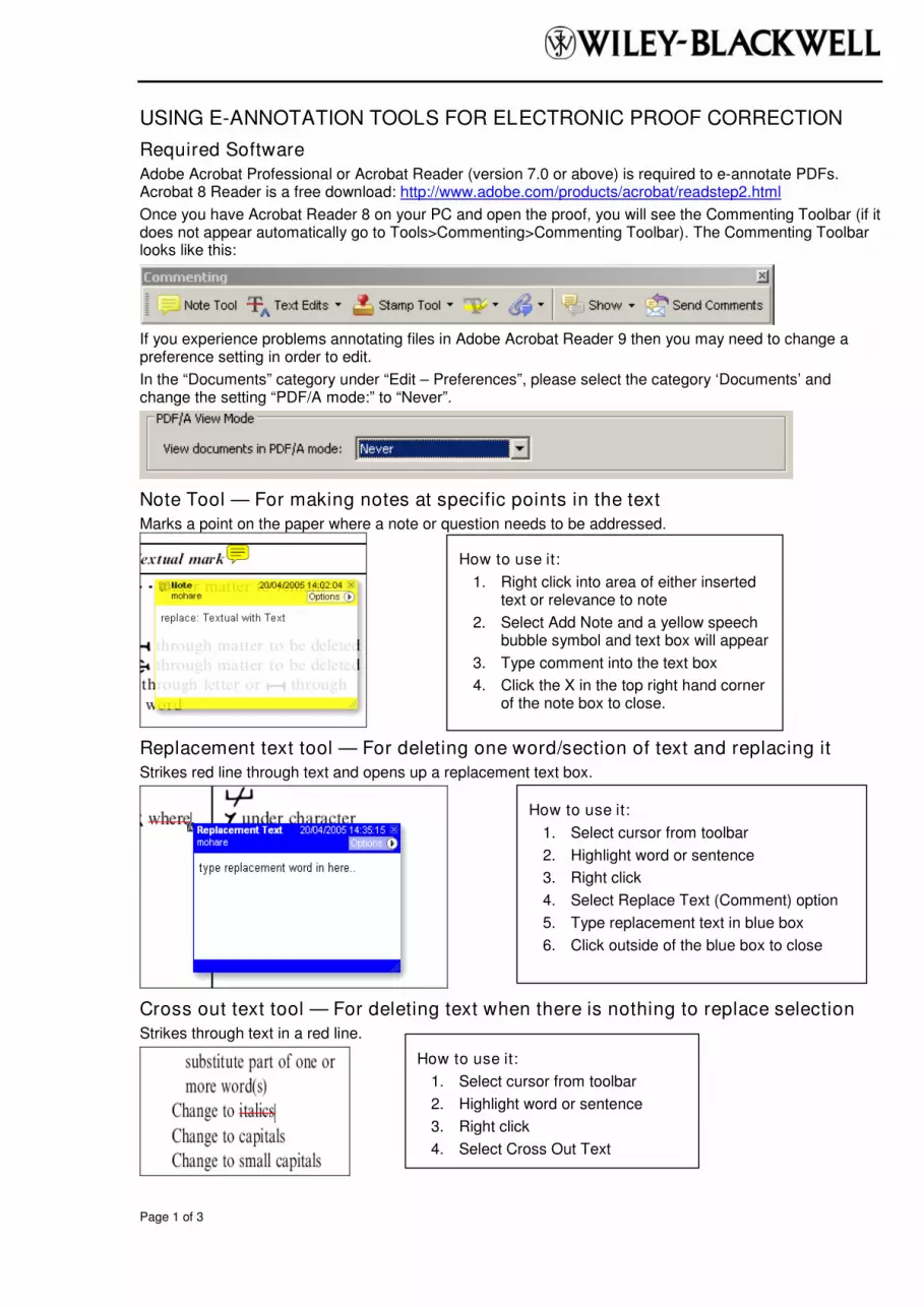
USING E-ANNOTATION TOOLS FOR ELECTRONIC PROOF CORRECTION
Required Software Adobe Acrobat Professional or Acrobat Reader (version 7.0 or above) is required to e-annotate PDFs. Acrobat 8 Reader is a free download: http://www.adobe.com/products/acrobat/readstep2.html
Once you have Acrobat Reader 8 on your PC and open the proof, you will see the Commenting Toolbar (if it does not appear automatically go to Tools>Commenting>Commenting Toolbar). The Commenting Toolbar looks like this:
If you experience problems annotating files in Adobe Acrobat Reader 9 then you may need to change a preference setting in order to edit.
In the “Documents” category under “Edit – Preferences”, please select the category ‘Documents’ and change the setting “PDF/A mode:” to “Never”.
Note Tool — For making notes at specific points in the text Marks a point on the paper where a note or question needs to be addressed.
Replacement text tool — For deleting one word/section of text and replacing it Strikes red line through text and opens up a replacement text box.
Cross out text tool — For deleting text when there is nothing to replace selection Strikes through text in a red line.
How to use it:
1. Right click into area of either inserted text or relevance to note
2. Select Add Note and a yellow speech bubble symbol and text box will appear
3. Type comment into the text box
4. Click the X in the top right hand corner of the note box to close.
How to use it:
1. Select cursor from toolbar
2. Highlight word or sentence
3. Right click
4. Select Replace Text (Comment) option
5. Type replacement text in blue box
6. Click outside of the blue box to close
How to use it:
1. Select cursor from toolbar
2. Highlight word or sentence
3. Right click
4. Select Cross Out Text

Page 2 of 3
Approved tool — For approving a proof and that no corrections at all are required.
Highlight tool — For highlighting selection that should be changed to bold or italic. Highlights text in yellow and opens up a text box.
Attach File Tool — For inserting large amounts of text or replacement figures as a files. Inserts symbol and speech bubble where a file has been inserted.
Pencil tool — For circling parts of figures or making freeform marks Creates freeform shapes with a pencil tool. Particularly with graphics within the proof it may be useful to use the Drawing Markups toolbar. These tools allow you to draw circles, lines and comment on these marks.
How to use it:
1. Click on the Stamp Tool in the toolbar
2. Select the Approved rubber stamp from the ‘standard business’ selection
3. Click on the text where you want to rubber stamp to appear (usually first page)
How to use it:
1. Select Highlighter Tool from the commenting toolbar
2. Highlight the desired text
3. Add a note detailing the required change
How to use it:
1. Select Tools > Drawing Markups > Pencil Tool
2. Draw with the cursor
3. Multiple pieces of pencil annotation can be grouped together
4. Once finished, move the cursor over the shape until an arrowhead appears and right click
5. Select Open Pop-Up Note and type in a details of required change
6. Click the X in the top right hand corner of the note box to close.
How to use it:
1. Click on paperclip icon in the commenting toolbar
2. Click where you want to insert the attachment
3. Select the saved file from your PC/network
4. Select appearance of icon (paperclip, graph, attachment or tag) and close
