sc35 promotes sustainable stress-induced alternative splicing of neuronal acetylcholinesterase mrna
TRANSCRIPT

ORIGINAL RESEARCH ARTICLE
SC35 promotes sustainable stress-induced alternativesplicing of neuronal acetylcholinesterase mRNAE Meshorer1, B Bryk1,4, D Toiber1, J Cohen1,2, E Podoly1,3, A Dori2 and H Soreq1
1Department of Biological Chemistry, The Hebrew University of Jerusalem, Jerusalem, Israel; 2Department of Neurology,Soroka University Medical Center, Beer-Sheva, Israel; 3The Wolfson Centre for Structural Biology, The Hebrew Universityof Jerusalem, Jerusalem, Israel
Long-lasting alternative splicing of neuronal acetylcholinesterase (AChE) pre-mRNA occursduring neuronal development and following stress, altering synaptic properties. To explore thecorresponding molecular events, we sought to identify mRNAs encoding for abundant splicingfactors in the prefrontal cortex (PFC) following stress. Here we show elevated levels of thesplicing factor SC35 in stressed as compared with naı̈ve mice. In cotransfections of COS-1 andHEK293 cells with an AChE minigene allowing 30 splice variations, SC35 facilitated a shift fromthe primary AChE-S to the stress-induced AChE-R variant, while ASF/SF2 caused the oppositeeffect. Transfection with chimeric constructs comprising of SC35 and ASF/SF2 RRM/RSdomains identified the SC35 RRM as responsible for AChE mRNA’s alternative splicing. Inpoststress PFC neurons, increased SC35 mRNA and protein levels coincided with selectiveincrease in AChE-R mRNA. In the developing mouse embryo, cortical progenitor cells in theventricular zone displayed transient SC35 elevation concomitant with dominance of AChE-Rover AChE-S mRNA. Finally, transgenic mice overexpressing human AChE-R, but not thoseoverexpressing AChE-S, showed significant elevation in neuronal SC35 levels, suggesting areciprocal reinforcement process. Together, these findings point to an interactive relationshipof SC35 with cholinergic signals in the long-lasting consequences of stress on nervoussystem plasticity and development.Molecular Psychiatry (2005) 10, 985–997. doi:10.1038/sj.mp.4001735; published online 25 August2005
Keywords: SC35; stress; acetylcholinesterase; alternative splicing; ASF/SF2
Long-lasting changes in alternative splicing in thenervous system1,2 are associated with disease,3 agingand trauma,4 as well as with cortical neurogenesis.5 Inrats, both adult and prenatal stress reduce learningperformance,6 suggesting, among other possibilities,parallel stress-induced changes in alternative spli-cing. However, the molecular mechanisms leading tosuch changes are still largely obscure. Missplicingevents are particularly prominent in human dis-eases.7–9 For example, impaired splicing patterns ofthe glutamate transporter gene EAAT2 is associatedwith sporadic amyotrophic lateral sclerosis (ALS).10
Changes in nitric oxide synthase (NOS) mRNAsplicing was also reported in reactive astrocytes inthe same disease.11 This suggests plausible involve-ment of components of the basic splicing machinery,
such as SR protein splicing factors, in ALS and othersplicing-associated diseases.
Both stress and neurogenesis involve extendedoverexpression phases of AChE-R, the stress-asso-ciated splice variant of the acetylcholine hydrolyzingenzyme, acetylcholinesterase (AChE).12,13 The stress-and development-associated modulations in AChEgene expression involve facilitated transcription,altered promoter usage, modified splicing patternsof the 30 variants of the AChE pre-mRNA, andincreased stability of the normally rare and unstableAChE-R mRNA transcript.12,14 This renders AChE pre-mRNA processing an appropriate model system forexploring the involvement of alternative splicingmodulations in the delayed effects of acute stress inthe adult and developing brain.
Splicing alterations likely involve changes inmembers of the serine-arginine (SR) rich proteins,implicated in alternative splicing and splice siteselection.15 SR proteins comprise of a structurallyand functionally related family, involved in multiplesteps of both constitutive and alternative splicing. SRproteins, like other eukaryotic proteins bindingsingle-stranded RNA, include one or more copies ofan RNA-binding domain of about 90 amino acids,known as RNA recognition motif (RRM).16 The RRM
Received 12 April 2005; revised 14 July 2005; accepted 19 July2005; published online 23 August 2005
Correspondence: Current address: E Meshorer, National CancerInstitute, National Institutes of Health, 41 Library Dr, Bethesda,MD 20892, USA. E-mail: [email protected] or H Soreq,Department of Biological Chemistry, The Hebrew University ofJerusalem, Jerusalem, Israel. E-mail: [email protected] address: EMBL Heidelberg, Meyerhofstrasse 1, Heidelberg
D-69117, Germany.
Molecular Psychiatry (2005) 10, 985–997& 2005 Nature Publishing Group All rights reserved 1359-4184/05 $30.00
www.nature.com/mp

structure consists of four strands and two helicesarranged in an alpha/beta sandwich, with a thirdhelix present during RNA binding in some cases.17
Also, characteristic of SR proteins are modularstructures with a conserved C-terminal arginine/serine-rich domain (RS domain) that interacts withcomponents of the basic splicing machinery.15
The RRMs recognize weakly conserved RNA se-quences, identified as either exonic or intronicsplicing enhancers (ESE/ISE) or suppressors (ESS/ISS).18 In vivo, the fine-tuned balance between SR-proteins, heterogeneous nuclear ribonucleic proteinparticles (hnRNPs), splice sites and enhancer/silencerelements is likely to be subject to modulations leadingto changes in the exon usage of pre-mRNA. The levelsof both SR-proteins and hnRNPs vary among tissues19
and can be further modulated by protein phosphor-ylation, leading to their release from intranuclearstorage compartments, such as the speckle domains atthe nuclear internal boundaries.20,21 Tissue-specificfactors, especially in the nervous system, also modifythese levels.3
Among the major SR proteins, ASF/SF2 contributesto 50 splice site selection, through protein–proteininteractions involving the RS domain,22 whereasSC35 is required for formation of the earliest ATP-dependent splicing complex with the U1 and U2snRNPs and the pre-mRNA. SC35 also interacts, viaits RS domain, with spliceosomal components boundto both the 50 and 30 splice sites during spliceosomeassembly. This forms a bridge between the 50 and 30
splice site binding components, U1 snRNP and U2AF.Identifying splicing-related proteins involved in
mammalian psychological stress-responses may notonly shed light on the molecular mechanism of AChEinvolvement in stress, but may have an imperativeimpact on the understanding of gene expressionpatterns in CNS pathologies in general includingchronic stress, post-traumatic stress disorder (PTSD)and other neurodegenerative disorders. Additionally,the levels of various neuromodulators are modifiedfollowing stress in the prefrontal cortex (PFC), one ofthe major areas involved in mammalian stressresponses.23 Examples include serotonin,24 dopa-mine,25 acetylcholine26 and glutamate.27 Functionalimplications of such changes involve, for example,impairment of spatial working memory.28 Therefore,we predicted that changes in the expression of SR-related proteins within the PFC might be a means toadapt to the new situation.
Here, we identified significant and long-lastingoverexpression of the splicing factor SC35 weeksafter stress. In cotransfection experiments with SC35and an AChE minigene, SC35 shifted alternativesplicing towards the stress-associated variant, AChE-R mRNA. The relationship between SC35 andAChE-R was further demonstrated in the mousebrain after stress and during embryonic development.To our knowledge, this is the first report of theinvolvement of a splicing factor in mammalian stressresponses.
Materials and methods
Adult animals
Naı̈ve FVB/N mice were kept at a 12 h dark/12 h lightdiurnal schedule and watered/fed ad libitum. Stressand corticosterone experiments included two 4-mindaily swim sessions or a daily intraperitoneal (i.p.)injection (10 mg/kg) of corticosterone (Sigma, SaintLouis, MO, USA) or saline for 4 consecutive days.Mice were killed 2 weeks thereafter, and brains weredissected on ice and frozen in liquid nitrogen. Bloodwas collected either from the orbital sinus underhalothane (Rhodia, Bristol, UK) anesthesia using anEDTA-containing capillary, or, upon decapitation,directly from the aorta into EDTA-containing centri-fuge tubes. Corticosterone levels were determined byradioimmunoassay (Coat-A-Count, DPC, Los Angeles,CA, USA) in plasma separated by slow centrifugation(500 g, 30 min, 41C).
Embryos
CD1 mice were kept at a 12 h dark/12 h light diurnalschedule and watered/fed ad libitum. Females weremated by housing with a male mouse overnight andexamined the next morning for the presence ofvaginal plugs to indicate conception, which wasdesignated embryonic day (E) 0. Embryos at E11, 13and 15 were collected for this study. At 3 min prior tokilling, pregnant dams were deeply anesthetized byan intramuscular injection of a ketamine (50 mg/kg)and xylazine (10 mg/kg) mixture. Embryos werequickly removed from the dams by hysterectomy,transferred to ice cold phosphate-buffered saline(PBS), and the embryonic sac was removed. Embryo-nic age was verified by measuring crown-rump length(CRL), and only embryos that exhibited close toaverage CRL values were employed. The embryoswere decapitated and whole heads were immersed in4% paraformaldehyde in PBS for 48 h at 41C,dehydrated in alcohol and embedded in paraffin.Coronal 4 mm sections through the developing soma-tosensory cortex were collected by adhesion toSuperfrosts-Plus slides (Menzel-Glaser, Braunsch-weig, Germany).
Cells
COS-1 or human embryonic kidney (HEK) 293 cellswere grown in six-well plates in a humidified atmo-sphere in Dulbecco’s modified Eagle’s medium(DMEM, Biological Industries) supplemented with10% fetal calf serum (FCS) and 2 mM L-glutamine at371C, 5% CO2. DNA transfections were carried outusing Lipofectamine-Plus (Gibco BRL Life Technolo-gies, Bethesda, MD, USA) with 1 mg plasmid DNA perwell as instructed. At 24 h post-transfections, cellswere harvested with RNA-Later reagent (Ambion,Austin, TX, USA) and RNA was extracted usingRNeasy Mini Kit (Qiagen, Hilden, Germany) anddiluted to 100 ng/ml.
SC35 in long-term stress responsesE Meshorer et al
986
Molecular Psychiatry

Real-time RT-PCR
LightCycler (Roche Diagnostics, Basel, Switzerland)equipped with dedicated software (ver. 3.5) was usedto amplify 200 ng samples of RNA template (SYBRGreen I RNA amplification kit, Roche). Primers(Sigma, Jerusalem, Israel) used in this study areshown in Table 1. In all experiments, values werenormalized to actin mRNA, which remained un-changed. Samples that displayed deviated levels ofactin mRNA by 1.5 or more were discarded.
In situ hybridization
Paraffin-embedded horizontal whole-brain sections ofstressed and control mice were used, essentially asdescribed.13 Synthetic 20-O-methyl 50-biotin-labeledRNA probe (Microsynth, Balgach, Switzerland) forSC35 was as described.29 For ASF/SF2, the probe 50-TGCGACUCCUGCUGUUGCUUCUGCUACGGCUUCUGCUACGACUACGGCUU-30 was designed using Oligosoftware (Molecular Biology Insights, Cascade, CO,USA). Streptavidin–fluorescein conjugate diluted1 : 100, an anti-AP converter and Fast Red substrate(all from Roche Diagnostics), were used for detection.Paraffin-embedded sections of embryonic brains weresimilarly processed, applying streptavidin–Cy5 con-jugate for detection of biotinylated probes directed toAChE-R mRNA, and antidigoxygenin–Cy3 conjugatefor detection of digoxygenin-labeled probes directedto AChE-S mRNA (Jackson ImmunoResearch Labora-tories, Cambridgeshire, UK).
Immunohistochemistry
Paraffin sections were deparaffinized with xylene,rehydrated in graded ethanol solutions and DDW, andincubated for 5 min in PBS. For antigen retrieval,slides were heated in a microwave oven at maximalpower (850 W, 10 min) in 500 ml of 0.01 M citricbuffer, pH 6.0, cooled for 5 min, and rinsed with PBS.Sections were incubated with anti-SC35 (Sigma;diluted to 1 : 50 in PBS with 0.5% Tween-20, 1.5 h,room temp.). Following a rinse with PBS andincubation with biotinylated anti-mouse IgG (VectorLaboratories, Burlingame, CA, USA; diluted 1 : 200 inPBS with 0.5% Tween-20, 1 h, room temp.), sectionswere rinsed with PBS and incubated with avidin-bound peroxidase complex (ABC Elite kit, VectorLaboratories, 1 h, room temp.) or Cy3-conjugatedstreptavidin (Jackson Immunoresearch). After rinsingwith 0.05 M Tris pH 7.6, the peroxidase was reactedfor 90 s with 0.05% diaminobenzidine (Sigma) sup-plemented with 0.05% nickel ammonium sulfate and0.006% H2O2 in 0.05 M Tris pH 7.6 for color inten-sification. The color reaction was terminated byrinsing with PBS, sections were dehydrated in ascendingconcentrations of alcohol, cleared in xylene andcovered with Eukitt (Calibrated Instruments,Hawthorne, NY, USA). T
able
1P
rim
er
sequ
en
ces
use
din
this
stu
dy
Gen
eA
ccess
ion
#F
orw
ard
pri
mer
Posi
tion
Revers
ep
rim
er
Posi
tion
SR
p20
Z85986
GC
CG
TG
TA
AG
AG
TG
GA
374–389
AA
GG
GC
TA
GT
TG
AC
TG
G540–556
SR
p30c
U30825
CA
GT
GT
CG
GC
TT
CG
TG
TG
GA
287–306
AG
AC
CG
AG
AC
CG
TG
AG
TA
GC
624–643
SR
p40
U30826
CG
AG
GT
GG
AA
GA
GG
TA
GA
GG
318–337
GG
CA
CG
GG
AG
AC
CT
AC
TA
AC
777–796
SR
p55
U30883
CG
GA
TG
CC
CA
CA
AG
GA
AC
GA
521–540
AC
CT
GG
AA
CG
GG
AG
CG
AC
TT
768–787
SF
2p
32
L04636
GA
AT
GG
GA
CA
GA
AG
CG
A417–433
GT
GG
TC
AT
AT
AA
GG
CC
CA
G774–792
U5-1
00
AF
026402
CA
GC
GT
CA
GG
CA
AT
TC
1291–1306
CC
TA
GC
GG
TT
TC
CC
AA
1511–1526
9G
8L
22253
GG
CG
CT
GG
CA
AA
GG
AG
AG
TT
166–185
CG
GC
TG
CT
TC
TT
GG
TC
GT
GA
715–724
SC
35
AF
250135
TC
CA
AG
TC
CA
AG
TC
CT
CC
TC
31–50
AC
TG
CT
CC
CT
CT
TC
TT
CT
GG
163–182
AS
F/S
F2
M69040
TC
GA
GT
CC
GC
GC
TT
TT
CG
99–117
GC
TT
CG
AG
GA
AA
CT
CC
AC
380–397
p54
NM
_004768
TC
TT
CT
GC
CT
GG
TG
GT
GG
AC
482–501
GG
GA
TG
GT
GA
GC
GG
GA
CA
AT
1175–1194
hn
RN
PA
1N
M_002136
AA
AG
TC
TC
TC
TT
CA
CC
CT
GC
81–100
AA
GT
GG
GC
AC
CT
GG
TC
TT
TG
390–409
AC
hE
-SA
F002993
CT
TC
TG
GA
AC
CG
CT
TC
CT
CC
CC
AA
AT
T7855–7881
GT
AG
TG
GT
CG
AA
CT
GG
TT
CT
TC
CA
GT
GC
AC
6916–6945
AC
hE
-RA
F002993
CT
TC
TG
GA
AC
CG
CT
TC
CT
CC
CC
AA
AT
T7855–7881
GG
TT
AC
AC
CT
GG
CG
GG
CT
CC
7779–7798
mb-
acti
nX
03672
CA
AT
TC
CA
TC
AT
GA
AG
TG
TG
AC
917–938
AT
CT
TG
AT
CT
TC
AT
GG
TG
CT
1047–1066
hb-
acti
nA
J251794/5
CA
CT
CT
TC
CA
GC
CT
TC
CT
TC
C1–21
CG
GA
CT
CG
TC
AT
AC
TC
CT
GC
TT
1–22
Sta
nd
ard
cu
rves
were
pro
du
ced
for
each
pri
mer
set
usi
ng
fou
rd
iffe
ren
td
ilu
tion
s(t
wo-,
fou
r-,
eig
ht-
or
16-f
old
)of
tem
pla
teR
NA
or
cD
NA
as
desc
ribed
.59
SC35 in long-term stress responsesE Meshorer et al
987
Molecular Psychiatry

Structural modelingThe RRM domains of SC35 and ASF/SF2 weremodeled using SWISS-MODEL30 and were super-imposed using Deep View spdbv 3.7.
Results
Long-lasting cooverexpression of SC35 and AChEfollowing stressTo search for splicing factors that might mediatestress-induced changes in neuronal long-lasting alter-native splicing, we subjected FVB/N mice to 4consecutive days of repeated swim sessions(2�4 min) and killed them 2 weeks later.13 Orbitalsinus blood of stressed mice served to quantifycorticosterone levels following the second swimsession as compared with naı̈ve, nonstressed mice.Plasma corticosterone levels rose by ca. five-foldfollowing stress (from 50713 to 259753 ng/ml,Po0.05, two-tailed Student’s t-test, Figure 1a), andreceded to close to normal levels (76725, P40.1) 2weeks later. In comparison, we subjected mice torepeated corticosterone (i.p., 10 mg/kg) or saline injec-tions. Plasma corticosterone rose by ca. four- andtwo-fold and receded to normal levels 2 weeks later(Figure 1a), demonstrating the peripherally transientnature of both these hormone and stress effects.
Using RNA extracted from the PFC of poststress ascompared to hormone-treated or naı̈ve brains, wetested the expression level of several abundant SR-related transcripts. Real-time RT-PCR quantificationinvolved SRp20, SRp30c, SRp40, SRp55, p54, 9G8,ASF/SF2 and SC35, as well as hnRNP A1, the U5-100 kDa and the 32 kDa subunit of SF2, SF2p32. Mostof the splicing factors tested did not display any long-lasting stress-induced expression difference (Figure1b). Exceptions were SC35 and U5-100 kDa, both ofwhich displayed significant sustained overexpression(Po0.05, Kruskal–Wallis test, Figure 1b). In addition,9G8 showed a slight, nonsignificant downregulation,and p54 and SF2p32 displayed nonsignificant upre-gulation. Neither saline nor corticosterone inducedsuch changes in PFC mRNAs (Figure 1b), suggestingthat the observed changes reflected a genuine long-lasting stress response.
To test whether the stress-associated increases inSC35 mRNA levels would occur in cultured cells aswell, we subjected COS-1 and HEK-293 cells to avariety of different stressors: UV (germicidal 254-nmUV lamp, 2 J/m2/s, 30 s), H2O2 (500mM), sorbitol(600 mM), dexamethasone (10mM) and cortisol(10 mM). Cells were harvested 6 h following treat-ments, total mRNA was extracted and subjected toreal-time RT-PCR analyses using primers for SC35 andb-actin. No change was found in the mRNA levels forSC35 in all of the different treatments (Figure 1c).
SC35 shifts AChE’s alternative splicing towardsAChE-R in transfected cellsThe long-lasting association between AChE-R andSC35 poststress increases could be either direct or
300
200
100
0
stress corticosterone
5
b
c
a
3
1
0.5
ct sal
Act
inU
V
hydr
ogen
per
oxid
eso
rbito
lco
rtico
ster
one
dexa
met
haso
ne UV
hydr
ogen
per
oxid
eso
rbito
lco
rtico
ster
one
dexa
met
haso
ne
SC
35S
F2p3
2U
5-10
0 kD
SR
p20
SR
p30c
SR
p40
SR
p55
p54
9G8
AS
F/S
F2hn
RN
P A
1
Act
inS
C35
AS
F/S
F2
cor
strs
sal-2
w
cor-2
w
strs-
2w
Pla
sma
cort
icos
tero
ne(n
g/m
l)
Fol
d di
ffere
nce
SC35
HEK293 COS-12
1
2
1
*
Figure 1 Prefrontal cortex following repeated organismalstress display long-term SC35 overexpression. (a) Plasmacorticosterone levels in control (ct, n¼ 4), saline-injected(sal, n¼ 3), corticosterone-injected (cor, n¼ 4) and stressedmice (4 left columns, n¼ 4) and 2 weeks later (2w, rightcolumns). (b) mRNA expression levels for a selection ofsplicing-related factors using real-time RT-PCR on RNAextracted from mouse prefrontal cortex 2 weeks followingrepeated stress (stress, n¼ 4) or 2 weeks followingrepeated corticosterone injections (corticosterone, n¼ 4).b-Actin (left, n¼ 4) was used as control. (c) SC35 mRNAexpression level following different cellular stressors inHEK293 (left, n¼ 8) and COS-1 (right, n¼ 8) cells. Whenanimals were used, the given value is an average of atleast three different measurements7standard deviationsfrom three or more different animals. Asterisks notestatistically significant differences from control (Po0.05,Kruskal–Wallis test).
SC35 in long-term stress responsesE Meshorer et al
988
Molecular Psychiatry

indirect. To distinguish between these possibilities,we scanned the AChE pre-mRNA sequence for ESEand ISE. To focus on potentially relevant sequences,we selected only those that were present in both thehuman and mouse ACHE genes. Selected sequencesinclude motifs for the splicing factors SC35, ASF/SF2and SRp20 in the 30 alternative splicing-prone regionof both human and mouse pre-mRNAs (Figure 2a).These can potentially serve both as ESEs or ESS. TheSC35 and SRp20 motifs are found within the intronbetween exons 5 and 6, whereas the ASF/SF2 motifappears in exon 6.
To test for causal relationships, we examined theinfluence of SC35, ASF/SF2 and SRp20 on thesplicing pattern of an ACHE-minigene construct,offering two 30 splice options31 (Figure 2b). SC35,ASF/SF2 or SRp20 were cotransfected with theACHE-minigene into COS-1 or HEK293 cells, both ofwhich possess low levels of endogenous AChEactivity.32
The CMV promoter was reported to generateprimarily ‘readthrough’ transcripts, for example,when inserted in front of the EDI minigene.33
Compatible with this tendency, when the CMV-ACHEminigene was cotransfected together with an insert-free plasmid or a GFP plasmid, much of the mRNAthat was generated was of the readthrough form,AChE-R. Cotransfections with SC35 further elevatedAChE-R mRNA levels in a dose-dependent manner,reflecting avoidance of the splice site between E4 andI4. Parallel slightly reduced AChE-S mRNA levelsindicated suppressed efficacy of the splicing eventlinking E4 with E6. Together, this induced a change ofover five-fold in the R/S ratio (Figure 2c, d, Po0.05,Kruskal–Wallis test). In contrast, cotransfections withASF/SF2 shifted splicing towards the AChE-S form,however, in a much more variable manner. Finally,cotransfection of the ACHE minigene with the SRprotein SRp20 showed no apparent change in the
AChE-R/AChE-S splicing ratio (Figure 2). There wasno detectable contribution of unprocessed RNA to ourmeasured AChE-R levels, since both reactions with noRT, and cell lines that produce only AChE-S yielded
5
4
3
2
1
00.25 0.5 0.75 1
SC35 (µg DNA)
AC
hE-R
/ A
ChE
-S r
atio
SC35
200 bp
4'
ASF
SRp20
hm
hm
hm
a
Ampr
ACHEmini-gene
CMV promoter SV40 OriRSV40 pA
ACHE
2 3 4 5 64'
2 3 4 5 2 3 4 6
AChE-R AChE-S
b
AChE-S
5
4
3
2
1
0.5
0.33
0.25
AChE-R
0.1 0.2 0.5 10.1 0.2
+GFP +SC35 (µg DNA) +ASF +SRp20
0.5 1
Fol
d di
ffere
nce
*
c
d
Figure 2 Transfected SC35 shifts alternative splicingtowards AChE-R mRNA. (a) Putative splicing factor bindingsites on the 30 region of AChE pre-mRNA. Consensussequences identified for SC35, ASF/SF2 and SRp20 inhuman (h) and mouse (m) AChE pre-mRNA are shown.Triangles note their position on the AChE primary tran-script. (b) Schematic diagram of the AChE minigene used ina cotransfection assay. Ampicillin resistance (Ampr), CMVpromoter, SV40 origin (OriR) and polyadenylation (pA) sitesand the AChE genomic DNA insert spanning the entirecoding sequence, from exon 2 to exon 6, including allintrons are noted. This insert allows production of eitherAChE-S or AChE-R transcripts, shown below. (c) Real-timeRT-PCR analysis of cotransfection of the AChE mini-genewith SRp20, SC35 and ASF/SF2. Cotransfected GFP servedas negative control (left). Columns show fold differencefrom control for AChE-R and AChE-S mRNA. SC35 wastransfected in a concentration-dependent manner. Numbersrepresent mg plasmid transfected. Asterisk notes Po0.05(Kruskal–Wallis test, n¼ 8). (d) AChE-R/AChE-S mRNAratio as a function of mg SC35 transfected.
SC35 in long-term stress responsesE Meshorer et al
989
Molecular Psychiatry
no AChE-R product (see Figure 3 below and data notshown).
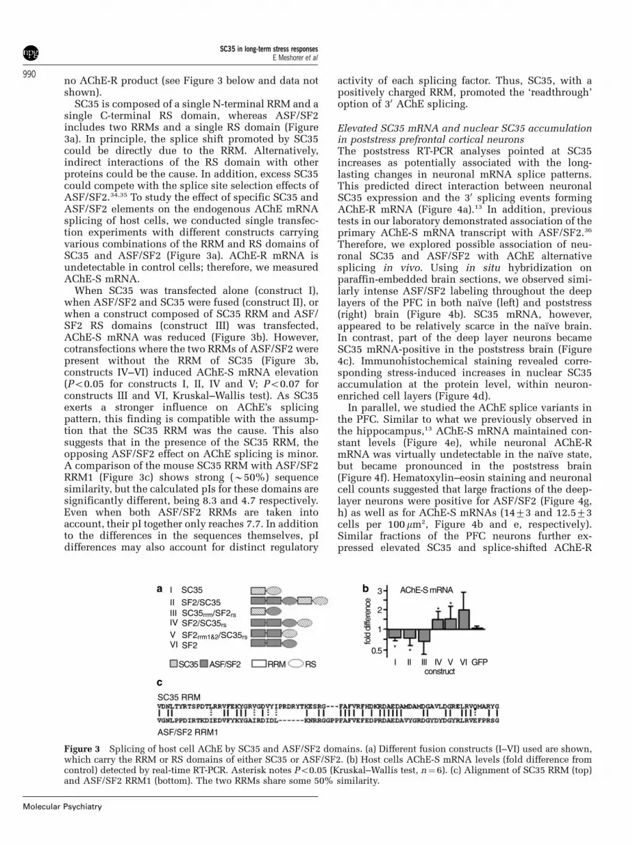
SC35 is composed of a single N-terminal RRM and asingle C-terminal RS domain, whereas ASF/SF2includes two RRMs and a single RS domain (Figure3a). In principle, the splice shift promoted by SC35could be directly due to the RRM. Alternatively,indirect interactions of the RS domain with otherproteins could be the cause. In addition, excess SC35could compete with the splice site selection effects ofASF/SF2.34,35 To study the effect of specific SC35 andASF/SF2 elements on the endogenous AChE mRNAsplicing of host cells, we conducted single transfec-tion experiments with different constructs carryingvarious combinations of the RRM and RS domains ofSC35 and ASF/SF2 (Figure 3a). AChE-R mRNA isundetectable in control cells; therefore, we measuredAChE-S mRNA.
When SC35 was transfected alone (construct I),when ASF/SF2 and SC35 were fused (construct II), orwhen a construct composed of SC35 RRM and ASF/SF2 RS domains (construct III) was transfected,AChE-S mRNA was reduced (Figure 3b). However,cotransfections where the two RRMs of ASF/SF2 werepresent without the RRM of SC35 (Figure 3b,constructs IV–VI) induced AChE-S mRNA elevation(Po0.05 for constructs I, II, IV and V; Po0.07 forconstructs III and VI, Kruskal–Wallis test). As SC35exerts a stronger influence on AChE’s splicingpattern, this finding is compatible with the assump-tion that the SC35 RRM was the cause. This alsosuggests that in the presence of the SC35 RRM, theopposing ASF/SF2 effect on AChE splicing is minor.A comparison of the mouse SC35 RRM with ASF/SF2RRM1 (Figure 3c) shows strong (B50%) sequencesimilarity, but the calculated pIs for these domains aresignificantly different, being 8.3 and 4.7 respectively.Even when both ASF/SF2 RRMs are taken intoaccount, their pI together only reaches 7.7. In additionto the differences in the sequences themselves, pIdifferences may also account for distinct regulatory
activity of each splicing factor. Thus, SC35, with apositively charged RRM, promoted the ‘readthrough’option of 30 AChE splicing.
Elevated SC35 mRNA and nuclear SC35 accumulationin poststress prefrontal cortical neuronsThe poststress RT-PCR analyses pointed at SC35increases as potentially associated with the long-lasting changes in neuronal mRNA splice patterns.This predicted direct interaction between neuronalSC35 expression and the 30 splicing events formingAChE-R mRNA (Figure 4a).13 In addition, previoustests in our laboratory demonstrated association of theprimary AChE-S mRNA transcript with ASF/SF2.36
Therefore, we explored possible association of neu-ronal SC35 and ASF/SF2 with AChE alternativesplicing in vivo. Using in situ hybridization onparaffin-embedded brain sections, we observed simi-larly intense ASF/SF2 labeling throughout the deeplayers of the PFC in both naı̈ve (left) and poststress(right) brain (Figure 4b). SC35 mRNA, however,appeared to be relatively scarce in the naı̈ve brain.In contrast, part of the deep layer neurons becameSC35 mRNA-positive in the poststress brain (Figure4c). Immunohistochemical staining revealed corre-sponding stress-induced increases in nuclear SC35accumulation at the protein level, within neuron-enriched cell layers (Figure 4d).
In parallel, we studied the AChE splice variants inthe PFC. Similar to what we previously observed inthe hippocampus,13 AChE-S mRNA maintained con-stant levels (Figure 4e), while neuronal AChE-RmRNA was virtually undetectable in the naı̈ve state,but became pronounced in the poststress brain(Figure 4f). Hematoxylin–eosin staining and neuronalcell counts suggested that large fractions of the deep-layer neurons were positive for ASF/SF2 (Figure 4g,h) as well as for AChE-S mRNAs (1473 and 12.573cells per 100mm2, Figure 4b and e, respectively).Similar fractions of the PFC neurons further ex-pressed elevated SC35 and splice-shifted AChE-R
SC35 RRM
ASF/SF2 RRM1
c
b
construct
AChE-S mRNA3
fold
diff
eren
ce
2
1
0.5I II III IV V VI GFP
**
**
a I SC35
II SF2/SC35III SC35rrm/SF2rs
SF2/SC35rs
SF2rrm1&2/SC35rsSF2
SC35 ASF/SF2 RRM RS
IVVVI
Figure 3 Splicing of host cell AChE by SC35 and ASF/SF2 domains. (a) Different fusion constructs (I–VI) used are shown,which carry the RRM or RS domains of either SC35 or ASF/SF2. (b) Host cells AChE-S mRNA levels (fold difference fromcontrol) detected by real-time RT-PCR. Asterisk notes Po0.05 (Kruskal–Wallis test, n¼ 6). (c) Alignment of SC35 RRM (top)and ASF/SF2 RRM1 (bottom). The two RRMs share some 50% similarity.
SC35 in long-term stress responsesE Meshorer et al
990
Molecular Psychiatry

mRNA following stress (1574 and 1776 cells per100mm2, respectively), with parallel increases at theSC35 protein level (Figure 4i). Together, this suggestsa stress-induced change from AChE-S mRNA, char-acteristic of the naı̈ve brain to an SC35-AChE-RmRNA association in the poststress PFC.
Transient SC35 overexpression coincides withAChE-R/AChE-S dominance in the developing brainMaternal stress during pregnancy was reported tocause long-lasting deleterious effects in the offspring,6
suggesting that stress affects brain development. Also,we have recently shown that neuronal progenitor
cells express both AChE-S and AChE-R, which exertdistinct effects on cortical development.37 To explorethe possibility that SC35–AChE interactions areinvolved, the developing mouse cortex was subjectedto comparative in situ hybridization and immunohis-tochemistry analyses.
Excitatory neurons in the cerebral neocortex emergefrom a pseudostratified neuroepithelium in theventricular zone (VZ) that lines the ventricular cavityof the developing cerebral wall.38 The postmitoticneurons that are generated migrate away from the VZto reach the outer surface of the cortical wall andsubsequently differentiate in the developing cortical
Figure 4 Stress-induced SC35 overexpression in prefrontal cortical neurons. (a) ACHE gene structure (top) and neuronal 30
alternatively spliced products (bottom). (b–c) Fluorescent in situ hybridization for ASF/SF2 (b) and SC35 (c) mRNAs.Confocal images shown are series projections. Positively labeled cytoplasms for ASF/SF2 mRNA is stained red (arrows) inboth naı̈ve (b, left) and stressed (right) mice. SC35 was difficult to detect in naı̈ve brains (c, left) but was notablyoverexpressed following stress (right). (d) Immunohistochemistry using monoclonal anti-SC35 antibody. Note the faintnuclear labeling of naı̈ve brain (left) as compared to the intense poststress labeling (right, arrows). (e–f) Fluorescent in situhybridization for AChE-R (e) and AChE-S (f) mRNAs. AChE-R displayed marked overexpression in the prefrontal cortex 2weeks following stress, while AChE-S remained unchanged. (g) Schematic diagram of a horizontal mouse brain section. Therectangle shows the area of the images taken. (h) Hematoxylin–eosin staining shows tissue morphology. Nuclei are labeleddark purple (arrows). (i) Cell counts per 100 square microns in control (c) and stressed (st) tissues. Fluorescent measurementswere as described.13 Asterisks note significant differences (Po0.005, two-talied Student’s t-test. Values represent counts fromat least 40 cells from at least three different animals).
SC35 in long-term stress responsesE Meshorer et al
991
Molecular Psychiatry

plate (Figure 5a). In the murine embryonic brain,immunohistochemistry demonstrated labeling of theSC35 protein primarily in round nuclei of cellsadjacent to the ventricular lumen, where DNAreplication occurs, that is, during mitosis of theneuroepithelial cells (Figure 5b). At the onset ofneurogenesis (E11), the AChE-R and AChE-S mRNAvariants colocalized, intensively expressed in thecytoplasm of the majority of neuroepithelial cellsborn in the VZ, as shown by in situ hybridization(Figure 5c). At E13, AChE-S mRNA labeling exhibiteda significant decrease (Po0.01, two-tailed Student’s t-test), while AChE-R mRNA labeling remained rela-tively unchanged, yielding an elevated AChE-R/AChE-S ratio. The AChE-R transcript decreased later,at E15 (Po0.001), becoming coleveled again withAChE-S. The number of SC35 expressing cellsincreased by 31% at E13 but declined back by 57%at E15 (Figure 5d). At this time period, AChE-SmRNA expression levels were downregulated withthe advance of neurogenesis, to 59 and 46% of thelevel at E11. The reduction in AChE-R mRNA wasmore abrupt, down to 95% at E13 and 37% at E15(Figure 5d), suggesting causal association in thismodified AChE-R/AChE-S ratio of the splicing factorSC35 and its target AChE pre-mRNA during neuro-genesis.
SC35 overexpression in AChE-R transgenic miceSC35 exerts an autoregulatory, alternative splicingmediated, control over its own mRNA levels.39
The long-term SC35 overexpression following stresstherefore raised the question whether changes incholinergic neurotransmission, such as those occur-ring under long-term stress reactions, may modify theSC35 level maintained by this mode of control.To challenge this hypothesis, we explored SC35expression in transgenic animals constitutivelyoverexpressing neuronal AChE-S or AChE-R. Miceoverexpressing the synaptic human AChE-S (TgSmice) show accelerated stress-related pathology andneuromuscular malfunctions,12,40 whereas AChE-R-expressing mice (TgR) display normal neuromuscularfunction and their brains are relatively protected fromthe stress-associated hallmarks of pathology.12 SC35immunohistochemistry was carried out on 10-mm-thick paraffin-embedded coronal sections (Figure 6a,b) of FVB/N controls (Figure 6c), TgS (Figure 6d) andTgR (Figure 6e) mice, and fluorescence intensity inthe labeled nuclei was measured in the motor andprefrontal cortices. In the stress-prone TgS mice, thepopulation distribution of neuronal SC35 levels wascomparable to that of strain-matched FVB/N controls(wt). In contrast, in the stress-protected TgR mice,larger fractions of cortical neurons presented high
Figure 5 Transient dominance of AChE-R over AChE-S mRNA correlates with increased expression of SC35 during braindevelopment. (a) Schematic diagram of the developing cortex, from top to bottom, at embryonic days (E) 11, 13 and 15.Rectangles represent the areas of the images taken. STR, striatum; CTX, cortex (somato-sensory); CP, cortical plate; VZ,ventricular zone; V, ventricle; Th, thalamus. (b) Immunolabeling of SC35 protein (grayscale). Arrows point to mitotic cellsexpressing SC35. (c) Confocal images showing in situ hybridization labeling of AChE-R (Cy5, red pseudocolor) and AChE-SmRNA (Cy3, green pseudocolor) during cortical development (from E11, top to E15, bottom). Merged figures demonstrate thecolocalization of the AChE transcripts at E11 and E15 as well as AChE-R mRNA dominance at E13. Bars¼ 25mm. (d)Quantified intensity of AChE-S (green) and AChE-R (red) mRNA labeling compared to their expression at E11 (top). Ratiobetween AChE-R and AChE-S mRNA (columns) is compared to changes in SC35 expressing mitotic figures (squares) fromE11 (top) to E15 (bottom). For quantification, at least 80 cells were analyzed from at least five different brains. See text forP-values.
SC35 in long-term stress responsesE Meshorer et al
992
Molecular Psychiatry

levels of nuclear SC35 (Figure 6f). Support of thisobservation comes from an analysis of the distributionof signal densities in neuronal nuclei for all sectionsanalyzed. Although the distributions for FVB/Nand TgR cortices overlap broadly, they do differsignificantly (Po0.001, F¼22.3, two-way ANOVA).Multiple comparison analysis confirmed that TgRmice are different in a statistically significant mannerfrom TgS or wt mice (Po0.05, Bonferroni test). Theseresults suggest that neuronal AChE-R overexpressionin the murine brain additionally reinforces an eleva-tion of neuronal expression of SC35 itself.
Discussion
Neuronal PFC expression of the ACHE gene providesan appropriate test case for studying the involvement
of SR proteins in long-lasting stress responses,because AChE pre-mRNA undergoes a multitude ofchanges. Others have shown that AChE mRNA levelsdepend, to a large extent, on the HuD RNA-bindingprotein.41 Our present findings add the SR proteinSC35, as a key causal element in the maintenance andsplicing decisions of neuronal AChE mRNA levels.
The ex vivo capacity of SC35 to affect the splicingpattern of AChE was demonstrated in this study intransfected COS-1 and HEK293 cells. Both are easilyamenable to transfection and possess extremely lowlevels of endogenous AChE, which allowed simpleinterpretation of the results. Our findings that SC35and AChE-R mRNA levels appear to be directlyinterrelated in the stressed adult brain, the develop-ing mouse brain and the brain of TgR transgenicmice support the theory that SC35 influences AChE
Figure 6 Elevated SC35 levels in TgR mice. (a) Coronal mouse brain section depicting the area analyzed in this experiment.(b) Immunohistochemistry for SC35 was conducted on paraffin-embedded brain sections (boxed area in (a)) from adult age-and gender-matched mice of the noted strains. Bar¼ 100 mm. (c) SC35 immunostaining in wt FVB/N mice. (d) SC35 in TgSmice. (e) SC35 in TgR mice. Bar¼ 10mm. (f) Distribution of fluorescence density (arbitrary units) in wt (empty bars), TgS (graybars) and TgR (red bars) mice. Values represent n¼ 180 cells from six different animals of each strain. Note that corticalneurons from TgR, but not TgS mice, include larger fractions of cells with intensive SC35 labeling as compared to wild-typeFVB/N controls. Asterisk notes Po0.005, two-way ANOVA.
SC35 in long-term stress responsesE Meshorer et al
993
Molecular Psychiatry

pre-mRNA splicing in the same manner in neurons,and that AChE-R excess reciprocally elevates neuro-nal SC35 levels.
Comparison, using real-time RT-PCR, of AChEmRNA levels in the PFC of naı̈ve, saline- andcorticosterone-injected or psychologically stressedmice showed that stress, but not repeated injectionsof saline or corticosterone, induced long-lastingchanges in neuronal SC35 expression. Thus, theinitial activation of the HPA axis appears insufficientto turn on and maintain the long-term stress-inducedchanges in neuronal SC35 expression. That cortisol ordexamethasone treatment failed to induce SC35overexpression in cultured cells supports this notion.SC35 was, however, markedly overexpressed in thestressed brain for at least 2 weeks and shifted, incultured cells, the alternative splicing of the ACHEmini-gene towards AChE-R mRNA. Correspondingly,expression of SC35 at the mitotic phase of progenitorcells paralleled a transient dominance of AChE-R overAChE-S expression during murine cortical develop-ment. As SC35 expression declined, the AChE-Rdominance faded, further suggesting a relationshipbetween the two. Reciprocally, transgenic AChE-Rdominance in adult mice induced chronic neuronalSC35 elevation, indicating a reinforcement mechan-ism whereby SC35 supports overproduction of AChE-R and vice versa.
Splicing-related proteins in general, and SC35 inparticular, may play a central role in mammalianstress responses. By controlling and modifying theexpression patterns of stress-related proteins as anadaptation strategy, they can modify relevant tran-scripts from their usually expressed forms to speciallyadapted ones.4 The PFC long-lasting overexpressionof SC35 in close association to that of AChE-R isconsistent with this hypothesis. SC35 is also inten-sively expressed in the developing neocortex, primar-ily in progenitor cells as they pass through the mitoticphase of the cell cycle and express AChE-R. Pro-longed overexpression of SC35 and the morphogenicAChE-R protein,12 as would be the case underprenatal stress, may possibly impair cortical devel-opment, compromising cognitive performance. Whileadditional, yet nonidentified, splicing-related pro-teins are likely involved (eg Tra2-b1),21 the contribu-tion of SC35 appears to be pivotal for changes incholinergic neurotransmission.
As a member of the SR protein family, SC35possesses both an RRM and an RS domain, andaffects both splice site selection and alternativesplicing.42 SC35 further promotes splicing events thatdestabilize its own mRNA, exercising a self-controlmechanism that limits its durability.39 This, in turn,raised the question of how SC35 mRNA maintains itslong-lasting high levels in the poststress brain. Aplausible explanation is that the balance betweenSC35 production and SC35 mRNA alternative spli-cing and/or degradation might be subject to choliner-gic regulation. In this case, the increases in SC35levels would not be due to the cholinergic hyper-
excitation, which accompanies the initiation of stressreactions. Rather, its sustainable excess would reci-procally be due to cholinergic down-tuning andprolonged AChE-R excess (as is the case in TgRtransgenic mice). Further studies would be requiredto characterize a wider profile of splicing regulators inthe TgR brain and find out if this profile is modifiedin this animal model.
An additional splicing-related factor that showed amore moderate overexpression was SF2p32, the32 kDa subunit of the splicing factor ASF/SF2. ASF/SF2 was also shown to affect both alternative splicingand choice of splice sites.43 Interestingly, ASF/SF2also antagonizes SC35 activity in other target se-quences.34,44 In addition, SF2p32 was shown toinhibit ASF/SF2 activity.45 Thus, elevation ofSF2p32 should reduce ASF/SF2 activity even underconditions of unchanged ASF/SF2 levels such as thepoststress PFC, where SF2p32 mRNA was slightlyelevated, and ASF/SF2 slightly reduced. Althoughnot significant, these changes are compatible with theassumption of suppressed ASF/SF2 activity underlong-term stress responses.
The antagonized ASF/SF2 and SC35 activities leadus to structurally compare between the RRMs of these
Figure 7 Molecular modeling of SC35 and ASF/SF2structures. (a) Molecular modeling of ASF/SF2 structure.(b) Molecular modeling of SC35 structure. (c) Superimposi-tion of ASF/SF2 (red) and SC35 (blue) structures. Note thestrong similarity, with the exception of the four prolinescluster (green). (d) Enlargement of the proline cluster ofASF/SF2, shown as dashed square in panel c.
SC35 in long-term stress responsesE Meshorer et al
994
Molecular Psychiatry

two splicing factors. Molecular modeling showed thatASF/SF2 (Figure 7a), but not SC35 (Figure 7b),includes a cluster of four proline residues that arepositioned close to each other in the extended regionprotruding from this RRM domain (Figure 7c, d).Polyproline segments that are part of RRMs areknown in other splicing factors, where they wereproposed to serve as protein–protein interactionmotifs during spliceosome assembly.46 Our currentstudy raises the possibility that the proline-richdomain in ASF/SF2 contributes toward the dynamicequilibrium balancing between AChE-R and AChE-S,possibly by facilitating ASF/SF2 interactions withother spliceosomal components. The determiningfactor is hence SC35 regulation, which by changingSC35 levels may tilt this balance towards over-production of AChE-R.
The mechanisms by which stress initiates themodifications in the composition of splicing-relatedproteins and the sustained elevation in SC35 is stillunknown; however, neuronal activity modulationsare likely involved.47 Acetylcholine levels are notablyelevated under stress,13 and acetylcholine modulatesthe activity of neurons responding to other neuro-transmitters as well (eg glutamate, GABA48). There-fore, the capacity to downregulate acetylcholinelevels throughout the brain by the soluble AChE-Rmay be important. In contrast, elevated levels of thesynapse-adhered AChE-S would only affect choliner-gic circuits, creating an imbalanced cholinergicstatus. That SC35 levels are elevated in TgR, but notTgS mice, may imply that cholinergic imbalancesimpair this process.
Apart from SC35 and Tra1b, neuronal stressmodifies the subcellular localization of the hetero-nuclear ribonucleoprotein hnRNP A1 through theMAP kinase cascade, thereby changing alternativesplicing patterns.49 This is especially relevant in ourcase, as the effects of SC35 and hnRNP A1 usuallyantagonize each other.35,50 Other splicing factors, suchas SRp30c and ASF/SF2, but not SC35, were shown tobe recruited, following cellular stress, to SAM68nuclear bodies (SNBs).51 Such recruitment can poten-tially alleviate competitive interactions of SC35 withits RNA substrates, assisting its function(s). Therelocalization of another such protein, hnRNP K, tothe cell cytoplasm also depends on the stress-activated ERK MAP-kinase pathway. In activated Tlymphocytes, cytoplasmic hnRNP K is involved in thealternative splicing of the transmembrane cell adhe-sion glycoprotein CD44 by retaining the exon v5sequence in the mature CD44 mRNA.52 Normaldevelopment of T lymphocytes, which is subject tostress-induced alterations, was shown to dependupon SC35,42 suggesting that the involvement ofSC35 in delayed stress responses extends beyondthe nervous system.
Alternative splicing of a growing number of geneproducts, including AChE, can be found in numeroustypes of tumors,53,54 probably reflecting a generalmodification of the processing machinery during
disease state.2,8 SC35 is overexpressed in a variety oftransformed murine cells.55 In humans, SC35 expres-sion is altered under HIV infection56 and duringpregnancy.57 ASF/SF2 as well is spatio-temporallyregulated in the uterine myometrium during preg-nancy,58 perhaps reflecting a more general phenom-enon for the regulation, amenable to therapeuticinterference, of splicing-related genes during variousstresses or under adaptation to altered conditions.
To conclude, we identified SC35 as a likely factormediating long-lasting alternative splicing of AChEgene expression in prefrontal cortical neurons follow-ing repeated stress and demonstrated close associa-tion of SC35 with AChE-R in cultured cells as well asin stressed, developing and transgenic animals.
Acknowledgements
We thank Dr Javier Caceres (Edinburgh) for kindlyproviding SC35 plasmids and Dr Bat-Sheva Kerem(Jerusalem) for SRp20 and ASF/SF2 plasmids. Thisstudy was supported by the Israel Science Fund (618/02-1), the European Union (QLK3-CT-2002-02062,LSHM-CT-2003-503330, DIP-G-3.2) and EURASNET,the Israel–US Binational Industrial Research andDevelopment (BIRD-F) to Ester Neurosciences Ltd(www.esterneuro.com) and Pharmacopeia, Inc. EMhas been an incumbent of a Lionel Perez predoctoralfellowship from the Israel Interdisciplinary Center forNeural Computation, the Hebrew University Rector’sdoctoral fellowship and a Golda Meir fellowship. BBhas been an incumbent of a Haselkorn Fellowship.AD has been an incumbent of a postdoctoral fellow-ship from the Israel Psychobiology Fund.
References
1 Grabowski PJ, Black DL. Alternative RNA splicing in the nervoussystem. Prog Neurobiol 2001; 65: 289–308.
2 Stamm S, Ben-Ari S, Rafalska I, Tang Y, Zhang Z, Toiber D et al.Function of alternative splicing. Gene 2005; 344: 1–20.
3 Dredge BK, Polydorides AD, Darnell RB. The splice of life:alternative splicing and neurological disease. Nat Rev Neurosci2001; 2: 43–50.
4 Meshorer E, Soreq H. Splicing modulations in senescence. AgingCell 2002; 1: 10–16.
5 Mitchelmore C, Kjaerulff KM, Pedersen HC, Nielsen JV, Rasmus-sen TE, Fisker MF et al. Characterization of two novel nuclearBTB/POZ domain zinc finger isoforms. Association with differ-entiation of hippocampal neurons, cerebellar granule cells, andmacroglia. J Biol Chem 2002; 277: 7598–7609.
6 Lemaire V, Koehl M, Le Moal M, Abrous DN. Prenatal stressproduces learning deficits associated with an inhibition ofneurogenesis in the hippocampus. Proc Natl Acad Sci USA2000; 97: 11032–11037.
7 Nissim-Rafinia M, Kerem B. Splicing regulation as a potentialgenetic modifier. Trends Genet 2002; 18: 123–127.
8 Stoilov P, Meshorer E, Gencheva M, Glick D, Soreq H, Stamm S.Defects in pre-mRNA processing as causes of and predisposition todiseases. DNA Cell Biol 2002; 21: 803–818.
9 Faustino NA, Cooper TA. Pre-mRNA splicing and human disease.Genes Dev 2003; 17: 419–437.
10 Lin CL, Bristol LA, Jin L, Dykes-Hoberg M, Crawford T, Clawson Let al. Aberrant RNA processing in a neurodegenerative disease: the
SC35 in long-term stress responsesE Meshorer et al
995
Molecular Psychiatry

cause for absent EAAT2, a glutamate transporter, in amyotrophiclateral sclerosis. Neuron 1998; 20: 589–602.
11 Catania MV, Aronica E, Yankaya B, Troost D. Increased expressionof neuronal nitric oxide synthase spliced variants in reactiveastrocytes of amyotrophic lateral sclerosis human spinal cord. JNeurosci 2001; 21: RC148.
12 Soreq H, Seidman S. Acetylcholinesterase—new roles for an oldactor. Nat Rev Neurosci 2001; 2: 294–302.
13 Meshorer E, Erb C, Gazit R, Pavlovsky L, Kaufer D, Friedman Aet al. Alternative splicing and neuritic mRNA translocationunder long-term neuronal hypersensitivity. Science 2002; 295:508–512.
14 Meshorer E, Toiber D, Zurel D, Sahly I, Dori A, Cagnano E et al.Combinatorial complexity of 50 alternative acetylcholinesterasetranscripts and protein products. J Biol Chem 2004; 279: 29740–29751.
15 Tacke R, Manley JL. Determinants of SR protein specificity. CurrOpin Cell Biol 1999; 11: 358–362.
16 Bandziulis RJ, Swanson MS, Dreyfuss G. RNA-binding proteins asdevelopmental regulators. Genes Dev 1989; 3: 431–437.
17 Birney E, Kumar S, Krainer AR. Analysis of the RNA-recognitionmotif and RS and RGG domains: conservation in metazoan pre-mRNA splicing factors. Nucleic Acids Res 1993; 21: 5803–5816.
18 Cartegni L, Chew SL, Krainer AR. Listening to silence andunderstanding nonsense: exonic mutations that affect splicing.Nat Rev Genet 2002; 3: 285–298.
19 Hanamura A, Caceres JF, Mayeda A, Franza Jr BR, Krainer AR.Regulated tissue-specific expression of antagonistic pre-mRNAsplicing factors. RNA 1998; 4: 430–444.
20 Mintz PJ, Spector DL. Compartmentalization of RNA processingfactors within nuclear speckles. J Struct Biol 2000; 129: 241–251.
21 Daoud R, Mies G, Smialowska A, Olah L, Hossmann KA, Stamm S.Ischemia induces a translocation of the splicing factor tra2-beta 1and changes alternative splicing patterns in the brain. J Neurosci2002; 22: 5889–5899.
22 Kramer A. The structure and function of proteins involved inmammalian pre-mRNA splicing. Annu Rev Biochem 1996; 65:367–409.
23 Buijs RM, Van Eden CG. The integration of stress by thehypothalamus, amygdala and prefrontal cortex: balance betweenthe autonomic nervous system and the neuroendocrine system.Prog Brain Res 2000; 126: 117–132.
24 Kawahara H, Yoshida M, Yokoo H, Nishi M, Tanaka M.Psychological stress increases serotonin release in the ratamygdala and prefrontal cortex assessed by in vivo microdialysis.Neurosci Lett 1993; 162: 81–84.
25 Hamamura T, Fibiger HC. Enhanced stress-induced dopaminerelease in the prefrontal cortex of amphetamine-sensitized rats.Eur J Pharmacol 1993; 237: 65–71.
26 Mark GP, Rada PV, Shors TJ. Inescapable stress enhancesextracellular acetylcholine in the rat hippocampus and prefrontalcortex but not the nucleus accumbens or amygdala. Neuroscience1996; 74: 767–774.
27 Bagley J, Moghaddam B. Temporal dynamics of glutamate efflux inthe prefrontal cortex and in the hippocampus following repeatedstress: effects of pretreatment with saline or diazepam. Neu-roscience 1997; 77: 65–73.
28 Mizoguchi K, Yuzurihara M, Ishige A, Sasaki H, Chui DH, Tabira T.Chronic stress induces impairment of spatial working memorybecause of prefrontal dopaminergic dysfunction. J Neurosci 2000;20: 1568–1574.
29 Sureau A, Soret J, Guyon C, Gaillard C, Dumon S, Keller M et al.Characterization of multiple alternative RNAs resulting fromantisense transcription of the PR264/SC35 splicing factor gene.Nucleic Acids Res 1997; 25: 4513–4522.
30 Schwede T, Kopp J, Guex N, Peitsch MC. SWISS-MODEL: anautomated protein homology-modeling server. Nucleic Acids Res2003; 31: 3381–3385.
31 Karpel R, Sternfeld M, Ginzberg D, Guhl E, Graessmann A, SoreqH. Overexpression of alternative human acetylcholinesteraseforms modulates process extensions in cultured glioma cells. JNeurochem 1996; 66: 114–123.
32 Velan B, Kronman C, Grosfeld H, Leitner M, Gozes Y, Flashner Y etal. Recombinant human acetylcholinesterase is secreted from
transiently transfected 293 cells as a soluble globular enzyme. CellMol Neurobiol 1991; 11: 143–156.
33 Kadener S, Fededa JP, Rosbash M, Kornblihtt AR. Regulationof alternative splicing by a transcriptional enhancer throughRNA pol II elongation. Proc Natl Acad Sci USA 2002; 99:8185–8190.
34 Gallego ME, Gattoni R, Stevenin J, Marie J, Expert-Bezancon A.The SR splicing factors ASF/SF2 and SC35 have antago-nistic effects on intronic enhancer-dependent splicing ofthe beta-tropomyosin alternative exon 6A. EMBO J 1997; 16:1772–1784.
35 Expert-Bezancon A, Sureau A, Durosay P, Salesse R, GroeneveldH, Lecaer JP et al. hnRNP A1 and the SR proteins ASF/SF2 andSC35 have antagonistic functions in splicing of beta-tropomyosinexon 6B. J Biol Chem 2004; 279: 38249–38259.
36 Lev-Lehman E, Deutsch V, Eldor A, Soreq H. Immature humanmegakaryocytes produce nuclear-associated acetylcholinesterase.Blood 1997; 89: 3644–3653.
37 Dori A, Cohen J, Silverman WF, Pollack Y, Soreq H. Functionalmanipulations of acetylcholinesterase splice variants highlightalternative splicing contributions to murine neocortical develop-ment. Cereb Cortex 2005; 15: 419–430.
38 Parnavelas JG. The origin and migration of cortical neurones: newvistas. Trends Neurosci 2000; 23: 126–131.
39 Sureau A, Gattoni R, Dooghe Y, Stevenin J, Soret J. SC35autoregulates its expression by promoting splicing events thatdestabilize its mRNAs. EMBO J 2001; 20: 1785–1796.
40 Meshorer E, Biton IE, Ben-Shaul Y, Ben-Ari S, Assaf Y, Soreq H etal. Chronic cholinergic imbalances promote brain diffusion andtransport abnormalities. FASEB J 2005; 19: 910–922.
41 Deschenes-Furry J, Belanger G, Perrone-Bizzozero N, Jasmin BJ.Post-transcriptional regulation of acetylcholinesterase mRNAs innerve growth factor-treated PC12 cells by the RNA-binding proteinHuD. J Biol Chem 2003; 278: 5710–5717.
42 Wang HY, Xu X, Ding JH, Bermingham Jr JR, Fu XD. SC35 plays arole in T cell development and alternative splicing of CD45. MolCell 2001; 7: 331–342.
43 Sun Q, Mayeda A, Hampson RK, Krainer AR, Rottman FM.General splicing factor SF2/ASF promotes alternative splicing bybinding to an exonic splicing enhancer. Genes Dev 1993; 7: 2598–2608.
44 Caceres JF, Stamm S, Helfman DM, Krainer AR. Regulation ofalternative splicing in vivo by overexpression of antagonisticsplicing factors. Science 1994; 265: 1706–1709.
45 Petersen-Mahrt SK, Estmer C, Ohrmalm C, Matthews DA, RussellWC, Akusjarvi G. The splicing factor-associated protein, p32,regulates RNA splicing by inhibiting ASF/SF2 RNA binding andphosphorylation. EMBO J 1999; 18: 1014–1024.
46 Kielkopf CL, Rodionova NA, Green MR, Burley SK. A novelpeptide recognition mode revealed by the X-ray structure of a coreU2AF35/U2AF65 heterodimer. Cell 2001; 106: 595–605.
47 West AE, Griffith EC, Greenberg ME. Regulation of transcriptionfactors by neuronal activity. Nat Rev Neurosci 2002; 3:921–931.
48 Pepeu G, Blandina P. The acetylcholine, GABA, glutamate trianglein the rat forebrain. J Physiol Paris 1998; 92: 351–355.
49 van der Houven van Oordt W, Diaz-Meco MT, Lozano J, KrainerAR, Moscat J, Caceres JF. The MKK(3/6)-p38-signaling cascadealters the subcellular distribution of hnRNP A1 and modulatesalternative splicing regulation. J Cell Biol 2000; 149: 307–316.
50 Rooke N, Markovtsov V, Cagavi E, Black DL. Roles for SR proteinsand hnRNP A1 in the regulation of c-src exon N1. Mol Cell Biol2003; 23: 1874–1884.
51 Denegri M, Chiodi I, Corioni M, Cobianchi F, Riva S, Biamonti G.Stress-induced nuclear bodies are sites of accumulation ofpre-mRNA processing factors. Mol Biol Cell 2001; 12: 3502–3514.
52 Weg-Remers S, Ponta H, Herrlich P, Konig H. Regulation ofalternative pre-mRNA splicing by the ERK MAP-kinase pathway.EMBO J 2001; 20: 4194–4203.
53 Caballero OL, de Souza SJ, Brentani RR, Simpson AJ. Alternativespliced transcripts as cancer markers. Dis Markers 2001; 17: 67–75.
54 Perry C, Sklan EH, Birikh K, Shapira M, Trejo L, Eldor A et al.Complex regulation of acetylcholinesterase gene expression inhuman brain tumors. Oncogene 2002; 21: 8428–8441.
SC35 in long-term stress responsesE Meshorer et al
996
Molecular Psychiatry

55 Maeda T, Furukawa S. Transformation-associated changes in geneexpression of alternative splicing regulatory factors in mousefibroblast cells. Oncol Rep 2001; 8: 563–566.
56 Maldarelli F, Xiang C, Chamoun G, Zeichner SL. The expression ofthe essential nuclear splicing factor SC35 is altered by humanimmunodeficiency virus infection. Virus Res 1998; 53: 39–51.
57 Nie GY, Li Y, Batten L, Griffiths B, Wang J, Findlay JK et al. Uterineexpression of alternatively spliced mRNAs of mouse splicingfactor SC35 during early pregnancy. Mol Hum Reprod 2000; 6:1131–1139.
58 Pollard AJ, Sparey C, Robson SC, Krainer AR, Europe-Finner GN.Spatio-temporal expression of the trans-acting splicingfactors SF2/ASF and heterogeneous ribonuclear proteinsA1/A1B in the myometrium of the pregnant human uterus:a molecular mechanism for regulating regional proteinisoform expression in vivo. J Clin Endocrinol Metab 2000; 85:1928–1936.
59 Bustin SA. Absolute quantification of mRNA using real-timereverse transcription polymerase chain reaction assays. J MolEndocrinol 2000; 25: 169–193.
SC35 in long-term stress responsesE Meshorer et al
997
Molecular Psychiatry