distinct cytotoxic mechanisms of pristine versus hydroxylated fullerene
TRANSCRIPT
Distinct Cytotoxic Mechanisms of Pristine versusHydroxylated Fullerene
Aleksandra Isakovic,* Zoran Markovic,† Biljana Todorovic-Markovic,† Nadezda Nikolic,† Sanja Vranjes-Djuric,†Marija Mirkovic,† Miroslav Dramicanin,† Ljubica Harhaji,‡ Nevena Raicevic,‡
Zoran Nikolic,§ and Vladimir Trajkovic¶,1
*Institute of Biochemistry, School of Medicine, University of Belgrade, 11000 Belgrade, Serbia and Montenegro; †Vinca Institute
of Nuclear Sciences, 11000 Belgrade, Serbia and Montenegro; ‡Institute for Biological Research, 11000 Belgrade, Serbia and Montenegro;
§Faculty of Physics, University of Belgrade, 11000 Belgrade, Serbia and Montenegro; and ¶Institute of Microbiology and Immunology,
School of Medicine, University of Belgrade, 11000 Belgrade, Serbia and Montenegro
Received November 18, 2005; accepted February 1, 2006
The mechanisms underlying the cytotoxic action of pure ful-
lerene suspension (nano-C60) and water-soluble polyhydroxylated
fullerene [C60(OH)n] were investigated. Crystal violet assay for
cell viability demonstrated that nano-C60 was at least three orders
of magnitude more toxic than C60(OH)n to mouse L929 fibrosar-
coma, rat C6 glioma, and U251 human glioma cell lines. Flow
cytometry analysis of cells stained with propidium iodide (PI),
PI/annexin V–fluorescein isothiocyanate, or the redox-sensitive dye
dihydrorhodamine revealed that nano-C60 caused rapid (observ-
able after few hours), reactive oxygen species (ROS)–associated
necrosis characterized by cell membrane damage without DNA
fragmentation. In contrast, C60(OH)n caused delayed, ROS-
independent cell death with characteristics of apoptosis, includ-
ing DNA fragmentation and loss of cell membrane asymmetry in
the absence of increased permeability. Accordingly, the anti-
oxidant N-acetylcysteine protected the cell lines from nano-C60
toxicity, but not C60(OH)n toxicity, while the pan-caspase inhi-
bitor z-VAD-fmk blocked C60(OH)n-induced apoptosis, but not
nano-C60–mediated necrosis. Finally, C60(OH)n antagonized, while
nano-C60 synergized with, the cytotoxic action of oxidative
stress–inducing agents hydrogen peroxide and peroxynitrite donor
3-morpholinosydnonimine. Therefore, unlike polyhydroxylated
C60 that exerts mainly antioxidant/cytoprotective and only mild
ROS-independent pro-apoptotic activity, pure crystallineC60 seems
to be endowed with strong pro-oxidant capacity responsible for
the rapid necrotic cell death.
Key Words: cytotoxicity; fullerene; C60; reactive oxygen species;
apoptosis; necrosis.
Water-soluble fullerene derivatives synthesized by attachingvarious functional groups (�OH,�COOH,�NH2) to the fuller-ene cage (C60) are promising candidates for many biomedicalapplications, including cytoprotection, DNA photocleavage,
enzyme inhibition, diagnostic imaging, and antimicrobial and
anticancer therapy (Bosi et al., 2003). Due to electrochemical
features that enable reaction with cell-damaging reactive
oxygen species (ROS) such as superoxide ðOd
2�Þ and hydroxyl
(dOH) radicals, the fullerene core behaves as a free radical
sponge with a protective effect in experimental ROS-dependent
neuronal death, both in vitro and in vivo (Dugan et al., 1997,
2001; Lotharius et al., 1999). Polyhydroxylated fullerenes
[C60(OH)n], also known as fullerols or fullerenols, are partic-
ularly efficient antioxidants, reducing ROS-mediated neuronal
death induced by engagement of glutamate receptors (Dugan
et al., 1996; Jin et al., 2000). Treatment with fullerols also
afforded protection against oxidative stress in the RAW 264.7
macrophage cell line and ischemia-reperfused rat lungs (Chen
et al., 2004), and significantly reduced doxorubicin toxicity
against human breast cancer cell lines (Bogdanovic et al.,
2004). On the other hand, fullerol itself caused RAW 264.7 cell
death (Chen et al., 2004) and suppressed proliferation of
human breast cancer cells in a cell line–dependent manner
(Bogdanovic et al., 2004). The mechanisms responsible for the
cytotoxic and cytostatic action of C60(OH)n in these studies
were not investigated, while its antiproliferative effect on rat
vascular smooth muscle cells was apparently associated with
inhibition of protein tyrosine kinase activity (Lu et al., 1998).Investigation of the biological properties of pure, under-
ivatized fullerene has been greatly hampered by its completelack of solubility in water. However, pure C60 can be broughtinto water by means of solvent extraction or simply by stirringover time, which results in formation of water-stable aggre-gates (Cheng et al., 2004; Deguchi et al., 2001). As theunintentional generation of these aggregates in aqueousenvironments is a possibility, their toxicological effects are ofgreat importance, particularly if C60 finds widespread use inconsumer products. Recently, Sayes et al. (2004) described theseveral orders of magnitude higher toxicity of pure C60 againsthuman dermal fibroblasts and liver carcinoma HepG2 cells, in
1 To whom correspondence should be addressed at Institute of Microbiology
and Immunology, School of Medicine, University of Belgrade, Dr. Subotica 1,
11000 Belgrade, Serbia and Montenegro. Fax: þ381 11 265 7258. E-mail:
� The Author 2006. Published by Oxford University Press on behalf of the Society of Toxicology. All rights reserved.For Permissions, please email: [email protected]
TOXICOLOGICAL SCIENCES 91(1), 173–183 (2006)
doi:10.1093/toxsci/kfj127
Advance Access publication February 13, 2006
by guest on July 6, 2014http://toxsci.oxfordjournals.org/
Dow
nloaded from
comparison with fullerol and other water-soluble fullerenes. Ina subsequent study, the same group reported that the cytotoxicactivity of C60 colloid was mediated through ROS-mediatedcell membrane lipid peroxidation (Sayes et al., 2005). Inaccordance with these data, a study performed in largemouthbass revealed a significant lipid peroxidation in brains of thisaquatic species after 48 h of exposure to underivatized C60
(Oberdorster, 2004).While the markedly higher cytotoxicity of pure C60 in
comparison with fullerol suggests distinct mechanisms for theinduction of cell death, the ability of pure or hydroxylated C60
to induce different types of cell death has not been directlycompared. Apoptosis and necrosis are two distinct forms of celldeath that have profoundly different implications for the sur-rounding tissues (reviewed in Edinger and Thompson, 2004).Apoptosis is characterized by chromatin condensation, activa-tion of caspases, and fragmentation of DNA without plasmamembrane breakdown, followed by packaging of the deceasedcell into apoptotic bodies that are recognized and removed byphagocytic cells in the absence of inflammation. On the otherhand, necrosis is typified by vacuolation of the cytoplasm,breakdown of the plasma membrane, and release of cellularcontents and pro-inflammatory molecules, resulting in theinduction of inflammation around the dying cell. Gaining aninsight into the type of cell death (apoptotic or necrotic) is im-portant for designing an effective therapeutic strategy againstcytotoxins, as apoptosis and necrosis apparently employ dif-ferent mechanisms for cell killing. For example, inhibition ofthe apoptotic cascade–initiating enzymes caspases usuallyblocks apoptosis, while caspase-independent necrosis in somecases can be forestalled by treatment with antioxidants(Edinger and Thompson, 2004).
The toxicity of fullerenes is an important characteristic fordefining and constraining their possible biomedical applica-tions, and a complete knowledge of the mechanisms underlyingfullerene-induced cell death is necessary for designing anefficient therapeutic strategy for its alleviation. In the presentstudy, we examined the hypothesis that pristine and hydroxyl-ated fullerenes might use distinct mechanisms for the inductionof cell death. To that effect, various tests have been employedfor analyzing the ability of pure and polyhydroxylated C60 toinduce apoptotic or necrotic cell death, and to assess the invol-vement of oxidative stress in the cytotoxicity of the twofullerene-based agents.
MATERIALS AND METHODS
Preparation and characterization of colloid and hydroxylated
fullerene. For preparation of fullerene colloid in water, we used a C60/70 ex-
tract of carbon soot (79% C60, 20% C70, 1% higher-order fullerenes) produced
by arc discharge (Markovic et al., 2003). C60/70 colloid (referred to as nano-C60
for reasons of convenience) was produced by evaporating tetrahydrofuran
(THF) from a mixture of water and molecularly dispersed C60/70 in THF
(Sigma, St. Louis, MO), using the procedure first described by Deguchi et al.
(2001) and modified by Fortner et al. (2005). The concentration of nano-C60
suspension in water was adjusted by evaporation to 10 lg/ml, as determined
from the absorption spectrum and using a gravimetric procedure. Polyhy-
droxylated fullerene, referred to as C60(OH)n, was prepared as previously
described (Zhao et al., 2004) from the same C60/70 extract of carbon soot used
for the nano-C60 preparation. Immediately after preparation, both nano-C60 and
C60(OH)n were transferred to light-protected glass bottles and stored at 4�Cuntil used for experiments. Images of nano-C60 were obtained upon evaporation
of diluted C60 colloid suspension on a 400-mesh carbon-coated copper grid
and imaging with a Phillips (FEI Europe B. V., The Netherland) EN 401
transmission electron microscope at 120 kV. The particle size distribution of
fullerene colloid was obtained using a Brookhaven Instruments (Holtsville, NY)
light-scattering system equipped with a BI-200SM goniometer, a BI-9000AT
correlator, a temperature controller, and a Coherent INNOVA 70C argon-ion
laser. Dynamic light-scattering measurements were performed using 135 mW
laser excitation on 514.5 nm at a 90� detection angle, and particle size
distribution was calculated using a Brookhaven Instruments particle-sizing
software. For the Fourier transform infrared (FTIR) spectroscopy analysis,
nano-C60 suspension and C60(OH)n solution were dried on silicon wafers until
thin films were formed. FTIR spectra were measured at room temperature in the
spectral range from 400 to 4000 cm�1, on a BOMEM spectrometer.
Cell Lines. The mouse fibrosarcoma cell line L929 was obtained from
the European Collection of Animal Cell Cultures (Salisbury, UK), while the
rat glioma cell line C6 and the human glioma cell line U251 were kindly
donated by Dr Pedro Tranque (Universidad de Castilla-La Mancha, Albacete,
Spain). The cell lines were maintained at 37�C in a humidified atmosphere with
5% CO2, in a HEPES (20mM)–buffered RPMI 1640 cell culture medium
(Sigma) supplemented with 5% fetal calf serum, 2mM L-glutamine, 50lM
2-mercaptoethanol, 10mM sodium pyruvate, and 100 IU/ml penicillin and
streptomycin (all from Sigma).
Experimental design. The cells were prepared for experiments using the
conventional trypsinization procedure with trypsin/EDTA and incubated in flat-
bottom 96-well or 6-well cell culture plates (Sarstedt, Newton, NC) for the cell
viability assessment or flow cytometry/lipid peroxidation analysis, respectively.
Cells (>95% viable, as determined by trypan blue staining) were seeded at
a rate of 1 3 104 per well (96-well plates) and 5 3 105 per well (6-well plates)
for the short-term treatment (0.5–4 h), or at 0.5 3 104 per well (96-well plates)
and 2.5 3 105 per well (6-well plates) for the long-term treatment (18–24 h).
After being rested for 18 h, cell cultures were washed to remove the small
number of nonadherent dead cells (<5%) and incubated in 200 ll (96-well
plates) or 4 ml (6-well plates) of cell culture medium. Cells were incubated
alone (control), or treated with fullerenes, antioxidant N-acetylcysteine (NAC),
pro-oxidants hydrogen peroxide and 3-morpholinosydnonimine (SIN-1), or
pan-caspase inhibitor z-VAD-fmk (all from Sigma), as described in detail in
Figures 2–5. To avoid photoexcitation of fullerenes, we tried to minimize their
exposure to ambient light, while all cell incubations were performed in the
dark. Cells were cultivated under conditions described in the previous section,
and cell culture conditions were identical for all cell lines. All treatments in
each experiment were performed and analyzed in triplicates, except for the flow
cytometry analysis in which single cultures were analyzed. Each experiment
was done at least three times.
Determination of cell viability and lactate dehydrogenase release. For
the assessment of cell viability, we used the crystal violet assay, which is based
on the inability of dead cells to remain adherent to cell culture plastic (Flick and
Gifford, 1984). After incubation, cells were washed with PBS to remove dead,
nonadherent cells. The remaining adherent, viable cells were fixed with
methanol and stained with 1% crystal violet solution at room temperature for
10 min. The plates were thoroughly washed with water, and crystal violet was
dissolved in 33% acetic acid. The absorbance of dissolved dye, corresponding
to the number of viable cells was measured in an automated microplate reader
at 570 nm. The release of intracellular enzyme lactate dehydrogenase (LDH), as
a marker of cell membrane damage, was determined exactly as previously
174 ISAKOVIC ET AL.
by guest on July 6, 2014http://toxsci.oxfordjournals.org/
Dow
nloaded from
described (Kaludjerovic et al., 2005). The results of both crystal violet and
LDH release assays were presented as a percent of the control value obtained in
untreated cells.
Detection of apoptosis and necrosis. Apoptotic cell death and necrotic
cell death were analyzed by double staining with annexin V–fluorescein
isothiocyanate (FITC) and propidium iodide (PI), in which annexin V bound to
the apoptotic cells with exposed phosphatidylserine, while PI labeled the
necrotic cells with a membrane damage. Staining was performed according to
the instructions from the manufacturer (BD Pharmingen, San Diego, CA), and
flow cytometry was conducted on a FACSCalibur flow cytometer (BD
Pharmingen). DNA fragmentation, another apoptotic marker that is not
characteristic for necrotic cells, was assessed by a flow cytometric analysis
of ethanol-fixed cells stained with PI as previously described (Kaludjerovic
et al., 2005). The percentage of apoptotic (annexinþ/PI� or PIlow hypodiploid
cells in sub-G1 fraction) and necrotic (annexinþ/PIþ) cells was determined
using CellQuest Pro software.
Measurement of ROS generation and lipid peroxidation. The produc-
tion of oxygen radicals was determined by measuring the intensity of green
fluorescence emitted by the redox-sensitive dye dihydrorhodamine 123 (DHR)
upon excitation at 488 nm. DHR (Sigma) was added to cell cultures 10 min
prior to fullerene treatment at a concentration of 1lM. At the end of incubation,
cells were detached by trypsinization and washed in PBS, and the fluorescence
intensity in treated cells was analyzed using a FACSCalibur flow cytometer
with a 488-nm argon laser. Alternatively, DHR fluorescences in nano-
C60–treated cell cultures and the cell-free suspension of nano-C60 in the cell
culture medium were compared using a fluorescence microplate reader
(Chameleon, Hidex, Finland) equipped with a 488-nm excitation filter and a
535-nm emission filter. Lipid peroxidation was measured using the colorimetric
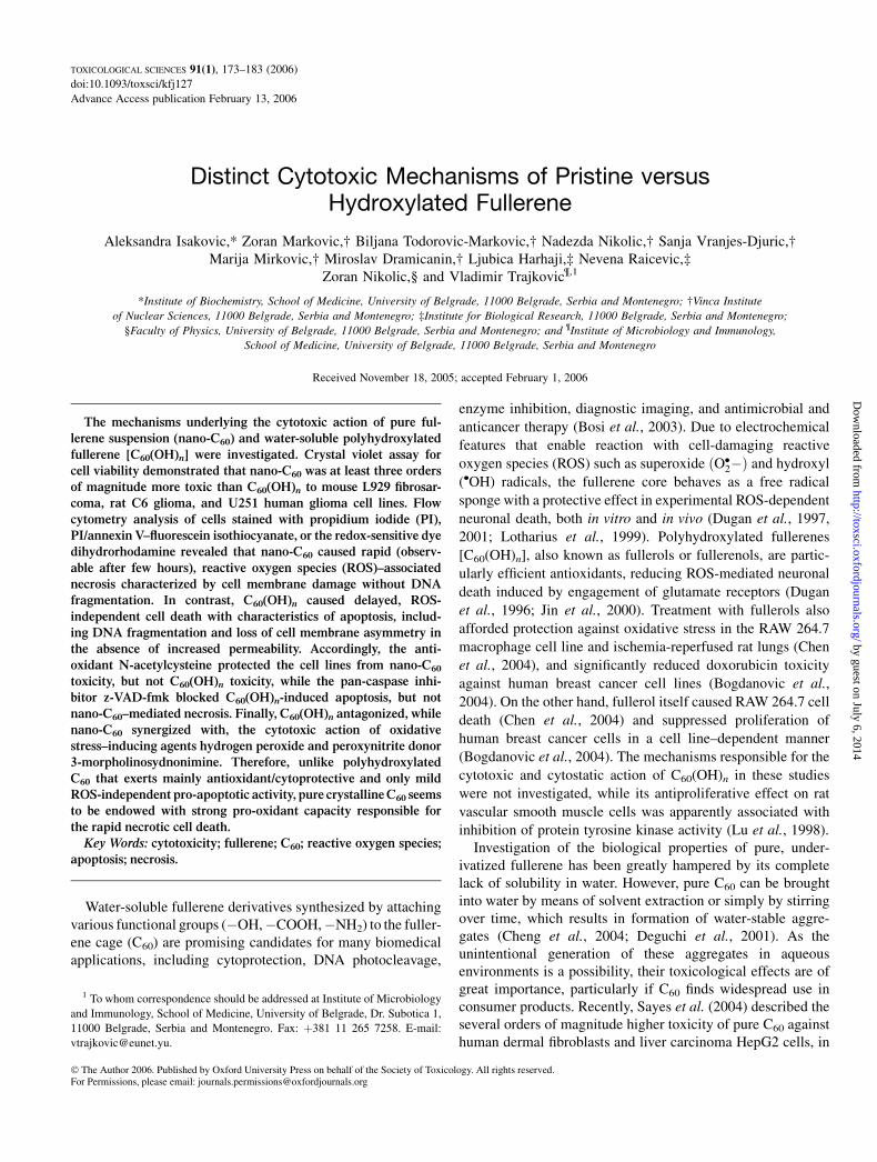
FIG. 1. Characterization of nano-C60 and C60(OH)n. (A) Images of nano-C60 crystals obtained by transmission electron microscopy. (B) Particle size
distribution of nano-C60 aggregates in water. Five thousand particles have been analyzed, and the highest number of particles (corresponding to a diameter of
96.3 nm) was normalized to 100. (C) UV/Vis spectra of nano-C60 (10 lg/ml) and C60(OH)n (1000 lg/ml). (D) FTIR spectra of nano-C60 and C60(OH)n. The arrows
indicate characteristic peaks at 526, 575, 1070, 1182, and 1608 cm-1.
CYTOTOXICITY OF PURE AND HYDROXY-C60 175
by guest on July 6, 2014http://toxsci.oxfordjournals.org/
Dow
nloaded from
thiobarbituric acid assay for malondialdehyde (MDA), as previously described
(Losa, 2003).
Analysis of synergism and antagonism in cytotoxic interactions. To
analyze the type (additive, synergistic, or antagonistic) of fullerene interaction
with the pro-oxidant agents H2O2 and SIN-1, or the interaction between nano-
C60 and C60(OH)n in inducing tumor cell death, cells were treated with each
agent alone and in combination. Six twofold dilutions were made from the
starting concentrations of 0.5 lg/ml nano-C60, 800 lg/ml C60(OH)n, 1mM
H2O2, and 1mM SIN-1, and the cytotoxic effect of each dose alone, as well as
its combination with the corresponding dose of appropriate agent, was tested
using the crystal violet assay. The results were expressed as a percent of
cytotoxic efficiency, and a combination index (CI) for mutually exclusive or
mutually nonexclusive interactions was calculated according to the method
designed by Chou and Talalay (1984). CI values ¼ 1, <1, or >1 indicate
additive, synergistic, or antagonistic interactions, respectively.
Statistical analysis. The statistical significance of the observed differences
was analyzed by t-test or ANOVA followed by the Student-Newman-Keuls test.
A p value < 0.05 was considered significant.
RESULTS
Characterization of Nano-C60 and C60(OH)n
The images obtained with the transmission electron micro-scope revealed that C60 in water formed nanocrystalline aggre-gates of approximately 100 nm (Fig. 1A). This is consistentwith the results of dynamic light-scattering measurements,which determined the average particle size to be 96.3 nm(Fig. 1B). The dynamic light-scattering analysis of C60(OH)nshowed that all particles were <5 nm (the detection limit), thusconfirming the absence of C60(OH)n aggregation (data notshown). The UV/Vis absorbance spectra of nano-C60 andC60(OH)n in Figure 1C indicate that fullerol does not havepronounced absorption bands in the UV region like nano-C60.Importantly, the spectra of our fullerene preparations are iden-tical to the previously published spectra of similarly preparedcommercial C60 and C60(OH)n (Fortner et al., 2005; Vilenoet al., 2005). The FTIR spectra of nano-C60 and C60(OH)n showcharacteristic vibrational modes of C60 at 526, 575, and 1182cm�1 (Kuzmany et al., 1995), as indicated by the arrows inFigure 1D. Expectedly, C60(OH)n has two broad absorptionbands at about 1070 and 1608 cm�1, which reflect the presenceof C–O and O–H covalent bonds, respectively. However, bothbands were also found in the nano-C60 spectrum, which is con-sistent with the data that some degree of derivatization appa-rently occurs at the surface of nano-C60 crystals (Sayes et al.,2005). A strong absorption band at 860 cm�1 in the FTIRspectrum of C60(OH)n is another indication of a C–O bond,
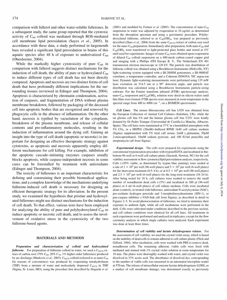
FIG. 2. Different cytotoxic efficiency and kinetics of nano-C60 and
C60(OH)n. (A) The cells were incubated for 24 h in the absence (control) or
presence of nano-C60 and C60(OH)n at different concentrations, and the cell
viability was assessed by crystal violet staining. (B) Light micrographs of
normal L929 cells (control) and L929 cells exposed to 1 lg/ml nano-C60 for 6 h.
(C) L929 cells were treated with 1 lg/ml nano-C60 (line) or 1000 lg/ml
C60(OH)n (bars), and the cell viability was analyzed by crystal violet assay at
the indicated time points (nano-C60) or after 6 h [C60(OH)n] (B). The values in
A are means from four independent experiments (SD values were <15% of the
corresponding means), while the values in C are mean ± SD of triplicate
measurements and are representative of three experiments (*p < 0.05).
176 ISAKOVIC ET AL.
by guest on July 6, 2014http://toxsci.oxfordjournals.org/
Dow
nloaded from
while the peaks observed in the region between 1200 and 1550cm�1 originate from the silicon substrate.
Nano-C60 and C60(OH)n Differ in the Efficacy and Kineticsof Their Cytotoxic Action
In the preliminary experiments, we used cells of differ-ent types and origin (rat primary astrocytes and fibroblasts,
rat/mouse peritoneal macrophages, C6 rat astrocytoma, U251human glioma, and L929 mouse fibrosarcoma cell line) toinvestigate if fullerenes might exert cell-selective toxicity. How-ever, repeated experiments in which the cell viability after 24-hincubation was evaluated by the crystal violet assay clearlydemonstrated that the toxicity of nano-C60 and C60(OH)n wasneither species/cell type specific, nor selective for primary ortransformed cells (data not shown). Having established that,for reasons of convenience and reproducibility, we used well-defined transformed cell lines (C6, U251, and L929) to deli-neate the cytotoxic mechanisms of the two different fullerenepreparations. To compare the cytotoxic efficiencies of nano-C60
and C60(OH)n, different doses of each agent were incubatedwith the L929, C6, or U251 cell line, and the number ofadherent, live cells was assessed by crystal violet staining after24 h. In accordance with the results of Sayes et al. (2005), thedata presented in Figure 2A clearly show that pristine fullerenewas at least three orders of magnitude more toxic to the testedcell lines than its hydroxylated counterpart [LC50 values were0.25 lg/ml and 800–1000 lg/ml for C60 and C60(OH)n,respectively]. Microscopic examination revealed a dramaticchange in the morphology of nano-C60–treated cells. In contrastto control cells, nano-C60–treated cells lost their processes andbecame round and detached from the culture well surface afteronly 6 h of treatment (Fig. 2B). The observed morphologicalchanges followed by cell detachment were indicative of rapidcell death. Accordingly, the crystal violet analysis of cellviability at various time points following nano-C60 additionrevealed an extremely fast kinetics of its cytotoxic action,reaching almost maximal efficiency after 6 h of cultivation(Fig. 2C, line). In the same time range, C60(OH)n failed to causeany discernible changes in cellular morphology (data notshown) or to induce cell death (Fig. 2C, bars). Therefore,pristine C60 was a much faster and more efficient cytotoxicagent than its hydroxylated derivative. Importantly, the cyto-toxic activity of crystalline C60 was completely unrelated to theresidual amount (<10% wt/wt) of organic solvent intercalatedinto its lattice (Sayes et al., 2005), as THF was without anytoxic effect (data not shown) even at concentrations 100-foldhigher (10 lg/ml) than its estimated residual presence (1 lg/mlnano-C60 contains <0.1 lg/ml THF).
Nano-C60 and C60(OH)n Induce Distinct Types of Cell Death
In the following experiments, we compared the ability ofnano-C60 and C60(OH)n to induce apoptotic or necrotic celldeath. To discriminate between the two distinct types of celldeath, we used double staining with annexin V–FITC and PI.Annexin V binds to the phosphatidylserine that is typicallyexposed at the outer side of cell membrane during apoptosis,while PI only enters the cells with membrane damage thatoccurs in necrotic cell death. Therefore, normal, healthy cellsare annexin�/PI� (Fig. 3A, 3B, lower left quadrant), apoptoticcells express phosphatidylserine, but have preserved membrane
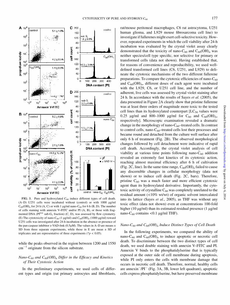
FIG. 3. Pure and hydroxylated C60 induce different types of cell death.
(A–D) U251 cells were incubated without (control) or with 1000 lg/ml
C60(OH)n for 24 h (A, C) or with 1 lg/ml nano-C60 for 6 h (B, D). The number
of cells staining with annexin V–FITC and/or PI (A, B), or those with frag-
mented DNA (PIlow sub-G1 fraction) (C, D), was assessed by flow cytometry.
(E) The cytotoxicity of nano-C60 (1 lg/ml) and C60(OH)n (1000 lg/ml) toward
U251 cells was investigated after 24-h incubation in the absence or presence of
the pan-caspase inhibitor z-VAD-fmk (0.5lM). The values in A–D are means ±
SD from three separate experiments, while those in E are mean ± SD of
triplicates and are representative of three experiments (*p < 0.05).
CYTOTOXICITY OF PURE AND HYDROXY-C60 177
by guest on July 6, 2014http://toxsci.oxfordjournals.org/
Dow
nloaded from
FIG. 4. The involvement of ROS in cytotoxic action of nano-C60. (A) C6 cells were incubated in the absence (control) or presence of nano-C60 (1 lg/ml) or
C60(OH)n (1000 lg/ml). The production of ROS was measured by flow cytometric analysis of DHR fluorescence after 3 h (nano-C60) or 18 h [C60(OH)n]. (B) The
viability of C6 cells incubated for 24 h without (control) or with C60(OH)n (1000 lg/ml), in the presence or absence of the antioxidant NAC (2mM). (C) The
influence of NAC (2mM) on various parameters determined after exposure of C6 cells to nano-C60 (1 lg/ml) for 3 h (DHR, MDA) or 6 h (viability, number of
annexinþ/PIþ cells, LDH release). (D) Light micrographs of L929 cells incubated for 24 h without (control) or with nano-C60 (1 lg/ml), in the presence or absence
of NAC (2mM). (E) The ability of nano-C60 (1 lg/ml) to generate ROS in C6 cultures and cell-free suspension in cell culture medium was assessed by measuring
DHR fluorescence in a microplate reader. The values in B, C, and E are presented as mean ± SD of triplicates and are representative of at least three experiments,
with the exception of values for DHR and annexinþ/PIþ cells in C, which are means ± SD from three separate experiments (*p < 0.05 refers to nano-C60 treatment
without NAC [C] or the corresponding DHR value obtained in cell-free conditions).
178 ISAKOVIC ET AL.
by guest on July 6, 2014http://toxsci.oxfordjournals.org/
Dow
nloaded from
integrity (annexinþ/PI�; lower right quadrant), while necroticcells with damaged membrane stain for both annexin V and PI(annexinþ/PIþ; upper right quadrant). The results presented inFigures 3A and 3B clearly show that the treatment of U251
cells with C60(OH)n led to a significant increase in the numberof apoptotic, but not necrotic, cells (Fig. 3A), while nano-C60
caused a massive increase in the number of necrotic, butnot apoptotic, cells (Fig. 3B). The analysis of cellular DNA
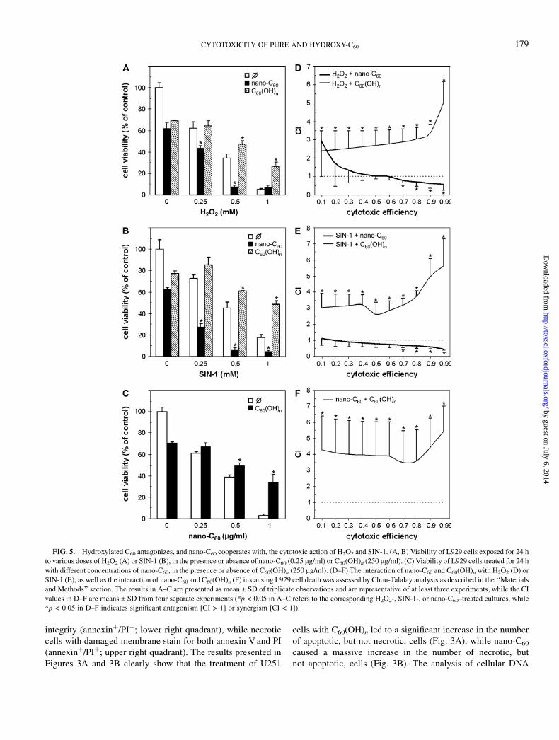
FIG. 5. Hydroxylated C60 antagonizes, and nano-C60 cooperates with, the cytotoxic action of H2O2 and SIN-1. (A, B) Viability of L929 cells exposed for 24 h
to various doses of H2O2 (A) or SIN-1 (B), in the presence or absence of nano-C60 (0.25 lg/ml) or C60(OH)n (250 lg/ml). (C) Viability of L929 cells treated for 24 h
with different concentrations of nano-C60, in the presence or absence of C60(OH)n (250 lg/ml). (D–F) The interaction of nano-C60 and C60(OH)n with H2O2 (D) or
SIN-1 (E), as well as the interaction of nano-C60 and C60(OH)n (F) in causing L929 cell death was assessed by Chou-Talalay analysis as described in the ‘‘Materials
and Methods’’ section. The results in A–C are presented as mean ± SD of triplicate observations and are representative of at least three experiments, while the CI
values in D–F are means ± SD from four separate experiments (*p < 0.05 in A–C refers to the corresponding H2O2-, SIN-1-, or nano-C60–treated cultures, while
*p < 0.05 in D–F indicates significant antagonism [CI > 1] or synergism [CI < 1]).
CYTOTOXICITY OF PURE AND HYDROXY-C60 179
by guest on July 6, 2014http://toxsci.oxfordjournals.org/
Dow
nloaded from
content, which is reduced in apoptosis due to DNA fragmen-tation, was consistent with the data from annexin/PI staining.Namely, the number of hypodiploid, presumably apoptotic,cells with fragmented DNA (sub-G1 fraction of PI-stainedcells) markedly increased upon the treatment of U251 cellswith fullerol (Fig. 3C), while pristine fullerene was withouteffect (Fig. 3D). Similar results were obtained with C6 andL929 cells (data not shown). The ability of nano-C60 to causecell membrane damage indicative of necrosis was also con-firmed by a significant increase in LDH release (159 ± 15%,n ¼ 3, p < 0.05), which was not observed after C60(OH)ntreatment. Due to a difference in the kinetics of cytotoxicaction of C60(OH)n and nano-C60 , the assessment of apoptosis/necrosis presented in Figures 3A and 3C and Figures 3B and3D was performed after 24 and 6 h, respectively. It should benoted, however, that the appearance of annexinþ cells in nano-C60–treated cultures could not be observed even at earliertime points (0.5–4 h; data not shown), which excluded thepossibility that some early apoptotic events were overlooked.Finally, we used z-VAD-fmk, a pharmacological inhibitor ofcaspase activation and subsequent apoptotic cell death, toevaluate the role of apoptosis in fullerene-mediated cytotoxic-ity. In accordance with the data presented in Figures 3A–3D,z-VAD-fmk exerted a significant protective effect in C60(OH)n-treated U251 cultures, but completely failed to rescue U251cells from the toxicity of nano-C60 (Fig. 3E). Therefore, pureC60 and C60(OH)n apparently employ distinct cytotoxicmechanisms resulting in the preferential induction of cas-pase-independent necrosis and caspase-dependent apoptosis,respectively.
Cytotoxicity of Nano-C60, but Not C60(OH)n, Dependson ROS Generation
We next investigated the involvement of oxidative stress inthe observed cytotoxic action of nano-C60 and C60(OH)n. Thetreatment of C6 cells with C60(OH)n for 18 h failed to inducedetectable intracellular production of ROS, as judged by un-altered intracellular DHR fluorescence (Fig. 4A, lower panel).On the other hand, a considerable increase in DHR fluores-cence was observed in C6 cells after 3 h of incubation withpristine C60 (Fig. 4A, upper panel), indicating a significantintracellular generation of ROS. It should be noted that a shortertime course was chosen for nano-C60 because of the prompt-ness of its cytotoxic action. Nevertheless, the inability ofC60(OH)n to generate ROS was confirmed in the experiments inwhich the incubation time was reduced from 18 h to 3 or 6 h(data not shown). We used a powerful antioxidant agent, NAC(Zafarullah et al., 2003), to examine the role of ROS pro-duction in fullerene-mediated cytotoxicity. While NAC wascompletely unable to prevent C60(OH)n-mediated tumor cellkilling (Fig. 4B), it markedly downregulated the production ofoxygen radicals and completely prevented viability loss,induction of necrosis (measured as number of annexinþ/PIþ
cells), and LDH release associated with nano-C60 treatment(Fig. 4C). Moreover, an increase in MDA levels in C6 cells,reflecting membrane lipid peroxidation, was readily observedupon addition of nano-C60 and efficiently blocked by NACtreatment (Fig. 4C). The protective effect of NAC was alsoobserved in U251 and L929 cells, and lasted for at least 24 h, asdemonstrated by crystal violet staining (data not shown) andthe completely preserved morphology of cells treated withnano-C60 (Fig. 4D). Because fluorescence-activated cell sortinganalysis could not distinguish cell-derived from nano-C60–produced oxygen radicals, we used a fluorescence microplatereader to detect DHR fluorescence in cell-free nano-C60
suspension. However, the results presented in Figure 4E showthat the time-dependent increase in DHR fluorescence in nano-C60–treated C6 cultures was slightly less pronounced than thatobserved in the absence of cells. Therefore, it appears thatintracellular increase in ROS production was not a cellularresponse to nano-C60 treatment, but rather depended on theability of pristine fullerene to directly generate oxygenradicals. Collectively, these results suggest that nano-C60–produced oxygen radicals are involved in lipid peroxidationand the consequent necrotic cell death, while C60(OH)n-triggered apoptosis seems to be ROS independent.
Nano-C60 and C60(OH)n Differently Cooperate with theCytotoxic Action of Oxidative Stress–Inducing Agents
Having demonstrated that the cytotoxicity of nano-C60, butnot C60(OH)n, depends on production of oxygen radicals, wewanted to examine if these fullerene preparations could in-fluence the toxicity of other ROS-generating agents. To thateffect, we assessed the cytotoxic effects of simultaneousapplication of nano-C60 or C60(OH)n with hydrogen peroxideand peroxynitrite donor SIN-1, the agents well known for theirability to induce oxidative stress–mediated cell death (Baueret al., 1998; Wang and Joseph, 1999). In contrast to pristine C60,C60(OH)n significantly improved the viability of L929 cellstreated with H2O2 or SIN-1 (Figs. 5A and 5B). We next sought toconfirm these observations by using mathematical analysis ofdrug interaction based on the approach by Chou and Talalay(1984). As shown in Figures 5D and 5E, the CI for combinationof C60(OH)n with H2O2 or SIN-1 was > 1 throughout the 0.1–0.99 efficiency range, thus confirming the putative ability ofhydroxylated fullerene to antagonize the cytotoxic action ofboth agents. On the other hand, the CI for combination of nano-C60 with H2O2 or SIN-1 was around or less than 1 (Figs. 5D and5E), indicating additive/synergistic cooperation in killing L929cells. Similar results were obtained with U251 and C6 cells(data not shown). These data suggest that pure C60 cooperateswith, while its hydroxylated derivative antagonizes, the cyto-toxic action of oxidative stress–inducing agents. Interestingly,C60(OH)n readily antagonized the oxidative stress–dependentcytotoxicity of C60 nanocrystals (Figs. 5C and 5F), thus furtherreinforcing the concept of cell-damaging/pro-oxidant versus
180 ISAKOVIC ET AL.
by guest on July 6, 2014http://toxsci.oxfordjournals.org/
Dow
nloaded from
cytoprotective/antioxidant activity of pristine fullerene and itshydroxylated counterpart, respectively.
DISCUSSION
The present study describes the rapid ROS-dependent, caspase-independent necrotic cell death that occurs within only few hoursof exposure to pure buckminsterfullerene aggregates (nano-C60).On the other hand, polyhydroxylated C60 caused delayed,ROS-independent, caspase-mediated apoptosis. It should benoted that the observed toxic effects were not restricted to thetransformed cell lines, as similar results were obtained in primaryrat astrocytes and fibroblasts, as well as in rat and mouse peri-toneal macrophages (Isakovic et al., unpublished observation).
While cytotoxic/antiproliferative effects of pristine C60 andits various water-soluble derivatives, including C60(OH)n, havebeen observed in different experimental systems (Bogdanovicet al., 2004; Bosi et al., 2004; Chen et al., 2004; Lu et al., 1998;Mashino et al., 2003; Sayes et al., 2004, 2005; Tsuchiya et al.,1996; Yang et al., 2002), their capacity for preferentialinduction of apoptosis or necrosis has not been compared.Although the release of LDH observed after 48-h incubation ofhuman dermal fibroblasts, astrocytes, or liver carcinoma cellswith nano-C60 (Sayes et al., 2005) is consistent with necroticcell death, secondary necrosis following an initially apoptoticdeath could not be excluded. A line of evidence from thepresent study, including the results of cell membrane asym-metry/integrity assessment by annexin V/PI staining or LDHrelease test, as well as the data from DNA fragmentationanalysis, clearly demonstrate that cell death induced by nano-C60 and C60(OH)n proceeds exclusively through the necroticand apoptotic pathways, respectively. Since aspartate-specificcysteine proteases, termed caspases, are apparently required forapoptotic, but not necrotic, cell death (Ferrari et al., 1999; Loset al., 2002), the protective effect of caspase inhibition inC60(OH)n-treated cells, but not C60-treated cells, furthersupports the view that pristine fullerene induces necrotic celldeath, as opposed to the mainly pro-apoptotic activity of itshydroxylated derivative. The mode of cell death might pro-foundly influence the healing of the surrounding tissue, sincenecrotic cell products could initiate additional injury bypromoting inflammation (Jaeschke et al., 2002). On the otherhand, in view of the immunostimulatory properties of necroticcells and resistance of tumor cells to apoptosis, it has beenproposed that necrosis might be more efficient than apoptosisin inducing tumor regression (Edinger and Thompson, 2004;Galluci et al., 1999; Reiter et al., 1999). Therefore, the abilityto rapidly induce necrotic, rather than apoptotic, cell deathcould pose a difficulty for counteracting the toxicity ofnanocrystalline C60, as well as potentially increase its effi-ciency in cancer therapy.
By using the redox-sensitive fluorescent dye DHR, MDAmeasurement, and cell incubation with the well-known antiox-
idant agent NAC, we have shown that nano-C60–inducednecrosis was exclusively a consequence of oxidative stress thatprobably resulted in lipid peroxidation–mediated damage of thecell membrane. The putative involvement of oxygen radicals innano-C60–induced necrosis is consistent with the ability ofanother antioxidant, L-ascorbic acid, to completely prevent lipidperoxidation and ensuing cell death in nano-C60–treatedcultures of human dermal fibroblasts, described recently bySayes et al. (2005). Based on their observation that C60
nanocrystals generate superoxide anions in a cell-free system(Sayes et al., 2004), the authors assumed that this feature ofnano-C60 could be solely responsible for the induction ofoxidative stress and subsequent cell death. However, it wasnot possible to conclude from these studies whether the targetcells contributed to production of oxygen radicals in response totreatment with pure C60. By comparing the nano-C60–evokedincrease in DHR fluorescence in tumor cell cultures and cell-freesolution, we have been able to provide some direct evidence insupport of the hypothesis by Sayes et al. (2004), as the cellsthemselves apparently did not contribute to ROS generation inour experiments. Moreover, we have demonstrated a slight, butsignificant, decrease in the ability of nano-C60 to generateoxygen radicals in the presence of cells, which might bea consequence of the cellular antioxidative defense. Indeed, ithas been shown that cells increase their production of antiox-idant glutathione in response to oxidative stress induced byexposure to pristine C60 (Sayes et al., 2005). However, it stillremains to be established if ROS production and subsequentdamage were limited to cell membrane, or nano-C60 couldsomehow gain access to the cell cytoplasm.
In contrast to nano-C60–triggered necrosis, apoptosis inducedby C60(OH)n proceeded entirely in a ROS-independent fashion.Furthermore, C60(OH)n significantly antagonized the cytotoxicaction of the oxidative stress–inducing agents H2O2 andperoxynitrite donor SIN-1, as demonstrated by the Chou-Talalay approach. This is consistent with the previously reportedability of C60(OH)n to prevent oxidative stress and subsequentcell death in various experimental settings (Chen et al., 2004;Dugan et al., 1996; Jin et al., 2000; Murugan et al., 2002; Tsaiet al., 1997), supporting a general view that C60(OH)n mainlyacts as a ‘‘free radical sponge’’ with only mild cytotoxic activity(Bosi et al., 2003). On the other hand, the present studyconfirmed the recently described capacity of nanocrystallineC60 to generate oxygen radicals in cell-free systems (Sayeset al., 2004), while extending this observation by demonstratingROS production in nano-C60–treated cells. This pro-oxidantactivity might account for our novel finding that pristine C60, incontrast to its hydroxylated derivative, readily cooperated ina synergistic fashion with H2O2 and peroxynitrite in theinduction of cell death. It seems conceivable to assume thatthe mechanisms underlying the pro- versus antioxidant effectsof pristine and hydroxylated fullerene might be related to dif-ferences in their chemical structures. Namely, the transmissionelectron microscopy images and light-scattering measurements
CYTOTOXICITY OF PURE AND HYDROXY-C60 181
by guest on July 6, 2014http://toxsci.oxfordjournals.org/
Dow
nloaded from
obtained in the present study confirm that a substantial fractionof pristine C60, unlike the fullerene core in intentionally water-solubilized derivatives, is contained in the interior of thenanocrystalline aggregates of approximately 90–100 nm in size(Fortner et al., 2005; Sayes et al., 2004). As a consequence, C60
in such conditions remains largely underivatized (>99%), andits high toxicity is therefore consistent with the finding that ROSgeneration and cytotoxicity decrease with increasing derivati-zation of the fullerene cage (Sayes et al., 2004). However, itremains to be revealed how this structural difference could leadto distinct oxidant and cytotoxic properties. To further empha-size the complexity of the oxidant behavior of fullerenes inbiological systems, we note that even polyhydroxylated full-erenes could generate oxygen radicals under certain circum-stances (e.g., upon photosensitization) (Kamat et al., 1998,2000). On the other hand, the ability of nano-C60 to produceROS does not seem to depend on the presence of light (Sayeset al., 2004), which is consistent with our results that exposure tovisible light or UV did not affect the cytotoxicity of nano-C60
(unpublished data).In contrast to the mainly antioxidant/cytoprotective and only
mild ROS-independent pro-apoptotic activity of polyhydroxy-lated C60, the present study clearly demonstrates the ability ofpure crystalline C60 to induce rapid ROS-dependent necrosisand to synergistically enhance the cytotoxicity of other oxi-dative stress–inducing agents. This is consistent with a recentstudy in which the presence of pristine C60 in water causedlipid peroxidation–mediated damage to fish brain (Oberdorster,2004), but the possible relevance of our findings for the in vivotoxicity of nano-C60 is still to be investigated. While thedifference in the cytotoxic mechanisms of pure versus hydrox-ylated C60 suggests distinct remediation for their unwarrantedbiological effects, their divergent cytotoxic/cytoprotectiveproperties also provide grounds for the development of full-erenes as either cytoprotective or anticancer therapeutics.
ACKNOWLEDGMENTS
This work was supported by the Ministry of Science and Environmental
Protection of the Republic of Serbia (Grant No. 145073). We thank Natasa
Bibic (Vinca Institute of Nuclear Sciences, Belgrade) for performing trans-
mission electron microscopy, Nebojsa Romcevic (Institute of Physics,
Belgrade) for the FTIR analysis, and Marija Mostarica Stojkovic and Zorica
Ramic (Institute of Microbiology and Immunology, School of Medicine,
Belgrade) for helpful discussion.
REFERENCES
Bauer, M. K., Vogt, M., Los, M., Siegel, J., Wesselborg, S., and Schulze-Osthoff,K.
(1998). Role of reactive oxygen intermediates in activation-induced CD95
(APO-1/Fas) ligand expression. J. Biol. Chem. 273, 8048–8055.
Bogdanovic, G., Kojic, V., Dordevic, A., Canadanovic-Brunet, J., Vojinovic-
Miloradov, M., and Baltic, V. V. (2004). Modulating activity of fullerol
C60(OH)22 on doxorubicin-induced cytotoxicity. Toxicol. In Vitro 18,
629–637.
Bosi, S., Da Ros, T., Spalluto, G., and Prato, M. (2003). Fullerene derivatives:
An attractive tool for biological applications. Eur. J. Med. Chem. 38,
913–923.
Bosi, S., Feruglio, L., Da Ros, T., Spalluto, G., Gregoretti, B., Terdoslavich, M.,
Decorti, G., Passamonti, S., Moro, S., and Prato, M. (2004). Hemolytic
effects of water-soluble fullerene derivatives. J. Med. Chem. 47, 6711–6715.
Chen, Y. W., Hwang, K. C., Yen, C. C., and Lai, Y. L. (2004). Fullerene
derivatives protect against oxidative stress in RAW 264.7 cells and ischemia-
reperfused lungs. Am. J. Physiol. Regul. Integr. Comp. Physiol. 287,
R21–R26.
Cheng, X., Kan, A. T., and Tomson, M. B. (2004). Naphthalene adsorption and
desorption from aqueous C60 fullerene. J. Chem. Eng. Data 49, 675–683.
Chou, T. C., and Talalay, P. (1984). Quantitative analysis of dose-effect
relationships: The combined effects of multiple drugs or enzyme inhibitors.
Adv. Enzyme Regul. 22, 27–55.
Deguchi, S., Rossitza, G. A., and Tsujii, K. (2001). Stable dispersions of
fullerenes, C60 and C70 in water. Preparation and characterization. Langmuir
17, 6013–6017.
Dugan, L. L., Gabrielsen, J. K., Yu, S. P., Lin, T. S., and Choi, D. W. (1996).
Buckminsterfullerenol free radical scavengers reduce excitotoxic and
apoptotic death of cultured cortical neurons. Neurobiol. Dis. 3, 129–135.
Dugan, L. L., Lovett, E. G., Quick, K. L., Lotharius, J., Lin, T. T., and
O’Malley, K. L. (2001). Fullerene-based antioxidants and neurodegenerative
disorders. Parkinsonism Relat. Disord. 7, 243–246.
Dugan, L. L., Turetsky, D. M., Du, C., Lobner, D., Wheeler, M., Almli, C. R.,
Shen, C. K., Luh, T. Y., Choi, D. W., and Lin, T. S. (1997). Carboxyfullerenes
as neuroprotective agents. Proc. Natl. Acad. Sci. U.S.A. 94, 9434–9439.
Edinger, A. L., and Thompson, C. B. (2004). Death by design: Apoptosis,
necrosis and autophagy. Curr. Opin. Cell Biol. 16, 663–669.
Ferrari, D., Los, M., Bauer, M. K., Vandenabeele, P., Wesselborg, S., and
Schulze-Osthoff, K. (1999). P2Z purinoreceptor ligation induces activation
of caspases with distinct roles in apoptotic and necrotic alterations of cell
death. FEBS Lett. 447, 71–75.
Flick, D. A., and Gifford, G. E. (1984). Comparison of in vitro cell cytotoxic
assays for tumor necrosis factor. J. Immunol. Methods 68, 167–175.
Fortner, J. D., Lyon, D. Y., Sayes, C. M., Boyd, A. M., Falkner, J. C., Hotze, E. M.,
Alemany, L. B., Tao, Y. J., Guo, W., Ausman, K. D., et al. (2005). C60 in
water: Nanocrystal formation and microbial response. Environ. Sci. Technol.
39, 4307–4316.
Galluci, S., Lolkema, M., and Matzinger, P. (1999). Natural adjuvants:
Endogenous activators of dendritic cells. Nat. Med. 5, 1249–1255.
Jaeschke, H., Gores, G. J., Cederbaum, A. I., Hinson, J. A., Pessayre, D., and
Lemasters, J. J. (2002). Mechanisms of hepatotoxicity. Toxicol. Sci. 65,
166–176.
Jin, H., Chen, W. Q., Tang, X. W., Chiang, L. Y., Yang, C. Y., Schloss, J. V.,
and Wu, J. Y. (2000). Polyhydroxylated C60, fullerenols, as glutamate
receptor antagonists and neuroprotective agents. J. Neurosci. Res. 62,
600–607.
Kaludjerovic, G., Miljkovic, D., Momcilovic, M., Djinovic, V., Mostarica
Stojkovic, M., Sabo, T., and Trajkovic, V. (2005). Novel platinum complexes
induce rapid tumor cell death in vitro. Int. J. Cancer 116, 479–486.
Kamat, J. P., Devasagayam, T. P., Priyadarsini, K. I., and Mohan, H. (2000).
Reactive oxygen species mediated membrane damage induced by fuller-
ene derivatives and its possible biological implications. Toxicology 155,
55–61.
Kamat, J. P., Devasagayam, T. P., Priyadarsini, K. I., Mohan, H., and Mittal, J. P.
(1998). Oxidative damage induced by the fullerene C60 on photosensitization
in rat liver microsomes. Chem-Biol. Interact. 114, 145–159.
Kuzmany, H., Winkler, R., and Pichler, T. (1995). Infrared spectroscopy of
fullerenes. J. Phys. Condens. Matter 7, 6601–6624.
182 ISAKOVIC ET AL.
by guest on July 6, 2014http://toxsci.oxfordjournals.org/
Dow
nloaded from
Los, M., Mozoluk, M., Ferrari, D., Stepczynska, A., Stroh, C., Renz, A.,
Herceg, Z., Wang, Z. Q., and Schulze-Osthoff, K. (2002). Activation and
caspase-mediated inhibition of PARP: A molecular switch between fibroblast
necrosis and apoptosis in death receptor signaling. Mol. Biol. Cell 13,
978–988.
Losa, G. A. (2003). Resveratrol modulates apoptosis and oxidation in human
blood mononuclear cells. Eur. J. Clin. Investig. 33, 818–823.
Lotharius, J., Dugan, L. L., and O’Malley, K. L. (1999). Distinct mechanisms
underlie neurotoxin-mediated cell death in cultured dopaminergic neurons.
J. Neurosci. 19, 1284–1293.
Lu, L. H., Lee, Y. T., Chen, H. W., Chiang, L. Y., and Huang, H. C. (1998). The
possible mechanisms of the antiproliferative effect of fullerenol, polyhy-
droxylated C60, on vascular smooth muscle cells. Br. J. Pharmacol. 123,
1097–1102.
Markovic, Z., Todorovic-Markovic, B., Marinkovic, M., and Nenadovic, T.
(2003). Temperature measurement of carbon arc plasma in helium. Carbon
41, 369–371.
Mashino, T., Nishikawa, D., Takahashi, K., Usui, N., Yamori, T., Seki, M.,
Endo, T., and Mochizuki, M. (2003). Antibacterial and antiproliferative activ-
ity of cationic fullerene derivatives. Bioorg. Med. Chem. Lett. 13, 4395–4397.
Murugan, M. A., Gangadharan, B., and Mathur, P. P. (2002). Antioxidative effect
of fullerenol on goat epididymal spermatozoa. Asian J. Androl. 4, 149–152.
Oberdorster, E. (2004). Manufactured nanomaterials (fullerenes, C60) induce
oxidative stress in the brain of juvenile largemouth bass. Environ. Health
Perspect. 112, 1058–1062.
Reiter, I., Krammer, G., and Schwamberger, G. (1999). Differential effect of
apoptotic versus necrotic tumor cells on macrophage antitumour activity.
J. Immunol. 163, 1730–1732.
Sayes, C., Fortner, J., Lyon, D., Boyd, A., Ausman, K., Tao, Y., Sitharaman, B.,
Wilson, L., West, J., and Colvin, V. L. (2004). The differential cytotoxicity
of water soluble fullerenes. Nano Lett. 4, 1881–1887.
Sayes, C. M., Gobin, A. M., Ausman, K. D., Mendez, J., West, J. L., and Colvin,
V. L. (2005). Nano-C60 cytotoxicity is due to lipid peroxidation. Biomaterials
26, 7587–7595.
Tsai, M. C., Chen, Y. H., and Chiang, L. Y. (1997). Polyhydroxylated C60,
fullerenol, a novel free-radical trapper, prevented hydrogen peroxide- and
cumene hydroperoxide-elicited changes in rat hippocampus in vitro.
J. Pharm. Pharmacol. 49, 438–445.
Tsuchiya, T., Oguri, I., Yamakoshi, Y. N., and Miyata, N. (1996). Novel
harmful effects of [60]fullerene on mouse embryos in vitro and in vivo.
FEBS Lett. 393, 139–145.
Vileno, B., Lekka, M., Sienkiewicz, A., Marcoux, P., Kulik, A. J., Kasas, S.,
Catsicas, S., Graczyk, A., and Forro, L. (2005). Singlet oxygen [(1)Delta(g)]-
mediated oxidation of cellular and subcellular components: ESR and AFM
assays. J. Phys. Condens. Matter 17, S1471–S1482.
Wang, H., and Joseph, J. A. (1999). Quantifying cellular oxidative stress by
dichlorofluorescein assay using microplate reader. Free Radic. Biol. Med. 27,
612–616.
Yang, X. L., Fan, C. H., and Zhu, H. S. (2002). Photo-induced cytotoxicity of
malonic acid [C60]fullerene derivatives and its mechanism. Toxicol. In Vitro
16, 41–46.
Zafarullah, M., Li, W. Q., Sylsvester, J., and Ahmad, M. (2003). Molecular
mechanisms of N-acetylcysteine actions. Cell. Mol. Life Sci. 60, 6–20.
Zhao, G. C., Zhang, P., Wei, X. W., and Yang, Z. S. (2004). Determination of
proteins with fullerol by a resonance light scattering technique. Anal.
Biochem. 334, 297–302.
CYTOTOXICITY OF PURE AND HYDROXY-C60 183
by guest on July 6, 2014http://toxsci.oxfordjournals.org/
Dow
nloaded from