disruption of epithelial cell-matrix interactions induces
TRANSCRIPT

Disruption of Epithelial Cell-Matrix Interactions Induces Apoptosis Steven M. Fr i sch a n d H u n t e r F ranc i s
La Jolla Cancer Research Foundation, La Jolla, California 92037
Abstract. Cell-matrix interactions have major effects upon phenotypic features such as gene regulation, cytoskeletal structure, differentiation, and aspects of cell growth control. Programmed cell death (apopto- sis) is crucial for maintaining appropriate cell number and tissue organization. It was therefore of interest to determine whether cell-matrix interactions affect apoptosis. The present report demonstrates that apop- tosis was induced by disruption of the interactions be- tween normal epithelial cells and extracellular matrix. We have termed this phenomenon "anoikis" Overex- pression of bcl-2 protected cells againstanoikis. Cel-
lular sensitivity to anoikis was apparently regulated: (a) anoikis did not occur in normal fibroblasts; (b) it was abrogated in epithelial cells by transformation with v-Ha-ras, v-src, or treatment with phorbol ester; (c) sensitivity to anoikis was conferred upon HTI080 cells or v-Ha-ms-transformed MDCK ceils by reverse- transformation with adenovirus Ela; (d) anoikis in MDCK cells was alleviated by the motility factor, scatter factor. The results suggest that the circumven- tion of anoikis accompanies the acquisition of an- chorage independence or cell motility.
C ELL-matrix interactions have major effects upon phenotypic features such as gene regulation; cyto- skeletal structure, differentiation, and aspects of cell
growth control (Adams and Watt, 1993; Blau and Baltimore, 1991; Ingber, 1993). In particular, determination of an- chorage dependence is an important function of cell-matrix interactions. The restriction of cell proliferation to matrix- interacting cells serves to prevent dysplasia; the circumven- tion of anchorage dependence plays an important role in tumorigenesis (Stoker et al., 1968).
In previous studies, the growth arrest induced by suspen- sion of fibroblasts was found to be reversible (Folkman and Moscona, 1978; Ben-Ze'ev et al., 1980). However, the fate of other cell types challenged similarly was not exam- ined. Programmed cell death (apoptosis; Tomei and Cope, 1991; M. Raft, 1992; Lee et al., 1993; Marx, 1993; Barinaga, 1993; Vaux, 1993) is crucial for maintaining ap- propriate cell number and tissue organization in certain cell types such as lymphocytes and neurons. We considered that possibility that, for certain cell types, lack of matrix attach- ment could stringently restrict inappropriate cell growth by inducing apoptosis. Without apoptosis, detached cells could possibly reattach to inappropriately localized matrices, in- cluding the matrix that they would eventually synthesize themselves, and resume growth. However, apoptosis occur- ring in detached cells would abrogate this escape mech- anism.
Address all correspondence to S. M. Frisch, La Jolla Cancer Research Foundation, 10901 N. Torrey Pines Road, La Jolla, CA 92037.
This article reports that a form of apoptosis that we call "anoikis" was induced by the disruption of cell-matrix inter- actions in two model epithelial cell lines. The authenticity of the apoptotic form of cell death was demonstrated by sev- eral criteria. Regulation of apoptosis thus appears to be a new function for cell-matrix interactions.
Aside from attachment to extraceUular matrix, epithelial cells are linked to each other via tight junctions, adherens junctions and desmosomes (Farquhar and Palade, 1963). These cells form the frst tissues to emerge in the embryo. Mesenchymal cells differentiate from the embryonic epi- thelia (Hay, 1990). Unlike the stationary epithelial cells, mesenchymal cells are invasive, motile, and do not adhere to each other. Certain tissue remodeling events, as well as oncogenic transformation, recapitulate the transition from epithelial to mesenchymal phenotype (Zuk et al., 1989; Behrens et al., 1991; Schmidt et al., 1993). We reasoned that cell motility or transformation might require matrix-inde- pendent survival. Because anoikis would be incompatible with these phenotypes, we tested the prediction that cell motility factors, transformation or fibroblastic (motile) phe- notypes might confer resistance to anoikis. Evidence con- firming these predictions is presented. Transformation or treatment with a cell motility factor conferred resistance to anoikis, while reverse-transformation conferred sensitivity; normal fibroblasts were resistant. These results suggest that the acquisition of anchorage-independent or motile pheno- types alleviates anoikis. Cellular sensitivity to anoikis can therefore be regulated.
© The Rockefeller University Press, 0021-9525/94/02/619/8 $2.00 The Journal of Ce|l Biology, Volume 124, Number 4, February 1994 619-626 619
Dow
nloaded from http://rupress.org/jcb/article-pdf/124/4/619/1401374/619.pdf by guest on 10 January 2022

Materials and Methods
Anoikis Assay
Cell lines were grown to confluence in 100-mm tissue culture dishes, unless otherwise indicated. Trypsinized cells (3 x 10⁶) were then counted and plated onto 1O0-mm petri dishes which had been coated with polyHEMA (Folkman and Moscona, 1978) (POlyHEMA plates were made by applying 4 ml of a 10 mg/ml solution of polyhydroxyethylmethacrylate [Aldrich Chemical Co., Milwaukee, WI] in ethanol onto the dish, drying in the tissue culture hood and repeating once, followed by extensive PBS washes). After 12 h (MDCK), 20 h (HaCat), or indicated times, cells were collected from POlyHEMA dishes by pipetting or from tissue culture dishes by scraping cells into the media in which they had been incubated. (In all tissue culture dish controls, floating cells were combined with the attached calls before DNA extraction, to determine spontaneous apoptosis.) Low molecular weight genomic DNA was extracted with 0.5 % Triton X-100, 10 mM EDTA, and 10 mM Tris, pH 7.4 (0.6 nil), phenol-chloroform extracted three times, ethanol precipitated and analyzed on a 1.5% agarose gel in TBE buffer. For the experiment in Fig. 1 a, MDCK cells were plated on tissue culture dishes containing 1 mg/mi of the peptide GRGDSP (Ruoslahti and Pierschbacher, 1986) (RGD) or the peptide GRGESP (CON) (LJCRF protein chemistry facility). Cells were harvested and processed after 8 h as above.
Antibodies and Western Blotting
Antibodies were obtained from the following sources: anti-lamin antibody 818A (Glass and Gerace, 1990), provided by L. Gerace, (Scripps Research Institute, La Jolla, CA); anti-bcl2 9716 (Reed et ai., 1991), provided by J. Reed (La Jolla Cancer Research Foundation); anti-Ela M73 (Harlow et ai., 1985) (Oncogene Science Inc., Manhasset, NY), anti-Elb 517 (White et al., 1988) provided by E. White (Rutgers University, New Brunswick, NJ). Ex- cept for nuclear lamin analysis, protein extracts were made by lysing directly in SDS sample buffer. Soluble lamin was obtained by clearing 0.5 % Triton X-100 cell extracts for 10 rain in a microfuge. Proteins were electro- blotted on Immobilon (Millipore Corp., Bedford, MA). After reaction with secondary antibody blots were developed using ECL reagent (Amersham Corp., Arlington Heights, IL).
Flow Cytometry
Flow cytometric analysis of DNA degradation in response to disruption of cell-matrix interactions. MDCK or HaCat cells (2 x 10⁶) were incubated on tissue culture or on polyHEMA-coated 100-ram dishes for 20 h, col- lected and fixed with 70% ethanol. After staining with 10 #g/ml propidium iodide, cells were analyzed on a Becton Dickinson FACS (Scripps Institute).
Retroviruses A retrovirus bearing the wild-type 243 aa E1A gene was constructed as fol- lows. The BstXI partial-PstI fragment from the plasmidl2Swt (Moran et al., 1986) (containing adenovirus map positions 610-1835) was three-way ligated with a synthetic oligonucleotide containing adenovirus map posi- tions 555-610 (with a synthetic 5' HindIH end) and Bluescript which had been cut with HindIII and PstI. This generated a plasmid containing com- plete 243 aa coding sequence without any upstream sequences from adeno-
virus. A HindIII-HpaI fragmem (555-1575) from this plasmid, (which ex- cluded 3' polyadenylation sequences from E/a) Was then made blunt-ended with Klenow enzyme and subeloned into the StuI site of the retrovirus vec- tor LNSX (Miller and Rosman, 1989). The packaging cell line gpE86 Was transiently transfected with this plasmid, and the resulting virus stock used to infect the amphotropic packaging cell line gpEaml2. G418 (500 #g/mi)- resistant producer cell lines were screened by Western blotting for ability to confer E1A expression on infected HT1080 cells. The amphotropic v-src retroviruses MOneoMT211 v-src (Warren and Nelson, 1987) and the bcl-2 retrovirus, ST44-1 (Hanada et al., 1993) were provided by S. Warren (Yale University, Now Haven, CT) and J. Reed, respectively.
Hoechst Staining
MDCK or HaCat cells (2 x 10⁶) were incubated on tissue culture or on polyHEMA-coated 100-ram dishes for 20 h, collected, fixed on glass cover- slips with methanol acetic acid (3:1), stained with 2.5 #g/ml of Hoechst 22358 and photographed on a Zeiss Axiovert microscope (Carl Zeiss Inc., Thornwood, NY).
Cell Lines
MDCK cells (1hub and Saier, 1979) were from ATCC. The M8 subclone of MDCK cells (Nelson and Veshnock, 1987; provided by W. J. Nelson, Stanford University, Stanford, CA) was used for the experiments with scat- ter factor, as it was found to be more morphologically responsive than MDCK from ATCC. HaCat cells (Ryle et al., 1989) were from R. Fusenig (German Cancer Research Center, Heidelberg, Germany) via A. Pentland (Washington University, St. Louis, MO); the bcl-2-, Ela-, and v-src-express- ing cell lines were constructed by infection with the retroviruses described above. Infections were done for 8 b in medium containing 4 #g/ml of poly- brene. After G418 selection (500 #g/ml, 2-3 wk), cell clones stably ex- pressing the gene were identified by Western blotting of SDS sample buffer lysates. The Elb-expressing derivative of Ela/H4 was made by cotransfection of the plasmid pCMVElb (White and Cipriani, 1990; provided by E. White, Rutgers University), with pSV4Ohyg (Frisch, S. M., unpublished data), se- lection in 250 ~g/ml hygromycin and screening of clones by Western blot- ting using the antibody 517. Ras-transformed MDCK ceils (Scolnick et al., 1976) were obtained from P. Insel (University of Califoria, San Diego, CA).
Scatter Factor
Purified, bacterially expressed human scatter factor Was generously pro- vided by G. Van de Woude (National Cancer Institute, Frederick, MD), (Bottaro et al., 1991), and used at 25 ng/rul final concentration in complete growth medium. Cells were pretreated for 36 h with scatter factor, at which time extensive scattering and appearance of fibroblastoid cell morphologies weFe s e e n .
Results
Disruption of Normal Epithelial Cell-Matrix Interactions Results in Apoptotic Cell Death Madin-Darby canine kidney epithelial cells (MDCK) or
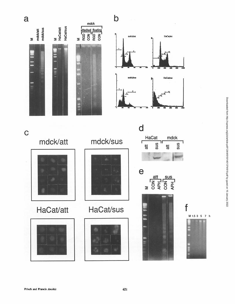
Figure 1. Effect of disruption of cell-matrix interactions on phenotypic features of apoptosis. (a, left and middle panels). Disruption of cell-matrix interactions by plating cells on a non-ionic surface: effects on internucleosomal DNA degradation. MDCK (leflpanel) or HaCat (middle panel) cells (3 x 10 ~) were plated onto tissue culture dishes (att) or onto petri dishes which had been coated with polyHEMA (sus); genomic DNA was analyzed as described in Materials and Methods. (M is the kilobase marker of GIBCO-BRL.) (a, right panel) Disruption of cell-matrix interactions by plating cells in the presence of RGD peptide. MDCK cells were plated on tissue culture dishes containing the peptide GRGDSP (RGD) or the peptide GRGESP (CON). Cells were harvested and processed after 8 h as above. (b) Flow cytometric analysis of cells treated as in a. (Apoptotic cells, bar 1; Go/G~ cells, bar 2; S-phase cells, bar 3; M phase cells, bar 4.) (c) Nuclear morphology before and after disruption of cell-matrix interactions. (d) Effect of disruption of cell-matrix interactions on the in- tegrity of nuclear lamins. MDCK or HaCat cells were incubated on wells of either tissue culture plastic (art) or polyHEMA (sus) and analyzed by Western blotting for soluble lamins as described in Materials and Methods. (e) Effect of aphidicolin on apoptosis. MDCK cells were plated on tissue culture dishes (art) or polyHEMA-coated dishes (sus) in the presence (APH) or absence (CON) of 5 #g/ml aphidicolin, a dose that inhibited DNA synthesis by >95% (data not shown). After 12 h, cells were collected and DNA was analyzed. (f) Time of commitment to anoikis. Confluent MDCK cells were trypsinized and plated on polyHEMA dishes for the times indicated above the lanes and then transferred to tissue culture dishes for the remainder of 7 h. Genomic DNAs were extracted and analyzed.
The Journal of Cell Biology, Volume 124, 1994 620
Dow
nloaded from http://rupress.org/jcb/article-pdf/124/4/619/1401374/619.pdf by guest on 10 January 2022
Frisch and Francis Anoikis 621
Dow
nloaded from http://rupress.org/jcb/article-pdf/124/4/619/1401374/619.pdf by guest on 10 January 2022
HaCat cells (spontaneously immortalized but nontrans- formed human keratinocytes) were plated on tissue culture plastic or on petri dishes that had been coated with poly- hydroxyethylmethacrylate (polyHEMA). polyHEMA has previously been used to prevent cell attachment because its uniformly nonionic nature prevents matrix deposition (Folk- man and Moscona, 1978). Agarose gel analysis of low mo- lecular weight DNA (Fig. 1 a) demonstrated a ladder of "o 200-bp multiple DNA fragments occurring in suspended but not atttached cells. This ladder has been shown to reflect internucleosomal DNA degradation, and is the currently ac- cepted hallmark of apoptosis (Wyllie, 1980). The DNA lad- der from "o3 x 106 cells was detectable within 7.5 h for MDCK or within 8.5 h for HaCat, and increased with incu- bation time on polyHEMA. Flow-cytometric analysis of propidium iodide-stained cells (Fig. 1 b) indicated that "o20% (MDCK) or ,o60% (HaCat) of the cells contained <2n DNA content after 18 h on polyHEMA. (These num- bers could in principle underestimate the fraction of apop- totic cells because mildly degraded DNA could score posi- tively in the flow cytometer, resulting in apparent 2n cells.)
To confirm that the extracellular signal for apoptosis was the lack of ceU-matrix interactions, fibronectin and vitronec- tin receptors of MDCK cells on tissue culture plastic were functionally blocked by the addition of the peptide GRGDSP (Ruoslahti and Pierschbacher, 1986). This peptide-induced apoptosis that was detectable in 8 h (Fig. 1 a) whereas a con- trol peptide that does not bind integrins (GRGESP) had no effect. We propose the term "anoikis" (an-o-EE-kis; Greek, meaning the state of being without a home) to describe the cells' apoptotic response to the absence of cell-matrix inter- actions.
Nuclear fragmentation, another diagnostic feature of apop- tosis (Wyllie, 1980), was also observed in cells incubated on polyHEMA (Fig. 1 c). Nuclear lamins have been reported to convert to a soluble form during apoptosis (Ucker et al., 1992) or to partially degrade into a 45-kD product (Kauf- mann, 1989), representing the u-helical domain common to lamins A, B, and C. Although increased soluble lamin level was not observed in suspended MDCK and HaCat cells (data not shown), the appearance of the 45-kD lamin degradation product was seen (Fig. 1 d), indicating some breakdown of the nuclear envelope. Generalized protein degradation was not observed on stained Western blot filters (data not shown).
Nonadherence has been shown to block DNA replication (Folkman and Moscona, 1978). To determine whether this inhibition sufficed to induce apoptosis, adherent MDCK cells were exposed to aphidicolin and analyzed for DNA degradation. No degradation was observed (Fig. 1 e) even when DNA replication was inhibited by >95 % (data not shown). Inhibition of DNA replication apparently was not the inducer of anoikis; it also did not protect cells against anoikis.
Certain forms of apoptosis are alleviated by inhibition of protein synthesis with drugs such as cycloheximide (Martin et al., 1988), implicating de novo expression of gene prod- uct(s) involved in cell suicide. Cycloheximide did not protect MDCK cells against DNA degradation in response to matrix detachment (data not shown). Several other forms of apopto- sis are resistant to cycloheximide (for example see Cotter et al., 1992) predicting the existence of both gene regulation- dependent and posttranslational pathways.
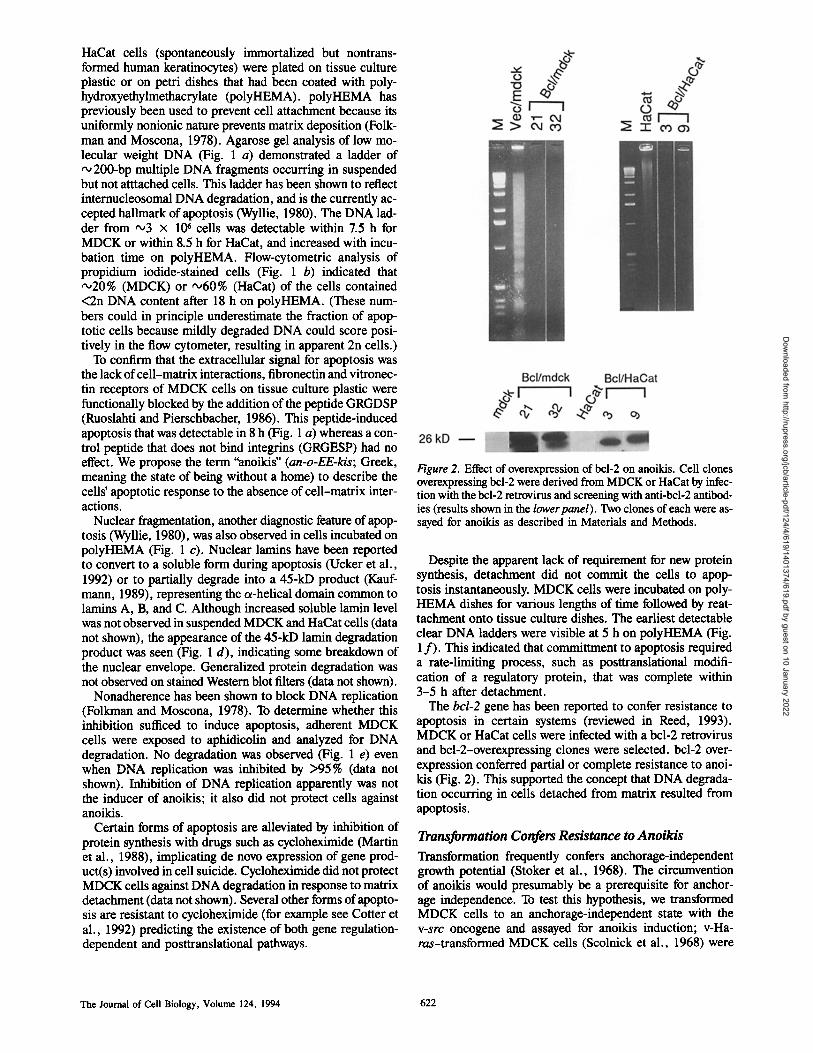
Figure 2. Effect of overexpression of bcl-2 on anoikis. Cell clones overexpressing bcl-2 were derived from MDCK or HaCat by infec- tion with the bcl-2 retrovirus and screening with anti-bcl-2 antibod- ies (results shown in the lower panel). Two clones of each were as- sayed for anoikis as described in Materials and Methods.
Despite the apparent lack of requirement for new protein synthesis, detachment did not commit the cells to apop- tosis instantaneously. MDCK cells were incubated on poly- HEMA dishes for various lengths of time followed by reat- tachment onto tissue culture dishes. The earliest detectable clear DNA ladders were visible at 5 h on polyHEMA (Fig. 1 f ) . This indicated that committment to apoptosis required a rate-limiting process, such as posttranslational modifi- cation of a regulatory protein, that was complete within 3-5 h after detachment.
The bcl-2 gene has been reported to confer resistance to apoptosis in certain systems (reviewed in Reed, 1993). MDCK or HaCat cells were infected with a bcl-2 retrovirus and bcl-2-overexpressing clones were selected, bcl-2 over- expression conferred partial or complete resistance to anoi- kis (Fig. 2). This supported the concept that DNA degrada- tion occurring in cells detached from matrix resulted from apoptosis.
Transformation Confers Resistance to Anoikis
Transformation frequently confers anchorage-independent growth potential (Stoker et al., 1968). The circumvention of anoikis would presumably be a prerequisite for anchor- age independence. To test this hypothesis, we transformed MDCK cells to an anchorage-independent state with the v-src oncogene and assayed for anoikis induction; v-Ha- ras-transformed MDCK cells (Scolnick et al., 1968) were
The Journal of Cell Biology, Volume 124, 1994 622
Dow
nloaded from http://rupress.org/jcb/article-pdf/124/4/619/1401374/619.pdf by guest on 10 January 2022
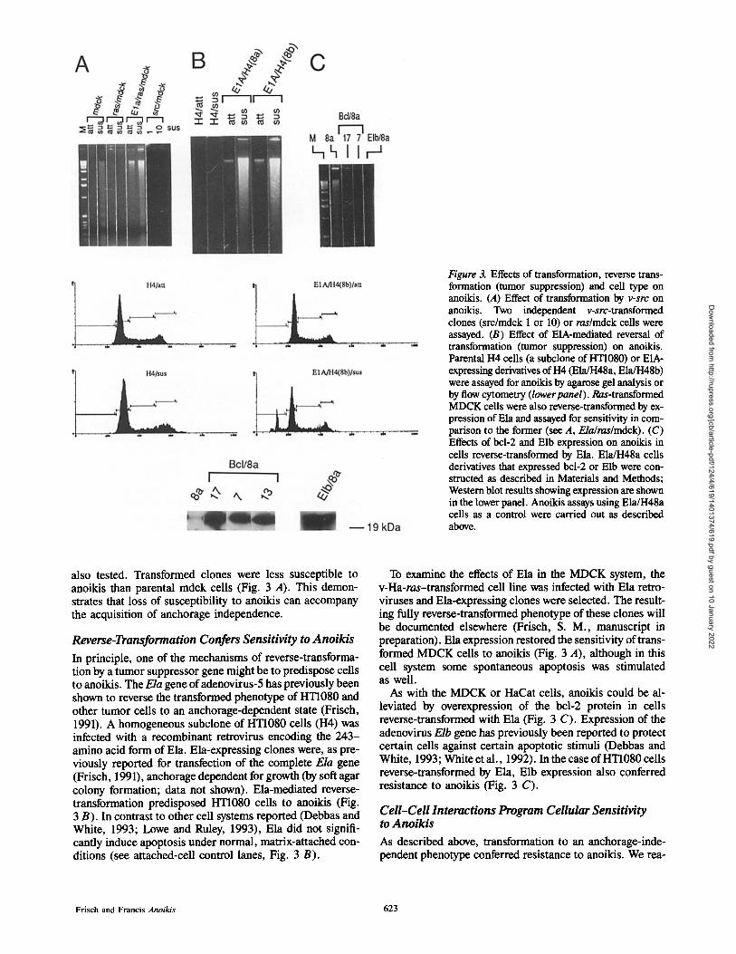
Figure 3. Effects of transformation, reverse trans- formation (tumor suppression) and cell type on anoikis. (A) Effect of transformation by v-src on anoikis. Two independent v-src-transformed clones (src/mdck 1 or 10) or ras/mdck cells were assayed. (B) Effect of E1A-mediated reversal of transformation (tumor suppression) on anoikis. Parental 1-14 cells (a subclone of HT1080) or E1A- expressing derivatives of 1-I4 (ElaJH48a, Ela/H48b) were assayed for anoikis by agarose gel analysis or by flow cytometry (lower panel). Ras-transformed MDCK cells were also reverse-transformed by ex- pression of Ela and assayed for sensitivity in com- parison to the former (see A, Ela/ras/mdck). (C) Effects of bcl-2 and Elb expression on anoikis in cells reverse-transformed by Ela. Ela/H48a cells derivatives that expressed bcl-2 or Elb were con- strutted as described in Materials and Methods; Western blot results showing expression are shown in the lower panel. Anoikis assays using Ela/H48a cells as a control were carded out as described above.
also tested. Transformed clones were less susceptible to anoikis than parental mdck cells (Fig. 3 A). This demon- strates that loss of susceptibility to anoikis can accompany the acquisition of anchorage independence.
Reverse-Transformation Confers Sensitivity to Anoikis
In principle, one of the mechanisms of reverse-transforma- tion by a tumor suppressor gene might be to predispose cells to anoikis. The E/a gene of adenovirus-5 has previously been shown to reverse the transformed phenotype of HT1080 and other tumor cells to an anchorage-dependent state (Frisch, 1991). A homogeneous subclone of HT1080 cells (H4) was infected with a recombinant retrovirus encoding the 243- amino acid form of Ela. Ela-expressing clones were, as pre- viously reported for transfection of the complete E/a gene (Frisch, 1991), anchorage dependent for growth (by soft agar colony formation; data not shown). Ela-mediated reverse- transformation predisposed HT1080 cells to anoikis (Fig. 3 B). In contrast to other cell systems reported (Debbas and White, 1993; Lowe and Ruley, 1993), Ela did not signifi- cantly induce apoptosis under normal, matrix-attached con- ditions (see attached-cell control lanes, Fig. 3 B).
To examine the effects of Ela in the MDCK system, the v-Ha-ras-transformed cell line was infected with Ela retro- viruses and Ela-expressing clones were selected. The result- ing fully reverse-transformed phenotype of these clones will be documented elsewhere (Frisch, S. M., manuscript in preparation). Ela expression restored the sensitivity of trans- formed MDCK cells to anoikis (Fig. 3 A), although in this cell system some spontaneous apoptosis was stimulated as well.
As with the MDCK or HaCat ceils, anoikis could be al- leviated by overexpression of the bcl-2 protein in cells reverse-transformed with Ela (Fig. 3 C). Expression of the adenovirus E/b gene has previously been reported to protect certain cells against certain apoptotic stimuli (Debbas and White, 1993; White et al., 1992). In the case of liT1080 cells reverse-transformed by Ela, Elb expression also conferred resistance to anoikis (Fig. 3 C).
CeU-CeU Interactions Program Cellular Sensitivity to Anoikis
As described above, transformation to an anchorage-inde- pendent phenotype conferred resistance to anoikis. We rea-
Frisch and Francis Anoikis 623
Dow
nloaded from http://rupress.org/jcb/article-pdf/124/4/619/1401374/619.pdf by guest on 10 January 2022
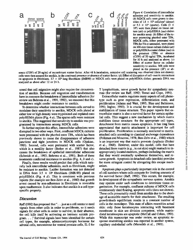
Figure 4. Correlation of intercellular adhesion and sensitivity to anoikis. (b) MDCK ceils were grown to den- sities of 1.8 x 10⁵ cells/cm 2 (dense) or 5.3 x 10⁴ (sparse). Cells (3 x 10⁴) were then plated on tissue cul- ture (att) or polyHEMA (sus) dishes for anoikis assay. (b) Effect of the tu- mor promoting phorbol ester TPA. MDCK cells, either pretreated with TPA for 5 h or untreated, were plated on 100-mm tissue culture dishes (att) or polyHEMA-coated dishes (sus) in the presence (TPA) or absence (CON) of 50 ng/ml TPA, incubated for 10 h and analyzed as above. (c) Effect of scatter factor on cellular sensitivity to anoikis. M8 cells were plated in the presence (SF) or ab-
sence (CON) of 20 ng/ml of recombinant scatter factor. After 48 h incubation, extensive cell scattering was observed (data not shown); cells were then assayed for anoikis, in the continued presence or absence of scatter factor. (d) Effect of disruption of cell-matrix interactions on apoptosis in fibroblasts. 3.7 x 10⁶ lung fibroblasts (IMR90) or MDCK cells were plated on polyHEMA dishes; genomic DNA was analyzed as above after 12 or 24 h.
soned that cell migration might also require the circumven- tion of anoikis. Because cell migration and transformation have in common the breakdown of intercellular adhesion (for review see Behrens et al., 1991, 1992), we reasoned that this breakdown might confer resistance to anoikis.
To determine whether interactions between cells served to modulate their sensitivity to anoikis, MDCK cells plated at either low or high density were trypsinized and replated onto polyHEMA plates (Fig. 4 a). The sparse cells were resistant to anoikis. This suggested that sensitivity to anoikis was pro- grammed by interactions among MDCK cells.
To further explore this effect, intercellular adhesions were disrupted in two other ways. First, confluent MDCK cultures were pretreated with the phorbol ester TPA, which has been previously shown to cause the disappearance of adherens junctions and tight junctions in MDCK cells (Ojakian, 1981). Second, cells were pretreated with scatter factor, which is a motility factor (Stoker et al., 1987) that also causes the breakdown of epithelial intercellular adhesions (Schmidt et al., 1993; Behrens et al., 1991). Both of these treatments conferred resistance to anoikis (Fig. 4, b and c).
Finally, these results would predict that cells which natu- rally lack intercellular adhesions, such as fibroblasts, might be resistant to anoikis. Ladder formation was not detected in DNA from 3.5 x 10⁶ fibroblasts (IMR-90) plated on polyHEMA (Fig. 4 d). This is consistent with previous reports (for example see Ben-Ze'ev et al., 1980) that growth arrest caused by non-adherence in fibroblasts is reversible upon readherence. It also indicates that anoikis is a cell type- specific property.
Discussion
Raft (1992) has proposed that" . . , just as a cell seems to need signals from other cells in order to proliferate, so it needs signals for other cells in order to survive; in their absence, the cell kills itself by activating an intrinsic suicide pro- g r a m . . . " Survival signals have been identified for certain cell types, for example, adrenocorticotropic hormone for adrenal cells, testosterone for ventral prostate cells, IL-2 for
T lymphoblasts, nerve growth factor for sympathetic neu- rons (for review see Raft, 1992; Tomei and Cope, 1991).
Extracellular matrix regulates many aspects of cell pheno- type such as gene expression, differentiation state, and proliferation (Adams and Watt, 1993; Blau and Baltimore, 1991; Ingber, 1993). It is crucial for the development and stabilization of tissue structure. This paper reports that ex- tracellular matrix is also a survival factor for certain epithe- lial cells. This suggest a new mechanism by which matrix stabilizes tissue structure: for the appropriate cell types, detachment from matrix causes apoptosis. It has long been appreciated that matrix attachment is important for cell proliferation. Proliferation is normally restricted to matrix- attached cells according to classical anchorage dependence (Folkman and Moscona, 1978; Ben-Ze'ev et al., 1980) which is an important way that matrix prevents dysplasia (Stoker et al., 1968). However, under this model, ceils that have detached from matrix (e.g., in cut skin) might reattach to in- appropriately located matrices, perhaps including the matrix that they would eventually synthesize themselves, and re- sume growth. Apoptosis in detached cells (anoikis) provides for more stringent control by abrogating this escape mech- anism.
Apoptosis plays a particularly important role in the control of cell numbers where cells compete for limiting amounts of the survival factor (Raft, 1992). This occurs, for example, in development of the sympathetic nervous system. Anoikis may operate under such conditions to determine tissue or- ganization. For example, confluent cultures of MDCK cells continuously shed floating, apoptotic cells (data not shown). These cells presumably result from anoikis due to the short- age of accessible matrix for their attachment. The resulting growth/death equilibrium results in a constant number of cells in the monolayer. This state of affairs resembles actual skin: only those keratinocytes interacting with basement membrane in skin are division competent, while matrix- distal keratinocytes are apoptotic (McCall and Cohen, 1991). While this manuscript was under review, an apoptotic re- sponse resembling anoikis was reported in another system, capillary endothelial cells (Meredith et al., 1993).
The Journal of Cell Biology, Volume 124, 1994 624
Dow
nloaded from http://rupress.org/jcb/article-pdf/124/4/619/1401374/619.pdf by guest on 10 January 2022
The acquisition of anchorage-independent growth during transformation (Stoker et al., 1968) would presumably re- quire that anoikis is abrogated. Transformation accom- plished this in the MDCK system. Elucidation of the mecha- nisms by which transformation abrogates anoikis may be critical for understanding the basis of anchorage (in)depen- dence. Clustering or ligand binding of 31 integrin has been shown to result in the phosphorylation of a protein kinase known as p125f'k; the latter is thus a candidate integrin sig- nal transducer (for review see Juliano and Haskill, 1993). V-src expression has been shown to cause constitutive hyper- phosphorylation of this molecule, perhaps playing a role in anchorage-independent growth. The involvement of p125 ~k in anoikis is currently being tested; hyperphosphorylation was observed in v-src-transformed MDCK cells (data not shown).
Altered cell-cell interactions could also be important for the effect of transformation on anoikis. Resistance to anoikis was conferred not only by transformation, but also by low cell density, TPA and scatter factor. All of these conditions cause the breakdown of normal epithelial cell-cell interac- tions (Zuk et al., 1989; Behrens et al., 1991, 1992; Schmidt et al., 1993; Ojakian, 1981). Although TPA and scatter fac- tor may affect cells in other unrelated ways, conferral of re- sistance by low cell density supports the role for cell-cell in- teractions. This breakdown and the ensuing resistance to anoikis is presumably important for tissue remodeling events involving conversions of epithelial ceils to temporarily or permanently motile states, such as epithelial-mesenchymal transitions. Alterations of cell-cell interactions by oncogenes could contribute to carcinogenesis through the abrogation of anoilds.
Reverse transformation by a tumor suppressor gene could in principle involve the conferral of cellular sensitivity to anoikis. We reported that the Ela gene of adenovirus is a tu- mor suppressor gene in a wide variety of human tumor cells (Frisch, 1991), as well as in v-ms-transformed MDCK cells (Frisch, S. M., manuscript in preparation). Results reported in this paper show that conferral of cellular sensitivity to anoikis is a possible mechanism for tumor suppression by Ela. The E/a gene has previously been reported to induce apoptosis independently of matrix effects (Debbas and White, 1993; Rao et al., 1992) and to sensitize cells to the apoptosis induced by tumor necrosis factor-or (White et al., 1992). As with the sensitization to anoilds, these effects are also blocked by bcl-2 or Elb. However, the Ela-induced apoptosis effect in rodent fibroblasts is mediated by wild- type 53 and blocked by mutant p53 (Debbas and White, 1993). By contrast, the anoikis effect occured in Ela-express- ing HT1080 cells, which are devoid of wild-type p53 (Ander- son, M. J., and E. Stanbridge, manuscript submitted for publication), and expression of mutant p53 did not block anoikis in MDCK cells (data not shown). Anoikis therefore differs from previously reported Ela-induced apoptosis in that it is contingent upon matrix detachment and appears to use a p53-independent apoptosis pathway.
The results identify a new function, regulation of anoilds, for several proteins whose contributions to carcinogenesis have previously been demonstrated. Cellular proteins that complex with Ela, to mediate both transformation (for re- view see Moran, 1993) and tumor suppression (Frisch, 1991) effects are likely to be key regulators of anoilds. The integrins (Ruoslahti, 1991), which can act as tumor suppres-
sors, (Giancotti and Ruoslahti, 1990) initiated or prevented the anoikis response, depending upon their ligand oc- cupancy. Activated protein kinase C (Colburn, 1980), the scatter factor receptor c-met (Rong et al., 1992) and bcl-2 (Reed et al., 1990) can contribute to transformation; each of these blocked the anoikis response. Proteins involved in in- tercellular adhesion, such as E-cadherin, are likely to be in- volved in programming sensitivity to anoikis, which is cur- rently being tested. Cellular adhesion molecules such as E-cadherin are important suppressors of carcinogenesis (Behrens et al., 1991, 1992). Re-examination of these classes of proteins in terms of anoikis regulation, as well as iden- tification of new proteins and elucidation of the signaling pathways involved, could prove important in understanding and controlling cancer.
In addition to those who provided cell lines, viruses, antibodies, and scatter factor listed in Materials and Methods, we would like to thank Matthew Schibler (La Jolla Cancer Research Foundation) (L.J.C.R.E) for help with microscopy, Muizz Hasham for artwork, John Joubran (Scripps) for flow cytometry, W. James Nelson (Stanford University), Erkki Ruoslahti and John Reed (L.J.C.R.E) for useful discussions, and J. Reed, E. Ruoslahti, E. Engvall, R. Oshima, M. Pfahl for reviewing the manuscript; D. Ucker was extremely helpful in advising about final revisions.
This work was supported by a Spaulding Foundation Pilot Project Award and National Institutes of Health grant R29 GM44573-04.
Received for publication 14 October 1993 and in revised form 9 December 1993.
References
Adams, J., and F. Watt. 1993. Regulation of development and differentiation by the extracellular matrix. Development (Camb.). 117:1183-1198.
Barinaga, M. 1993. Death gives birth to the nervous system. But how? Science (Wash. DC). 259:762-763.
Behrens, J., K. Weidner, U. Frixen, J. Schipper, M. Sachs, N. Arakaki, Y. Dailmhara, and W. Birchmeier. 1991. The role of E-cadherin and scatter factor in tumor invasion and cell motility. In Cell Motility Factors. I. Gold- berg, editor. Birkhauser Verlag, Basel.
Behrens, J., U. Frixen, J. Schipper, M. Weidner, and W. Birchmeier. 1992. Cell adhesion in invasion and metastasis. Sere. Cell Biol. 3:169-178.
Ben-Ze'ev, A., S. Farmer, and S. Penman. 1980. Protein synthesis requires cell-surface contact while nuclear events respond to cell shape on anchorage dependent fibroblasts. Cell. 21:365-372.
Blan, H., and D. Baltimore. 1991. Differentiation requires continuous regula- tion. J. Cell Biol. 112:781-783.
Bottaro, D., J. Rubin, A. Faletto, T. Chain, G. Kmiecik, G. Van de Woude, and S. Aaronson. 1991. The hepatocyte growth factor receptor is the c-met oncogene product. Science (Wash. DC). 152:802-804.
Colburn, N. 1980. Carcinogenesis-a Comprehensive Survey. Vol. 5. T. Slagu, editor. Raven Press, New York. 33-56.
Cotter, T., J. Glynn, F. Echeverri, and D. Green. 1992. The induction of apop- tosis occurs in all phases of the cell cycle. Anticancer Res. 12:773-780.
Debbas, M., and E. White. 1993. Wild-type p53 mediates apoptosis by Ela, which is inhibited by Elb. Genes & Dev. 7:546-555.
Dion, L., T. Gindhart, and N. Colburn. 1988. Four day duration of tumor pro- motor exposure required to transform JB6 promotion sensitive cells to an- chorage independence. Cancer Res. 48:7126-7131.
Duke, R., R. Chervenak, and J. Cohen. 1983. Endogenous nuclease induced DNA fragmentation: an early event in cell-mediated cytolysis. Proc. Natl. Acad. Sci. USA. 80:6361-6365.
Farquhar, M., and G. Palade. 1963. Junctional complexes in various epithelia. J. Cell Biol. 17:375-412.
Folkman, J., and A. Moscona. 1978. The role of cell shape in growth control. Nature (Lond.). 273:345-349.
Frisch, S. 1991. Antioncogenic effect of adenovirus Ela in human tumor cells. Proc. Natl. Acad. Sci. USA. 88:9077-9081.
Giancotti, F., and E. Ruoslahti. 1990. Elevated levels of fibronectin receptor suppress the transformed phenotype of CHO cells. Cell. 60:849-859.
Glass, J., and L. Gerace. 1990. Lamins A and C bind and assemble at the sur- face of mitotic chromosomes. J. Cell Biol. 111:1047-1057.
Gumbiner, B. 1992. Epithelial morphogenesis. Cell. 69:385-387. Gumbiner, B., B. Stevenson, and A. Grimaldi. 1988. The role of cell adhesion
molecule uvomorulin in the formation and maintenance of the epithelial junc- tional complex. J. Cell Biol. 107:1575-1587.
Frisch and Francis Anoikis 625
Dow
nloaded from http://rupress.org/jcb/article-pdf/124/4/619/1401374/619.pdf by guest on 10 January 2022
Hanada, M., S. Krajewski, S. Tanaka, D. Cazals-Hatem, B. Spengler, R. Ross, J. Biedler, and J. Reed. 1993. Regulation of bcl-2 oncoprotein levels with differentiation of human neuroblastuma cells. Cancer Res. 5:4978-4986.
Harlow, E., B. Franza, and C. Schiey. 1985. Monoclonai antibodies specific for Ela proteins: heterogeneity in Ela products. J. Virol. 3:533-546.
Hay, E. 1990. Epitheliai-mesenchymal transitions. Semin. Dev. Biol. 1: 347-356.
Ingber, D. 1993. Cellular tensegrity: defining new rules of biological design that govern the cytoskeletun. J. Cell Sci. 104:613-623.
Juliano, R., and S. Haskill. 1993. Signal transduction from the extracellular ma- trix. J. Cell Biol. 120:577-585.
Kaufman, S. 1989. Induction of endonucleolytic DNA cleavage in human acute myelogenous leukemia cells by etuposide, camptuthecin and other cytotoxic anticancer drugs. Cancer Res. 49:5870-5878.
Lee, S., S. Christakos, and M. Small. 1993. Apoptusis and signal transduction: clues to a molecular mechanism. Curr. Opin. Cell Biol. 5:286-294.
Li, Y., J. Ansamma, M. Bhargava, E. Rosen, T. Nakamura, and I. Goldberg. 1992. Effect of scarer factor and hepatucyte growth factor on motility and morphology of MDCK cells. In Vitro Cell Dev. Biol. 28A:364-368.
Lowe, S., and H. Rule)'. 1993. Stabilization of the p53 tumor suppressor is in- duced by Ela and accompanies apoptosis. Genes & Dev. 7:535-543.
Martin, D., R. E. Schmidt, P. S. DiSefano, O. H. Lowry, J. G. Carter, and E. M. Jonson, Jr. 1988. Inhibitors of protein synthesis and RNA synthesis prevent neuronal death caused by nerve growth factor deprivation. J. Cell Biol. 106:829.
Marx, J. 1993. Call death studies yield cancer clues. Science (Wash. DC). 259:760-761.
McCall, C., and J. Cohen. 1991. Programmed cell death in terminally differen- tiating keratinocytes. J. Invest. Dermatol. 97:111-115.
Meredith, J., B. Fazeli, and M. Schwartz. 1993. The extracellular matrix as a survival factor. Mol. Biol. Cell. 4:953-961.
Miller, A., and G. Rosman. 1989. New retrovirai vectors for gene transfer and expression. BioTechniques. 7:980-990.
Montesano, R., G. Schaller, and L. Orci. 1991. Induction of epithelial tubular morphogenesis in vitro by fibroblast-derived soluble factors. Cell. 66: 697-71 I.
Moran, E. 1993. DNA tumor virus transforming proteins and the cell cycle. Curt. Opin. Gen. Dev. 3:63-70.
Moran, E., B. Zerler, T. Harrison, and M. Mathews. 1986. Identification of separate domains in the Ela gene for immortalization and the activation of virus early genes. Mol. Cell. Biol. 6:3470-3480.
Nelson, W., and P. Veshnock. 1986. Dynamics of membrane-cytuskeletun (fo- drin) organization during development of polarity in MDCK cells. J. Cell Biol. 103:1751-1756.
Ojakian, G. 1981. Tumor promoter-induced changes in the permeability of epi- thelial cell tight junctions. Cell. 23:95-103.
Raft, M. 1992. Social controls on cell survival and cell death. Nature (Load.). 356:397-400.
Rao, L., M. Debbas, P. Sabbatini, D. Hockenbery, S. Korsmeyer, and E. White. 1992. The adenovirus Ela proteins induce apoptusis, which is in- hibited by the Elb 19-kD and bcl-2 proteins. Proc. Natl. Acad. Sci. USA. 89:7742-7746.
Reed, J. 1993. Bcl-2 and the regulation of programmed cell death. J. Cell Biol. 124:1-6.
Reed, J., L. Meister, S. Tanaka, M. Cuddy, S. Yum, C. Geyer, and D. Pleas- ure. 1991. Differential expression of bcl-2 proto-oncogene in neuroblas-
tuma and other human tumor liines of neural origin. Cancer Res. 51:6529- 6538.
Reed, J., S. Halden, C. Croce, and M. Cuddy. 1990. Complementation by bcl-2 and c-Ha-ras oncogenes in malignant transformation of rat embryo fibro- blasts. Mol. Cell. Biol. 10:4370-4374.
Rong, S., M. Bodescot, D. Blair, J. Dunn, T. Nakamura, K. Mizuno, M. Park, A. Chart, S. Aaronson, and G. VandeWoude. 1992. Tumorigenicity of the met protu-oncogene and the gene for hepatucyte growth factor. Mol. Cell. Biol. 12:5153-5158.
Ruoslahti, E. 1991. Integrins. J. Clin. Invest. 87:1-5. Ruoslahti, E., and M. Pierschbacher. 1986. Arg-Gly-Asp: a versatile cell rec-
ognition signal. Cell. 44:517-518. Ryle, C., D. Breitkxeutz, H. Stark, I. Leigh, P. Steinert, D. Roop, and N. Fuse-
nig. 1989. Density-dependent modulation of synthesis of keratins 1 + 10 in the human keratinocyte line HaCat and in ras-transfected tumorigenic clones. Differentiation. 40:42-54.
Schmidt, J., P. Piepenhagen, and W. J. Nelson. 1993. Modulation of epithelial morpbogenesis and cell fate by cell-to-cell signals and regulated cell adhe- sion. Semin. Cell Biol. 4:161-173.
Scolnick, E., D. Williams, J. Maryak, W. Vass, R. Goldberg, and W. Parks. 1976. Type C particle-positive and type C particle-negative rat cell lines: characterization of the coding capacity of endogenous sarcoma virus-specific RNA. Virology. 20:570-576.
Slavkin, H., and R. Greulich. 1975. Extracellular Matrix Influences on Gene Expression. Academic Press, New York.
Stoker, M., E. Gherards, M. Perryman, and J. Gray. 1987. Scatter Factor is a fibroblast-derived modulator of epithelial cell motility. Nature (Lond.). 327:239-242.
Stoker, M., C. O'Neill, S. Berryman, and V. Waxman. 1968. Anchorage and growth regulation in normal and virus-transformed cells. Int. J. Cancer 3:683-693.
Taub, M., and M. Saier. 1979. An established but differentiated kidney epithe- lial cell line (MDCK). Methods. Enzymol. LVIII:552-560.
Tomei, L., and F. Cope. 1991. Apoptosis: the Molecular Basis of Cell Death. Cold Spring Harbor Laboratory Press, Cold Spring Harbor, NY.
Ucker, D., J. Meyers, and P. Obermiller. 1992. Activation-driven T cell death. J. lmmunol. 149:1583-1592.
Vaux, M. 1993. Toward an understanding of the molecular mechanisms of physiologic cell death. Proc. Natl. Acad. Sci. USA. 90:786-789.
Warren, S., and W. J. Nelson. 1987. Non-mitogenic morphoregulatury action of pp60 v'~ on multicellular epithelial structures. Mol. Cell. Biol. 7: 1326-1335.
White, E., A. Denton, and B. Stillman. 1988. Role of the Elb 19,000 kD tumor antigen in regulating early gene expression. J. Virol. 62:3445-3454.
White, E., and R. Cipriani. 1990. Role of Elb proteins in transformation: al- tered organization of intermediate filaments in transformed cells that express the 19 kD protein. Mol. Cell. Biol. 10:120-130.
White, E., P. Sabbutini, M. Debbas, W. Wold, D. Kusher, and L. Gooding. 1992. The 19 kD Elb transforming protein inhibits programmed cell death and prevents cytolysis by tumor necrosis factor-alpha. Mol. Cell Biol. 12:2570-2580.
Wyllie, A. 1980. Glucocorticoid-induced thymocyte apoptusis is associated with endogenous nuclease activation. Nature (Load.). 284:555-556.
Zuk, A., K. Matlin, and E. Hay. 1989. Type I collagen Gel induces MDCK cells to become fusiform in shape and lose apical-basal polarity. J. Cell Biol. 108:903-919.
The Journal of Cell Biology, Volume 124, 1994 626
Dow
nloaded from http://rupress.org/jcb/article-pdf/124/4/619/1401374/619.pdf by guest on 10 January 2022