discovery of selective glucocorticoid receptor modulators by multiplexed reporter screening
TRANSCRIPT
Discovery of selective glucocorticoid receptormodulators by multiplexed reporter screeningAnthony N. Gerbera,b, Kiriko Masunoa, and Marc I. Diamondc,d,1
aDepartment of Medicine, bCardiovascular Research Institute, and Departments of cNeurology and dCellular and Molecular Pharmacology,University of California, San Francisco, CA 94143-2280
Edited by Keith R. Yamamoto, University of California, San Francisco, CA, and approved January 21, 2009 (received for review December 3, 2008)
Glucocorticoids are widely used to suppress inflammation andtreat various immune-mediated diseases. Some glucocorticoid re-ceptor (GR)-regulated genes mediate the therapeutic response,whereas others cause debilitating side effects. To discover selectivemodulators of the GR response, we developed a high-throughput,multiplexed system to monitor regulation of 4 promoters simul-taneously. An initial screen of 1,040 natural products and Food andDrug Administration-approved drugs identified modulators thatcaused GR to regulate only a subset of its target promoters. Somecompounds selectively inhibited GR-mediated gene activationwithout altering the repression of cytokine expression by GR. Thisapproach will facilitate identification of genes and small moleculesthat augment beneficial effects of GR and diminish deleteriousones. Our results have important implications for the developmentof GR modulators and the identification of cross-talk pathwaysthat control selective GR gene regulation.
fluorescent protein � high-throughput screen � inhibitor �selective modulator � combinatorial therapy
G lucocorticoid receptor (GR) belongs to the nuclear recep-tor (NR) family of intracellular ligand-regulated transcrip-
tion factors and is expressed ubiquitously in humans (1). Hor-mones such as cortisol activate GR, causing its nucleartranslocation, interaction with coregulators, and binding tospecific genomic sites to regulate transcription (2, 3). Thismediates the broad systemic effects of GR signaling and under-lies glucocorticoid treatment of diverse immune-mediated dis-eases such as asthma and rheumatoid arthritis. However, severedose-limiting side effects occur, including osteoporosis, musclewasting, and diabetes (4). No existing drugs induce only bene-ficial effects of GR.
The beneficial and harmful effects of glucocorticoids are dueto selective activation or repression of particular genes by GR.This selectivity is based in part on tissue-specific factors andcross-talk pathways (5). For example, GR reduces the expressionof certain inflammatory cytokines by inhibiting other transcrip-tion factors such as AP-1 and NF-�b (6). Conversely, GRincreases expression of RANKL, a gene coregulated by thevitamin D receptor that activates bone resorption by osteoclasts(7). Controlling GR activity at certain tissues or promoters hasprofound therapeutic implications.
Efforts to achieve this goal have focused primarily on devel-oping selective GR ligands that induce a subset of GR activities(8, 9). However, it is not yet possible to predict GR generegulation based on ligand design, and it remains uncertainwhether new ligands can produce therapeutically relevant tran-scriptional selectivity. Moreover, transcription-based screens forGR modulators generally measure GR activation at a singleexperimental promoter (10). This does not allow efficient iden-tification of molecules that produce promoter-specific responses.Accordingly, although numerous GR agonists with differentpotencies and/or modes of delivery are in clinical use, doseequivalency generally results in similar clinical responses andside effects (11). Nonligand modulation of GR is an alternative
strategy to achieve the desired transcriptional output, potentiallyenabling tissue or promoter-specific GR effects.
To address this problem, we developed a high-throughputsystem to measure GR activity simultaneously at 4 promoters.This permits discovery of genes or molecules that alter GRsignaling in a promoter-specific fashion. We have applied thissystem to identify selective modulators of the GR response in aninitial screen of 1,040 natural products and Food and DrugAdministrtion (FDA)-approved compounds.
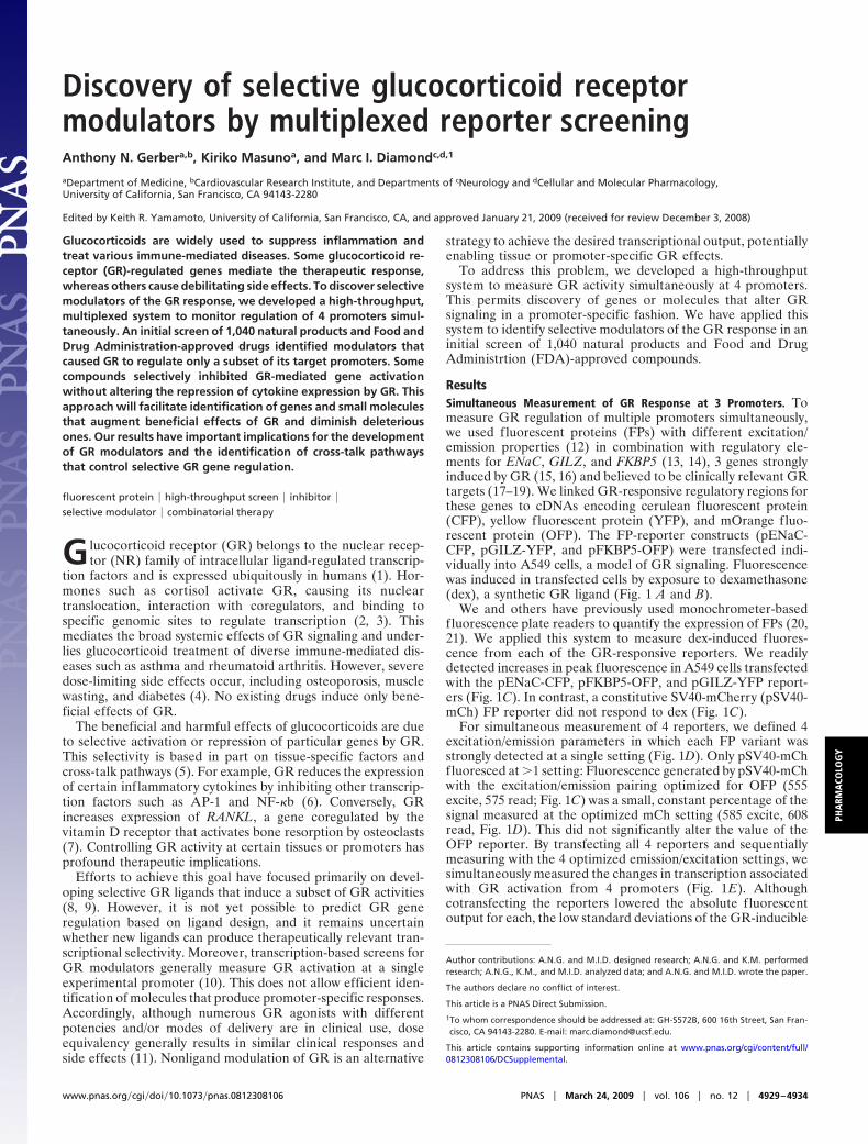
ResultsSimultaneous Measurement of GR Response at 3 Promoters. Tomeasure GR regulation of multiple promoters simultaneously,we used fluorescent proteins (FPs) with different excitation/emission properties (12) in combination with regulatory ele-ments for ENaC, GILZ, and FKBP5 (13, 14), 3 genes stronglyinduced by GR (15, 16) and believed to be clinically relevant GRtargets (17–19). We linked GR-responsive regulatory regions forthese genes to cDNAs encoding cerulean fluorescent protein(CFP), yellow fluorescent protein (YFP), and mOrange fluo-rescent protein (OFP). The FP-reporter constructs (pENaC-CFP, pGILZ-YFP, and pFKBP5-OFP) were transfected indi-vidually into A549 cells, a model of GR signaling. Fluorescencewas induced in transfected cells by exposure to dexamethasone(dex), a synthetic GR ligand (Fig. 1 A and B).
We and others have previously used monochrometer-basedfluorescence plate readers to quantify the expression of FPs (20,21). We applied this system to measure dex-induced fluores-cence from each of the GR-responsive reporters. We readilydetected increases in peak fluorescence in A549 cells transfectedwith the pENaC-CFP, pFKBP5-OFP, and pGILZ-YFP report-ers (Fig. 1C). In contrast, a constitutive SV40-mCherry (pSV40-mCh) FP reporter did not respond to dex (Fig. 1C).
For simultaneous measurement of 4 reporters, we defined 4excitation/emission parameters in which each FP variant wasstrongly detected at a single setting (Fig. 1D). Only pSV40-mChfluoresced at �1 setting: Fluorescence generated by pSV40-mChwith the excitation/emission pairing optimized for OFP (555excite, 575 read; Fig. 1C) was a small, constant percentage of thesignal measured at the optimized mCh setting (585 excite, 608read, Fig. 1D). This did not significantly alter the value of theOFP reporter. By transfecting all 4 reporters and sequentiallymeasuring with the 4 optimized emission/excitation settings, wesimultaneously measured the changes in transcription associatedwith GR activation from 4 promoters (Fig. 1E). Althoughcotransfecting the reporters lowered the absolute fluorescentoutput for each, the low standard deviations of the GR-inducible
Author contributions: A.N.G. and M.I.D. designed research; A.N.G. and K.M. performedresearch; A.N.G., K.M., and M.I.D. analyzed data; and A.N.G. and M.I.D. wrote the paper.
The authors declare no conflict of interest.
This article is a PNAS Direct Submission.
1To whom correspondence should be addressed at: GH-S572B, 600 16th Street, San Fran-cisco, CA 94143-2280. E-mail: [email protected].
This article contains supporting information online at www.pnas.org/cgi/content/full/0812308106/DCSupplemental.
www.pnas.org�cgi�doi�10.1073�pnas.0812308106 PNAS � March 24, 2009 � vol. 106 � no. 12 � 4929–4934
PHA
RMA
COLO
GY
reporters relative to the level of activation (Fig. 1E) implied thateach would clearly indicate GR activity in screens. There was noevidence of fluorescence resonance energy transfer between thedifferent reporters.
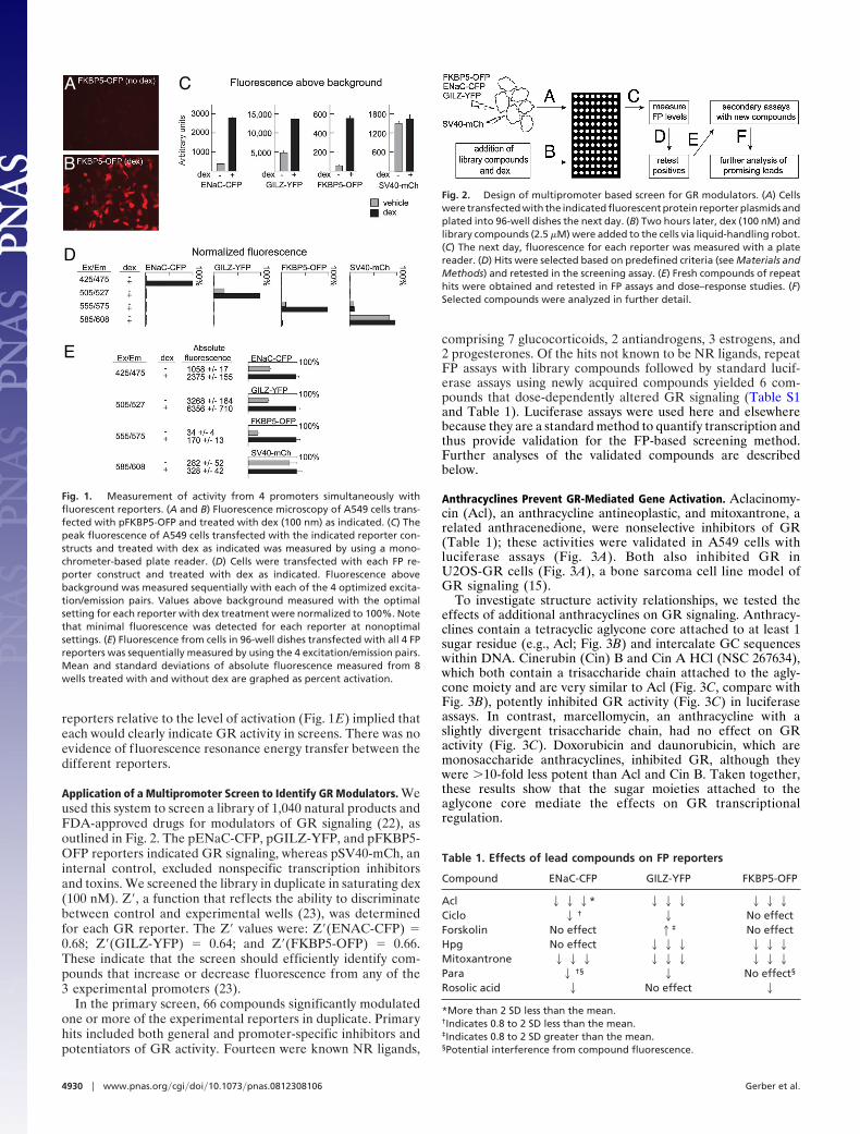
Application of a Multipromoter Screen to Identify GR Modulators. Weused this system to screen a library of 1,040 natural products andFDA-approved drugs for modulators of GR signaling (22), asoutlined in Fig. 2. The pENaC-CFP, pGILZ-YFP, and pFKBP5-OFP reporters indicated GR signaling, whereas pSV40-mCh, aninternal control, excluded nonspecific transcription inhibitorsand toxins. We screened the library in duplicate in saturating dex(100 nM). Z�, a function that reflects the ability to discriminatebetween control and experimental wells (23), was determinedfor each GR reporter. The Z� values were: Z�(ENAC-CFP) �0.68; Z�(GILZ-YFP) � 0.64; and Z�(FKBP5-OFP) � 0.66.These indicate that the screen should efficiently identify com-pounds that increase or decrease fluorescence from any of the3 experimental promoters (23).
In the primary screen, 66 compounds significantly modulatedone or more of the experimental reporters in duplicate. Primaryhits included both general and promoter-specific inhibitors andpotentiators of GR activity. Fourteen were known NR ligands,
comprising 7 glucocorticoids, 2 antiandrogens, 3 estrogens, and2 progesterones. Of the hits not known to be NR ligands, repeatFP assays with library compounds followed by standard lucif-erase assays using newly acquired compounds yielded 6 com-pounds that dose-dependently altered GR signaling (Table S1and Table 1). Luciferase assays were used here and elsewherebecause they are a standard method to quantify transcription andthus provide validation for the FP-based screening method.Further analyses of the validated compounds are describedbelow.
Anthracyclines Prevent GR-Mediated Gene Activation. Aclacinomy-cin (Acl), an anthracycline antineoplastic, and mitoxantrone, arelated anthracenedione, were nonselective inhibitors of GR(Table 1); these activities were validated in A549 cells withluciferase assays (Fig. 3A). Both also inhibited GR inU2OS-GR cells (Fig. 3A), a bone sarcoma cell line model ofGR signaling (15).
To investigate structure activity relationships, we tested theeffects of additional anthracyclines on GR signaling. Anthracy-clines contain a tetracyclic aglycone core attached to at least 1sugar residue (e.g., Acl; Fig. 3B) and intercalate GC sequenceswithin DNA. Cinerubin (Cin) B and Cin A HCl (NSC 267634),which both contain a trisaccharide chain attached to the agly-cone moiety and are very similar to Acl (Fig. 3C, compare withFig. 3B), potently inhibited GR activity (Fig. 3C) in luciferaseassays. In contrast, marcellomycin, an anthracycline with aslightly divergent trisaccharide chain, had no effect on GRactivity (Fig. 3C). Doxorubicin and daunorubicin, which aremonosaccharide anthracyclines, inhibited GR, although theywere �10-fold less potent than Acl and Cin B. Taken together,these results show that the sugar moieties attached to theaglycone core mediate the effects on GR transcriptionalregulation.
A
B
C
D
E
Fig. 1. Measurement of activity from 4 promoters simultaneously withfluorescent reporters. (A and B) Fluorescence microscopy of A549 cells trans-fected with pFKBP5-OFP and treated with dex (100 nm) as indicated. (C) Thepeak fluorescence of A549 cells transfected with the indicated reporter con-structs and treated with dex as indicated was measured by using a mono-chrometer-based plate reader. (D) Cells were transfected with each FP re-porter construct and treated with dex as indicated. Fluorescence abovebackground was measured sequentially with each of the 4 optimized excita-tion/emission pairs. Values above background measured with the optimalsetting for each reporter with dex treatment were normalized to 100%. Notethat minimal fluorescence was detected for each reporter at nonoptimalsettings. (E) Fluorescence from cells in 96-well dishes transfected with all 4 FPreporters was sequentially measured by using the 4 excitation/emission pairs.Mean and standard deviations of absolute fluorescence measured from 8wells treated with and without dex are graphed as percent activation.
Fig. 2. Design of multipromoter based screen for GR modulators. (A) Cellswere transfected with the indicated fluorescent protein reporter plasmids andplated into 96-well dishes the next day. (B) Two hours later, dex (100 nM) andlibrary compounds (2.5 �M) were added to the cells via liquid-handling robot.(C) The next day, fluorescence for each reporter was measured with a platereader. (D) Hits were selected based on predefined criteria (see Materials andMethods) and retested in the screening assay. (E) Fresh compounds of repeathits were obtained and retested in FP assays and dose–response studies. (F)Selected compounds were analyzed in further detail.
Table 1. Effects of lead compounds on FP reporters
Compound ENaC-CFP GILZ-YFP FKBP5-OFP
Acl 222* 222 222Ciclo 2 † 2 No effectForskolin No effect 1‡ No effectHpg No effect 222 222Mitoxantrone 222 222 222Para 2 †§ 2 No effect§
Rosolic acid 2 No effect 2
*More than 2 SD less than the mean.†Indicates 0.8 to 2 SD less than the mean.‡Indicates 0.8 to 2 SD greater than the mean.§Potential interference from compound fluorescence.
4930 � www.pnas.org�cgi�doi�10.1073�pnas.0812308106 Gerber et al.
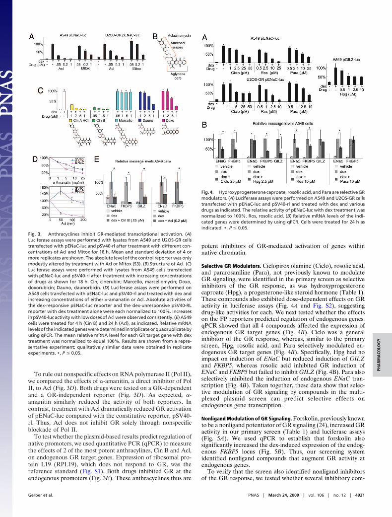
To rule out nonspecific effects on RNA polymerase II (Pol II),we compared the effects of �-amanitin, a direct inhibitor of PolII, to Acl (Fig. 3D). Both drugs were tested on a GR-dependentand a GR-independent reporter (Fig. 3D). As expected, �-amanitin similarly reduced the activity of both reporters. Incontrast, treatment with Acl dramatically reduced GR activationof pENaC-luc compared with the constitutive reporter, pSV40-rl. Thus, Acl does not inhibit GR solely through nonspecificblockade of Pol II.
To test whether the plasmid-based results predict regulation ofnative promoters, we used quantitative PCR (qPCR) to measurethe effects of 2 of the most potent anthracylines, Cin B and Acl,on endogenous GR target genes. Expression of ribosomal pro-tein L19 (RPL19), which does not respond to GR, was thereference standard (Fig. S1). Both drugs inhibited GR at theendogenous promoters (Fig. 3E). These anthracyclines thus are
potent inhibitors of GR-mediated activation of genes withinnative chromatin.
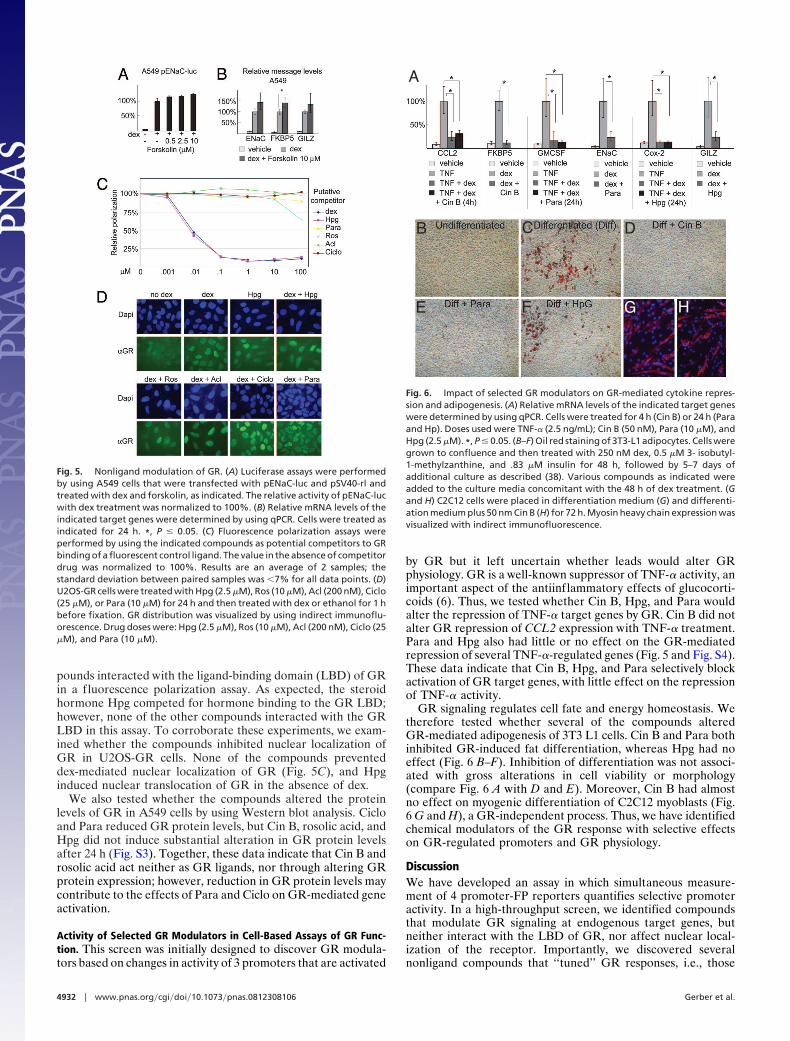
Selective GR Modulators. Ciclopirox olamine (Ciclo), rosolic acid,and pararosaniline (Para), not previously known to modulateGR signaling, were identified in the primary screen as selectiveinhibitors of the GR response, as was hydroxyprogesteronecaproate (Hpg), a progesterone-like steroid hormone (Table 1).These compounds also exhibited dose-dependent effects on GRactivity in luciferase assays (Fig. 4A and Fig. S2), suggestingdrug-like activities for each. We next tested whether the effectson the FP reporters predicted regulation of endogenous genes.qPCR showed that all 4 compounds affected the expression ofendogenous GR target genes (Fig. 4B). Ciclo was a generalinhibitor of the GR response, whereas, similar to the primaryscreen, Hpg, rosolic acid, and Para selectively modulated en-dogenous GR target genes (Fig. 4B). Specifically, Hpg had noimpact on induction of ENaC but reduced induction of GILZand FKBP5, whereas rosolic acid inhibited GR induction ofENaC and FKBP5 but failed to inhibit GILZ (Fig. 4B). Para alsoselectively inhibited the induction of endogenous ENaC tran-scription (Fig. 4B). Taken together, these data show that selec-tive modulation of GR signaling by compounds in the multi-plexed plasmid screen can predict selective effects onendogenous gene transcription.
Nonligand Modulation of GR Signaling. Forskolin, previously knownto be a nonligand potentiator of GR signaling (24), increased GRactivity in our primary screen (Table 1) and luciferase assays(Fig. 5A). We used qPCR to establish that forskolin alsosignificantly increased the dex-induced expression of the endog-enous FKBP5 locus (Fig. 5B). Thus, our screening systemidentified nonligand compounds that augment GR activity atendogenous genes.
To verify that the screen also identified nonligand inhibitorsof the GR response, we tested whether several inhibitory com-
Fig. 3. Anthracyclines inhibit GR-mediated transcriptional activation. (A)Luciferase assays were performed with lysates from A549 and U2OS-GR cellstransfected with pENaC-luc and pSV40-rl after treatment with different con-centrations of Acl and Mitox for 18 h. Mean and standard deviation of 4 ormore replicates are shown. The absolute level of the control reporter was onlymodestly altered by treatment with Acl or Mitox (S3). (B) Structure of Acl. (C)Luciferase assays were performed with lysates from A549 cells transfectedwith pENaC-luc and pSV40-rl after treatment with increasing concentrationsof drugs as shown for 18 h. Cin, cinerubin; Marcello, marcellomycin; Doxo,doxorubicin; Dauno, daunorbicin. (D) Luciferase assays were performed onA549 cells transfected with pENaC-luc and pSV40-rl and treated with dex andincreasing concentrations of either �-amanatin or Acl. Absolute activities ofthe dex-responsive pENaC-luc reporter and the dex-unresponsive pSV40-RLreporter with dex treatment alone were each normalized to 100%. Increasesin pSV40-luc activity with low doses of Acl were observed consistently. (E) A549cells were treated for 4 h (Cin B) and 24 h (Acl), as indicated. Relative mRNAlevels of the indicated genes were determined in triplicate or quadruplicate byusing qPCR. The mean relative mRNA level for each GR target gene with dextreatment was normalized to equal 100%. Results are shown from a repre-sentative experiment; qualitatively similar data were obtained in replicateexperiments. *, P � 0.05.
Fig. 4. Hydroxyprogesterone caproate, rosolic acid, and Para are selective GRmodulators. (A) Luciferase assays were performed on A549 and U2OS-GR cellstransfected with pENaC-luc and pSV40-rl and treated with dex and variousdrugs as indicated. The relative activity of pENaC-luc with dex treatment wasnormalized to 100%. Ros, rosolic acid. (B) Relative mRNA levels of the indi-cated genes were determined by using qPCR. Cells were treated for 24 h asindicated. *, P � 0.05.
Gerber et al. PNAS � March 24, 2009 � vol. 106 � no. 12 � 4931
PHA
RMA
COLO
GY
pounds interacted with the ligand-binding domain (LBD) of GRin a fluorescence polarization assay. As expected, the steroidhormone Hpg competed for hormone binding to the GR LBD;however, none of the other compounds interacted with the GRLBD in this assay. To corroborate these experiments, we exam-ined whether the compounds inhibited nuclear localization ofGR in U2OS-GR cells. None of the compounds preventeddex-mediated nuclear localization of GR (Fig. 5C), and Hpginduced nuclear translocation of GR in the absence of dex.
We also tested whether the compounds altered the proteinlevels of GR in A549 cells by using Western blot analysis. Cicloand Para reduced GR protein levels, but Cin B, rosolic acid, andHpg did not induce substantial alteration in GR protein levelsafter 24 h (Fig. S3). Together, these data indicate that Cin B androsolic acid act neither as GR ligands, nor through altering GRprotein expression; however, reduction in GR protein levels maycontribute to the effects of Para and Ciclo on GR-mediated geneactivation.
Activity of Selected GR Modulators in Cell-Based Assays of GR Func-tion. This screen was initially designed to discover GR modula-tors based on changes in activity of 3 promoters that are activated
by GR but it left uncertain whether leads would alter GRphysiology. GR is a well-known suppressor of TNF-� activity, animportant aspect of the antiinflammatory effects of glucocorti-coids (6). Thus, we tested whether Cin B, Hpg, and Para wouldalter the repression of TNF-� target genes by GR. Cin B did notalter GR repression of CCL2 expression with TNF-� treatment.Para and Hpg also had little or no effect on the GR-mediatedrepression of several TNF-�-regulated genes (Fig. 5 and Fig. S4).These data indicate that Cin B, Hpg, and Para selectively blockactivation of GR target genes, with little effect on the repressionof TNF-� activity.
GR signaling regulates cell fate and energy homeostasis. Wetherefore tested whether several of the compounds alteredGR-mediated adipogenesis of 3T3 L1 cells. Cin B and Para bothinhibited GR-induced fat differentiation, whereas Hpg had noeffect (Fig. 6 B–F). Inhibition of differentiation was not associ-ated with gross alterations in cell viability or morphology(compare Fig. 6 A with D and E). Moreover, Cin B had almostno effect on myogenic differentiation of C2C12 myoblasts (Fig.6 G and H), a GR-independent process. Thus, we have identifiedchemical modulators of the GR response with selective effectson GR-regulated promoters and GR physiology.
DiscussionWe have developed an assay in which simultaneous measure-ment of 4 promoter-FP reporters quantifies selective promoteractivity. In a high-throughput screen, we identified compoundsthat modulate GR signaling at endogenous target genes, butneither interact with the LBD of GR, nor affect nuclear local-ization of the receptor. Importantly, we discovered severalnonligand compounds that ‘‘tuned’’ GR responses, i.e., those
Fig. 5. Nonligand modulation of GR. (A) Luciferase assays were performedby using A549 cells that were transfected with pENaC-luc and pSV40-rl andtreated with dex and forskolin, as indicated. The relative activity of pENaC-lucwith dex treatment was normalized to 100%. (B) Relative mRNA levels of theindicated target genes were determined by using qPCR. Cells were treated asindicated for 24 h. *, P � 0.05. (C) Fluorescence polarization assays wereperformed by using the indicated compounds as potential competitors to GRbinding of a fluorescent control ligand. The value in the absence of competitordrug was normalized to 100%. Results are an average of 2 samples; thestandard deviation between paired samples was �7% for all data points. (D)U2OS-GR cells were treated with Hpg (2.5 �M), Ros (10 �M), Acl (200 nM), Ciclo(25 �M), or Para (10 �M) for 24 h and then treated with dex or ethanol for 1 hbefore fixation. GR distribution was visualized by using indirect immunoflu-orescence. Drug doses were: Hpg (2.5 �M), Ros (10 �M), Acl (200 nM), Ciclo (25�M), and Para (10 �M).
A
B C D
E F G H
Fig. 6. Impact of selected GR modulators on GR-mediated cytokine repres-sion and adipogenesis. (A) Relative mRNA levels of the indicated target geneswere determined by using qPCR. Cells were treated for 4 h (Cin B) or 24 h (Paraand Hp). Doses used were TNF-� (2.5 ng/mL); Cin B (50 nM), Para (10 �M), andHpg (2.5 �M). *, P � 0.05. (B–F) Oil red staining of 3T3-L1 adipocytes. Cells weregrown to confluence and then treated with 250 nM dex, 0.5 �M 3- isobutyl-1-methylzanthine, and .83 �M insulin for 48 h, followed by 5–7 days ofadditional culture as described (38). Various compounds as indicated wereadded to the culture media concomitant with the 48 h of dex treatment. (Gand H) C2C12 cells were placed in differentiation medium (G) and differenti-ation medium plus 50 nm Cin B (H) for 72 h. Myosin heavy chain expression wasvisualized with indirect immunofluorescence.
4932 � www.pnas.org�cgi�doi�10.1073�pnas.0812308106 Gerber et al.
that caused GR to regulate only a subset of its normal spectrumof target genes and cellular responses. This work thus hasimportant implications for drug discovery and genetic studies toelucidate factors and signaling pathways that selectively regulateGR function.
Power of a MultiPromoter Readout. Transcriptional regulation isusually measured in high throughput with a single experimentalpromoter driving an enzyme-based reporter or FP (25–27).Laser-based systems and high-content microscopy can be used tomeasure multiple cellular processes but have not been widelyapplied for multiplexed measurement of transcription (28). Theease of measurement, cost, and low variability of FP expressionoffer advantages over screening systems that directly measuremRNA levels (29). Our system can also use mixtures of cell typesharboring distinct FP reporters, thereby distinguishing cell se-lective modulators. Thus, our system is an important, low-costalternative to current systems that use a single reporter ordirectly measure mRNA levels.
Most importantly, the multiplexed system identified com-pounds that differentially modulated GR activity at a subset of3 promoters. Although the screen was not designed to identifythem, several of the compounds inhibited GR activation but notthe repression of inflammatory cytokines. By choosing promot-ers to include both beneficial and harmful targets, it should bepossible to identify more efficiently small molecules that tunereceptor output to a desired pattern through effects on relevantcross-talk pathways.
In the screen, there were 66 primary hits, of which 14 weresteroid-like NR ligands. Six additional hits were validated asnonsteroidal GR modulators. Thus, the overall screening effi-ciency was 30%. However, the percentage of nonsteroidal hitsthat validated as dose-dependent GR regulators was only 11% (6of 52). The false positive rate was in part due to edge effects,which have been described (30). In the future, specific libraryand plate configurations can be used to reduce variability.
Inhibitors of GR. Anthracyclines were identified as inhibitors ofthe GR response. The most potent of these, Acl and Cin B, bothcontain a trisaccharide chain attached to the anthracyclineaglycone core that mediates DNA intercalation and is critical forthe cytotoxic properties of this drug class (31). Although an-thracyclines inhibit the activity of RNA polymerases (32), theeffects we observed cannot be ascribed to nonspecific inhibitionof transcription (Fig. 3D). The impact of the trisaccharide chainon GR inhibition also indicates that Acl and Cin B do not blockthe GR response simply through competing for DNA binding viathe aglycone core, a mechanism previously reported for inhibi-tion of SP-1 by other anthracyclines (33, 34). Rather, intercala-tion of the anthracycline aglycone core with DNA may allowspecific interactions between the attached sugars and DNA-associated proteins such as transcriptional cofactors and chro-matin remodeling complexes that regulate subsets of the Pol IItranscriptome.
Selective Modulators of the GR Response. Progestins can bind GRand mediate transcriptional effects (35). We identified hy-droxyprogesterone caproate as a selective regulator of GR in theprimary screen, and at endogenous genes. We also identified 7known glucocorticoids as selectively regulating GR in the pri-mary screen (Table S2). Thus, the multipromoter assay can beharnessed to identify selective ligands that specify a desiredtranscriptional output.
Rosolic acid and Para, which share a common structure of acentral carbon bound to 3 phenyl groups, also selectively mod-ulated the GR response in the primary screen and at endogenousgenes. Para was previously reported to inhibit androgen receptor(AR) signaling (29), and 2 additional compounds with structural
similarity to Para and rosolic acid also inhibited GR in luciferaseassays (Fig. S5). It will be of interest to test whether thecompounds identified here selectively regulate other nuclearreceptors and to determine their mechanisms of action. It doesnot appear that their predominant mode of action is via inter-actions with the LBD of GR or through preventing the nuclearlocalization of GR, suggesting that GR cross-talk pathways aremediating the effects.
ConclusionSelective modulation of GR and other nuclear receptors is amajor therapeutic goal. Most efforts to achieve selective mod-ulation have been directed at the LBD of each receptor, whichrepresents only a single facet of the nuclear receptor’s activityand does not directly address receptor interactions with cross-talk pathways (36, 37). In contrast, the multiplexed reporterscreen we have described here is a powerful approach foridentifying ligands that specify a desired transcriptional outputor for identifying and targeting therapeutically relevant receptorcross-talk pathways. Subsequent analysis using qPCR or mi-croarrays can rapidly determine whether hits have bona fideimpact on receptor signaling. For putative GR modulators, thereare also numerous cell-based assays, such as repression ofcytokine expression and modulation of cell phenotype, to testphysiologic function. Our application of such assays here sug-gests that GR signaling can be altered in a potentially beneficialfashion through combinations of traditional ligands and nonli-gand modulators. We anticipate that broad interrogation of NRcross-talk pathways in compound screens will lead to improvedtherapies in which physiologic responses are tuned via combi-nations of classical receptor ligands and heterologousmodulators.
Materials and MethodsPlasmids. mCherry and mOrange, cerulean, pENaC-luc, pFKBP5-luc and pGILZ-luc were gifts from R. Tsien (University of California, San Diego), D. Piston(Vanderbilt University, Nashville), C. Thomas (University of Iowa, Iowa City), T.Scammell (University of South Alabama, Mobile), and K. Yamamoto (Univer-sity of California, San Francisco), respectively. pSV40-RL is from Promega.Luciferase coding regions in pENaC-luc, pFKBP5-luc, pGILZ-luc, and pSV40-RLwere replaced with various FPs by using standard techniques. When necessary,site-directed mutagenesis used the QuikChange kit and protocol (Promega).
Cell Culture. A549 (ATCC), U2OS-GR (gift of Keith Yamamoto’s laboratory),and C2C12 (ATCC) cells were grown in high-glucose DMEM supplementedwith glutamine, penicillin, streptomycin, and 5% or 10% FBS (HyClone). Dextreatment was at 100 nm unless otherwise indicated. Culture and differ-entiation of 3T3-L1 cells (ATCC) was as described (38). C2C12 cells werecultured in DMEM supplemented with 2% heat-inactivated horse serum(GIBCO) to induce differentiation.
Chemicals. �-Amanitin, 3-isobutyl-1-methylzanthine, Hpg, mitoxantrone, dex,DMSO, forskolin, rosolic acid, Para, and Ciclo were from Sigma. TNF-� wasfrom Roche. Acl, doxorubicin, daunorubicin, marcellomycin, Cin B, and Cin AHCL (NSC 267694) were from the National Cancer Institute/DTP Open ChemicalRepository (http://dtp.nci.nih.gov).
Transfections. Approximately 3 � 105 cells were plated in 6-well dishes. Thenext day, 500 �L of Lipofectamine 2000 (Invitrogen)/plasmid DNA (4 �g total)complex was added to 1.5 mL of DMEM with pen/strep and 5% FBS (A549 cells)or to 1.5 mL of Optimem (U2OS cells). After 3–4 h of incubation, media werereplaced with standard growth media.
Luciferase Assays. Luciferase assays used the Promega Dual-Luciferase Re-porter system and protocol. Briefly, cells were transfected with firefly lucif-erase plasmids and pSV40-RL at a ratio of 10:1. The next day, cells were splitinto 96-well dishes and treated with dex and various drugs. After 12–18 h, cellswere harvested in 50 �L of PLB. Luminescence was detected from 10 �L oflysate by using an Ultra Evolution plate reader (Tecan).
Gerber et al. PNAS � March 24, 2009 � vol. 106 � no. 12 � 4933
PHA
RMA
COLO
GY
GR Binding. Compound binding to the GR LBD was assessed by using fluores-cence polarization competition (P2816; Invitrogen). Polarization with no com-petitor was normalized to 100%.
Compound Screen. A549 cells were transfected with an equal mixture ofpENaC-CFP, pGILZ-YFP, and pFKBP5-OFP (1.3 �g of each) or with pSV40-ChFP(4 �g). The next day, cells were mixed (�1.8 � 104 of the triple-transfectedcells; �2 � 103 cells transfected with pSV40-ChFP alone) and replated into96-well plates (3603; Costar). Two hours later, library compounds (2.5 �M) anddex (100 nM) were added by using a Biomek robot. The next day, cells werefixed for 4 min in 4% paraformaldehyde. Fluorescence was measured for eachreporter by using a Safire plate reader (Tecan). Hits were scored based on themean and SD of the experimental wells on each plate. Criteria for a hit wereif 1 reporter was �1.5 times the SD from the mean, or if 2 reporters were �0.8times the SD from the mean. Wells were generally excluded if SV40-ChFP was�2 times the SD from the mean in a single plate, or 0.8 times the SD induplicate plates. Compounds with intrinsic fluorescence were not scored forreporters with overlapping fluorescence.
Quantitative PCR. Total RNA isolation used TRIzol and Pure Link (Invitrogen).Random-primed cDNA was prepared from 1 �g of total RNA by usingMMLV-RT (Promega). Resultant cDNA (50 ng) was used per 50-�L reactioncontaining 1.25 units of TaqDNA polymerase (Invitrogen), 1.5 mM MgCl2, 300
nM concentrations of each primer (sequences available on request), 0.15 mMdNTP mix, and 0.2� SYBR green dye (Molecular Probes) in 1� PCR buffer(Qiagen). Real-time PCR used an Applied Biosystems 7700 machine and wasanalyzed by using the ��Ct method (Applied Biosystems Prism 7700 UsersBulletin No. 2). RPL19 expression was used for data normalization. Relativemessage levels between samples were compared by using nonparametricrank-sum tests. Data from representative experiments are shown; qualita-tively similar data were obtained in replicate experiments.
Immunohistochemistry. Cells were fixed in ice-cold methanol (�GR) or 4% PFA[myosin heavy chain (MyHC)] and washed three times in PBS, incubated witha polyclonal GR antibody (1 �g/mL; 3579; Abcam) or MF20 (MyHC), washed inPBS, and incubated with secondary antibody (Invitrogen). Oil-red staining wasperformed as described (38).
ACKNOWLEDGMENTS. We thank Jeremy Jones, Sebastian Meijsing, StefanTaubert, Abigail Kroch, and other members of M.I.D.’s and Keith R. Yamamo-to’s laboratories for stimulating discussions and critical comments on themanuscript and David Pearce, David Erle, and Monica and Nicholas Gerber fortheir critiques. A.N.G. was supported by National Institutes of Health GrantK08 HL077159, a SABRe award (Sandler Foundation), and an American LungAssociation of California research grant. M.I.D. was supported by NationalInstitutes of Health Grants R01 NS50284-01A1 (National Institute of Neuro-logical Disorders and Stroke) and R01CA131226-01 (National Cancer Institute).
1. McMaster A, Ray DW (2007) Modelling the glucocorticoid receptor and producingtherapeutic agents with anti-inflammatory effects but reduced side-effects. ExpPhysiol 92:299–309.
2. Yamamoto KR, Darimont BD, Wagner RL, Iniguez-Lluhi JA (1998) Building transcrip-tional regulatory complexes: Signals and surfaces. Cold Spring Harb Symp Quant Biol63:587–598.
3. So AY, Chaivorapol C, Bolton EC, Li H, Yamamoto KR (2007) Determinants of cell- andgene-specific transcriptional regulation by the glucocorticoid receptor. PLoS Genet3:e94.
4. Schacke H, Docke W-D, Asadullah K (2002) Mechanisms involved in the side effects ofglucocorticoids. Pharmacol Ther 96:23–43.
5. Kassel O, Herrlich P (2007) Crosstalk between the glucocorticoid receptor and othertranscription factors: Molecular aspects. Mol Cell Endocrinol 275:13–29.
6. Smoak KA, Cidlowski JA (2004) Mechanisms of glucocorticoid receptor signalingduring inflammation. Mech Ageing Dev 125:697–706.
7. Kim S, Yamazaki M, Zella LA, Shevde NK, Pike JW (2006) Activation of receptoractivator of NF-{kappa}B ligand gene expression by 1,25-dihydroxyvitamin D3 is me-diated through multiple long-range enhancers. Mol Cell Biol 26:6469–6486.
8. Rosen J, Miner JN (2005) The search for safer glucocorticoid receptor ligands. EndocrRev 26:452–464.
9. Cole TJ, Mollard R (2007) Selective glucocorticoid receptor ligands. Med Chem 3:494–506.
10. Fan F, Wood KV (2007) Bioluminescent assays for high-throughput screening. ASSAYDrug Dev Technol 5:127–136.
11. Adams N, Lasserson TJ, Cates CJ, Jones PW (2007) Fluticasone versus beclomethasoneor budesonide for chronic asthma in adults and children. Cochrane Database Syst RevCD002310.
12. Shaner NC, Steinbach PA, Tsien RY (2005) A guide to choosing fluorescent proteins. NatMethods 2:905–909.
13. Mick VE, et al. (2001) The {{alpha}}-subunit of the epithelial sodium channel is analdosterone-induced transcript in mammalian collecting ducts, and this transcriptionalresponse is mediated via distinct cis-elements in the 5�-flanking region of the gene. MolEndocrinol 15:575–588.
14. Hubler TR, Scammell JG (2004) Intronic hormone response elements mediateregulation of FKBP5 by progestins and glucocorticoids. Cell Stress Chaperones9:243–252.
15. Rogatsky I, et al. (2003) Target-specific utilization of transcriptional regulatory surfacesby the glucocorticoid receptor. Proc Natl Acad Sci USA 100:13845–13850.
16. Wang JC, et al. (2004) Chromatin immunoprecipitation (ChIP) scanning identifiesprimary glucocorticoid receptor target genes. Proc Natl Acad Sci USA 101:15603–15608.
17. Woodruff PG, et al. (2007) Genome-wide profiling identifies epithelial cell genesassociated with asthma and with treatment response to corticosteroids. Proc Natl AcadSci USA 104:15858–15863.
18. Ayroldi E, et al. (2007) GILZ mediates the antiproliferative activity of glucocorticoids bynegative regulation of Ras signaling. J Clin Invest 117:1605–1615.
19. Wang HC, et al. (2000) Oxidative stress disrupts glucocorticoid hormone-dependenttranscription of the amiloride-sensitive epithelial sodium channel alpha-subunit inlung epithelial cells through ERK-dependent and thioredoxin-sensitive pathways.J Biol Chem 275:8600–8609.
20. Richards B, et al. (2002) Stable expression of Anthozoa fluorescent proteins in mam-malian cells. Cytometry 48:106–112.
21. Pollitt SK, et al. (2003) A rapid cellular FRET assay of polyglutamine aggregationidentifies a novel inhibitor. Neuron 40:685–694.
22. Abbott A (2002) Neurologists strike gold in drug screen effort. Nature 417:109.23. Zhang JH, Chung TD, Oldenburg KR (1999) A simple statistical parameter for use in
evaluation and validation of high throughput screening assays. J Biomol Screen4:67–73.
24. Liu JL, Papachristou DN, Patel YC (1994) Glucocorticoids activate somatostatin genetranscription through co-operative interaction with the cyclic AMP signalling pathway.Biochem J 301 (Pt 3):863–869.
25. Necela BM, Cidlowski JA (2003) Development of a flow cytometric assay to studyglucocorticoid receptor-mediated gene activation in living cells. Steroids 68:341–350.
26. Suzuki T, et al. (2006) Screening of novel nuclear receptor agonists by a convenientreporter gene assay system using green fluorescent protein derivatives. Phytomedicine13:401–411.
27. Sonneveld E, Jansen HJ, Riteco JAC, Brouwer A, van der Burg B (2005) Development ofandrogen- and estrogen-responsive bioassays, members of a panel of human cellline-based highly selective steroid-responsive bioassays. Toxicol Sci 83:136–148.
28. Auld DS, et al. (2006) Fluorescent protein-based cellular assays analyzed by laser-scanning microplate cytometry in 1,536-well plate format. Methods Enzymol 414:566–589.
29. Hieronymus H, et al. (2006) Gene expression signature-based chemical genomicprediction identifies a novel class of HSP90 pathway modulators. Cancer Cell10:321–330.
30. Lundholt BK, Scudder KM, Pagliaro L (2003) A simple technique for reducing edgeeffect in cell-based assays. J Biomol Screen 8:566–570.
31. Binaschi M, et al. (2001) Anthracyclines: Selected new developments. Curr Med ChemAnticancer Agents 1:113–130.
32. Long BH, Willis CE, Prestayko AW, Crooke ST (1982) Effect of anthracycline analogueson the appearance of newly synthesized total RNA and messenger RNA in the cyto-plasm of erythroleukemia cells. Mol Pharmacol 22:152–157.
33. Punchihewa C, De Alba A, Sidell N, Yang D (2007) XR5944: A potent inhibitor ofestrogen receptors. Mol Cancer Ther 6:213–219.
34. Martin B, Vaquero A, Priebe W, Portugal J (1999) Bisanthracycline WP631 inhibitsbasal and Sp1-activated transcription initiation in vitro. Nucleic Acids Res 27:3402–3409.
35. Thomas CP, Liu KZ, Vats HS (2006) Medroxyprogesterone acetate binds the glucocor-ticoid receptor to stimulate {alpha}-ENaC and sgk1 expression in renal collecting ductepithelia. Am J Physiol 290:F306–F312.
36. Bai C, Schmidt A, Freedman LP (2003) Steroid hormone receptors and drug discovery:Therapeutic opportunities and assay designs. Assay Drug Dev Technol 1:843–852.
37. Kremoser C, Albers M, Burris TP, Deuschle U, Koegl M (2007) Panning for SNuRMs:Using cofactor profiling for the rational discovery of selective nuclear receptor mod-ulators. Drug Discov Today 12:860–869.
38. Wang JC, et al. (2006) Novel arylpyrazole compounds selectively modulate glucocor-ticoid receptor regulatory activity. Genes Dev 20:689–699.
4934 � www.pnas.org�cgi�doi�10.1073�pnas.0812308106 Gerber et al.