discovery of isoerianin analogues as promising anticancer agents
TRANSCRIPT

DOI: 10.1002/cmdc.201000456
Discovery of Isoerianin Analogues as Promising AnticancerAgents
Samir Messaoudi,[a] Abdallah Hamze,[a] Olivier Provot,[a] Bret Tr�guier,[a]
Jordi Rodrigo De Losada,[a] J�r�me Bignon,[b] Jian-Miao Liu,[b] Joanna Wdzieczak-Bakala,[b]
Sylviane Thoret,[b] Jo�lle Dubois,[b] Jean-Daniel Brion,[a] and Mouad Alami*[a]
Introduction
Combretastatin A-4 (CA-4, 1a; Figure 1) is a natural cis-stilbene
that was isolated by Pettit et al. in 1989 from the South African
willow tree Combretum caffrum.[1] Because of its structural sim-
plicity and strong anticancer properties, CA-4 is presently one
of the most promising and more heavily studied compounds.
CA-4 has been found to be a potent inhibitor of tubulin poly-
merization, as it binds to the colchicine binding site and exerts
significant cytotoxicity toward a wide range of human cancer
cell lines, including multidrug-resistant cancer cells.[2] However,
its poor pharmacokinetics profile, resulting from its high lipo-
philicity and low aqueous solubility, limits its efficacy in vivo.[3]
The corresponding water-soluble disodium phosphate pro-
drug (CA-4P,[4, 5] 1b) and the amino derivative (AVE-8062,[3] 1c)
are the preferred leads for human studies. They have been
demonstrated to cause vascular
shutdown in established tumors
in vivo, consistent with an anti-
vascular mechanism of action.
CA-4P, either as a single agent or
in combination therapy, is cur-
rently undergoing several ad-
vanced clinical trials worldwide
for the treatment of age-related
macular degeneration (AMD)[6]
and anaplastic thyroid cancer.[7]
Despite the remarkable anticanc-
er activity of these stilbenes, the
Z-configured double bond is
prone to isomerization to the
more thermally stable E isomer
during storage and administra-
tion, resulting in a dramatic loss
of cytotoxicity.[8]
The cytotoxic activity of a series of 23 new isoerianin deriva-
tives with modifications on both the A and B rings was stud-
ied. Several compounds exhibited excellent antiproliferative ac-
tivity at nanomolar concentrations against a panel of human
cancer cell lines. The most cytotoxic compound, isoerianin (3),
strongly inhibits tubulin polymerization in the micromolar
range. Moreover, isoerianin leads to G2/M phase cell-cycle
arrest in H1299 and K562 cancer cells, and strongly induces
apoptosis. Isoerianin also disrupts the vessel-like structures
formed by human umbilical vein endothelial cells (HUVECs) in
vitro, suggesting that this compound may act as a vascular dis-
rupting agent. It clearly appears that in this compound series,
the 1,1-ethane bridge encountered in isoerianin derivatives
can replace the 1,2-ethane bridge of natural erianin with no
loss of activity. This reinforces the bioisosteric replacement ap-
proach in the combretastatin series previously reported by our
research group.
Figure 1. Rational drug design from natural products CA-4 (1a) and erianin to isocombretastatins 2 and isoerianinderivatives 3.
[a] Dr. S. Messaoudi, Dr. A. Hamze, Dr. O. Provot, B. Tr�guier,
Dr. J. Rodrigo De Losada, Prof. J.-D. Brion, Dr. M. Alami
Universit� Paris-Sud, CNRS, BioCIS-UMR 8076
Laboratoire de Chimie Th�rapeutique, Facult� de Pharmacie
5 Rue J.-B. Cl�ment, Ch�tenay-Malabry, 92296 (France)
Fax: (+33)1.46.83.58.28
E-mail : [email protected]
[b] Dr. J. Bignon, Dr. J.-M. Liu, Dr. J. Wdzieczak-Bakala, S. Thoret, Dr. J. Dubois
Institut de Chimie des Substances Naturelles, UPR 2301, CNRS
Avenue de la Terrasse, 91198 Gif-sur-Yvette (France)
488 � 2011 Wiley-VCH Verlag GmbH&Co. KGaA, Weinheim ChemMedChem 2011, 6, 488 – 497
MED

As a part of our search for novel tubulin polymerization in-
hibitors,[9,10] we recently synthesized and evaluated the biologi-
cal properties of a new series of 1,1-diarylethylene derivatives
with general structure 2 (Figure 1).[11] We demonstrated that
bioisosteric replacement of the (Z)-1,2-ethylene portion with a
1,1-ethylene bridge resulted in retention of biological activi-
ties.[12] Three representative compounds, isoCA-4 (2a), iso-
NH2CA-4 (2b), and isoFCA-4 (2c) emerged as lead compounds.
They display antiproliferative activity with GI50 values ranging
from 2 to 10 nm against various human cancer cell lines. Flow
cytometric analysis indicated that these drugs act as antimitot-
ic substances and arrest the cell cycle in the G2/M phase.[11]
Moreover, 1,1-diarylethylenes[13] of type 2 are stable (no iso-
merization) and easy to synthesize[14] without the need to con-
trol the (Z)-olefin geometry.
On the basis of these bioisosteric considerations, we hy-
pothesized that compounds of type 3 bearing a 1,1-diaryl-
ethane scaffold could be as active as the natural isomer eria-
nin, which displays important anticancer activities.[15] Moreover,
compounds 3 could be regarded as simplified analogues of
podophyllotoxin,[16] a highly potent cytotoxic and antimitotic
substance. We evaluated the biological effects of reducing the
1,1-diarylethylene double bond on biological activity through a
small library of novel reduced isoCA-4 analogues. Preliminary
evaluation of these compounds in terms of their cytotoxicity
toward various human cancer cell lines, inhibition of tubulin
assembly, cell cycle, and apoptosis were studied. The interest-
ing biological results obtained with compound 3a (isoeria-
nin)—high cytotoxicity and potent inhibition of tubulin assem-
bly—further validated the bioisosteric replacements in which a
1,1-ethane moiety mimics a 1,2-ethane bridge.
Results and Discussion
Chemistry
Isoerianin derivatives 3a and 5–26 were prepared by reduction
of 1,1-diarylethylenes 2[11] readily obtained via palladium-cata-
lyzed reactions of N-tosylhydrazones with various aryl hal-
ides[14,17] (Scheme 1). The water-soluble phosphate prodrug 4
was obtained from isoCA-4 by dibenzyl phosphate phosphory-
lation of the phenolic function followed by hydrogenolysis and
the concomitant reduction sequence (H2, Pd/C in EtOAc). The
preparation of compounds 27 and 28 involved a reaction se-
quence in three steps: Wittig olefination of phenstatin silyl
ether,[18] cleavage of the O�Si bond with tetra-n-butylammoni-
um fluoride (TBAF) and finally reduction of the C=C double
bond.
Biological evaluation
In vitro cell growth inhibition
All the synthesized compounds were tested in a preliminary
cytotoxic assay with the HCT-116 human colon carcinoma cell
line, using CA-4, isoCA-4, and erianin as reference compounds.
The results are reported in Table 1.
Several isoerianin analogues retained potent growth inhibi-
tory activity at nanomolar concentrations against cancer cells.
In particular, the best results were observed with compounds
3a (isoerianin), 14, 15, 17, 27, and 28. Interestingly, both enan-
tiomers of 3a, obtained after separation by chiral HPLC,
showed similar antiproliferative potencies against HCT-116 cells
[GI50 values: (+)-3, 26 nm ; (�)-3, 29 nm] . The water-soluble
phosphate derivative 4 retained the activity shown by the
parent molecule 3a (GI50=100 nm).
The 3,4,5-trimethoxyphenyl group appears to be essential.
Seemingly minor changes, such as replacing the 3,4,5-trime-
thoxyphenyl (compound 17) with 3,5-dimethoxyphenyl (com-
pound 21) or 3-ethoxy-4-methoxyphenyl (compound 22),
caused an approximate 5- to 100-fold decrease in cytotoxicity.
Variations of the B ring of isoerianin derivatives had a signifi-
cant impact on cytotoxicity. Introduction of 3-fluoro-4-methox-
yphenyl, 3-amino-4-methoxyphenyl, or 6-N-methylindole sub-
stituents as the B ring (see 5, 17, and 15, respectively) provid-
ed the most promising cytotoxic agents of this series. More-
over, compounds 27 and 28, with CH2CN or CF2 groups at the
benzylic position, respectively, showed cytotoxic activities simi-
lar to that of 3a, suggesting that small substituents at the ben-
zylic position are favorable for cytotoxicity. In contrast, diaryl-
methane compound 29, bearing no substituent at the benzylic
position, is not active against HCT-116 cells. From these consid-
erations, it appears that reduc-
tion of the double bond of
isoCA-4 derivatives, leading to
substituted benzylic drugs, is
well tolerated for cytotoxicity,
and some variations at the B
ring are also permitted.
Cytotoxicity and inhibition of
tubulin polymerization for
selected compounds
To characterize the cytotoxicity
profiles of these compounds, we
investigated the effect of the
more active substances 3a, 15,
Scheme 1. Synthesis of isoerianin analogues 3–28 : a) Pd(OAc)2 (5 mol%), XPhos (10 mol%), tBuOLi (2 equiv), diox-ane, 90 8C, 3–4 h; b) H2, Pd/C, EtOAc, 20 8C, overnight; c) 1. (EtO)2POCH2CN, LiHMDS, 2. TBAF, THF, 3. H2, Pd/C,EtOAc, 20 8C, overnight; d) 1. (EtO)2POCHF2, LiHMDS, 2. TBAF, THF, 3. H2, Pd/C, EtOAc, 20 8C, overnight.
ChemMedChem 2011, 6, 488 – 497 � 2011 Wiley-VCH Verlag GmbH&Co. KGaA, Weinheim www.chemmedchem.org 489
Isoerianin Analogues

17, 27, and 28 (GI50<60 nm) as well as the water-soluble pro-
drug 4 on the proliferation of four tumor cell lines [myeloge-
nous leukemia (K562), non-small-cell lung carcinoma (H1299),
human breast cancer (MDA-MB435), and hormone-independ-
ent breast cancer (MDA-MB231)] as well as normal primary
human umbilical vein endothelial cells (HUVEC).
The screening results revealed that the selected compounds
strongly inhibit the growth of all examined tumor cell lines,
and this effect does not depend on the cell type (Table 2).
Again, isoCA-4 and CA-4 themselves were slightly more cyto-
toxic than any other compound toward the same cancer cell
lines, but the GI50 values of selected derivatives did not differ
from that observed for isoerianin. Therefore, for antiprolifera-
tive activities the 1,1-ethane bridge appears to be a suitable re-
placement for the 1,2-ethane group in this series. To investi-
gate whether these compounds exert their activities by inter-
acting with microtubules, their effects on the in vitro polymeri-
zation of tubulin were examined. Tubulin was purified from
porcine brain tissue according to a slight modification of the
protocol reported by Gu�nard and colleagues.[19] All selected
compounds inhibited tubulin polymerization with IC50 values
from 3 to 30 mm (Table 2). Compounds 15, 17, 27, and 28 are
slightly less active as inhibitors of tubulin assembly than isoCA-
4 and CA-4 (IC50 : 1 and 2 mm, respectively). However, compari-
son of the IC50 values for isoerianin 3a and CA-4 or isoCA-4 in-
dicates that 3a is similar to isoCA-4 and may act as a cytotoxic
agent through inhibition of tubulin polymerization.
Table 1. Cytotoxic activity of isoerianin derivatives against HCT-116 cells.[a]
Compd GI50 [nm][b] Compd GI50 [nm][b] Compd GI50 [nm][b]
3 28�2 13 1200�89 23 300�28
4 100�8.5 14 60�4.4 24 5000�337
5 85�6.3 15 29�1.7 25 2000�184
6 120�9.4 16 4000�336 26 5200�445
7 5500�328 17 60�4.5 27 50�3.1
8 4000�259 18 5000�321 28 30�1.8
9 450�38 19 300�26 29[31] 6000�487
10 250�17 20 250�18 1a CA-4[c] 2�0.2
11 700�55 21 300�23 2a isoCA-4[c] 2�0.15
12 90�4.2 22 6000�446 erianin[c] 44�3
[a] HCT-116 human colon carcinoma cells. [b] Compound concentration required to decrease cell growth by 50% following treatment with drug for 72 h;values represent the average �SEM of three experiments. [c] The GI50 values for CA-4, isoCA-4, and erianin were determined in this study.
490 www.chemmedchem.org � 2011 Wiley-VCH Verlag GmbH&Co. KGaA, Weinheim ChemMedChem 2011, 6, 488 – 497
MED M. Alami et al.

Cell-cycle analysis and apoptosis
Because molecules that exhibit effects toward tubulin polymer-
ization cause alteration of cell-cycle parameters with preferen-
tial G2/M blockade, flow cytometry analysis was performed to
determine the effect of the most active compound 3a on K562
and H1299 cells (Figure 2). This effect was evaluated after 24 h
treatment with 3a at various concentrations. The lowest con-
centration of 3a (5 nm) induced no alteration in the distribu-
tion of H1299 and K562 cells through the cell cycle, whereas at
a 10-fold higher concentration (50 nm) 3a arrested the majori-
ty of cells in the G2/M phase (87% for H1299, 70% for K562).
The observed effects of 3a on cell-cycle progression correlate
well with its strong antiproliferative and antitubulin activities.
This result is in agreement with the similar properties reported
previously for the majority of antimitotic agents.
Cell-cycle arrest in the G2/M phase is frequently followed by
DNA fragmentation and other morphological features of apop-
tosis. Therefore, we investigated the effect of 3a, 17, 27, and
28 at various concentrations (5, 10, 50, and 100 nm) on the in-
duction of apoptosis in H1299 and K562 cancer cells by using
standard assays for caspases 3
and 7.[20] The activity of caspases
3 and 7 was measured by moni-
toring the cleavage of the pro-
fluorescent DEVD peptide–rhod-
amine 110 substrate in cancer
cells. The results presented in
Figure 3 show that treatment of
cancer cells with compounds 3a,
17, 27, and 28 promotes the
cleavage of pro-caspases in
H1299 and K562 cells. Notably,
all isoerianin derivatives were
more active as apoptosis-induc-
ing agents in the H1299 cell line
than in the K562 line, previously
described as being resistant to
apoptosis induction by a variety
of cytotoxic agents.[21] A pro-
nounced processing of pro-cas-
pases was observed at a concen-
tration of 50 nm, indicating that
this effect is dose-dependent.
Isoerianin 3a at 50 nm seems to
be the most promising apoptotic
agent in this series because it
elicited a 12- to 65-fold increase
in caspase activity in K562 and
Table 2. Cytotoxic activity and inhibition of tubulin polymerization of selected compounds.
Compd GI50 [nm][a] ITP IC50 [mm][c]
HCT-116[b] K562[b] H1299[b] MB435[b] MB231[b] HUVEC[b]
3 (isoerianin) 28�2 25�1.6 40�2.8 35�2.2 35�2.1 45�3.5 3�0.54 100�8.5 47�2.8 50�3.2 54�3.5 41�2.5 70�4.6 ND[e]
15 29�1.7 32�1.7 42�2.9 33�2.1 32�1.8 15�0.9 20�417 60�4.5 20�1.4 60�3.6 50�3.2 50�3.1 30�2.2 13�227 50�3.1 15�0.9 40�2.4 35�1.8 35�1.9 25�1.8 30�728 30�1.8 12�1.1 30�1.8 35�2.1 35�2.2 25�1.7 23�51a (CA-4)[d] 2�0.2 4�0.21 5�0.21 3�0.15 3�0.17 2.5�0.19 1�0.12a (isoCA-4)[d] 2�0.15 5�0.26 5�0.18 4�0.27 4�0.31 1.5�0.1 2�0.3erianin[d] 44�3 36�2.2 42�2.9 31�1.8 49�2.9 10�0.9 1.5�0.2
[a] Compound concentration required to decrease cell growth by 50% following treatment with drug for 72 h; values represent the average �SEM ofthree experiments. [b] HCT-116: colon carcinoma; K562: myelogenous leukemia; H1299: non-small-cell lung carcinoma; MDA-MB435: breast cancer ; MDA-MB231: hormone-independent breast cancer ; HUVEC: human umbilical vein endothelial cell. [c] ITP: inhibition of tubulin polymerization. IC50 : compoundconcentration required to decrease the rate of microtubule assembly by 50%; values represent the average �SEM of three experiments. [d] The GI50 andIC50 values (cytotoxicity and ITP, respectively) for CA-4, isoCA-4, and erianin were determined in this study. [e] Not determined.
Figure 2. Effect on cell cycle as determined by flow cytometry: untreated cells, a) K562 and b) H1299, were takenas control ; treatment of c) K562 and d) H1299 cells with isoerianin (3a) at a concentration of 50 nm.
ChemMedChem 2011, 6, 488 – 497 � 2011 Wiley-VCH Verlag GmbH&Co. KGaA, Weinheim www.chemmedchem.org 491
Isoerianin Analogues
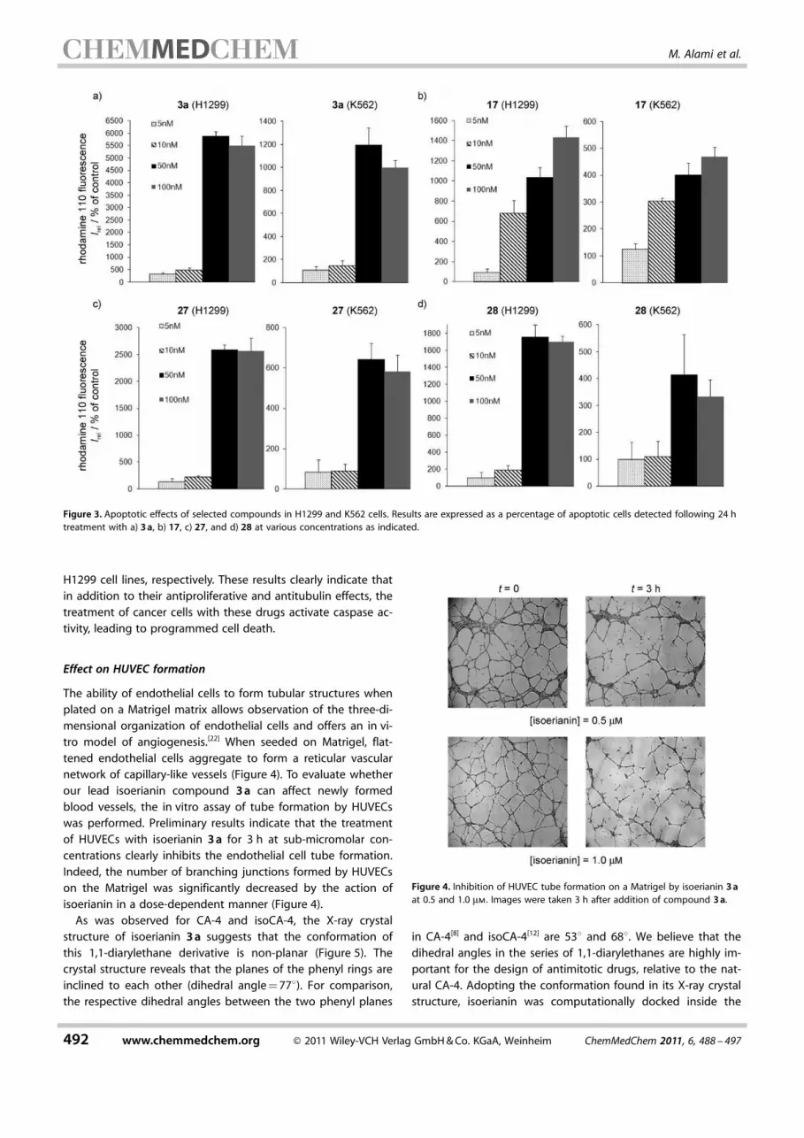
H1299 cell lines, respectively. These results clearly indicate that
in addition to their antiproliferative and antitubulin effects, the
treatment of cancer cells with these drugs activate caspase ac-
tivity, leading to programmed cell death.
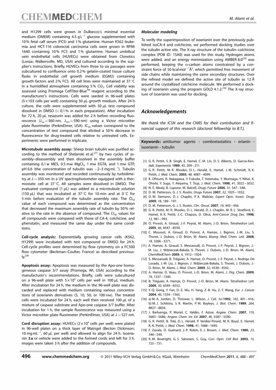
Effect on HUVEC formation
The ability of endothelial cells to form tubular structures when
plated on a Matrigel matrix allows observation of the three-di-
mensional organization of endothelial cells and offers an in vi-
tro model of angiogenesis.[22] When seeded on Matrigel, flat-
tened endothelial cells aggregate to form a reticular vascular
network of capillary-like vessels (Figure 4). To evaluate whether
our lead isoerianin compound 3a can affect newly formed
blood vessels, the in vitro assay of tube formation by HUVECs
was performed. Preliminary results indicate that the treatment
of HUVECs with isoerianin 3a for 3 h at sub-micromolar con-
centrations clearly inhibits the endothelial cell tube formation.
Indeed, the number of branching junctions formed by HUVECs
on the Matrigel was significantly decreased by the action of
isoerianin in a dose-dependent manner (Figure 4).
As was observed for CA-4 and isoCA-4, the X-ray crystal
structure of isoerianin 3a suggests that the conformation of
this 1,1-diarylethane derivative is non-planar (Figure 5). The
crystal structure reveals that the planes of the phenyl rings are
inclined to each other (dihedral angle=778). For comparison,
the respective dihedral angles between the two phenyl planes
in CA-4[8] and isoCA-4[12] are 538 and 688. We believe that the
dihedral angles in the series of 1,1-diarylethanes are highly im-
portant for the design of antimitotic drugs, relative to the nat-
ural CA-4. Adopting the conformation found in its X-ray crystal
structure, isoerianin was computationally docked inside the
Figure 3. Apoptotic effects of selected compounds in H1299 and K562 cells. Results are expressed as a percentage of apoptotic cells detected following 24 htreatment with a) 3a, b) 17, c) 27, and d) 28 at various concentrations as indicated.
Figure 4. Inhibition of HUVEC tube formation on a Matrigel by isoerianin 3a
at 0.5 and 1.0 mm. Images were taken 3 h after addition of compound 3a.
492 www.chemmedchem.org � 2011 Wiley-VCH Verlag GmbH&Co. KGaA, Weinheim ChemMedChem 2011, 6, 488 – 497
MED M. Alami et al.

colchicine binding site. For this purpose, the X-ray structure of
tubulin–colchicine complex was used (PDB ID: 1SA0).[23]
Figure 6 shows the docking-derived superimposition of iso-
erianin, isoCA-4, and colchicine (green, blue, and orange, re-
spectively). We recently published the close fit between isoCA-
4 and the colchicine X-ray structure,[12] and as expected, isoer-
ianin 3a shows good superimposition with both of them in
the binding site (Figure 6). As previously reported for a set of
colchicine site inhibitors,[24] the 3,4,5-trimethoxyphenyl moiet-
ies occupy very similar Cartesian space. Hydroxy and methoxy
groups belonging to the rest of the system are also well fitted.
Hydroxy groups belonging to isoerianin and isoCA-4 show a
hydrogen bond with the backbone of Val181 as proposed by
Nguyen et al.[24]
Conclusions
We synthesized a series of 1,1-diarylethane compounds bear-
ing structural similarity to the isocombretastatins and evaluat-
ed their biological activities. As was observed for the CA-4
series, we demonstrated that it is possible to transfer the B
ring of natural erianin from the C2 to the C1 position with no
loss of activity. The most biologically active agent was isoeria-
nin 3, obtained from the catalytic reduction of isoCA-4, with a
minimum inhibitory concentration at the nanomolar level
against various human cancer cell lines. Furthermore, the anti-
tubulin activity of isoerianin was found to be similar to that of
the parent compound erianin as well as CA-4 and isoCA-4,
which may indicate a similarity in the mechanism of action.
This result contradicts the previously reported conclusions re-
garding the necessity for tubulin to recognize the sp2 hybridi-
zation[18] introduced by the stilbene or 1,1-diarylethylene
bridges. Flow cytometric analysis indicated that isoerianin ar-
rests the cell cycle in the G2/M phase in H1299 and K562 cells.
Furthermore, isoerianin and other derivatives were character-
ized as strongly apoptotic agents promoting the cleavage of
pro-caspase 3 in K562 and H1299 cells.
From a chemical point of view, the preparation of isoerianin
3a is particularly simple and was carried out by the racemic
catalytic reduction of isoCA-4, as both enantiomers display
similar cytotoxicities. In vivo tests of water-soluble phosphate
4 are currently underway in our research group to elucidate
the anticancer activity of this compound in mice.
Experimental Section
Chemistry
Melting points (mp) were recorded on a B�chi B-450 apparatusand are uncorrected. NMR spectra were performed on a BrukerAMX 200 (1H, 200MHz ; 13C, 50MHz), Bruker AVANCE 300 orBruker AVANCE 400 (1H, 400 MHz ; 13C, 100 MHz). Unless otherwisestated, CDCl3 was used as solvent. Chemical shifts (d) are reportedin ppm, and the following abbreviations are used: singlet (s), dou-blet (d), triplet (t), multiplet (m). Elemental analyses (C, H, N) wereperformed with a PerkinElmer 240 analyzer at the microanalysisservice of the Faculty of Pharmacy at Ch�tenay-Malabry (France),and are within 0.4% of the theoretical values unless otherwisestated. Mass spectra were obtained using a Bruker Esquire electro-spray ionization apparatus. Thin-layer chromatography (TLC) wasperformed on silica gel 60 plates with a fluorescent indicator andvisualized under a UVP Mineralight UVGL-58 lamp (l=254 nm) andwith a 7% solution of phosphomolybdic acid in EtOH. Flash chro-matography was performed with silica gel 60 (40–63 mm, 230–400mesh ASTM) at medium pressure (200 mbar). All solvents were dis-tilled and stored over 4 � molecular sieves before use. All reagentswere obtained from commercial suppliers unless otherwise stated.Organic extracts were, in general, dried over MgSO4 or Na2SO4.
Synthesis of isoerianin derivatives 3–28
Isoerianin derivatives 3a and 5–26 were prepared from the reduc-tion of known 1,1-diarylethylenes 2,[11,25] as follows: A solution of1,1-diarylethylenes 2 (1 mmol) in EtOAc (15 mL) was hydrogenated,at atmospheric pressure, in the presence of Pd/C (5 mg). After fil-tration, the solution was concentrated under reduced pressure,and the residue was purified by flash chromatography.
2-Methoxy-5-[1-(3,4,5-trimethoxyphenyl)ethyl]phenol (isoerianin
3a): (311 mg, 98%); Rf=0.4 (cyclohexane/EtOAc 7:3); 1H NMR(300MHz, CDCl3): d=1.58 (d, 3H, J=7.2 Hz), 3.81 (s, 9H), 3.83 (s,3H), 3.88 (q, 1H, J=7.2 Hz), 5.60 (s, 1H), 6.43 (s, 2H), 6.70 (dd, 1H,
Figure 5. X-ray crystal structure of isoerianin 3a ; non-hydrogen atoms are la-beled using a crystallographic numbering scheme.
Figure 6. Putative binding mode of isoerianin (green), isoCA-4 (blue), andcolchicine (orange) in the colchicine binding site.
ChemMedChem 2011, 6, 488 – 497 � 2011 Wiley-VCH Verlag GmbH&Co. KGaA, Weinheim www.chemmedchem.org 493
Isoerianin Analogues

J=10.2 Hz, J=2.2 Hz), 6.78 (d, 1H, J=10.2 Hz), 6.81 (d, 1H, J=2.2 Hz); 13C NMR (75MHz, CDCl3): d=21.0, 43.4, 54.9, 55.1 (2), 59.8,103.6 (2), 109.5, 112.8, 117.8, 127.8, 135.2, 138.7, 141.2 (2), 143.9,144.4, 152.0; MS (ESI) [M+Na]+=341; Anal. calcd for C18H22O5 : C67.91, H 6.97, found: C 67.91, H 6.92.
2-Methoxy-5-[1-(3,4,5-trimethoxyphenyl)ethyl]phenyldihydrogen
phosphate (4): (279 mg, 70%); Rf=0.1 (MeOH/EtOAc 2:98);1H NMR (300MHz, (CD3)2CO): d=1.40 (m, 3H), 3.50 (s, 3H), 3.61 (s,9H), 3.95 (m, 1H), 6.40 (m, 2H), 6.75–6.90 (m, 2H), 7.10–7.30 (m,1H); 13C NMR (75 MHz, (CD3)2CO): d=25.3, 48.0, 59.4 (3), 63.5,108.7 (2), 116.7, 124.3, 127.8, 140.5, 142.9, 144.2, 146.1, 153.1, 157.2(2); MS (ESI�) [M�H]�=397; Anal. calcd for C18H23O8P: C 54.27, H5.827, found: C 54.09, H 5.73.
5-[1-(3-Fluoro-4-methoxyphenyl)ethyl]-1,2,3-trimethoxybenzene
(5): (310 mg, 97%); Rf=0.6 (cyclohexane/EtOAc 7:3); 1H NMR(300MHz, CDCl3): d=1.50 (d, 3H, J=7.2 Hz), 3.73 (s, 6H), 3.74 (s,3H), 3.78 (s, 3H), 3.95 (q, 1H, J=7.2 Hz), 6.32 (s, 2H), 6.68–6.90 (m,3H); 13C NMR (75MHz, CDCl3): d=22.0, 44.1, 56.1, (2) 56.3, 60.8,104.6 (2), 113.3, 115.2 (d, J=18.1 Hz), 122.8 (d, J=3.0 Hz), 136.4,139.4, 141.7 (2), 145.7, 150.6, 153.1, 153.8; MS (ESI) [M+Na]+=343;Anal. calcd for C18H21FO4 : C 67.49, H 6.61, found: C 67.39, H 6.52.
1,2,3-Trimethoxy-5-[1-(4-methoxyphenyl)ethyl]benzene (6):
(281 mg, 93%); Rf=0.5 (CH2Cl2) ;1H NMR (300MHz, CDCl3): d=1.50
(d, 3H, J=7.2 Hz), 3.66 (s, 6H), 3.72 (s, 3H), 3.74 (s, 3H), 3.95 (q,1H, J=7.2 Hz), 6.30 (s, 2H), 6.75 (d, 1H, J=7.2 Hz), 7.05 (d, 1H, J=10.2 Hz); 13C NMR (75 MHz, CDCl3): d=22.2, 44.2, 55.2, 56.1 (2),60.8, 104.3 (2), 113.7 (2), 128.4 (2), 136.2, 138.3, 142.5 (2), 153.0,157.9; MS (ESI) [M+Na]+=325; Anal. calcd for C18H2O4 : C 71.50, H7.33, found: C 71.39, H 7.26.
1,2,3-Trimethoxy-5-(1-p-tolylethyl)benzene (7): (285 mg, 99%);Rf=0.5 (CH2Cl2) ;
1H NMR (300MHz, CDCl3): d=1.52 (d, 3H, J=
7.2 Hz), 2.20 (s, 3H), 3.70 (s, 6H), 3.72 (s, 3H), 3.94 (q, 1H, J=7.2 Hz), 6.35 (s, 2H), 6.98–7.05 (m, 4H); 13C NMR (75 MHz, CDCl3):d=21.0, 22.1, 44.7, 56.1 (2), 60.8, 104.7 (2), 127.3 (2) 128.7 (2),135.6, 136.3, 142.3, 143.2, 154.2 (2); MS (ESI) [M+Na]+=309; Anal.calcd for C18H22O3 : C 75.50, H 7.74, found: C 75.40, H 7.66.
5-[1-(3,4,5-Trimethoxyphenyl)ethyl]benzo[d][1,3]dioxole (8):
(272 mg, 86%); Rf=0.4 (CH2Cl2) ;1H NMR (300MHz, CDCl3): d=1.52
(d, 3H, J=7.2 Hz), 3.75 (s, 9H), 4.00 (q, 1H, J=7.2 Hz), 5.80 (s, 2H),6.35 (s, 2H), 6.62–6.68 (m, 3H); 13C NMR (75MHz, CDCl3): d=22.1,44.7, 56.1 (2), 60.8, 100.8, 104.6 (2), 108.2, 120.2, 136.3, 140.3, 142.1,145.8, 147.6, 153.0 (2); MS (ESI) [M+Na]+=339; Anal. calcd forC18H20O5 : C 68.34, H 6.37, found: C 68.29, H 6.31.
2-[1-(3,4,5-Trimethoxyphenyl)ethyl]naphthalene (9): (293 mg,91%); Rf=0.5 (CH2Cl2) ;
1H NMR (300MHz, CDCl3): d=1.62 (d, 3H,J=7.2 Hz), 3.70 (s, 6H), 3.74 (s, 3H), 4.16 (q, 1H, J=7.2 Hz), 6.38 (s,2H), 7.22 (dd, 1H, J=8.5 Hz, J=2.2 Hz), 7.28–7.42 (m, 2H), 7.61 (s,1H), 7.64–7.77 (m, 3H); 13C NMR (75MHz, CDCl3): d=21.9, 45.1,56.1 (2), 60.6, 104.9, 125.3, 125.5, 126.0, 126.7, 127.6, 127.8, 128.0,132.2, 133.6, 136.4, 141.2, 143.3, 153.2 (2); MS (ESI) [M+Na]+=345;Anal. calcd for C21H22O3 : C 78.23, H 6.88, found: C 78.19, H 6.84.
6-[1-(3,4,5-Trimethoxyphenyl)ethyl]quinoline (10): (174 mg,54%); Rf=0.4 (Et2O);
1H NMR (300 MHz, CDCl3): d=1.76 (d, 3H, J=7.2 Hz), 3.76 (s, 3H), 3.80 (s, 6H), 4.36 (q, 1H, J=7.0 Hz), 6.61 (s,2H), 7.53 (dd, 1H, J=4.4 Hz, J=8.3 Hz), 7.70 (dd, 1H, J=1.8 Hz, J=8.8 Hz), 7.87 (s, 1H), 7.96 (d, 1H, J=8.8 Hz), 8.36 (d, 1H, J=8.0 Hz),8.81 (dd, 1H, J=1.5 Hz, J=4.3 Hz); 13C NMR (75MHz, MeOD): d=22.3, 46.2, 56.6 (2C), 61.1, 106.2 (2C), 122.6, 126.4, 128.9, 129.9,132.1, 138.3, 143.4, 146.7 (2), 150.6, 154.6 (2C); MS (APCI) [M+H]+
=324; Anal. calcd for C20H21NO3 : C 74.28, N 4.33, H 6.55, found: C74.18, N 4.21, H 6.42.
3-Methoxy-6-[1-(3,4,5-trimethoxyphenyl)ethyl]benzene-1,2-diol
(11): (317 mg, 95%); Rf=0.45 (cyclohexane/EtOAc 1:1); 1H NMR(CDCl3 300MHz): d=1.57 (d, 3H, J=7.2 Hz), 3.82 (s, 9H), 3.86 (s,3H), 4.39 (q, 1H, J=7.0 Hz), 5.37 (s, 2H), 6.44 (d, 1H, J=8.6 Hz),6.50 (s, 2H), 6.65 (d, 1H, J=8.6 Hz); 13C NMR (75 MHz, CDCl3): d=21.0, 37.9, 56.1 (3C), 60.8, 102.5, 104.7 (2C), 117.8, 126.1, 132.3,136.2, 141.6, 141.9, 145.3, 153.0 (2C); MS (APCI) [M+H]+=335;Anal. calcd for C18H22O6 : C 64.66, H 6.63, found: C 64.40, H 6.48.
2-Methoxy-5-[1-(3,4,5-trimethoxyphenyl)ethyl]phenyl acetate
(12): (324 mg, 90%); Rf=0.5 (CH2Cl2) ;1H NMR (300MHz, CDCl3):
d=1.51 (d, 3H, J=7.2 Hz), 2.22 (s, 3H), 3.73(s, 3H), 3.74 (s, 6H),3.75 (s, 3H), 3.95 (q, 1H, J=7.2 Hz), 6.32 (s, 2H), 6.79–6.83 (m, 2H),6.97 (dd, 1H, J=8.4 Hz, J=1.7 Hz); 13C NMR (75 MHz, CDCl3): d=20.7, 22.1, 38.7, 55.9, 56.1 (2), 60.8, 104.6 (2), 112.2, 121.9, 125.6,136.3, 138.8, 139.5, 141.9, 149.3, 153.1 (2), 169.0; MS (ESI) [M+Na]+
=383; Anal. calcd for C20H24O6 : C 66.65, H 6.71, found: C 66.49, H6.63.
2-Methoxy-5-[1-(3,4,5-trimethoxyphenyl)ethyl]phenyl diethylcar-
bamate (13): (379 mg, 91%); Rf=0.5 (CH2Cl2) ;1H NMR (300MHz,
CDCl3): d=1.10–1,30 (m, 6H), 1.50 (d, 3H, J=7.2 Hz), 3.25–3.45 (m,4H), 3.72 (s, 6H), 3.74 (s, 3H), 3.75 (s, 3H), 3.95 (q, 1H, J=7.2 Hz),6.32 (s, 2H), 6.78 (d, 1H, J=8.4 Hz), 6.86–6.95 (m, 2H); 13C NMR(75MHz, CDCl3): d=12.7 (2), 20.9, 22.1, 35.7, 42.9, (2), 55.9, 56.3(2), 67.8, 105.6 (2), 113.2, 120.9, 126.6, 135.9, 136.3, 137.8, 140.1,149.3, 153.1 (2), 156.2.0; MS (ESI) [M+Na]+=440; Anal. calcd forC23H31NO6 : C 66.17, N 3.35, H 7.48, found: C 66.04, N 3.19, H 7.33.
5-Methoxy-2-[1-(3,4,5-trimethoxyphenyl)ethyl]pyridine (14):
(302 mg, 99%); Rf=0.35 (cyclohexane/EtOAc 95:5); 1H NMR(300MHz, CDCl3): d=1.63 (d, 3H, J=7.2 Hz), 3.75 (s, 3H), 3.81 (s,6H), 3.89 (s, 3H), 4.11 (q, 1H, J=7.2 Hz), 6.55 (s, 2H), 6.75 (d, 1H,J=8.6 Hz), 7.59 (dd, 1H, J=2.3 Hz, J=8.6 Hz), 8.04 (d, 1H, J=
2.3 Hz); 13C NMR (75MHz, CDCl3): d=22.1, 22.0, 43.1, 54.1, 56.6(2C), 61.1, 105.8 (2C), 111.4, 136.2, 137.5, 139.9, 143,4, 146.1, 154.5,164.3 (2C); MS (APCI) [M+H]+=304; Anal. calcd for C17H21NO4 : C67.31, N 4.62, H 6.98, found: C 67.25, N 4.55, H 6.92.
1-Methyl-5-[1-(3,4,5-trimethoxyphenyl)ethyl]-1H-indole (15):
(192 mg, 59%); Rf=0.75 (cyclohexane/EtOAc 6:4); 1H NMR(300MHz, CDCl3): d=1.67 (d, 3H, J=7.2 Hz), 3.74 (s, 3H), 3.76 (s,9H), 4.19 (q, 1H, J=7.1 Hz), 6.38 (dd, 1H, J=0.6 Hz, J=3.0 Hz),6.55 (s, 2H), 7.05 (dd, 1H, J=1.4 Hz, J=8.5 Hz), 7.10 (d, 1H, J=3.1 Hz), 7.26 (d, 1H, J=8.5 Hz), 7,45 (s, 1H); 13C NMR (100MHz,CDCl3): d=22.9, 32.8, 46.3, 56.5 (2C), 61.1, 101.6, 106.4 (2C), 110.0,112.6, 119.7, 122.8, 130.1, 137.0, 138.2 (2), 145.4, 154.2 (2C); MS(APCI) [M+H]+=326; Anal. calcd for C20H23NO3 : C 73.82, N 4.30, H7.12, found: C 73.73, N 4.18, H 7.04.
3-[1-(3,4,5-Trimethoxyphenyl)ethyl]-1H-indole (16): (247 mg,79%); Rf=0.4 (CH2Cl2) ;
1H NMR (300MHz, CDCl3): d=1.55 (d, 3H,J=7.2 Hz), 3.72 (s, 6H), 3.74 (s, 3H), 4.20 (q, 1H, J=7.1 Hz), 6.43 (s,2H), 6.92–6.99 (m, 3H), 7.06–7.09 (dd, 1H, J=8.1 Hz, J=0.9 Hz),7.15 (d, 1H, J=7.8 Hz), 7.27 (d, 1H, J=7.8 Hz), 7.95 (s, 1H);13C NMR (75 MHz, CDCl3): d=21.8, 34.7, 56.2 (2C), 56.6, 105.3 (2C),111.0, 115.4, 119.0, 120.2, 122.3, 127.2, 134.6, 136.2, 136.4, 153.0(2C); MS (ESI) [M+Na]+=334; Anal. calcd for C19H21NO3 : C 73.29, N4.50, H 6.80, found: C 73.09, N 4.33, H 6.60.
2-Methoxy-5-[1-(3,4,5-trimethoxyphenyl)ethyl]benzenamine
(17): (171 mg, 58%); Rf=0.15 (cyclohexane/EtOAc 7:3); 1H NMR(300MHz, CDCl3): d=1.57 (d, 3H, J=7.2 Hz), 3.82 (s, 12H), 3.98 (q,1H, J=7.2 Hz), 6,44 (s, 2H), 6,56 (d, 1H, J=2.1 Hz), 6,60 (dd, 1H,
494 www.chemmedchem.org � 2011 Wiley-VCH Verlag GmbH&Co. KGaA, Weinheim ChemMedChem 2011, 6, 488 – 497
MED M. Alami et al.

J=8.2 Hz, J=2.1 Hz), 6,72 (d, 1H, J=8.2 Hz); 13C NMR (75MHz,CDCl3): d=22.2, 44.4, 55.5, 56.1 (2), 60.9, 104.6 (2), 110.2, 114.5,117.1, 135.9, 136.2, 139.0, 142.6, 145.8, 153.0 (2); MS (ESI) [M+Na]+
=318; Anal. calcd for C18H23NO4 : C 68.12, N 4.41, H 7.30, found: C68.00, N 4.29, H 7.19.
4-Methoxy-2-[1-(3,4,5-trimethoxyphenyl)ethyl]benzenamine
(18): (282 mg, 89%); Rf=0.2 (CH2Cl2) ;1H NMR (300MHz, CDCl3):
d=1.50 (d, 3H, J=7.2 Hz), 3.71 (s, 6H), 3.72 (s, 3H), 3.74 (s, 3H),3.85 (q, 1H, J=7.2 Hz), 6.33 (s, 2H), 6.55 (d, 1H, J=8.4 Hz), 6.60(dd, 1H, J=8.4 Hz, J=2.7 Hz), 6.80 (d, 1H, J=2.7 Hz); 13C NMR(75MHz, CDCl3): d=21.7, 40.7, 55.7, 56.1 (2C), 60.8, 104.5, 111.6,114.1 117.2, 131.8, 136.5, 137.9, 141.2 (2), 153.0, 153.4 (2); MS (ESI)[M+Na]+=340; Anal. calcd for C18H23NO4 : C 68.12, N 4.41, H 7.30,found: C 68.07, N 4.31, H 7.35.
5-Methoxy-2-[1-(3,4,5-trimethoxyphenyl)ethyl]benzenamine
(19): (273 mg, 86%); Rf=0.2 (CH2Cl2) ;1H NMR (300MHz, CDCl3):
d=1.50 (d, 3H, J=7.2 Hz), 3.63 (s, 3H), 3.70 (s, 6H), 3.74 (s, 3H),3.85 (q, 1H, J=7.2 Hz), 6.14 (m, 1H), 6.32 (m, 3H), 7.06 (d, 1H, J=8.4 Hz); 13C NMR (75MHz, CDCl3): d=21.0, 39.1, 54.1, 55.1 (2C),59.8, 101.1, 102.8, 103.3 (2), 121.4, 126.8 (2), 135.4, 140.9, 144.5,152.4 (2), 158.1; MS (ESI) [M+H]+=318; Anal. calcd for C18H23NO4 :C 68.12, N 4.41, H 7.30, found: C 68.02, N 4.4.28, H 7.23.
2-Methoxy-5-[1-(7-methoxybenzo[d][1,3]dioxol-5-yl)ethyl]phenol
(20): (239 mg, 79%); Rf=0.8 (CH2Cl2/cyclohexane 8:2); 1H NMR(300MHz, CDCl3): d=1.55 (d, 3H, J=7.2 Hz), 3.85 (s, 3H), 3.86 (s,3H), 3.95 (q, 1H, J=7.2 Hz), 5.60 (m, 1H), 5.90 (s, 2H), 6.39 (m, 2H),6.69 (dd, 1H, J=8.4 Hz, J=1.8 Hz), 6.77 (d, H, J=8.4 Hz), 79 (d, 1H,J=1.8 Hz); 13C NMR (75 MHz, CDCl3): d=22.1, 44.1, 55.9, 56.6,101.2, 101.6, 107.0, 110.5, 113.7, 118.7, 139.7, 141.4, 143.3, 144.8,145.4, 148.8; MS (APCI) [M+H]+=303; Anal. calcd for C17H18O5 : C67.54, H 6.00, found: C 67.20, H 5.87.
5-[1-(3,5-Dimethoxyphenyl)ethyl]-2-methoxybenzenamine (21):
(252 mg, 88%); Rf=0.5 (cyclohexane/EtOAc 6:4); 1H NMR(300MHz, CDCl3): d=1.56 (d, 3H, J=7.2 Hz), 3.76 (s, 6H), 3.82 (s,3H), 3.95 (q, 1H, J=7.2 Hz), 6.29 (t, 1H, J=2.3 Hz), 6.39 (d, 2H, J=2.3 Hz), 6.57 (d, 1H, J=2.1 Hz), 6.61 (dd, 1H, J=2.1 Hz, J=8.2 Hz),6.71 (d, 1H, J=8.2 Hz); 13C NMR (75MHz, CDCl3): d=21.8 44.4,55.2 (2C), 55.5, 97.5, 105.9 (2C), 110.2, 114.5, 117.2, 135.8, 138.8,145.7, 149.4, 160.6 (2C); MS (APCI) [M+H]+=288; Anal. calcd forC17H21NO3 : C 71.06, N 4.87, H 7.37, found: C 70.98, N 4.67, H 7.28.
5-[1-(3-Ethoxy-4-methoxyphenyl)ethyl]-2-methoxybenzenamine
(22): (242 mg, 80%); Rf=0.6 (cyclohexane/EtOAc 6:4); 1H NMR(300MHz, CDCl3): d=1.44 (t, 3H, J=7.0 Hz), 1.58 (d, 3H, J=
7.2 Hz), 3.82 (s, 3H), 3.85 (s, 3H), 3.97 (q, 1H, J=7.2 Hz), 4.06 (q,2H, J=7.0 Hz), 6.56 (d, 1H, J=2.2 Hz), 6.59 (ddd, 1H, J=0.5 Hz, J=2.2 Hz, J=8.2 Hz), 6.72 (d, 1H, J=8.2 Hz), 6.76 (s, 1H), 6.79 (d, 1H,J=1.7 Hz), 6.81 (dd, 1H, J=0.6 Hz, J=8.1 Hz); 13C NMR (75MHz,CDCl3): d=14.8, 22.2, 43.6, 55.5, 56.0, 64.3, 110.2, 111.3, 112.8,114.5, 117.2, 119.3, 135.8, 139.5, 145.7, 147.3, 147.5, 148.0; MS(APCI) [M+H]+=302; Anal. calcd for C18H23NO3 : C 71.73, N 4.65, H7.69, found: C 71.58, N 4.39, H 7.49.
5-[1-(3,5-Dimethoxyphenyl)ethyl]-2-methoxyphenol (23):
(251 mg, 87%); Rf=0.5 (CH2Cl2) ;1H NMR (300MHz, CDCl3): d=1.62
(d, 3H, J=7.2 Hz), 3.86–3.70 (m, 6H), 3.89 (s, 3H), 4.03 (q, 1H, J=7.2 Hz), 5.64 (s, 1H), 6.34 (t, 1H, J=2.2 Hz), 6.43 (d, 2H, J=2.2 Hz),6.76 (dd, 1H, J=8.3, J=2.0 Hz), 6.81 (d, 1H, J=8.3 Hz), 6.87 (d, 1H,J=1.9 Hz); 13C NMR (75MHz, CDCl3): d=21.8, 44.4, 55.3 (2), 56.0,97.6, 106.1 (2), 110.5, 113.9, 118.9, 139.5, 144.9, 145.4, 149.1, 160.7(2); MS (APCI) [M+H]+=289; Anal. calcd for C17H20O4 : C 70.81, H6.99, found: C 70.74, H 6.96.
5-[1-(2,3-Dimethoxyphenyl)ethyl]-2-methoxyphenol (24):
(233 mg, 81%); Rf=0.49 (CH2Cl2) ;1H NMR (300MHz, CDCl3): d=
1.51 (d, 3H, J=7.1 Hz), 3.65 (s, 3H), 3.81 (s, 6H), 4.48 (q, 1H, J=7.2 Hz), 6.71–6.82 (m, 5H), 6.97 (t, 1H, J=8.0 Hz); 13C NMR(75MHz, CDCl3): d=21.7, 37.0, 55.7, 56.0, 60.6, 110.2, 110.4, 114.0119.0, 119.8, 123.8, 140.0, 140.5, 144.7, 145.3, 146.5, 152.7; Anal.calcd for C17H20O4 : C 70.81, H 6.99, found: C 70.58, H 6.94.
5-[1-(3-Ethoxy-4-methoxyphenyl)ethyl]-2-methoxyphenol (25):
(214 mg, 71%); Rf=0.5 (CH2Cl2) ;1H NMR (300MHz, CDCl3): d=1.33
(t, 3H, J=7.0 Hz), 1.48 (d, 3H, J=7.2 Hz), 3.74 (s, 6H), 3.92 (m, 3H),5.58 (s, 1H), 6.59 (dd, 1H, J=8.3, 2.1 Hz), 6.60- 6.71 (m, 5H);13C NMR (75 MHz, CDCl3): d=14.8, 22.1, 43.7, 55.9 (2), 64.2, 110.5,111.3, 112.6, 113.85, 118.74, 119.24, 139.16, 140.16, 144.78, 145.38,147.54, 148.06, 153.62; MS (APCI) [M+H]+=303; Anal. calcd forC18H22O4 : C 71.50, H 7.33, found: C 71.22, H 7.06.
2-Methoxy-5-[1-(2,3,4-trimethoxyphenyl)ethyl]phenol (26):
(267 mg, 84%); Rf=0.2 (cyclohexane/EtOAc 9:1); 1H NMR(300MHz, CDCl3): d=1.48 (d, 3H, J=7.2 Hz), 3.59 (s, 3H), 3.70 (s,3H), 3.75 (s, 3H), 3.77 (s, 3H), 4.25 (q, 1H, J=7.2 Hz), 6.50–6.60 (m,2H), 6.60–6.65 (d, 1H, J=8.3 Hz), 6.70 (d, 1H, J=1.9 Hz), 6.79 (d,1H, J=8.6 Hz); 13C NMR (75 MHz, CDCl3): d=21.8, 37.0, 55.8, 55.9,60.6, 60.8, 107.1, 110.4, 114.2, 118.2, 121.7, 132.7, 140.4, 142.2,144.9, 146.1, 151.4, 151.9; MS (ESI) [M�H]�=317; Anal. calcd forC18H22O5 : C 67.91, H 6.97, found: C 67.87, H 6.89.
3-(3-Hydroxy-4-methoxyphenyl)-3-(3,4,5-trimethoxyphenyl)pro-
panenitrile (27): (121 mg, 50%); Rf=0.4 (cyclohexane/EtOAc 5:5);1H NMR (300MHz, CDCl3): d=2.97 (d, 2H, J=7.5 Hz), 3.83 (s, 9H),3.88 (s, 3H), 4.21 (t, 1H, J=7.6 Hz), 5.61 (s, 1H), 6.42 (s, 2H), 6.73–6.83 (m, 3H); 13C NMR (75MHz, CDCl3): d=24.4, 46.8, 55.9, 56.1(2), 60.8, 104.6 (2), 110.7, 113.6, 118.5, 118.9, 134.3, 137.1, 137.2,145.8 (2), 153.4 (2); MS (ESI) [M+Na]+=366; Anal. calcd forC19H21NO5 : C 66.46, N 4.08, H 6.16, found: C 66.39, N 3.99, H 6.09.
5-[2,2-Difluoro-1-(3,4,5-trimethoxyphenyl)ethyl]-2-methoxyphe-
nol (28): (220 mg, 62%); Rf=0.25 (cyclohexane/EtOAc 7:3); 1H NMR(300MHz, CDCl3): d=3.76 (s, 9H), 3.83 (s, 3H), 4.15 (td, 1H, J=15.8 Hz, J=4.2 Hz), 5.51 (s, 1H), 6.15 (td, 1H, J=55.9 Hz, J=4.2 Hz),6.42 (s, 2H), 6.70–6.83 (m, 3H); 13C NMR (100 MHz, CDCl3): d=54.5(t, J=20.7 Hz), 55.9, 56.1 (2), 60.8, 106.1 (2), 110.6, 115.1, 120.5,116.8 (t, J=244.5), 130.0, 132.7, 137.3, 145.6, 145.9, 153.2 (2);19F NMR: (188MHz, CDCl3 decoupled): �116.09, �116.12; Anal.calcd for C18H20F2O5 : C 61.01, H 5.69, found: C 60.81, H 5.46.
Synthesis of erianin: Erianin was prepared from the Pd/C(5 mol%) catalytic reduction of CA-4[26] in MeOH for 3 h at roomtemperature.
5-(3,4,5-Trimethoxyphenethyl)-2-methoxyphenol (erianin):
(223 mg, 70%); Rf=0.35 (cyclohexane/EtOAc 8:2); 1H NMR(300MHz, CDCl3): d=2.82 (s, 4H), 3.83 (s, 9H), 3.86 (s, 3H), 5.64 (s,1H), 6.38 (s, 2H), 6.64 (dd, 1H, J=2.1 Hz, J=8.2 Hz), 6.77 (d, 1H,J=8.2 Hz), 6.81 (d, 1H, J=2.0 Hz); 13C NMR (75MHz, CDCl3): d=37.3, 38.4, 56.0 (3C), 60.9, 105.4 (2C), 110.6, 114.7, 119.8, 135.0,136.1, 137.6, 144.9, 145.5, 153.0 (2C); MS (ESI) [M+Na]+=341; Anal.calcd for C18H22O5 : C 67.91, H 6.97, found: C 67.87, H 6.94.
Biology
Cell culture and proliferation assay
Cancer cell lines were obtained from the American Type CultureCollection (ATCC; Rockville, MD, USA) and were cultured accordingto the supplier’s instructions. Briefly, MDA-MB231, MDA-MB435,
ChemMedChem 2011, 6, 488 – 497 � 2011 Wiley-VCH Verlag GmbH&Co. KGaA, Weinheim www.chemmedchem.org 495
Isoerianin Analogues
and H1299 cells were grown in Dulbecco’s minimal essentialmedium (DMEM) containing 4.5 gL�1 glucose supplemented with10% fetal calf serum (FCS) and 1% glutamine. Human K562 leuke-mia and HCT-116 colorectal carcinoma cells were grown in RPMI1640 containing 10% FCS and 1% glutamine. Human umbilicalvein endothelial cells (HUVECs) were obtained from Clonetics(Lonza; Walkersville, MD, USA) and cultured according to the sup-plier’s instructions. Briefly, HUVECs from three to six passages weresubcultured to confluence onto 0.2% gelatin-coated tissue cultureflasks in endothelial cell growth medium (EGM2) containinggrowth factors and 2% FCS. All cell lines were maintained at 37 8Cin a humidified atmosphere containing 5% CO2. Cell viability wasassessed using Promega CellTiter-BlueTM reagent according to themanufacturer’s instructions. Cells were seeded in 96-well plates(5103 cells per well) containing 50 mL growth medium. After 24 hculture, the cells were supplemented with 50 mL test compounddissolved in DMSO (<0.1% in each preparation). After incubationfor 72 h, 20 mL resazurin was added for 2 h before recording fluo-rescence (lex=560 nm, lem=590 nm) using a Victor microtiterplate fluorimeter (PerkinElmer, USA). IC50 values correspond to theconcentration of test compound that elicited a 50% decrease influorescence for drug-treated cells relative to untreated cells. Ex-periments were performed in triplicate.
Microtubule assembly assay: Sheep brain tubulin was purified ac-cording to the method of Shelanski et al.[27] by two cycles of as-sembly–disassembly and then dissolved in the assembly buffercontaining 0.1m MES, 0.5 mm MgCl2, 1 mm EGTA, and 1 mm GTP,pH 6.6 (the concentration of tubulin was ~2–3 mgmL�1). Tubulinassembly was monitored and recorded continuously by turbidime-try at l=350 nm in a UV spectrophotometer equipped with a ther-mostatic cell at 37 8C. All samples were dissolved in DMSO. Theevaluated compound (1 mL) was added to a microtubule solution(150 mL) that was incubated at 37 8C for 10 min and at 0 8C for5 min before evaluation of the tubulin assembly rate. The CI50value of each compound was determined as the concentrationthat decreased the maximum assembly rate of tubulin by 50% rel-ative to the rate in the absence of compound. The CI50 values forall compounds were compared with those of CA-4, colchicine, andphenstatin, and measured the same day under the same condi-tions.
Cell-cycle analysis: Exponentially growing cancer cells (K562,H1299) were incubated with test compound or DMSO for 24 h.Cell-cycle profiles were determined by flow cytometry on a FC500flow cytometer (Beckman–Coulter, France) as described previous-ly.[28]
Apoptosis assay: Apoptosis was measured by the Apo-one homo-geneous caspase 3/7 assay (Promega, WI, USA) according to themanufacturer’s recommendations. Briefly, cells were subculturedon a 96-well plate with 5104 cells per well in 100 mL medium.After incubation for 24 h, the medium in the 96-well plate was dis-carded and replaced with medium containing various concentra-tions of isoerianin derivatives (5, 10, 50, or 100 nm). The treatedcells were incubated for 24 h, each well then received 100 mL of amixture of caspase substrate and Apo-one caspase 3/7 buffer. Afterincubation for 1 h, the sample fluorescence was measured using aVictor microtiter plate fluorimeter (PerkinElmer, USA) at l=527 nm.
Cord disruption assay: HUVECs (2104 cells per well) were platedin 96-well plates on a thick layer of Matrigel (Becton Dickinson;10 mgmL�1, 60 mL per well) and allowed to align for 24 h. Isoeria-nin 3a or vehicle were added to the formed cords and left for 3 h.Images were taken 3 h after the addition of compounds.
Molecular modeling
To verify the superimposition of isoerianin over the previously pub-lished isoCA-4 and colchicine, we performed docking studies overthe tubulin active site. The X-ray structure of the tubulin–colchicinecomplex (PDB ID: 1SA0) was used for this study. Hydrogen atomswere added, and an energy minimization using AMBER 8.0[29] wasperformed, keeping the a-carbon atoms constrained by a con-straint force of 50 kcalmol�1�2, which permitted free movement ofside chains while maintaining the same secondary structures. Overthe refined model we defined the active site of tubulin as 12 �around the crystallized colchicine molecule. We performed a dock-ing of isoerianin using the program GOLD 4.1.2.[30] The X-ray struc-ture of isoerianin was used for docking.
Acknowledgements
We thank the ICSN and the CNRS for their contribution and fi-
nancial support of this research (doctoral fellowship to B.T.).
Keywords: antitumor agents · combretastatins · erianin ·
isoerianin · tubulin
[1] G. R. Pettit, S. B. Singh, E. Hamel, C. M. Lin, D. S. Alberts, D. Garcia-Ken-dall, Experientia 1989, 45, 209–211.
[2] G. R. Pettit, M. R. Rhodes, D. L. Herald, E. Hamel, J. M. Schmidt, R. K.Pettit, J. Med. Chem. 2005, 48, 4087–4099.
[3] K. Ohsumi, R. Nakagawa, Y. Fukuda, T. Hatanaka, Y. Morinaga, Y. Nihei, K.Ohishi, Y. Suga, Y. Akiyama, T. Tsuji, J. Med. Chem. 1998, 41, 3022–3032.
[4] N. E. Mealy, B. Lupone, M. Balcell, Drugs Future 2006, 31, 547–548.[5] D. M. Patterson, G. J. S. Rustin, Drugs Future 2007, 32, 1025–1032.[6] D. W. Siemann, D. J. Chaplin, P. A. Walicke, Expert Opin. Invest. Drugs
2009, 18, 189–197.[7] D. M. Patterson, G. J. S. Rustin, Clin. Oncol. 2007, 19, 443–456.[8] G. R. Pettit, M. R. Rhodes, D. L. Herald, D. J. Chaplin, M. R. L. Stratford, E.
Hamel, R. K. Pettit, J.-C. Chapuis, D. Oliva, Anti-Cancer Drug Des. 1998,13, 981–993.
[9] O. Provot, A. Giraud, J.-F. Peyrat, M. Alami, J.-D. Brion, Tetrahedron Lett.
2005, 46, 8547–8550.[10] C. Mousset, A. Giraud, O. Provot, A. Hamze, J. Bignon, J.-M. Liu, S.
Thoret, J. Dubois, J.-D. Brion, M. Alami, Bioorg. Med. Chem. Lett. 2008,18, 3266–3271.
[11] A. Hamze, A. Giraud, S. Messaoudi, O. Provot, J.-F. Peyrat, J. Bignon, J.-M. Liu, J. Wdzieczak-Bakala, S. Thoret, J. Dubois, J.-D. Brion, M. Alami,ChemMedChem 2009, 4, 1912–1924.
[12] S. Messaoudi, B. Tr�guier, A. Hamze, O. Provot, J.-F. Peyrat, J. Rodrigo DeLosada, J.-M. Liu, J. Bignon, J. Wdzieczak-Bakala, S. Thoret, J. Dubois, J.-D. Brion, M. Alami, J. Med. Chem. 2009, 52, 4538–4542.
[13] A. Hamze, D. Veau, O. Provot, J.-D. Brion, M. Alami, J. Org. Chem. 2009,74, 1337–1340.
[14] B. Tr�guier, A. Hamze, O. Provot, J.-D. Brion, M. Alami, Tetrahedron Lett.
2009, 50, 6549–6552.[15] Y.-Q. Gong, Y. Fan, D.-Z. Wu, H. Yang, Z.-B. Hu, Z.-T. Wang, Eur. J. Cancer
2004, 40, 1554–1565.[16] a) M. A. Jordan, D. Thrower, L. Wilson, J. Cell. Sci.1992, 102, 401–416;
b) M. J. Schilstra, S. R. Martin, P. M. Bayleys, J. Biol. Chem. 1989, 264,8827–8834.
[17] J. Barluenga, P. Moriel, C. Vald�s, F. Aznar, Angew. Chem. 2007, 119,5683–5686; Angew. Chem. Int. Ed. 2007, 46, 5587–5590.
[18] G. R. Pettit, B. Toki, D. L. Herald, P. Verdier-Pinard, M. R. Boyd, E. Hamel,R. K. Pettit, J. Med. Chem. 1998, 41, 1688–1695.
[19] F. Zavala, D. Gu�nard, J.-P. Robin, E. J. Brown, J. Med. Chem. 1980, 23,546–549.
[20] K. M. Boatright, G. S. Salvesen, S. Guy, Curr. Opin. Cell Biol. 2003, 15,725–731.
496 www.chemmedchem.org � 2011 Wiley-VCH Verlag GmbH&Co. KGaA, Weinheim ChemMedChem 2011, 6, 488 – 497
MED M. Alami et al.
[21] a) M. P. Chang, J. Bramhall, S. Graves, B. Bonavida, B. J. Wisnieski, J. Biol.Chem. 1989, 264, 15261–15267; b) A. McGahon, R. Bissonnette, M.
Schmitt, K. M. Cotter, D. R. Green, T. G. Cotter, Blood 1994, 83, 1179–1187; c) A. J. McGahon, W. K. Nishioka, S. J. Martin, A. Mahboubi, T. G.Cotter, D. R. Green, J. Biol. Chem. 1995, 270, 22625–22631; d) R. M.
Gangemi, M. Tiso, C. Marchetti, A. B. Severi, M. Fabbi, Cancer Chemother.
Pharmacol. 1995, 36, 385–392; e) L. Dubrez, F. Goldwasser, P. Genne, Y.Pommier, E. Solary, Leukemia 1995, 9, 1013–1024; f) S. Ray, G. Bullock,
G. Nunez, C. Tang, A. M. Ibrado, Y. Huang, K. Bhalla, Cell Growth Differ.
1996, 7, 1617–1623.
[22] K. A. Hotchkiss, A. W. Ashton, R. Mahmood, R. G. Russel, J. A. Sparano,E. L. Schwartz, Mol. Cancer Ther. 2002, 1, 1191–1200.
[23] R. B. Ravelli, B. Gigant, P. A. Curmi, I. Jourdain, S. Lachkar, A. Sobel, M.
Knossow, Nature 2004, 427-432, 198–202.[24] T. L. Nguyen, C. McGrath, A. R. Hermone, J. C. Burnett, D. W. Zaharevitz,
B. W. Day, P. Wipf, E. Hamel, R. A. Gussio, J. Med. Chem. 2005, 48, 6107–6116.
[25] M. Alami, S. Messaoudi, A. Hamze, O. Provot, J.-D. Brion, J.-M. Liu, J.
Bignon, J. Bakala, WO 2009/147217A1, PCT/EP2009/056885.
[26] A. Giraud, O. Provot, A. Hamze, J.-D. Brion, M. Alami, Tetrahedron Lett.
2008, 49, 1107–1110.[27] M. L. Shelanski, F. Gaskin, C. R. Cantor, Proc. Natl. Acad. Sci. USA 1973,
70, 765–768.[28] C. Venot, M. Maratrat, C. Dureuil, E. Conseiller, L. Bracco, L. Debussche,
EMBO J. 1998, 17, 4668–4679.[29] D. A. Case, T. A. Darden, T. E. Cheatham III, C. L. Simmerling, J. Wang,
R. E. Duke, R. Luo, K. M. Merz, B. Wang, D. A. Pearlman, M. Crowley, S.Brozell, V. Tsui, H. Gohlke, J. Mongan, V. Hornak, G. Cui, P. Beroza, C.Schafmeister, J. W. Caldwell, W. S. Ross, P. A. Kollman (2004), AMBER 8,University of California, San Francisco.
[30] M. L. Verdonk, J. C. Cole, M. J. Hartshorn, C. W. Murray, R. D. Taylor, Pro-teins Struct. Funct. Bioinf. 2003, 52, 609–623.
[31] N. L’Hermite, A. Giraud, O. Provot, J.–F. Peyrat, M. Alami, J.-D. Brion, Tet-rahedron 2006, 62, 11994–12002.
Received: October 22, 2010Revised: December 14, 2010Published online on January 14, 2011
ChemMedChem 2011, 6, 488 – 497 � 2011 Wiley-VCH Verlag GmbH&Co. KGaA, Weinheim www.chemmedchem.org 497
Isoerianin Analogues