fluoro-modified chemotactic peptides: fmlf analogues
TRANSCRIPT
Journal of Peptide ScienceJ. Peptide Sci. 10: 67–81 (2004)Published online 18 September 2003 in Wiley InterScience (www.interscience.wiley.com). DOI: 10.1002/psc.515
Fluoro-modified Chemotactic Peptides: fMLF Analogues‡
BEATE KOKSCH,a* CHRISTINA DAHL,a GABOR RADICS,a ANDREAS VOCKS,b KLAUS ARNOLD,b
JURGEN ARNHOLD,b JOACHIM SIELERc and KLAUS BURGERa*
a Department of Organic Chemistry, University of Leipzig, 04103 Leipzig, Germanyb Department of Medical Physics and Biophysics, University of Leipzig, 04103 Leipzig, Germanyc Department of Inorganic Chemistry, University of Leipzig, 04103 Leipzig, Germany
Received 4 November 2002Revised 28 May 2003
Abstract: A small library of peptide analogues of the chemotactic tripeptide For-Met-Leu-Phe-NH2 modifiedby substitution of Leu at position 2 by three different fluorinated amino acids varying in content of fluorine,length of the fluorinated side chain, and alkylation degree at the α-carbon atom was synthesized. Theinfluence of the fluorine substitution on the biological activity was investigated by measuring the oxidativeactivity of neutrophils using a luminol-dependent chemiluminescence assay. Copyright 2003 EuropeanPeptide Society and John Wiley & Sons, Ltd.
Keywords: Cα-difluoroethylglycine; Cα-difluoromethylalanine; Cα-trifluoromethylalanine;chemiluminescence; chemotactic peptides fluoro-modified peptides
INTRODUCTION
Neutrophils are involved in the first line of defenceagainst bacterial infections [1,2]. They are activatedby the binding of chemoattractants to their sur-face receptors. As a consequence, the neutrophilsmove toward the bacteria from which the chemoat-tractants originate. This directed movement of cellsalong a chemical gradient is called chemotaxis [3].
The hydrophobic N-formyl-tripeptide For-Met-Leu-Phe-OH (fMLF) is one of the early recog-nized chemoattractants [4,5], which is producedby Escherichia coli amongst others. It was cho-sen as the model peptide for measuring chemo-taxis and lysosomal enzyme release. Binding offMLF to G protein-coupled N-formylpeptide recep-tor (FPR) triggers chemotaxis and a subsequent
* Correspondence to: Beate Koksch and Klaus Burger , Departmentof Organic Chemistry, University of Leipzig, Johannisallee 29,04103 Leipzig, Germany; e-mail: [email protected];[email protected]‡ Dedicated to Professor J. C. Tatlow on the occasion of his 80thbirthday.Contract/grant sponsor: Deutsche Forschungsgemeinschaft; Con-tract/grant number: Bu 277-22-1.
cascade of biochemical events: lysosomal enzymesecretion, i.e. the production of β-glucosaminidase,β-glucuronidase and bacteriocytic proteins, as wellas the activation of NADPH oxidase with produc-tion of toxic oxygen metabolites, such as superoxideanion radical, hydrogen peroxide and hypochlorousacid, which is considered to be one of the primaryphysiological responses to bacterial invasion andtissue injury [6–8].
The cellular mechanism by which neutrophilsrapidly move to sites of infection signalled bychemoattractants still remains enigmatic, but thereis some evidence that the chemoattractant pep-tide fMLF interacts with a neutrophil recep-tor [9] with a specificity for hydrophobic N-formylated peptides thanks to a hydrophobic pocketin the receptor where the Leu side chain fits[1,10]. Structure–activity studies and conforma-tional analysis [11] revealed that this pocket islarge relative to the size of the Leu isobutylgroup, since N-formylmethionyl-1-aminocyclohexyl-1-carbonyl-phenylalanine methylester (For-Met-Ac6c-Phe-OMe) and N-formylmethionyl-Cα,α-di-n-butylglycyl-phenylalanine methylester (For-Met-
Copyright 2003 European Peptide Society and John Wiley & Sons, Ltd.
68 KOKSCH ET AL.
Dbg-Phe-OMe) are at least as active as the parentsequence [10,12,13]. Furthermore, chemical modi-fications provided evidence that the Met side chainfits into a hydrophobic pocket of limited depth [14].Likewise, the fit of the third pocket to the aromaticring side chain of the C-terminal Phe residue seemsto be very tight [15,16]. Modification of the par-ent sequence fMLF has given clear evidence forthe requirement of hydrophobic residues in all ofthe three positions [1,14,15]. Position 2 is the mostversatile for constructing libraries of chemotactic N-formyltripeptides of the type For-Met-Xaa-Phe-OMewith high structural variability.
Recently, it was disclosed that hydrophobic inter-actions are far more essential in ligand–receptorinteractions than assumed, while the influence ofhydrogen bonding was overestimated [17,18]. Themechanism of these hydrophobic interactions, how-ever, was never elucidated in detail [19]. Side-chain phenyl groups play an essential role in lig-and–receptor interactions. When the aromatic sys-tem interacts with the side chains of Val, Leu, Ile andAla, the aromatic π-system functions as a hydrogenbond acceptor. These interactions are denoted asCH/π . This concept has recently been establishedby Nishio et al. [20]. In the series CF3, CHF2, CH2F,CH3 only CHF2 and CH2F can act as a hydrogenbond donor as well as a hydrogen bond acceptor[21,22], with the CHF2 group being the most potenthydrogen bond donor in this series [23–25]. Further-more, fluoroalkyl groups may act as a coordinativesite in metal complexes.
In this context, incorporation of fluoro-modifiedamino acids into peptides is of current inter-est, since it represents an efficient strategy todelay proteolytic degradation, to stabilize secondarystructure elements and to improve lipophilicity.Fluoro-modification seems to be complementary toother existing stabilization methods [26]. There-fore, fluoro-modification might serve as a ‘finalpush’ towards higher stability after rational designand might also improve ligand–receptor interactions[27]. Herein, a method is described for the incor-poration of fluoro-modified amino acids, such as
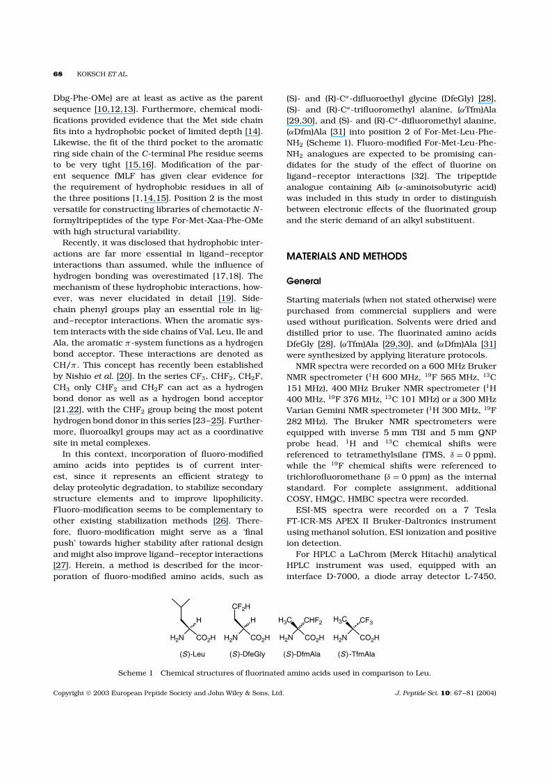
(S)- and (R)-Cα-difluoroethyl glycine (DfeGly) [28],(S)- and (R)-Cα-trifluoromethyl alanine, (αTfm)Ala[29,30], and (S)- and (R)-Cα-difluoromethyl alanine,(αDfm)Ala [31] into position 2 of For-Met-Leu-Phe-NH2 (Scheme 1). Fluoro-modified For-Met-Leu-Phe-NH2 analogues are expected to be promising can-didates for the study of the effect of fluorine onligand–receptor interactions [32]. The tripeptideanalogue containing Aib (α-aminoisobutyric acid)was included in this study in order to distinguishbetween electronic effects of the fluorinated groupand the steric demand of an alkyl substituent.
MATERIALS AND METHODS
General
Starting materials (when not stated otherwise) werepurchased from commercial suppliers and wereused without purification. Solvents were dried anddistilled prior to use. The fluorinated amino acidsDfeGly [28], (αTfm)Ala [29,30], and (αDfm)Ala [31]were synthesized by applying literature protocols.
NMR spectra were recorded on a 600 MHz BrukerNMR spectrometer (1H 600 MHz, 19F 565 MHz, 13C151 MHz), 400 MHz Bruker NMR spectrometer (1H400 MHz, 19F 376 MHz, 13C 101 MHz) or a 300 MHzVarian Gemini NMR spectrometer (1H 300 MHz, 19F282 MHz). The Bruker NMR spectrometers wereequipped with inverse 5 mm TBI and 5 mm QNPprobe head. 1H and 13C chemical shifts werereferenced to tetramethylsilane (TMS, δ = 0 ppm),while the 19F chemical shifts were referenced totrichlorofluoromethane (δ = 0 ppm) as the internalstandard. For complete assignment, additionalCOSY, HMQC, HMBC spectra were recorded.
ESI-MS spectra were recorded on a 7 TeslaFT-ICR-MS APEX II Bruker-Daltronics instrumentusing methanol solution, ESI ionization and positiveion detection.
For HPLC a LaChrom (Merck Hitachi) analyticalHPLC instrument was used, equipped with aninterface D-7000, a diode array detector L-7450,
H2N CO2H
H
H2N CO2H
H
CF2H
H2N CO2H
H3C CF3
(S)-Leu (S)-DfeGly (S)-TfmAla
H2N CO2H
H3C CHF2
(S)-DfmAla
Scheme 1 Chemical structures of fluorinated amino acids used in comparison to Leu.
Copyright 2003 European Peptide Society and John Wiley & Sons, Ltd. J. Peptide Sci. 10: 67–81 (2004)
FLUORO-MODIFIED CHEMOTACTIC PEPTIDES 69
and a pump A-B L-7100. Eluant system parameterswere (gradient method A): flow 1.000 ml/min, eluantA: 95% H2O, 5% MeCN, 0.1% TFA; eluant B:95% MeCN, 5% H2O, 0.1% TFA; gradient: 0.0 min(100% A, 0.0% B), 30.0 min (0% A, 100% B),40.0 min (100% A, 0% B); column: Vydac C4, 10 µm,4.6 × 250 (Separation Products).
For flash chromatography, silica gel (32–63 µm)was used with solvent systems given in the text.
On TLC the compounds were visualized byspraying the plate with a mixture of ceric(IV)sulphate (0.2%), ammonium molybdate (5%) andH2SO4 (5%) in water, followed by heating.
Luminol was a product from Boehringer-Mannheim (Heidelberg, Germany). Chemicals forneutrophil isolation and purification, i.e. Hanks’balanced salt solution without phenol red, Ficoll-Hypaque, dextran, heparin and the stimulatorFor-Met-Leu-Phe-OH were purchased from Sigma(Deisenhofen, Germany).
Tripeptides were dissolved in dimethylsulphoxideat 10−2 mol/l. These stock solutions were dividedinto aliquots and stored at −20 °C. Working solu-tions of tripeptides were prepared by dilution withHanks’ medium immediately before use.
Peptide Synthesis
Peptide coupling with DIC/HOAt (method A). Tothe stirred solution of 1 eq of PG-Xaa-OH (PG:protecting group) in DMF (N ,N-dimethylformamide),1.2 eq HOAt (1-hydroxy-7-aza-1,2,3-benzotriazole)and 1.2 eq of DIC (N ,N ′-diisopropylcarbodiimide)were added at room temperature. After 5 min 1.2 eqof H-Yaa-PG and 1 eq of DIEA (diisopropyl ethylamine) were added. The progress of the reaction wasmonitored by HPLC or 19F-NMR spectrometry. Theorganic phase was evaporated in vacuo. The residuewas partitioned between a 10% citric acid solutionand ethyl acetate. The phases were separated andthe organic phase was washed with 10% citric acid(2x), sat. NaCl solution (3x), 10% NaHCO3 solution(3x) and sat. NaCl solution. (3x). The organic layerwas dried with MgSO4, filtered and concentratedunder reduced pressure. The peptides were purifiedby flash chromatography using the solvent mixturesgiven in the text.
Peptide coupling via mixed anhydrides (methodB). To a stirred solution of 2.2 eq of PG-Xaa-OH inDMF, at −30 °C 2 eq of NMM (N-methyl morpholine)was added. After stirring for 15 min at −15 °C,2 eq of CAIBE (isobutyl chloroformate), and after
10 min 1 eq of H-Yaa-PG, were added. The reactiontemperature was kept below 0 °C for 1 h, and thenthe mixture was warmed up to room temperature.The progress of the reaction was monitored by HPLCor 19F-NMR spectrometry. For work-up procedureand purification, see method A.
Nα-Formylation of H-Met-Xaa-Phe-NH2 (methodC). To 1 eq of HCO2
−. +H2-Xaa-PG dissolved inDMF, 1.2 eq of CMF (cyanomethyl formate) [33] and1 eq of triethylamine were added and stirred for 2 h.After completion of the reaction (monitored by HPLC)the solvent was evaporated in vacuo. The formylpeptides were purified by flash chromatographyusing the solvent mixtures given in the text. In thecase of 6a and 6b, where chromatography was notpossible because of the formation of almost insolubleaggregates, the solid was dispersed in water andfiltrated on a 45 µm filter. The salt-free, formylatedpeptides were washed down from the filter withmethanol. After evaporating the solvent, the solidmaterials were lyophilized.
Deprotection of the Z-group. To a solution of Z-Xaa-Phe-NH2 in methanol, Pd/C (10%) was added.The mixture was stirred under an atmosphere ofhydrogen until the reaction was complete (19F-NMR or TLC analysis). After filtration, methanolwas evaporated under reduced pressure. The crudepeptide was used for the subsequent couplingreaction without further purification.
Deprotection of the Boc-group. Boc-Met-Xaa-Phe-NH2 was stirred in the presence of an excess ofconcentrated formic acid. After 30 min the formicacid was evaporated under reduced pressure andthe residue was subjected to Nα-formylation withoutfurther purification.
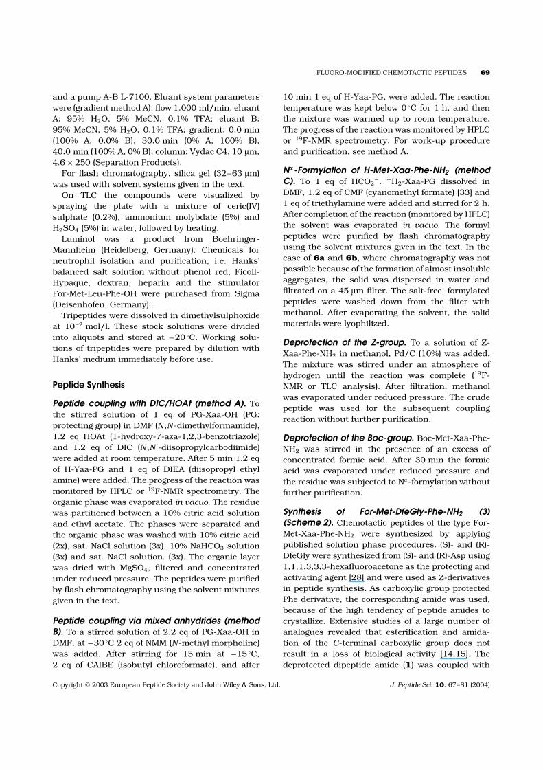
Synthesis of For-Met-DfeGly-Phe-NH2 (3)(Scheme 2). Chemotactic peptides of the type For-Met-Xaa-Phe-NH2 were synthesized by applyingpublished solution phase procedures. (S)- and (R)-DfeGly were synthesized from (S)- and (R)-Asp using1,1,1,3,3,3-hexafluoroacetone as the protecting andactivating agent [28] and were used as Z-derivativesin peptide synthesis. As carboxylic group protectedPhe derivative, the corresponding amide was used,because of the high tendency of peptide amides tocrystallize. Extensive studies of a large number ofanalogues revealed that esterification and amida-tion of the C-terminal carboxylic group does notresult in a loss of biological activity [14,15]. Thedeprotected dipeptide amide (1) was coupled with
Copyright 2003 European Peptide Society and John Wiley & Sons, Ltd. J. Peptide Sci. 10: 67–81 (2004)
70 KOKSCH ET AL.
Boc-Met-OH on treatment with DIC/HOAt to give theBoc-protected tripeptide amide (2). The N-terminalamino group of 2 was deprotected with concen-trated formic acid and subsequently Nα-formylatedon treatment with CMF/TEA (triethylamine) [33]to give the fluoro-modified chemotactic tripeptideamide (3).
H-(S)-DfeGly-Phe-NH2 (1a). The dipeptide 1a wassynthesized from Z-(S)-DfeGly-OH [22] (273 mg,1.0 mmol) and H-Phe-NH2 (197 mg, 1.2 mmol)applying method A. The Z-protected dipeptidewas purified by flash chromatography (eluant:CHCl3/MeOH, 10 : 1). Z-deprotection was achievedusing the above mentioned general route. Yield of1a: 122 mg (43%).
1H-NMR (d6-DMSO, 400 MHz) δ ppm: 1.67–1.85,1.86–2.03 (m, 2H, DfeGly CβH2), 1.94 (s, 2H, DfeGlyNH2), 2.82 (dd, 1H, 2J = 13.6 Hz, 3J = 9.2 Hz, PheCβH2), 3.04 (dd, 1H, 2J = 13.6 Hz, 3J = 4.8 Hz,Phe CβH2), 3.27 (dd, 1H, 3J = 9.2 Hz, 3J = 4.8 Hz,DfeGly CαH), 4.47 (m, 1H, Phe CαH), 5.99 (tdd,1H, 2J = 57.0 Hz, 3J = 5.7 Hz, 3J = 4.0 Hz, CF2H),7.11 (s, 1H, Phe NH2), 7.14–7.29 (m, 5H, Ar-Phe),7.48 (s, 1H, Phe NH2), 8.11 (d, 1H, 3J = 8.2 Hz,Phe NH). 13C-NMR (d6-DMSO, 101 MHz) δ ppm:37.72 (Phe Cβ ), 38.6 (overl. m, DfeGly Cβ ), 50.13(t, 3J = 5.7 Hz, DfeGly Cα), 53.10 (Phe Cα), 116.88(t, 1J = 236.8 Hz, CF2H) 126.12–137.65 (Ar-Phe),172.66 (C O, DfeGly), 173.27 (C O, Phe). 19F-NMR (d6-DMSO, 376 MHz) δ ppm: −114.4 (ddd,1F, 2J = 280.8 Hz, 2J = 57.0 Hz, 3J = 22.2 Hz, 3J =14.4 Hz, CF2H), −115.3 (ddd, 1F, 2J = 280.8 Hz,2J = 56.8 Hz, 3J = 17.2 Hz, 3J = 14.2 Hz, CF2H).
Analytical HPLC: Rt = 4.71 min. Calculated MW =285.29 for C13H17F2N3O2.. ESI-MS: m/z [M +H]+ 286.13606 (286.13616), [M + Na]+ 308.11757(308.11810), [2M + H]+ 571.26562 (571.26504),[2M + Na]+ 593.24846 (593.24699).
Met DfeGly Phe
Z OH H NH2
Z NH2
H NH2Boc OH
NH2Boc
NH2H
DIC, HOAt
HCOOH
DIC, HOAt
NH2ForCMF / TEA
1
2
3
H2, Pd / C
Scheme 2 Synthesis of peptides 1–3.
H-(R)-DfeGly-Phe-NH2 (1b). For synthesis of 1bsee 1a. Yield of 1b: 134 mg (47%). 1H-NMR (d6-DMSO, 600 MHz) δ ppm: 1.71–1.85, 1.88–1.99(m, 2H, DfeGly CβH2), 2.78 (dd, 1H, 2J = 13.7 Hz,3J = 9.5 Hz, Phe CβH2), 3.06 (dd, 1H, 2J = 13.7 Hz,3J = 4.6 Hz, Phe CβH2), 3.41 (m, 1H, DfeGly CαH),3.79–4.30 (br s, 2H, DfeGly NH2), 4.50 (m, 1H, PheCαH), 5.87 (tdd, 1H, 2J = 56.6 Hz, 3J = 5.7 Hz, 3J =4.0 Hz, CF2H), 7.14 (s, 1H, Phe NH2), 7.16–7.28 (m,5H, Ar-Phe), 7.48 (s, 1H, Phe NH2), 8.32 (d, 1H, 3J =7.9 Hz, Phe NH). 13C-NMR (d6-DMSO, 151 MHz) δ
ppm: 37.72 (overl., Phe Cβ , DfeGly Cβ ), 49.32 (DfeGlyCα), 53.41 (Phe Cα), 116.32 (t, 1J = 233.7 Hz, CF2H),126.26–137.72 (Ar-Phe), 171.72 (C O, DfeGly),172.59 (C O, Phe). 19F-NMR (d6-DMSO, 376 MHz) δ
ppm: −114.3 (ddd, 1F, 1J = 282.0 Hz, 2J = 56.6 Hz,3J = 20.3 Hz, 3J = 14.9 Hz, CF2H), −115.1 (ddd,br.1F, br. 1J = 282.0 Hz, 2J = 57 Hz, CF2H).
Analytical HPLC: Rt = 7.31 min. Calculated MW =285.29 for C13H17F2N3O2. ESI-MS: m/z [M +H]+ 286.13607 (286.13616), [2M + H]+ 571.26581(571.26504).
Boc-Met-(S)-DfeGly-Phe-NH2 (2a). Tripeptide 2awas synthesized from Boc-Met-OH (249 mg,1.0 mmol) and H-(S)-DfeGly-Phe-NH2 (1a) (342 mg,1.2 mmol) by applying method A. Purificationby column chromatography (eluant: CHCl3/MeOH,15 : 1). Yield of 2a: 305 mg (59%).
1H-NMR (d6-DMSO, 400 MHz) δ ppm: 1.37 (s,9H, C(CH3)3), 1.67–1.88 (m, 2H, Met CβH), 2.02(s, 3H, Met CH3), 2.03–2.24 (m, 2H, DfeGly CβH2),2.35–2.47 (m, 2H, Met Cγ H2), 2.81 (dd, 1H, 2J =13.8 Hz, 3J = 9.1 Hz, Phe CβH2), 3.00 (dd, 1H,2J = 13.8 Hz, 3J = 4.8 Hz, Phe CβH2), 3.90–4.00 (m,1H, DfeGly CαH), 4.35–4.47 (m, 2H, Met CαH, PheCαH), 5.94 (tdd, 1H, 2J = 56.5 Hz, 3J = 5.9 Hz,3J =3.8 Hz, CF2H), 7.08 (overl., 2H, Met NH, Phe NH2),7.14–7.28 (m, 5H, Ar-Phe), 7.43 (s, 1H, Phe NH2),8.10 (d, 1H, 3J = 7.7 Hz, Phe NH), 8.17 (d, 1H, 3J =7.7 Hz, DfeGly NH). 13C-NMR (d6-DMSO, 101 MHz)δ ppm: 14.52 (Met CH3), 28.04 (C(CH3)3), 29.59 (MetCγ ), 31.03 (Met Cβ ), 36.32 (DfeGly Cβ ), 37.33 (PheCβ ), 47.55 (DfeGly Cα), 53.24 (Met Cα), 53.81 (PheCα), 115.90 (t, 1J = 236.9 Hz, CF2H), 126.16–137.63(Ar-Phe). 19F-NMR (d6-DMSO, 376 MHz) δ ppm:−114.4 (ddd, 1F, 1J = 282.3 Hz, 2J = 56.5 Hz, 3J =21.9 Hz, 3J = 14.2 Hz, CF2H), −115.3 (br ddd, 1F,1J = 282.3 Hz, 2J = 56 Hz, CF2H).
Analytical HPLC: Rt = 16.93 min. TLC (CHCl3/MeOH, 15 : 1): Rf = 0.11. Calculated MW =516.61 for C23H34F2N4O5S. ESI-MS: m/z [M +H]+ 517.22901 (517.22907), [M + Na]+ 539.21076
Copyright 2003 European Peptide Society and John Wiley & Sons, Ltd. J. Peptide Sci. 10: 67–81 (2004)
FLUORO-MODIFIED CHEMOTACTIC PEPTIDES 71
(539.21102), [M + K]+ 555.18588 (555.18496),[2M + Na]+ 1055.43128 (1055.43282), [2M + K]+
1071.41125 (1071.40676), [3M + Na]+ 1571.64627(1571.65462).
Boc-Met-(R)-DfeGly-Phe-NH2 (2b). Tripeptide 2bwas synthesized from Boc-Met-OH (249 mg,1.0 mmol) and H-(R)-DfeGly-Phe-NH2 (342 mg,1.2 mmol) using method A. Purification by columnchromatography (eluant: CHCl3/MeOH, 15 : 1). Yieldof 2b: 237 mg (46%).
1H-NMR (d6-DMSO, 600 MHz) δ ppm: 1.37 (s, 9H,C(CH3)3), 1.70–1.87 (m, 2H, Met CβH2), 1.87–2.04(m, 2H, DfeGly CβH2), 2.02 (s, 3H, Met CH3),2.39–2.48 (m, 2H, Met Cγ H2), 2.74 (dd, 1H, 2J =13.4 Hz, 3J = 10.5 Hz, Phe CβH2), 3.08 (1H, dd,2J = 13.4 Hz, 3J = 14.2 Hz, Phe CβH2), 3.96 (1H,m, Met CαH), 4.39 (1H, m, DfeGly CαH), 4.45 (m,1H, Phe CαH), 5.69 (br. tdd, 1H, 2J = 56 Hz, CF2H),7.07 (d, 1H, 3J = 7.2 Hz, Met NH), 7.15 (s, 1H,Phe NH2), 7.16–7.26 (m, 5H, Ar-Phe), 7.40 (s, 1H,Phe NH2), 8.11 (d, 1H, 3J = 8.1 Hz, DfeGly NH),8.16 (d, 1H, 3J = 8.8 Hz, Phe NH). 13C-NMR (d6-DMSO, 151 MHz) δ ppm: 14.10 (Met CH3), 27.70(C(CH3)3), 29.18 (Met Cγ ), 30.70 (Met Cβ ), 35.42(t, 2J = 20.4 Hz, DfeGly Cβ ), 37.13 (Phe Cβ ), 47.17(DfeGly Cα), 53.41 (overl. br. s, Met Cα, Phe Cα),77.93 (C(CH3)3), 115.53 (t, 1J = 233.4 Hz, CF2H),125.80–137.46 (Ar-Phe), 155.08 (C O, Boc), 169.04(C O, DfeGly), 171.33 (C O, Met), 172.16 (C O,Phe). 19F-NMR(d6-DMSO, 376 MHz) δ ppm: −113.9(ddd, 1F, 2J = 282.4 Hz, 2J = 56.5 Hz, 3J = 21.0 Hz,3J = 14.8 Hz, CF2H), −115.0 (br ddd, 1F, 2J =282.4 Hz, 2J = 56 Hz, CF2H).
Analytical HPLC: Rt = 17.05 min. TLC (CHCl3/MeOH, 15 : 1): Rf = 0.17. Calculated MW = 516.61for C23H34F2N4O5S. ESI-MS: m/z [M + Na]+
539.21109 (539.21102), [2M + Na]+ 1055.43142(1055.43282), [2M + K]+ 1071.40762 (1071.40676),[3M + Na]+ 1571.65698 (1571.65462), [4M + Na]+
2087.87048 (2087.87641).
For-Met-(S)-DfeGly-Phe-NH2 (3a). Tripeptide 2a(516 mg, 1.0 mmol) was deprotected with concen-trated formic acid as described above and thenNα-formylated by method C to give 3a. Yield of 3a:204 mg (46%).
1H-NMR (d6-DMSO, 600 MHz) δ ppm: 1.72–1.80,1.83–1.91 (m, 2H, Met CβH2), 2.02 (s, 3H, MetCH3), 2.03–2.24 (s, 2H, DfeGly CβH2), 2.36–2.47(m, 2H, Met Cγ H2), 2.82 (dd, 1H, 2J = 13.9 Hz,3J = 9.2 Hz, Phe CβH2), 3.01 (dd, 1H, 2J = 13.9 Hz,3J = 4.7 Hz, Phe CβH2), 4.33–4.45 (overl. m, 3H,
Phe CαH, DfeGly CαH, Met CαH), 5.96 (tdd, 1H,2J = 56.2 Hz, 3J = 5.7 Hz, 3J = 3.8 Hz, CF2H), 7.10(s, 1H, Phe NH2), 7.16–7.28 (m. 5H, Ar-Phe), 7.40(s, 1H, Phe NH2), 8.01 (d, 1H, 3J = 8.2 Hz, Phe NH),8.03 (s, 1H, H-For), 8.33 (d, 1H, 3J = 8.2 Hz, DfeGlyNH), 8.35 (d, 1H, 3J = 8.2 Hz, Met NH). 13C-NMR(d6-DMSO, 151 MHz) δ ppm: 14.16 (Met Cε), 28.92(Met Cγ ), 31.15 (Met Cβ ), 35.41 (t, 2J = 22.2 Hz,DfeGly Cβ ), 36.96 (Phe Cβ ), 47.40 (DfeGly Cα), 50.17(Met Cα), 53.45 (Phe Cα), 115.72 (t, 1J = 238.6 Hz,CF2H), 125.81–137.28 (Ar-Phe), 160.80 (C O, For),169.16 (C O, DfeGly), 170.34 (C O, Met), 172.06(C O, Phe). 19F-NMR (d6-DMSO, 376 MHz) δ ppm:−114.4 (ddd, 1F, 2J = 282.4 Hz, 2J = 56.6 Hz, 3J =22.2 Hz, 3J = 14.4 Hz, CF2H), −115.4 (ddd, 1F, 2J =282.4 Hz, 2J = 55.8 Hz, 3J = 16.4 Hz, 3J = 13.3 Hz,CF2H).
Analytical HPLC: Rt = 11.75 min. TLC (CHCl3/MeOH/acetone, 5 : 1 : 1): Rf = 0.37. Calculated MW =444.50 for C19H26F2N4O4S. ESI-MS: m/z [M +Na]+ 467.15414 (467.15350), [2M + Na]+ 911.32037(911.31724).
For-Met-(R)-DfeGly-Phe-NH2 (3b). Tripeptide 2b(516 mg, 1.0 mmol) was deprotected with concen-trated formic acid as described above and thenNα-formylated by applying method C to give 3b.Yield of 3b: 222 mg (50%).
1H-NMR (d6-DMSO, 600 MHz) δ ppm: 1.72–2.06(overl. m, 4H, DfeGly CβH2, Met CβH2), 2.02 (s,3H, Met CH3), 2.56–2.40 (m, 2H, Met Cγ H2), 2.76(dd, 1H, 2J = 13.8 Hz, 3J = 10.6 Hz, Phe CβCH2),3.07 (dd, 1H, 2J = 13.8 Hz, 3J = 4.2 Hz, Phe CβH2),4.33–4.48 (m, 3H, overl.-Phe CαH, DfeGly CαH,Met CαH), 5.73 (br tdd, 1H, 2J = 56 Hz, 3J = 5 Hz,CF2H), 7.14 (s, 1H, Phe NH2), 7.15–7.27 (m, 5H,Ar-Phe), 7.40 (s, 1H, Phe NH2), 8.03 (s, 1H, H-For), 8.21 (d, 1H, 3J = 8.7 Hz, Phe NH), 8.40 (overl.d, 2H, DfeGly NH, Met NH). 13C-NMR (d6-DMSO,151 MHz) δ ppm: 14.10 (Met CH3), 28.88 (Met Cγ )31.03 (Met Cβ ), 35.25 (t, 2J = 20.4 Hz, DfeGly Cβ ),37.06 (Phe Cβ ), 47.20 (br. s, DfeGly Cα), 50.42(Met Cα), 53.50 (Phe Cα), 115.61 (t, 1J = 236.7 Hz,CF2H), 125.79–137.54 (Ar-Phe), 160.94 (C O, For),169.06 (C O, DfeGly), 170.31 (C O, Met), 172.25(C O, Phe). 19F-NMR (d6-DMSO, 376 MHz) δ ppm:−114.0 (ddd, 1F, 2J = 282.4 Hz, 2J = 56.3 Hz, 3J =20.8 Hz, 3J = 14.5 Hz, CF2H), −115.2 (ddd, 1F, 2J =282.4 Hz, 2J = 56.2 Hz, 3J = 18.2 Hz, 3J = 13.4 Hz,CF2H).
Analytical HPLC: Rt = 13.00 min. TLC (CHCl3/MeOH/acetone, 5 : 1 : 1): Rf = 0.37. Calculated MW =444.50 for C19H26F2N4O4S. ESI-MS: m/z [M +
Copyright 2003 European Peptide Society and John Wiley & Sons, Ltd. J. Peptide Sci. 10: 67–81 (2004)
72 KOKSCH ET AL.
Na]+ 467.15368 (467.15350), [2M + Na]+ 911.31950(911.31779), [3M + Na]+ 1355.48134 (1355.48207).
Synthesis of For-Met-(αTfm)Ala-Phe-NH2 (6)
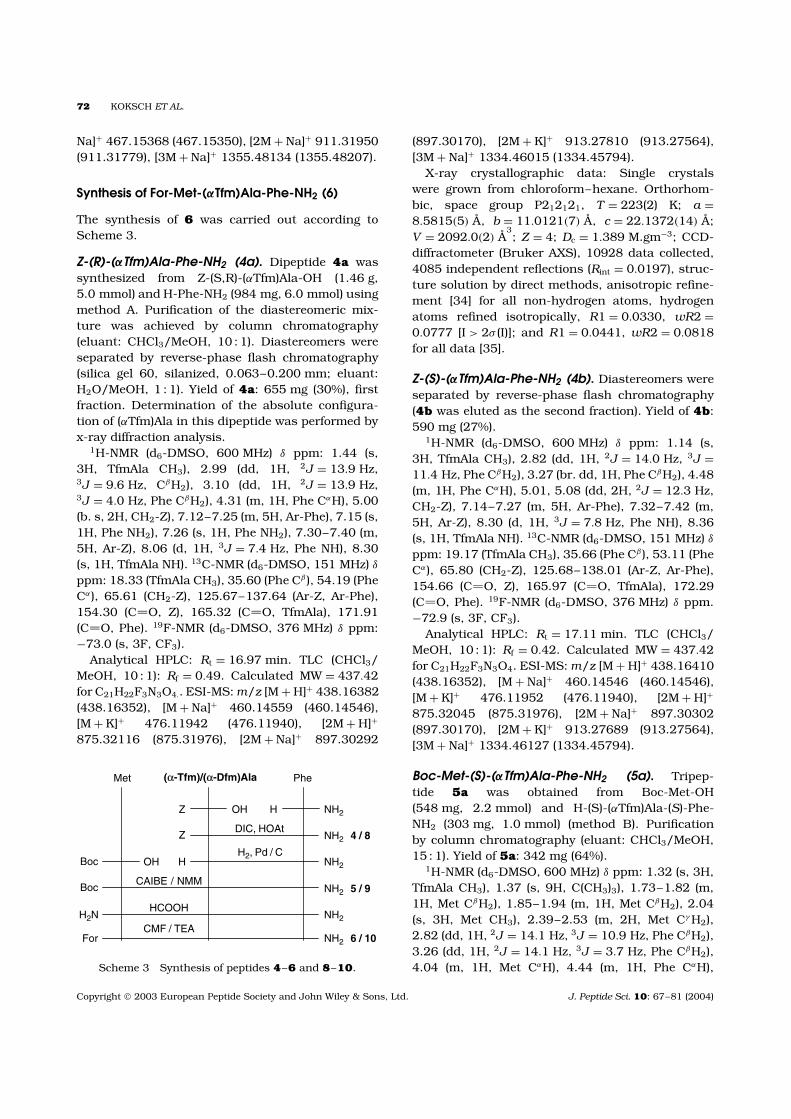
The synthesis of 6 was carried out according toScheme 3.
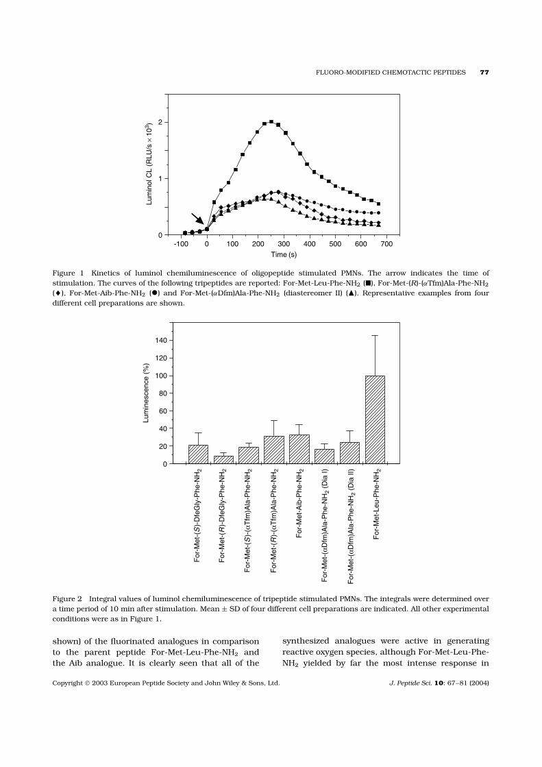
Z-(R)-(αTfm)Ala-Phe-NH2 (4a). Dipeptide 4a wassynthesized from Z-(S,R)-(αTfm)Ala-OH (1.46 g,5.0 mmol) and H-Phe-NH2 (984 mg, 6.0 mmol) usingmethod A. Purification of the diastereomeric mix-ture was achieved by column chromatography(eluant: CHCl3/MeOH, 10 : 1). Diastereomers wereseparated by reverse-phase flash chromatography(silica gel 60, silanized, 0.063–0.200 mm; eluant:H2O/MeOH, 1 : 1). Yield of 4a: 655 mg (30%), firstfraction. Determination of the absolute configura-tion of (αTfm)Ala in this dipeptide was performed byx-ray diffraction analysis.
1H-NMR (d6-DMSO, 600 MHz) δ ppm: 1.44 (s,3H, TfmAla CH3), 2.99 (dd, 1H, 2J = 13.9 Hz,3J = 9.6 Hz, CβH2), 3.10 (dd, 1H, 2J = 13.9 Hz,3J = 4.0 Hz, Phe CβH2), 4.31 (m, 1H, Phe CαH), 5.00(b. s, 2H, CH2-Z), 7.12–7.25 (m, 5H, Ar-Phe), 7.15 (s,1H, Phe NH2), 7.26 (s, 1H, Phe NH2), 7.30–7.40 (m,5H, Ar-Z), 8.06 (d, 1H, 3J = 7.4 Hz, Phe NH), 8.30(s, 1H, TfmAla NH). 13C-NMR (d6-DMSO, 151 MHz) δ
ppm: 18.33 (TfmAla CH3), 35.60 (Phe Cβ ), 54.19 (PheCα), 65.61 (CH2-Z), 125.67–137.64 (Ar-Z, Ar-Phe),154.30 (C O, Z), 165.32 (C O, TfmAla), 171.91(C O, Phe). 19F-NMR (d6-DMSO, 376 MHz) δ ppm:−73.0 (s, 3F, CF3).
Analytical HPLC: Rt = 16.97 min. TLC (CHCl3/MeOH, 10 : 1): Rf = 0.49. Calculated MW = 437.42for C21H22F3N3O4.. ESI-MS: m/z [M + H]+ 438.16382(438.16352), [M + Na]+ 460.14559 (460.14546),[M + K]+ 476.11942 (476.11940), [2M + H]+
875.32116 (875.31976), [2M + Na]+ 897.30292
Met Phe
Z OH H NH2
Z NH2 4 / 8
H NH2Boc OH
NH2Boc
NH2H2N
DIC, HOAt
H2, Pd / C
HCOOH
CAIBE / NMM
NH2ForCMF / TEA
5 / 9
6 / 10
(α-Tfm)/(α-Dfm)Ala
Scheme 3 Synthesis of peptides 4–6 and 8–10.
(897.30170), [2M + K]+ 913.27810 (913.27564),[3M + Na]+ 1334.46015 (1334.45794).
X-ray crystallographic data: Single crystalswere grown from chloroform–hexane. Orthorhom-bic, space group P212121, T = 223(2) K; a =8.5815(5) A, b = 11.0121(7) A, c = 22.1372(14) A;V = 2092.0(2) A
3; Z = 4; Dc = 1.389 M.gm−3; CCD-
diffractometer (Bruker AXS), 10928 data collected,4085 independent reflections (Rint = 0.0197), struc-ture solution by direct methods, anisotropic refine-ment [34] for all non-hydrogen atoms, hydrogenatoms refined isotropically, R1 = 0.0330, wR2 =0.0777 [I > 2σ (I)]; and R1 = 0.0441, wR2 = 0.0818for all data [35].
Z-(S)-(αTfm)Ala-Phe-NH2 (4b). Diastereomers wereseparated by reverse-phase flash chromatography(4b was eluted as the second fraction). Yield of 4b:590 mg (27%).
1H-NMR (d6-DMSO, 600 MHz) δ ppm: 1.14 (s,3H, TfmAla CH3), 2.82 (dd, 1H, 2J = 14.0 Hz, 3J =11.4 Hz, Phe CβH2), 3.27 (br. dd, 1H, Phe CβH2), 4.48(m, 1H, Phe CαH), 5.01, 5.08 (dd, 2H, 2J = 12.3 Hz,CH2-Z), 7.14–7.27 (m, 5H, Ar-Phe), 7.32–7.42 (m,5H, Ar-Z), 8.30 (d, 1H, 3J = 7.8 Hz, Phe NH), 8.36(s, 1H, TfmAla NH). 13C-NMR (d6-DMSO, 151 MHz) δ
ppm: 19.17 (TfmAla CH3), 35.66 (Phe Cβ ), 53.11 (PheCα), 65.80 (CH2-Z), 125.68–138.01 (Ar-Z, Ar-Phe),154.66 (C O, Z), 165.97 (C O, TfmAla), 172.29(C O, Phe). 19F-NMR (d6-DMSO, 376 MHz) δ ppm.−72.9 (s, 3F, CF3).
Analytical HPLC: Rt = 17.11 min. TLC (CHCl3/MeOH, 10 : 1): Rf = 0.42. Calculated MW = 437.42for C21H22F3N3O4. ESI-MS: m/z [M + H]+ 438.16410(438.16352), [M + Na]+ 460.14546 (460.14546),[M + K]+ 476.11952 (476.11940), [2M + H]+
875.32045 (875.31976), [2M + Na]+ 897.30302(897.30170), [2M + K]+ 913.27689 (913.27564),[3M + Na]+ 1334.46127 (1334.45794).
Boc-Met-(S)-(αTfm)Ala-Phe-NH2 (5a). Tripep-tide 5a was obtained from Boc-Met-OH(548 mg, 2.2 mmol) and H-(S)-(αTfm)Ala-(S)-Phe-NH2 (303 mg, 1.0 mmol) (method B). Purificationby column chromatography (eluant: CHCl3/MeOH,15 : 1). Yield of 5a: 342 mg (64%).
1H-NMR (d6-DMSO, 600 MHz) δ ppm: 1.32 (s, 3H,TfmAla CH3), 1.37 (s, 9H, C(CH3)3), 1.73–1.82 (m,1H, Met CβH2), 1.85–1.94 (m, 1H, Met CβH2), 2.04(s, 3H, Met CH3), 2.39–2.53 (m, 2H, Met Cγ H2),2.82 (dd, 1H, 2J = 14.1 Hz, 3J = 10.9 Hz, Phe CβH2),3.26 (dd, 1H, 2J = 14.1 Hz, 3J = 3.7 Hz, Phe CβH2),4.04 (m, 1H, Met CαH), 4.44 (m, 1H, Phe CαH),
Copyright 2003 European Peptide Society and John Wiley & Sons, Ltd. J. Peptide Sci. 10: 67–81 (2004)
FLUORO-MODIFIED CHEMOTACTIC PEPTIDES 73
7.09 (s, 1H, Phe NH2), 7.14–7.27 (m, 5H, Ar-Phe),7.20 (s, 1H, Phe NH2), 7.78 (d, 1H, 3J = 8.3 Hz, PheNH), 8.66 (s, 1H, TfmAla NH). 13C-NMR(d6-DMSO,151 MHz) δ ppm: 14.30 (Met CH3), 18.43 (TfmAlaCH3), 27.66 (C(CH3)3), 29.19 (Met Cγ ) 29.98 (Met Cβ ),35.77 (Phe Cβ ), 53.19 (Phe Cα), 53.48 (Met Cα), 60.59(q, 2J = 30 Hz, TfmAla Cα), 78.08 (C(CH3)3), 124.09(q, 1J = 287 Hz, TfmAla CF3), 125.72–137.73 (Ar-Phe), 155.31 (C O, Boc), 165.38 (C O, TfmAla),171.96 (C O, Phe), 172.25 (C O, Met). 19F-NMR(d6-DMSO, 376 MHz) δ ppm: −72.8 (s, 3F, CF3).
Analytical HPLC: Rt = 17.98 min. TLC (CHCl3/MeOH, 15 : 1): Rf = 0.30. Calculated MW = 534.60for C23H33F3N4O5S. ESI-MS: m/z [M + Na]+
557.20154 (557.20160), [2M + H]+ 1069.43430(1069.43203), [2M + Na]+ 1091.41467(1091.41397), [2M + K]+ 1107.39077 (1107.38791),[3M + Na]+ 1625.62529 (1625.62635), [3M + K]+
1641.60696 (1641.60029), [4M + Na]+ 2159.84978(2159.83873).
Boc-Met-(R)-(αTfm)Ala-Phe-NH2 (5b). Tripeptide5b was obtained from Boc-Met-OH (548 mg,2.2 mmol) and H-(R)-(αTfm)Ala-Phe-NH2 (303 mg,1.0 mmol) (method B). Purification by column chro-matography (eluant: CHCl3/MeOH, 15 : 1). Yield of5b: 208 mg (39%).
1H-NMR (d6-DMSO, 600 MHz) δ ppm: 1.36 (s, 9H,C(CH3)3), 1.45 (s, 3H, TfmAla, CH3), 1.71–1.88 (m,2H, Met CβH2), 2.03 (s, 3H, Met CH3), 2.37–2.48(m, 2H, Met Cγ H2), 2.95 (dd, 1H, 2J = 14.1 Hz,3J = 9.2 Hz, Phe CβH2), 3.13 (dd, 1H, 2J = 14.1 Hz,3J = 4.7 Hz, Phe CβH2), 4.06 (m, 1H, Met CαH),4.30 (m, 1H, Phe CαH), 7.12 (d, 1H, 3J = 7.0 Hz,Met NH), 7.13–7.26 (m, 5H, Ar-Phe), 7.70 (d, 1H,3J = 7.7 Hz, Phe NH), 8.67 (s, 1H, TfmAla NH). 13C-NMR (d6-DMSO, 151 MHz) δ ppm: 14.19 (Met CH3),17.54 (TfmAla, CH3), 27.67 (C(CH3)3), 29.09 (MetCγ ) 30.24 (Met Cβ ), 35.71 (Phe Cβ ), 53.21 (MetCα), 54.13 (Phe Cα), 61.08 (q, 2J = 29 Hz, TfmAlaCα), 78.05 (C(CH3)3), 124.06 (q, 1J = 286 Hz, TfmAlaCF3), 125.72–137.52 (Ar-Phe), 155.27 (C O, Boc),164.69 (C O,-TfmAla), 171.63 (C O, Met), 172.11(C O, Phe). 19F-NMR (d6-DMSO, 376 MHz) δ ppm:−73.1 (s, 3F, CF3).
Analytical HPLC: Rt = 17.94 min. TLC (CHCl3/MeOH, 15 : 1): Rf = 0.30. Calculated MW =534.60 for C23H33F3N4O5S. ESI-MS: m/z [M +H]+ 535.21968 (535.21965), [M + Na]+ 557.20191(557.20160), [M + K]+ 573.17578 (573.17553),[2M + Na]+ 1091.41677 (1091.41397), [2M + K]+
1107.39187 (1107.38791), [3M + Na]+ 1625.63540(1625.62635), [4M + Na]+ 2159.83542 (2159.83873).
For-Met-(S)-(αTfm)Ala-Phe-NH2 (6a). Tripeptide 5a(267 mg, 0.5 mmol) was deprotected by the abovementioned general route and then Nα-formylatedby applying method C to give 6a. Purificationby column chromatography (eluant: CHCl3/MeOH,15 : 1). Yield of 6a: 155 mg (67%).
1H-NMR (d6-DMSO, 600 MHz) δ ppm: 1.29 (s, 3H,TfmAla CH3), 1.73–1.82, 1.95–2.03 (m, 2H, MetCβH2), 2.05 (s, 3H, Met CH3), 2.38–2.45 (m, 2H, MetCγ H2), 2.82 (dd, 1H, 2J = 14.2 Hz, 3J = 10.7 Hz, PheCβH2), 3.25 (dd, 1H, 2J = 14.2 Hz, 3J = 4.1 Hz, PheCβH2), 4.41–4.49 (overl. m, 2H, Met CαH, Phe CαH),7.07 (s, 1H, Phe NH2), 7.15–7.29 (m, 5H, Ar-Phe),7.81 (d, 1H, 3J = 8.4 Hz, Phe NH), 8.01 (s, 1H, ForH), 8.45 (d, 1H, 3J = 7.3 Hz, Met NH), 8.86 (s, 1H,TfmAla NH). 13C-NMR (d6-DMSO, 151 MHz) δ ppm:14.33 (Met CH3), 18.50 (TfmAla CH3), 28.99 (Met Cγ )30.58 (Met Cβ ), 35.82 (Phe Cβ ), 50.57 (Met Cα), 53.19(Phe Cα), 60.62 (q, 2J = 29 Hz, TfmAla Cα), 124.12(q, 1J = 286 Hz, TfmAla CF3), 125.73–137.77 (Ar-Phe), 161.01 (C O, For), 165.31 (C O, TfmAla),171.22 (C O, Met), 172.00 (C O, Phe). 19F-NMR(d6-DMSO, 376 MHz) δ-ppm: 72.7 (s, 3F, CF3).
Analytical HPLC: Rt = 13.51 min. TLC (CHCl3/MeOH, 15 : 1): Rf = 0.15. Calculated MW =462.49 for C19H25F3N4O4S. ESI-MS: m/z [M + H]+
463.16247 (463.16214), [M + Na]+ 485.14425(485.14408), [M + K]+ 501.11852 (501.11802),[2M + Na]+ 947.30160 (947.29894), [2M + K]+
963.27412 (963.27288), [3M + Na]+ 1409.45676(1409.45381), [4M + Na]+ 1871.62426(1871.60867).
For-Met-(R)-(αTfm)Ala-Phe-NH2 (6b). Tripeptide5b (267 mg, 0.5 mmol) was deprotected as describedabove and then Nα-formylated by method C to give6b. Purification by column chromatography (eluant:CHCl3/MeOH, 15 : 1). Yield of 6b: 169 mg (73%).
1H-NMR (d6-DMSO, 600 MHz) δ ppm: 1.46 (s, 3H,TfmAla CH3), 1.72–1.81, 1.87–1.96 (m, 2H, MetCβH2), 2.03 (s, 3H, Met CH3), 2.37–2.48 (m, 2H,Met Cγ H2), 2.96 (dd, 1H, 2J = 14.1 Hz, 3J = 9.0 Hz,Phe CβH2), 3.11 (dd, 1H, 2J = 14.1 Hz, 3J = 4.9 Hz,Phe CβH2), 4.30 (m, 1H, Phe CαH), 4.49 (m, 1H,Met CαH), 7.13–7.27 (m, 5H, Ar-Phe), 7.72 (d, 1H,3J = 7.6 Hz, Phe NH), 8.03 (s, 1H, For H), 8.38 (d,1H, 3J = 7.8 Hz, Met NH), 8.89 (s, 1H, TfmAlaNH).13C-NMR (d6-DMSO, 151 MHz) δ ppm: 14.21 (MetCH3), 17.77 (TfmAla CH3), 28.80 (Met Cγ ) 30.99(Met Cβ ), 35.73 (Phe Cβ ), 50.30 (Met Cα), 54.06(Phe Cα), 61.13 (q, 2J = 30 Hz, TfmAla Cα), 124.06(q, 1J = 286 Hz, TfmAla CF3), 125.72–137.47 (Ar-Phe), 160.89 (C O, For), 164.63 (C O, TfmAla),
Copyright 2003 European Peptide Society and John Wiley & Sons, Ltd. J. Peptide Sci. 10: 67–81 (2004)
74 KOKSCH ET AL.
171.05 (C O, Met), 171.67 (C O, Phe). 19F-NMR(d6-DMSO, 376 MHz) δ ppm: −72.9 (s, 3F, CF3).
Analytical HPLC: Rt = 13.49 min. TLC (CHCl3/MeOH, 15 : 1): Rf = 0.17. Calculated MW =462.49 for C19H25F3N4O4S. ESI-MS: m/z [M +H]+ 463.16268 (463.16214), [M + Na]+ 485.14433(485.14408), [M + K]+ 501.11869 (501.11802),[2M + Na]+ 947.30112 (947.29894), [2M + K]+
963.27793 (963.27288), [3M + Na]+ 1409.45730(1409.45381).
For-Met-Aib-Phe-NH2 (7)
The synthesis of 7 was carried out according toScheme 3.
1H-NMR (d6-DMSO, 400 MHz) δ ppm: 1.17 (s,3H, Aib CH3), 1.21 (s, 3H, Aib CH3), 1.69–1.81,1.85–1.97 (m, 2H, Met CβH2), 2.04 (s, 3H, MetCH3), 2.36–2.49 (m, 2H, Met Cγ H2), 2.89 (dd, 1H,2J = 14.0 Hz, 3J = 10.3 Hz, Phe CβH2), 3.16 (dd,1H, 2J = 14.0 Hz, 3J = 4.3 Hz, Phe CβH2), 4.25–4.35(m,2H, Phe CαH, Met CαH), 7.06–7.28 (m, 7H, Ar-Phe, Phe NH2), 7.41 (d, 1H, 3J = 8.4 Hz, Phe NH),8.04 (s, 1H, For H), 8.23 (s, 1H, Aib NH), 8.35(d, 1H, 3J = 7.6 Hz, Met NH). 13C-NMR (d6-DMSO,101 MHz) δ 14.59 (Met CH3), 24.27 (Aib CH3), 25.05(Aib CH3), 29.33 (Met Cγ ), 31.23 (Met Cβ ), 36.17 (PheCβ ), 51.15 (Met Cα), 53.87 (Phe Cα), 55.90 (Aib Cα),126.01–138.33 (Ar-Phe), 161.39 (C O, For), 171.03(C O, Met), 172.87 (C O, Phe), 173.15 (C O, Aib).
Analytical HPLC: Rt = 11.25 min. CalculatedMW = 408.52 for C19H28N4O4S. ESI-MS: m/z [M +Na]+ 431.17296 (431.17235), [M + K]+ 447.14712(447.14629), [2M + Na]+ 839.35793 (839.35492),
Synthesis of For-Met-(αDfm)Ala-Phe-NH2 (10)
The synthesis of 10 was carried out according toScheme 3.
Z-(αDfm)Ala-Phe-NH2 (Diastereomer I) (8a).Dipeptide 8 was synthesized from Z-(R,S)-(αDfm)Ala-OH (273 mg, 1.0 mmol) and H-Phe-NH2
(197 mg, 1.2 mmol). Yield of 8a: 134 mg (32%), firstfraction. Separation of the diastereomers by MPLC(medium-pressure liquid chromatography).
1H-NMR (d6-DMSO, 400 MHz) δ ppm: 1.27 (brs, 3H, DfmAla CH3), 2.95 (dd, 1H, 2J = 13.9 Hz,3J = 8.7 Hz, Phe CβH2), 3.04 (dd, 1H, 2J = 13.9 Hz,3J = 4.9 Hz, Phe CβH2), 4.34 (ddd, 1H, 3J = 8.7 Hz,3J = 7.9 Hz, 3J = 4.9 Hz, Phe CαH), 4.93–5.07 (br.m, 2H, CH2-Z), 6.23 (br. t, 1H, 2J = 57 Hz, CF2H),7.10–7.40 (overl. m, 12H, Ar-Z, Ar-Phe, NH2-Phe),7.81 (d, 1H, 3J = 7.9 Hz, Phe NH), 7.96 (br. s,
DfmAla NH). 13C-NMR (d6-DMSO, 101 MHz) δ ppm:16.33 (DfmAla CH3), 36.56 (Phe Cβ ), 54.11 (Phe Cα),60.21 (t, 2J = 20 Hz, DfmAla Cα), 113.84 (t, 1J =247 Hz, CF2H), 126.18–137.92 (Ar-Phe), 155.25(C O, Z), 168.94 (C O, DfmAla), 172.36 (C O,Phe). 19F-NMR (d6-DMSO, 376 MHz) δ ppm: −126.7(dd, 1F, 2J = 276.4 Hz, 2J = 56.8 Hz, CF2H), −130.4(dd, 1F, 2J = 276.4 Hz, 2J = 56.8 Hz, CF2H).
Analytical HPLC: Rt = 15.53 min. CalculatedMW = 419.43 for C21H23F2N3O4. ESI-MS: m/z [M +H]+ 420.17377 (420.17294), [M + Na]+ 442.15539(442.15488), [M + K]+ 458.13036 (458.12882),[2M + Na]+ 861.32199 (861.32000).
Z-(αDfm)Ala-Phe-NH2 (Diastereomer II) (8b).Dipeptide 8b was synthesized from Z-(R,S)-DfmAla-OH (273 mg, 1.0 mmol) and H-Phe-NH2 (197 mg,1.2 mmol). Yield of 8b: 125 mg (30%), secondfraction. Separation of the diastereomers by MPLC.
1H-NMR(d6-DMSO, 600 MHz) δ ppm: 1.04 (brs, 3H, DfmAla CH3), 2.85 (dd, 1H, 2J = 13.9 Hz,3J = 10.7 Hz, Phe CβH2), 3.21 (dd, 1H, 2J = 13.9 Hz,3J = 4.0 Hz, Phe CβH2), 4.47 (ddd, 1H, 3J = 10.7 Hz,3J = 8.6 Hz, 3J = 4.0 Hz, Phe CαH), 5.02 (br. m, 2H,CH2-Z), 6.19 (br t, 1H, 2J = 56 Hz, CF2H), 7.12–7.42(12H, overl. m, Ar-Z, Ar-Phe, NH2-Phe), 8.07 (br.s, 1H, DfmAla NH), 8.13 (d, 1H, d, 3J = 8.6 Hz,Phe NH). 13C-NMR (d6-DMSO, 151 MHz) δ 16.91(DfmAla CH3), 36.28 (Phe Cβ ), 53.24 (Phe Cα), 59.75(t, 2J = 21 Hz, DfmAla Cα), 113.92 (t, 1J = 246 Hz,CF2H), 126.14–138.33 (Ar-Phe), 155.64 (C O, Z),169.38 (C O, DfmAla), 172.66 (C O, Phe). 19F-NMR (d6-DMSO, 376 MHz) δ ppm: −128.1 (dd, 1F,2J = 275.1 Hz, 2J = 55.6 Hz, CF2H), −130.2 (dd, 1F,2J = 276.4 Hz, 2J = 56.8 Hz, CF2H).
Analytical HPLC: Rt = 15.79 min. CalculatedMW = 419.43 for C21H23F2N3O4. ESI-MS: m/z [M +H]+ 420.17335 (420.17294), [M + Na]+ 442.15535(442.15488), [M + K]+ 458.12966 (458.12882),[2M + H]+ 839.34149 (839.33805), [2M + Na]+
861.32169 (861.32000).
Boc-Met-(αDfm)Ala-Phe-NH2 (diastereomer I)(9a). 9a was synthesized from Boc-Met-OH (548 mg,2.2 mmol) and H-(αDfm)Ala-Phe-NH2 (diastereomerI) (285 mg, 1.0 mmol) (method B). Purificationby column chromatography (eluant: CHCl3/MeOH,20 : 1). Yield of 9a: 268 mg (52%).
1H-NMR (d6-DMSO, 400 MHz) δ ppm: 1.29 (br s,3H, DfmAla CH3), 1.36 (s, 9H, C(CH3)3), 1.70–1.88(m, 2H, Met CβH2), 2.02 (s, 3H, Met CH3), 2.38–2.47(m, 2H, Met Cγ H2), 2.91 (dd, 1H, 2J = 14.1 Hz,3J = 9.1 Hz, Phe CβH2), 3.08 (dd, 1H, 2J = 14.1 Hz,
Copyright 2003 European Peptide Society and John Wiley & Sons, Ltd. J. Peptide Sci. 10: 67–81 (2004)
FLUORO-MODIFIED CHEMOTACTIC PEPTIDES 75
3J = 5.0 Hz, Phe CβH2), 4.03 (m, 1H, Met CαH) 4.26(m, 1H, Phe CαH), 6.25 (br. t, 1H, 2J = 57 Hz, CF2H),7.07 (d, 1H, 3J = 6.8 Hz, Met NH), 7.10–7.28 (overl.m, 7H, Ar-Phe, Phe NH2), 7.61 (d, 1H, 3J = 7.9 Hz,Phe NH), 8.41 (s, 1H, DfmAla NH). 13C-NMR (d6-DMSO, 101 MHz) δ ppm: 14.60 (Met CH3), 16.10(DfmAla, CH3), 28.11 (C(CH3)3), 29.40 (Met Cγ ),31.30 (Met Cβ ), 36.46 (Phe Cβ ), 53.40 (Met Cα),54.48 (Phe Cα), 60.42 (t, 2J = 30 Hz, DfmAla Cα),78.49 (C(CH3)3), 113.63 (t, 1J = 249 Hz, CF2H),126.15–138.10 (Ar-Phe), 168.35 (C O, DfmAla),172.16 (C O, Phe), 172.71 (C O, Met). 19F-NMR(d6-DMSO, 376 MHz) δ ppm: −126.5 (dd, 1F, 2J =276.4 Hz, 2J = 56.4 Hz, CF2H), −129.4 (dd, 1F,2J = 276.4 Hz, 2J = 57.2 Hz, CF2H).
Analytical HPLC: Rt = 16.54 min. TLC (CHCl3/MeOH, 15 : 1): Rf = 0.30. Calculated MW = 516.61for C23H34F2N4O5S. ESI-MS: m/z [M + Na]+
539.21171 (539.21102), [2M + Na]+ 1055.43515(1055.4322).
Boc-Met-(αDfm)Ala-Phe-NH2 (diastereomer II)(9b). Tripeptide 9b was synthesized from Boc-Met-OH (548 mg, 2.2 mmol) and H-(αDfm)Ala-Phe-NH2
(diastereomer II) (285 mg, 1.0 mmol) (Method B).Purification by column chromatography (eluant:CHCl3/MeOH, 20 : 1). Yield of 9b: 330 mg (64%).
1H-NMR (d6-DMSO, 400 MHz) δ ppm: 1.18 (br. s,3H, DfmAla CH3), 1.37 (s, 9H, C(CH3)3), 1.68–1.83(m, 1H, Met CβH2), 1.83–1.96 (m, 1H, Met CβH2),2.04 (s, 3H, Met CH3), 2.37–2.54 (m, 2H, m, MetCγ H2), 2.82 (dd, 1H, dd, 2J = 14.0 Hz, 3J = 10.3 Hz,Phe CβH2), 3.21 (dd, 1H, 2J = 14.0 Hz, 3J = 3.4 Hz,Phe CβH2), 4.03 (m, 1H, Met CαH) 4.43 (m, 1H,Phe CαH), 6.20 (br. t, 1H, 2J = 57 Hz, CF2H), 7.10(d, 1H, 3J = 7.1 Hz, Met NH), 7.00–7.30 (overl. m,7H, Ar-Phe, Phe NH2), 7.69 (d, 1H, 3J = 8.4 Hz, PheNH), 8.47 (s, 1H, DfmAla NH). 13C-NMR (d6-DMSO,101 MHz) δ ppm: 14.61 (Met CH3), 16.17 (DfmAlaCH3), 28.03 (C(CH3)3), 29.50 (Met Cγ ), 30.54 (MetCβ ), 36.50 (Phe Cβ ), 52.91 (Phe Cα), 53.23 (Met Cα),59.86 (t, 2J = 32 Hz, DfmAla Cα), 78.40 (C(CH3)3),113.37 (t, 1J = 254 Hz, CF2H), 126.12–138.03 (Ar-Phe), 168.92 (C O, DfmAla), 172.75 (C O, Phe),173.05 (C O, Met). 19F-NMR (d6-DMSO, 376 MHz)δ ppm: −126.7 (dd, 1F, 2J = 275.7 Hz, 2J = 56.2 Hz,CF2H), −130.3 (dd, 1F, 2J = 275.7 Hz, 2J = 57.0 Hz,CF2H).
Analytical HPLC: Rt = 16.79 min. TLC (CHCl3/MeOH, 15 : 1): Rf = 0.32. Calculated MW = 516.61for C23H34F2N4O5S. ESI-MS: m/z [M + Na]+
539.21169 (539.21102), [2M + Na]+ 1055.43586(1055.43227).
For-Met-(αDfm)Ala-Phe-NH2 (diastereomer I)(10a). Tripeptide 9a (258 mg, 0.5 mmol) wasdeprotected as described above and then Nα-formylated by method C to give 10a. Purificationby column chromatography (eluant: CHCl3/MeOH,15 : 1). Yield of 10a: 173 mg (78%).
1H-NMR (d6-DMSO, 600 MHz) δ ppm: 1.29 (br. s,3H, DfmAla CH3), 1.73–1.81 (m, 1H, Met CβH2),1.85–1.92 (m, 1H, Met CβH2), 2.03 (s, 3H, MetCH3), 2.37–2.47 (m, 2H, Met Cγ H2), 2.92 (dd, 1H,2J = 14.0 Hz, 3J = 8.9 Hz, Phe CβH2), 3.05 (dd, 1H,2J = 14.0 Hz, 3J = 5.0 Hz, Phe CβH2), 4.28 (dt, 1H,dt, 3J = 8.3 Hz, 3J = 5.0 Hz, Met CαH), 4.45 (dt,1H, 3J = 7.8 Hz, 3J = 5.2 Hz, Phe CαH), 6.27 (t,1H, 2J = 56.2 Hz, CF2H), 7.12 (br s, 1H, Phe NH2),7.13–7.26 (overl. m, 6H, Ar-Phe, Phe NH2), 7.63(d, 1H, 3J = 7.8 Hz, Phe NH), 8.03 (s, 1H, For H),8.36 (br. d, 1H, 3J = 8 Hz, Met NH), 8.61 (s, 1H,DfmAla NH). 13C-NMR (d6-DMSO, 151 MHz) δ ppm:14.58 (Met CH3), 16.17 (DfmAla CH3), 29.11 (MetCγ ), 31.49 (Met Cβ ), 36.40 (Phe Cβ ), 50.48 (Met Cα),54.24 (Phe Cα), 60.35 (t, 2J = 21 Hz, DfmAla Cα),113.51 (t, 1J = 241 Hz, CF2H), 126.13–137.98 (Ar-Phe), 161.28 (C O, For), 168.32 (C O, DfmAla),171.72 (C O, Met), 172.12 (C O, Phe). 19F-NMR(d6-DMSO, 565 MHz) δ ppm: −126.5 (dd, 1F, 2J =277.0 Hz, 2J = 56.2 Hz, CF2H), −129.5 (dd, 1F,2J = 277.0 Hz, 2J = 56.5 Hz, CF2H).
Analytical HPLC: Rt = 11.61 min. TLC (CHCl3/MeOH, 15 : 1): Rf = 0.14. Calculated MW = 444.50for C19H26F2N4O4S. ESI-MS: m/z [M + Na]+
467.15402 (467.15350), [2M + Na]+ 911.32037(911.31724).
For-Met-(αDfm)Ala-Phe-NH2 (diastereomer II)(10b). Tripeptide 9b (258 mg, 0.5 mmol) wasdeprotected as described above and then Nα-formylated by method C to give 10b. Purificationby column chromatography (eluant: CHCl3/MeOH,15 : 1). Yield of 10b: 144 mg (65%).
1H-NMR (d6-DMSO, 600 MHz) δ 1.15 (br. s,3H, DfmAla CH3), 1.71–1.80 (m, 1H, Met CβH2),1.95–2.02 (m, 1H, Met CβH2), 2.05 (s, 3H, MetCH3), 2.37–2.49 (m, 2H, Met Cγ H2), 2.83 (dd, 1H,2J = 14.0 Hz, 3J = 10.2 Hz, Phe CβH2), 3.20 (dd, 1H,2J = 14.0 Hz, 3J = 4.1 Hz, Phe CβH2), 4.43 (overl. m,2H, Met CαH, Phe Cα), 6.22 (t, 1H, 2J = 56.6 Hz,CF2H), 7.05 (br. s, 1H, Phe NH2), 7.16–7.27 (overl.m, 6H, Ar-Phe, Phe NH2), 7.71 (d, 1H, 3J = 8.3 Hz,Phe NH), 8.04 (s, 1H, For H), 8.40 (br. d, 1H,3J = 7 Hz, Met NH), 8.67 (s, 1H, DfmAla NH). 13C-NMR (d6-DMSO, 151 MHz) δ ppm: 14.66 (Met CH3),16.19 (DfmAla CH3), 29.44 (Met Cγ ), 31.10 (Met Cβ ),
Copyright 2003 European Peptide Society and John Wiley & Sons, Ltd. J. Peptide Sci. 10: 67–81 (2004)
76 KOKSCH ET AL.
36.49 (Phe Cβ ), 50.67 (Met Cα), 53.29 (Phe Cα), 59.86(t, 2JCF = 23 Hz, DfmAla Cα), 113.30 (t, 1J = 241 Hz,CF2H), 126.19–138.06 (Ar-Phe), 161.42 (C O, For),168.97 (C O, DfmAla), 172.22 (C O, Met), 172.43(C O, Phe). 19F-NMR(d6-DMSO, 565 MHz) δ ppm:−126.5 (dd, 1F, 2J = 276.1 Hz, 2J = 56.5 Hz, CF2H),−130.5 (dd, 1F, 2J = 276.1 Hz, 2J = 56.5 Hz, CF2H).
Analytical HPLC: Rt = 12.21 min. TLC (CHCl3/MeOH, 15 : 1): Rf = 0.12. Calculated MW = 444.50for C19H26F2N4O4S. ESI-MS: m/z [M + Na]+
467.15403 (467.15350), [2M + Na]+ 911.32061(911.31724).
Cell preparation. Polymorphonuclear leukocytes(PMNs, neutrophils) were isolated from freshlyheparinized (10 U/ml) blood of healthy volun-teers. The preparation included a dextran-enhancedsedimentation of red blood cells, Ficoll-Hypaquedensity centrifugation, lysis of remaining red bloodcells with distilled water and washing of cellswith Hanks’ balanced salt solution. PMNs werestored in Hanks’ medium at a concentration of4 × 106 cells/ml at 4 °C. The cells were used within2 h after preparation. For each experiment, PMNswere purified from at least three different donors.
Luminol chemiluminescence. All luminescencemeasurements were performed on a microplateluminometer MicroLumat LB 96 P (EG & G Berthold,Wildbad, Germany) using white microtitre plates.Cells (105 cells/well) were preincubated with lumi-nol (5 × 10⁻⁵ mol/l, final concentration) at 37 °C for5 min. 50 µl of a working solution of tripeptide wasadded to 200 µl cell suspension in order to acti-vate the cells via the formyl peptide receptor. Theluminol-dependent chemiluminescence, which indi-cates the oxidative activity of stimulated PMNs, wasfollowed during the next 11 min. All experimentswere run in triplicate.
RESULTS AND DISCUSSION
Analogues of the chemotactic peptide fMLF contain-ing a fluorinated amino acid at position 2 insteadof Leu were synthesized to impose stereochemicalrestrictions on the peptide backbone as well as toinvestigate the effect of fluorine on the biologicalactivity of small peptides. The corresponding Aibpeptide was synthesized and studied in the biolog-ical assay as a reference in view of the fact thatits structural properties and biological activity havebeen very well investigated [36,37]. Incorporation of
an αTfm and αDfm group, respectively, into aminoacids is known to induce considerable polariza-tion effects on neighbouring substituents. Due tothe high electron density, fluoroalkyl substitutionimplies the capability of peptide interaction withreceptor subsites in a manner which is completelydifferent from that of non-fluorinated analogues.Moreover, substitution of the Cα-proton of an aminoacid by both the Tfm and Dfm groups was foundto exert conformational restrictions on the peptidechain. Thus, backbone torsion angles determinedfrom several crystal structures indicate the forma-tion of β-turns [38,39]. These structural alterationsresult in an increased proteolytic stability of peptides[40].
Bacterial peptides related to the fMLF-prototypeact via binding to the formyl peptide receptors (FPR).Upon ligand binding to FPR different signallingevents are induced, resulting in activation of NADPHoxidase [41–44]. Furthermore, NADPH oxidasereduces molecular oxygen to superoxide anionradicals and gives rise to the formation of all otherreactive oxygen species produced by PMNs [45].These reactive oxygen species modify luminol underthe emission of light. Therefore, luminol-dependentchemiluminescence is a very sensitive method fordetecting newly generated reactive oxygen speciesby these cells. fMLF and related peptides act at thestart of this cascade. The FPR receptor recognizesnot only fMLF but also a variety of other structurallyrelated peptides [46–48].
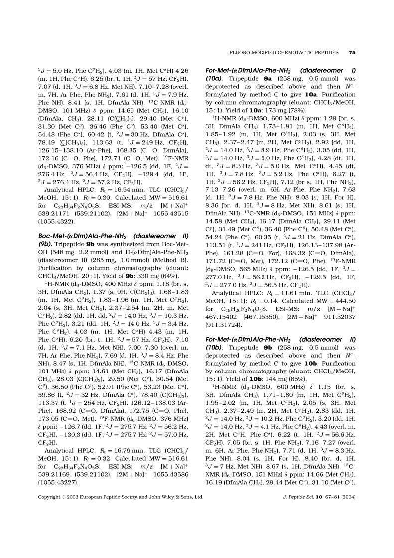
Typical kinetics of the luminol-dependent chemi-luminescence of tripeptide stimulated PMNs areshown in Figure 1.
In particular, the chemotactic tripeptide fMLF isknown to produce several time-resolved maximaof luminescence upon PMN stimulation while theintensity of these maxima depends on cell concen-tration and the cell state [49,50]. In accordancewith those published data, luminescence curves ofthe fluorinated analogues investigated here exhib-ited two maxima under our experimental conditions.The first maximum was usually found during thesecond minute after cell stimulation. In most cases,this maximum looked like a shoulder. The secondmaximum was more evident. With our peptides itwas found during the fifth minute after the additionof the cell stimulator.
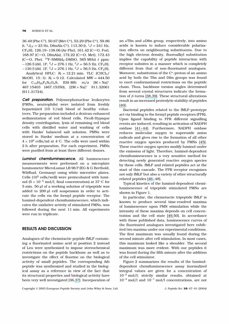
Figure 2 summarizes the results of the luminol-dependent chemiluminescence assay (normalizedintegral values are given for a concentration of10−6 mol/l; strictly similar results, obtained at10−5 mol/l and 10−7 mol/l concentrations, are not
Copyright 2003 European Peptide Society and John Wiley & Sons, Ltd. J. Peptide Sci. 10: 67–81 (2004)
FLUORO-MODIFIED CHEMOTACTIC PEPTIDES 77
0 100-100 200 300 400 500 600 7000
1
2
Lum
inol
CL
(RLU
/s ×
103 )
Time (s)
Figure 1 Kinetics of luminol chemiluminescence of oligopeptide stimulated PMNs. The arrow indicates the time ofstimulation. The curves of the following tripeptides are reported: For-Met-Leu-Phe-NH2 (�), For-Met-(R)-(αTfm)Ala-Phe-NH2
(♦), For-Met-Aib-Phe-NH2 (ž) and For-Met-(αDfm)Ala-Phe-NH2 (diastereomer II) (�). Representative examples from fourdifferent cell preparations are shown.
0
20
40
60
80
100
120
140
Lum
ines
cenc
e (%
)
For
-Met
-(S
)-D
feG
ly-P
he-N
H2
For
-Met
-(R
)-D
feG
ly-P
he-N
H2
For
-Met
-(S
)-(α
Tfm
)Ala
-Phe
-NH
2
For
-Met
-(R
)-(α
Tfm
)Ala
-Phe
-NH
2
For
-Met
-(αD
fm)A
la-P
he-N
H2
(Dia
I)
For
-Met
-(αD
fm)A
la-P
he-N
H2
(Dia
II)
For
-Met
-Aib
-Phe
-NH
2
For
-Met
-Leu
-Phe
-NH
2
Figure 2 Integral values of luminol chemiluminescence of tripeptide stimulated PMNs. The integrals were determined overa time period of 10 min after stimulation. Mean ± SD of four different cell preparations are indicated. All other experimentalconditions were as in Figure 1.
shown) of the fluorinated analogues in comparisonto the parent peptide For-Met-Leu-Phe-NH2 andthe Aib analogue. It is clearly seen that all of the
synthesized analogues were active in generatingreactive oxygen species, although For-Met-Leu-Phe-NH2 yielded by far the most intense response in
Copyright 2003 European Peptide Society and John Wiley & Sons, Ltd. J. Peptide Sci. 10: 67–81 (2004)
78 KOKSCH ET AL.
Figure 3 Crystal structure of 7-(R)-(αTfm)Ala-Phe-NH2 (4a). The intramolecular H-bond is indicated by a dashed line.
0-100 100 200 300 400 500 600 7000
1
2
Time (s)
Lum
inol
CL
(RLU
/s ×
103 )
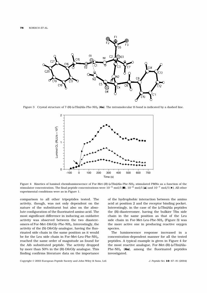
Figure 4 Kinetics of luminol chemiluminescence of For-Met-(R)-(αTfm)Ala-Phe-NH2 stimulated PMNs as a function of thestimulator concentration. The final peptide concentrations were 10−5 mol/l (�), 10−6 mol/l (�) and 10−7 mol/l (♦). All otherexperimental conditions were as in Figure 1.
comparison to all other tripeptides tested. Theactivity, though, was not only dependent on thenature of the substituent but also on the abso-lute configuration of the fluorinated amino acid. Themost significant difference in inducing an oxidativeactivity was observed between the two diastere-omers of For-Met-DfeGly-Phe-NH2. Interestingly, theactivity of the (S)-DfeGly-analogue, having the fluo-rinated side chain in the same position as it wouldbe for the Leu side chain in For-Met-Leu-Phe-NH2,reached the same order of magnitude as found forthe Aib substituted peptide. The activity droppedby more than 50% in the (R)-DfeGly analogue. Thisfinding confirms literature data on the importance
of the hydrophobic interaction between the aminoacid at position 2 and the receptor binding pocket.Interestingly, in the case of the (αTfm)Ala peptidesthe (R)-diastereomer, having the bulkier Tfm sidechain in the same position as that of the Leuside chain in For-Met-Leu-Phe-NH2 (Figure 3) wasthe more active one in producing reactive oxygenspecies.
The luminescence response increased in aconcentration-dependent manner for all the testedpeptides. A typical example is given in Figure 4 forthe most reactive analogue, For-Met-(R)-(αTfm)Ala-Phe-NH2 (4a), among the fluorinated peptidesinvestigated.
Copyright 2003 European Peptide Society and John Wiley & Sons, Ltd. J. Peptide Sci. 10: 67–81 (2004)
FLUORO-MODIFIED CHEMOTACTIC PEPTIDES 79
CONCLUSION
Site-specific incorporated, highly functionalizedamino acids within biologically active peptidescan serve as valuable biophysical probes for theinvestigation of structure–function relationships.Usually, the incorporation of fluorine atoms dra-matically influences the physical properties ofamino acids and proteins as well as providingthe opportunity for studying conformational prop-erties, peptide/protein–membrane interactions, ormetabolic processes by 19F-NMR [26,51]. Fur-thermore, the incorporation of fluoroalkyl aminoacids into peptides is known to result in anincreased resistance towards proteolysis as well asstabilization of secondary structures [40,52].
A small library of chemotactic peptide analoguesof For-Met-Leu-Phe-NH2 substituted at position2 by different fluorinated amino acids varyingin the content of fluorine, the length of thefluorinated side chain, and alkylation degree at theα-carbon atom was synthesized and the influenceof the fluorine substitution on the biologicalactivity was investigated. In a luminol-dependentchemiluminescence assay all of the synthesized For-Met-Leu-Phe-NH2 analogues showed activity in thegeneration of reactive oxygen species. The activitywas not only dependent on the nature of thesubstituent but also on the absolute configurationof the fluorinated amino acid. Remarkably, in thecases of the (αTfm) and DfeGly analogues, thediastereomer having the fluorinated, bulkier sidechain in the same position as it would be for theLeu side chain in For-Met-Leu-Phe-NH2 was themore active one. The (αDfm) Ala analogue seemsto work in the same way; however, the configurationof the fluorinated amino acid has not been provenby crystal structure analysis yet.
This finding shows again the importance of thehydrophobic interaction between the amino acidat position 2 and the receptor binding pocket.Furthermore, these results establish that chemo-tactic peptide analogues which favour folded back-bone conformations are biologically highly active[1,10,37]. In any case, activity of the fluoroalkylsubstituted analogues of For-Met-Leu-Phe-NH2, e.g.in inducing chemotaxis, lysosomal enzyme releaseand histamine release, has to be further investi-gated as it appears that analogues that are highlyactive in generating reactive oxygen species are notnecessarily active in chemotaxis. The Aib-modifiedanalogue investigated here had already been shownto be highly active for the release of lysozyme from
rabbit neutrophils [37]. As the shape of lumines-cence curves for all of the fluoro-modified peptidestested was comparable to that of the Aib analogue, acommon mechanism of cell activation for all of thesestimulators can be assumed. Obviously, the prepa-ration of fluoroalkyl substituted For-Met-Leu-Phe-NH2 analogues resulted in structurally constrainedpeptidomimetics active in the generation of reac-tive oxygen species and bearing a fluorine labelfor NMR spectroscopy. Therefore, these conceptuallynew analogues of the prototype of the chemotacticpeptide family provide the opportunity to study thereceptor bound conformation of these signal pep-tides by 19F-NMR.
Acknowledgements
This work was supported by Deutsche Forschungs-gemeinschaft (Bu 277-22-1) and the Fonds of theChemical Industry.
REFERENCES
1. Becker EL, Freer RJ, Toniolo C, Balaram P. Thespecificity of the chemotactic formyl peptide receptorof rabbit neutrophils. In Membrane Receptors andCellular Regulation, Czech MP, Kahn CR (eds). Liss:New York, 1985; 129–134.
2. Tornoe CW, Sengelov H, Meldal M. Solid-phasesynthesis of chemotactic peptides using α-azido acids.J. Peptide Sci. 2000; 6: 314–320.
3. Pirrung MC, Drabik SJ, Ahamed J, Ali H. Cagedchemotactic peptides. Bioconj. Chem. 2000; 11:679–681.
4. Schiffmann E, Corcoran BA, Wahl SM. N-Formyl-methionyl peptides as chemoattractants forleukocytes. Proc. Natl. Acad. Sci. USA 1975; 72:1059–1062.
5. Schiffmann E, Showell HJ, Corcoran BA, Ward PA,Smith E, Becker EL. The isolation and partialcharacterization of neutrophil chemotactic factorsfrom Escherichia coli. J. Immunol. 1975; 114:1831–1837.
6. Toniolo C, Formaggio F, Crisma M, ValleG, Boesten WHJ, Schoemaker HE, Kamphuis J,Temussi PA, Becker EL, Precigoux G. Bioactive andmodel peptides characterized by the helicogenic(αMe)Phe residue. Tetrahedron 1993; 49: 3641–3653.
7. Becker EL. The formylpeptide receptor of theneutrophil. Am. J. Path. 1987; 129: 15–24.
8. McPhail LC, Harvath L. Signal transduction inneutrophil oxidative metabolism and chemotaxis.
Copyright 2003 European Peptide Society and John Wiley & Sons, Ltd. J. Peptide Sci. 10: 67–81 (2004)
80 KOKSCH ET AL.
In The Natural Immune System: The Neutrophil,Abramson JS, Wheeler JG (eds). Oxford UniversityPress: Oxford, 1993; 63–77.
9. Torrini I, Zecchini GP, Paradisi MP, Lucente G,Gavuzzo E, Mazza F, Pochetti G, Spisani F, Guliani AL.Synthesis and properties of chemotactic peptideanalogs. II. HCO-Met-Leu-Phe-OMe analogs contain-ing cyclic α, α-disubstituted amino acids as Met andPhe mimicking residues. Int. J. Peptide Protein Res.
1991; 38: 495–504.10. Toniolo C, Crisma M, Valle G, Bonora GM, Polinelli S,
Becker EL, Freer RJ, Prasad S, Roa RB, Balaram P,Sukumar M. Conformationally restricted formylmethionyl tripeptide chemoattractants: a three-dimensional structure-activity study of analogsincorporating a Cα,α-dialkylated glycine at position 2.Peptide Res. 1989; 2: 275–281.
11. Benedetti E. X-ray crystallography of peptides.Biopolymers (Peptide Sci.) 1996; 40: 3–44.
12. Sukumar MP, Raz AP, Balaram P, Becker EL. A highlyactive chemotactic peptide analog incorporating theunusual residue 1-aminocyclohexanecarboxylic acidat position 2. Biochem. Biophys. Res. Commun. 1985;128: 339–344.
13. Prasad S, Rao RB, Bergstrand H, Lundquist B,Becker EL, Balaram P. Conformation-activity correla-tions of chemotactic tripeptide analogs incorporatingdialkyl residues with linear and cyclic alkyl sidechainsat position 2. Int. J. Peptide Protein Res. 1996; 48:312–318.
14. Freer RJ, Day AR, Schiffmann E, Aswanikumar S,Showell HJ, Becker EL. Further studies on the struc-tural requirements for synthetic peptide chemoattrac-tants. Biochemistry 1980; 19: 2404–2409.
15. Freer RJ, Day AR, Muthukumaraswamy N, Pinon D,Wu A, Showell HJ, Becker EL. Formyl peptidechemoattractants. A model of the receptor in rabbitneutrophils. Biochemistry 1982; 21: 257–263.
16. Day AR, Radding JA, Freer RJ, Showell HJ, Becker EL,Schiffmann E, Corcoran B. Synthesis and bindingcharacteristics of an intrinsically radio-labeled chemo-tactic acyl tripeptide: Nα-formyl-norleucyl-leucyl-phenylalanine. FEBS Lett. 1977; 77: 291–294.
17. Shimohigashi Y. In Opioid Peptides, Medicinal
Chemistry, Rapka RS, Bernett G, Hawks RL (eds).NIDA Research Monograph 1986; 69: 65.
18. Hruby VJ, Li G, Haskell-Luevano C, Shenderovich M.Design of peptides, proteins, and peptidomimetic inChi space. Biopolymers 1997; 43: 219–266.
19. Fujita T, Nose T, Matsushima A, Okada K, Asai D,Yamauchi Y, Shirasu N, Honda T, Shigehiro D,Shimohigashi Y. Synthesis of a complete set ofL-difluorophenylalanines, L-(F2)Phe, as molecularexplorers for the CH/π interaction between peptideligand and receptor. Tetrahedron Lett. 2000; 41:923–927.
20. Nishio M, Umezawa Y, Hirota M, Takeuchi Y. TheCH/π interaction: significance in molecularrecognition. Tetrahedron 1995; 51: 8665–8701.
21. Erickson JA, Mc Loughlin JI. Hydrogen bond donorproperties of the difluoromethyl group. J. Org. Chem.1995; 60: 1626–1631.
22. Caminati W, Melandri S, Moreschini P, Favero PG. TheC-F· · ·H-C ‘anti-hydrogen bond’ in the gas phase:microwave structure of the difluoromethane dimer.Angew. Chem. Int. Ed. 1999; 38: 2924–2925.
23. Howard JAK, Hoy VJ, O’Hagan D, Smith GT. Howgood is fluorine as hydrogen bond acceptor.Tetrahedron 1996; 52: 12 613–12 622.
24. O’Hagan D, Rzepta HS. Some influences of fluorine inbioorganic chemistry. J. Chem. Soc., Chem. Commun.1997; 645–652.
25. Dunitz JD, Taylor R. Organic fluorine hardly everaccepts hydrogen bonds. Chem. Eur. J. 1997; 3:89–98.
26. Tang Y, Ghirlanda G, Vaidehi N, Kua J, Mainz TD,Goddard WA, III, Degrado WF, Tirrell DA. Stabilizationof coiled-coil peptide domains by introduction oftrifluoroleucine. Biochemistry 2000; 40: 2790–2796.
27. Degrado WF, Summa CM, Pavone V, Nastri F, Lom-bardi A. De novo design and structural character-ization of proteins and metalloproteins. Annu. Rev.Biochem. 1999; 68: 779–819.
28. Winkler D, Burger K. Synthesis of enantiomericallypure D- and L-armentomycin and its difluoro analoguesfrom aspartic acid. Synthesis 1996; 1419–1421.
29. Sewald N, Hollweck W, Mutze K, Schierlinger K, Sey-mour LC, Gaa K, Burger K, Koksch B, Jakubke HD.Peptide modification by introduction of α-trifluoromethyl substituted amino acids. Amino Acids1995; 8: 187–195.
30. Sewald N, Burger K. Synthesis of β-fluoro-containingamino acids. In Fluorine-containing Amino Acids,Synthesis and Properties, Kukhar VP, Soloshonok VA(eds). Wiley: Chichester 1995; 139–162.
31. Osipov SN, Golubev AS, Sewald N, Michel T, KolomietsAF, Fokin AV, Burger K. A new strategy for thesynthesis of β-difluoromethyl substituted α-hydroxyand α-amino acids. J. Org. Chem. 1996; 61:7521–7528.
32. Preliminary results have been published: Moroni M,Koksch B, Burger K. Analogues of chemotacticpeptides containing fluorinated amino acids. Peptides;Proceedings of the 26th European Peptide Symposium,Martinez J, Fehrentz J-A (eds.). EDK: Paris 2001;689–690.
33. Duczek W, Deutsch J, Vieth S, Niclas H-S. A simpleand convenient synthesis of N-formyl amino acidesters under mild conditions. Synthesis 1996; 37–38.
34. Sheldrick GM. SHELXS 97. Program Used to Solve andto Refine Structure. University of Gottingen: Gottingen,Germany, 1997.
35. Crystallographic data have been deposited atthe Cambridge Crystallographic Data Centre,
Copyright 2003 European Peptide Society and John Wiley & Sons, Ltd. J. Peptide Sci. 10: 67–81 (2004)
FLUORO-MODIFIED CHEMOTACTIC PEPTIDES 81
CCDC210454. Copies of this information may beobtained free of charge from The Director, CCDC,12 Union Road, Cambridge, CB2 1EZ, UK (fax:+44 1233 336033, e-mail: [email protected] orhttp://www.ccdc.cam.ac.uk).
36. Bardi R, Piazzesi AM, Toniolo C, Antony Raj P,Raghothama S, Balaram P. Solid state and solutionconformation of Boc-L-Met-Aib-L-Phe-OMe. Int. J.Peptide Protein Res. 1986; 27: 229–238.
37. Iqbal M, Balaram P, Showell HJ, Freer RJ, Becker EL.Conformationally constrained chemotactic peptideanalogs of high biological activity. FEBS Lett. 1984;165: 171–174.
38. Koksch B, Sewald N, Burger K, Jakubke H-D. Peptidemodification by incorporation of α-trifluoromethylsubstituted amino acids. Amino Acids 1996; 11:425–434.
39. Michel T, Koksch B, Osipov SN, Golubev AN, Sieler J,Burger K. Peptide synthesis with α-(difluoromethyl)-substituted amino acids. Coll. Czech. Chem. Commun.2000; 67: 1533–1553.
40. Koksch B, Sewald N, Hofmann H-J, Burger K,Jakubke H-D. Proteolytically stable peptides byincorporation of α-Tfm amino acids. J Peptide Sci.1997; 3: 157–167.
41. Watson F, Robinson JJ, Edwards SW. Sequentialphospholipase activation in the stimulation of theneutrophil NADPH-oxidase. FEMS Microbiol. Immunol.1992; 105: 239–248.
42. Curnutte JT, Erikson RW, Ding J, Badwey JA.Reciprocal interactions between protein kinase Cand components of the NADPH-oxidase complexmay regulate superoxide production by neutrophilsstimulated by a phorbol ester. J. Biol. Chem. 1994;269: 10 813–10 819.
43. Waite KA, Wallin R, Qualliotine-Mann D, McPhail LC.Phosphatidic acid-mediated phosphorylation of theNADPH oxidase component p47phox. J. Biol. Chem.1997; 272: 15 569–15 578.
44. Arnhold J, Benard S, Kilian U, Reichl S, Schiller J,Arnold K. Modulation of luminol chemiluminescenceof fmet-leu-phe-stimulated neutrophils by affectingdephosphorylation and the metabolism of phospha-tidic acid. Luminescence 1999; 14: 1–9.
45. Segal AW, Abo A. The biochemical basis of the NADPHoxidase of phagocytes. Trends Biochem. Sci. 1993; 18:43–47.
46. Le Y, Yang Y, Cui Y, Yazawa H, Gong W, Qiu C,Wang JM. Receptors for chemotactic formyl peptidesas pharmacological targets. Int. Immunopharmacol.2002; 2: 1–13.
47. Le Y, Oppenheim JJ, Wang JM. Pleiotropic roles offormyl peptide receptors. Cytokine Growth Factor Rev.2001; 12: 91–105.
48. Dalpiaz A, Scatturin A, Vertuani G, Pecoraro R,Borea PA, Varani K, Traniello S, Spisani S. Met-Ile-Phe-Leu derivatives: full and partial agonists of humanneutrophil formylpeptide receptors. Eur. J. Pharmacol.2001; 411: 327–333.
49. Bender JG, van Epps DE. Analysis of the bimodalchemiluminescence pattern stimulated in humanneutrophils by chemotactic factors. Infect. Immunol.1983; 41: 1062–1070.
50. Dahlgren C. Polymorphonuclear leukocyte chemi-luminescence induced by formylmethionyl-leucyl-phenylalanine and phorbol myristate acetate: effectsof catalase and superoxide dismutase. Agent Actions1987; 21: 104–112.
51. Ulrich AS. In Encyclopedia of Spectroscopy andSpectrometry, Lindon J, Tranter G, Holmes J (eds).Academic Press: New York, 2000; 813.
52. Koksch B, Sewald N, Jakubke H-D, Burger K. InBiomedical Frontiers of Fluorine Chemistry, Ojima I,McCarthy JR, Welch JT (eds). ACS Symposium Series,American Chemical Society: Washington, DC, 1996;639: 42–58.
Copyright 2003 European Peptide Society and John Wiley & Sons, Ltd. J. Peptide Sci. 10: 67–81 (2004)