lipophilic guanylhydrazone analogues as promising
TRANSCRIPT
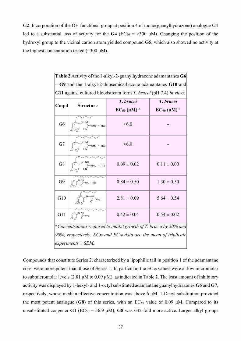
1
Lipophilic Guanylhydrazone Analogues As Promising Trypanocidal Agents: An Extended
SAR Study
Vasiliki Pardalia, Erofili Giannakopouloua, Dimitrios-Ilias Balourdasa, Vassilios Myrianthopoulosa,
Martin C. Taylorb, Marina Šekutorc, Kata Mlinarić-Majerskic, John M. Kellyb, and Grigoris Zoidisa,*
aSchool of Health Sciences, Faculty of Pharmacy, Department of Pharmaceutical Chemistry,
National and Kapodistrian University of Athens, Panepistimiopolis-Zografou, GR-15771 Athens,
Greece; bDepartment of Infection Biology, London School of Hygiene and Tropical Medicine,
Keppel Street, London WC1E 7HT, UK; cDepartment of Organic Chemistry and Biochemistry,
Ruđer Bošković Institute, Bijenička cesta 54, 10 000 Zagreb, Croatia
*Corresponding author:
Prof. Grigoris Zoidis ([email protected])
Keywords: Adamantane, S-adenosylmethionine decarboxylase (AdoMetDC), guanylhydrazones,
structure-activity relationships, trypanocidal agents, Kernel-based Partial Least Squares regression,
SZmap, Hydration analysis, Docking-scoring calculations
Abstract
In this report, we extend the SAR analysis of a number of lipophilic guanylhydrazone analogues with
respect to in vitro growth inhibition of Trypanosoma brucei and Trypanosoma cruzi. Sleeping
sickness and Chagas disease, caused by the tropical parasites T. brucei and T. cruzi, constitute a
significant socioeconomic burden in low-income countries of sub-Saharan Africa and Latin America,
respectively. Drug development is under-funded. Moreover, current treatments are outdated and
difficult to administer, while drug resistance is an emerging concern. The synthesis of adamantane-
based compounds that have potential as antitrypanosomal agents, is extensively reviewed. The critical
role of the adamantane ring was further investigated by synthesizing and testing a number of novel
lipophilic guanylhydrazones. The introduction of hydrophobic bulky substituents onto the
adamantane ring generated the most active analogues, illustrating the synergistic effect of the
lipophilic character of the C1 side chain and guanylhydrazone moiety on trypanocidal activity. The
n-decyl C1-substituted compound G8 (R = C10H21) proved to be the most potent adamantane
derivative against T. brucei with activity in the nanomolar range (EC50=90 nM). Molecular

2
simulations were also performed to better understand the structure-activity relationships between the
studied guanylhydrazone analogues and their potential enzyme target.
1. Introduction
Neglected tropical diseases (NTDs) constitute a diverse group of chronic bacterial, protozoan, fungal,
viral and helminth infections [1,2] in 149 countries worldwide [3]. It is estimated that more than a
billion people — one-sixth of the world’s population, are affected by NTDs in tropical and sub-
tropical regions. These infections are considered to be one of the leading causes of high morbidity
and mortality in underdeveloped countries [4]. Leishmaniasis, Human African Trypanosomiasis
(HAT), Chagas disease and malaria are the most serious and prominent protozoan infections,
accounting for the highest death toll among the existing NTDs annually [5,6]. In this review, we will
focus on the two trypanosomiases, HAT and Chagas disease, caused by protozoan parasites of the
genus Trypanosoma.
1.1 Human African trypanosomiasis
Human African trypanosomiasis (HAT), also known as sleeping sickness, is a vector-borne disease
that affects poor populations in the African continent. It is caused by infection with the protozoan
parasite Trypanosoma brucei and is transmitted by the bite of tsetse flies of the genus Glossina [7].
The disease is found in 36 countries of sub-Saharan Africa and about 65 million people are at risk of
developing the infection [8].
Two morphologically identical subspecies of T. brucei are responsible for the onset of the disease in
humans. They display a different host range, exhibit dissimilar virulence profiles, occur in separate
geographical regions, and display distinct epidemiological characteristics and clinical manifestations
[9]. Trypanosoma brucei rhodesiense causes an acute and rapidly progressing form of the disease,
known as rhodesiense HAT (rHAT), which is localized to 13 countries in eastern and southern Africa
[10]. It is a zoonotic disease [11], that is transmitted by tsetse flies of the Glossina morsitans group
3
[12] and passes directly or indirectly from animals or insects to humans. Trypanosoma brucei
gambiense leads to a slow-progressing and chronic form of the disease, Gambian sleeping sickness
or gambiense HAT (gHAT), which is found predominantly in 24 countries of western and central
Africa [10]. The palpalis group is the vector of T. b. gambiense, with the main carriers being Glossina
palpalis palpalis and Glossina palpalis gambiensis [13]. gHAT is an anthroponotic disease which is
transmitted predominantly from person to person, although pigs and some wild animals have been
reported as smaller reservoirs [11]. Infections from T. b. gambiense are the most common and
represent over 97% of current cases [9].
Three severe outbreaks of the disease have been recorded in Africa. The first occurred between 1896
and 1906 with 800,000 deaths. The second began in 1920, peaking in 1930 with 70,000 registered
cases, and ended in the late 1940s. The most recent widespread outbreak began in the 1970s and
lasted until the end of the 1990s [14,15]. Incidence data from the World Health Organization (WHO)
for 2009 showed a total of 9,878 new cases, with a sharp drop off observed in 2014. Nowadays, only
the Democratic Republic of the Congo (DRC) reports an average of more than 1,000 new cases per
year, comprising over 70% of the total recorded T. b. gambiense cases. However, the real number of
people affected by this disease is estimated to be much higher due to under-reporting [8,16,17].
Tsetse flies are hematophagous, cyclical vectors of HAT [18]. Congenital transmission has also been
demonstrated in newborn children within the first days of life and in older children born in
nonendemic regions from infected mothers [19-22]. Transmission through sexual contact has been
described once when the infection was passed from a male with confirmed gHAT to a woman who
had never been to an endemic country [21-23]. Furthermore, mechanical transmission has also been
reported via other blood-sucking insects, whereas accidental infections may occur from needle stick
injuries in laboratories, or through blood transfusion [18,23].
HAT is clinically defined by two distinct stages. During the early hemolymphatic stage, trypanosomes
spread and proliferate in the blood and the lymph. In the second or late meningoencephalitic stage,
parasites are able to cross the blood-brain barrier (BBB) and invade the central nervous system (CNS)
[16]. Although the progression of each stage involves different clinical features, it is often difficult to
distinguish reliably between the two stages and they merge into each other, especially upon infection
with T. b. rhodesiense. rHAT progresses rapidly and parasite invasion of the CNS occurs in a few
weeks, causing death within months of infection. On the other hand, the neurological stage of gHAT
generally starts several months or even years after infection, and the disease can take years or decades
before becoming lethal [9,24].
After the bite from a tsetse fly, a chancre also known as a trypanome – a local skin reaction, develops
at the feeding point. This occurs in about 20% of patients with rHAT, but is rarely observed in gHAT
cases [10,11,25]. After an initial asymptomatic period that lasts one to three weeks in rhodesiense

4
infection, and usually some weeks longer in gHAT, patients develop nonspecific symptoms such as
fever with chills, fatigue, headache, arthralgia, weight loss and a general sense of discomfort and
unease. Splenomegaly, or less frequently hepatomegaly, has been observed, while enlargement of the
posterior cervical lymph nodes, known as Winterbottom’s sign, is common in Gambian sleeping
sickness [10,11,16]. The meningoencephalitic stage involves symptoms from almost all regions of
the nervous system. These clinical manifestations are categorized by psychiatric, motor, sensory
disturbances and sleep abnormalities, the main features that gave the disease its name. Deep and
painful hyperesthesia is the leading sensory symptom, known as Kerandel’s sign. The disease causes
deregulation of the circadian clock characterized by daytime somnolence followed by nocturnal
insomnia. As the disease progresses, patients display an uncontrollable desire to sleep and alterations
of sleep structure. During the final stage, cerebral edema, coma and systemic organ failure are
observed, leading to sudden death [10,11,16,26-28].
The first issue in HAT diagnosis is to identify the disease as soon as possible by confirming the
presence of parasites in the peripheral blood, or in other infected tissues such as the lymph nodes.
Subsequently, the key step constitutes the accurate staging of the disease [29]. Τhe non-specific signs
and symptoms are insufficient for an accurate diagnosis. In rHAT, identification of trypanosomes is
feasible because of the high levels of parasitemia, by microscopic examination of blood, or by a
lymph node aspirate. The presence of the trypanosomal chancre is also typical of infection with T. b.
rhodesiense. In contrast, gambiense infection is typified by cyclical parasitemia, reflecting a process
of antigenic variation by the parasite, and parasitological diagnosis has poor sensitivity [7,24].
However, rapid and reliable serological tests (e.g., Card Agglutination Test for Trypanosomiasis
(CATT), HAT Sero-K-SeT, SD Bioline HAT), based on the detection of specific antibodies, can be
used for mass population screening [16,30,31]. Identification of the late stage of both types of HAT
is based on the examination of cerebrospinal fluid (CSF) collected by lumbar puncture. WHO has
defined 5 white blood cells (WBC) per μL as a threshold, or the presence of trypanosomes in the CSF,
or both, as indicators that the infection has progressed to the CNS-stage. In gambiense infections, 20
cells per μL is used as the cutoff point [30]. Concentrations of immunoglobulin M (IgM) in the CSF,
neopterin and combined panels of cytokines and chemokines, released after the tissue damage, have
also been evaluated as biomarkers of HAT, and they reflect the severity of neurological signs [32,33].
Recognition of the causative T. brucei sub-species and correct staging of the disease are critical for
choosing the appropriate chemotherapeutic regimen [10,34]. Drugs used for the first-stage disease
are not able to cure second-stage disease, since therapy against the late stage requires drugs that travel
across the BBB. Treatment of the first stage of sleeping sickness is limited to two drugs, pentamidine
and suramin [35].
5
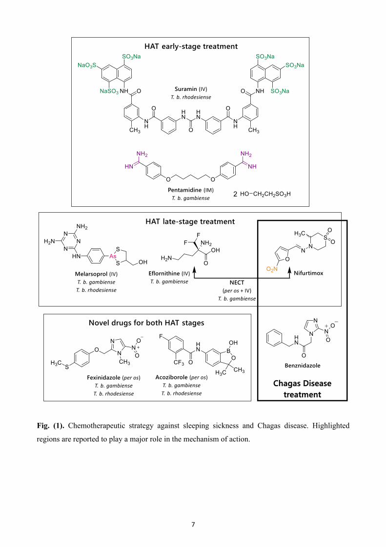
Pentamidine isethionate (Fig. (1)) is an aromatic diamidine and was registered as the drug of choice
for the treatment of infection caused by T. b. gambiense in 1940. As a positively charged molecule,
pentamidine interacts electrostatically with cellular polyanions. The drug seems to bind to the minor
groove of DNA and to be involved in the disruption of both kinetoplast (kDNA) and nuclear DNA.
kDNA is thought to be the primary target with drug activity linked to inhibition of the mitochondrial
topoisomerase II. It has also been suggested that diamidines may act through other mechanisms, such
as inhibition of the enzyme S-adenosylmethionine decarboxylase (AdoMetDC), or by deregulation
of oxidative phosphorylation. [34,36-40].
Suramin (Fig. (1)) is a polysulphonated naphthylamine and has been used as a standard treatment for
the hemolymphatic stage of rHAT. Suramin appears to bind to glycolytic enzymes through
electrostatic interactions and inhibit them, thus interrupting glycolysis. In a recent study,
dysregulation in cytokinesis was observed following suramin treatment, while parasites with more
than two nuclei were abundant, indicating a possible new mode of action. Anaphylactic shock, renal
failure, skin lesions, neurological complications, such as peripheral neuropathy, and bone marrow
toxicity have been reported as the main adverse reactions [34,36,37,39,40].
Melarsoprol (Fig. (1)) is a trivalent arsenical and the only effective drug for the second stage of both
forms of sleeping sickness. It was first used in 1949. Melarsoprol is a prodrug which is converted, in
trypanosomes, into the active form melarsen oxide. The active metabolite seems to engage in stable
interactions with sulfhydryl groups and has a high affinity for thiols such as trypanothione, affecting
the redox (reduction–oxidation) balance of the parasites. It also interacts with a variety of glycolytic
and pentose phosphate pathway enzymes, and thus, inhibits ATP synthesis. The use of melarsoprol
is linked with a high frequency of severe and life-threatening adverse effects, with an overall mortality
rate of 5%. This drug is very toxic, and patients may develop a severe post-treatment reactive
encephalopathy (PTRE) with mortality rates that climb to 50%. [9,34,36,37,39,40].
Eflornithine (Fig. (1)) was originally registered as an anticancer drug, but in the 1980s, it was found
to be an effective therapeutic solution for the second stage of gHAT. It is a specific and irreversible
inhibitor of ornithine decarboxylase (ODC), the first enzyme involved in the biosynthesis of
polyamines, which are essential for cell growth and proliferation. The main adverse effects reported
are fever, pruritus, hypertension, bone marrow suppression, gastrointestinal symptoms and rarely,
seizures. [34,36,37,39,40].
Combination regimens of melarsoprol, eflornithine and nifurtimox were assessed for the treatment of
HAT. Nifurtimox is registered to treat patients with American trypanosomiasis (below). Nifurtimox-
eflornithine combination therapy (NECT) (Fig. (1)) was found to be the most effective and now
constitutes the first-line therapy for the late stage of gambiense infection. Nifurtimox is a prodrug that
6
is bioactivated through reduction of the nitro group, yielding nitro-anion radicals and other active
metabolites. NECT is based on the synergistic actions of the two drugs; nifurtimox leads to oxidative
stress and widespread damage to macromolecules, while eflornithine reduces the levels of
trypanothione, the key redox-protecting metabolite. Administration includes oral nifurtimox with a
daily dose of 15 mg/kg, three times a day for 10 days, and eflornithine infusions of 400 mg/kg per
day given every 12 hours for 7 days. NECT produced a marked improvement in terms of
administration (56 infusions reduced to 14), high cure rates, low mortality rates and less adverse side
effects, especially compared to melarsoprol. Shorter hospitalization is also required, and the cost of
the therapeutic schedule has been significantly reduced. NECT-treated patients may be affected by
musculoskeletal and abdominal pain, tremors, vomiting and nausea, headache and gastrointestinal
disorders [36,41-43].
Two new drugs are currently undergoing clinical trials, fexinidazole and acoziborole (Fig. (1)).
Fexinidazole is a prodrug that is rapidly metabolized in vivo – through two different pathways,
cytochrome P450 and a monooxygenase – into two metabolites, a sulfoxide and a sulfone, both of
which exhibit trypanocidal activity. It is the only orally administered drug that is effective against
both stages of HAT caused by T. b. gambiense and T. b. rhodesiense. This drug has been deemed
broadly acceptable due to the easy administration (one oral dose per day for 10 days), its safety
profile, its reduced cost in terms of synthesis, and the fact that there is no requirement for disease
staging. Fexinidazole has received marketing authorization in the DCR for the treatment of gHAT,
and has successfully entered phase 3 and phase 2 clinical trials in patients infected with gHAT of any
stage and chronic Chagas disease, respectively [44-47].
Acoziborole (SCYX-7158) is another oral drug candidate that has been tested as a treatment for T. b.
gambiense and T. b. rhodesiense infections. After phase 1 clinical trial, completed in 2015, it was
confirmed that the drug is able to access the CNS, so may have potential against both disease stages.
Phase IIb/III clinical trials have been ongoing in the DCR since 2016 [46-48].
A series of amidine-containing compounds have also been studied for their effect on T. brucei and
some of them were evaluated in animal models and/or in clinical trials. However, the trials were
discontinued due to high toxicity.
7
Fig. (1). Chemotherapeutic strategy against sleeping sickness and Chagas disease. Highlighted
regions are reported to play a major role in the mechanism of action.
8
1.2 American trypanosomiasis
Chagas disease, or American trypanosomiasis, is a systemic, life-threatening zoonotic infection [49],
caused by the protozoan parasite Trypanosoma cruzi. The disease is endemic in 21 continental Latin
American countries, with an estimated 6-8 million people infected, mainly in poor rural areas,
resulting in over 10,000 deaths annually. Chagas disease is also becoming a global public health
problem. The migration of chronically infected individuals has spread the disease beyond its natural
geographical boundaries, particularly to the USA and Europe [50]. The main route of transmission to
humans is via the blood-sucking insect vector. Infection begins after mucous membranes,
conjunctivae, or breaks in the skin become contaminated with parasites contained in the faeces of
various species of the triatomine bug. However, other routes of transmission also exist; these include
blood transfusion, organ transplantation, and vertical transmission from mother to infant [51,52].
Numerous outbreaks of the disease related to oral transmission, via food or liquid products
contaminated with T. cruzi, have also been reported [49]. T. cruzi is able to invade almost all nucleated
mammalian cells and has a wide host range.
Progression of the disease is characterized by two distinct clinical phases: an acute phase, and a
chronic phase that develops over many years. The acute phase has a duration of 4-8 weeks and occurs
shortly after the initial infection. This first stage is asymptomatic in 60 to 70% of patients. In the
others, symptoms include inflammation at the inoculation site, fever, enlarged lymph nodes,
splenomegaly and the highly characteristic unilateral palpebral oedema (Romaña sign). The infection
is considered to be life-long, with 30% to 40% of patients subsequently developing chronic stage
pathology, often 20 to 30 years after the acute infection. Symptoms can include cardiac disorders,
mainly cardiomyopathy and arrhythmias, as well as gastrointestinal and/or neurological alterations
that can result in death [51,53].
Currently, two drugs are available for the treatment of Chagas disease; nifurtimox (see above) and
the most commonly used, benznidazole (BZ) (Fig. (1)). Recent findings suggest that BZ is responsible
for the induction of potentially lethal double-stranded breaks in parasite DNA. This mechanism of
action is based on reactive metabolites that are generated following nitroreductase mediated drug
activation, which can damage DNA and other macromolecules, and result in the incorporation of
oxidized nucleotides during DNA replication [54]. However, both drugs are only partially effective
and relatively toxic, which, along with the prolonged administration required, limit their use [55].
There are no vaccines available against these two parasitic diseases. Vaccine development remains a
daunting challenge, particularly in the case of HAT, where antigenic variation results in frequently
changing surface glycoproteins that facilitate immune evasion. Thus, the major ways to prevent
disease transmission are vector control and effective treatment. New and improved therapeutic
options for both sleeping sickness and Chagas disease have been limited. The available drugs have
9
been in use for many years and are associated with poor efficacy, emergence of resistance, difficulties
with administration, and severe adverse effects. Despite the progress made in the development of
fexinidazole and, to a lesser extent, of acoziborole, both diseases continue to compromise public
health in some of the poorest regions of the world. More efforts, focused on the discovery of new
classes of therapeutics, are required [46,56,57].
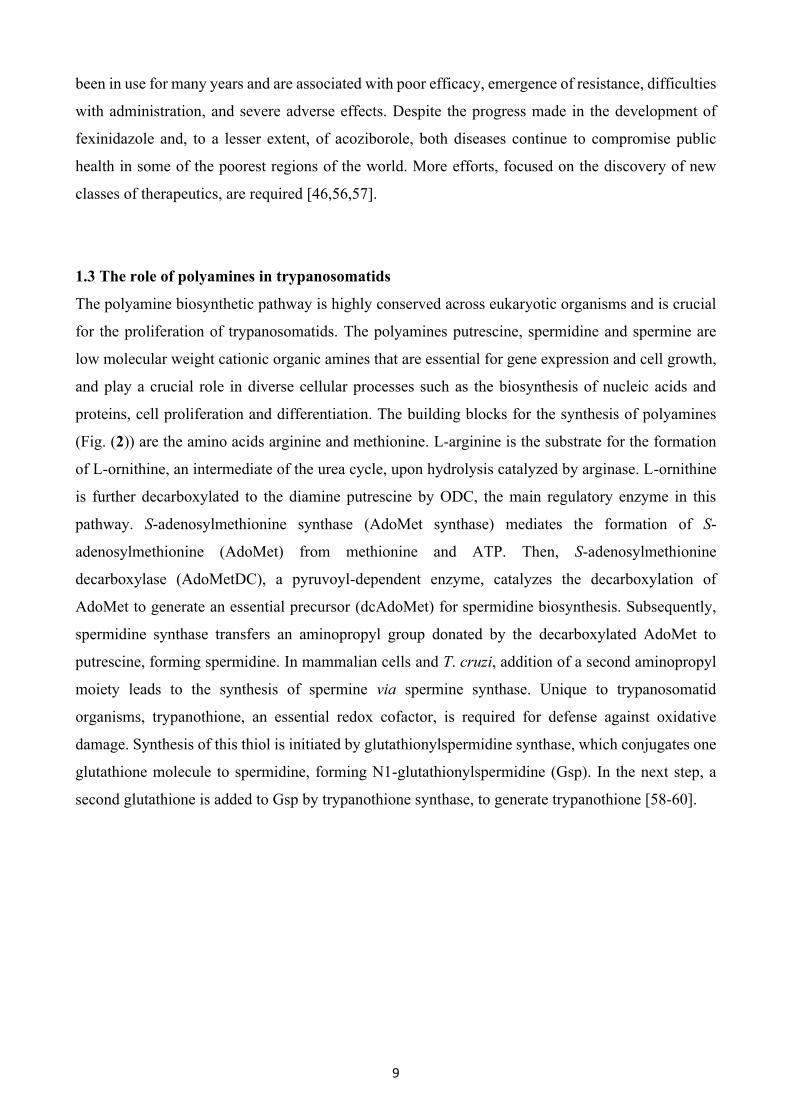
1.3 The role of polyamines in trypanosomatids
The polyamine biosynthetic pathway is highly conserved across eukaryotic organisms and is crucial
for the proliferation of trypanosomatids. The polyamines putrescine, spermidine and spermine are
low molecular weight cationic organic amines that are essential for gene expression and cell growth,
and play a crucial role in diverse cellular processes such as the biosynthesis of nucleic acids and
proteins, cell proliferation and differentiation. The building blocks for the synthesis of polyamines
(Fig. (2)) are the amino acids arginine and methionine. L-arginine is the substrate for the formation
of L-ornithine, an intermediate of the urea cycle, upon hydrolysis catalyzed by arginase. L-ornithine
is further decarboxylated to the diamine putrescine by ODC, the main regulatory enzyme in this
pathway. S-adenosylmethionine synthase (AdoMet synthase) mediates the formation of S-
adenosylmethionine (AdoMet) from methionine and ATP. Then, S-adenosylmethionine
decarboxylase (AdoMetDC), a pyruvoyl-dependent enzyme, catalyzes the decarboxylation of
AdoMet to generate an essential precursor (dcAdoMet) for spermidine biosynthesis. Subsequently,
spermidine synthase transfers an aminopropyl group donated by the decarboxylated AdoMet to
putrescine, forming spermidine. In mammalian cells and T. cruzi, addition of a second aminopropyl
moiety leads to the synthesis of spermine via spermine synthase. Unique to trypanosomatid
organisms, trypanothione, an essential redox cofactor, is required for defense against oxidative
damage. Synthesis of this thiol is initiated by glutathionylspermidine synthase, which conjugates one
glutathione molecule to spermidine, forming N1-glutathionylspermidine (Gsp). In the next step, a
second glutathione is added to Gsp by trypanothione synthase, to generate trypanothione [58-60].
10
Fig. (2). Polyamine and trypanothione biosynthetic pathway.
In general, the enzymes involved in polyamine biosynthesis differ between mammalian cells and
parasites in terms of their structural features, or their presence or absence from the host. Therefore,
they emerge as potential targets for the development of trypanocidal agents. Of these enzymes, ODC
and AdoMetDC, which regulate polyamine homeostasis, constitute druggable targets [61-63]. As
previously described, ODC is inhibited irreversibly by eflornithine, and other approaches toward the
inhibition of ODC in T. brucei are being considered. T. cruzi lacks this enzyme [64]. However, both
11
parasites do express AdoMetDC. Inhibitors of AdoMetDC have been reported that have potent
activity against T. brucei and T. cruzi in vitro, as well as in mouse models [65-71]. The
trypanosomatid AdoMetDC differs from the respective human enzyme, as it possesses a unique
regulatory mechanism. The enzyme is allosterically activated by formation of a heterodimer between
the active subunit and a catalytically inactive paralogous pseudoenzyme, referred to as a prozyme.
Heterodimerization increases enzyme activity by three orders of magnitude. This novel regulation of
the polyamine pathway in trypanosomatids suggests that selective inhibition of T. brucei AdoMetDC
may be feasible, supporting the value of the target to combat HAT [72].
1.4 Drug design
Adamantane
The adamantane scaffold is a polycyclic cage moiety, characterized by high symmetry and a unique
geometry of three fused cyclohexanes in a chair-like conformation. It is a rigid and practically strain-
free structure that has been applied as a valuable structural subunit in medicinal chemistry for many
years [73]. The adamantane moiety can either be incorporated into existing drugs to improve their
pharmacokinetic and pharmacodynamic profile, or used as a starting compound for the development
of novel therapeutic agents. Therefore, it has been widely used in drug discovery strategies. More
specifically, it has been demonstrated that the addition of the adamantyl moiety into a molecule
enhances its overall lipophilicity and subsequently increases cell membrane permeability of the drug
candidate [73-75]. Another example of adamantane’s usefulness in medicinal chemistry is the
increased affinity for lipophilic receptors that it confers, presumably due to greater hydrophobic
interactions, giving rise to higher selectivity. Adamantane can be used to add steric bulk to a
compound and to protect nearby functional groups from metabolic cleavage, thus offering greater
stability and prolonged duration of action [73,75,76]. Finally, due to its rigidity and bulkiness, this
cage scaffold can accommodate β-cyclodextrins (β-CD), resulting in inclusion complexes, or act
mechanically as a blocker for several ion channels [77,78]. Because of all the above, the adamantyl
subunit constitutes a privileged building block that provides drug-like properties to lead compounds.
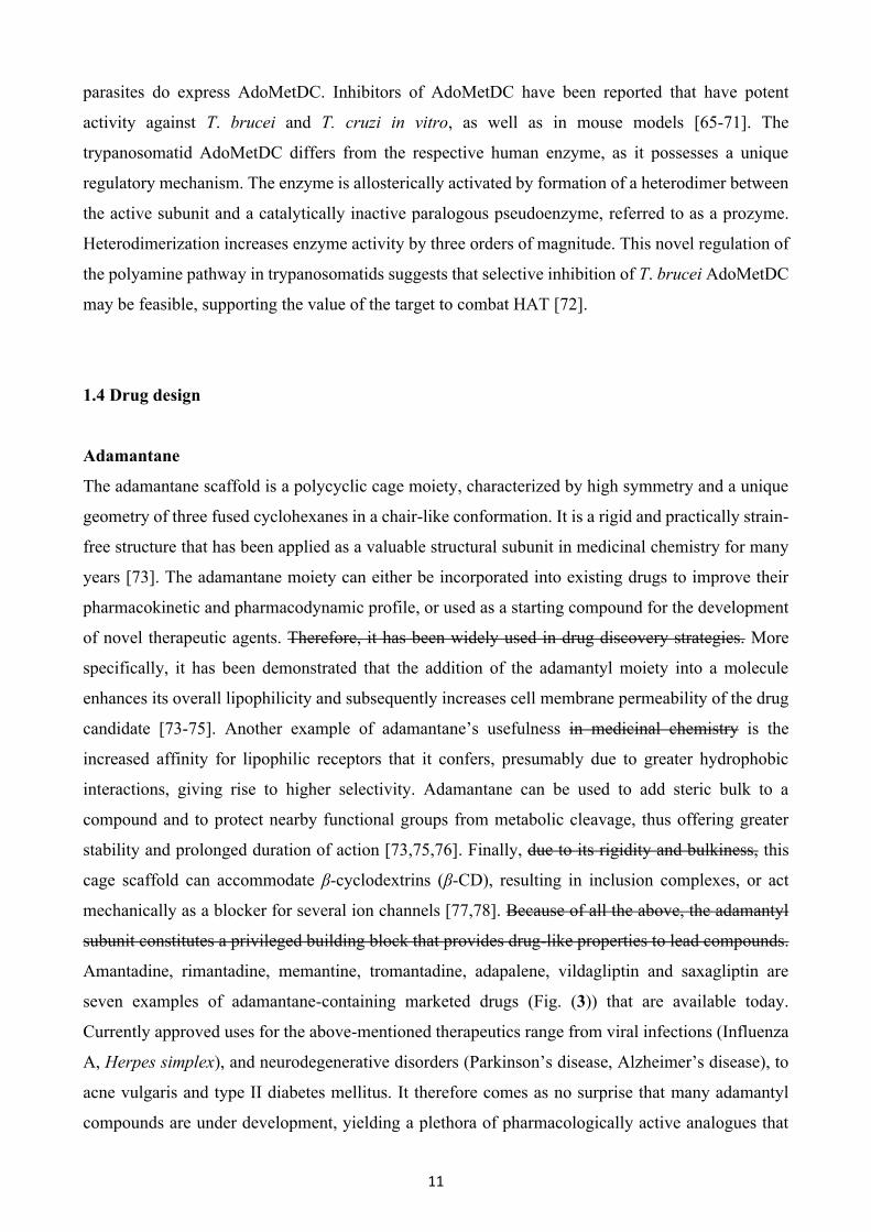
Amantadine, rimantadine, memantine, tromantadine, adapalene, vildagliptin and saxagliptin are
seven examples of adamantane-containing marketed drugs (Fig. (3)) that are available today.
Currently approved uses for the above-mentioned therapeutics range from viral infections (Influenza
A, Herpes simplex), and neurodegenerative disorders (Parkinson’s disease, Alzheimer’s disease), to
acne vulgaris and type II diabetes mellitus. It therefore comes as no surprise that many adamantyl
compounds are under development, yielding a plethora of pharmacologically active analogues that
12
have been studied for various medical conditions, including iron overload disease, cancer, CNS
diseases, inflammation, hypertension, hyperglycemia, malaria and tuberculosis [74-76,78-81].
Fig. (3). Clinically approved adamantane derivatives and their pharmacological profiles.
Adamantane-based guanylhydrazone analogues
Over the years, we have synthesized a variety of structurally diverse adamantane derivatives that
target influenza A virus, T. brucei and T. cruzi [96-103]. In continuation of our studies, we were
interested in developing further active adamantane-based compounds that could inhibit the
proliferation of the above-mentioned trypanosomatids. Intrigued by the in vitro and in vivo
trypanocidal properties of several guanylhydrazone analogues, that could act as potential AdoMetDC
inhibitors [104-112], and following a molecular hybridization approach, we designed derivatives of
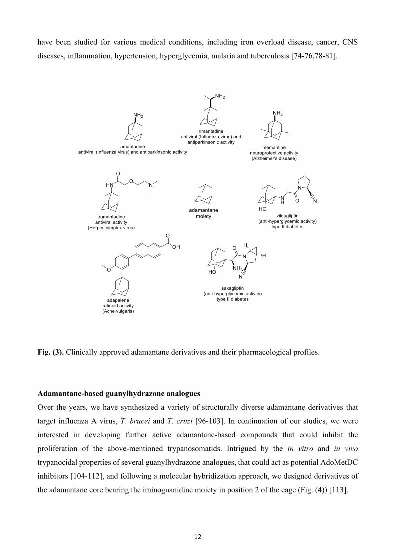
the adamantane core bearing the iminoguanidine moiety in position 2 of the cage (Fig. (4)) [113].
13
Fig. (4). Structures of adamantane-substituted guanylhydrazones G1 – G25 described in the present
study [112-114].
From this perspective, The design strategy was aimed at combining the increased lipophilicity of the
adamantane cage structure with the trypanocidal properties activity of the guanylhydrazone moiety
to generate compounds with better antiparasitic activity. The general approach for this class of
compounds was to use the adamantane component as a lipid carrier for the BBB to counterbalance
the potential transport limitations conferred by the conjugated polar iminoguanidine side chain. Thus,
improving their BBB permeability enables enhanced CNS penetration and increased concentration in
brain tissue, which is of crucial importance for the treatment of the second stage HAT. Finally, the
incorporation of the guanylhydrazone group into this class of compounds should have the added
advantage of structural similarity with the guanidine pharmacophore of the licensed trypanocidal drug
pentamidine. To study the impact of changing the guanylhydrazone position and to evaluate the
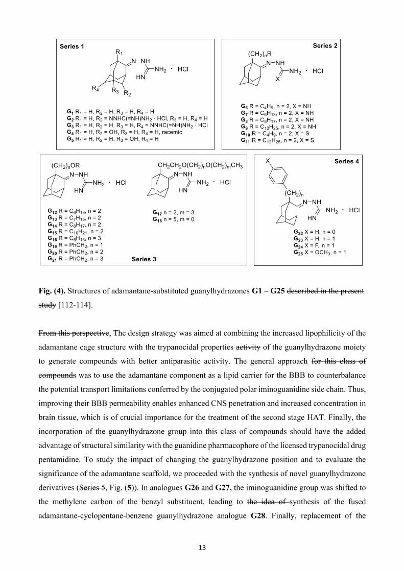
significance of the adamantane scaffold, we proceeded with the synthesis of novel guanylhydrazone
derivatives (Series 5, Fig. (5)). In analogues G26 and G27, the iminoguanidine group was shifted to
the methylene carbon of the benzyl substituent, leading to the idea of synthesis of the fused
adamantane-cyclopentane-benzene guanylhydrazone analogue G28. Finally, replacement of the
14
adamantane cage structure by the tricyclic scaffolds of fluorene and dibenzocycloheptene, and the
bendable 4-phenyl cyclohexane, afforded led to the analogues G29, G30 and G31, respectively.
Fig. (5). Structures of novel guanylhydrazone derivatives G26 – G31, synthesized in the present
study.
Here, we report the synthesis and structural characterization of novel guanylhydrazones (G26 – G31,
Fig. (5)), with their subsequent in vitro biological evaluation against bloodstream form T. brucei and
T. cruzi epimastigotes. We also report, for the first time, the results of the trypanocidal activity of
previously synthesized adamantane guanylhydrazones (G1 – G5, Fig. (4)) that have been used as
butyrylcholinesterase inhibitors. Finally, we provide an overview on the chemistry and
pharmacological activity of adamantane-based guanylhydrazone derivatives (G6 – G25, Fig. (4)),
synthesized in the recent past, by our research group. Yet Another objective of the study was to
evaluate the effect of structural modifications and steric parameters on antiparasitic activity and to
correlate this activity with possible interactions in the active site of AdoMetDC, using docking-
scoring calculations and Quantitative Structure-Activity Relationship (QSAR) studies.
15
2. Experimental
2.1 General Experimental methods and techniques
Melting points were determined using a Büchi capillary apparatus and are uncorrected. NMR
experiments were performed to elucidate the structure and determine the purity of the newly
synthesized compounds. 1H NMR and 2D NMR spectra (COSY, HSQC, HSQC-DEPT and HMBC)
were recorded on a Bruker Ultrashield™ Plus Avance III 600 spectrometer (600.11 MHz, 1H NMR).
13C NMR spectra were recorded on a Bruker Ultrashield™ Plus Avance III 600 spectrometer (150.9
MHz, 13C NMR). Chemical shifts δ (delta) are reported in parts per million (ppm) downfield from
the NMR solvent, with the tetramethylsilane or solvent (DMSO-d6) as internal standard. Data
processing including Fourier transformation, baseline correction, phasing, peak peaking and
integrations were performed using MestReNova software v.12.0.0. Splitting patterns are designated
as follows: s, singlet; br s, broad singlet; d, doublet; t, triplet; q, quartet; dd, doublet of doublets; dt,
doublet of triplets; dq, doublet of quartets; td, triplet of doublets; tt, triplet of triplets; ttd, triplet of
triplets of doublets; qd, quartet of doublets; quin, quintet; complex m, complex multiplet. Coupling
constants (J) are expressed in units of Hertz (Hz). The spectra were recorded at 305 K (32 °C) unless
otherwise specified. The solvent used to obtain the spectra was deuterated dimethyl sulfoxide,
DMSO-d6 (quin, 2.50 ppm, 1H NMR; septet, 39.52 ppm, 13C NMR). Elemental analyses (C, H, N)
were performed by the Service Central de Microanalyse at CNRS (France), and were within ± 0.4%
of the theoretical values. Elemental analysis results for the tested compounds correspond to ˃95%
purity. The commercial reagents were purchased from Alfa Aesar, Sigma-Aldrich, and Merck, and
were used without further purification. Organic solvents used were of the highest purity.
2.2 Synthetic procedure for the preparation of analogues G26 – G31
Aminoguanidine bicarbonate (1.5 mmol) was suspended in 18 mL of absolute ethanol, and it was
progressively dissolved by dropwise acidification with concentrated hydrochloric acid, until pH was
adjusted to 2. Subsequently, the appropriate ketone (1.5 mmol) was added and the mixture was
refluxed under stirring for 5 h at 105 – 110 °C. After completion of the reaction time, ethanol was
evaporated under reduced pressure and the residue was treated with dry Et2O. The resulting product
was cooled at 5 °C for 1 h and filtered off in vacuo. The precipitate was washed with dry Et2O and
dried to obtain the desired analogues G26 – G31 as crystals.
Tricyclo[3.3.1.13,7]decan-1-yl(phenyl)methylene)guanylhydrazone hydrochloride (G26)

16
Product G26 was obtained using 500 mg (2.08 mmol) of 1-adamantyl phenyl ketone and 283 mg
(2.08 mmol) aminoguanidine bicarbonate in 25 mL of absolute EtOH, resulting in 690 mg of white
crystalline solid. Yield: almost quantitative; mp >250 °C (from EtOH/dry Et2O); 1H NMR (600.11
MHz, DMSO-d6) δ (ppm) [1.57 (dq, 3H, J1=12.2 Hz, J2=2.1 Hz), 1.66 (dt, 3H, J1=12.5 Hz, J2=3.1
Hz), H4-Ad, H6-Ad, H10-Ad], 1.75 (d, 3H, J=3.0 Hz, H2-Ad, H8-Ad, H9-Ad), 1.96 (~quin, 3H, J=3.2
Hz, H3-Ad, H5-Ad, H7-Ad), 7.09 (dd, 2H, J1=8.1 Hz, J2=1.5 Hz, H2', H6'), 7.32-8.01 (br s, 3H,
NHC(NH)NH2), 7.47-7.54 (complex m, 3H, H3', H4', H5'), 9.25 (s, 1H, NHC(NH)NH2); 13C NMR
(150.9 MHz, DMSO-d6) δ (ppm) 27.6 (C3-Ad, C5-Ad, C7-Ad), 36.0 (C4-Ad, C6-Ad, C10-Ad), 39.4
(C2-Ad, C8-Ad, C9-Ad), 127.6 (C2', C6'), 129.0 (C3', C5'), 129.1 (C4'), 131.3 (C1'), 155.7
(NHC(NH)NH2), 164.6 (C=N). Anal. calcd. For C18H25ClN4, C, 64.95; H, 7.57; N, 16.83. Found: C,
65.00; H, 7.50; N, 16.79.
Tricyclo[3.3.1.13,7]decan-2-yl(phenyl)methylene)guanylhydrazone hydrochloride (G27)
Product G27 was obtained using 500 mg (2.08 mmol) of 2-adamantyl phenyl ketone and 283 mg
(2.08 mmol) aminoguanidine bicarbonate in 25 mL of absolute EtOH, resulting in 680 mg of white
crystalline solid. Yield: almost quantitative; mp >250 °C (from EtOH/dry Et2O);1H NMR (600.11
MHz, DMSO-d6) δ (ppm) 1.45 (d, 2H, J=12.3 Hz, H4e-Ad, H9e-Ad), 1.66 (d, 2H, J=3.1 Hz, H6-Ad),
1.69-1.78 (complex m, 4H, H8-Ad, H10-Ad), 1.79 (~quin, 1H, J=3.1 Hz, H5-Ad), 1.86 (~quin, 1H,
J=3.0 Hz, H7-Ad), 1.93 (dd, 2H, J1=12.8 Hz, J2=2.8 Hz, H4a-Ad, H9a-Ad), 2.04 (q, 2H, J=2.9 Hz, H1-
Ad, H3-Ad), 2.92 (s, 1H, H2-Ad), 7.09-7.29 (br s, 3H, NHC(NH)NH2), 7.28 (dd, 2H, J1=8.2 Hz,
J2=1.4 Hz, H2', H6'), 7.47 (td, 1H, J1=7.4 Hz, J2=1.4 Hz, H4'), 7.51 (~dd, 2H, J1=8.1, 6.6 Hz, J2=1.5
Hz, H3', H5'), 9.74 (s, 1H, NHC(NH)NH2); 13C NMR (150.9 MHz, DMSO-d6) δ (ppm) 27.2 (C5-Ad),
27.3 (C7-Ad), 28.6 (C1-Ad, C3-Ad), 32.0 (C4-Ad, C9-Ad), 37.1 (C6-Ad), 37.9 (C8-Ad, C10-Ad), 50.1
(C2-Ad), 126.9 (C2', C6'), 129.3 (C3', C5'), 129.4 (C4'), 155.8 (NHC(NH)NH2), 159.5 (C=N). Anal.
calcd. For C18H25ClN4, C, 64.95; H, 7.57; N, 16.83. Found: C, 64.91; H, 7.63; N, 16.85.
Adamantane[1,2-a]inden-11-ylidene)guanylhydrazone hydrochloride (G28)
Product G28 was obtained using 500 mg (2.10 mmol) of adamantane[1,2-a]inden-11-one and 286 mg
(2.10 mmol) aminoguanidine bicarbonate in 25 mL of absolute EtOH, resulting in 575 mg of white
crystalline solid. Yield: 83%; mp 230 – 232 °C (from EtOH/dry Et2O); 1H NMR (600.11 MHz,
DMSO-d6) δ (ppm) 1.51 (d, 2H, J=12.5 Hz, H9e-Ad, H13e-Ad), 1.61-2.08 (complex m, 7H, H5, H7,
H8, H12), 2.31 (t, 4H, J=11.2 Hz, H6, H9a, H10, H13a), 2.81 (s, 1H, H10a), 7.29 (t, 1H, J=7.2 Hz, H3),
7.48 (t, 1H, J=7.4 Hz, H2), 7.83 (d, 1H, J=7.7 Hz, H1), 7.84-8.11 (br s, 3H, NHC(NH)NH2), 8.22 (d,
1H, J=7.6 Hz, H4), 10.33 (s, 1H, NHC(NH)NH2); 13C NMR (150.9 MHz, DMSO-d6) δ (ppm) 27.0
(C8), 27.1 (C6), 27.6 (C10), 32.2 (C7), 36.3 (C9), 36.5 (C12), 42.3 (C4b), 46.7 (C13), 47.8 (C5), 82.6
17
(C10a), 122.2 (C4), 126.0 (C2), 128.0 (C3), 133.8 (C1), 136.1 (C4a), 156.5 (C=N), 158.2
(NHC(=NH)NH2), 162.5 (C11a). Anal. calcd. For C18H23ClN4, C, 65.34; H, 7.01; N, 16.93. Found: C,
65.40; H, 7.08; N, 16.88.
2-(9H-Fluoren-9-ylidene)guanylhydrazone hydrochloride (G29)
Product G29 was obtained using 500 mg (2.77 mmol) of 9H-fluoren-9-one and 377 mg (2.77 mmol)
aminoguanidine bicarbonate in 33 mL of absolute EtOH, resulting in 695 mg of yellow crystalline
solid. Yield: 92%; mp 238 – 240 °C (from EtOH/dry Et2O); 1H NMR (600.11 MHz, DMSO-d6) δ
(ppm) [7.35 (td, 1H, J1=7.4 Hz, J2=0.9 Hz), 7.39 (td, 1H, J1=7.6 Hz, J2=1.2 Hz), H2, H7], [7.44 (td,
1H, J1=7.5 Hz, J2=1.2 Hz), 7.54 (t, 1H, J=7.5 Hz), H3, H6], [7.79 (d, 1H, J=7.5 Hz), 7.88 (d, 1H,
J=7.5 Hz), H4, H5], 8.11 [(d, 1H, J=7.5 Hz), 8.76 (d, 1H, J=7.7 Hz), H1, H8], 8.23-8.67 (br s, 3H,
NHC(NH)NH2), 11.45 (s, 1H, NHC(NH)NH2); 13C NMR (150.9 MHz, DMSO-d6) δ (ppm) 120.2,
120.9 (C4, C5), 122.7, 127.4 (C1, C8), 128.2, 128.3 (C2, C7), 130.7, 131.9 (C3, C6), 136.3, 139.1
(C8a/C9a), 141.8 (C4a, C4b), 148.2 (C=N), 157.3 (NHC(=NH)NH2). Anal. calcd. For C14H13ClN4, C,
61.65; H, 4.80; N, 20.54. Found: C, 61.70; H, 4.89; N, 20.49.
2-(10,11-Dihydro-5H-dibenzo[a,d][7]annulen-5-ylidene)guanylhydrazone hydrochloride (G30)
Product G30 was obtained using 1.0 g (4.80 mmol) of 10,11-dihydro-5H-dibenzo[a,d][7]annulen-5-
one (dibenzosuberone) and 653 mg (4.80 mmol) aminoguanidine bicarbonate in 58 mL of absolute
EtOH, resulting in 405 mg of beige crystalline solid. Yield: 28%; mp 155-157 °C (from EtOH/dry
Et2O); 1H NMR (600.11 MHz, DMSO-d6) δ (ppm) [2.87 (dd, 2H, J1=17.8 Hz, J2=10.0 Hz), 3.06 (d,
1H, J=13.6 Hz), 3.26 (d, 1H, J=17.2 Hz), H10, H11], 7.13 (d, 1H, J=7.7 Hz, H1 or H9), [7.22 (t, 1H,
J=7.4 Hz), 7.23 (t, 1H, J=7.1 Hz), H3, H7], 7.29 (d, 1H, J=7.4 Hz, H4 or H6), 7.33 (t, 1H, J=7.3 Hz,
H2 or H8), 7.41 (d, 1H, J=7.3 Hz, H1 or H9), 7.43 (t, 1H, J=7.8 Hz, H2or H8), 7.55-8.13 (br s, 3H,
NHC(NH)NH2), 7.75 (d, 1H, J=7.7 Hz, H4/H6), 10.13 (s, 1H, NHC(NH)NH2); 13C NMR (150.9 MHz,
DMSO-d6) δ (ppm) 31.1, 33.5 (C10, C11), 126.1, 126.6 (C3, C7), 127.3, 128.8 (C2, C8), 129.3, 129.6
(C4, C6), 130.6, 130.8 (C1, C9), 133.4, 135.2 (C4a, C5a), 138.4, 139.4 (C9a, C11a), 156.2 (C=N), 159.1
(NHC(=NH)NH2). Anal. calcd. For C16H17ClN4, C, 63.89; H, 5.70; N, 18.63. Found: C, 63.93; H,
5.82; N, 18.66.
2-(4-Phenylcyclohexylidene)guanylhydrazone hydrochloride (G31)
Product G31 was obtained using 500 mg (2.87 mmol) of 4-phenylcyclohexan-1-one and 391 mg (2.87
mmol) aminoguanidine bicarbonate in 34 mL of absolute EtOH, resulting in 760 mg of white
crystalline solid. Yield: almost quantitative; mp 218 – 220 °C (from EtOH/dry Et2O); 1H NMR
(600.11 MHz, DMSO-d6) δ (ppm) 1.59 (qd, 1H, J1=12.9 Hz, J2=4.1 Hz, H2a or H6a), 1.64 (qd, 1H,
18
J1=12.7 Hz, J2=4.3 Hz, H3a or H5e), 1.98 (~ttd, 2H, J1=9.3 Hz, J2=6.3, 5.8 Hz, J3=3.3 Hz, H2a or H6a,
H3a or H5e), 2.14 (td, 1H, J1=14.1 Hz, J2=5.1 Hz, H2e or H6e), 2.42 (td, 1H, J1=13.5 Hz, J2=4.8 Hz,
H3e or H5a), 2.50-2.55 (m, 1H, H3e or H5a), 2.85 (tt, 1H, J1=12.2 Hz, J2=3.5 Hz, H4), 3.15 (dq, 1H,
J1=14.7 Hz, J2=3.0 Hz, H2e or H6e), 7.25 (d, 2H, J=7.4 Hz, H2', H6'), 7.28 (t, 2H, J=7.5 Hz, H3', H5'),
7.48-7.95 (br s, 3H, NHC(=NH)NH2), 8.84 (s, 0.25H, NHC(=NH)NH2) 11.34 (s, 0.75H,
NHC(=NH)NH2); 13C NMR (150.9 MHz, DMSO-d6) δ (ppm) 27.3, 32.5 (C2, C6), 33.8, 34.3 (C3, C5),
42.2 (C4), 126.1 (C4'), 126.6 (C2', C6'), 128.3 (C3', C5'), 145.5 (C1'), 156.2 (NHC(=NH)NH2), 158.8
(C=N), 159.2 (C=N). Anal. calcd. For C13H19ClN4, C, 58.53; H, 7.18; N, 21.00. Found: C, 58.50; H,
7.25; N, 21.07.
2.3 Trypanocidal assays
For compounds G1 – G25: A bloodstream form of T. brucei brucei (strain 427, derivative 221) was
cultured in 25 mL flasks at 37 °C in modified Iscove’s medium (pH 7.4) [115]. To assess activity,
parasites were grown for 3 days in the presence of test compounds and the EC50 and EC90 values were
determined (concentrations that inhibit growth by 50% and 90%, respectively). In these experiments,
the densities of untreated cultures increased from 0.25 × 10⁴ to 4 × 10⁶ cells mL−1. After determination
of cell densities at each drug concentration with a hemocytometer (Weber Scientific International
Ltd.), drug sensitivity was expressed as a percentage of growth of control cells. Data presented are
the mean of triplicate experiments ± SEM.
For compounds G26 – G31: Bloodstream form T. brucei (strain 427, derivative 221) were cultured
in modified Iscove’s medium, as outlined previously [116]. Eight-point potency curves were
performed in 96-well plates (200 μL volumes), and the compound concentrations that inhibited
growth by 50% (EC50) and 90% (EC90) were determined. Parasites were first diluted to 2.5 × 10⁴
mL−1, compounds were added at range of concentrations, and the plates incubated at 37 °C. Resazurin
was added after 48 h, and the plates incubated for a further 16 h. T. cruzi epimastigotes (strain CL
Brener) were cultured as previously described [117]. Trypanocidal activity was determined in
microtiter plates as outlined above, with the following modifications. Experiments were initiated by
seeding the parasites at 2.5 × 10⁵ mL−1, and after the addition of test compounds, cultured at 28 °C
for 4 days. Resazurin was added, the plates were incubated for a further 2 days, and then assessed as
above. Fluorescence intensities were determined using a BMG FLUOstar Omega (excitation 545 nm,
emission 590 nm). Data were analyzed using Graph Pad Prism 7 software. Values are expressed as
EC50 ± SD and are the average of three independent replicates.
2.4 In vitro cytotoxicity assays on rat skeletal myoblast L6 cells
19
Cytotoxicity against L6 cells was assessed using microtiter plates. Briefly, cells were seeded in
triplicate at 1 × 10⁴ mL−1 in growth medium containing different compound concentrations. The plates
were incubated for 6 days at 37 °C and resazurin was then added to each well. After a further 8 h of
incubation, the fluorescence was determined using a BMG FLUOstar Omega plate reader.
2.5 Computer-aided drug design
The discovery of new drugs is undoubtedly an expensive and time-consuming multi-step process.
However, it is accelerated through our increased understanding of the fundamental principles of
protein-ligand interactions, in addition to the integration of a computer-aided drug design (CADD)
strategy. CADD strategies involve application of computer modelling technologies, which ultimately
aid in selection and filtering of candidate compounds, generation of hypotheses on their mode of
action, and also facilitate their further development [118,119]. Over the last decades, due to
advancements in computer technology, CADD has been considered a rapidly growing field and plays
a crucial role in drug development projects, from hit identification to lead optimization of novel,
potentially therapeutically valuable small molecules [120-123]. CADD strategies can be mainly
classified into three broad categories; the ligand-based (LBDD) and the structure-based (SBDD)
approaches, as well as fragment-based drug discovery (FBDD), a newly emerging lead discovery
method [124,125]. LBDD approaches utilize structure and activity data from a set of known active
and inactive compounds in order to retrieve other potential molecular scaffolds based on similarity
measures, common pharmacophores or descriptor values. LBDD methods include, amongst others,
similarity and substructure searching, quantitative structure-activity relationships (QSAR), as well as
pharmacophore and three-dimensional (3D) shape matching [126,127]. Contrarily, SBDD approaches
are based on direct calculation of protein-ligand interactions utilizing the 3D structure of the
biological target, determined through X-ray crystallography and NMR, or through homology
modeling, and include ligand docking-based scoring, molecular dynamics and de novo design [128].
Notably, both of the above mentioned knowledge-driven approaches can be synergistically integrated
to improve the drug design process [129]. In this current study, we are mainly focusing on QSAR
analysis via 2D fingerprint similarity and docking-based scoring, one methodology from each CADD
approach.
2.6 Docking-based scoring
Introduction

20
The docking process involves the prediction of ligand conformation and posing, as well as binding
interactions within the targeted protein site. This is a rather challenging task by itself, since even
relatively simple organic molecules may contain many conformational degrees of freedom, or show
a diversity of binding modes. Thorough sampling of ligand orientation within the binding domain of
the target is provided through stochastic search techniques such as the Monte Carlo algorithm [130].
Rapid and accurate assessment of protein-ligand complexes is pivotal for docking approaches, since
a typical ligand docking experiment generates hundreds to thousands of ligand conformations [131].
Thus, an efficient scoring function is essential to rank order different docked poses for each ligand,
and ultimately to discriminate the valid binding mode predictions from the invalid ones [132].
Evaluation of docked poses is achieved by four distinct classes of scoring functions that are currently
applied in computational drug design: force-field-based, empirical, knowledge-based and a rather
new approach, machine learning-based scoring functions [133], with the first being the scoring
function of our choice.
Protocol
Prior to any docking experiment, protein and ligand databases need to be prepared. The crystal
structure of T. brucei AdoMetDC/prozyme heterodimer (PDB structure: 5tvf) was computationally
cleaved to include only AdoMetDC in complex with the bound ligand, CGP. Then, Protein
Preparation Wizard [134] workflow was executed, as previously thoroughly described in detail
previously [135], generating a structurally sound model ensuring accuracy on all downstream
modeling simulations. Preparation of the guanylhydrazone analogues was performed using the
LigPrep tool [136]. LigPrep generates precise, energy minimized 3D molecular structures with
correct protonation states, tautomerism and enumeration of all possible stereoisomers. Docking grid
preparations and docking calculations were accomplished as previously described [135]. Two
different grids were generated using no scaling for protein van der Waals (VdW) radii and an extended
ligand centering box of 10 × 10 × 10 Å and 15 × 15 × 15 Å, respectively, to ensure proper fitting of
the large aliphatic chains of some guanylhydrazone analogues. In this study, the Glide algorithm [137]
was utilized, which is considered to be one of the most efficient and accurate virtual screening tools
[138] for structure-based virtual screening of the guanylhydrazone analogues inside the AdoMetDC
binding site. All docking calculations were performed by using VdW scaling of 0.8 for ligand atoms,
a maximum of 3 poses per ligand and extra-precision (XP), a rather extensive, accurate and time- and
CPU- demanding mode [139].
2.7 QSAR analysis
21
Introduction
Although QSAR was first developed more than 55 years ago [140], it still remains one of the most
promising research areas in medicinal chemistry and cheminformatics. QSAR aims to reveal a
quantitative correlation between the structural features of chemical entries and their many different
properties, such as continuous (pIC50, pEC50, Ki, etc.), categorical/binary (active, inactive, toxic,
nontoxic, etc.) and biological/toxicological. To this end, the main strategy involves the generation of
a statistical linear or non-linear regression model that matches experimental values to a diverse set of
molecular descriptors determined from the structure of the compounds [141]. Chemical descriptors
constitute the basis of QSAR modeling and reflect a plethora of chemical structure representation.
These molecular descriptors have been categorized into different classes, including constitutional,
geometrical, topological, quantum chemical and so on, with 2D (two-dimensional) being the most
popular among drug developers. Molecular descriptors compose the basis of QSAR modeling and
represent different levels of chemical structure representation ranging from single physico-chemical
properties (1D QSAR) to three-dimensional structures (3D QSAR), and even more intricate,
multidimensional approaches like simulation of induced fit and solvation scenarios (5D and 6D
QSAR) [142]. The success of QSAR analysis depends on the quality of the input data, selection of
appropriate descriptors and statistical methods to validate the developed model. QSAR analysis has
yielded quite a few success stories, regarding including currently approved drugs [143-145]. In the
framework of this study, the Canvas cheminformatics software package was utilized [146], in an
attempt to connect ones and zeros with reliable SAR predictions. Canvas provides a wide range of
applications for structural and data analysis, including 2D and 3D fingerprints, similarity searching,
as well as building regression and classification models.
Protocol
Preparation of the guanylhydrazone analogues was performed with Ligprep, as previously described
[135], with the exception of generating only the most probable ionization and tautomeric state of each
analogue, chosen on the basis of energetic considerations. Then, prepared compounds were
introduced into the Canvas interface and the complete set of 498 physicochemical, topological,
Ligfilter and Qikprop descriptors were determined for all entries. A total of nine fingerprints were
calculated using default atom typing and scaling parameters. Seven 2D fingerprints, (linear, radial,
dendritic, MOLPRINT 2D, atom pairs, atom triplets, and topological torsions) and two 3D
pharmacophore fingerprints (2p and 3p pharmacophore), with Tanimoto similarity were used for
ranking. A detailed description of each fingerprint has been realized established by Sastry and
colleagues [147]. Statistical regression models were then generated, utilizing three diverse and well-
established statistical methods, namely Multiple Linear Regression (MLR), Partial Least Squares

22
regression (PLS) and Kernel-based Partial Least Squares regression (KPLS), in order to successfully
and efficiently correlate the cell-based in vitro activity of the guanylhydrazone analogues on against
T. brucei to the aforementioned molecular descriptor and 2D/3D fingerprints. The initial approach
included the implementation of all guanylhydrazone analogues in the training set. As expected, the
compounds with none or marginal activity against T. brucei (G12, G14, G15 and G20) seemed to
impede the generation of an accurate and reliable model. Indicatively, standard deviation (SD), as
well as the coefficient of determination (R2), resulted from MLR analysis, and proved that the model
containing all analogues was quite questionable (SD = 1.526; R2 = 0.5720). Therefore, in order to
improve the predictivity of the model, only active compounds were utilized. A training set of 27
active compounds were analysed through MLR, PLS and KPLS regression functions with a 20% of
random test set assignment for model evaluation. The option of five best subsets was used for MLR,
attempting to find the highest-ranking model containing said number of independent variables,
utilizing a simulated annealing Monte Carlo technique. For each MLR model generated, the option
of “best subsets” was chosen, attempting to find the highest-ranking models containing five
independent variables. Default settings were used for the simulated annealing Monte Carlo technique.
Selection of the best MLR model was performed through careful evaluation of each model,
considering taking into account intercorrelation between selected independent variables. For both
PLS and KPLS, four factors were used. Hierarchical clustering of the guanylhydrazone analogues
was attempted, based on the 2D fingerprint of atom pairs. Analogues were divided into three clusters,
which were then statistically processed as previously described. Note that this approach did not yield
any meaningful results.
3. Results and Discussion
3.1 Chemistry
Synthesis of Series 1 compounds
For the synthesis of the starting ketones 3 and 4 (Scheme 1), adamantan-2-one 1 was first converted
to 4-oxahomoadamantan-5-one 2 via a selenium dioxide mediated oxidation. Lactone 2 was heated
23
with a 50% sulfuric acid and yielded a mixture consisting of the axial and the equatorial isomer of 3,
in an approximate ratio of 5 : 1. Oxidation of the latter with chromic acid afforded the adamantane
dione 4 [148].
Scheme 1. Synthetic route to the starting compounds 3 and 4 and synthesis of mono and
bis(guanylhydrazone) adamantanes G1, G2 and G4. Reagents and conditions: (a) SeO2, H2O2, t-
BuOH, 96%; (b) H2SO4 (50%), 5 h, 90 °C; (c) H2CrO4; (d) H2NNHC(=NH)NH2 · HCl, EtOH, reflux,
5 h, 99% (G1), 97% (G2) and 94% (G4) [114,148].
The synthetic route for the formation of starting compounds 9 and 10 is described in Scheme 2.
Oxygen-containing adamantane analogues are provided by functionalization of the adamantane core
with the use of oxidizing agents. Initially the adamantane hydroxylation was performed upon
treatment with fuming sulfuric acid (H2O7S2). The products of the oxidative procedure are three diols
obtained in different ratio. In fuming acid with a SO3 percentage of 20%, 1,4-adamantane diol 6 is
the main product in an overall yield of 45 – 50%. 1,3-Diol 7 and 2,6-diol 8 were also formed with 15
– 20% and 5 – 10% yields, respectively. The mixture of diols 6, 7 and 8 was subjected to oxidation
with chromic acid to give 5-hydroxyadamantan-2-one/1-hydroxyadamantan-4-one 9 and
adamantane-2,6-dione 10 [149].
24
Scheme 2. Synthetic route to the starting compounds 9 and 10 and synthesis of mono and
bis(guanylhydrazone) adamantanes G3 and G5, respectively. Reagents and conditions: (a) 20% SO3
(10 °C), 20 min, 25 °C then 100 °C, 2 h; (b) CrO3, H2O, 30 min, 70 °C; (c) H2NNHC(=NH)NH2 ·
HCl, EtOH, reflux, 5 h, 98% (G3) and 99% (G5) [114,149].
Adamantyl guanylhydrazones G1 – G5 used in this study were synthesized according to the
previously published procedure by condensing the corresponding adamantanone with
aminoguanidine hydrochloride (Schemes 1 and 2) almost quantitatively. Note that compounds G2
and G4 (Scheme 1) have been isolated as a mixture of diastereoisomers and derivative G2 has three
possible diastereoisomers while derivative G4 has four possible diastereoisomers. However, for
guanylhydrazones G1 (Scheme 1), G3 and G5 (Scheme 2), only one diastereomeric form is possible
due to molecular symmetry [114].
Synthesis of Series 2 compounds
The synthesis of 1,2-disubstituted adamantane derivatives was the key step for the preparation of
analogues described in this study and was achieved by rearrangement of the protoadamantane
framework [150]. The starting compound, protoadamantanone 13, was synthesized by Majerski’s
group for the first time in 1979. As shown in Scheme 3, the commercially available adamantan-1-ol
11 reacted with lead tetraacetate and iodine in benzene as a solvent, and the iodo ketone 12 that was
generated was refluxed with potassium hydroxide to provide protoadamantanone 13 in yields ranging
from 71% to 82% [151]. The amount of the unreacted starting alcohol was decreased by heating the
25
mixture composed of adamantan-1-ol 11, lead tetraacetate and iodine for 2 h at 75 – 76 °C. The target
compound 13 was then produced, almost pure, in excellent yields [152].
Scheme 3. Synthesis of protoadamantanone. Reagents and conditions: (a) Pb(OAc)4, I2, C6H6, 70 –
75 °C (b) KOH, MeOH, reflux [151].
As depicted in Scheme 4, synthesis of the starting ketones 18a – d [112] proceeded with formation
of the acetylenic alcohols 14a – d. More specifically, protoadamantanone 13 reacted with the
appropriate lithiated acetylide, which was prepared in situ by addition of a substituted acetylene to a
solution of butyllithium, to give the desired compounds 14a – d as a mixture of endo/exo isomers (1
: 1) in an overall yield of 82 – 95%. Catalytic hydrogenolysis of the latter with Adams’ catalyst
yielded the corresponding saturated isomeric alcohols 15a – d. The mixture was refluxed with formic
acid and the formate esters immediately underwent an intramolecular metathesis to give analogues
16a – d. Subsequent saponification of the intermediates 16a – d in the presence of an aqueous sodium
hydroxide solution led to alcohols 17a – d in excellent yields which underwent Jones oxidation to
form the target 1-alkyl-adamantan-2-ones 18a – d almost quantitatively [112].
For the synthesis of 1-alkyl-adamantane 2-guanylhydrazones G6 – G9, the starting ketones 18a – d
were refluxed with aminoguanidine bicarbonate in the presence of concentrated hydrochloric acid
(pH ~ 2) and ethanol as a solvent. The target compounds G6 – G9 were obtained in an overall yield
of 85 to 94% [112].
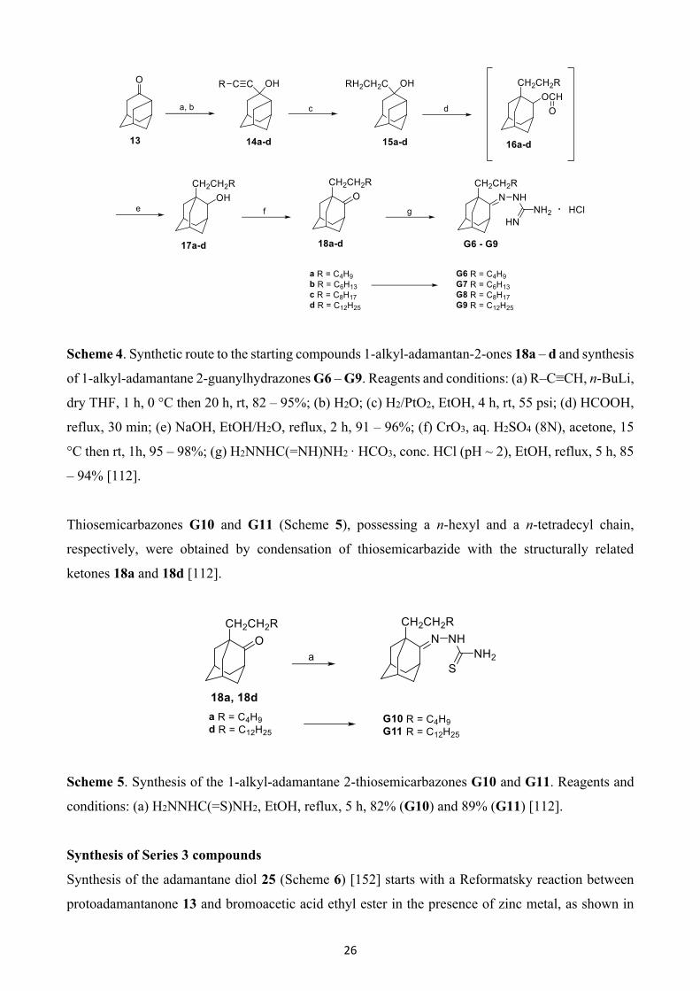
26
Scheme 4. Synthetic route to the starting compounds 1-alkyl-adamantan-2-ones 18a – d and synthesis
of 1-alkyl-adamantane 2-guanylhydrazones G6 – G9. Reagents and conditions: (a) R–C≡CH, n-BuLi,
dry THF, 1 h, 0 °C then 20 h, rt, 82 – 95%; (b) H2O; (c) H2/PtO2, EtOH, 4 h, rt, 55 psi; (d) HCOOH,
reflux, 30 min; (e) NaOH, EtOH/H2O, reflux, 2 h, 91 – 96%; (f) CrO3, aq. H2SO4 (8N), acetone, 15
°C then rt, 1h, 95 – 98%; (g) H2NNHC(=NH)NH2 · HCO3, conc. HCl (pH ~ 2), EtOH, reflux, 5 h, 85
– 94% [112].
Thiosemicarbazones G10 and G11 (Scheme 5), possessing a n-hexyl and a n-tetradecyl chain,
respectively, were obtained by condensation of thiosemicarbazide with the structurally related
ketones 18a and 18d [112].
Scheme 5. Synthesis of the 1-alkyl-adamantane 2-thiosemicarbazones G10 and G11. Reagents and
conditions: (a) H2NNHC(=S)NH2, EtOH, reflux, 5 h, 82% (G10) and 89% (G11) [112].
Synthesis of Series 3 compounds
Synthesis of the adamantane diol 25 (Scheme 6) [152] starts with a Reformatsky reaction between
protoadamantanone 13 and bromoacetic acid ethyl ester in the presence of zinc metal, as shown in
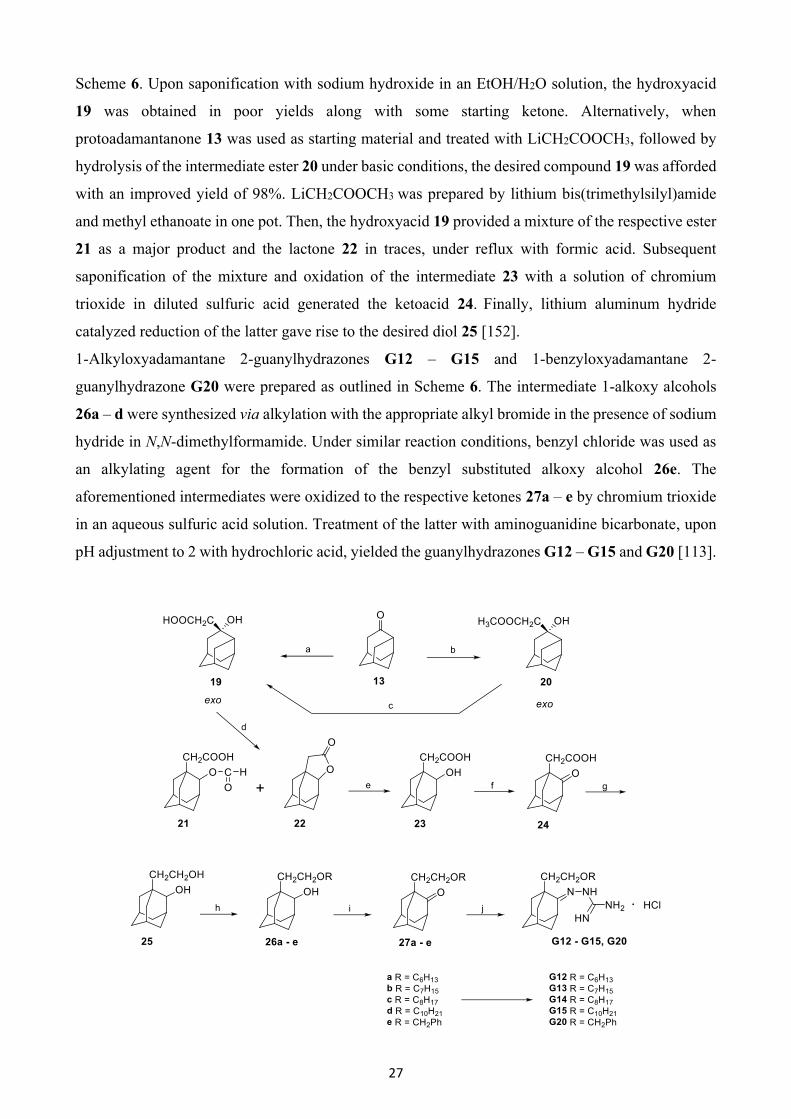
27
Scheme 6. Upon saponification with sodium hydroxide in an EtOH/H2O solution, the hydroxyacid
19 was obtained in poor yields along with some starting ketone. Alternatively, when
protoadamantanone 13 was used as starting material and treated with LiCH2COOCH3, followed by
hydrolysis of the intermediate ester 20 under basic conditions, the desired compound 19 was afforded
with an improved yield of 98%. LiCH2COOCH3 was prepared by lithium bis(trimethylsilyl)amide
and methyl ethanoate in one pot. Then, the hydroxyacid 19 provided a mixture of the respective ester
21 as a major product and the lactone 22 in traces, under reflux with formic acid. Subsequent
saponification of the mixture and oxidation of the intermediate 23 with a solution of chromium
trioxide in diluted sulfuric acid generated the ketoacid 24. Finally, lithium aluminum hydride
catalyzed reduction of the latter gave rise to the desired diol 25 [152].
1-Alkyloxyadamantane 2-guanylhydrazones G12 – G15 and 1-benzyloxyadamantane 2-
guanylhydrazone G20 were prepared as outlined in Scheme 6. The intermediate 1-alkoxy alcohols
26a – d were synthesized via alkylation with the appropriate alkyl bromide in the presence of sodium
hydride in N,N-dimethylformamide. Under similar reaction conditions, benzyl chloride was used as
an alkylating agent for the formation of the benzyl substituted alkoxy alcohol 26e. The
aforementioned intermediates were oxidized to the respective ketones 27a – e by chromium trioxide
in an aqueous sulfuric acid solution. Treatment of the latter with aminoguanidine bicarbonate, upon
pH adjustment to 2 with hydrochloric acid, yielded the guanylhydrazones G12 – G15 and G20 [113].

28
Scheme 6. Synthesis of the starting adamantane diol 25 and synthetic procedure for the preparation
of the 1-alkyloxy and 1-benzyloxyadamantane 2-guanylhydrazones G12 – G15 and G20,
respectively. Reagents and conditions: (a) i) BrCH2COOEt, Zn, C6H6, reflux; ii) NaOH, EtOH/H2O,
90 °C, 2.5 h, (HCl), 25%; (b) [(CH3)3Si]2NLi, CH3COOCH3, dry THF, –75 °C, 20 min and –60 °C,
30 min; (c) NaOH, EtOH/H2O, 90 °C, 2.5 h, (HCl), 98%; (d) HCOOH, reflux, 30 min; (e) NaOH,
EtOH/H2O, 90 °C, 2.5 h, (HCl), 87%; (f) CrO3, aq. H2SO4 (1M), acetone, 15 °C then rt, 24h, 92%;
(g) LiAlH4, dry Et2O, reflux; (h) For compounds 26a – d: i) NaH, dry DMF, 20 min, rt; ii) RBr, dry
DMF, 16 h, 105 °C, 69 – 87%; For compound 26e: i) NaH, dry DMF, 20 min, rt; ii) PhCH2Cl, dry
DMF, 2 h, rt; iii) 1 h, 60 °C; iv) 16 h, rt, 82%; (i) CrO3, aq. H2SO4 (8N), acetone, 15 °C then rt, 1h,
quantitative yield; (j) H2NNHC(=NH)NH2 · HCO3, conc. HCl (pH ~ 2), EtOH, reflux, 6 h, 56 – 89%
[113,152].
For the synthesis of the (butoxyethoxy)ethyl substituted adamantane 2-guanylhydrazone G17
(Scheme 7) [113], the adamantane diol 25 was first treated with sodium hydride and the resulting
alkoxide anion was subsequently reacted with ethyl bromoacetate. The intermediate ethanoate was
saponified, and upon acidification with concentrated hydrochloric acid, the hydroxy acid 28 was
produced with an overall yield of 85%. The latter was reduced with lithium aluminum hydride to the
respective diol 29, which was alkylated with 1-bromobutane in the presence of sodium hydride in
poor yields. Jones oxidation of the alcohol 30, followed by coupling of the precursor ketone 31 with
aminoguanidine bicarbonate, as previously described, led to the desired compound G17 [113].
Scheme 7. Synthetic procedure for the preparation of 1-(butoxyethoxy)ethyladamantano 2-
guanylhydrazone G17. Reagents and conditions: (a) i) NaH, dry DMF, 20 min, rt; ii)
BrCH2COOCH2CH3, dry DMF, 8 h, 110 °C; (b) NaOH, EtOH/H2O, 2 h, 100 °C, conc. HCl, 85%;
(c) LiAlH4, dry THF, reflux, 8 h, 96%; (d) i) NaH, dry DMF, 20 min, rt; ii) CH3CH2CH2CH2Br, dry

29
DMF, 15 h, 105 – 110 °C, 18%; (e) CrO3, aq. H2SO4 (8N), acetone, 15 °C then rt, 30 min, 89%; (f)
H2NNHC(=NH)NH2 · HCO3, conc. HCl (pH ~ 2), EtOH, reflux, 6 h, 79% [113].
As depicted in Scheme 8 [113], a five-step synthetic route was applied for the preparation of the
guanylhydrazone G18. Starting from diol 25, the ethyl ester 32 was obtained by alkylation with ethyl
5-bromovalerate. Then, conversion of 32 to diol 33 was carried out by reduction mediated by lithium
aluminum hydride. Subsequent alkylation of 33, to the corresponding methoxy congener 34 was
achieved with methyl iodide and sodium hydride as a base. Employment of the above-mentioned
procedure, oxidation of 34 with Jones reagent and condensation of the intermediate ketone 35 with
aminoguanidine bicarbonate afforded the analogue G18 in 69% yield [113].
Scheme 8. Synthetic procedure for the preparation of 1-(methoxypentyloxy)ethyladamantano 2-
guanylhydrazone G18. Reagents and conditions: (a) i) NaH, dry DMF, 30 min, rt; ii)
Br(CH2)4COOCH2CH3, dry DMF, 15 h, 105 °C, 32%; (b) LiAlH4, dry Et2O, reflux, 15 h, 88%; (c) i)
NaH, dry DMF, 20 min, rt; ii) CH3I, dry DMF, 24 h, rt, 40%; (d) CrO3, aq. H2SO4 (8N), acetone, 15
°C then rt, 30 min, 89%; (e) H2NNHC(=NH)NH2 · HCO3, conc. HCl (pH ~ 2), EtOH, reflux, 5 h,
69% [113].
The synthetic route to 1-benzyloxyadamantane 2-guanylhydrazone G19 [113] is depicted in Scheme
9. For the preparation of the adamantane diol 37 [100], protoadamantanone 13 was treated with
dimethylsulfonium methylide to give a mixture of endo/exo epimers of the epoxide 36 in a 1:15 ratio.
Then, oxirane ring opening under acidic conditions in acetone produced the target compound 37.
Alternatively, the cleavage of oxirane rings is pronounced in the presence of formic acid. Reaction of
epoxide 36 with formic acid, upon heating, yielded the respective formate diester, which was further
saponified to provide diol 37 [100].
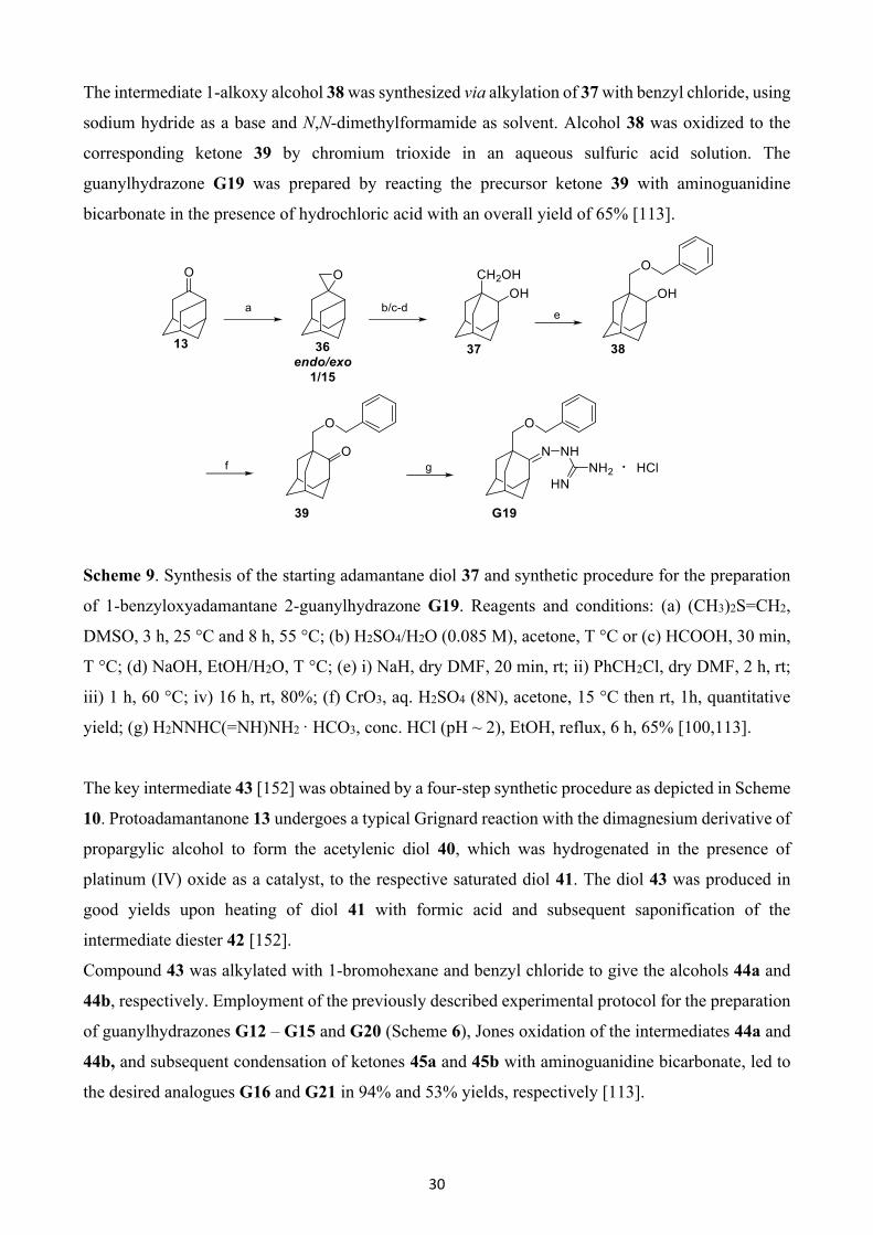
30
The intermediate 1-alkoxy alcohol 38 was synthesized via alkylation of 37 with benzyl chloride, using
sodium hydride as a base and N,N-dimethylformamide as solvent. Alcohol 38 was oxidized to the
corresponding ketone 39 by chromium trioxide in an aqueous sulfuric acid solution. The
guanylhydrazone G19 was prepared by reacting the precursor ketone 39 with aminoguanidine
bicarbonate in the presence of hydrochloric acid with an overall yield of 65% [113].
Scheme 9. Synthesis οf the starting adamantane diol 37 and synthetic procedure for the preparation
of 1-benzyloxyadamantane 2-guanylhydrazone G19. Reagents and conditions: (a) (CH3)2S=CH2,
DMSO, 3 h, 25 °C and 8 h, 55 °C; (b) H2SO4/H2O (0.085 M), acetone, T °C or (c) HCOOH, 30 min,
T °C; (d) NaOH, EtOH/H2O, T °C; (e) i) NaH, dry DMF, 20 min, rt; ii) PhCH2Cl, dry DMF, 2 h, rt;
iii) 1 h, 60 °C; iv) 16 h, rt, 80%; (f) CrO3, aq. H2SO4 (8N), acetone, 15 °C then rt, 1h, quantitative
yield; (g) H2NNHC(=NH)NH2 · HCO3, conc. HCl (pH ~ 2), EtOH, reflux, 6 h, 65% [100,113].
The key intermediate 43 [152] was obtained by a four-step synthetic procedure as depicted in Scheme
10. Protoadamantanone 13 undergoes a typical Grignard reaction with the dimagnesium derivative of
propargylic alcohol to form the acetylenic diol 40, which was hydrogenated in the presence of
platinum (IV) oxide as a catalyst, to the respective saturated diol 41. The diol 43 was produced in
good yields upon heating of diol 41 with formic acid and subsequent saponification of the
intermediate diester 42 [152].
Compound 43 was alkylated with 1-bromohexane and benzyl chloride to give the alcohols 44a and
44b, respectively. Employment of the previously described experimental protocol for the preparation
of guanylhydrazones G12 – G15 and G20 (Scheme 6), Jones oxidation of the intermediates 44a and
44b, and subsequent condensation of ketones 45a and 45b with aminoguanidine bicarbonate, led to
the desired analogues G16 and G21 in 94% and 53% yields, respectively [113].
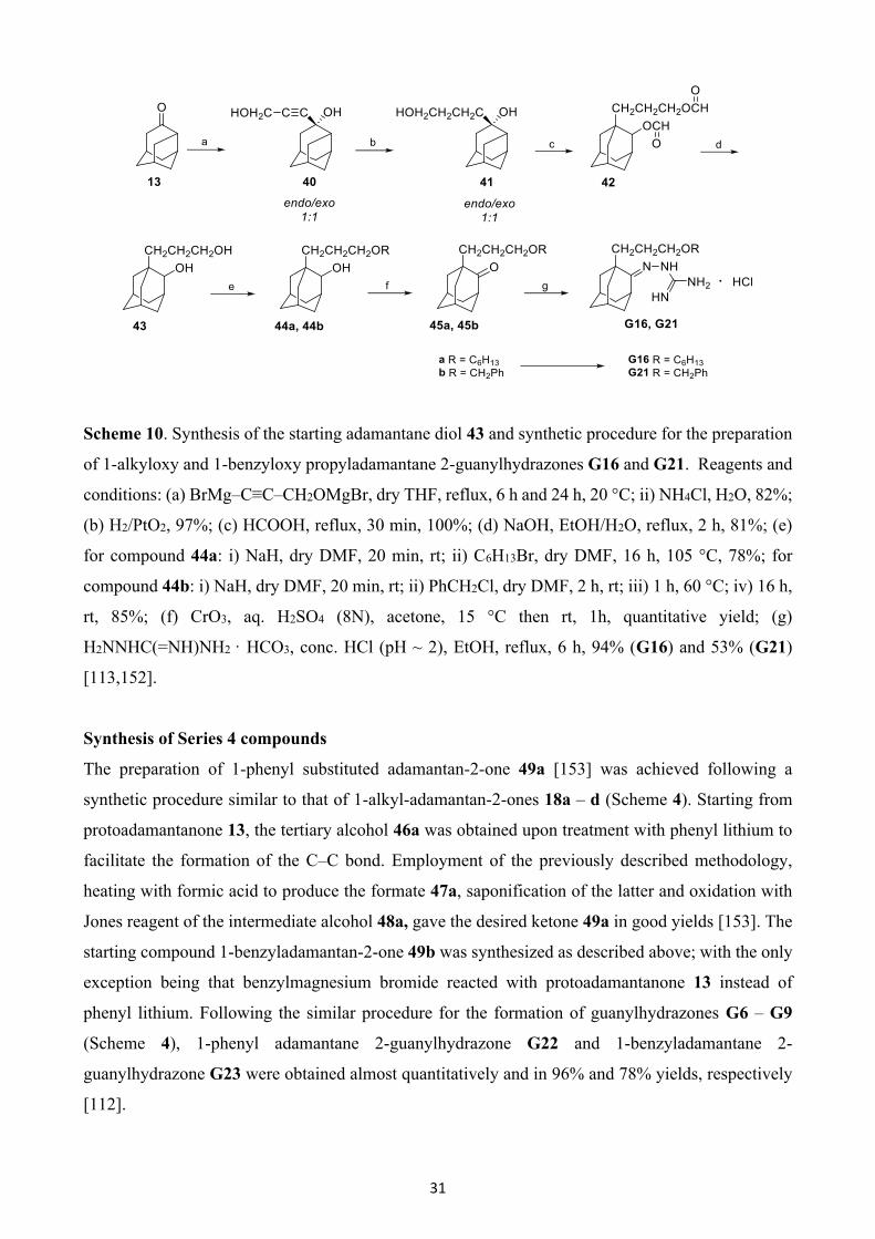
31
Scheme 10. Synthesis of the starting adamantane diol 43 and synthetic procedure for the preparation
of 1-alkyloxy and 1-benzyloxy propyladamantane 2-guanylhydrazones G16 and G21. Reagents and
conditions: (a) BrMg–C≡C–CH2OMgBr, dry THF, reflux, 6 h and 24 h, 20 °C; ii) NH4Cl, H2O, 82%;
(b) H2/PtO2, 97%; (c) HCOOH, reflux, 30 min, 100%; (d) NaOH, EtOH/H2O, reflux, 2 h, 81%; (e)
for compound 44a: i) NaH, dry DMF, 20 min, rt; ii) C6H13Br, dry DMF, 16 h, 105 °C, 78%; for
compound 44b: i) NaH, dry DMF, 20 min, rt; ii) PhCH2Cl, dry DMF, 2 h, rt; iii) 1 h, 60 °C; iv) 16 h,
rt, 85%; (f) CrO3, aq. H2SO4 (8N), acetone, 15 °C then rt, 1h, quantitative yield; (g)
H2NNHC(=NH)NH2 · HCO3, conc. HCl (pH ~ 2), EtOH, reflux, 6 h, 94% (G16) and 53% (G21)
[113,152].
Synthesis of Series 4 compounds
The preparation of 1-phenyl substituted adamantan-2-one 49a [153] was achieved following a
synthetic procedure similar to that of 1-alkyl-adamantan-2-ones 18a – d (Scheme 4). Starting from
protoadamantanone 13, the tertiary alcohol 46a was obtained upon treatment with phenyl lithium to
facilitate the formation of the C–C bond. Employment of the previously described methodology,
heating with formic acid to produce the formate 47a, saponification of the latter and oxidation with
Jones reagent of the intermediate alcohol 48a, gave the desired ketone 49a in good yields [153]. The
starting compound 1-benzyladamantan-2-one 49b was synthesized as described above; with the only
exception being that benzylmagnesium bromide reacted with protoadamantanone 13 instead of
phenyl lithium. Following the similar procedure for the formation of guanylhydrazones G6 – G9
(Scheme 4), 1-phenyl adamantane 2-guanylhydrazone G22 and 1-benzyladamantane 2-
guanylhydrazone G23 were obtained almost quantitatively and in 96% and 78% yields, respectively
[112].
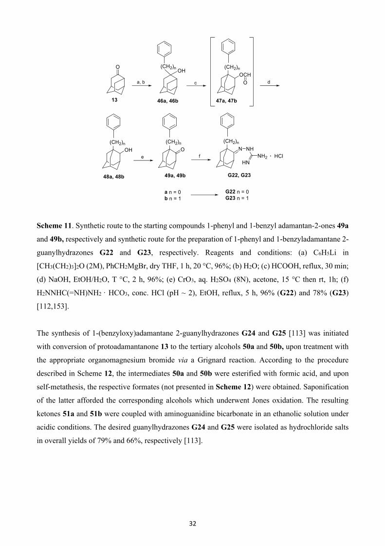
32
Scheme 11. Synthetic route to the starting compounds 1-phenyl and 1-benzyl adamantan-2-ones 49a
and 49b, respectively and synthetic route for the preparation of 1-phenyl and 1-benzyladamantane 2-
guanylhydrazones G22 and G23, respectively. Reagents and conditions: (a) C6H5Li in
[CH3(CH2)3]2O (2M), PhCH2MgBr, dry THF, 1 h, 20 °C, 96%; (b) H2O; (c) HCOOH, reflux, 30 min;
(d) NaOH, EtOH/H2O, T °C, 2 h, 96%; (e) CrO3, aq. H2SO4 (8N), acetone, 15 °C then rt, 1h; (f)
H2NNHC(=NH)NH2 · HCO3, conc. HCl (pH ~ 2), EtOH, reflux, 5 h, 96% (G22) and 78% (G23)
[112,153].
The synthesis of 1-(benzyloxy)adamantane 2-guanylhydrazones G24 and G25 [113] was initiated
with conversion of protoadamantanone 13 to the tertiary alcohols 50a and 50b, upon treatment with
the appropriate organomagnesium bromide via a Grignard reaction. According to the procedure
described in Scheme 12, the intermediates 50a and 50b were esterified with formic acid, and upon
self-metathesis, the respective formates (not presented in Scheme 12) were obtained. Saponification
of the latter afforded the corresponding alcohols which underwent Jones oxidation. The resulting
ketones 51a and 51b were coupled with aminoguanidine bicarbonate in an ethanolic solution under
acidic conditions. The desired guanylhydrazones G24 and G25 were isolated as hydrochloride salts
in overall yields of 79% and 66%, respectively [113].
33
Scheme 12. Synthetic procedure for the preparation of 1-(benzyloxy)adamantane 2-
guanylhydrazones G24 and G25. Reagents and conditions: (a) XC6H4MgBr, dry THF; (b) HCOOH,
reflux, 30 min; (c) NaOH, EtOH/H2O, 2 h, T °C; (d) CrO3, aq. H2SO4 (8N), acetone, 15 °C then rt,
30 min, 93% (51a) and 91% (51b); (e) H2NNHC(=NH)NH2 · HCO3, conc. HCl (pH ~ 2), EtOH,
reflux, 5 h, 79% (G24) and 66% (G25) [113].
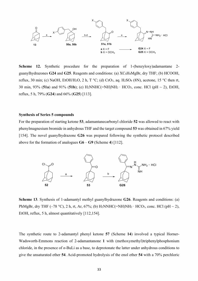
Synthesis of Series 5 compounds
For the preparation of starting ketone 53, adamantanecarbonyl chloride 52 was allowed to react with
phenylmagnesium bromide in anhydrous THF and the target compound 53 was obtained in 67% yield
[154]. The novel guanylhydrazone G26 was prepared following the synthetic protocol described
above for the formation of analogues G6 – G9 (Scheme 4) [112].
Scheme 13. Synthesis of 1-adamantyl methyl guanylhydrazone G26. Reagents and conditions: (a)
PhMgBr, dry THF (–78 °C), 2 h, rt, Ar, 67%; (b) H2NNHC(=NH)NH2 · HCO3, conc. HCl (pH ~ 2),
EtOH, reflux, 5 h, almost quantitatively [112,154].
The synthetic route to 2-adamantyl phenyl ketone 57 (Scheme 14) involved a typical Horner-
Wadsworth-Emmons reaction of 2-adamantanone 1 with (methoxymethyl)triphenylphosphonium
chloride, in the presence of n-BuLi as a base, to deprotonate the latter under anhydrous conditions to
give the unsaturated ether 54. Acid-promoted hydrolysis of the enol ether 54 with a 70% perchloric
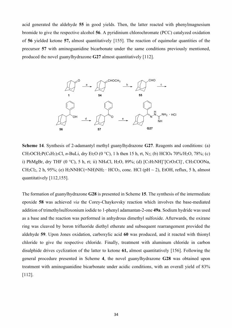
34
acid generated the aldehyde 55 in good yields. Then, the latter reacted with phenylmagnesium
bromide to give the respective alcohol 56. A pyridinium chlorochromate (PCC) catalyzed oxidation
of 56 yielded ketone 57, almost quantitatively [155]. The reaction of equimolar quantities of the
precursor 57 with aminoguanidine bicarbonate under the same conditions previously mentioned,
produced the novel guanylhydrazone G27 almost quantitatively [112].
Scheme 14. Synthesis of 2-adamantyl methyl guanylhydrazone G27. Reagents and conditions: (a)
CH3OCH2P(C6H5)3Cl, n-BuLi, dry Et2O (0 °C), 1 h then 15 h, rt, N2; (b) HClO4 70%/H2O, 78%; (c)
i) PhMgBr, dry THF (0 °C), 5 h, rt; ii) NH4Cl, H2O, 89%; (d) [C5H5NH]+[CrO3Cl]-, CH3COONa,
CH2Cl2, 2 h, 95%; (e) H2NNHC(=NH)NH2 · HCO3, conc. HCl (pH ~ 2), EtOH, reflux, 5 h, almost
quantitatively [112,155].
The formation of guanylhydrazone G28 is presented in Scheme 15. The synthesis of the intermediate
epoxide 58 was achieved via the Corey-Chaykovsky reaction which involves the base-mediated
addition of trimethylsulfoxonium iodide to 1-phenyl adamantan-2-one 49a. Sodium hydride was used
as a base and the reaction was performed in anhydrous dimethyl sulfoxide. Afterwards, the oxirane
ring was cleaved by boron trifluoride diethyl etherate and subsequent rearrangement provided the
aldehyde 59. Upon Jones oxidation, carboxylic acid 60 was produced, and it reacted with thionyl
chloride to give the respective chloride. Finally, treatment with aluminum chloride in carbon
disulphide drives cyclization of the latter to ketone 61, almost quantitatively [156]. Following the
general procedure presented in Scheme 4, the novel guanylhydrazone G28 was obtained upon
treatment with aminoguanidine bicarbonate under acidic conditions, with an overall yield of 83%
[112].
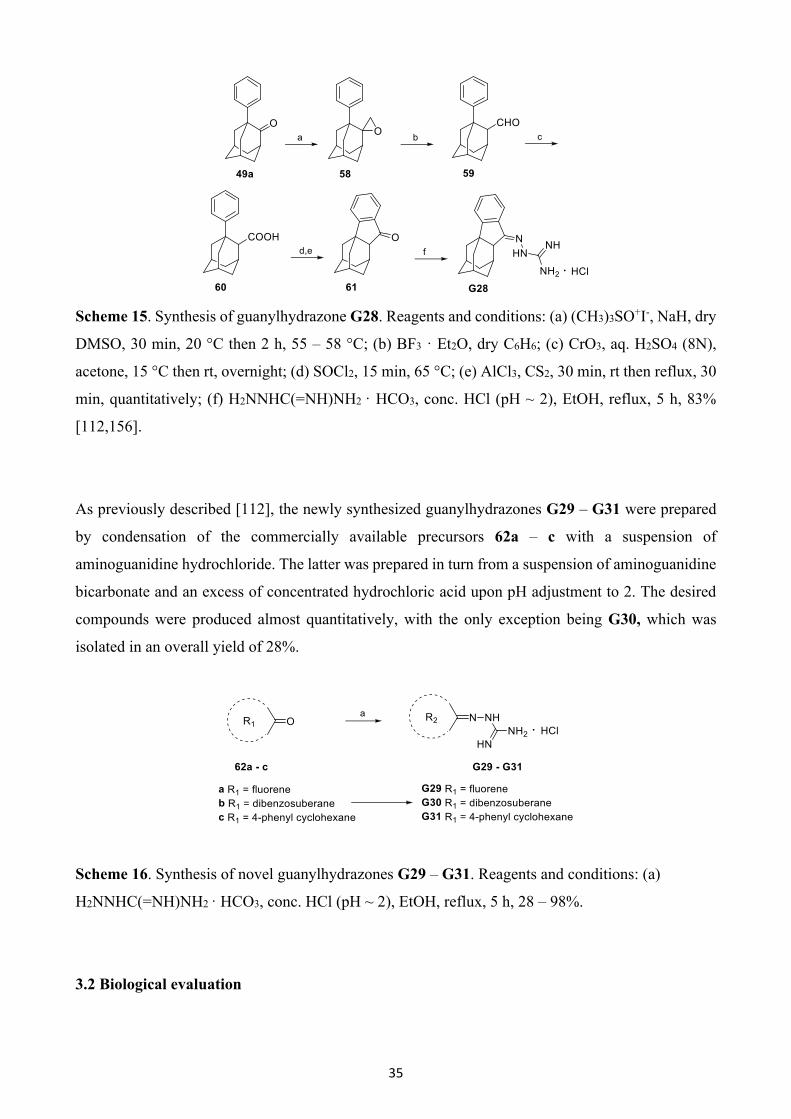
35
Scheme 15. Synthesis of guanylhydrazone G28. Reagents and conditions: (a) (CH3)3SO+I-, NaH, dry
DMSO, 30 min, 20 °C then 2 h, 55 – 58 °C; (b) BF3 · Et2O, dry C6H6; (c) CrO3, aq. H2SO4 (8N),
acetone, 15 °C then rt, overnight; (d) SOCl2, 15 min, 65 °C; (e) AlCl3, CS2, 30 min, rt then reflux, 30
min, quantitatively; (f) H2NNHC(=NH)NH2 · HCO3, conc. HCl (pH ~ 2), EtOH, reflux, 5 h, 83%
[112,156].
As previously described [112], the newly synthesized guanylhydrazones G29 – G31 were prepared
by condensation of the commercially available precursors 62a – c with a suspension of
aminoguanidine hydrochloride. The latter was prepared in turn from a suspension of aminoguanidine
bicarbonate and an excess of concentrated hydrochloric acid upon pH adjustment to 2. The desired
compounds were produced almost quantitatively, with the only exception being G30, which was
isolated in an overall yield of 28%.
Scheme 16. Synthesis of novel guanylhydrazones G29 – G31. Reagents and conditions: (a)
H2NNHC(=NH)NH2 · HCO3, conc. HCl (pH ~ 2), EtOH, reflux, 5 h, 28 – 98%.
3.2 Biological evaluation
36
Series 1-5 analogues (G1 – G31) were assessed based on their ability to inhibit proliferation of
cultured bloodstream form T. brucei in vitro. Series 5 analogues (G26 – G31) were also tested against
T. cruzi epimastigotes in vitro. The EC50 and EC90 values for each compound are presented in Tables
1 – 5. As shown, 20 (G3, G8 – G11, G13, G16 – G19, G21, G22 – G25, G26 – G29 and G31) out
of the 31 tested compounds had promising EC50 values in the low micromolar to sub micromolar
range against T. brucei. Most of the compounds were evaluated as hydrochloride salts, with the only
exception being analogues G10 and G11, which were tested as free bases.
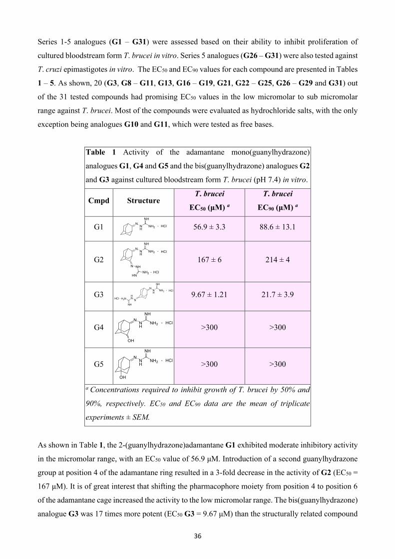
Table 1 Activity of the adamantane mono(guanylhydrazone)
analogues G1, G4 and G5 and the bis(guanylhydrazone) analogues G2
and G3 against cultured bloodstream form T. brucei (pH 7.4) in vitro.
Cmpd Structure T. brucei
EC50 (μM) a
T. brucei
EC90 (μM) a
G1
56.9 ± 3.3 88.6 ± 13.1
G2
167 ± 6 214 ± 4
G3
9.67 ± 1.21 21.7 ± 3.9
G4
>300 >300
G5
>300 >300
a Concentrations required to inhibit growth of T. brucei by 50% and
90%, respectively. EC50 and EC90 data are the mean of triplicate
experiments ± SEM.
As shown in Table 1, the 2-(guanylhydrazone)adamantane G1 exhibited moderate inhibitory activity
in the micromolar range, with an EC50 value of 56.9 μM. Introduction of a second guanylhydrazone
group at position 4 of the adamantane ring resulted in a 3-fold decrease in the activity of G2 (EC50 =
167 μM). It is of great interest that shifting the pharmacophore moiety from position 4 to position 6
of the adamantane cage increased the activity to the low micromolar range. The bis(guanylhydrazone)
analogue G3 was 17 times more potent (EC50 G3 = 9.67 μM) than the structurally related compound
37
G2. Incorporation of the OH functional group at position 4 of mono(guanylhydrazone) analogue G1
led to a substantial loss of activity for the G4 (EC50 = >300 μM). Changing the position of the
hydroxyl group to the vicinal carbon atom yielded compound G5, which also showed no activity at
the highest concentration tested (~300 μM).
Table 2 Activity of the 1-alkyl-2-guanylhydrazone adamantanes G6
– G9 and the 1-alkyl-2-thiosemicarbazone adamantanes G10 and
G11 against cultured bloodstream form T. brucei (pH 7.4) in vitro.
Cmpd Structure T. brucei
EC50 (μM) a
T. brucei
EC90 (μM) a
G6
>6.0 -
G7
>6.0 -
G8
0.09 ± 0.02 0.11 ± 0.00
G9
0.84 ± 0.50 1.30 ± 0.50
G10
2.81 ± 0.09 5.64 ± 0.54
G11
0.42 ± 0.04 0.54 ± 0.02
a Concentrations required to inhibit growth of T. brucei by 50% and
90%, respectively. EC50 and EC90 data are the mean of triplicate
experiments ± SEM.
Compounds that constitute Series 2, characterized by a lipophilic tail in position 1 of the adamantane
core, were more potent than those of Series 1. In particular, the EC50 values were at low micromolar
to submicromolar levels (2.81 μM to 0.09 μM), as indicated in Table 2. The least amount of inhibitory
activity was displayed by 1-hexyl- and 1-octyl substituted adamantane guanylhydrazones G6 and G7,
respectively, whose median effective concentration was above 6 μM. 1-Decyl substitution provided
the most potent analogue (G8) of this series, with an EC50 value of 0.09 μM. Compared to its
unsubstituted congener G1 (EC50 = 56.9 μM), G8 was 632-fold more active. Larger alkyl groups
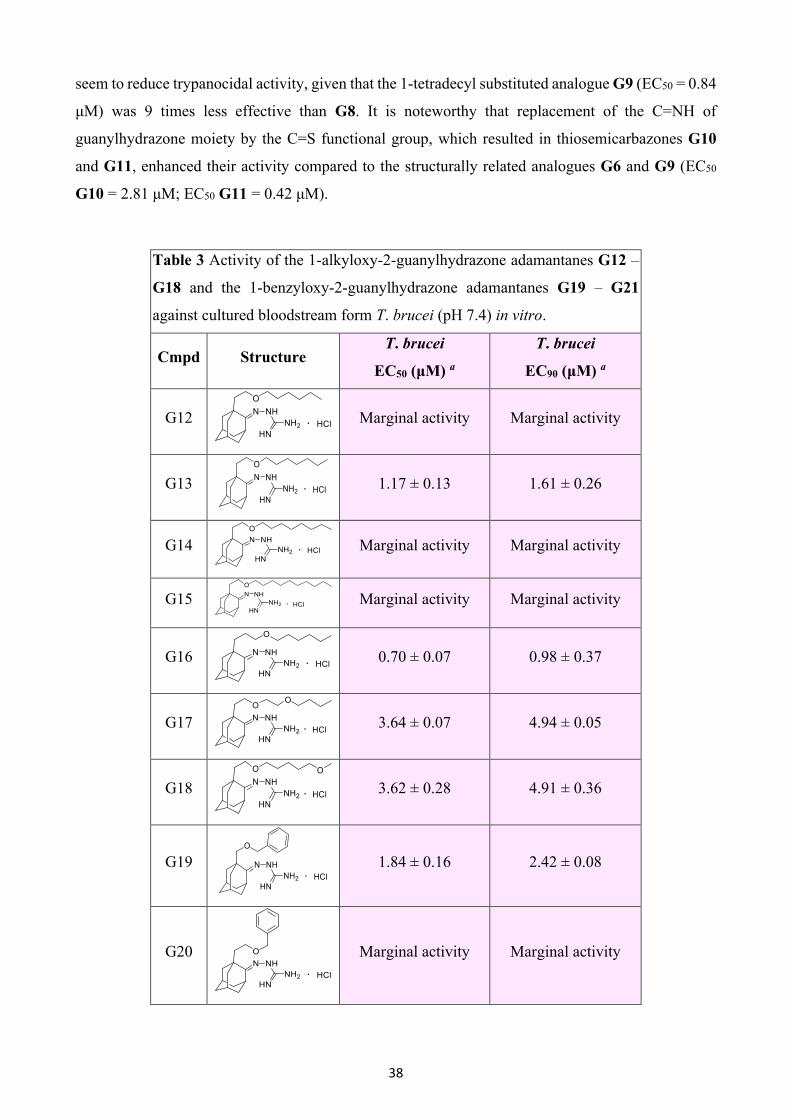
38
seem to reduce trypanocidal activity, given that the 1-tetradecyl substituted analogue G9 (EC50 = 0.84
μM) was 9 times less effective than G8. It is noteworthy that replacement of the C=NH of
guanylhydrazone moiety by the C=S functional group, which resulted in thiosemicarbazones G10
and G11, enhanced their activity compared to the structurally related analogues G6 and G9 (EC50
G10 = 2.81 μM; EC50 G11 = 0.42 μM).
Table 3 Activity of the 1-alkyloxy-2-guanylhydrazone adamantanes G12 –
G18 and the 1-benzyloxy-2-guanylhydrazone adamantanes G19 – G21
against cultured bloodstream form T. brucei (pH 7.4) in vitro.
Cmpd Structure T. brucei
EC50 (μM) a
T. brucei
EC90 (μM) a
G12
Marginal activity Marginal activity
G13
1.17 ± 0.13 1.61 ± 0.26
G14
Marginal activity Marginal activity
G15
Marginal activity Marginal activity
G16
0.70 ± 0.07 0.98 ± 0.37
G17
3.64 ± 0.07 4.94 ± 0.05
G18
3.62 ± 0.28 4.91 ± 0.36
G19
1.84 ± 0.16 2.42 ± 0.08
G20
Marginal activity Marginal activity
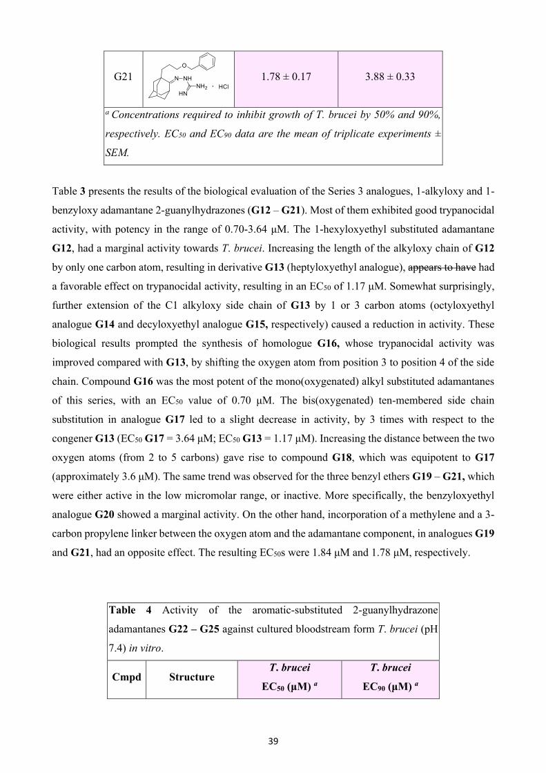
39
G21
1.78 ± 0.17 3.88 ± 0.33
a Concentrations required to inhibit growth of T. brucei by 50% and 90%,
respectively. EC50 and EC90 data are the mean of triplicate experiments ±
SEM.
Table 3 presents the results of the biological evaluation of the Series 3 analogues, 1-alkyloxy and 1-
benzyloxy adamantane 2-guanylhydrazones (G12 – G21). Most of them exhibited good trypanocidal
activity, with potency in the range of 0.70-3.64 μM. The 1-hexyloxyethyl substituted adamantane
G12, had a marginal activity towards T. brucei. Increasing the length of the alkyloxy chain of G12
by only one carbon atom, resulting in derivative G13 (heptyloxyethyl analogue), appears to have had
a favorable effect on trypanocidal activity, resulting in an EC50 of 1.17 μM. Somewhat surprisingly,
further extension of the C1 alkyloxy side chain of G13 by 1 or 3 carbon atoms (octyloxyethyl
analogue G14 and decyloxyethyl analogue G15, respectively) caused a reduction in activity. These
biological results prompted the synthesis of homologue G16, whose trypanocidal activity was
improved compared with G13, by shifting the oxygen atom from position 3 to position 4 of the side
chain. Compound G16 was the most potent of the mono(oxygenated) alkyl substituted adamantanes
of this series, with an EC50 value of 0.70 μM. The bis(oxygenated) ten-membered side chain
substitution in analogue G17 led to a slight decrease in activity, by 3 times with respect to the
congener G13 (EC50 G17 = 3.64 μM; EC50 G13 = 1.17 μM). Increasing the distance between the two
oxygen atoms (from 2 to 5 carbons) gave rise to compound G18, which was equipotent to G17
(approximately 3.6 μM). The same trend was observed for the three benzyl ethers G19 – G21, which
were either active in the low micromolar range, or inactive. More specifically, the benzyloxyethyl
analogue G20 showed a marginal activity. On the other hand, incorporation of a methylene and a 3-
carbon propylene linker between the oxygen atom and the adamantane component, in analogues G19
and G21, had an opposite effect. The resulting EC50s were 1.84 μM and 1.78 μM, respectively.
Table 4 Activity of the aromatic-substituted 2-guanylhydrazone
adamantanes G22 – G25 against cultured bloodstream form T. brucei (pH
7.4) in vitro.
Cmpd Structure T. brucei
EC50 (μM) a
T. brucei
EC90 (μM) a
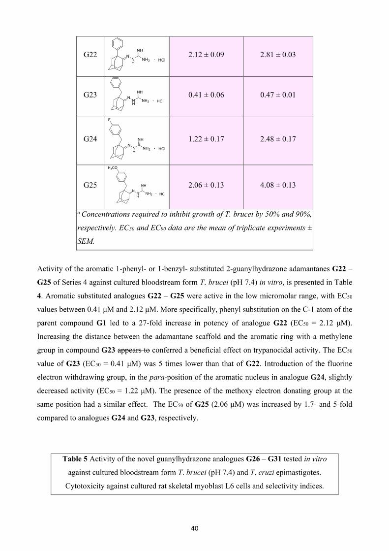
40
G22
2.12 ± 0.09 2.81 ± 0.03
G23
0.41 ± 0.06 0.47 ± 0.01
G24
1.22 ± 0.17 2.48 ± 0.17
G25
2.06 ± 0.13 4.08 ± 0.13
a Concentrations required to inhibit growth of T. brucei by 50% and 90%,
respectively. EC50 and EC90 data are the mean of triplicate experiments ±
SEM.
Activity of the aromatic 1-phenyl- or 1-benzyl- substituted 2-guanylhydrazone adamantanes G22 –
G25 of Series 4 against cultured bloodstream form T. brucei (pH 7.4) in vitro, is presented in Table
4. Aromatic substituted analogues G22 – G25 were active in the low micromolar range, with EC50
values between 0.41 μM and 2.12 μM. More specifically, phenyl substitution on the C-1 atom of the
parent compound G1 led to a 27-fold increase in potency of analogue G22 (EC50 = 2.12 μM).
Increasing the distance between the adamantane scaffold and the aromatic ring with a methylene
group in compound G23 appears to conferred a beneficial effect on trypanocidal activity. The EC50
value of G23 (EC50 = 0.41 μM) was 5 times lower than that of G22. Introduction of the fluorine
electron withdrawing group, in the para-position of the aromatic nucleus in analogue G24, slightly
decreased activity (EC50 = 1.22 μM). The presence of the methoxy electron donating group at the
same position had a similar effect. The EC50 of G25 (2.06 μM) was increased by 1.7- and 5-fold
compared to analogues G24 and G23, respectively.
Table 5 Activity of the novel guanylhydrazone analogues G26 – G31 tested in vitro
against cultured bloodstream form T. brucei (pH 7.4) and T. cruzi epimastigotes.
Cytotoxicity against cultured rat skeletal myoblast L6 cells and selectivity indices.

41
Cm
pd Structure
T. brucei
EC50 (μM)
a
T. brucei
EC90
(μM) a
SI
T.
brucei b
T. cruzi
EC50
(μM) a
T. cruzi
EC90
(μM) a
SI
T.
cruzi b
L6 cells
CC50
(μM) c
G26
1.62 ±
0.06
2.16 ±
0.02 - - - - -
G27
1.35 ±
0.09
1.71 ±
0.02 3.4
5.40 ±
1.17 11.2 ± 1 0.9
4.65 ±
0.54
G28
1.99 ±
0.06
2.72 ±
0.12 6.5
3.47 ±
0.15
4.90 ±
0.12 3.7
12.9 ±
0.3
G29
6.88 ±
0.72
13.1 ±
0.7 3.7
6.14 ±
0.33
11.6 ±
0.3 4.2
25.5 ±
0.6
G30
>30 - - >30 - - -
G31
14.3 ± 1.2 21.1 ±
0.6 13 >30 - - 186 ± 11
a Concentrations required to inhibit growth of T. brucei and T. cruzi by 50% and 90%,
respectively. EC50 and EC90 data are the mean of triplicate experiments ± SEM. b Selectivity
indices were calculated as the ratio of the CC50 for L6 cells to EC50 for T. brucei or T. cruzi,
respectively. c Cytotoxicity was determined by establishing the concentration required to
inhibit growth of cultured L6 cells by 50% (CC50). Data are the mean of triplicate
experiments ± SEM.
The novel analogues G26 – G31 were tested for their trypanocidal potency against both cultured
bloodstream form T. brucei and T. cruzi epimastigotes in vitro. The resulting EC50 and EC90 values
are presented in Table 5. Cytotoxicity values (CC50) and selectivity indices (SIs) were also
determined. The compounds bearing an adamantane core (G26 – G28) were the most active, being
in the low micromolar range, while the replacement of the adamantane scaffold led to compounds
with decreased potency, in a higher micromolar level, with the exception of compound G30, which
was inactive. More specifically, compounds G26 – G29 and G31 exhibited considerable activity
against T. brucei with EC50 values ranging from 1.35 to 14.3 μM. With the G26 analogue, the
guanylhydrazone group was shifted in the methylene carbon of the benzyl substituent, with respect
to the parent compound G23. This structural modification resulted in a higher EC50 value by 4 times
(EC50 G26 = 1.62 μM; EC50 G23 = 0.41 μM). The respective 2-substituted analogue G27 was
42
equipotent to G26 (EC50 G27 = 1.35 μM). Ring closure by formation of the C1 – C2’ bond of G27
resulted in the adamantane-cyclopentane-benzene fused analogue G28, which exhibited a similar
inhibitory activity (EC50 G28 = 1.99 μM). Replacing the adamantane component with a second
benzene ring lowered the activity observed for of the analogue G29 by 3.5 fold (EC50 G29 = 6.88
μM), while changing the linker cyclopentane ring to the bulkier cycloheptane in compound G30
abolished the activity. Finally, the use of a completely different and non-rigid scaffold, like the one
of such as the 4-phenyl cyclohexane (G31), seems to compromise disfavor activity compared to the
structurally related analogue G29, as G31 was 2-fold less effective.
Most of the newly synthesized compounds were also tested against T. cruzi epimastigotes. Derivatives
G27, G28 and G29 were the most potent, with EC50 values being 5.40 μM, 3.47 μM and 6.14 μM,
respectively. On the other hand, G30 and G31 showed no inhibition in vitro at 30 μM, which was the
highest concentration tested. The cytotoxicity of the target compounds against mammalian cells was
determined using the rat skeletal myoblast L6 cell line. The CC50 values ranged between 4.65 for
analogue G27 and 186 μM for G31. Moreover, selectivity indices observed for T. brucei were
generally higher than those observed for T. cruzi, as shown in Table 5.
3.3 Computational studies
S-Adenosylmethionine Decarboxylase
Recently, Volkov and colleagues [157] elucidated the crystal structure of the T. brucei
AdoMetDC/prozyme heterodimer in complex with a carbonimidic dihydrazide derivative well-
known for its AdoMetDC inhibition activity, namely CGP 40215 (CGP) [108]. Structural data from
the binding site of the protein-ligand complex reveal that CGP forms several interactions, including
a bidentate H-bond with E266 and monodentate H-bonds with the backbone carbonyl of L83, the side
chain hydroxyls of S249 and Y243, and with C100. Additionally, CGP is further stabilized through
π-π stacking between Y243 and F28, as well as cation-π interactions with F28 (Fig. (6)). Due to the
structural similarity of CGP with our guanylhydrazone analogues, it was thus hypothesized that both
will share a common binding pattern, rendering these as interesting and novel in drug development
for AdoMetDC. In order to test this hypothesis, in silico docking experiments were conducted.
Furthermore, guanylhydrazone compound characterisation and model validation was obtained
through an extended SAR and QSAR analysis.
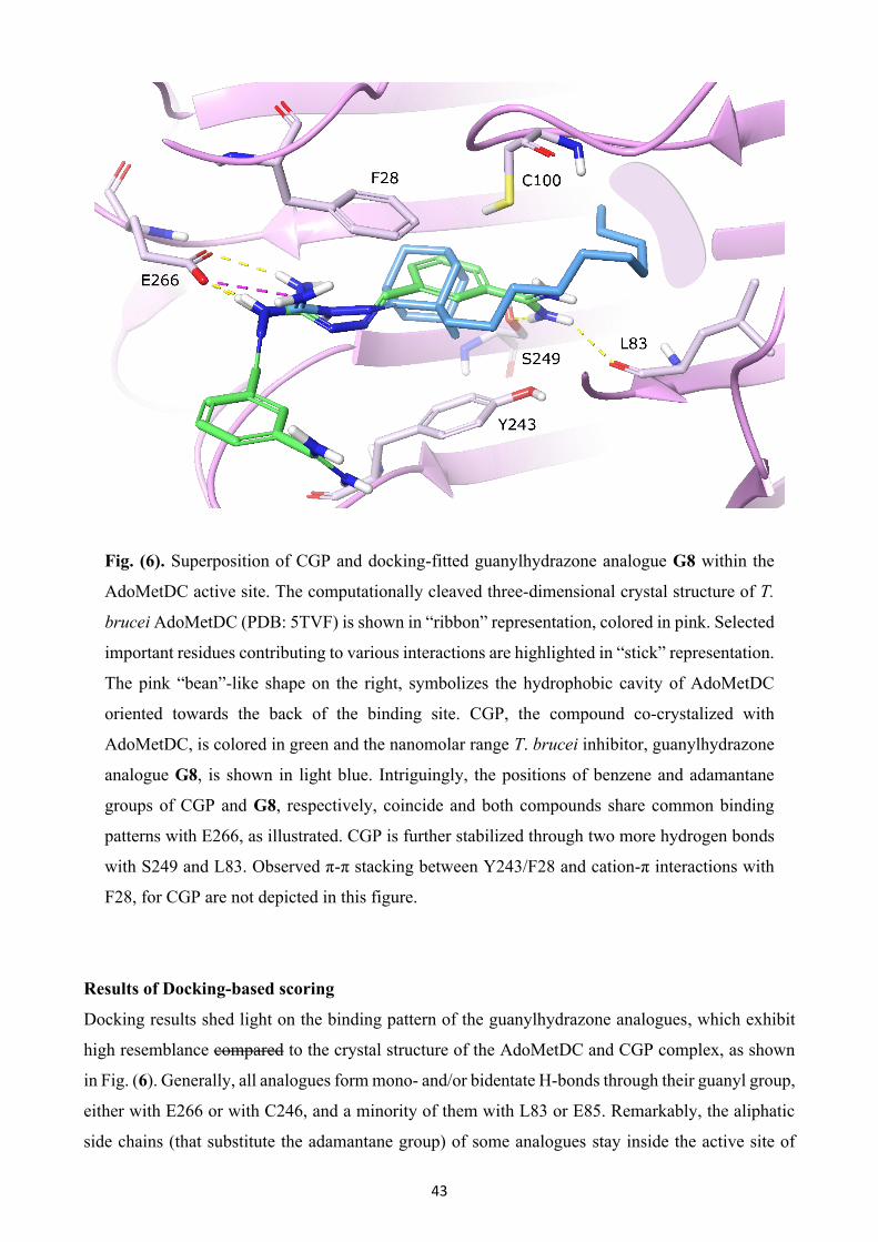
43
Fig. (6). Superposition of CGP and docking-fitted guanylhydrazone analogue G8 within the
AdoMetDC active site. The computationally cleaved three-dimensional crystal structure of T.
brucei AdoMetDC (PDB: 5TVF) is shown in “ribbon” representation, colored in pink. Selected
important residues contributing to various interactions are highlighted in “stick” representation.
The pink “bean”-like shape on the right, symbolizes the hydrophobic cavity of AdoMetDC
oriented towards the back of the binding site. CGP, the compound co-crystalized with
AdoMetDC, is colored in green and the nanomolar range T. brucei inhibitor, guanylhydrazone
analogue G8, is shown in light blue. Intriguingly, the positions of benzene and adamantane
groups of CGP and G8, respectively, coincide and both compounds share common binding
patterns with E266, as illustrated. CGP is further stabilized through two more hydrogen bonds
with S249 and L83. Observed π-π stacking between Y243/F28 and cation-π interactions with
F28, for CGP are not depicted in this figure.
Results of Docking-based scoring
Docking results shed light on the binding pattern of the guanylhydrazone analogues, which exhibit
high resemblance compared to the crystal structure of the AdoMetDC and CGP complex, as shown
in Fig. (6). Generally, all analogues form mono- and/or bidentate H-bonds through their guanyl group,
either with E266 or with C246, and a minority of them with L83 or E85. Remarkably, the aliphatic
side chains (that substitute the adamantane group) of some analogues stay inside the active site of
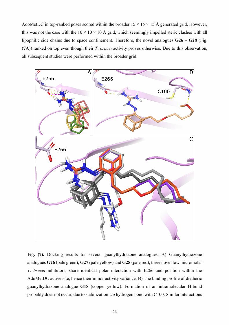
44
AdoMetDC in top-ranked poses scored within the broader 15 × 15 × 15 Å generated grid. However,
this was not the case with the 10 × 10 × 10 Å grid, which seemingly impelled steric clashes with all
lipophilic side chains due to space confinement. Therefore, the novel analogues G26 – G28 (Fig.
(7A)) ranked on top even though their T. brucei activity proves otherwise. Due to this observation,
all subsequent studies were performed within the broader grid.
Fig. (7). Docking results for several guanylhydrazone analogues. A) Guanylhydrazone
analogues G26 (pale green), G27 (pale yellow) and G28 (pale red), three novel low micromolar
T. brucei inhibitors, share identical polar interaction with E266 and position within the
AdoMetDC active site, hence their minor activity variance. B) The binding profile of dietheric
guanylhydrazone analogue G18 (copper yellow). Formation of an intramolecular H-bond
probably does not occur, due to stabilization via hydrogen bond with C100. Similar interactions
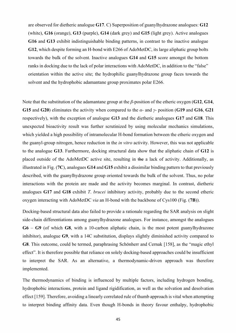
45
are observed for dietheric analogue G17. C) Superposition of guanylhydrazone analogues: G12
(white), G16 (orange), G13 (purple), G14 (dark grey) and G15 (light grey). Active analogues
G16 and G13 exhibit indistinguishable binding patterns, in contrast to the inactive analogue
G12, which despite forming an H-bond with E266 of AdoMetDC, its large aliphatic group bolts
towards the bulk of the solvent. Inactive analogues G14 and G15 score amongst the bottom
ranks in docking due to the lack of polar interactions with AdoMetDC, in addition to the “false”
orientation within the active site; the hydrophilic guanylhydrazone group faces towards the
solvent and the hydrophobic adamantane group proximates polar E266.
Note that the substitution of the adamantane group at the β-position of the etheric oxygen (G12, G14,
G15 and G20) eliminates the activity when compared to the α- and γ- position (G19 and G16, G21
respectively), with the exception of analogue G13 and the dietheric analogues G17 and G18. This
unexpected bioactivity result was further scrutinized by using molecular mechanics simulations,
which yielded a high possibility of intramolecular H-bond formation between the etheric oxygen and
the guanyl-group nitrogen, hence reduction in the in vitro activity. However, this was not applicable
to the analogue G13. Furthermore, docking structural data show that the aliphatic chain of G12 is
placed outside of the AdoMetDC active site, resulting in the a lack of activity. Additionally, as
illustrated in Fig. (7C), analogues G14 and G15 exhibit a dissimilar binding pattern to that previously
described, with the guanylhydrazone group oriented towards the bulk of the solvent. Thus, no polar
interactions with the protein are made and the activity becomes marginal. In contrast, dietheric
analogues G17 and G18 exhibit T. brucei inhibitory activity, probably due to the second etheric
oxygen interacting with AdoMetDC via an H-bond with the backbone of Cys100 (Fig. (7B)).
Docking-based structural data also failed to provide a rationale regarding the SAR analysis on slight
side-chain differentiations among guanylhydrazone analogues. For instance, amongst the analogues
G6 – G9 (of which G8, with a 10-carbon aliphatic chain, is the most potent guanylhydrazone
inhibitor), analogue G9, with a 14C substitution, displays slightly diminished activity compared to
G8. This outcome, could be termed, paraphrasing Schönherr and Cernak [158], as the “magic ethyl
effect”. It is therefore possible that reliance on solely docking-based approaches could be insufficient
to interpret the SAR. As an alternative, a thermodynamic-driven approach was therefore
implemented.
The thermodynamics of binding is influenced by multiple factors, including hydrogen bonding,
hydrophobic interactions, protein and ligand rigidification, as well as the solvation and desolvation
effect [159]. Therefore, avoiding a linearly correlated rule of thumb approach is vital when attempting
to interpret binding affinity data. Even though H-bonds in theory favour enthalpy, hydrophobic

46
interactions favor entropy and conformational changes are unfavorable for entropy. One needs to
consider that enthalpy and entropy of an intricate molecular system are vaguely interconnected. The
theory of Enthalpy−Entropy Compensation (EEC) [160,161], which states that the enthalpy gain of a
rigid protein-ligand complex is compensated by an entropy loss caused by the decrease of the
available states for the system, provides partial explanation. However, as Wienen-Schmidt and
colleagues showed, this is not always the case. Highest flexibility ligands can display,
counterintuitively, the entropically most favored binding. Thus, the loss of degrees of freedom of the
ligand does not necessarily monopolize the thermodynamic profile [162]. Adequate explanation for
this unexpected thermodynamic outcome could be offered by the role of water molecules, which also
strongly affect protein and ligand stability through their dipolar nature and the entropic gain
associated with the hydrophobic effect [163]. Water molecules of the protein-ligand complex can be
classified on the basis of their energetics into either high energy, unstable water molecules trapped
inside hydrophobic cavities or stable water molecule that are stabilized through extensive interactions
with the protein over the corresponding solvent accessible surface [164]. Computational tools such
as SZmap [165] from Openeye, have been used to incorporate the water network in drug design.
SZmap (solvent-Zap-mapping) is capable of quantitatively predicting water positions and their
energetics, and is potentially a useful tool for identifying significant favorable or unfavorable regions
of solvent thermodynamics in the binding site [166]. The hydration analysis via SZmap for the active
site of AdoMetDC was performed as previously described [167] and the results were visually
inspected.
Predictably, SZmap solvation mapping analysis yielded interesting results that compensated for the
limitations of a merely docking-scoring approach. The binding site of AdoMetDC is rich in both
stable and unstable water molecules, which through their displacement influence binding
thermodynamics. Since all analogues are comprised of bulky hydrophobic groups, such as aliphatic
hydrocarbons, adamantane and benzene moieties, water molecules are inevitably displaced. With the
introduction of the polar guanylhydrazone group, which ideally contributes to favorable enthalpy via
structural water molecule displacement, elucidating the complete thermodynamic binding profile
becomes complex. Specifically, for G8, a nanomolar-range T. brucei inhibitor, SZmap data showed
that the adamantane group as well as the 10C side chain displace unstable, high-energy water
molecules (Fig. (8)), thus contributing to the increase of entropy.
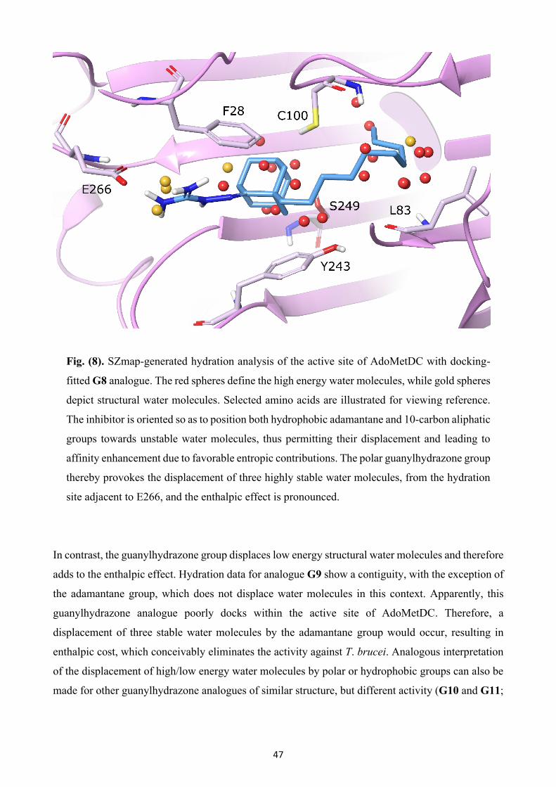
47
Fig. (8). SZmap-generated hydration analysis of the active site of AdoMetDC with docking-
fitted G8 analogue. The red spheres define the high energy water molecules, while gold spheres
depict structural water molecules. Selected amino acids are illustrated for viewing reference.
The inhibitor is oriented so as to position both hydrophobic adamantane and 10-carbon aliphatic
groups towards unstable water molecules, thus permitting their displacement and leading to
affinity enhancement due to favorable entropic contributions. The polar guanylhydrazone group
thereby provokes the displacement of three highly stable water molecules, from the hydration
site adjacent to E266, and the enthalpic effect is pronounced.
In contrast, the guanylhydrazone group displaces low energy structural water molecules and therefore
adds to the enthalpic effect. Hydration data for analogue G9 show a contiguity, with the exception of
the adamantane group, which does not displace water molecules in this context. Apparently, this
guanylhydrazone analogue poorly docks within the active site of AdoMetDC. Therefore, a
displacement of three stable water molecules by the adamantane group would occur, resulting in
enthalpic cost, which conceivably eliminates the activity against T. brucei. Analogous interpretation
of the displacement of high/low energy water molecules by polar or hydrophobic groups can also be
made for other guanylhydrazone analogues of similar structure, but different activity (G10 and G11;

48
G24 and G25; G2 and G3). These differences could be related to EEC and should be further
investigated.
Results of QSAR analysis
All statistical methods comprised of only the active analogues yielded substantial model fidelity,
based on their statistical properties, as illustrated in Table 6.
Table 6. Statistical values of Multiple Linear Regression (MLR), Partial Least Squares
regression (PLS) and Kernel-based Partial Least Squares regression (KPLS) regression
models.
Statistical Regression
Model
Training
set Test set R² Q² SD RMSE
MLR 22 5 0.9339 0.8270 0.2510 0.3569
PLS 22 5 0.8890 0.3573 0.3350 0.5466
KPLS 22 5 0.9662 0.9105 0.1752 0.2922
Interestingly, the most accurate regression model was generated through KPLS (R² = 0.9662; Q² =
0.9105). PLS had less predictive accuracy (R² = 0.8890; Q² = 0.3575). MLR analysis also produced
an acceptable model (R² = 0.9339; Q² = 0.8270). However, it was challenged by notable inter-
correlation within the Canvas-derived optimal subset of descriptors, thus rendering the respective
MLR model as one of limited predicting capacity. Out of the molecular descriptors identified as
significant by the statistical analysis, the most meaningful were the following; average vertex distance
degree, Balaban distance connectivity index, conformation independent QPlogS, Gutman molecular
topological and topological charge index of order 9. Details description of these descriptors can be
acquired from the Handbook of Molecular Descriptors, by Todeschini and colleagues [168].
Fundamentally, molecular descriptors, due to the involvement of mathematics, usually lack direct
structural interpretation of a molecule. Since chemical information is transformed through various
procedures into sole numeric values, deducing tangible characteristics about the structure of a
compound is rather difficult. That said, it is important to construct a reliable and highly predictable
model, regardless of its ability to interpret structural features. Such a model would enable rapid and
accurate activity predictions, even for novel compounds currently non-synthesized, and provide
proof-of-concept for further drug development approaches.
Experimental and predicted EC50 values of active guanylhydrazone analogues for T. brucei are shown
in Table 7.
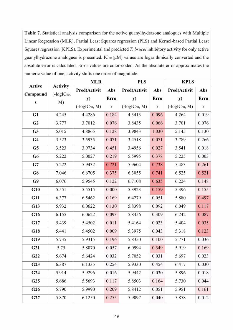
49
Table 7. Statistical analysis comparison for the active guanylhydrazone analogues with Multiple
Linear Regression (MLR), Partial Least Squares regression (PLS) and Kernel-based Partial Least
Squares regression (KPLS). Experimental and predicted T. brucei inhibitory activity for only active
guanylhydrazone analogues is presented. IC50 (μΜ) values are logarithmically converted and the
absolute error is calculated. Error values are color-coded. As the absolute error approximates the
numeric value of one, activity shifts one order of magnitude.
Active
Compound
s
Activity
(-logIC50,
M)
MLR PLS KPLS
Pred(Activit
y)
(-logIC50, M)
Abs
Erro
r
Pred(Activit
y)
(-logIC50, M)
Abs
Erro
r
Pred(Activit
y)
(-logIC50, M)
Abs
Erro
r
G1 4.245 4.4286 0.184 4.3413 0.096 4.264 0.019
G2 3.777 3.7012 0.076 3.8435 0.066 3.701 0.076
G3 5.015 4.8865 0.128 3.9843 1.030 5.145 0.130
G4 3.523 3.5935 0.071 3.4518 0.071 3.789 0.266
G5 3.523 3.9734 0.451 3.4956 0.027 3.541 0.018
G6 5.222 5.0027 0.219 5.5995 0.378 5.225 0.003
G7 5.222 5.9432 0.721 5.9604 0.738 5.483 0.261
G8 7.046 6.6705 0.375 6.3055 0.741 6.525 0.521
G9 6.076 5.9545 0.122 6.7108 0.635 6.224 0.148
G10 5.551 5.5515 0.000 5.3923 0.159 5.396 0.155
G11 6.377 6.5462 0.169 6.4279 0.051 5.880 0.497
G13 5.932 6.0622 0.130 5.8398 0.092 6.049 0.117
G16 6.155 6.0622 0.093 5.8456 0.309 6.242 0.087
G17 5.439 5.4502 0.011 5.4164 0.023 5.404 0.035
G18 5.441 5.4502 0.009 5.3975 0.043 5.318 0.123
G19 5.735 5.9315 0.196 5.8350 0.100 5.771 0.036
G21 5.75 5.8070 0.057 6.0994 0.349 5.919 0.169
G22 5.674 5.6424 0.032 5.7052 0.031 5.697 0.023
G23 6.387 6.1335 0.254 5.9330 0.454 6.417 0.030
G24 5.914 5.9296 0.016 5.9442 0.030 5.896 0.018
G25 5.686 5.5693 0.117 5.8503 0.164 5.730 0.044
G26 5.790 5.9990 0.209 5.8412 0.051 5.951 0.161
G27 5.870 6.1250 0.255 5.9097 0.040 5.858 0.012
50
G28 5.701 5.6679 0.033 5.7783 0.077 5.590 0.111
G29 5.162 4.9830 0.179 4.8028 0.360 5.082 0.080
G30 4.523 4.5656 0.043 4.7354 0.213 4.407 0.116
G31 4.845 5.4141 0.569 4.8898 0.045 5.069 0.224
As can be observed, all three regression models yielded medium to relatively high absolute error
values for analogues G7 and G8. A possible explanation for this observation can be, again, related to
the effect of the solvent. Software implementing the ligand-based drug discovery approach, such as
Canvas, are exclusively focused on the ligand structure and can thus seriously neglect the exact
thermodynamic effect that individual water molecules might have on the binding affinity of small
molecules that can displace them. The displacement of water molecules affects each compound
differently, hence the almost absolute agreement for analogues G2, G17, G18, G22, G24 among all
statistical approaches. Generated statistical data should be considered as a stepping-stone for further
investigation.
Conclusion
In summary, the data obtained from this extended SAR study illustrate: (i) The synergistic effect of
the lipophilic character of the C1 substitution and C2 guanylhydrazone moiety on trypanocidal
activity. (ii) The critical role of the adamantane moiety for trypanocidal activity. Replacement of the
adamantane cage structure by the tricyclic scaffolds of fluorene and dibenzocycloheptene, and the
bendable 4-phenyl cyclohexane decreases activity against T. brucei. (iii) The great importance of the
nature and length of the second substitution to antiparasitic activity. Replacement of the lipophilic
alkyl chain at of the adamantane-based guanylhydrazones with a more polar component (alkyloxy,
hydroxyl, aminoguanidine) results in a substantial reduction in trypanocidal properties. Finally, a
number of diverse approaches were undertaken to understand the interplay between the structure of
the promising bioactive compounds and the activity against their putative protozoan target,
AdoMetDC. Chemoinformatics-based models afforded a series of molecular descriptors that might
account for the observed bioactivity and sustain the design of new derivatives. In addition, molecular
docking simulations, enhanced by a hydration analysis, derived interesting thermodynamic insights
into the role of structural factors, such as intramolecular hydrogen bonding or displacement of
energetically unfavorable water molecules on the observed binding affinity. The design of new
compounds with improved bioactivity against AdoMetDC could greatly benefit from such rational
approaches. Efforts towards this direction are currently underway.
51
LIST OF ABBREVIATIONS
3D : Three-dimensional
AdoMet : S-adenosylmethionine
AdoMetDC : S-adenosylmethionine decarboxylase
BBB : blood-brain barrier
BZ : benznidazole
CADD : Computer-aided drug design
CATT : card agglutination test for trypanosomiasis
CNS : central nervous system
CSF : cerebrospinal fluid
CC50 : half maximal cytotoxic concentration, concentration required to cause cytotoxic effect by 50%
or to inhibit growth of cultured L6 cells by 50%
CGP : derivative CGP 40215 (AdoMetDC inhibitor)
DMF : N,N-dimethylformamide
DMSO : dimethyl sulfoxide
DRC : Democratic Republic of Congo
EC50 : half maximal effective concentration, concentration required to give half-maximal response or
concentration required to inhibit growth of T. brucei and T. cruzi by 50%
EEC : Enthalpy−Entropy Compensation
Et2O : diethyl ether
EtOH : ethanol
FBDD : Fragment-based drug discovery
gHAT : gambiense HAT
Gsp : glutathionylspermidine
HAT : Human African Trypanosomiasis
LBDD : Ligand-based drug design
IgM : Immunoglobulin M
IM : intramuscular
IV : intravenous
KPLS : Kernel-based Partial Least Squares
MLR : Multiple Linear Regression

52
NECT : nifurtimox-eflornithine combination treatment
NTDs : neglected tropical diseases
ODC : ornithine decarboxylase
PLS : Partial Least Squares
PTRE : post-treatment reactive encephalopathy
QSAR : Quantitative structure-activity relationship
rHAT : rhodesiense HAT
SBDD : Structure-based drug design
SD : Standard deviation
SI : selectivity index
SZMAP : solvent-Zap-mapping
T. brucei : Trypanosoma brucei
THF : tetrahydrofuran
VdW : van der Waals
WBC : white blood cells
WHO : World Health Organization
ACKNOWLEDGEMENTS:
G.Z. would like to thank Empirikion Foundation for the financial support. V.P. would like to thank
the State Scholarships Foundation of Greece for providing her a Ph.D. fellowship (MIS-5000432,
NSRF 2014–2020). K.M.M. and M.Š. thank the Croatian Ministry of Science, Education and Sports
for the financial support of this study (grant number 098-0982933-2911).
CONFLICT OF INTEREST
There are no conflicts to declare.
FUNDING:
Croatian Ministry of Science, Education and Sports (grant number 098-0982933-2911).
CONSENT FOR PUBLICATION:
53
All the authors consent for this publication.
REFERENCES
[1] Mackey TK, Liang BA, Cuomo R, Hafen R, Brouwer KC, Lee DE. Emerging and reemerging
neglected tropical diseases: a review of key characteristics, risk factors, and the policy and innovation
environment. Clin Microbiol Rev 2014; 27(4): 949-79.
[2] Addisu A, Adriaensen W, Balew A, et al. Neglected tropical diseases and the sustainable
development goals: an urgent call for action from the front line. BMJ Glob Health 2019; 4(1):
e001334.
[3] Peeling RW, Boeras DI, Nkengasongb J. Re-imagining the future of diagnosis of Neglected
Tropical Diseases. Comput Struct Biotechnol J 2017; 15: 271-4.
[4] Mitra AK, Mawson AR. Neglected Tropical Diseases: epidemiology and global burden. Trop Med
Infect Dis 2017; 2(3): 36.
[5] Varikuti S, Jha BK, Volpedo G, et al. Host-directed drug therapies for Neglected Tropical
Diseases caused by protozoan parasites. Front Microbiol 2018; 9: 2655.
[6] Hotez PJ, Kamath A. Neglected tropical diseases in sub-saharan Africa: review of their
prevalence, distribution, and disease burden. PLoS Negl Trop Dis 2009; 3(8): e412.
[7] Kennedy PGE. Update on human African trypanosomiasis (sleeping sickness). J Neurol 2019;
266(9): 2334-37.
[8] World Health Organization, Trypanosomiasis, human African (sleeping sickness) [updated 2019
April 12; cited 2019 August]. Available from: (https://www.who.int/news-room/fact-
sheets/detail/trypanosomiasis-human-african-(sleeping-sickness)).
[9] Bottieau E, Clerinx J. Human African Trypanosomiasis: progress and stagnation. Infect Dis Clin
North Am 2019; 33(1): 61-77.
[10] Büscher P, Cecchi G, Jamonneau V, Priotto G. Human African trypanosomiasis. Lancet 2017;
390(10110): 2397-2409.
[11] Malvy D, Chappuis F. Sleeping sickness. Clin Microbiol Infect 2011; 17(7): 986-95.
[12] Geiger A, Ravel S, Mateille T, et al. Vector competence of Glossina palpalis gambiensis for
Trypanosoma brucei s.l. and genetic diversity of the symbiont Sodalis glossinidius. Mol Biol Evol
2007; 24(1): 102-9.
[13] Geiger A, Cuny G, Frutos R. Two tsetse fly species, Glossina palpalis gambiensis and Glossina
morsitans morsitans, carry genetically distinct populations of the secondary symbiont Sodalis
glossinidius. Appl Environ Microbiol 2005; 71(12): 8941-3.
[14] Steverding D. The history of African trypanosomiasis. Parasit Vectors 2008; 1(1): 3.

54
[15] Welburn SC, Molyneux DH, Maudlin I. Beyond tsetse--implications for research and control of
Human African Trypanosomiasis epidemics. Trends Parasitol 2016; 32(3): 230-41.
[16] Kennedy PGE, Rodgers J. Clinical and neuropathogenetic aspects of Human African
Trypanosomiasis. Front Immunol 2019; 10: 39.
[17] Franco JR, Cecchi G, Priotto G, et al. Monitoring the elimination of human African
trypanosomiasis: Update to 2016. PLoS Negl Trop Dis 2018; 12(12): e0006890.
[18] Franco JR, Simarro PP, Diarra A, Jannin JG. Epidemiology of human African trypanosomiasis.
Clin Epidemiol 2014; 6: 257-75.
[19] Lindner AK, Priotto G. The unknown risk of vertical transmission in sleeping sickness--a
literature review. PLoS Negl Trop Dis 2010; 4(12): e783.
[20] Gaillot K, Lauvin MA, Cottier JP. Vertical transmission of human African trypanosomiasis:
Clinical evolution and brain MRI of a mother and her son. PLoS Negl Trop Dis 2017; 11(7):
e0005642.
[21] Biteau N, Asencio C, Izotte J, et al. Trypanosoma brucei gambiense Infections in mice lead to
tropism to the reproductive organs, and horizontal and vertical transmission. PLoS Negl Trop Dis
2016; 10(1): e0004350.
[22] Rocha G, Martins A, Gama G, Brandão F, Atouguia J. Possible cases of sexual and congenital
transmission of sleeping sickness. Lancet. 2004; 363(9404): 247.
[23] Migchelsen SJ, Büscher P, Hoepelman AI, Schallig HD, Adams ER. Human African
trypanosomiasis: a review of non-endemic cases in the past 20 years. Int J Infect Dis 2011; 15(8):
e517-24.
[24] Kennedy PGE. Clinical features, diagnosis, and treatment of human African trypanosomiasis
(sleeping sickness). Lancet Neurol 2013; 12(2): 186-94.
[25] Chappuis F, Loutan L, Simarro P, Lejon V, Büscher P. Options for field diagnosis of human
african trypanosomiasis. Clin Microbiol Rev 2005; 18(1): 133-46.
[26] Kennedy PGE, Human African trypanosomiasis of the CNS: current issues and challenges. J
Clin Invest 2004; 113(4): 496-504.
[27] Checchi F, Funk S, Chandramohan D, Haydon DT, Chappuis F. Updated estimate of the duration
of the meningo-encephalitic stage in gambiense human African trypanosomiasis. BMC Res Notes
2015; 8: 292.
[28] Mogk S, Boßelmann CM, Mudogo CN, Stein J, Wolburg H, Duszenko M. African trypanosomes
and brain infection - the unsolved question. Biol Rev Camb Philos Soc 2017; 92(3): 1675-87.
[29] Njamnshi AK, Gettinby G, Kennedy PGE. The challenging problem of disease staging in human
African trypanosomiasis (sleeping sickness): a new approach to a circular question. Trans R Soc Trop
Med Hyg 2017; 111(5): 199-203.
55
[30] Bonnet J, Boudot C, Courtioux B. Overview of the diagnostic methods used in the field for
Human African Trypanosomiasis: what could change in the next years? Biomed Res Int 2015; 2015:
583262.
[31] Büscher P, Mertens P, Leclipteux T, et al. Sensitivity and specificity of HAT Sero-K-SeT, a
rapid diagnostic test for serodiagnosis of sleeping sickness caused by Trypanosoma brucei
gambiense: a case-control study. Lancet Glob Health 2014; 2(6): e359-63.
[32] Ponte-Sucre A. An overview of Trypanosoma brucei infections: an intense host-parasite
interaction. Front Microbiol 2016; 7: 2126.
[33] Kato CD, Matovu E, Mugasa CM, Nanteza A, Alibu VP. The role of cytokines in the
pathogenesis and staging of Trypanosoma brucei rhodesiense sleeping sickness. Allergy Asthma Clin
Immunol 2016; 12: 4.
[34] Barrett MP, Boykin DW, Brun R, Tidwell RR. Human African trypanosomiasis:
pharmacological re-engagement with a neglected disease. Br J Pharmacol 2007; 152(8): 1155-71.
[35] Steverding D. Sleeping Sickness and Nagana Disease Caused by Trypanosoma brucei. In:
Marcondes C, Eds. Arthropod Borne Diseases. Cham: Springer 2017; pp. 277-97.
[36] Babokhov P, Sanyaolu AO, Oyibo WA, Fagbenro-Beyioku AF, Iriemenam NC. A current
analysis of chemotherapy strategies for the treatment of human African trypanosomiasis. Pathog Glob
Health 2013; 107(5): 242-52.
[37] Thomas JA, Baker N, Hutchinson S, et al. Insights into antitrypanosomal drug mode-of-action
from cytology-based profiling. PLoS Negl Trop Dis 2018; 12(11): e0006980.
[38] Zhang Y, Li Z, Pilch DS, Leibowitz MJ. Pentamidine inhibits catalytic activity of group I intron
Ca.LSU by altering RNA folding. Nucleic Acids Res 2002; 30(13): 2961-71.
[39] Jacobs RT, Nare B, Phillips MA. State of the art in African trypanosome drug discovery. Curr
Top Med Chem 2011; 11(10): 1255-74.
[40] Denise H, Barrett MP. Uptake and mode of action of drugs used against sleeping sickness.
Biochem Pharmacol 2001; 61(1): 1-5.
[41] Franco JR, Simarro PP, Diarra A, Ruiz-Postigo JA, Samo M, Jannin JG. Monitoring the use of
nifurtimox-eflornithine combination therapy (NECT) in the treatment of second stage gambiense
human African trypanosomiasis. Res Rep Trop Med 2012; 3: 93-101.
[42] Eperon G, Balasegaram M, Potet J, Mowbray C, Valverde O, Chappuis F. Treatment options for
second-stage gambiense human African trypanosomiasis. Expert Rev Anti Infect Ther 2014; 12(11):
1407-17.
[43] Kansiime F, Adibaku S, Wamboga C. A multicentre, randomised, non-inferiority clinical trial
comparing a nifurtimox-eflornithine combination to standard eflornithine monotherapy for late stage
56
Trypanosoma brucei gambiense human African trypanosomiasis in Uganda. Parasit Vectors 2018;
11(1): 105.
[44] Deeks ED. Fexinidazole: first global approval. Drugs 2019; 79(2): 215-220.
[45] Chappuis F. Oral fexinidazole for human African trypanosomiasis. Lancet 2018; 391(10116):
100-102.
[46] Baker CH, Welburn SC. The long wait for a new drug for Human African Trypanosomiasis.
Trends Parasitol 2018; 34(10): 818-27.
[47] Nagle AS, Khare S, Kumar AB, et al. Recent developments in drug discovery for leishmaniasis
and human African trypanosomiasis. Chem Rev 2014; 114(22): 11305-47.
[48] Steketee PC, Vincent IM, Achcar F, et al. Benzoxaborole treatment perturbs S-adenosyl-L-
methionine metabolism in Trypanosoma brucei. PLoS Negl Trop Dis 2018; 12(5): e0006450.
[49] Lewis MD, Francisco AF, Jayawardhana S, Langston H, Taylor MC, Kelly JM. Imaging the
development of chronic Chagas disease after oral transmission. Sci Rep 2018; 8(1): 11292.
[50] World Health Organization, Chagas disease (American trypanosomiasis). [updated 2019 April
17; cited 2019 August]. Available from: https://www.who.int/en/news-room/fact-
sheets/detail/chagas-disease-(american-trypanosomiasis).
[51] Pérez-Molina JA, Perez AM, Norman FF, Monge-Maillo B, López-Vélez R. Old and new
challenges in Chagas disease. Lancet Infect Dis 2015; 15(11): 1347-56.
[52] Stevens L, Dorn PL, Schmidt JO, Klotz JH, Lucero D, Klotz SA. Kissing bugs. The vectors of
Chagas. Adv Parasitol 2011; 75: 169-92.
[53] Rassi A Jr, Rassi A, Marin-Neto JA. Chagas disease. Lancet 2010; 375(9723): 1388-1402.
[54] Rajão MA, Furtado C, Alves CL, et al. Unveiling benznidazole's mechanism of action through
overexpression of DNA repair proteins in Trypanosoma cruzi. Environ Mol Mutagen 2014; 55(4):
309-21.
[55] Bermudez J, Davies C, Simonazzi A, Real JP, Palma S. Current drug therapy and pharmaceutical
challenges for Chagas disease. Acta Trop 2016; 156: 1-16.
[56] Field MC, Horn D, Fairlamb AH, et al. Anti-trypanosomatid drug discovery: an ongoing
challenge and a continuing need. Nat Rev Microbiol 2017; 15(7): 447.
[57] Stich A, Ponte-Sucre A, Holzgrabe U. Do we need new drugs against human African
trypanosomiasis? Lancet Infect Dis 2013; 13(9): 733-4.
[58] Willert E, Phillips MA. Regulation and function of polyamines in African trypanosomes. Trends
Parasitol 2012; 28(2): 66-72.
[59] Phillips MA. Polyamines in protozoan pathogens. J Biol Chem 2018; 293(48): 18746-56.
[60] Müller S, Coombs GH, Walter RD. Targeting polyamines of parasitic protozoa in chemotherapy.
Trends Parasitol 2001; 17(5): 242-9.
57
[61] Keiser J, Stich A, Burri C. New drugs for the treatment of human African trypanosomiasis:
research and development. Trends Parasitol 2001; 17(1): 42-9.
[62] Heby O, Persson L, Rentala M. Targeting the polyamine biosynthetic enzymes: a promising
approach to therapy of African sleeping sickness, Chagas' disease, and leishmaniasis. Amino Acids
2007; 33(2): 359-66.
[63] Birkholtz LM, Williams M, Niemand J, Louw AI, Persson L, Heby O. Polyamine homoeostasis
as a drug target in pathogenic protozoa: peculiarities and possibilities. Biochem J 2011; 438(2): 229-
44.
[64] Smithson DC, Lee J, Shelat AA, Phillips MA, Guy RK. Discovery of potent and selective
inhibitors of Trypanosoma brucei ornithine decarboxylase. J Biol Chem 2010; 285(22): 16771-81.
[65] Tekwani BL, Bacchi CJ, Secrist JA 3rd, Pegg AE. Irreversible inhibition of S-
adenosylmethionine decarboxylase of Trypanosoma brucei brucei by S-adenosylmethionine
analogues. Biochem Pharmacol 1992; 44(5): 905-11.
[66] Hirth B, Barker RH Jr, Celatka CA, et al. Discovery of new S-adenosylmethionine decarboxylase
inhibitors for the treatment of Human African Trypanosomiasis (HAT). Bioorg Med Chem Lett 2009;
19(11): 2916-9.
[67] Barker RH Jr, Liu H, Hirth B, et al. Novel S-adenosylmethionine decarboxylase inhibitors for
the treatment of human African trypanosomiasis. Antimicrob Agents Chemother 2009; 53(5): 2052-
8.
[68] Bacchi CJ, Barker RH Jr, Rodriguez A, et al. Trypanocidal activity of 8-methyl-5'-{[(Z)-4-
aminobut-2-enyl]-(methylamino)}adenosine (Genz-644131), an adenosylmethionine decarboxylase
inhibitor. Antimicrob Agents Chemother 2009; 53(8): 3269-72.
[69] Volkov OA, Cosner CC, Brockway AJ, et al. Identification of Trypanosoma brucei AdoMetDC
inhibitors using a high-throughput mass spectrometry-based assay. ACS Infect Dis 2017; 3(7): 512-
26.
[70] Volkov OA, Brockway AJ, Wring SA, et al. Species-selective pyrimidineamine inhibitors of
Trypanosoma brucei S-adenosylmethionine decarboxylase. J Med Chem 2018; 61(3): 1182-203.
[71] Brockway AJ, Volkov OA, Cosner CC, et al. Synthesis and evaluation of analogs of 5'-(((Z)-4-
amino-2-butenyl)methylamino)-5'-deoxyadenosine (MDL 73811, or AbeAdo) - An inhibitor of S-
adenosylmethionine decarboxylase with antitrypanosomal activity. Bioorg Med Chem 2017; 25(20):
5433-40.
[72] Velez N, Brautigam CA, Phillips MA. Trypanosoma brucei S-adenosylmethionine
decarboxylase N terminus is essential for allosteric activation by the regulatory subunit prozyme. J
Biol Chem 2013; 288(7): 5232-40.
58
[73] Lamoureux G, Artavia G. Use of the adamantane structure in medicinal chemistry. Curr Med
Chem 2010; 17(26): 2967-78.
[74] Wanka L, Iqbal K, Schreiner PR. The lipophilic bullet hits the targets: medicinal chemistry of
adamantane derivatives. Chem Rev 2013; 113(5): 3516-604.
[75] Van der Schyf CJ, Geldenhuys WJ. Polycyclic compounds: ideal drug scaffolds for the design
of multiple mechanism drugs? Neurotherapeutics 2009; 6(1): 175-86.
[76] Stockdale TP, Williams CM. Pharmaceuticals that contain polycyclic hydrocarbon scaffolds.
Chem Soc Rev 2015; 44(21): 7737-63.
[77] Štimac A, Šekutor M, Mlinarić-Majerski K, Frkanec L, Frkanec R. Adamantane in drug delivery
systems and surface recognition. Molecules 2017; 22(2): E297.
[78] Geldenhuys WJ, Malan SF, Bloomquist JR, Marchand AP, Van der Schyf CJ. Pharmacology
and structure-activity relationships of bioactive polycyclic cage compounds: a focus on
pentacycloundecane derivatives. Med Res Rev 2005; 25(1): 21-48.
[79] Liu J, Obando D, Liao V, Lifa T, Codd R. The many faces of the adamantyl group in drug design.
Eur J Med Chem 2011; 46(6): 1949-63.
[80] Grillaud M, Bianco A. Multifunctional adamantane derivatives as new scaffolds for the
multipresentation of bioactive peptides. J Pept Sci 2015; 21(5): 330-45.
[81] Horvat S, Mlinaric-Majerski K, Glavas-Obrovac L, et al. Tumor-cell-targeted methionine-
enkephalin analogues containing unnatural amino acids: design, synthesis, and in vitro antitumor
activity. J Med Chem 2006; 49(11): 3136-42.
[82] Li Z, Khaliq M, Zhou Z, Post CB, Kuhn RJ, Cushman M. Design, synthesis, and biological
evaluation of antiviral agents targeting flavivirus envelope proteins. J Med Chem 2008; 51(15): 4660-
71.
[83] Ajdačić V, Senerovic L, Vranić M, et al. Synthesis and evaluation of thiophene-based
guanylhydrazones (iminoguanidines) efficient against panel of voriconazole-resistant fungal isolates.
Bioorg Med Chem 2016; 24(6): 1277-91.
[84] Ajdačić V, Lazić J, Mojičević M, Šegan S, Nikodinović-Runić J, Opsenica I. Antibacterial and
antifungal properties of guanylhydrazones. J Serb Chem Soc 2017; 82(6): 641-49.
[85] Gadad AK, Mahajanshetti CS, Nimbalkar S, Raichurkar A. Synthesis and antibacterial activity
of some 5-guanylhydrazone/thiocyanato-6-arylimidazo[2,1-b]-1,3,4-thiadiazole-2-sulfonamide
derivatives. Eur J Med Chem 2000; 35(9): 853-7.
[86] Bairwa R, Kakwani M, Tawari NR. Novel molecular hybrids of cinnamic acids and
guanylhydrazones as potential antitubercular agents. Bioorg Med Chem Lett 2010; 20(5): 1623-5.
59
[87] Silva FP, Dantas BB, Faheina Martins GV, de Araújo DA, Vasconcellos ML. Synthesis and
anticancer activities of novel guanylhydrazone and aminoguanidine tetrahydropyran derivatives.
Molecules 2016; 21(6): E671.
[88] Andreani A, Burnelli S, Granaiola M, et al. Synthesis and antitumor activity of
guanylhydrazones from 6-(2,4-Dichloro-5-nitrophenyl)imidazo[2,1-b]thiazoles and 6-
Pyridylimidazo[2,1-b]thiazoles. J Med Chem 2006; 49(26): 7897-901.
[89] Kamal A, Kashi Reddy M, Viswanath A. The design and development of imidazothiazole-
chalcone derivatives as potential anticancer drugs. Expert Opin Drug Discov 2013; 8(3): 289-304.
[90] de Oliveira CBJ, Neto DCF, de Almeida JSFD, et al. Synthesis of new quinoline-piperonal
hybrids as potential drugs against Alzheimer’s disease. Int J Mol Sci 2019; 20(16): 3944.
[91] Neto DCF, de Souza Ferreira M, da Conceição Petronilho E, et al. A new guanylhydrazone
derivative as a potential acetylcholinesterase inhibitor for Alzheimer's disease: synthesis, molecular
docking, biological evaluation and kinetic studies by nuclear magnetic resonance. RSC Adv 2017; 7:
33944-52.
[92] Soll RM, Lu T, Tomczuk B, et al. Amidinohydrazones as guanidine bioisosteres: application to
a new class of potent, selective and orally bioavailable, non-amide-based small-molecule thrombin
inhibitors. Bioorg Med Chem Lett 2000; 10(1): 1-4.
[93] Barron S, Lewis B, Wellmann K, et al. Polyamine modulation of NMDARs as a mechanism to
reduce effects of alcohol dependence. Recent Pat CNS Drug Discov 2012; 7(2): 129-44.
[94] Kathuria D, Chourasiya SS, Wani AA, Singh M, Sahoo SC, Bharatam PV. Geometrical
isomerism in guanabenz free base: synthesis, characterization, crystal structure, and theoretical
studies. Cryst Growth Des 2019; 19(6): 3183-91.
[95] Wang J, Grishin AV, Ford HR. Experimental anti-inflammatory drug semapimod inhibits TLR
signaling by targeting the TLR chaperone gp96. J Immunol 2016; 196(12): 5130-7.
[96] Pardali V, Giannakopoulou E, Konstantinidi A, Kolocouris A, Zoidis G. 1,2-Αnnulated
Adamantane Heterocyclic Derivatives as Anti-Influenza Α Virus Agents. Croat Chem Acta 2019;
92(2): 211-28.
[97] Fytas G, Zoidis G, Taylor MC, Kelly JM, Tsatsaroni A, Tsotinis A. Novel 2,6-diketopiperazine-
derived acetohydroxamic acids as promising anti-Trypanosoma brucei agents. Future Med Chem
2019; 11(11): 1259-66.
[98] Kolocouris N, Zoidis G, Foscolos GB, et al. Design and synthesis of bioactive adamantane spiro
heterocycles. Bioorg Med Chem Lett 2007; 17(15): 4358-62.
[99] Zoidis G, Tsotinis A, Kolocouris N, et al. Design and synthesis of bioactive 1,2-annulated
adamantane derivatives. Org Biomol Chem 2008; 6(17): 3177-85.
60
[100] Zoidis G, Kolocouris N, Naesens L, De Clercq E. Design and synthesis of 1,2-annulated
adamantane piperidines with anti-influenza virus activity. Bioorg Med Chem 2009; 17(4): 1534-41.
[101] Zoidis G, Kolocouris N, Kelly JM, Prathalingam SR, Naesens L, De Clercq E. Design and
synthesis of bioactive adamantanaminoalcohols and adamantanamines. Eur J Med Chem 2010;
45(11): 5022-30.
[102] Fytas C, Zoidis G, Tzoutzas N, Taylor MC, Fytas G, Kelly JM. Novel lipophilic
acetohydroxamic acid derivatives based on conformationally constrained spiro carbocyclic 2,6-
diketopiperazine scaffolds with potent trypanocidal activity. J Med Chem 2011; 54(14): 5250-4.
[103] Zoidis G, Tsotinis A, Tsatsaroni A, et al. Lipophilic conformationally constrained spiro
carbocyclic 2,6-diketopiperazine-1-acetohydroxamic acid analogues as trypanocidal and
leishmanicidal agents: An extended SAR study. Chem Biol Drug Des 2018; 91(2): 408-21.
[104] Ulrich P, Cerami A. Trypanocidal 1,3-arylene diketone bis(guanylhydrazone)s. Structure-
activity relationships among substituted and heterocyclic analogues. J Med Chem 1984; 27(1): 35-
40.
[105] Bitonti AJ, Dumont JA, McCann PP. Characterization of Trypanosoma brucei brucei S-
adenosyl-L-methionine decarboxylase and its inhibition by Berenil, pentamidine and methylglyoxal
bis(guanylhydrazone). Biochem J 1986; 237(3): 685-9.
[106] Sundberg RJ, Dahlhausen DJ, Manikumar G, et al. Cationic antiprotozoal drugs. Trypanocidal
activity of 2-(4'-formylphenyl)imidazo[1,2-a]pyridinium guanylhydrazones and related derivatives of
quaternary heteroaromatic compounds. J Med Chem 1990; 33(1): 298-307.
[107] Messeder JC, Tinoco LW, Figueroa-Villar JD, Souza EM, Santa Rita R, de Castro SL, Aromatic
guanyl hydrazones: Synthesis, structural studies and in vitro activity against Trypanosoma cruzi.
Bioorg Med Chem Lett 1995; 5(24): 3079-84.
[108] Bacchi CJ, Brun R, Croft SL, Alicea K, Bühler Y. In vivo trypanocidal activities of new S-
adenosylmethionine decarboxylase inhibitors. Antimicrob Agents Chemother 1996; 40(6): 1448-53.
[109] Brun R, Bühler Y, Sandmeier U, et al. In vitro trypanocidal activities of new S-
adenosylmethionine decarboxylase inhibitors. Antimicrob Agents Chemother 1996; 40(6): 1442-7.
[110] Borges MN, Messeder JC, Figueroa-Villar JD. Synthesis, anti-Trypanosoma cruzi activity and
micelle interaction studies of bisguanylhydrazones analogous to pentamidine. Eur J Med Chem 2004;
39(11): 925-9.
[111] Ellis S, Sexton DW, Steverding D. Trypanotoxic activity of thiosemicarbazone iron chelators.
Exp Parasitol 2015; 150: 7-12.
[112] Papanastasiou I, Tsotinis A, Kolocouris N, Prathalingam SR, Kelly JM. Design, synthesis, and
trypanocidal activity of new aminoadamantane derivatives. J Med Chem 2008; 51(5): 1496-500.
61
[113] Papanastasiou I, Tsotinis A, Zoidis G, Kolocouris N, Prathalingam SR, Kelly JM. Design and
synthesis of Trypanosoma brucei active 1-alkyloxy and 1-benzyloxyadamantano 2-
guanylhydrazones. ChemMedChem 2009; 4(7): 1059-62.
[114] Šekutor M, Mlinarić-Majerski K, Hrenar T, Tomić S, Primožič I. Adamantane-substituted
guanylhydrazones: novel inhibitors of butyrylcholinesterase. Bioorg Chem 2012; 41-42: 28-34.
[115] Hirumi H, Hirumi K. Continuous cultivation of Trypanosoma brucei blood stream forms in a
medium containing a low concentration of serum protein without feeder cell layers. J Parasitol 1989;
75(6): 985-9.
[116] Taylor MC, McLatchie AP, Kelly JM. Evidence that transport of iron from the lysosome to the
cytosol in African trypanosomes is mediated by a mucolipin orthologue. Mol Microbiol 2013; 89(3):
420-32.
[117] Kendall G, Wilderspin AF, Ashall F, Miles MA, Kelly JM. Trypanosoma cruzi glycosomal
glyceraldehyde-3-phosphate dehydrogenase does not conform to the 'hotspot' topogenic signal model.
EMBO J 1990; 9(9): 2751-8.
[118] Sliwoski G, Kothiwale S, Meiler J, Lowe EW Jr, Computational methods in drug discovery.
Pharmacol Rev 2014; 66(1): 334-95.
[119] Kapetanovic IM. Computer-aided drug discovery and development (CADDD): in silico-
chemico-biological approach. Chem Biol Interact 2008; 171(2): 165-76.
[120] Baig MH, Ahmad K, Roy S, et al. Computer aided drug design: success and limitations. Curr
Pharm Des 2016; 22(5): 572-81.
[121] Talele TT, Khedkar SA, Rigby AC. Successful applications of computer aided drug discovery:
moving drugs from concept to the clinic. Curr Top Med Chem 2010; 10(1): 127-41.
[122] Lionta E, Spyrou G, Vassilatis DK, Cournia Z. Structure-based virtual screening for drug
discovery: principles, applications and recent advances. Curr Top Med Chem 2014; 14(16): 1923-38.
[123] Macalino SJ, Gosu V, Hong S, Choi S. Role of computer-aided drug design in modern drug
discovery. Arch Pharm Res 2015; 38(9): 1686-701.
[124] Erlanson DA. Introduction to fragment-based drug discovery. Top Curr Chem 2012; 317: 1-32.
[125] Tanaka D. [Fragment-based drug discovery: concept and aim]. Yakugaku Zasshi 2010; 130(3):
315-23.
[126] Bak A, Kozik V, Smolinskic A, Jampilekd J. Multidimensional (3D/4D-QSAR) probability-
guided pharmacophore mapping: investigation of activity profile for a series of drug absorption
promoters. RSC Adv 2016; 6(80): 76183-205.
[127] Devillers J. Methods for building QSARs. Methods Mol Biol 2013; 930: 3-27.
[128] Lavecchia A, Di Giovanni C. Virtual screening strategies in drug discovery: a critical review.
Curr Med Chem 2013; 20(23): 2839-60.
62
[129] McGaughey GB, Sheridan RP, Bayly CI, et al. Comparison of topological, shape, and docking
methods in virtual screening. J Chem Inf Model 2007; 47(4): 1504-19.
[130] Hart TN, Read RJ. A multiple-start Monte Carlo docking method. Proteins 1992; 13(3): 206-
22.
[131] Irwin JJ, Shoichet BK. Docking screens for novel ligands conferring new biology. J Med Chem
2016; 59(9): 4103-20.
[132] Kitchen DB, Decornez H, Furr JR, Bajorath J. Docking and scoring in virtual screening for
drug discovery: methods and applications. Nat Rev Drug Discov 2004; 3(11): 935-49.
[133] Li J, Fu A, Zhang L. An overview of scoring functions used for protein-ligand interactions in
molecular docking. Interdiscip Sci 2019; 11(2): 320-328.
[134] Schrödinger Release 2018-1: Schrödinger Suite 2018-1 Protein Preparation Wizard; Epik,
Schrödinger, LLC, New York, NY, 2018; Impact, Schrödinger, LLC, New York, NY, 2018; Prime,
Schrödinger, LLC, New York, NY, 2018.
[135] Myrianthopoulos V, Lambrinidis G, Mikros E. In silico screening of compound libraries using
a consensus of orthogonal methodologies. Methods Mol Biol 2018; 1824: 261-77.
[136] Schrödinger Release 2018-1: Ligprep, Schrödinger, LLC, New York, NY, 2018.
[137] Schrödinger Release 2018-1: Glide, Schrödinger, LLC, New York, NY, 2018.
[138] Friesner RA, Banks JL, Murphy RB, et al. Glide: a new approach for rapid, accurate docking
and scoring. 1. Method and assessment of docking accuracy. J Med Chem 2004; 47(7): 1739-49.
[139] Friesner RA, Murphy RB, Repasky MP, et al. Extra precision glide: docking and scoring
incorporating a model of hydrophobic enclosure for protein-ligand complexes. J Med Chem 2006;
49(21): 6177-96.
[140] Hansch C, Fujita T. p-σ-π Analysis. A method for the correlation of biological activity and
chemical structure. J Am Chem Soc 1964; 86(8): 1616-26.
[141] Cherkasov A, Muratov EN, Fourches D, et al. QSAR modeling: where have you been? Where
are you going to? J Med Chem 2014; 57(12): 4977-5010.
[142] Lill MA, Multi-dimensional QSAR in drug discovery. Drug Discov Today 2007; 12(23-24):
1013-7.
[143] Golbraikh A, Wang XS, Zhu H, Tropsha A. Predictive QSAR modeling: methods and
applications in drug discovery and chemical risk assessment. In: Leszczynski J, Kaczmarek-Kedziera
A, Puzyn T, Papadopoulos MG, Reis H, Shukla MK, Eds. Handbook of Computational Chemistry.
Cham: Springer 2017; pp. 2303-40.
[144] Peter SC, Dhanjal JK, Malik V, Radhakrishnan N, Jayakanthan M, Sundar D. Quantitative
structure-activity relationship (QSAR): modeling approaches to biological applications. Encycl
Bioinforma Comput Biol 2019; 2: 661-76.
63
[145] Roy K, Kar S, Das RN. SAR and QSAR in drug discovery and chemical design—Some
examples. Understanding the Basics of QSAR for Applications in Pharmaceutical Sciences and Risk
Assessment, 2015; pp. 427-53.
[146] Schrödinger Release 2018-1: Canvas, Schrödinger, LLC, New York, NY, 2018.
[147] Sastry M, Lowrie JF, Dixon SL, Sherman W. Large-scale systematic analysis of 2D fingerprint
methods and parameters to improve virtual screening enrichments. J Chem Inf Model 2010; 50(5):
771-84.
[148] Faulkner D, McKervey MA. The π-route to substituted adamantanes. Part I. J Chem Soc C
1971; 3906-10.
[149] Geluk HW, Schlatmann JLMA. Hydride transfer reactions of the adamantyl cation (IV):
Synthesis of 1,4‐ and 2,6‐substituted adamantanes by oxidation with sulfuric acid. Rec Trav Chim
1971; 88(13): 516-20.
[150] Hrdina R. Directed C–H functionalization of the adamantane framework. Synthesis 2019;
51(03): 629-42.
[151] Majerski Z, Hameršak Z. Rearrangement of bridgehead alcohols to polycyclic ketones by
fragmentation‐cyclization: 4‐protoadamantanone (tricyclo‐[4.3.1.03,8]decan‐4‐one). Org Synth 1988;
50: 958-62.
[152] Kolocouris N, Zoidis G, Fytas C. Facile synthetic routes to 2-oxo-1-adamantanalkanoic acids.
Synlett 2007; 7(7): 1063-66.
[153] Chyi Tseng C, Handa I, Abdel-Sayed AN, Bauer L. N-[(aryl substitute adamantane)alkyl] 2-
mercaptoacetamidines, their corresponding disulfides and 5-phosphorothioates. Tetrahedron 1988;
44(7): 1893-904.
[154] Endo Y, Sawabe T, Taoda Y. Electronic Effects of Icosahedral Carboranes. Retentive
Solvolysis of (1,2-Dicarba-closo-dodecaboran-1-yl)benzyl p-Toluenesulfonates. J Am Chem Soc
2000; 122(1): 180-1.
[155] Natarajan A, Joy A, Kaanumalle LS, Scheffer JR, Ramamurthy V. Enhanced enantio- and
diastereoselectivity via confinement and cation binding: yang photocyclization of 2-
benzoyladamantane derivatives within zeolites. J Org Chem 2002; 67(24): 8339-50.
[156] Papanastasiou I, Tsotinis A, Kolocouris N, Nikas SP, Vamvakides A. New aminoadamantane
derivatives with antiproliferative activity. Med Chem Res 2014; 23(4): 1966-75.
[157] Volkov OA, Kinch L, Ariagno C, et al. Relief of autoinhibition by conformational switch
explains enzyme activation by a catalytically dead paralog. Elife 2016; 5: e20198.
[158] Schönherr H, Cernak T. Profound methyl effects in drug discovery and a call for new C-H
methylation reactions. Angew Chem Int Ed Engl 2013; 52(47): 12256-67.

64
[159] Klebe G. Applying thermodynamic profiling in lead finding and optimization. Nat Rev Drug
Discov 2015; 14(2): 95-110.
[160] Dunitz JD. Win some, lose some: enthalpy-entropy compensation in weak intermolecular
interactions. Chem Biol 1995; 2(11): 709-12.
[161] Lumry R, Rajender S. Enthalpy-entropy compensation phenomena in water solutions of
proteins and small molecules: a ubiquitous property of water. Biopolymers 1970; 9(10): 1125-227.
[162] Wienen-Schmidt B, Jonker HRA, Wulsdorf T, et al, Paradoxically, most flexible ligand binds
most entropy-favored: intriguing impact of ligand flexibility and solvation on drug-kinase binding. J
Med Chem 2018; 61(14): 5922-33.
[163] Motiejunas D, Wade RC. Structural, energetic, and dynamic aspects of ligand–receptor
interactions, In: Triggle DJ, Taylor JB. Eds. Comprehensive Medicinal Chemistry II. Oxford: Elsevier
2007; pp. 193-213.
[164] Spyrakis F, Ahmed MH, Bayden AS, Cozzini P, Mozzarelli A, Kellogg GE. The roles of water
in the protein matrix: a largely untapped resource for drug discovery. J Med Chem 2017; 60(16):
6781-827.
[165] SZMAP, 1.2.1 OpenEye Scientific Software Inc. , Santa Fe, NM, USA, 2015.
[166] Bayden AS, Moustakas DT, Joseph-McCarthy D, Lamb ML. Evaluating free energies of
binding and conservation of crystallographic waters using SZMAP. J Chem Inf Model 2015; 55(8):
1552-65.
[167] Myrianthopoulos V, Gaboriaud-Kolar N, Tallant C, et al. Discovery and optimization of a
selective ligand for the switch/sucrose nonfermenting-related bromodomains of polybromo protein-
1 by the use of virtual screening and hydration analysis. J Med Chem 2016; 59(19): 8787-803.
[168] Todeschini R, Consonni V. Handbook of Molecular Descriptors. Methods and Principles in
Medicinal Chemistry. 2000: WILEY‐VCH Verlag GmbH.