differential roles for signal transducers and activators of transcription 5a and 5b in prl...
TRANSCRIPT
Differential Roles for Signal Transducers andActivators of Transcription 5a and 5b in PRLStimulation of ER� and ER� Transcription
JONNA FRASOR, KYUNGSOO PARK, MICHAEL BYERS, CARLOS TELLERIA, TOSHIO KITAMURA,LI-YUAN YU-LEE, JEAN DJIANE, OK-KYONG PARK-SARGE, AND GEULA GIBORI
Department of Physiology and Biophysics (J.F., C.T., G.G.), University of Illinois at Chicago, Chicago,Illinois 60612; Department of Physiology (K.P., M.B., O.-K.P.-S.), University of Kentucky, Lexington,Kentucky 40536; Department of Hemopoietic Factors (T.K.), University of Tokyo, Tokyo 108-8639,Japan; Departments of Medicine, Molecular and Cell Biology, and Immunology (L.-Y.Y.-L.), BaylorCollege of Medicine, Houston, Texas 77030-3411; and Department of Biologie Cellulaire (J.D.),Institut National de Recherche Agronomique, Jouy-en-Josas, F-78350 France
PRL has been shown to stimulate mRNA expres-sion of both ER� and ER� in the rat corpus luteumand decidua of pregnancy. To investigate whetherPRL may stimulate ER expression at the level oftranscription and which transcription factors maymediate this stimulation, we have cloned the 5�-flanking regions of both rat ER genes. A constitu-tively active PRL receptor (PRL-RCA) stimulatedboth ER� and ER� promoter activity, indicatingthat PRL is acting to stimulate ER transcription.Putative signal transducer and activator of tran-scription (Stat)5 response elements were identifiedat �189 in the ER� promoter and at �330 in theER� promoter. Mutation of these response ele-ments or overexpression of dominant negativeStat5 prevented stimulation of ER� and ER� pro-
moter activity, indicating that PRL regulation of ERexpression requires both intact Stat5 binding sitesas well as functional Stat5. Interestingly, eitherStat5a or Stat5b could stimulate ER� transcriptionwhile stimulation of ER� occurred only in the pres-ence of Stat5b. Through mutational analysis, a sin-gle nucleotide difference between the ER� andER� Stat5 response elements was shown to beresponsible for the lack of Stat5a-mediated stim-ulation of ER�. These findings indicate that PRLstimulation of ER expression occurs at the level oftranscription and that PRL regulation of ER� canbe mediated by either Stat5a or Stat5b, while reg-ulation of ER� appears to be mediated only byStat5b. (Molecular Endocrinology 15: 2172–2181,2001)
IN THE PREGNANT rat, E2 is a potent tropic hor-mone, which stimulates both progesterone biosyn-
thesis and luteal cell hypertrophy (1). However, thestimulatory effect of E2 depends upon previous expo-sure of the corpus luteum to PRL or PRL-related hor-mones from placental origin (2). This prerequisite wasshown to be due to PRL stimulation of E2 bindingactivity and mRNA levels for both ER� and ER� (2, 3).In addition to the corpus luteum, PRL has been shownto stimulate E2 binding activity or mRNA levels in therat decidua (4), mammary gland (5), and liver (6). Themechanism of PRL action on ER expression, however,is not known.
Although PRL has been shown to activate multiplesignaling pathways, including MAPK (7–11), PKC�
(12), c-src (13–16), and PI3K (17–20), the major andmost comprehensively studied pathway activated byPRL is the janus kinase 2/signal transducer and acti-
vator of transcription 5 (Jak2/Stat5) pathway. PKC�may be involved in PRL regulation of relaxin expres-sion in the rat corpus luteum (12) whereas PI3K and/orMAPK may regulate PIM-1 expression in Nb2 cells(11). However, the mechanisms of by which PRL reg-ulates gene expression through these pathways arenot fully understood. In contrast, PRL has clearly beenshown to regulate gene transcription through theJak2/Stat5 pathway. This pathway has been impli-cated in the regulation of numerous genes by PRL,including milk proteins in the mammary gland (21),�2-macroglobulin in the corpus luteum (22), sodium-dependent bile acid cotransporter in rat liver (23), theCIS gene promoter in COS cells (24), the 3�-hydroxy-steroid dehydrogenase gene promoter in HeLa cells (25),the PRL receptor (PRL-R) gene in insulin-producingINS-1 cells (26), and the aP2 promoter in NIH-3T3cells (27).
In the general Jak/Stat signaling paradigm, thePRL-R dimerizes upon ligand binding (28). This causesactivation of the tyrosine kinase, Jak2, which under-goes autophosphorylation and subsequently phos-phorylates the receptor on tyrosine residues (28, 29).The phosphorylated tyrosines on the receptors andJak2 become docking sites for the SH2 domains of
Abbreviations: CA-5a, CA-5b, Constituitively activatedStat5a and Stat5b; DN-5a, dominant negative Stat5a; �-gal,�-galactosidase; GAS, �-interferon-activating sequence;Jak2, janus kinase 2; Mut-5b, mutant Stat5b; PRL-R, PRLreceptor; PRL-RCA , constitutively active PRL-R; Stat5, signaltransducer and activator of transcription 5; WCE, whole-cellextracts.
0888-8809/01/$03.00/0 Molecular Endocrinology 15(12):2172–2181Printed in U.S.A. Copyright © 2001 by The Endocrine Society
2172
Stat proteins (30, 31). Jak2 can thus phosphorylateand activate the recruited Stat proteins. The phospho-tyrosine residues on the Stat proteins can serve asdocking sites for the SH2 domain of another Statprotein so that Stats can either homo- or heterodimer-ize and translocate to the nucleus (32). By binding tocognate response elements located upstream of theirresponsive genes, Stat proteins can interact with basaltranscriptional machinery and thereby regulate tran-scription (33).
Two forms of Stat5, Stat5a and Stat5b, were shownto transduce PRL signaling (33, 34). Although encodedby different genes, they are approximately 95% ho-mologous at the protein level. These proteins containa single conserved tyrosine residue in the C terminus(Y694 in Stat5a and Y699 in Stat5b), which becomesphosphorylated by Jak2 in response to PRL and isnecessary for regulation of gene transcription (33, 34).Both Stat5a and Stat5b recognize the same DNA bind-ing site, or GAS site (�-interferon-activating sequence;TTCNNNGAA), and can mediate PRL-induced tran-scription (34). Stat5a and Stat5b have been knockedout, either independently or together, and several keydifferences between these two transcription factorswere observed (35, 36). Without Stat5a, mammarygland maturation and function is impaired, while malepatterns of liver function appear to be disrupted whenStat5b is absent (35–37). The reproductive phenotypein these mice is not clear. In the single Stat5a or Stat5bknockouts, no reproductive defects were observed,while the double knockout was infertile (36). However,in another Stat5b knockout, the ability to maintainpregnancy was reduced (35).
One of the major functions of PRL in luteal functionis to stimulate ER expression and thereby maintainluteal responsiveness to E2 (2). To examine whetherthis stimulation occurs at the level of transcription, a2-kb genomic fragment of the rat ER� promoter regionwas isolated and sequenced. The rat ER� promoterwas also cloned. Sequence analysis has revealed thatboth contain putative Stat5 response elements. Bothpromoters were found to be stimulated by PRL and torequire intact Stat5 binding sites and functional Stat5.However, PRL stimulation of ER� could be mediatedby Stat5b only. The lack of ER� responsiveness toStat5a was found to be due to a single nucleotidedifference in the ER� Stat5 response element.
RESULTS
Our laboratory has previously shown, using semiquan-titative RT-PCR, that PRL stimulates both ER� andER� mRNA levels in corpora lutea of pregnant rats andin primary cultures of luteinized granulosa cells (3).Since this method does not provide information as tothe differential levels of expression between twogenes, we have used quantitative real-time RT-PCR.Known amounts of rat ER� and ER� cDNA were used
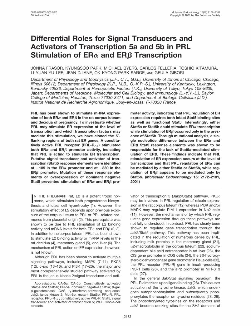
to generate standard curves for analysis of mRNAlevels in experimental samples amplified in parallelreactions (Fig. 1A). In corpora lutea on d 7 of preg-
Fig. 1. Standard Curves for Real-Time Quantitative PCRA, Known amounts of ER� and ER� cDNA, ranging from
103 to 107 copies/�l and 103 to 106 copies/�l, respectively,were amplified as described in Materials and Methods. Thecrossing point represents the number of cycles required toreach a chosen level of fluorescence, at which all standardsand samples were in the linear range of amplification. Thecrossing point was then plotted against the number of copiesof cDNA/�l, and the linear regression equation through thedata points was used to determine the number of copies ofER� or ER� in reverse transcribed RNA samples. B, Real-time quantitative PCR was carried out for ER� and ER� usingmRNA from corpora lutea of pregnant rats, hypophysecto-mized rats, and hypophysectomized rats treated with PRL. C,Real-time quantitative PCR was carried out for ER� and ER�using mRNA from primary luteinized granulosa cells that hadbeen treated with 1 �g/ml PRL for 12 h.
Frasor et al. • Stat5-Mediated Transcription of ER� and ER� Mol Endocrinol, December 2001, 15(12):2172–2181 2173
nancy, approximately 45,000 copies of ER� and 550copies of ER� were detected in samples correspond-ing to 1 ng of RNA (Fig. 1B). In rats, hypophysecto-mized on d 3 of pregnancy, ER� and ER� levels werereduced to approximately 30% of the control levels.Sustained treatment with PRL induced a 3.5-fold in-duction of ER� expression and a 2-fold induction ofER� expression. In contrast to corpora lutea, in whichthere was approximately 70 times more ER� than ER�,luteinized granulosa cells cultured for 72 h expressedonly 8 times more ER� than ER�, with approximately850 copies of ER� and 120 copies of ER� per ng ofstarting RNA (Fig. 1C). After a 12-h treatment withPRL, a 2.4-fold increase in ER� and a 1.8-fold increasein ER� expression was observed. These findings con-firm that PRL can stimulate both ER� and ER� mRNAexpression and further demonstrate a much higherlevel of ER� than ER� in corpora lutea of pregnancyand in primary luteinized granulosa cells.
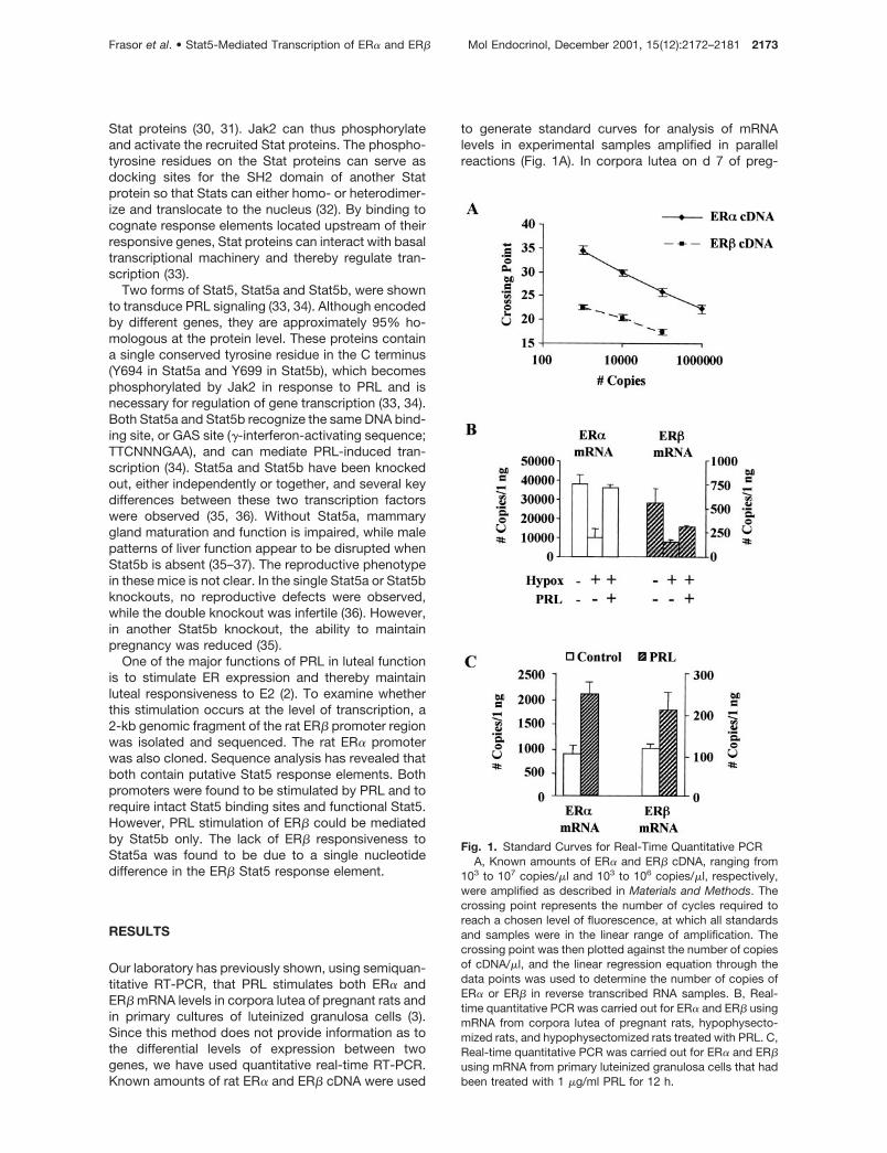
To investigate whether PRL can regulate ER expres-sion at the level of transcription, the ER� and ER�5�-flanking regions were cloned, and promoter-reporter constructs were prepared as described inMaterials and Methods. A putative Stat5 response el-ement (5�-TTCTAGGAA-3�), which represents a per-fect consensus Stat5 binding site, was located at�180 bp in the ER� promoter region. In addition, aputative Stat5 response element (3�-TTCTGGTAA-5�)with one nucleotide difference (underlined) from the
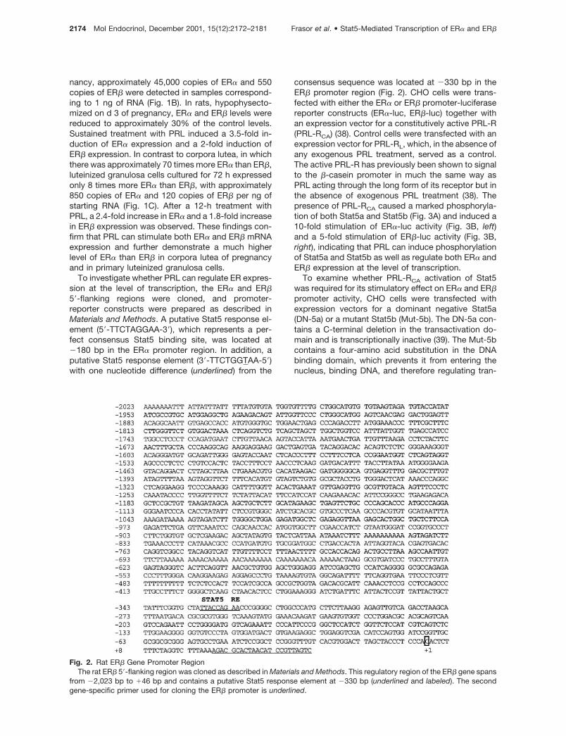
consensus sequence was located at �330 bp in theER� promoter region (Fig. 2). CHO cells were trans-fected with either the ER� or ER� promoter-luciferasereporter constructs (ER�-luc, ER�-luc) together withan expression vector for a constitutively active PRL-R(PRL-RCA) (38). Control cells were transfected with anexpression vector for PRL-RL, which, in the absence ofany exogenous PRL treatment, served as a control.The active PRL-R has previously been shown to signalto the �-casein promoter in much the same way asPRL acting through the long form of its receptor but inthe absence of exogenous PRL treatment (38). Thepresence of PRL-RCA caused a marked phosphoryla-tion of both Stat5a and Stat5b (Fig. 3A) and induced a10-fold stimulation of ER�-luc activity (Fig. 3B, left)and a 5-fold stimulation of ER�-luc activity (Fig. 3B,right), indicating that PRL can induce phosphorylationof Stat5a and Stat5b as well as regulate both ER� andER� expression at the level of transcription.
To examine whether PRL-RCA activation of Stat5was required for its stimulatory effect on ER� and ER�promoter activity, CHO cells were transfected withexpression vectors for a dominant negative Stat5a(DN-5a) or a mutant Stat5b (Mut-5b). The DN-5a con-tains a C-terminal deletion in the transactivation do-main and is transcriptionally inactive (39). The Mut-5bcontains a four-amino acid substitution in the DNAbinding domain, which prevents it from entering thenucleus, binding DNA, and therefore regulating tran-
Fig. 2. Rat ER� Gene Promoter RegionThe rat ER� 5�-flanking region was cloned as described in Materials and Methods. This regulatory region of the ER� gene spans
from �2,023 bp to �46 bp and contains a putative Stat5 response element at �330 bp (underlined and labeled). The secondgene-specific primer used for cloning the ER� promoter is underlined.
2174 Mol Endocrinol, December 2001, 15(12):2172–2181 Frasor et al. • Stat5-Mediated Transcription of ER� and ER�
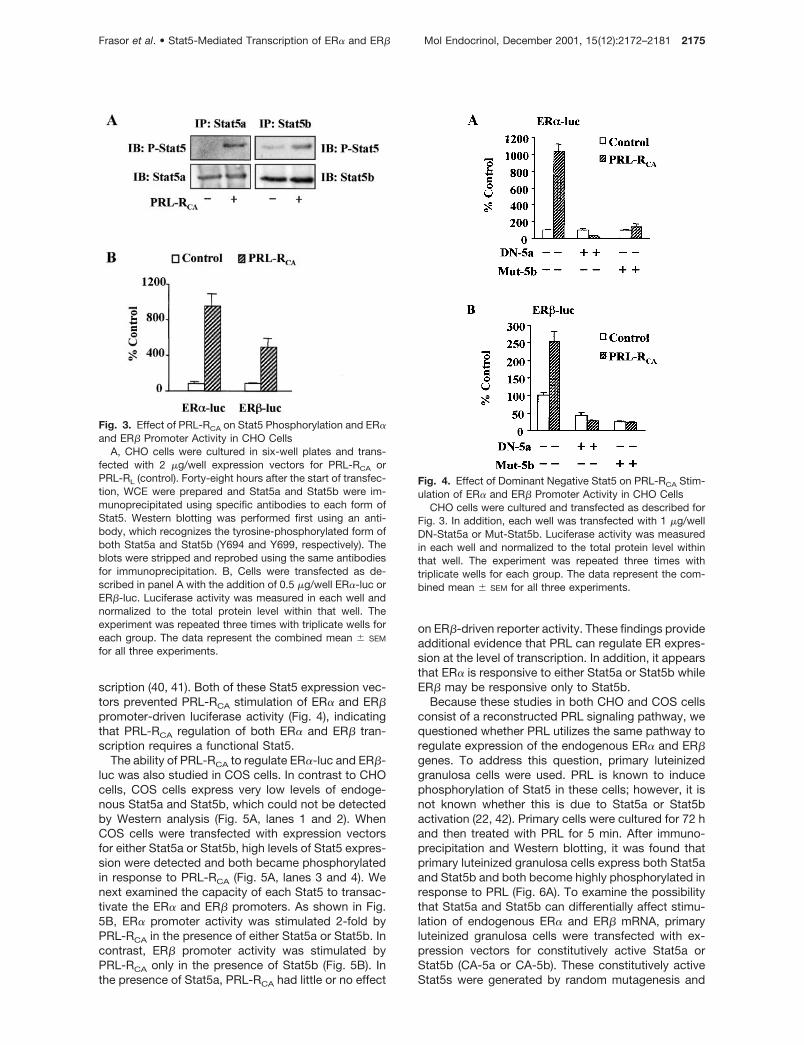
scription (40, 41). Both of these Stat5 expression vec-tors prevented PRL-RCA stimulation of ER� and ER�promoter-driven luciferase activity (Fig. 4), indicatingthat PRL-RCA regulation of both ER� and ER� tran-scription requires a functional Stat5.
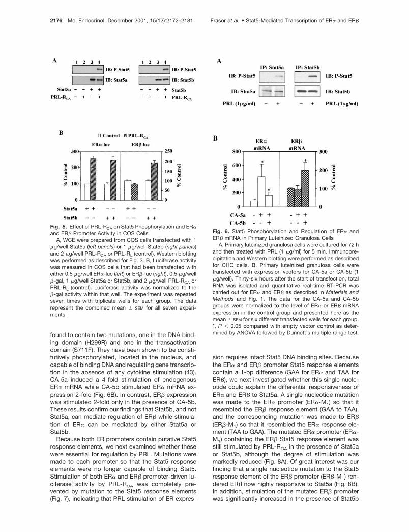
The ability of PRL-RCA to regulate ER�-luc and ER�-luc was also studied in COS cells. In contrast to CHOcells, COS cells express very low levels of endoge-nous Stat5a and Stat5b, which could not be detectedby Western analysis (Fig. 5A, lanes 1 and 2). WhenCOS cells were transfected with expression vectorsfor either Stat5a or Stat5b, high levels of Stat5 expres-sion were detected and both became phosphorylatedin response to PRL-RCA (Fig. 5A, lanes 3 and 4). Wenext examined the capacity of each Stat5 to transac-tivate the ER� and ER� promoters. As shown in Fig.5B, ER� promoter activity was stimulated 2-fold byPRL-RCA in the presence of either Stat5a or Stat5b. Incontrast, ER� promoter activity was stimulated byPRL-RCA only in the presence of Stat5b (Fig. 5B). Inthe presence of Stat5a, PRL-RCA had little or no effect
on ER�-driven reporter activity. These findings provideadditional evidence that PRL can regulate ER expres-sion at the level of transcription. In addition, it appearsthat ER� is responsive to either Stat5a or Stat5b whileER� may be responsive only to Stat5b.
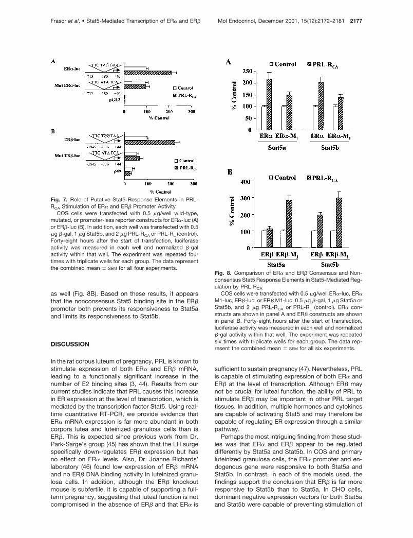
Because these studies in both CHO and COS cellsconsist of a reconstructed PRL signaling pathway, wequestioned whether PRL utilizes the same pathway toregulate expression of the endogenous ER� and ER�genes. To address this question, primary luteinizedgranulosa cells were used. PRL is known to inducephosphorylation of Stat5 in these cells; however, it isnot known whether this is due to Stat5a or Stat5bactivation (22, 42). Primary cells were cultured for 72 hand then treated with PRL for 5 min. After immuno-precipitation and Western blotting, it was found thatprimary luteinized granulosa cells express both Stat5aand Stat5b and both become highly phosphorylated inresponse to PRL (Fig. 6A). To examine the possibilitythat Stat5a and Stat5b can differentially affect stimu-lation of endogenous ER� and ER� mRNA, primaryluteinized granulosa cells were transfected with ex-pression vectors for constitutively active Stat5a orStat5b (CA-5a or CA-5b). These constitutively activeStat5s were generated by random mutagenesis and
Fig. 3. Effect of PRL-RCA on Stat5 Phosphorylation and ER�and ER� Promoter Activity in CHO Cells
A, CHO cells were cultured in six-well plates and trans-fected with 2 �g/well expression vectors for PRL-RCA orPRL-RL (control). Forty-eight hours after the start of transfec-tion, WCE were prepared and Stat5a and Stat5b were im-munoprecipitated using specific antibodies to each form ofStat5. Western blotting was performed first using an anti-body, which recognizes the tyrosine-phosphorylated form ofboth Stat5a and Stat5b (Y694 and Y699, respectively). Theblots were stripped and reprobed using the same antibodiesfor immunoprecipitation. B, Cells were transfected as de-scribed in panel A with the addition of 0.5 �g/well ER�-luc orER�-luc. Luciferase activity was measured in each well andnormalized to the total protein level within that well. Theexperiment was repeated three times with triplicate wells foreach group. The data represent the combined mean � SEM
for all three experiments.
Fig. 4. Effect of Dominant Negative Stat5 on PRL-RCA Stim-ulation of ER� and ER� Promoter Activity in CHO Cells
CHO cells were cultured and transfected as described forFig. 3. In addition, each well was transfected with 1 �g/wellDN-Stat5a or Mut-Stat5b. Luciferase activity was measuredin each well and normalized to the total protein level withinthat well. The experiment was repeated three times withtriplicate wells for each group. The data represent the com-bined mean � SEM for all three experiments.
Frasor et al. • Stat5-Mediated Transcription of ER� and ER� Mol Endocrinol, December 2001, 15(12):2172–2181 2175
found to contain two mutations, one in the DNA bind-ing domain (H299R) and one in the transactivationdomain (S711F). They have been shown to be consti-tutively phosphorylated, located in the nucleus, andcapable of binding DNA and regulating gene transcrip-tion in the absence of any cytokine stimulation (43).CA-5a induced a 4-fold stimulation of endogenousER� mRNA while CA-5b stimulated ER� mRNA ex-pression 2-fold (Fig. 6B). In contrast, ER� expressionwas stimulated 2-fold only in the presence of CA-5b.These results confirm our findings that Stat5b, and notStat5a, can mediate regulation of ER� while stimula-tion of ER� can be mediated by either Stat5a orStat5b.
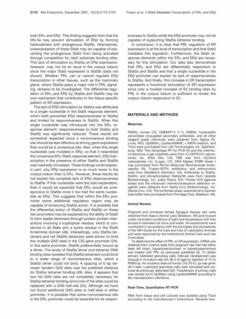
Because both ER promoters contain putative Stat5response elements, we next examined whether thesewere essential for regulation by PRL. Mutations weremade to each promoter so that the Stat5 responseelements were no longer capable of binding Stat5.Stimulation of both ER� and ER� promoter-driven lu-ciferase activity by PRL-RCA was completely pre-vented by mutation to the Stat5 response elements(Fig. 7), indicating that PRL stimulation of ER expres-
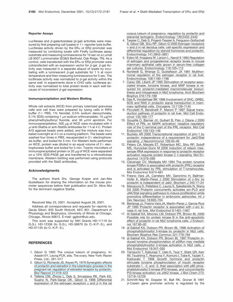
sion requires intact Stat5 DNA binding sites. Becausethe ER� and ER� promoter Stat5 response elementscontain a 1-bp difference (GAA for ER� and TAA forER�), we next investigated whether this single nucle-otide could explain the differential responsiveness ofER� and ER� to Stat5a. A single nucleotide mutationwas made to the ER� promoter (ER�-M1) so that itresembled the ER� response element (GAA to TAA),and the corresponding mutation was made to ER�(ER�-M1) so that it resembled the ER� response ele-ment (TAA to GAA). The mutated ER� promoter (ER�-M1) containing the ER� Stat5 response element wasstill stimulated by PRL-RCA in the presence of Stat5aor Stat5b, although the degree of stimulation wasmarkedly reduced (Fig. 8A). Of great interest was ourfinding that a single nucleotide mutation to the Stat5response element of the ER� promoter (ER�-M1) ren-dered ER� now highly responsive to Stat5a (Fig. 8B).In addition, stimulation of the mutated ER� promoterwas significantly increased in the presence of Stat5b
Fig. 5. Effect of PRL-RCA on Stat5 Phosphorylation and ER�and ER� Promoter Activity in COS Cells
A, WCE were prepared from COS cells transfected with 1�g/well Stat5a (left panels) or 1 �g/well Stat5b (right panels)and 2 �g/well PRL-RCA or PRL-RL (control). Western blottingwas performed as described for Fig. 3. B, Luciferase activitywas measured in COS cells that had been transfected witheither 0.5 �g/well ER�-luc (left) or ER�-luc (right), 0.5 �g/well�-gal, 1 �g/well Stat5a or Stat5b, and 2 �g/well PRL-RCA orPRL-RL (control). Luciferase activity was normalized to the�-gal activity within that well. The experiment was repeatedseven times with triplicate wells for each group. The datarepresent the combined mean � SEM for all seven experi-ments.
Fig. 6. Stat5 Phosphorylation and Regulation of ER� andER� mRNA in Primary Luteinized Granulosa Cells
A, Primary luteinized granulosa cells were cultured for 72 hand then treated with PRL (1 �g/ml) for 5 min. Immunopre-cipitation and Western blotting were performed as describedfor CHO cells. B, Primary luteinized granulosa cells weretransfected with expression vectors for CA-5a or CA-5b (1�g/well). Thirty-six hours after the start of transfection, totalRNA was isolated and quantitative real-time RT-PCR wascarried out for ER� and ER� as described in Materials andMethods and Fig. 1. The data for the CA-5a and CA-5bgroups were normalized to the level of ER� or ER� mRNAexpression in the control group and presented here as themean � SEM for six different transfected wells for each group.*, P � 0.05 compared with empty vector control as deter-mined by ANOVA followed by Dunnett’s multiple range test.
2176 Mol Endocrinol, December 2001, 15(12):2172–2181 Frasor et al. • Stat5-Mediated Transcription of ER� and ER�
as well (Fig. 8B). Based on these results, it appearsthat the nonconsensus Stat5 binding site in the ER�promoter both prevents its responsiveness to Stat5aand limits its responsiveness to Stat5b.
DISCUSSION
In the rat corpus luteum of pregnancy, PRL is known tostimulate expression of both ER� and ER� mRNA,leading to a functionally significant increase in thenumber of E2 binding sites (3, 44). Results from ourcurrent studies indicate that PRL causes this increasein ER expression at the level of transcription, which ismediated by the transcription factor Stat5. Using real-time quantitative RT-PCR, we provide evidence thatER� mRNA expression is far more abundant in bothcorpora lutea and luteinized granulosa cells than isER�. This is expected since previous work from Dr.Park-Sarge’s group (45) has shown that the LH surgespecifically down-regulates ER� expression but hasno effect on ER� levels. Also, Dr. Joanne Richards’laboratory (46) found low expression of ER� mRNAand no ER� DNA binding activity in luteinized granu-losa cells. In addition, although the ER� knockoutmouse is subfertile, it is capable of supporting a full-term pregnancy, suggesting that luteal function is notcompromised in the absence of ER� and that ER� is
sufficient to sustain pregnancy (47). Nevertheless, PRLis capable of stimulating expression of both ER� andER� at the level of transcription. Although ER� maynot be crucial for luteal function, the ability of PRL tostimulate ER� may be important in other PRL targettissues. In addition, multiple hormones and cytokinesare capable of activating Stat5 and may therefore becapable of regulating ER expression through a similarpathway.
Perhaps the most intriguing finding from these stud-ies was that ER� and ER� appear to be regulateddifferently by Stat5a and Stat5b. In COS and primaryluteinized granulosa cells, the ER� promoter and en-dogenous gene were responsive to both Stat5a andStat5b. In contrast, in each of the models used, thefindings support the conclusion that ER� is far moreresponsive to Stat5b than to Stat5a. In CHO cells,dominant negative expression vectors for both Stat5aand Stat5b were capable of preventing stimulation of
Fig. 7. Role of Putative Stat5 Response Elements in PRL-RCA Stimulation of ER� and ER� Promoter Activity
COS cells were transfected with 0.5 �g/well wild-type,mutated, or promoter-less reporter constructs for ER�-luc (A)or ER�-luc (B). In addition, each well was transfected with 0.5�g �-gal, 1 �g Stat5b, and 2 �g PRL-RCA or PRL-RL (control).Forty-eight hours after the start of transfection, luciferaseactivity was measured in each well and normalized �-galactivity within that well. The experiment was repeated fourtimes with triplicate wells for each group. The data representthe combined mean � SEM for all four experiments.
Fig. 8. Comparison of ER� and ER� Consensus and Non-consensus Stat5 Response Elements in Stat5-Mediated Reg-ulation by PRL-RCA
COS cells were transfected with 0.5 �g/well ER�-luc, ER�M1-luc, ER�-luc, or ER� M1-luc, 0.5 �g �-gal, 1 �g Stat5a orStat5b, and 2 �g PRL-RCA or PRL-RL (control). ER� con-structs are shown in panel A and ER� constructs are shownin panel B. Forty-eight hours after the start of transfection,luciferase activity was measured in each well and normalized�-gal activity within that well. The experiment was repeatedsix times with triplicate wells for each group. The data rep-resent the combined mean � SEM for all six experiments.
Frasor et al. • Stat5-Mediated Transcription of ER� and ER� Mol Endocrinol, December 2001, 15(12):2172–2181 2177
both ER� and ER�. This finding suggests then that theDN-5a may prevent stimulation of ER� by formingheterodimers with endogenous Stat5b. Alternatively,overexpression of these Stats may be capable of pre-venting the endogenous Stat5 from being activatedthrough competition for Jak2 substrate binding sites.This lack of stimulation by Stat5a on ER� expression,however, may not be an issue in the corpus luteumsince the major Stat5 expressed is Stat5b (data notshown). Whether PRL can or cannot regulate ER�transcription in other tissues, such as the mammarygland, where Stat5a plays a major role in PRL signal-ing, remains to be investigated. The differential regu-lation of ER� and ER� by Stat5a and Stat5b may beone mechanism that contributes to the tissue-specificpattern of ER expression.
The lack of ER� stimulation by Stat5a was attributedto a single nucleotide in the Stat5 response element,which both prevented ER� responsiveness to Stat5aand limited its responsiveness to Stat5b. When thissingle nucleotide was introduced into the ER� re-sponse element, responsiveness to both Stat5a andStat5b was significantly reduced. These results aresomewhat expected since a nonconsensus bindingsite should be less effective at driving gene expressionthan would be a consensus site. Also, when this singlenucleotide was mutated in ER�, so that it resembledthe consensus ER� Stat5 response element, ER� tran-scription in the presence of either Stat5a and Stat5bwas markedly increased. These findings could explain,in part, why ER� expression is so much lower in thecorpus luteum than is ER�. However, these results donot explain the complete lack of ER� responsivenessto Stat5a. If this nucleotide were the sole explanation,then it would be expected that ER� would be unre-sponsive to Stat5a once it too had the same nucleo-tide as ER�. This suggests that within the ER� pro-moter some additional regulatory region may becapable of enhancing Stat5a action. It is possible thatthe differential action of Stat5a and Stat5b on thesetwo promoters may be explained by the ability of Stat5to form stable tetramers through protein-protein inter-actions involving a tryptophan residue, which is con-served in all Stats and a lysine residue in the Stat5N-terminal domain (48). Interestingly, only Stat5a tet-ramers and not Stat5b tetramers were shown to bindthe multiple GAS sites in the CIS gene promoter (24).In this same promoter, Stat5b preferentially bound asa dimer. The study of Stat5a dimer and tetramer DNAbinding sites revealed that Stat5a tetramers could bindto a wider range of nonconsensus sites, which aStat5a dimer could not bind. A spacing of 6 bp be-tween tandem GAS sites was the preferred distancefor Stat5a tetramer binding (49). Also, it appears thattwo full GAS sites are not completely necessary forStat5a tetramer binding since one of the sites could bereplaced with a GAS half-site (24). Although we havenot found additional GAS sites or half-sites in eitherpromoter, it is possible that some nonconsensus sitein the ER� promoter could be essential for its respon-
siveness to Stat5a while the ER� promoter may not becapable of supporting Stat5a tetramer binding.
In conclusion, it is clear that PRL regulation of ERexpression is at the level of transcription and that Stat5mediates this regulation. Furthermore, the Stat5 re-sponse elements within the ER� and ER� are neces-sary for this stimulation. Our data also demonstratethat ER� and ER� are differentially responsive toStat5a and Stat5b and that a single nucleotide in theER� promoter can explain its lack of responsivenessto Stat5a. And finally, this increase in ER transcriptionrepresents a functional stimulation of ER expressionsince only a modest increase of E2 binding sites byPRL in the corpus luteum is sufficient to render thecorpus luteum responsive to E2.
MATERIALS AND METHODS
Materials
PMSG, human CG, DMEM/F12 (1:1), DMEM, horseradishperoxidase conjugated secondary antibodies, and all otherreagent grade chemicals were obtained from Sigma (St.Louis, MO). OptiMem, LipofectAMINE, �-MEM medium, andTrizol were purchased from Life Technologies, Inc. (Gaithers-burg, MD). The Advantage RT-for-PCR kit and the chemilu-minescence �-gal substrate were from CLONTECH Labora-tories, Inc. (Palo Alto, CA). FBS was from HyCloneLaboratories, Inc. (Logan, UT). DNA Master SYBR Green Iwas purchased from Roche Molecular Biochemicals (India-napolis, IN). Trypsin-EDTA, antibiotics, and antimycoticswere from Mediatech (Herndon, VA). Antibodies to Stat5a,Stat5b, and phosphorylated Stat5a/5b were from UpstateBiotechnology, Inc. (Lake Placid, NY). Protein A/G agarosebeads and the enhanced chemiluminescence detection re-agents were obtained from Santa Cruz Biotechnology, Inc.(Santa Cruz, CA). The luciferase assay substrate and reporterlysis buffer were purchased from Promega Corp. (Madison, WI).
Animal Models
Pregnant and immature female Sprague Dawley rats wereobtained from Sasco Animal Labs (Madison, WI) and housedunder controlled conditions of light and temperature with freeaccess to standard rat chow and water. All experiments wereconducted in accordance with the principles and proceduresof the NIH Guide for the Care and Use of Laboratory Animalsand were approved by the Institutional Animal Care and UseCommittee.
To determine the effect of PRL on ER expression, mRNA wasobtained from corpora lutea from pregnant rats that had eitherbeen left intact, hypophysectomized, or hypophysectomizedand treated with PRL as previously published (3). To obtainprimary luteinized granulosa cells, follicular development wasinduced in immature rats (24–26 d of age) by injection of 15 IUPMSG ip. An ovulatory dose of human CG (10 IU, ip) was given48 h later. Luteinized granulosa cells were harvested and cul-tured as previously described (42). Transfection of primary cellswas carried out in OptiMem using LipofectAMINE according tothe manufacturer’s directions.
Real-Time, Quantitative RT-PCR
RNA from tissue and cell cultures was isolated using Trizolaccording to the manufacturer’s instructions. Reverse tran-
2178 Mol Endocrinol, December 2001, 15(12):2172–2181 Frasor et al. • Stat5-Mediated Transcription of ER� and ER�

scription was carried out using reagents from the AdvantageRT-for-PCR kit according to the manufacturer’s instructions.One microgram of total RNA was used for the reverse tran-scription reaction, and the product was diluted to a finalvolume of 100 �l by adding diethyl pyrocarbonate-treatedH2O. To generate standard curves for quantitative PCR,rat ER� and ER� cDNA, which was kindly provided by Dr.Maruyama (50), were diluted to concentrations ranging from103 to 107 copies/�l. Five-microliter aliquots of standards ordiluted reverse transcription products were combined with 2�l 10� DNA Master SYBR Green I, 1.6 �l MgCl2 (3 mM finalconcentration), and specific primers for rat ER� or ER� (0.5�M final concentration). The primers used have been previ-ously published (3). Reactions were carried out in glass cap-illary tubes in a total volume of 20 �l. The DNA Master SYBRGreen I mix contains Taq DNA polymerase, reaction buffer,deoxynucleotide triphosphate, 10 mM MgCl2, and SYBRGreen I dye, which is a specific fluorescence dye for double-stranded DNA. PCR reactions were performed in the RocheLightcycler instrument and the accompanying software wasused for data analysis (Roche Molecular Biochemicals,Mannheim, Germany). After a 2-min denaturation, PCR cy-cles were carried out as follows: 0 sec at 95 C, 10 sec at theannealing temperature, and 15 sec at 72 C. For ER�, 40cycles at an annealing temperature of 63 C were used; forER�, 35 cycles at an annealing temperature of 69 C wereused. At the end of each cycle, the amount of double-stranded DNA was monitored by measuring the level of SYBRGreen I fluorescence. After the completion of all cycles, alevel of fluorescence was selected at which all of the stan-dards and samples were within the linear range of amplifica-tion. The crossing point, or the number of cycles necessaryfor each sample or standard to obtain the selected level offluorescence, was calculated using the Roche Lightcyclersoftware. Based on these crossing points, a standard curvewas generated, and the number of ER� or ER� copies wascalculated for each sample. The data presented represent thenumber of copies of ER� or ER� in 1 ng of total RNA.
Cloning of the ER� and ER� Promoters
The 5�-flanking region of the rat ER� gene was cloned usingthe rat PromoterFinder DNA Walking kit (CLONTECH Labo-ratories, Inc.). Five different genomic libraries were generatedby digesting genomic DNA with 5 different restriction en-zymes, namely EcoRV, ScaI, DraI, PvuII, and SspI followed byligation with a specifically designed PromoterFinder adapter.The ER� promoter region was amplified by nested PCR usingthe five different genomic libraries as templates and two setsof primers designed according to the published sequence ofthe ER� promoter (51). The first PCR reaction was carried outusing the following primers: 5�-CCACTCATAAATCTCTT-GGTAACGGC-3� and 5�-GAAGGAAGGAATGTGCTCGAAT-GATC-3�. A second PCR reaction was carried out usingproduct from the first reaction and the following primers:5�-CTGGGGTTGCAATTAGTC-ATTTAGGC-3� and 5�-TCGC-GAATTCGAGTGGCGCGGTGTGTGATCAAG-3�. The secondprimer also included an attached EcoRI site for subsequentcloning. All five sources of genomic DNA yielded an amplifiedproduct of the expected size (880 bp). The PCR productswere pooled, and the internal KpnI site at �769 in the ER�promoter region and the added EcoRI site were used tosubclone the PCR product into the pBluescript DNA vector(Stratagene, La Jolla, CA). Subcloning of the ER� promoterregion into the pGL3-basic luciferase reporter vector (Pro-mega Corp.) was carried out utilizing the KpnI and BglII sitesin the pGL3 vector and the KpnI and BamHI sites in thepBluescript vector. Both strands of the ER� promoter gen-erated from different colonies were sequenced. Sequenceanalysis revealed two differences from the originally pub-lished sequence (G to C at �494, A to G at �346) (51).
To isolate the regulatory region of the rat ER� gene, weused the touchdown PCR amplification approach using the
GenomeWalker kit (CLONTECH Laboratories, Inc.) accordingto the manufacturer’s procedure. Two gene-specific primerswere designed against the most 5�-end of sequences of therat ER� mRNA (gene-specific primer 1: 5�-AAGCTGCAAA-GATTACCCACGACTA-3� and gene-specific primer 2: 5�-GACTAACGGATGTTAGTGCGTCTT-3�) (52). Thus, the ex-pected gene-regulatory DNA would contain 46 bp of the5�-end of the ER� mRNA. The primary PCR amplification wascarried out using the combined GenomeWalker libraries (1 �l)and the primer set of gene-specific primer 1 and adapterprimer 1, under PCR conditions of 72 C for 4 min (7 cycles)and 67 C for 4 min (33 cycles). The secondary PCR amplifi-cation was carried out using 1 �l of the diluted primary PCRproducts (1:100) and the primer set of gene-specific primer 2and adapter primer 2, under PCR conditions of 72 C for 4 min(5 cycles) and 67 C (22 cycles). This procedure yielded twoprominent PCR fragments (�1 and �2 kb) that were subse-quently isolated, subcloned into PCR2.1 T/A overhang vector(CLONTECH Laboratories, Inc.), and sequenced using M13forward and backward primers. Both contained the adapter 2sequences at their 5�-end and the gene-specific primer 2 attheir 3�-end. The inserts of these clones were isolated byrestriction digestion using EcoRV/SpeI and subsequent fill-inreactions with Klenow, and inserted into the SmaI arms of thepUBT-luc vector (53). For these studies, the �1 kb promoterregion of the rat ER� gene was used.
Mutations to ER� and ER� Promoters
The first set of mutations made to the ER� and ER� promot-ers consisted of six and five nucleotides, respectively, beingchanged to abolish the Stat5 binding sites. Oligonucleotideprimers for these mutations were made as follows (mutatednucleotides underlined): ER� 5�-GCCAAGGGGGCTG-GAGTTTCTTGATATCATGCTGA-TTCTAGTGGTGCTACT-GCCG -3� and ER� 5�-ATTACTGCTTATTTCGGTGCTATGA-TATCAACCCGGGGCCTGGCCCATGC-3�. The second set ofmutations made to the ER� and ER� promoters consisted ofa single nucleotide being changed. The consensus Stat5 siteof the ER� promoter (TTCnnnGAA) was changed so that itresembled the Stat5 response element of the ER� promoter(TTCnnnTAA). The nonconsensus ER� Stat5 response ele-ment (TTCnnnTAA) was mutated so that it would resemblethe ER� Stat5 response element (TTCnnnGAA). Oligonucle-otide primers for these mutations were made as follows(mutated nucleotides underlined): ER� 5�-GGCTGGAGTT-TCTTCTAGTAAT-GCTGATTCTAGTGG-3� and ER� 5�-CGGTGCTATTCCCAGAACCCGG-GGCCTGG-3�. All muta-tions were made using the QuikChange Site-directedMutagenesis kit according to the manufacturer’s directions(Stratagene). The presence of the correct mutations wasconfirmed by DNA sequencing.
Culture and Transfection of CHO and COS Cells
CHO and COS cells were routinely cultured in �-MEM andDMEM/F12 (1:1), respectively. All media were supplementedwith 10% FBS, 100 IU/ml penicillin G, 100 �g/ml streptomy-cin, and 0.25 �g/ml Amphotericin. Cultures were carried outat 37 C in a 5% CO2, humidified atmosphere. For transienttransfections, 100,000 cells were seeded per well in six-wellplates and cultured for 24 h. Both CHO and COS cells weretransfected using calcium phosphate DNA precipitation andwere approximately 50% confluent at the start of transfection(54). In general, a total of 4–5 �g DNA were transfected perwell, and the total amount of DNA was equalized with emptyvector when necessary. Twenty-four hours after the start oftransfection, media were changed to standard culture mediasupplemented with 1% FBS, and cells were cultured for anadditional 24 h at 5% CO2. The entire length of the experi-ments was standardized to 48 h from the start of transfection.
Frasor et al. • Stat5-Mediated Transcription of ER� and ER� Mol Endocrinol, December 2001, 15(12):2172–2181 2179
Reporter Assays
Luciferase and �-galactosidase (�-gal) activities were mea-sured by first preparing cell lysates in 1� reporter lysis buffer.Luciferase activity driven by the ER� or ER� promoter wasmeasured by combining lysate with Firefly luciferase assaysubstrate and measuring luminescence for 10 sec on a LumatLB 9507 Luminometer (EG&G Berthold, Oak Ridge, TN). As acontrol, cells transfected with the ER� or ER� promoter werecotransfected with an expression vector for �-gal. �-gal Ac-tivity was measured in a separate aliquot of lysate by incu-bating with a luminescent �-gal substrate for 1 h at roomtemperature and then measuring luminescence for 5 sec. Theluciferase activity was normalized to �-gal activity within thesame well. In experiments done in CHO cells, luciferase ac-tivity was normalized to total protein levels in each well be-cause of inconsistent �-gal expression.
Immunoprecipitation and Western Blotting
Whole cell extracts (WCE) from primary luteinized granulosacells and cell lines were prepared by lysing cells in RIPAbuffer (1� PBS, 1% Nonidet, 0.5% sodium deoxycholate,0.1% SDS) containing 1 �M sodium orthovanadate, 10 �g/mlphenylmethylsulfonyl fluoride, and 30 �l/ml aprotinin. Forimmunoprecipitation, 500 �g of WCE were incubated with 4�l anti-Stat5a or anti-Stat5b antibodies for 1 h at 4 C. ProteinA/G agarose beads were added, and the mixture was incu-bated overnight at 4 C on a rocking platform. The beads werewashed four times in PBS, resuspended in 2� electrophore-sis buffer, and boiled for 5 min. For Western blots performedon WCE, protein was diluted in an equal volume of 2� elec-trophoresis buffer and boiled for 5 min. Twenty microliters ofimmunoprecipitated protein or 20 �g of WCE were separatedon a 10% SDS-PAGE gel and transferred to a nitrocellulosemembrane. Western blotting was performed using protocolsprovided with the Stat5 antibodies.
Acknowledgments
The authors thank Drs. George Kuiper and Jan-AkeGustafsson for sharing the information on the mouse pro-moter sequences before their publication and Dr. Alice Muifor the dominant negative Stat5a.
Received May 23, 2001. Accepted August 28, 2001.Address all correspondence and requests for reprints to:
Geula Gibori, 835 South Wolcott, M/C 901, Department ofPhysiology and Biophysics, University of Illinois at Chicago,Chicago, Illinois 60612. E-mail: [email protected].
This work was supported by NIH Grants HD-11119 (toG.G.), HD-12356 (to G.G.), HD-36879 (to O.-K.P.-S.), andHD-01135 (to O.-K.P.-S.).
REFERENCES
1. Gibori G 1993 The corpus luteum of pregnancy. In:Adashi EY, Leung PCK, eds. The ovary. New York: RavenPress, Ltd.; 261–317
2. Gibori G, Richards JS, Keyes PL 1979 Synergistic effectsof prolactin and estradiol in the luteotropic process in thepregnant rat: regulation of estradiol receptor by prolactin.Biol Reprod 21:419–423
3. Telleria CM, Zhong L, Deb S, Srivastava RK, Park KS,Sugino N, Park-Sarge OK, Gibori G 1998 Differentialexpression of the estrogen receptors � and � in the rat
corpus luteum of pregnancy: regulation by prolactin andplacental lactogens. Endocrinology 139:2432–2442
4. Tessier C, Deb S, Prigent-Tessier A, Ferguson-GottschallS, Gibori GB, Shiu RP, Gibori G 2000 Estrogen receptors� and � in rat decidua cells: cell-specific expression anddifferential regulation by steroid hormones and prolactin.Endocrinology 141:3842–3851
5. Edery M, Imagawa W, Larson L, Nandi S 1985 Regulationof estrogen and progesterone receptor levels in mousemammary epithelial cells grown in serum-free collagengel cultures. Endocrinology 116:105–112
6. Norstedt G, Wrange O, Gustafsson JA 1981 Multihor-monal regulation of the estrogen receptor in rat liver.Endocrinology 108:1190–1196
7. Carey GB, Liberti JP 1995 Stimulation of receptor-asso-ciated kinase, tyrosine kinase, and MAP kinase is re-quired for prolactin-mediated macromolecular biosyn-thesis and mitogenesis in Nb2 lymphoma. Arch BiochemBiophys 316:179–189
8. Das R, Vonderhaar BK 1996 Involvement of SHC, GRB2,SOS and RAS in prolactin signal transduction in mam-mary epithelial cells. Oncogene 13:1139–1145
9. Piccoletti R, Bendinelli P, Maroni P 1997 Signal trans-duction pathway of prolactin in rat liver. Mol Cell Endo-crinol 135:169–177
10. Goupille O, Barnier JV, Guibert B, Paly J, Djiane J 2000Effect of PRL on MAPK activation: negative regulatoryrole of the C-terminal part of the PRL receptor. Mol CellEndocrinol 159:133–146
11. Buckley AR 2000 Transcriptional regulation of pim-1 byprolactin: independence of a requirement for Jak2/Statsignaling. J Neuroimmunol 109:40–46
12. Peters CA, Maizels ET, Robertson MC, Shiu RP, SoloffMS, Hunzicker-Dunn M 2000 Induction of relaxin mes-senger RNA expression in response to prolactin receptoractivation requires protein kinase C � signaling. Mol En-docrinol 14:576–590
13. Clevenger CV, Medaglia MV 1994 The protein tyrosinekinase P59fyn is associated with prolactin (PRL) receptorand is activated by PRL stimulation of T-lymphocytes.Mol Endocrinol 8:674–681
14. Fresno Vara JA, Carretero MV, Geronimo H, Ballmer-Hofer K, Martin-Perez J 2000 Stimulation of c-Src byprolactin is independent of Jak2. Biochem J 345:17–24
15. Mangoura D, Pelletiere C, Leung S, Sakellaridis N, WangDX 2000 Prolactin concurrently activates src-PLD andJAK/Stat signaling pathways to induce proliferation whilepromoting differentiation in embryonic astrocytes. Int JDev Neurosci 18:693–704
16. Berlanga JJ, Fresno Vara JA, Martin-Perez J, Garcia-RuizJP 1995 Prolactin receptor is associated with c-src ki-nase in rat liver. Mol Endocrinol 9:1461–1467
17. Al-Sakkaf KA, Mooney LM, Dobson PR, Brown BL 2000Possible role for protein kinase B in the anti-apoptoticeffect of prolactin in rat Nb2 lymphoma cells. J Endocri-nol 167:85–92
18. al-Sakkaf KA, Dobson PR, Brown BL 1996 Activation ofphosphatidylinositol 3-kinase by prolactin in Nb2 cells.Biochem Biophys Res Commun 221:779–784
19. al-Sakkaf KA, Dobson PR, Brown BL 1997 Prolactin in-duced tyrosine phosphorylation of p59fyn may mediatephosphatidylinositol 3-kinase activation in Nb2 cells. JMol Endocrinol 19:347–350
20. Yamauchi T, Kaburagi Y, Ueki K, Tsuji Y, Stark GR, KerrIM, Tsushima T, Akanuma Y, Komuro I, Tobe K, Yazaki Y,Kadowaki T 1998 Growth hormone and prolactinstimulate tyrosine phosphorylation of insulin receptorsubstrate-1, -2, and -3, their association with p85 phos-phatidylinositol 3-kinase (PI3-kinase), and concomitantlyPI3-kinase activation via JAK2 kinase. J Biol Chem 273:15719–15726
21. Schmitt-Ney M, Doppler W, Ball RK, Groner B 1991�-Casein gene promoter activity is regulated by the
2180 Mol Endocrinol, December 2001, 15(12):2172–2181 Frasor et al. • Stat5-Mediated Transcription of ER� and ER�
hormone-mediated relief of transcriptional repressionand a mammary-gland-specific nuclear factor. Mol CellBiol 11:3745–3755
22. Dajee M, Kazansky AV, Raught B, Hocke GM, Fey GH,Richards JS 1996 Prolactin induction of the �2-macro-globulin gene in rat ovarian granulosa cells: Stat 5 acti-vation and binding to the interleukin-6 response element.Mol Endocrinol 10:171–184
23. Ganguly TC, O’Brien ML, Karpen SJ, Hyde JF, Suchy FJ,Vore M 1997 Regulation of the rat liver sodium-depen-dent bile acid cotransporter gene by prolactin. Mediationof transcriptional activation by Stat5. J Clin Invest 99:2906–2914
24. Verdier F, Rabionet R, Gouilleux F, Beisenherz-Huss C,Varlet P, Muller O, Mayeux P, Lacombe C, GisselbrechtS, Chretien S 1998 A sequence of the CIS gene promoterinteracts preferentially with two associated STAT5Adimers: a distinct biochemical difference betweenSTAT5A and STAT5B. Mol Cell Biol 18:5852–5860
25. Feltus FA, Groner B, Melner MH 1999 Stat5-mediatedregulation of the human type II 3�-hydroxysteroid dehy-drogenase/�5-�4 isomerase gene: activation by prolac-tin. Mol Endocrinol 13:1084–1093
26. Galsgaard ED, Nielsen JH, Moldrup A 1999 Regulation ofprolactin receptor (PRLR) gene expression in insulin-producing cells. Prolactin and growth hormone activateone of the rat prlr gene promoters via STAT5a andSTAT5b. J Biol Chem 274:18686–18692
27. Nanbu-Wakao R, Fujitani Y, Masuho Y, Muramatu M,Wakao H 2000 Prolactin enhances CCAAT enhancer-binding protein-beta (C/EBP�) and peroxisome prolifera-tor-activated receptor gamma (PPAR�) messenger RNAexpression and stimulates adipogenic conversion ofNIH-3T3 cells. Mol Endocrinol 14:307–316
28. Rui H, Lebrun JJ, Kirken RA, Kelly PA, Farrar WL 1994JAK2 activation and cell proliferation induced by anti-body-mediated prolactin receptor dimerization. Endocri-nology 135:1299–1306
29. Lebrun JJ, Ali S, Sofer L, Ullrich A, Kelly PA 1994 Prolactin-induced proliferation of Nb2 cells involves tyrosine phos-phorylation of the prolactin receptor and its associatedtyrosine kinase JAK2. J Biol Chem 269:14021–14026
30. Goupille O, Daniel N, Bignon C, Jolivet G, Djiane J 1997Prolactin signal transduction to milk protein genes:carboxy-terminal part of the prolactin receptor and itstyrosine phosphorylation are not obligatory for JAK2 andSTAT5 activation. Mol Cell Endocrinol 127:155–169
31. Pezet A, Ferrag F, Kelly PA, Edery M 1997 Tyrosinedocking sites of the rat prolactin receptor required forassociation and activation of stat5. J Biol Chem 272:25043–25050
32. Ali S 1998 Prolactin receptor regulates Stat5 tyrosinephosphorylation and nuclear translocation by two sepa-rate pathways. J Biol Chem 273:7709–7716
33. Gouilleux F, Wakao H, Mundt M, Groner B 1994 Prolactininduces phosphorylation of Tyr694 of Stat5 (MGF), aprerequisite for DNA binding and induction of transcrip-tion. EMBO J 13:4361–4369
34. Liu X, Robinson GW, Gouilleux F, Groner B, Hen-nighausen L 1995 Cloning and expression of Stat5 andan additional homologue (Stat5b) involved in prolactinsignal transduction in mouse mammary tissue. Proc NatlAcad Sci USA 92:8831–8835
35. Udy GB, Towers RP, Snell RG, Wilkins RJ, Park SH, RamPA, Waxman DJ, Davey HW 1997 Requirement ofSTAT5b for sexual dimorphism of body growth rates andliver gene expression. Proc Natl Acad Sci USA 94:7239–7244
36. Teglund S, McKay C, Schuetz E, van Deursen JM, Stra-vopodis D, Wang D, Brown M, Bodner S, Grosveld G, IhleJN 1998 Stat5a and Stat5b proteins have essential andnonessential, or redundant, roles in cytokine responses.Cell 93:841–850
37. Grimley PM, Dong F, Rui H 1999 Stat5a and Stat5b:fraternal twins of signal transduction and transcriptionalactivation. Cytokine Growth Factor Rev 10:131–157
38. Gourdou I, Gabou L, Paly J, Kermabon AY, Belair L,Djiane J 1996 Development of a constitutively activemutant form of the prolactin receptor, a member of thecytokine receptor family. Mol Endocrinol 10:45–56
39. Mui AL, Wakao H, Kinoshita T, Kitamura T, Miyajima A1996 Suppression of interleukin-3-induced gene expres-sion by a C-terminal truncated Stat5: role of Stat5 inproliferation. EMBO J 15:2425–2433
40. Luo G, Yu-Lee L 1997 Transcriptional inhibition by Stat5.Differential activities at growth-related vs. differentiation-specific promoters. J Biol Chem 272:26841–26849
41. Herrington J, Rui L, Luo G, Yu-Lee LY, Carter-Su C 1999A functional DNA binding domain is required for growthhormone-induced nuclear accumulation of Stat5B. J BiolChem 274:5138–5145
42. Zhong L, Parmer TG, Robertson MC, Gibori G 1997Prolactin-mediated inhibition of 20�-hydroxysteroid de-hydrogenase gene expression and the tyrosine kinasesystem. Biochem Biophys Res Commun 235:587–592
43. Onishi M, Nosaka T, Misawa K, Mui AL, Gorman D,McMahon M, Miyajima A, Kitamura T 1998 Identificationand characterization of a constitutively active STAT5 mu-tant that promotes cell proliferation. Mol Cell Biol 18:3871–3879
44. Gibori G, Richards JS, Keyes PL 1979 Prolactin controlof receptor for estradiol in corpora lutea of pregnant rats.Adv Exp Med Biol 112:53–58
45. Byers M, Kuiper GG, Gustafsson JA, Park-Sarge OK1997 Estrogen receptor-� mRNA expression in rat ovary:down-regulation by gonadotropins. Mol Endocrinol 11:172–182
46. Sharma SC, Clemens JW, Pisarska MD, Richards JS1999 Expression and function of estrogen receptor sub-types in granulosa cells: regulation by estradiol and for-skolin. Endocrinology 140:4320–4334
47. Krege JH, Hodgin JB, Couse JF, Enmark E, Warner M,Mahler JF, Sar M, Korach KS, Gustafsson JA, Smithies O1998 Generation and reproductive phenotypes of micelacking estrogen receptor�. Proc Natl Acad Sci USA95:15677–15682
48. John S, Vinkemeier U, Soldaini E, Darnell Jr JE, LeonardWJ 1999 The significance of tetramerization in promoterrecruitment by Stat5. Mol Cell Biol 19:1910–1918
49. Soldaini E, John S, Moro S, Bollenbacher J, Schindler U,Leonard WJ 2000 DNA binding site selection of dimericand tetrameric Stat5 proteins reveals a large repertoire ofdivergent tetrameric Stat5a binding sites. Mol Cell Biol20:389–401
50. Maruyama K, Endoh H, Sasaki-Iwaoka H, Kanou H, Shi-maya E, Hashimoto S, Kato S, Kawashima H 1998 Anovel isoform of rat estrogen receptor � with 18 aminoacid insertion in the ligand binding domain as a putativedominant negative regular of estrogen action. BiochemBiophys Res Commun 246:142–147
51. Freyschuss B, Grandien K 1996 The 5� flank of the ratestrogen receptor gene: structural characterization andevidence for tissue- and species-specific promoter utili-zation. J Mol Endocrinol 17:197–206
52. O’Brien ML, Park K, In Y, Park-Sarge OK 1999 Charac-terization of estrogen receptor-� (ER�) messenger ribo-nucleic acid and protein expression in rat granulosa cells.Endocrinology 140:4530–4541
53. de Martin R, Strasswimmer J, Philipson L 1993 A newluciferase promoter insertion vector for the analysis ofweak transcriptional activities. Gene 124:137–138
54. Southern PJ, Berg P 1982 Transformation of mammaliancells to antibiotic resistance with a bacterial gene undercontrol of the SV40 early region promoter. J Mol ApplGenet 1:327–341
Frasor et al. • Stat5-Mediated Transcription of ER� and ER� Mol Endocrinol, December 2001, 15(12):2172–2181 2181