asvin 2008 srp tranlation er
TRANSCRIPT
SRP Keeps Polypeptides Translocation-Competent by Slowing Translation toMatch Limiting ER-Targeting SitesAsvin K.K. Lakkaraju,1,3 Camille Mary,1,3 Anne Scherrer,1 Arthur E. Johnson,2 and Katharina Strub1,*1Departement de biologie cellulaire, Universite de Geneve, Sciences III, 1211 Geneva, Switzerland2Department of Molecular and Cellular Medicine, Texas A&M University System Health Science Center, College Station,
TX 77843-1114, USA3These authors contributed equally to the work.*Correspondence: [email protected]
DOI 10.1016/j.cell.2008.02.049
SUMMARY
SRP is essential for targeting nascent chains to theendoplasmic reticulum, and it delays nascent chainelongation in cell-free translation systems. However,the significance of this function has remained un-clear. We show that efficient protein translocationinto the ER is incompatible with normal cellular trans-lation rates due to rate-limiting concentrations ofSRP receptor (SR). We complemented mammaliancells depleted of SRP14 by expressing mutant ver-sions of the protein lacking the elongation arrestfunction. The absence of a delay caused inefficienttargeting of preproteins leading to defects in secre-tion, depletion of proteins in the endogenous mem-branes, and reduced cell growth. The detrimentaleffects were reversed by either reducing the cellularprotein synthesis rate or increasing SR expression.SRP therefore ensures that nascent chains remaintranslocation competent during the targeting timewindow dictated by SR. Since SRP-signal sequenceaffinities vary, the delay may also regulate which pro-teins are preferentially targeted.
INTRODUCTION
Efficient delivery of proteins to their subcellular locations and to
the outside of the cell is important to maintain cell function and
organization. Thus, cells have developed competent mecha-
nisms to deliver proteins to their target sites. The universally con-
served signal recognition particle (SRP) and its membrane-asso-
ciated receptor (SRP receptor [SR] or docking protein) are
responsible for cotranslational targeting of secretory and mem-
brane proteins to the endoplasmic reticulum (ER).
SRP-mediated targeting is achieved via a series of ordered
steps that are closely coordinated (for review, see Keenan
et al., 2001; Pool, 2005). The hydrophobic signal sequence,
a common hallmark of ER-targeted proteins, is first recognized
by the SRP54 subunit of SRP, and their association causes a
440 Cell 133, 440–451, May 2, 2008 ª2008 Elsevier Inc.
delay in the elongation of the nascent chain that is termed the
elongation arrest (Walter and Blobel, 1981). The ribosome-
nascent chain-SRP complex (RNC-SRP) is then docked to the
ER membrane through the interaction of SRP with SR (Gilmore
et al., 1982; Meyer et al., 1982), both in their GTP-bound form
(Connolly and Gilmore, 1986). After docking of the ribosome
onto the protein-conducting channel (translocon), SRP and SR
dissociate from the ribosome and from each other by hydrolyzing
their bound GTPs (Connolly et al., 1991), and translation resumes
at its normal speed. Such a coordinated mechanism requires
SRP and SR to switch dynamically between multiple functional
states in response to cargo binding (Shan et al., 2004).
Mammalian SRP is composed of a single RNA and six protein
subunits and can be divided into two domains. The heterodimer
SRP9/14 and the 50 and 30 ends of the SRP RNA form the Alu do-
main, which holds the elongation arrest function (Siegel and
Walter, 1985). SRP lacking the Alu domain or SRP9/14 can still
promote translocation in cell-free assays, albeit at reduced
efficiency. However, it lacks the capacity to bind RNCs lacking
signal sequences (Hauser et al., 1995; Powers and Walter,
1996). A C-terminal truncation of murine SRP14 abrogates the
elongation arrest function but does not interfere with its ribo-
some-binding capacity (Thomas et al., 1997). A similar truncation
in S. cerevisiae SRP14 (Srp14pD29) also leads to the loss of the
elongation arrest function (Mason et al., 2000). These results
suggest an elongation arrest-specific role for the C-terminal
region in SRP14.
Cryoelectron microscopy images of SRP bound to artificially
arrested ribosomes showed that the Alu domain of SRP is lo-
cated in the elongation factor-binding site (Halic et al., 2004).
Furthermore, SRP was found to interact with the ribosome at
the step of the EF2-catalyzed translocation of the tRNA from
the A site to the P site in yeast and in mammalian cells (Ogg
and Walter, 1995; Lakkaraju et al., 2007). The Alu domain might
then delay the elongation cycle by preventing the binding of EF2.
Crosslinking studies in ongoing translation revealed that the
interactions between the Alu domain and the ribosome are
dynamic and change upon signal sequence recognition
(Terzi et al., 2004).
The elongation arrest function has been studied mostly in
cell-free translation/translocation systems. In the heterologous
translation/translocation assay using wheat germ lysate and ca-
nine microsomes and SRP, signal sequence recognition by SRP
induces an arrest in the elongation of several ER-targeted pro-
teins at one or multiple sites in the nascent chain (Walter and Blo-
bel, 1981; Meyer et al., 1982; Lipp et al., 1987; Okun and Shields,
1992; Wolin and Walter, 1993). In the homologous mammalian
and yeast translation/translocation systems, SRP causes a delay
in the accumulation of full-length protein rather than an accumu-
lation of arrested fragments (Wolin and Walter, 1989; Mason
et al., 2000), although specific pause sites of the ribosome could
be revealed at the level of the mRNA. The delay in elongation of
the nascent chain by SRP leads to the stacking of ribosomes at
the pause sites (Wolin and Walter, 1988, 1989, 1993).
Abrogating the elongation arrest function of SRP reduces the
translocation efficiency in the heterologous as well as in the
yeast homologous translation/translocation systems (Siegel
and Walter, 1986; Thomas et al., 1997; Mason et al., 2000), lead-
ing to the hypothesis that the elongation arrest activity may
increase the time window of opportunity for the SRP-RNC com-
plex to interact with SR (Siegel and Walter, 1985). A time
window for SRP-mediated targeting also served as a parameter
to develop a mathematical model of the translation/translocation
process, which predicted that translation inhibition is not
required for efficient translocation in vivo unless SR concentra-
tions would be strongly limiting (Rapoport et al., 1987). The
mutant S. cerevisiae strain expressing an elongation arrest-
defective version of the SRP14 subunit failed to reveal growth
and translocation defects under normal conditions (Mason
et al., 2000). The strain is temperature sensitive for growth,
and, at nonpermissive temperature, small amounts of the untar-
geted precursor protein of Pho8p could be detected whereas
DPAPB was still translocated efficiently even though both pro-
teins are SRP dependent for ER targeting (Ng et al., 1996). The
ubiquitin-assisted translocation assay (Johnsson and Varshav-
sky, 1994) also revealed a defect in the tight coupling of transla-
tion and translocation.
Although absent in many bacteria, the Alu domain of SRP is
otherwise highly conserved in evolution, consistent with its hav-
ing an important function. It therefore remained a key task to un-
derstand the significance of the function for protein translocation
into the ER. Based on previous SRP protein depletion experi-
ments (Lakkaraju et al., 2007), we developed a complementation
assay in mammalian cells. It was used to investigate the pheno-
types caused by mutations in human SRP14 that abrogate ex-
clusively the elongation arrest function of SRP in cell-free assays
without interfering with its signal recognition and targeting activ-
ities. The mutant versions of the h14 subunit assembled well and
restored normal SRP levels but caused significant growth and
translocation defects. The defects could be rescued (1) by spe-
cifically slowing down the elongation step in protein synthesis or
(2) by increasing the cellular levels of the receptor subunits SRa
and SRß. Our results demonstrate that the elongation arrest ac-
tivity has an essential function in mammalian cells. SRP reduces
the elongation rate of nascent chains to maximize the in vivo ef-
ficiency of protein translocation into the ER through a limited
number of targeting sites. In doing so, SRP may also function
in a regulatory role by favoring the targeting of RNCs whose
signal sequences bind to SRP with high affinity.
RESULTS
A Short Basic Region in SRP14 Is Essentialfor the Elongation Arrest Function of SRPThe C-terminal region of human SRP14 (h14) is composed
mainly of highly conserved basic amino acid (aa) residues in po-
sitions 96–107, followed by an alanine tail that is unique to pri-
mates (Figure 1A) and dispensable for elongation arrest activity
(Bovia et al., 1997). In murine SRP14, truncation of the C-terminal
residues 91 to 110 abrogated elongation arrest activity of SRP
(Thomas et al., 1997). Crystal structures of the protein-RNA com-
plex revealed that amino acid residues 93 to 95 make contacts
with SRP9 (Weichenrieder et al., 2000), whereas residues after
amino acid 95 could not be traced. This suggested that residues
past amino acid 95 might be important for elongation arrest ac-
tivity. We therefore decided to change the basic amino acid res-
idues 96–107 in h14 (Figure 1A) either completely (h14A12) or
partially (h14A5, h14A6-12). Since h14 functions in complex
with h9, we purified the recombinant h14 proteins as a heterodi-
meric complex with recombinant h9 as described (Terzi et al.,
2004).
To analyze the effects of the mutations, it was necessary to re-
constitute particles from wild-type (WT) and mutated h9/14 pro-
teins together with all other SRP proteins and synthetic SRP
RNA. The activities of the particles were assayed by adding
the reconstitution reactions directly to wheat germ lysate pro-
grammed for translation with synthetic mRNAs encoding prepro-
lactin (a secreted protein) and a truncated form of cyclin D (a cy-
tosolic protein). The relative inhibition of preprolactin synthesis
as compared to cyclin D synthesis was monitored to determine
elongation arrest activity.
As expected, particles reconstituted with h9/14 (RCWT)
showed maximal elongation arrest activity confirming the as-
sembly of active SRP in vitro (Figure 1B), whereas the negative
controls (�RC and RC(�14)) displayed strongly reduced elonga-
tion arrest activities. The results of the negative controls con-
firmed that our assay system was dependent on exogenous
SRP and that h9/14 is essential for the activity. Of the three mu-
tated proteins we analyzed, h9/14A12 and h9/14A5 lacked the
capacity to reconstitute elongation arrest-competent particles
whereas h9/14A6-12 was able to do so, albeit slightly less effi-
ciently than the WT protein. With insulin as a secretory protein
in the elongation arrest assay, we obtained the same results
(data not shown).
Although unlikely based on structure information, h14A12 and
h14A5 might fail to assemble into SRP. To examine this possibil-
ity, we fractionated the reconstitution reactions on glycerol gra-
dients. The fractionation profiles of h14 and the synthetic SRP
RNA were comparable in the three reconstitution reactions
(Figure S1 available online). Hence, h9/14A12 and h9/14A5 are
assembly competent.
Next, we examined processing of preprolactin into prolactin in
the presence of microsomes (Figure 1C). All the reconstituted
particles were able to promote translocation of preprolactin,
confirming that they possessed intact signal recognition and
targeting activities. Importantly, the lack of elongation arrest ac-
tivity reduced the translocation efficiency. RCA6-12 promoted
efficient translocation.
Cell 133, 440–451, May 2, 2008 ª2008 Elsevier Inc. 441
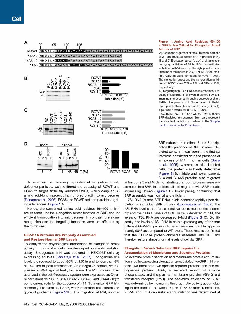
To examine the targeting capacities of elongation arrest-
defective particles, we monitored the capacity of RCWT and
RCA5 to target artificially arrested RNCs, which carry an 86
amino acid-long nascent chain of preprolactin, to microsomes
(Flanagan et al., 2003). RCA5 and RCWT had comparable target-
ing efficiencies (Figure 1D).
Hence, the conserved amino acid residues 96–100 in h14
are essential for the elongation arrest function of SRP and for
efficient translocation into microsomes. In contrast, the signal
recognition and the targeting functions were not affected by
the mutations.
GFP-h14 Proteins Are Properly Assembledand Restore Normal SRP LevelsTo analyze the physiological importance of elongation arrest
activity in mammalian cells, we developed a complementation
assay. Endogenous h14 was depleted in HEK293T cells by
expressing shRNAs (Lakkaraju et al., 2007). Endogenous h14
levels are reduced to about 50% at 120 hr and to less than 5%
at 144–168 hr post-transfection. As a negative control, we ex-
pressed shRNA against firefly luciferase. The h14 proteins char-
acterized in the cell-free assay system were expressed as C-ter-
minal fusions with GFP (G14, G14A12, G14A5, and G14A6-12) to
complement cells for the absence of h14. To monitor GFP-h14
assembly into functional SRP, we fractionated cell extracts on
glycerol gradients (Figure S1B). The migration of h19, another
Figure 1. Amino Acid Residues 96–100
in SRP14 Are Critical for Elongation Arrest
Activity of SRP
(A) Sequence alignment of the C-terminal portions
of WT and mutated human SRP14 proteins (h14).
(B and C) Elongation arrest (black) and transloca-
tion (gray) activities of SRPs (RCs) reconstituted
with different h14 proteins. The right panels: quan-
tification of the results (n R 3). EKRM: 0.2 eq/reac-
tion. Activities were normalized to RCWT (100%).
The elongation arrest and the translocation activi-
ties of RCWT were 72% ± 7% and 79% ± 10%,
respectively.
(D) Targeting of pPL86-RNCs to microsomes. Tar-
geting efficiencies (T [%]) were monitored by sed-
imenting microsomes through a sucrose cushion.
EKRM: 1 eq/reaction; S: Supernatant, P: Pellet.
Right panel: Quantification of the assays (n = 3).
T [%] was normalized to RCWT (100%).
�RC: buffer. RC(�14): SRP without h9/14. EKRM:
SRP-depleted microsomes. Error bars represent
the standard deviation as defined in the Supple-
mental Experimental Procedures.
SRP subunit, in fractions 5 and 6 desig-
nated the presence of SRP. In mock-de-
pleted cells, h14 was seen in the first six
fractions consistent with the presence of
an excess of h14 in human cells (Bovia
et al., 1995), whereas in h14-depleted
cells, the protein was hardly detectable
(Figure S1B, middle and lower panels).
G14 and G14A5 proteins also migrated
in fractions 5 and 6, demonstrating that both proteins were as-
sembled into SRP. In addition, all h19 migrated with SRP in cells
expressing G14A5 (Figure S1B, lower panel), confirming that
SRP assembly was normal and efficient.
7SL RNA (human SRP RNA) levels decrease rapidly upon de-
pletion of individual SRP proteins (Lakkaraju et al., 2007). The
7SL RNA level is therefore a sensitive tool to monitor the assem-
bly and the cellular levels of SRP. In cells depleted of h14, the
levels of 7SL RNA are decreased 9-fold (Figure S1C). Signifi-
cantly, the levels of 7SL RNA in cells expressing any of the four
different GFP-h14 protein chimeras were restored to approxi-
mately 90% as compared to WT levels. These results confirmed
that the GFP-h14 protein chimeras assemble into SRP and
thereby restore almost normal levels of cellular SRP.
Elongation Arrest-Defective SRP Impairs theAccumulation of Membrane and Secreted ProteinsTo examine protein secretion and membrane protein accumula-
tion in cells expressing elongation arrest-defective GFP-h14 pro-
teins, we monitored two specific reporter proteins and one en-
dogenous protein: SEAP, a secreted version of alkaline
phosphatase, and the plasma membrane proteins VSV-G and
transferrin receptor (TfnR). The secretion efficiency of SEAP
was determined by measuring the enzymatic activity accumulat-
ing in the medium between 144 and 168 hr after transfection.
VSV-G and TfnR cell-surface accumulation was determined at
442 Cell 133, 440–451, May 2, 2008 ª2008 Elsevier Inc.
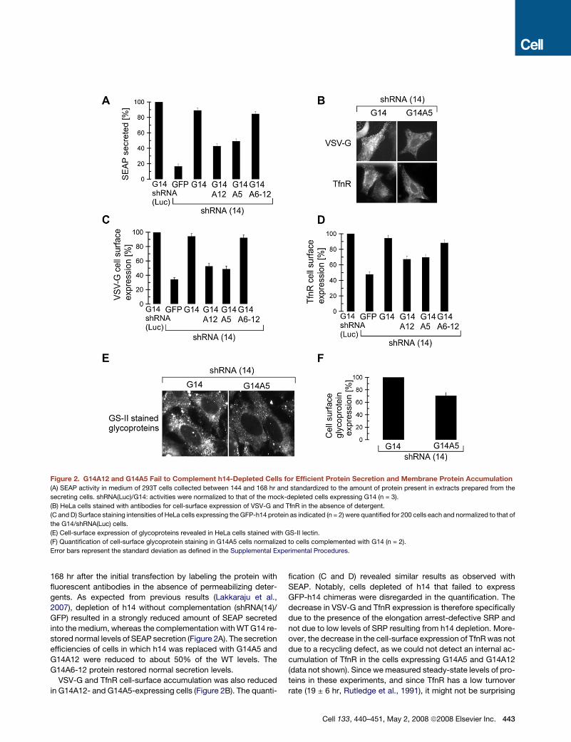
Figure 2. G14A12 and G14A5 Fail to Complement h14-Depleted Cells for Efficient Protein Secretion and Membrane Protein Accumulation
(A) SEAP activity in medium of 293T cells collected between 144 and 168 hr and standardized to the amount of protein present in extracts prepared from the
secreting cells. shRNA(Luc)/G14: activities were normalized to that of the mock-depleted cells expressing G14 (n = 3).
(B) HeLa cells stained with antibodies for cell-surface expression of VSV-G and TfnR in the absence of detergent.
(C and D) Surface staining intensities of HeLa cells expressing the GFP-h14 protein as indicated (n = 2) were quantified for 200 cells each and normalized to that of
the G14/shRNA(Luc) cells.
(E) Cell-surface expression of glycoproteins revealed in HeLa cells stained with GS-II lectin.
(F) Quantification of cell-surface glycoprotein staining in G14A5 cells normalized to cells complemented with G14 (n = 2).
Error bars represent the standard deviation as defined in the Supplemental Experimental Procedures.
168 hr after the initial transfection by labeling the protein with
fluorescent antibodies in the absence of permeabilizing deter-
gents. As expected from previous results (Lakkaraju et al.,
2007), depletion of h14 without complementation (shRNA(14)/
GFP) resulted in a strongly reduced amount of SEAP secreted
into the medium, whereas the complementation with WT G14 re-
stored normal levels of SEAP secretion (Figure 2A). The secretion
efficiencies of cells in which h14 was replaced with G14A5 and
G14A12 were reduced to about 50% of the WT levels. The
G14A6-12 protein restored normal secretion levels.
VSV-G and TfnR cell-surface accumulation was also reduced
in G14A12- and G14A5-expressing cells (Figure 2B). The quanti-
fication (C and D) revealed similar results as observed with
SEAP. Notably, cells depleted of h14 that failed to express
GFP-h14 chimeras were disregarded in the quantification. The
decrease in VSV-G and TfnR expression is therefore specifically
due to the presence of the elongation arrest-defective SRP and
not due to low levels of SRP resulting from h14 depletion. More-
over, the decrease in the cell-surface expression of TfnR was not
due to a recycling defect, as we could not detect an internal ac-
cumulation of TfnR in the cells expressing G14A5 and G14A12
(data not shown). Since we measured steady-state levels of pro-
teins in these experiments, and since TfnR has a low turnover
rate (19 ± 6 hr, Rutledge et al., 1991), it might not be surprising
Cell 133, 440–451, May 2, 2008 ª2008 Elsevier Inc. 443
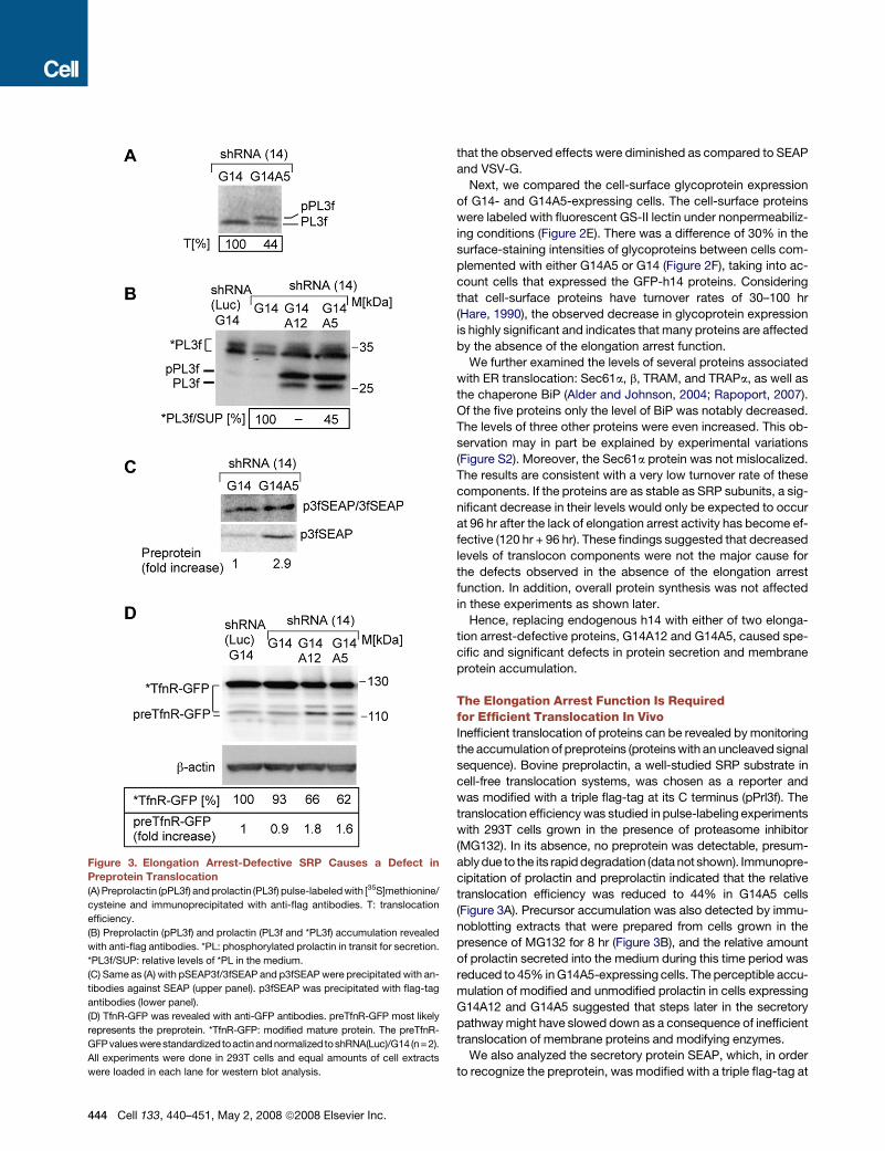
Figure 3. Elongation Arrest-Defective SRP Causes a Defect in
Preprotein Translocation
(A) Preprolactin (pPL3f) and prolactin (PL3f) pulse-labeled with [35S]methionine/
cysteine and immunoprecipitated with anti-flag antibodies. T: translocation
efficiency.
(B) Preprolactin (pPL3f) and prolactin (PL3f and *PL3f) accumulation revealed
with anti-flag antibodies. *PL: phosphorylated prolactin in transit for secretion.
*PL3f/SUP: relative levels of *PL in the medium.
(C) Same as (A) with pSEAP3f/3fSEAP and p3fSEAP were precipitated with an-
tibodies against SEAP (upper panel). p3fSEAP was precipitated with flag-tag
antibodies (lower panel).
(D) TfnR-GFP was revealed with anti-GFP antibodies. preTfnR-GFP most likely
represents the preprotein. *TfnR-GFP: modified mature protein. The preTfnR-
GFP valueswere standardizedtoactinandnormalizedtoshRNA(Luc)/G14 (n= 2).
All experiments were done in 293T cells and equal amounts of cell extracts
were loaded in each lane for western blot analysis.
444 Cell 133, 440–451, May 2, 2008 ª2008 Elsevier Inc.
that the observed effects were diminished as compared to SEAP
and VSV-G.
Next, we compared the cell-surface glycoprotein expression
of G14- and G14A5-expressing cells. The cell-surface proteins
were labeled with fluorescent GS-II lectin under nonpermeabiliz-
ing conditions (Figure 2E). There was a difference of 30% in the
surface-staining intensities of glycoproteins between cells com-
plemented with either G14A5 or G14 (Figure 2F), taking into ac-
count cells that expressed the GFP-h14 proteins. Considering
that cell-surface proteins have turnover rates of 30–100 hr
(Hare, 1990), the observed decrease in glycoprotein expression
is highly significant and indicates that many proteins are affected
by the absence of the elongation arrest function.
We further examined the levels of several proteins associated
with ER translocation: Sec61a, b, TRAM, and TRAPa, as well as
the chaperone BiP (Alder and Johnson, 2004; Rapoport, 2007).
Of the five proteins only the level of BiP was notably decreased.
The levels of three other proteins were even increased. This ob-
servation may in part be explained by experimental variations
(Figure S2). Moreover, the Sec61a protein was not mislocalized.
The results are consistent with a very low turnover rate of these
components. If the proteins are as stable as SRP subunits, a sig-
nificant decrease in their levels would only be expected to occur
at 96 hr after the lack of elongation arrest activity has become ef-
fective (120 hr + 96 hr). These findings suggested that decreased
levels of translocon components were not the major cause for
the defects observed in the absence of the elongation arrest
function. In addition, overall protein synthesis was not affected
in these experiments as shown later.
Hence, replacing endogenous h14 with either of two elonga-
tion arrest-defective proteins, G14A12 and G14A5, caused spe-
cific and significant defects in protein secretion and membrane
protein accumulation.
The Elongation Arrest Function Is Requiredfor Efficient Translocation In VivoInefficient translocation of proteins can be revealed by monitoring
the accumulation of preproteins (proteins with an uncleaved signal
sequence). Bovine preprolactin, a well-studied SRP substrate in
cell-free translocation systems, was chosen as a reporter and
was modified with a triple flag-tag at its C terminus (pPrl3f). The
translocation efficiency was studied in pulse-labeling experiments
with 293T cells grown in the presence of proteasome inhibitor
(MG132). In its absence, no preprotein was detectable, presum-
ably due to the its rapid degradation (data not shown). Immunopre-
cipitation of prolactin and preprolactin indicated that the relative
translocation efficiency was reduced to 44% in G14A5 cells
(Figure 3A). Precursor accumulation was also detected by immu-
noblotting extracts that were prepared from cells grown in the
presence of MG132 for 8 hr (Figure 3B), and the relative amount
of prolactin secreted into the medium during this time period was
reduced to 45% in G14A5-expressing cells. The perceptible accu-
mulation of modified and unmodified prolactin in cells expressing
G14A12 and G14A5 suggested that steps later in the secretory
pathway might have slowed down as a consequence of inefficient
translocation of membrane proteins and modifying enzymes.
We also analyzed the secretory protein SEAP, which, in order
to recognize the preprotein, was modified with a triple flag-tag at
its very N terminus (p3fSEAP). The relative size difference be-
tween the precursor and the mature SEAP proteins is very small,
and the pulse-labeled proteins immunoprecipated with SEAP
antibodies therefore migrated together in SDS-PAGE. However,
the specific analysis of the [35S]methionine-labeled preprotein
immunoprecipitated with the flag antibodies showed its accu-
mulation in G14A5-expressing cells during the pulse of 3 min
(Figure 3C). A similar precursor accumulation was also revealed
in extracts from cells grown in the presence of MG132 for 8 hr
(not shown).
To examine the accumulation of preTfnR, we used a plasmid
expressing the TfnR-GFP fusion protein (Figure 3D). We ob-
served an accumulation of unmodified TfnR-GFP, which most
likely represents preTfnR-GFP, in cells with elongation arrest-de-
fective SRP. Moreover, accumulation of mature TfnR-GFP was
decreased by 40% in the cells expressing G14A12 and G14A5,
consistent with the results shown previously for the endogenous
TfnR (Figure 2D).
These results demonstrated that the elongation arrest function
is essential for efficient translocation in vivo and that its absence
leads to defects in protein secretion and membrane protein
accumulation.
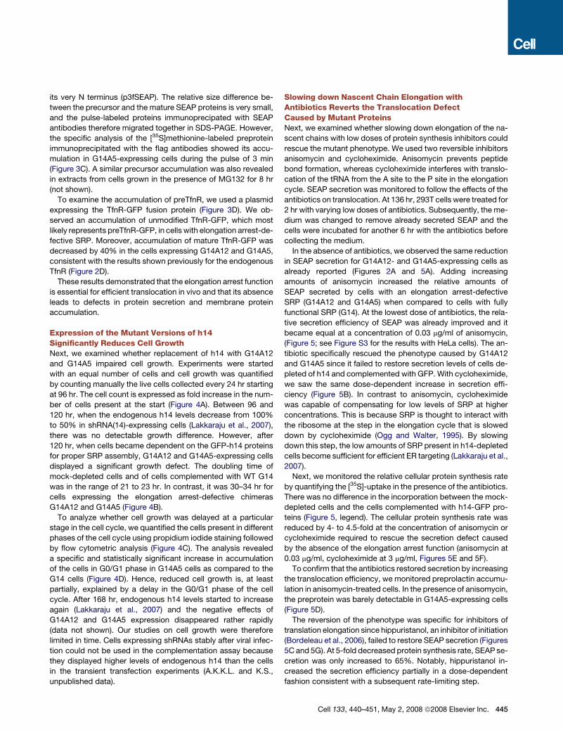
Expression of the Mutant Versions of h14Significantly Reduces Cell GrowthNext, we examined whether replacement of h14 with G14A12
and G14A5 impaired cell growth. Experiments were started
with an equal number of cells and cell growth was quantified
by counting manually the live cells collected every 24 hr starting
at 96 hr. The cell count is expressed as fold increase in the num-
ber of cells present at the start (Figure 4A). Between 96 and
120 hr, when the endogenous h14 levels decrease from 100%
to 50% in shRNA(14)-expressing cells (Lakkaraju et al., 2007),
there was no detectable growth difference. However, after
120 hr, when cells became dependent on the GFP-h14 proteins
for proper SRP assembly, G14A12 and G14A5-expressing cells
displayed a significant growth defect. The doubling time of
mock-depleted cells and of cells complemented with WT G14
was in the range of 21 to 23 hr. In contrast, it was 30–34 hr for
cells expressing the elongation arrest-defective chimeras
G14A12 and G14A5 (Figure 4B).
To analyze whether cell growth was delayed at a particular
stage in the cell cycle, we quantified the cells present in different
phases of the cell cycle using propidium iodide staining followed
by flow cytometric analysis (Figure 4C). The analysis revealed
a specific and statistically significant increase in accumulation
of the cells in G0/G1 phase in G14A5 cells as compared to the
G14 cells (Figure 4D). Hence, reduced cell growth is, at least
partially, explained by a delay in the G0/G1 phase of the cell
cycle. After 168 hr, endogenous h14 levels started to increase
again (Lakkaraju et al., 2007) and the negative effects of
G14A12 and G14A5 expression disappeared rather rapidly
(data not shown). Our studies on cell growth were therefore
limited in time. Cells expressing shRNAs stably after viral infec-
tion could not be used in the complementation assay because
they displayed higher levels of endogenous h14 than the cells
in the transient transfection experiments (A.K.K.L. and K.S.,
unpublished data).
Slowing down Nascent Chain Elongation withAntibiotics Reverts the Translocation DefectCaused by Mutant ProteinsNext, we examined whether slowing down elongation of the na-
scent chains with low doses of protein synthesis inhibitors could
rescue the mutant phenotype. We used two reversible inhibitors
anisomycin and cycloheximide. Anisomycin prevents peptide
bond formation, whereas cycloheximide interferes with translo-
cation of the tRNA from the A site to the P site in the elongation
cycle. SEAP secretion was monitored to follow the effects of the
antibiotics on translocation. At 136 hr, 293T cells were treated for
2 hr with varying low doses of antibiotics. Subsequently, the me-
dium was changed to remove already secreted SEAP and the
cells were incubated for another 6 hr with the antibiotics before
collecting the medium.
In the absence of antibiotics, we observed the same reduction
in SEAP secretion for G14A12- and G14A5-expressing cells as
already reported (Figures 2A and 5A). Adding increasing
amounts of anisomycin increased the relative amounts of
SEAP secreted by cells with an elongation arrest-defective
SRP (G14A12 and G14A5) when compared to cells with fully
functional SRP (G14). At the lowest dose of antibiotics, the rela-
tive secretion efficiency of SEAP was already improved and it
became equal at a concentration of 0.03 mg/ml of anisomycin,
(Figure 5; see Figure S3 for the results with HeLa cells). The an-
tibiotic specifically rescued the phenotype caused by G14A12
and G14A5 since it failed to restore secretion levels of cells de-
pleted of h14 and complemented with GFP. With cycloheximide,
we saw the same dose-dependent increase in secretion effi-
ciency (Figure 5B). In contrast to anisomycin, cycloheximide
was capable of compensating for low levels of SRP at higher
concentrations. This is because SRP is thought to interact with
the ribosome at the step in the elongation cycle that is slowed
down by cycloheximide (Ogg and Walter, 1995). By slowing
down this step, the low amounts of SRP present in h14-depleted
cells become sufficient for efficient ER targeting (Lakkaraju et al.,
2007).
Next, we monitored the relative cellular protein synthesis rate
by quantifying the [35S]-uptake in the presence of the antibiotics.
There was no difference in the incorporation between the mock-
depleted cells and the cells complemented with h14-GFP pro-
teins (Figure 5, legend). The cellular protein synthesis rate was
reduced by 4- to 4.5-fold at the concentration of anisomycin or
cycloheximide required to rescue the secretion defect caused
by the absence of the elongation arrest function (anisomycin at
0.03 mg/ml, cycloheximide at 3 mg/ml, Figures 5E and 5F).
To confirm that the antibiotics restored secretion by increasing
the translocation efficiency, we monitored preprolactin accumu-
lation in anisomycin-treated cells. In the presence of anisomycin,
the preprotein was barely detectable in G14A5-expressing cells
(Figure 5D).
The reversion of the phenotype was specific for inhibitors of
translation elongation since hippuristanol, an inhibitor of initiation
(Bordeleau et al., 2006), failed to restore SEAP secretion (Figures
5C and 5G). At 5-fold decreased protein synthesis rate, SEAP se-
cretion was only increased to 65%. Notably, hippuristanol in-
creased the secretion efficiency partially in a dose-dependent
fashion consistent with a subsequent rate-limiting step.
Cell 133, 440–451, May 2, 2008 ª2008 Elsevier Inc. 445
Figure 4. The Absence of the Elongation Arrest Function Impairs Cell Growth
(A) 293T cells were plated at equal densities and live cells counted at the times indicated (n = 3). h14 depletion starts at 120 hr (Lakkaraju et al., 2007). No increased
cell death was observed with GA12 and GA5 cells (see 0–300 arbitrary units [AU] in C).
(B) The doubling time was calculated for cell counts between 120 and 168 hr. Doubling times of 30–34 hr represent a 50%–60% increase as compared to 21 hr of
the mock-treated cells.
(C) Propidium iodide-stained 293T cells were analyzed for cell-cycle progression using FACSort. FI: fluorescence intensity in AU.
(D) Quantification of cells in different cell-cycle phases (n = 5). The sum of cells in the three phases was set to 100%. p < 0.05 for cells in G0/G1 phase indicating
statistical significance of the data at 95% confidence levels.
These results demonstrated that a delay in elongation is es-
sential for efficient translocation of preproteins and indicated
the presence of a rate-limiting step subsequent to the formation
of the SRP-RNC complex.
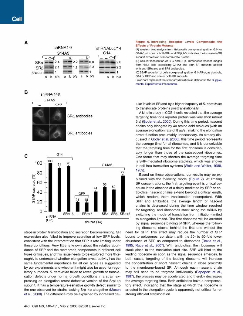
Cellular SR Levels Are Rate Limiting in TargetingTo determine whether SR was rate-limiting in targeting, we ex-
pressed exogenous human SRa and SRß from two separate
plasmids transfected into HeLa cells at the same time as the
expression plasmids for the GFP-h14 chimeras. When ex-
pressed together, the levels of SRa and SRb were increased
by 2.4- and 2.1-fold, respectively, as compared to the endoge-
nous proteins, and both proteins were found associated with
the ER (Figures 6A and 6B). The subunits expressed individually
also accumulated stably and were properly localized (Figure 6
446 Cell 133, 440–451, May 2, 2008 ª2008 Elsevier Inc.
and data not shown). As before, without the elongation arrest
function, SEAP secretion was reduced to 44% as compared to
control cells (Figure 6C). Expression of individual receptor sub-
units increased SEAP secretion only slightly. Expression of
both subunits rescued secretion to 72% as compared to cells
not depleted of h14. These results showed that the availability
of SR is rate limiting in targeting.
In the presence of the elongation arrest function, when all
nascent chains become successfully targeted to the translocon,
and in the absence of exogenous h14 (at low levels of SRP), the
expression of SRa and SRß did not significantly change the
secretion efficiency (Figure 6C, G14 and GFP). Similar results
were obtained in a set of experiments that were done in 293T cells
with the receptor subunits expressed as fusion proteins tagged
with GFP (GFP-SRa) and three flag epitopes (SRß-3f, Figure S4).

DISCUSSION
The results of these studies demonstrate that elongation arrest
activity has a fundamental role in maintaining the structure and
Figure 5. The Mutant Phenotypes of G14A12 and
G14A5 Are Specifically Rescued by Slowing down
Nascent Chain Elongation
(A–C) SEAP secretion from 293T cells in the presence of aniso-
mycin (ANM), cycloheximide (CHX), or hippuristanol. For each
concentration, the secretion activities were normalized to that
of shRNA(Luc)/G14 cells (n = 3). The absolute activities of
mock-depleted cells were decreased to 11%, 16%, and
15% at 0.03 mg/ml of anisomycin, at 3 mg/ml cycloheximide,
and at 0.75 mM hippuristanol, respectively, as compared to
control cells.
(D) Preprotein accumulation in the presence of anisomycin at
168 hr. Equal amounts of cell extracts were displayed by SDS-
PAGE and pPL3f revealed with anti-flag antibodies (n = 2).
(E–G) Effects of antibiotics on the cellular protein synthesis
rate in mock-depleted 293T cells. Cells were labeled for
15 min with [35S]methionine/cysteine, and labeled proteins
were acid-precipitated and quantified (n = 3). In parallel exper-
iments we found that the [35S]-uptake in shRNA(Luc)/G14,
shRNA(14)/G14, and shRNA(14)/G14A5 cells without antibi-
otics was 31,354; 33,047; and 29,149 total counts/106 cells,
respectively, indicating that protein synthesis was unaffected
by GFP-h14A5 expression.
Error bars represent the standard deviation as defined in the
Supplemental Experimental Procedures.
function of mammalian cells. Its absence leads to
the depletion of proteins in the endogenous mem-
brane system and to the reduction of protein secre-
tion with profound consequences on cell growth.
SRP lacking elongation arrest activity causes de-
fects in protein translocation that are reversed
when the overall rate of nascent chain elongation
is decreased by 4-fold. This result demonstrates
that when the cellular translation elongation rate
of signal sequence-containing proteins exceeds
the rate of targeting those proteins to the ER mem-
brane, translocation into the ER does not proceed
efficiently. The rate-limiting factor in targeting in
vivo is the SRP receptor: if its concentration is in-
sufficient, delays in the targeting of RNCs to the
ER occur because of a shortage of operational tar-
geting sites. As a consequence, not all nascent
chains will reach the translocon in time for success-
ful engagement into translocation. But importantly,
this effect may also serve a regulatory purpose.
Since SRP will dissociate preferentially from SRP-
RNC complexes with low affinity while awaiting
a free SR, nascent chains will be targeted and pro-
cessed more frequently at the ER if their SRP-signal
sequence affinity is high rather than low.
In pancreatic microsomes, SR is about 2-fold
less abundant than active translocons (Guth et al.,
2004), consistent with a rate-limiting function in
secretion. Moreover, the translocon might become
rate-limiting at more than 2-fold increased SR levels, providing
an explanation for the incomplete rescue of translocation in
these experiments. Exogenous SR expression in WT cells did
not improve secretion efficiency, suggesting that subsequent
Cell 133, 440–451, May 2, 2008 ª2008 Elsevier Inc. 447
steps in protein translocation and secretion become limiting. SR
expression also failed to improve secretion at low SRP levels,
consistent with the interpretation that SRP is rate limiting under
these conditions. Very little is known about the relative abun-
dance of SRP and the membrane components in different cell
types or tissues, and this issue needs to be explored more thor-
oughly to understand whether elongation arrest activity has the
same fundamental importance for all cell types as suggested
by our experiments and whether it might also be used for regu-
latory purposes. S. cerevisiae failed to reveal growth or translo-
cation defects under normal growth conditions in a strain ex-
pressing an elongation arrest-defective version of the Srp14p
subunit. It has a temperature-sensitive growth defect similar to
the one observed for strains lacking Srp14p altogether (Mason
et al., 2000). The difference may be explained by increased cel-
Figure 6. Increasing Receptor Levels Compensate the
Effects of Protein Mutants
(A) Western blot analysis from HeLa cells coexpressing either G14 or
G14A5 with one or both SRa and SRb. b/a indicates the increase in SR
subunit expression standardized to b-actin.
(B) Cellular localization of SRa and SRb. Immunofluorescent images
from HeLa cells expressing G14A5 and both SR subunits labeled
with anti-SRa and anti-SRß antibodies.
(C) SEAP secretion of cells coexpressing either G14A5 or, as controls,
G14 or GFP and one or both SR subunits.
Error bars represent the standard deviation as defined in the Supple-
mental Experimental Procedures.
lular levels of SR and by a higher capacity of S. cerevisiae
to translocate proteins posttranslationally.
A kinetic study in COS-1 cells revealed that the average
targeting time for a reporter protein was very short (about
5 s) (Goder et al., 2000). During this time period, nascent
chains only elongate by 40 amino acid residues (with an
average elongation rate of 8 aa/s), making the elongation
arrest function presumably unnecessary. As already dis-
cussed in Goder et al. (2000), this time period represents
the average time for all ribosomes, and it is conceivable
that the targeting time for the first ribosome is consider-
ably longer than those of the subsequent ribosomes.
One factor that may shorten the average targeting time
is SRP-mediated ribosome stacking, which was shown
in cell-free translation systems (Wolin and Walter, 1988,
1989).
Based on these observations, our results may be ex-
plained with the following model (Figure 7). At limiting
SR concentrations, the first targeting event is critical be-
cause in the absence of a delay mediated by SRP or an-
tibiotics, nascent chains extend beyond a critical length,
which renders them translocation incompetent. With
SRP and antibiotics, the average length of nascent
chains is decreased during the time window required
for targeting, and ribosomes stack along the mRNA by
switching the mode of translation from initiation-limited
to elongation-limited. The first ribosome will be arrested
by signal sequence binding of SRP, whereas the follow-
ing ribosome stacks behind the first one without the
need for SRP. This effect may reduce the number of SRP
bound to polysomes, consistent with the 20- to 50-fold lower
abundance of SRP as compared to ribosomes (Bovia et al.,
1995; Raue et al., 2007). With antibiotics, the ribosomes will
stack close to the translation start site. SRP will bind to the
leading ribosome as soon as the signal sequence emerges. In
both cases, targeting of the leading ribosome will increase
the concentration of short nascent chains in close proximity
to the membrane-bound SR. Although each nascent chain
may still need to be targeted individually (Rapoport et al.,
1987), the process may be accelerated and thereby decreases
the average targeting time. Both antibiotics have a compensa-
tory effect, indicating that the stage at which the ribosome is
arrested in the elongation cycle is apparently not critical for re-
storing efficient translocation.
448 Cell 133, 440–451, May 2, 2008 ª2008 Elsevier Inc.
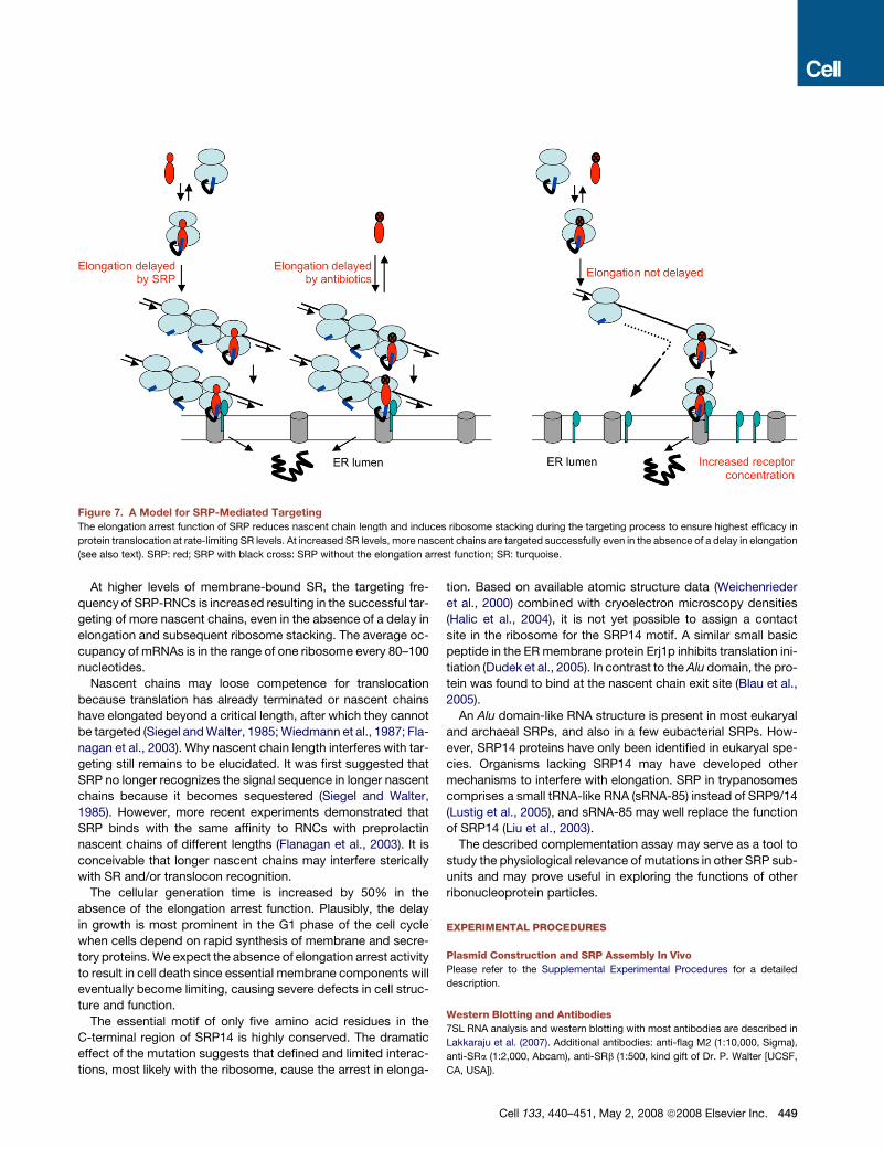
Figure 7. A Model for SRP-Mediated Targeting
The elongation arrest function of SRP reduces nascent chain length and induces ribosome stacking during the targeting process to ensure highest efficacy in
protein translocation at rate-limiting SR levels. At increased SR levels, more nascent chains are targeted successfully even in the absence of a delay in elongation
(see also text). SRP: red; SRP with black cross: SRP without the elongation arrest function; SR: turquoise.
At higher levels of membrane-bound SR, the targeting fre-
quency of SRP-RNCs is increased resulting in the successful tar-
geting of more nascent chains, even in the absence of a delay in
elongation and subsequent ribosome stacking. The average oc-
cupancy of mRNAs is in the range of one ribosome every 80–100
nucleotides.
Nascent chains may loose competence for translocation
because translation has already terminated or nascent chains
have elongated beyond a critical length, after which they cannot
be targeted (Siegel and Walter, 1985; Wiedmann et al., 1987; Fla-
nagan et al., 2003). Why nascent chain length interferes with tar-
geting still remains to be elucidated. It was first suggested that
SRP no longer recognizes the signal sequence in longer nascent
chains because it becomes sequestered (Siegel and Walter,
1985). However, more recent experiments demonstrated that
SRP binds with the same affinity to RNCs with preprolactin
nascent chains of different lengths (Flanagan et al., 2003). It is
conceivable that longer nascent chains may interfere sterically
with SR and/or translocon recognition.
The cellular generation time is increased by 50% in the
absence of the elongation arrest function. Plausibly, the delay
in growth is most prominent in the G1 phase of the cell cycle
when cells depend on rapid synthesis of membrane and secre-
tory proteins. We expect the absence of elongation arrest activity
to result in cell death since essential membrane components will
eventually become limiting, causing severe defects in cell struc-
ture and function.
The essential motif of only five amino acid residues in the
C-terminal region of SRP14 is highly conserved. The dramatic
effect of the mutation suggests that defined and limited interac-
tions, most likely with the ribosome, cause the arrest in elonga-
tion. Based on available atomic structure data (Weichenrieder
et al., 2000) combined with cryoelectron microscopy densities
(Halic et al., 2004), it is not yet possible to assign a contact
site in the ribosome for the SRP14 motif. A similar small basic
peptide in the ER membrane protein Erj1p inhibits translation ini-
tiation (Dudek et al., 2005). In contrast to the Alu domain, the pro-
tein was found to bind at the nascent chain exit site (Blau et al.,
2005).
An Alu domain-like RNA structure is present in most eukaryal
and archaeal SRPs, and also in a few eubacterial SRPs. How-
ever, SRP14 proteins have only been identified in eukaryal spe-
cies. Organisms lacking SRP14 may have developed other
mechanisms to interfere with elongation. SRP in trypanosomes
comprises a small tRNA-like RNA (sRNA-85) instead of SRP9/14
(Lustig et al., 2005), and sRNA-85 may well replace the function
of SRP14 (Liu et al., 2003).
The described complementation assay may serve as a tool to
study the physiological relevance of mutations in other SRP sub-
units and may prove useful in exploring the functions of other
ribonucleoprotein particles.
EXPERIMENTAL PROCEDURES
Plasmid Construction and SRP Assembly In Vivo
Please refer to the Supplemental Experimental Procedures for a detailed
description.
Western Blotting and Antibodies
7SL RNA analysis and western blotting with most antibodies are described in
Lakkaraju et al. (2007). Additional antibodies: anti-flag M2 (1:10,000, Sigma),
anti-SRa (1:2,000, Abcam), anti-SRb (1:500, kind gift of Dr. P. Walter [UCSF,
CA, USA]).
Cell 133, 440–451, May 2, 2008 ª2008 Elsevier Inc. 449
Cell Culture
Human HeLa (CCL-2) and HEK293T cells were grown at 37�C in Dulbecco’s
modified Eagle’s medium (DMEM) supplemented with 10% fetal calf serum
(Sigma). Equal numbers of cells were initially transfected with shRNA(14)
and, for mock depletion, with shRNA(Luc). After 24 hr, cells were selected
for 24 hr in puromycin (3 mg/ml). At 72 hr, cells were plated at equal numbers
into 6 cm dishes and transfected with GFP-h14-expressing plasmids (or
GFP as a control) and, if applicable, with a plasmid expressing a reporter pro-
tein. Antibiotics were added initially for 2 hr at 136 hr, then the medium was
changed and the cells were incubated for another 6 hr with antibiotics before
collecting the medium and lysing the cells (Lakkaraju et al., 2007). The cell
number was quantified manually by counting the number of live cells. Growth
rates were calculated for every 24 hr. Cell-cycle assays are described in the
Supplemental Experimental Procedures.
SEAP Assay, Glycoprotein, VSV-G, and TfnR-GFP Cell-Surface
Quantification and Microscopy
Methods are described in Lakkaraju et al. (2007). Cell-surface glycoprotein
staining is described in the Supplemental Experimental Procedures.
Pulse Labeling
At 168 hr after initial transfection, MG132 (25 mM) was added and after 45 min,
293T cells were labeled with 200 mCi/ml of [35S]methionine/cysteine mix
(Hartmann Analytic) for 3 min. Cells were lysed with 200 ml of 1% SDS in
0.1M Tris (pH 8) directly in the plate. The lysate was transferred into a micro-
tube and heated to 70�C with occasional vortexing until the lysate became
less viscous. The sample was diluted (3-fold) in the IP buffer (150 mM
NaCl, 1% NP-40, 0.5% deoxycholate, 0.1% SDS 50 mM Tris-Hcl [pH 8],
1 mM EDTA, and 13 protease inhibitor cocktail) and the labeled proteins in-
cubated overnight with the antibodies. The antibodies were immobilized on
protein G-Sepharose (40 ml) for 4 hr. The precipitates were washed three
times with IP buffer for 15 min, and the bound proteins displayed by 12%
SDS-PAGE. Visualization and quantification were done with the Bio-Rad
phosphorimager.
Protein and RNA Expression, Purification, Reconstitution of SRP,
and Activity Assays
Proteins and synthetic SRP RNA were expressed and purified as described in
the Supplemental Experimental Procedures and in Huck et al. (2004) and
Terzi et al. (2004). The targeting assay was done as described in Flanagan
et al. (2003). Cycloheximide, SRP, and EKRM were used at concentrations
of 500 mM, 200 nM, and 1 eq/reaction, respectively. EKRM membranes
were presaturated as described in Schaletzky and Rapoport (2006). The pro-
tein contents of the supernatants and pellets were analyzed on a 15% Tricine
gel and the radioactivity was detected and quantified with a Bio-Rad phos-
phorimager.
SUPPLEMENTAL DATA
Supplemental Data contain Supplemental Experimental Procedures, Supple-
mental References, and four figures and can be found with this article online
at http://www.cell.com/cgi/content/full/133/3/440/DC1/.
ACKNOWLEDGMENTS
We thank A. Chassot, Y. Miao, and Y. Shao for technical assistance; Drs. J.
Pelletier, R.S. Hegde, and P. Walter for providing reagents; and Dr. D. Picard
for suggestions on the manuscript. This work was supported by grants from
the Swiss National Science Foundation, the Canton of Geneva, and the MEDIC
Foundation. Support from NIH grants R01GM26494 and the Robert A. Welch
Foundation (Chair grant BE-0017) are also acknowledged (A.E.J.).
Received: October 18, 2007
Revised: January 8, 2008
Accepted: February 14, 2008
Published: May 1, 2008
450 Cell 133, 440–451, May 2, 2008 ª2008 Elsevier Inc.
REFERENCES
Alder, N.N., and Johnson, A.E. (2004). Cotranslational membrane protein
biogenesis at the endoplasmic reticulum. J. Biol. Chem. 279, 22787–22790.
Blau, M., Mullapudi, S., Becker, T., Dudek, J., Zimmermann, R., Penczek, P.A.,
and Beckmann, R. (2005). ERj1p uses a universal ribosomal adaptor site to co-
ordinate the 80S ribosome at the membrane. Nat. Struct. Mol. Biol. 12, 1015–
1016.
Bordeleau, M.E., Mori, A., Oberer, M., Lindqvist, L., Chard, L.S., Higa, T., Bel-
sham, G.J., Wagner, G., Tanaka, J., and Pelletier, J. (2006). Functional charac-
terization of IRESes by an inhibitor of the RNA helicase eIF4A. Nat. Chem. Biol.
2, 213–220.
Bovia, F., Fornallaz, M., Leffers, H., and Strub, K. (1995). The SRP9/14 subunit
of the signal recognition particle (SRP) is present in more than 20-fold excess
over SRP in primate cells and exists primarily free but also in complex with
small cytoplasmic Alu RNAs. Mol. Biol. Cell 6, 471–484.
Bovia, F., Wolff, N., Ryser, S., and Strub, K. (1997). The SRP9/14 subunit of the
human signal recognition particle binds to a variety of Alu-like RNAs and with
higher affinity than its mouse homolog. Nucleic Acids Res. 25, 318–326.
Connolly, T., and Gilmore, R. (1986). Formation of a functional ribosome-mem-
brane junction during translocation requires the participation of a GTP-binding
protein. J. Cell Biol. 103, 2253–2261.
Connolly, T., Rapiejko, P.J., and Gilmore, R. (1991). Requirement of GTP
hydrolysis for dissociation of the signal recognition particle from its receptor.
Science 252, 1171–1173.
Dudek, J., Greiner, M., Muller, A., Hendershot, L.M., Kopsch, K., Nastainczyk,
W., and Zimmermann, R. (2005). ERj1p has a basic role in protein biogenesis at
the endoplasmic reticulum. Nat. Struct. Mol. Biol. 12, 1008–1014.
Flanagan, J.J., Chen, J.C., Miao, Y., Shao, Y., Lin, J., Bock, P.E., and Johnson,
A.E. (2003). Signal recognition particle binds to ribosome-bound signal
sequences with fluorescence-detected subnanomolar affinity that does not
diminish as the nascent chain lengthens. J. Biol. Chem. 278, 18628–18637.
Gilmore, R., Blobel, G., and Walter, P. (1982). Protein translocation across the
endoplasmic reticulum. I. Detection in the microsomal membrane of a receptor
for the signal recognition particle. J. Cell Biol. 95, 463–469.
Goder, V., Crottet, P., and Spiess, M. (2000). In vivo kinetics of protein target-
ing to the endoplasmic reticulum determined by site-specific phosphorylation.
EMBO J. 19, 6704–6712.
Guth, S., Volzing, C., Muller, A., Jung, M., and Zimmermann, R. (2004). Protein
transport into canine pancreatic microsomes: a quantitative approach. Eur. J.
Biochem. 271, 3200–3207.
Halic, M., Becker, T., Pool, M.R., Spahn, C.M., Grassucci, R.A., Frank, J., and
Beckmann, R. (2004). Structure of the signal recognition particle interacting
with the elongation-arrested ribosome. Nature 427, 808–814.
Hare, J.F. (1990). Mechanisms of membrane protein turnover. Biochim. Bio-
phys. Acta 1031, 71–90.
Hauser, S., Bacher, G., Dobberstein, B., and Lutcke, H. (1995). A complex of
the signal sequence binding protein and the SRP RNA promotes translocation
of nascent proteins. EMBO J. 14, 5485–5493.
Huck, L., Scherrer, A., Terzi, L., Johnson, A.E., Bernstein, H.D., Cusack, S.,
Weichenrieder, O., and Strub, K. (2004). Conserved tertiary base pairing
ensures proper RNA folding and efficient assembly of the signal recognition
particle Alu domain. Nucleic Acids Res. 32, 4915–4924.
Johnsson, N., and Varshavsky, A. (1994). Split ubiquitin as a sensor of protein
interactions in vivo. Proc. Natl. Acad. Sci. USA 91, 10340–10344.
Keenan, R.J., Freymann, D.M., Stroud, R.M., and Walter, P. (2001). The signal
recognition particle. Annu. Rev. Biochem. 70, 755–775.
Lakkaraju, A.K., Luyet, P.P., Parone, P., Falguieres, T., and Strub, K. (2007). In-
efficient targeting to the endoplasmic reticulum by the signal recognition par-
ticle elicits selective defects in post-ER membrane trafficking. Exp. Cell Res.
313, 834–847.
Lipp, J., Dobberstein, B., and Haeuptle, M.T. (1987). Signal recognition particle
arrests elongation of nascent secretory and membrane proteins at multiple
sites in a transient manner. J. Biol. Chem. 262, 1680–1684.
Liu, L., Ben-Shlomo, H., Xu, Y.X., Stern, M.Z., Goncharov, I., Zhang, Y., and
Michaeli, S. (2003). The trypanosomatid signal recognition particle consists
of two RNA molecules, a 7SL RNA homologue and a novel tRNA-like molecule.
J. Biol. Chem. 278, 18271–18280.
Lustig, Y., Goldshmidt, H., Uliel, S., and Michaeli, S. (2005). The Trypanosoma
brucei signal recognition particle lacks the Alu-domain-binding proteins: puri-
fication and functional analysis of its binding proteins by RNAi. J. Cell Sci. 118,
4551–4562.
Mason, N., Ciufo, L.F., and Brown, J.D. (2000). Elongation arrest is a physiolog-
ically important function of signal recognition particle. EMBO J. 19, 4164–
4174.
Meyer, D.I., Krause, E., and Dobberstein, B. (1982). Secretory protein translo-
cation across membranes-the role of the ‘‘docking protein’. Nature 297, 647–
650.
Ng, D.T., Brown, J.D., and Walter, P. (1996). Signal sequences specify the
targeting route to the endoplasmic reticulum membrane. J. Cell Biol. 134,
269–278.
Ogg, S.C., and Walter, P. (1995). SRP samples nascent chains for the pres-
ence of signal sequences by interacting with ribosomes at a discrete step dur-
ing translation elongation. Cell 81, 1075–1084.
Okun, M.M., and Shields, D. (1992). Translocation of preproinsulin across the
endoplasmic reticulum membrane. The relationship between nascent poly-
peptide size and extent of signal recognition particle-mediated inhibition of
protein synthesis. J. Biol. Chem. 267, 11476–11482.
Pool, M.R. (2005). Signal recognition particles in chloroplasts, bacteria, yeast
and mammals. Mol. Membr. Biol. 22, 3–15.
Powers, T., and Walter, P. (1996). The nascent polypeptide-associated com-
plex modulates interactions between the signal recognition particle and the
ribosome. Curr. Biol. 6, 331–338.
Rapoport, T.A. (2007). Protein translocation across the eukaryotic endoplas-
mic reticulum and bacterial plasma membranes. Nature 450, 663–669.
Rapoport, T.A., Heinrich, R., Walter, P., and Schulmeister, T. (1987). Mathe-
matical modeling of the effects of the signal recognition particle on translation
and translocation of proteins across the endoplasmic reticulum membrane.
J. Mol. Biol. 195, 621–636.
Raue, U., Oellerer, S., and Rospert, S. (2007). Association of protein biogene-
sis factors at the yeast ribosomal tunnel exit is affected by the translational
status and nascent polypeptide sequence. J. Biol. Chem. 282, 7809–7816.
Rutledge, E.A., Mikoryak, C.A., and Draper, R.K. (1991). Turnover of the trans-
ferrin receptor is not influenced by removing most of the extracellular domain.
J. Biol. Chem. 266, 21125–21130.
Schaletzky, J., and Rapoport, T.A. (2006). Ribosome binding to and dissocia-
tion from translocation sites of the endoplasmic reticulum membrane. Mol.
Biol. Cell 17, 3860–3869.
Shan, S.O., Stroud, R.M., and Walter, P. (2004). Mechanism of association and
reciprocal activation of two GTPases. PLoS Biol. 2, e320. 10.1371/journal.
pbio.0020320.
Siegel, V., and Walter, P. (1985). Elongation arrest is not a prerequisite for
secretory protein translocation across the microsomal membrane. J. Cell
Biol. 100, 1913–1921.
Siegel, V., and Walter, P. (1986). Removal of the Alu structural domain from sig-
nal recognition particle leaves its protein translocation activity intact. Nature
320, 81–84.
Terzi, L., Pool, M.R., Dobberstein, B., and Strub, K. (2004). Signal recognition
particle Alu domain occupies a defined site at the ribosomal subunit interface
upon signal sequence recognition. Biochemistry 43, 107–117.
Thomas, Y., Bui, N., and Strub, K. (1997). A truncation in the 14 kDa protein of
the signal recognition particle leads to tertiary structure changes in the RNA
and abolishes the elongation arrest activity of the particle. Nucleic Acids
Res. 25, 1920–1929.
Walter, P., and Blobel, G. (1981). Translocation of proteins across the endo-
plasmic reticulum III. signal recognition protein (SRP) causes signal se-
quence-dependent and site-specific arrest of chain elongation that is released
by microsomal membranes. J. Cell Biol. 91, 557–561.
Weichenrieder, O., Wild, K., Strub, K., and Cusack, S. (2000). Structure and as-
sembly of the Alu domain of the mammalian signal recognition particle. Nature
408, 167–173.
Wiedmann, M., Kurzchalia, T.V., Bielka, H., and Rapoport, T.A. (1987). Direct
probing of the interaction between the signal sequence of nascent preprolactin
and the signal recognition particle by specific cross-linking. J. Cell Biol. 104,
201–208.
Wolin, S.L., and Walter, P. (1988). Ribosome pausing and stacking during
translation of a eukaryotic mRNA. EMBO J. 7, 3559–3569.
Wolin, S.L., and Walter, P. (1989). Signal recognition particle mediates a tran-
sient elongation arrest of preprolactin in reticulocyte lysate. J. Cell Biol. 109,
2617–2622.
Wolin, S.L., and Walter, P. (1993). Discrete nascent chain lengths are required
for the insertion of presecretory proteins into microsomal membranes. J. Cell
Biol. 121, 1211–1219.
Cell 133, 440–451, May 2, 2008 ª2008 Elsevier Inc. 451