dialysis membranes for acute kidney injury - mdpi
TRANSCRIPT
�����������������
Citation: Raharjo, Y.; Zainol Abidin,
M.N.; Ismail, A.F.; Fahmi, M.Z.;
Saiful; Elma, M.; Santoso, D.; Haula’,
H.; Habibi, A.R. Dialysis Membranes
for Acute Kidney Injury. Membranes
2022, 12, 325. https://doi.org/
10.3390/membranes12030325
Academic Editor: Faheem
Hassan Akhtar
Received: 30 January 2022
Accepted: 7 March 2022
Published: 15 March 2022
Publisher’s Note: MDPI stays neutral
with regard to jurisdictional claims in
published maps and institutional affil-
iations.
Copyright: © 2022 by the authors.
Licensee MDPI, Basel, Switzerland.
This article is an open access article
distributed under the terms and
conditions of the Creative Commons
Attribution (CC BY) license (https://
creativecommons.org/licenses/by/
4.0/).
membranes
Review
Dialysis Membranes for Acute Kidney InjuryYanuardi Raharjo 1,* , Muhammad Nidzhom Zainol Abidin 2, Ahmad Fauzi Ismail 3, Mochamad Zakki Fahmi 1,Saiful 4, Muthia Elma 5 , Djoko Santoso 6 , Hamizah Haula’ 1 and Ahlan Riwahyu Habibi 1
1 Membrane Science and Technology Research Group (MSTRG), Chemistry Department, Faculty of Science andTechnology, Universitas Airlangga, Surabaya 60115, Indonesia; [email protected] (M.Z.F.);[email protected] (H.H.); [email protected] (A.R.H.)
2 Department of Chemistry, Faculty of Science, Universiti Malaya, Jalan Profesor Diraja Ungku Aziz,Kuala Lumpur 50603, Malaysia; [email protected]
3 Advanced Membrane Technology Research Centre (AMTEC), Universiti Teknologi Malaysia,Skudai 81310, Malaysia; [email protected]
4 Chemistry Department, Faculty of Mathematics and Natural Science, Syiah Kuala University,Banda Aceh 23111, Indonesia; [email protected]
5 Chemical Engineering Department, Lambung Mangkurat University, Jl. A. Yani KM 36,Banjarbaru 70123, Indonesia; [email protected]
6 Division of Nephrology and Hypertension, Dr. Soetomo Hospital, Faculty of Medicine, Universitas Airlangga,Surabaya 60115, Indonesia; [email protected]
* Correspondence: [email protected]
Abstract: Mortality and morbidity rates among critically ill septic patients having acute kidney injury(AKI) are very high, considering the total number of deaths after their admission. Inappropriateselection of the type of continuous renal replacement therapy and inadequate therapy become theimmediate causes of these issues. Dialysis is a commonly used treatment intended to prolong thelife of AKI patients. Dialysis membranes, which are the core of dialysis treatment, must be properlyselected to ensure fair treatment to the patients. The accumulation of certain types of moleculesmust be dealt with using the right membrane. Whether it is low-flux, high-flux, or adsorptivetype, the dialysis membrane should be chosen depending on the condition of the patients. Theselection of dialysis membranes should also be based on their effect on the treatment outcomesand well-being. All these options are needed to serve the patients of different clinical settings. Theuse of dialysis membranes is not restricted to conventional haemodialysis, but rather they can beemployed in haemoperfusion, haemofiltration, haemodiafiltration, or a combination of any two ofthem. This review focuses in-depth on different types of dialysis membranes, their characteristics,and approaches in addressing the issues encountered in patients having AKI with sepsis and/ormultiorgan failure in intensive care units.
Keywords: acute kidney injury; haemodialysis membrane; mixed matrix membrane; haemoperfusion;adsorption
1. Introduction
Membrane technology is growing rapidly. At the beginning of the use of this technol-ogy, membranes were widely applied to support the development of science and technologyin theoretical physics and chemistry, rather than for commercial use. Due to various techno-logical benefits, researchers began developing membrane applications for human survival.One of the applications is for kidney disease treatment, namely haemodialysis (HD) througha principle of dialysis, whereby the membrane acts as an artificial kidney. Haemodialysis isnot intended to heal the patient but to prolong the life of the patient in acute and chronicconditions. Acute kidney injury (AKI) and acute-on-chronic kidney failure (ACKF) areconditions when the patients experience a sudden negative change in blood quality causedby the accumulation of toxins.
Membranes 2022, 12, 325. https://doi.org/10.3390/membranes12030325 https://www.mdpi.com/journal/membranes
Membranes 2022, 12, 325 2 of 15
Specifically, AKI is a condition in which the concentration of creatinine in serumincreases >50% more than its normal concentration in a very short time (<7 days). Theurine output becomes less than 0.5 mL/kg/h for more than 6 h. Based on the NationalKidney Foundation (NKF) in the USA, around 37 million people have been affected bykidney disease, affecting 15% of the adult population. There are different conditions ofAKI, namely prerenal, renal, and postrenal. In prerenal and postrenal conditions, the bloodflow to the kidney and the urine flow from the kidney is affected. Meanwhile, the renalcondition can be caused by glomerulonephritis, blood clotting, and blood vessel disease,whereby the kidney fails to purify the blood. Patients with AKI and ACKF have to betreated using HD [1].
Acute Dialysis Quality Initiative has classified AKI based on the consensual risk,injury, failure, loss of kidney function, and end-stage kidney disease (RIFLE). The RIFLEclassification is based on serum creatinine (sCr) and urine output (UO). Based on theglomerular filtration rate, the risk, injury, and failure are occurring while the sCr increases1.5, 2, and 3 times, respectively. The patients are defined to have the loss of kidney functionand the end-stage kidney disease when their kidney function has been lost for more than4 weeks and more than 3 months, respectively. Based on the UO, the risk, injury, and failureare classified with less than 0.5 mL/kg/h for 6 h, less than 0.5 mL/kg/h for 12 h, and lessthan 0.3 mL/kg/h for 24 h or anuria for 12 h, respectively. The RIFLE is used as a guidelineto diagnose and justify the patient’s condition for kidney conditions [2]. In March 2012, theNational Kidney Foundation–Kidney Disease Outcomes Quality Initiative completed theAKI regulation for adults and paediatrics [3].
There are two dialysis treatments for AKI patients, namely intermittent haemodialysis(IHD) and continuous renal replacement therapy (CRRT). The main focus of the dialysistreatments in AKI is to remove excess water and waste. IHD is used for a short periodof time (3–4 h), while CRRT is conducted continuously (24 h) for several days. CRRT isapplicable for critically ill patients having different catabolic states, systemic inflammatorysyndromes with or without sepsis, and other organ failures. CRRT does not cause abruptvariations in fluid removal or osmolality, ensuring good clearance of solute and betterhaemodynamic tolerance due to the slower liquid flow rate [4].
A kidney is a vital organ to clean the body fluid from acidic, organic, and metabolicwaste through a series of urine production stages that cover water and toxin clearance.Kidney failure is one of the significant health problems of the world population sufferingfrom the disease. It refers to the incapability of kidneys to perform their essential tasks:eliminating waste products from body metabolisms (i.e., urea, creatinine, and excesswater) and maintaining electrolyte balance in the body. It is commonly caused by certainconditions, such as diseases (i.e., diabetes, hypertension) and injuries that induce sepsis orsystemic inflammatory response syndrome. AKI is a sudden reduction of kidney functionwithin 48 h, indicated by the increasing concentration of creatinine in serum of equal toor more than 0.3 mg/dL, the increasing percentage of creatinine of equal to or more than50%, or the reduction of excreted urine of less than 0.5 mL/kg per hour for more than6 h [5]. AKI can be detected by symptoms like pallor, leukonychia, pulmonary oedema,raised blood pressure, peripheral oedema, pleural effusion, tiredness, loin pain, anorexia,itching, nausea, vomiting, and haematuria. Hence, a patient with AKI is required to receiveimmediate treatment from the doctor to keep him alive.
Haemofiltration (HF), haemodialysis (HD), and haemodiafiltration (HDF) are amongthe treatment options for chronic kidney failure conditions like AKI and ACKF. The selec-tion of dialysis membrane is very important to achieve adequate treatment. The patientshave to be assessed in terms of their quality of blood and the amount of uremic toxinspresent prior to HD. Failure to choose the suitable dialysis membrane may cause an adverseeffect and worsen the patient’s condition.
Uremic toxins are defined as the products of metabolism that accumulate in thebody and their accumulation is associated with uraemia due to renal degradation and/orimpaired excretory capacity. Based on the physicochemical characteristics, uremic toxins
Membranes 2022, 12, 325 3 of 15
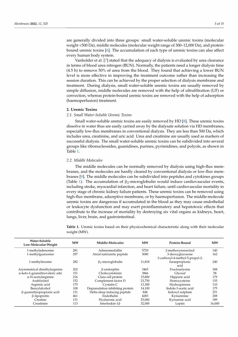
are generally divided into three groups: small water-soluble uremic toxins (molecularweight <500 Da), middle molecules (molecular weight range of 300–12,000 Da), and protein-bound uremic toxins [6]. The accumulation of each type of uremic toxins can also affectevery human body system.
Vanholder et al. [7] stated that the adequacy of dialysis is evaluated by urea clearancein terms of blood urea nitrogen (BUN). Normally, the patients need a longer dialysis time(4.5 h) to remove 50% of urea from the blood. They found that achieving a lower BUNlevel is more effective in improving the treatment outcome rather than increasing thesession duration. This can be achieved by the proper selection of dialysis membrane andtreatment. During dialysis, small water-soluble uremic toxins are usually removed bysimple diffusion, middle molecules are removed with the help of ultrafiltration (UF) orconvection, whereas protein-bound uremic toxins are removed with the help of adsorption(haemoperfusion) treatment.
2. Uremic Toxins2.1. Small Water-Soluble Uremic Toxins
Small water-soluble uremic toxins are easily removed by HD [8]. These uremic toxinsdissolve in water thus are easily carried away by the dialysate solution via HD membranes,especially low-flux membranes in conventional dialysis. They are less than 500 Da, whichincludes urea, creatinine, and uric acid. Urea and creatinine are usually used as markers ofsuccessful dialysis. The small water-soluble uremic toxins can be subdivided into severalgroups like ribonucleosides, guanidines, purines, pyrimidines, and polyols, as shown inTable 1.
2.2. Middle Molecules
The middle molecules can be normally removed by dialysis using high-flux mem-branes, and the molecules are hardly cleared by conventional dialysis or low-flux mem-branes [9]. The middle molecules can be subdivided into peptides and cytokines groups(Table 1). The accumulation of β2-microglobulin would induce cardiovascular events,including stroke, myocardial infarction, and heart failure, until cardiovascular mortality inevery stage of chronic kidney failure patients. These uremic toxins can be removed usinghigh-flux membrane, adsorptive membrane, or by haemoperfusion. The middle-moleculeuremic toxins are dangerous if accumulated in the blood as they may cause endothelialor leukocyte dysfunction and may exert proinflammatory and hepatotoxic effects thatcontribute to the increase of mortality by destroying six vital organs as kidneys, heart,lungs, liver, brain, and gastrointestinal.
Table 1. Uremic toxins based on their physicochemical characteristic along with their molecularweight (MW).
Water-SolubleLow-Molecular-Weight MW Middle-Molecules MW Protein-Bound MW
1-methyladenosine 281 Adrenomedullin 5729 2-methoxyresorcinol 1401-methylguanosine 297 Atrial natriuretic peptide 3080 3-deoxyglucosone 162
1-methylinosine 282 β2-microglobulin 11,8183-carboxyl-4-methyl-5-propyl-2-
furanpropionicacid
240
Asymmetrical dinethylarginine 202 β-endorphin 3465 Fructoselysine 308α-keto-δ-guanidinovaleric adic 151 Cholecystokinin 3866 Glyoxal 58
α-N-acetylarginine 216 Clara cell protein 15,800 Hippuric acid 179Arab(in)itol 152 Complement factor D 23,750 Homocysteine 135
Arginnic acid 175 Cystatin C 13,300 Hydroquinone 110Benzylalcohol 108 Degranulation inhibiting protein 14,100 Indole-3-acetic acid 175
β-guanidinopropionic acid 131 Delta-sleep inducing peptide 848 Indoxyl sulphate 251β-lipoprotin 461 Endothelin 4283 Kynurenine 208
Creatine 131 Hyaluronic acid 25,000 Kynurenic acid 189Creatinine 113 Interleukin-1β 32,000 Leptin 16,000
Membranes 2022, 12, 325 4 of 15
Table 1. Cont.
Water-SolubleLow-Molecular-Weight MW Middle-Molecules MW Protein-Bound MW
Cytidine 234 Interleukin-6 24,500 Melatonin 126Dimethylglycine 103 κ-Ig light chain 25,000 Methylglyoxal 72
Erythritol 122 λ-Ig light chain 25,000 Nε-(carboxymethyl)lysine 204γ-guanidinobutyric acid 145 Leptin 16,000 p-cresol 108
Guanidine 59 Methionine-enkephalin 555 Pentosidine 342Guanidinoacetic acid 117 Neuropeptide 4272 Phenol 94
Guanidinosuccinic acid 175 Parathyroid hormone 9225 P-OH hippuric acid 195Hypoxanthine 136 Retinol-binding protein 21,200 Putrescine 88
Malondialdehyde 71 Tumor necrosis factor-α 26,000 Quinolinic acid 167Mannitol 182 Retinol-binding protein 21,200
Methyguanidine 73 Spermidine 145Myoinositol 180 Spermine 202
N2,N2-dimethylguanosine 311N4-acetylcytidine 285
N6-methyladenosine 281N6-threonylcarbamoyladenosine 378
Orotic acid 174Orotidine 288Oxalate 90
Phenylacetylgluatmine 264Pseudouridine 244
Symmetrical dimethylarginine 202Sorbitol 182
Taurocyamine 174Threitol 122Thymine 126
Uracil 112Urea 60
Uric acid 168Uridine 244
Xanthine 152Xanthosine 284
2.3. Protein-Bound Uremic Toxins
The protein-bound uremic toxins can increase the body’s susceptibility to infectionand cardiovascular complications. According to Sakai [10], there are a total of 25 protein-bound uremic toxins, which can be divided into phenols, hippurates, indoles, peptides,polyamines, and advanced glycation end-products (AGE) groups (Table 1). These uremictoxins can cause cardiorenal syndrome. Although they are small-sized molecules, theirtendency to bind with proteins has made them difficult to be removed using a conventionaldialyser. Hence, these toxins can be effectively removed using either haemoperfusion orHDF, which combines dialysis and adsorption mechanisms.
3. Dialysis Membrane
Haemodialysis is a replacement therapy for kidney failure patients to remove excessmetabolic waste or uremic toxins from blood, such as water, sodium, potassium, hydrogen,urea, creatinine, uric acid, and other substances through a semi-permeable membrane,where the processes like diffusion, osmosis, and UF occur without losing the essentialsubstances, such as glucose, electrolytes, and amino acids [11]. The principle of HD involvesthe movements of solutes and water from blood across the membrane into the dialysate.Large molecules (i.e., blood cells and proteins) are retained inside the blood. In contrast,smaller molecules (i.e., urea, creatinine, and other biological waste) will pass through thesmall pores of the membrane into the dialysate. Diffusion and UF are two fundamentalprocesses that ensure continuous renal therapy. Diffusion refers to the movement oftypically small solutes from a compartment in which they are in high concentration toanother compartment in which they are in low concentration. Meanwhile, UF is a processwhereby water molecules (blood plasma) are forced to move across a semi-permeable
Membranes 2022, 12, 325 5 of 15
membrane by a pressure gradient. The rate of UF depends on the porosity of the membraneand the hydrostatic pressure of the blood, which depends on blood flow [12].
There are two compartments in the dialyser to contain blood and dialysate, separatedby membranes. Dialysate flows in counter-current to the blood flow on the other side ofthe membrane to maximise solute concentration gradient for efficient diffusion. Diffusiveclearance of a solute depends on its molecular weight and electrical charge, as well as theblood-dialysate concentration gradient, blood and dialysate flow rates, and the membranecharacteristics in terms of diffusion coefficient [13]. Small molecules such as urea will moveeasily along the concentration gradient into the dialysate most of the time. However, moresignificant- or middle-sized molecules, which are believed to cause uraemia, are hardlyremoved by this process. Meanwhile, UF or convection is very effective for the removalof fluid along with middle-sized molecules from the blood into the dialysate and acrossthe membrane.
Since the first creation of HD membrane, this membrane has been derived frompolymeric materials. Membrane separation technology is effective because it is flexible,requires little energy, does not alter the molecular structure of the separated substance, canbe operated at room temperature, and does not require additional chemical substancesduring separation [14]. The dialysis membrane must possess a selective transport propertythat can withstand larger species and skip smaller species through the membrane.
Several properties are required for a dialysis membrane, such as high solute permeabil-ity, high water permeability, the balance between solute and water permeability, mechanicalstrength in wet state, satisfactory biocompatibility, and low cost. HD membranes maybecome wet through contact with blood, where there will be a change in inner diameter,thickness, or length, and the membranes must also have excellent mechanical strength. Thebest membranes applied in HD are membranes with a large pore area, strong, stretchable,thin, and lightweight [6].
In general, membranes used for HD application are hollow fibre rather than flat sheetmembranes. This is due to many drawbacks of a flat sheet membrane, such as frequentfouling, resulting in reduced membrane performance. In addition, the membrane also hasa relatively small surface area when applied in HD. Compared to the flat sheet membrane,a hollow fibre membrane offers several advantages for HD. The performance of hollowfibre membrane is better due to its higher total surface area [15]. The surface area of thehollow fibre membrane has a surface area density of 3000 m2/m3 compared to a flat sheetmembrane with a surface area density of 400 m2/m3. A hollow fibre membrane also has astronger mechanical structure than a flat sheet membrane. However, to produce a goodmembrane, the hollow fibre must be thin with a very small diameter (about 200 nm) so thatmore toxins in the body can be eliminated from human blood [16].
A cellulose-based membrane without modification was used in the earlier devel-opment of HD membrane. This membrane is homogeneous, symmetric, and has goodperformance for small water-soluble molecules. In the 1970s, a synthetic membrane dialyserwas developed with higher water permeability for the purpose of blood filtration. Themost noticeable changes of this development are larger pore size, a thicker wall structure,higher hydrophobicity, more uniform pore size and distribution, and a more asymmetricmembrane structure. Those changes influenced the performance of HD membranes tobecome better and more stable over a longer period [17]. Various synthetic membranes havebeen used clinically for HD, which are usually referred to as a single polymer name, includingpolysulfone (PSf), polyethersulfone (PES), polyamide, poly(aryl ether sulfone), polycarbonate,polyacrylonitrile, polymethylmethacrylate, and poly(ethylene-co-vinyl alcohol).
Hoenich [13] reviewed the fabrication of polyvinylidene fluoride (PVDF) hollow fibremembranes via the non-solvent induction phase method to be used for HD. Their studyindicated that the hollow fibre membranes made of PVDF have advantages in mechanicalproperties and water flux. However, the membrane did not have anticoagulant propertiesand was easily fouled by protein. In other research, poly (lactic acid) (PLA) was used as thebase material of HD membranes. PLA is one of the eco-friendly bioplastic materials that
Membranes 2022, 12, 325 6 of 15
are easily broken, easily processed, and cheap. Many studies in the field of health materialsused PLA. It is a polymer with hydrophilic properties and low electrical conductivity.During dialysis, pollutants in the body (e.g., proteins and microorganisms) are easilyadsorbed and stored in the membrane matrix [18].
Meanwhile, PSf as a membrane is advantageous due to its high chemical resistanceand is also not reactive in mineral acids, alkalis, and salts. Furthermore, PSf has excellentresistance over a wide temperature range (75–125 ◦C) and pH range (1–13). The maindrawback is its higher fouling tendency than hydrophilic membranes [19]. Therefore,surface modifications are needed to solve the existing issues of hydrophobic membranes.Some materials can be added or blended to the membrane to solve this problem, suchas using hydrophilic polymer, biomaterials, sorbent, and inorganic nanoparticles. Otheroptions include surface coating and functionalisation.
Blending polymers is interpreted as a physical mixture that is not covalently bondedby accumulating the properties of different polymers into a single membrane [20]. Thistechnique is the most widely used in the development of HD membranes, specifically toincrease the hydrophilicity and biocompatibility of synthetic membranes. The optimumratio of the hydrophilic materials blended to the hydrophobic polymer can be determinedby the permeability and selectivity of the resultant membrane. The mixture of two ormore materials can produce homogeneous (polymers miscible in all compositions) andheterogeneous (polymers not miscible in all compositions) membranes.
Researchers have carried out the blending of hydrophobic polymers like PSf and PESusing a hydrophilic polymer like polyvinylpyrrolidone (PVP) [20,21]. A combination ofboth polymers may produce a patient-friendly membrane. PVP is a highly hydrophilicpolymer without hydroxyl carbon and is non-ionic. PVP is known for its ability to inhibitprotein adsorption on the membrane surface; therefore, it can increase the antifoulingproperty and biocompatibility of the membrane [22,23]. The higher PVP loading in thedope composition increased the water flux and improved the biocompatibility of the mem-brane [24]. Other hydrophilising agents besides PVP are polyethylene glycol (PEG) andpolypropylene glycol (PPG) that utilise water-soluble solvents, such as dimethylformamide(DMF), dimethylacetamide (DMAc), N-methylpyrrolidone (NMP), and dimethyl sulfoxide(DMSO) [25].
Biomaterials or biological compounds are also added to HD membranes as additivesto increase membrane biocompatibility. Heparin [26] and vitamin E [27] have been success-fully blended into hydrophobic polymers, such as PSf and PES. Other added materials canbe used as adsorbents, like activated carbon (charcoal) [21], zeolite [28], and hydroxyap-atite (HAP) [29]. Zeolites, on the other hand, are microporous, aluminosilicate mineralscommonly used as commercial adsorbents and catalysts. Wernert et al. [26] successfullyutilised zeolites in the development of HD membranes. The membrane could eliminateabout 67% creatinine and 29% p-cresol. In addition, zeolite can be used and added to theHD membrane to clear middle-molecule toxins [27].
Recently, inorganic nanoparticles like carbon nanoparticles (carbon nanotube andgraphene) and metal oxide nanoparticles (titanium dioxide and iron oxide) were used asnanofillers in HD membranes [28,29]. Nanoparticles are particles with a size between 1and 100 nm. The application of these particles is based on their large surface area andwater transport properties. They can increase membrane resistance towards chemicaldegradation, thermal stability, and fouling.
Similar to dialyzers, membranes can be categorised based on their flux and efficiency.A low-flux membrane is termed as having a UF coefficient of <10 mL/h/mmHg, whereasa high-flux membrane is a membrane with a UF coefficient of >20 mL/h/mmHg withmiddle-molecule (i.e., β2-microglobulin) clearance of >20 mL/min. On the other hand,membrane efficiency is determined based on the mass transfer area coefficient of urea,KoAurea. Membrane with KoA < 500 mL/min is called a low-efficiency membrane, whereasthe one with KoA > 600 mL/min is known as a high-efficiency membrane. Although thereare specific values to dictate which one is a high- or low-flux membrane, and whether
Membranes 2022, 12, 325 7 of 15
it has high or low efficiency, membranologists often describe the flux and efficiency ofthe membranes in different terms (i.e., pure water flux and percentage removal of uremictoxins). Therefore, one could only tell which category the membrane belongs to based onthe comparison with various developed membranes reported in research articles.
3.1. Low-Flux Membrane
Cellulosic membranes are usually denoted as low-flux membranes despite their pro-nounced hydrophilic nature. This is due to their symmetric structure and small mean poresize [2,14]. The uniform resistance acted upon the entire membrane wall makes cellulosicmembranes suitable for diffusion of small water-soluble molecules, such as urea. The latterproduced modified cellulosic membranes (i.e., cellulose acetate) with a larger mean poresize compared to the unmodified cellulosic membrane (22 µm). This resulted in higherporosity of the membranes. However, their weak hydraulic permeability still limits theseparation performance [30]. They are incapable of extending their molecular weightrange of solutes that can be removed. Their relatively denser structure is impermeable tomiddle-molecule uremic toxins. The low flux does not help the convection of large proteinspassing through the membrane.
In addition, some unmodified synthetic membranes also display low permeability ofmiddle molecules, such as proteins. PES and PSf membranes are prevalently employedfor blood purification [20]. Despite the advantages of PES- and PSf-based membranes(e.g., excellent oxidative and hydrolytic stability and good mechanical properties [31]),their progress in HD application is always limited by their hydrophobic properties. Manystudies have concluded that membrane fouling is directly related to hydrophobicity [32].Membrane fouling consistently remains one of the greatest challenges to HD treatment.Fouling is caused by the deposition or adsorption of solutes like proteins on the mem-brane surface and into membrane pores [33]. This phenomenon subsequently reduces themembrane flux and disrupts the separation performance of the membrane.
Vilar and Farrington [34] concluded that low-flux membranes allow efficient diffusiveremoval of small molecular weight molecules like urea. Nevertheless, the membranesshow poor convective removal of middle molecules. Despite the disadvantages of low-flux membranes, many studies have shown that the use of this type of membrane is stillrelevant to some patients if the dialysis adequacy (KT/V > 1.2) is fulfilled [35]. However, forpatients having chronic kidney failure with a high accumulation of uremic toxins, high-fluxmembranes are necessary to achieve minimum adequacy of the dialysis treatment.
3.2. High-Flux Membrane
High-flux membranes are highly permeable and biocompatible, usually made upof synthetic polymers with certain modifications to increase the hydrophilicity of themembranes [36]. The high permeability of synthetic membranes is also contributed bytheir larger mean pore size that offers a higher UF rate at low pressure. The additionalfeature of this type of membrane over low-flux membranes is the enhanced convectiveremoval of middle molecules while maintaining excellent removal of small molecularweight molecules via diffusion [37]. The main requirement for the HD setting in high-fluxmembranes is the use of a high-quality dialysate solution that is made up of ultrapurewater with no detectable endotoxins (<0.03 endotoxin units per mL) to minimise chronicinflammation [38].
For PSf and PES membranes, the large amount of low molecular weight moleculeremoval is mainly because of the asymmetric structure and the higher UF coefficient, whichare contributed by the larger pore size and higher porosity of the membranes [39]. Nev-ertheless, HD membranes must also be assessed in terms of their capacity to eliminatepotentially deleterious middle molecules. Very few studies have successfully removedmiddle molecules efficiently. Yu et al. [40] produced a highly permeable thin-film nanofi-brous composite membrane consisting of an ultrathin hydrophilic active layer of chemicallycross-linked PVA and an electrospun PAN nanofibrous supporting layer. The membrane
Membranes 2022, 12, 325 8 of 15
exhibited excellent selectivity by removing 45.8% of middle-molecular weight toxins. Italso possessed good mechanical strength and comparable haemocompatibility.
In addition to improved middle-molecule clearance, a study by Mortada et al. [41]showed that high-flux PSf membranes are more superior than those of low-flux PSf mem-branes in terms of removing accumulated metals in the blood of kidney failure patientsduring HD, especially cadmium and lead. This is very helpful as cadmium and lead aretoxic and can potentially induce adverse effects on the patients.
Despite their advantages, membranes with too high flux often trigger the loss of watercontent in blood among dialysis patients as excessive UF may deplete the blood volume.This exposes the patients to the risk of hypotension [34]. Therefore, the dialysis dosemust be properly controlled as recommended by nephrologists. Kidney failure patients,especially those in high-risk groups, are suggested to use high-flux membranes to ensuretheir long survival [42]. Besides, studies show that high-flux membranes can minimise theoccurrence of uremic syndromes, such as amyloidosis, dyslipidemia, polyneuropathy, andinfection [43]. However, for the patients having an AKI with a high accumulation of uremictoxins in a short time, high-flux membranes are not necessary to achieve dialysis adequacy,whereas the patients with AKI have a problem with the haemodynamic system.
4. Haemofiltration and Haemodiafiltration
HF and HDF generally use high-flux membranes to remove larger-sized uremic toxins.High-flux membranes are known for high generation of hydrostatic pressure, in whichuremic toxins are significantly removed together with plasma filtrate by convection. Thesemembranes must ensure that the filtrate contains high concentrations of uremic toxins;otherwise, the treatment would not be efficient and might render hypertension. A plasma-like electrolyte solution made up of Type I water (ultrapure water) is used to replace thelost plasma filtrate.
Another concern is the susceptibility of essential proteins (i.e., albumin) to be removeddue to the high-pressure gradient. Thus, the chosen membrane must possess a well-compromised flux (UF coefficient) to attain the intended selectivity. A slow and continuousHF or HDF is usually performed on patients with AKI [44]. Although most developedmembranes are meant for HD, the reported data on their sieving properties could serveas useful information for the potential use in HF and HDF. Compared to HD, HF couldachieve excellent middle-molecule clearance of up to 80%, while HDF that utilises diffusionand convection could remove both small and large uremic toxins more efficiently.
5. Adsorptive Membrane/Sorbent
Adsorption can be categorised as either chemisorption or physisorption. Bondingin chemisorption is stronger than physisorption as chemisorption is simply a chemicalreaction with interactions between adsorbates and chemical bonding groups on the sur-face of adsorbents. In contrast, physisorption involves electrostatic interactions betweenadsorbates and the surface of adsorbents. Generally, the time needed to reach adsorptionequilibrium is shorter for physisorption compared to chemisorption.
5.1. Sorbent in Haemodialysis
In HD, adsorption has been proposed as one of the alternative methods to eliminateuremic toxins, and most studies used adsorptive membranes to remove protein-boundtoxins, which are hardly removed via diffusion and UF. Zeolite Y successfully removed p-cresol as protein-bounded uremic toxins both with and without modification [45]. Inspiredby the concept of the adsorptive membrane, several attempts have been made to forma mixed matrix membrane (MMM) using the HDF principle. In general, MMM is acombination of organic and inorganic components comprised of the integration of fillersor adsorbents into the polymer matrix. The MMMs combine the selectivity of inorganicparticles or sorbents with the high productivity of filtration membrane and have beenapplied to separate and recover proteins or enzymes. Based on the structure, MMM can
Membranes 2022, 12, 325 9 of 15
be divided into two categories: dense and porous structures. A dense structured MMMis mostly used in gas separation, pervaporation, and fuel cell applications. Meanwhile,a porous structured MMM is designed for adsorption purposes. The fillers that can beincorporated into the MMM are nanomaterials, such as zeolite, carbon nanotube, metal-organic framework, charcoal, and others. These nanomaterials can also be functionalisedin order to adopt the fillers to provide selectivity and absorptivity towards the targetedmolecules [46]. Hence, there are two mechanisms (i.e., diffusion and adsorption) in onemembrane module offered by MMM. Excess water can be removed by diffusion mechanism,and excess uremic toxins (cytokines) can be cleaned by the adsorption mechanism.
Tijink et al. [47] established a novel approach in blood cleaning by focusing on theimprovement of the adsorption capacity of a membrane. They fabricated dual-layer (flatsheet) MMMs, which combined diffusion and adsorption in a single step. Activated carbon(AC) was incorporated in the mixture of PES and PVP. A dual-layer MMM was fabricated,in which a particle-free membrane layer was formed on top of an MMM layer containingAC. The dual-layer MMM had lower water permeability (~350 L/m2·h·bar) compared tothat of the single-layer MMM (1800 350 L/m2·h·bar). The MMMs adsorbed 29 mg/g of ACat an equilibrium concentration of 0.05 mg/mL. When tested towards human plasma for4 h, the MMMs were able to remove more than 80% of creatinine and para-aminohippuricacid, which is a protein-bound toxin.
A year after, Tijink et al. [48] used the same formulation to develop a dual-layerhollow fibre MMM with the same goal of removing creatinine and protein-bound toxins(i.e., hippuric acid (HA), indoxyl sulphate (IS), and p-cresyl sulphate (pCS)). The waterpermeability of the MMM was 58.4 350 L/m2·h·bar. Compared to the previous dual-layerflat sheet membrane, the dual-layer hollow fibre MMM developed in this study achieved ahigher adsorption capacity of creatinine (100 mg/g of AC) at the equilibrium concentrationof 0.05 mg/mL. The maximum adsorption capacity of creatinine, HA, and IS was 3064,134, and 350 mg/g of AC, respectively. In the human blood plasma adsorption study,the MMM maintained 83% removal of creatinine, and it adsorbed 60% pCS, 90% IS, and95% HA after 4 h of incubation. In the presence of albumin, IS and pCS were poorlyremoved by adsorption and diffusion even after 6 h of operation. In contrast, the amountadsorbed was higher for all protein-bound toxins during the convection experiment due tothe pressure difference. Despite the better performance, albumin partially passed throughthe membrane during the convection experiment.
In 2016, another attempt was made by Pavlenko et al. [49]. They developed a low-fluxdual-layer hollow fibre MMM with a smaller dimension. A commercial carbon-basedsorbent (mesoporous Norit A Supra) with an average pore size of 3 nm was embedded inthe MMM. The Norit A Supra-incorporated MMM showed promising results, where theadsorption capacity of creatinine was 2579 mg/m2 and the removal of IS and pCS was 30%and 125% better in comparison to the first generation MMM and up to 100% as comparedto particle-free industrial membranes [48]. It was stated that the low UF coefficient of theMMM (3.35 mL/m2/h/mmHg) and a molecular weight-cut off around 12 kDa preventedalbumin leakage while achieving excellent removal of protein-bound toxins.
Other previously used adsorbents in the membrane include hydroxy appetite (HAP).Although its application in HD is yet to be seen, HAP has good adsorption to protein andis usually used in the medical field due to its biocompatibility and bioactivity [29]. HAPcan be incorporated into an HD membrane to improve the membrane’s adsorption capacitytowards middle-sized proteins. In general, MMM is very beneficial for the removal ofuremic toxins that cannot be removed by conventional dialyses, such as larger-molecular-weight and protein-bound uremic toxins. It offers an alternative way to overcome thelimitations of diffusion coefficient, sieving properties, and flux of the membrane. Thecombination of adsorption and diffusion carries an advantage on HD applications. Theincorporation of sorbents within a membrane is a very effective approach for efficientblood purification [50]. The integration of adsorbent into a dialysis membrane significantlyimproved the performance of the membrane. The development of this method is ensured to
Membranes 2022, 12, 325 10 of 15
be safe as the cellular components of blood do not interact with the embedded sorbent [51].This method revolutionises the previous method of adsorption, for instance, in blooddetoxification techniques (e.g., haemoperfusion).
5.2. Haemoperfusion
Haemoperfusion is the adsorption of molecules onto the surface of a biocompatibleadsorbent via direct contact in an extracorporeal circuit, as opposed to removing toxinsand excess body fluids through a semi-permeable membrane [52]. The sorbent has tobe sufficiently biocompatible to enable direct contact without the destruction of bloodelements. Sorbents can come from synthetic or natural materials. The natural sorbentsusually used are natural zeolite and carbons like charcoal and AC. Meanwhile, the syntheticsorbents can be obtained from the polymerisation of monomers, synthetic zeolite that canbe modified to control the structure of the internal pore system, and ion-exchange resin [53].Sorbents can be in granule form, powder, spheres, flakes, and cylindrical pellets. Theyare solid particles with a single particle diameter between 50 µm and 1.2 cm. The surface-area-to-volume ratio is extremely high in sorbent particles with a surface area varyingfrom 300 to 1200 m2/g. The adsorbents are also defined as microporous (pore size < 20 Å),mesoporous (pore size between 20 and 500 Å), and macroporous (pore size > 500 Å). Thesorbents must have high selectivity and good transport properties to catch up with thetargeted analyte fast.
The US Food and Drugs Administration (FDA) has classified the haemoperfusionsystem into two classes of medical device: class II and class III. A device is considered aclass II medical device when it is used for treating poison and drug overdoses. In contrast,a class III medical device is used to treat patients with liver deficiencies, such as in hepaticcoma [54]. Based on the principles and benefits of haemoperfusion, it is possible to beapplied for removing uremic toxins during HD.
The development of haemoperfusion for HD was continued using zeolites in adsorbingcreatinine (representing the small water-soluble molecules) and IS (representing the protein-bound uremic toxins). It was reported that 0.025 g of 940-zeolite powders could eliminate91% of 2 µmol creatinine in 5 min. Meanwhile, PES/zeolite membrane could adsorb 4948 µgcreatinine per g membrane and 550 µg IS per g membrane. The adsorption mechanism wasproposed to be via electrostatic attraction [55].
6. Roles in Removing Cytokines and Endotoxins
The presence of cytokines and endotoxins in the bloodstreams of patients with AKI andsepsis poses a threat to the patients’ life. In the development of haemoperfusion for HD, theresearchers in [54] examined carbon nanomaterials in terms of their adsorption capabilitiesfor blood cleansing. Non-porous carbon represented by graphene nanoplatelets (GNPs)and porous carbon polymorphs representing mesoporous carbon was compared to observetheir adsorption capacity for cytokines. It was found that the adsorption kinetics of the non-porous carbon significantly outperformed mesoporous carbon. The GNPs could completelyremove cytokines from the blood after 5 min. Moreover, the GNPs also maintained goodperformance when embedded into cryogels and polytetrafluoroethylene [54].
In recent years, a well-established sorption cartridge, namely CytoSorb (distributedby LINC Medical) is integrated with dialysis machines with the intent to adsorb cytokines.It is designed specifically to treat patients with sepsis. This new technology has shownexcellent removal of a wide range of crucial cytokines that cannot be eliminated usingexisting blood purification processes [56]. The cartridge consists of porous polymer beadsthat capture cytokines depending on the level of cytokines in the blood. The higher theconcentration of cytokines, the faster the adsorption rate. The cartridge is for single use butcan withstand up to 7 days of continuous use.
A multicentre study conducted by Basu et al. [56] on 43 patients provided the mostreliable clinical evidence of cytokine removal using CytoSorb. It was reported that CytoSorbsignificantly reduced the concentration of IL-6 (49.1%), IL-1ra (36.5%), and IL-8 (30.2%) in
Membranes 2022, 12, 325 11 of 15
the patients’ blood. Single-centre clinical studies (<20 patients) performed in India, Italy,and Germany suggested that the patients with predicted high mortality could survive ifthey were given CytoSorb less than 24 h after admission. The high overall survival of75% and 62.5% were reported, where the procalcitonin levels, sepsis-related organ failureassessment score, and simplified acute physiology score decreased. The reduction of thescores justified the positive impact of using CytoSorb on the haemodynamic parameters,which is important for patients with AKI and sepsis. In terms of safety, there were noserious device-related adverse effects observed during the treatment.
Another way to remove cytokines is using specific HD membranes. Due to thehuge size of cytokines as part of middle molecules, high cut-off membranes with a largerpore size (>0.01 µm) are used [46]. These membranes are intended to effectively removemolecules from 20 kDa to 50 kDa. It was clinically proven that high cut-off membranescould effectively remove cytokines, particularly IL-4, IL-6, IL-8, and IL-12, without elimi-nating albumin. The clearance of cytokines resulted in substantial enhancement of organdysfunction and haemodynamic condition.
On the other hand, an increased level of endotoxins is usually observed in patientswith AKI. Among the patients, endotoxins are found circulating in the bloodstream afterencountering severe traumas. After a few times undergoing dialysis treatment, endotoxinsoriginated from bacterial lipopolysaccharides (i.e., Gram-negative bacteria) found in thedialysate may accumulate in the blood due to their diffusion into the blood during thetreatment. This would elevate the level of endotoxins in the patient’s blood. To removethe circulating endotoxins from the patient’s blood, Toray Medical Co. Ltd. (Tokyo, Japan)has developed Toraymyxin, a polymyxin B-immobilised fibre blood purification column.The product was approved by the Japanese National Health Insurance system to treatendotoxemia and septic shock. Polymyxin B is known for its bactericidal effect againstGram-negative bacteria. Besides, it can inhibit the effects of endotoxins via binding withthe active site (lipid A domain) of the endotoxin molecules. For selective adsorption ofendotoxins, polymyxin B is immobilised on the surface of polymeric fibres (membrane).Mortality risk studies on various targeted groups revealed that the use of Toraymyxinsignificantly reduced the mortality risk ratio [50].
Due to the serious threats imposed by cytokines and endotoxins, the focus is now ondeveloping HD membranes that could remove these two types of molecules simultane-ously from blood. oXiris membrane was developed recently, which comprised of AN69membrane, surface-treated with polyethylenimine, and grafted with heparin on the innermembrane surface. AN69 membrane alone can remove large molecular weight molecules,including cytokines and uremic toxins, by membrane binding [57]. To tackle the problemof endotoxin accumulation, the membrane was surface-treated with polyethylenimine asthis polymer can adsorb endotoxins. The permanent grafting of heparin on the membranesurface inhibited blood coagulation by adsorbing antithrombin III (ATIII) from blood,forming a stable ATIII-thrombin complex as high as ~270 ng/mL compared to ~10 ng/mLwhen using non-heparin-grafted AN69 membrane. The grafted heparin has been proposedto catalyse the conversion of ATIII into a potent anticoagulant in the bloodstream. Theimproved membrane thrombogenicity could minimise the problem of membrane clotting,which is the most frequent technical complication encountered during continuous renalreplacement therapy for patients with AKI.
To date, oXiris is the only membrane (dialyser) with great removal of both endotox-ins (68%) and cytokines (>90%) [58]. This membrane can function as a dialyser in HDor as an adsorbent in haemoperfusion. Clinical improvements, such as increased arte-rial pressure and reduced norepinephrine dose, have been observed following the useof oXiris membrane on patients with AKI. These lead to improved organ function andhaemodynamic stability.
Membranes 2022, 12, 325 12 of 15
7. Combination of Haemodialysis and Haemoperfusion
Ghezi et al. [53] studied the combination of two dialysers in series, called pairedfiltration dialysis (PFD) technique or HDF mode. This technique has been developed dueto the weakness of high-flux HD, which is the process interference between diffusion andconvection as they occur simultaneously, thus decreasing their respective efficiency. Instudying a series of dialysis modes, the following mathematical reasoning is used [59];
Coefficient of haemodiafiltration: Khdf = Kuf + Kd (1)
Coefficient of UF or convection: Kuf = Quf (Co/Ci) (2)
However, Co < Ci, and therefore Co/Ci < 1.0 (3)
Then, Kuf < Quf (4)
Consequently, [Kd + Kuf] < ([Kd] + [Kuf]) (5)
where Khdf is the coefficient of haemodiafiltration; Kuf is the coefficient of UF; Kd is thecoefficient of diffusion; Quf is UF flow rate; Co is concentration out; Ci is concentration in;[ ] is one chamber only; ([ ] + [ ]) is one dialyzer with two chambers.
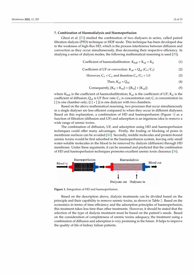
Based on the above mathematical reasoning, two processes that occur simultaneouslyin a single dialyser are less efficient compared to when they occur in different dialysers.Based on this explanation, a combination of HD and haemoperfusion (Figure 1) as afunction of filtration (diffusion and UF) and adsorption is an ingenious idea to remove awide range of uremic toxins.
The combination of diffusion, UF, and adsorption using HD and haemoperfusiontechniques could offer many advantages. Firstly, the fouling or blocking of pores inmembrane surfaces can be avoided [40]. Secondly, middle molecules and protein-bounduremic toxins would be first adsorbed in the haemoperfusion system, leaving only smallwater-soluble molecules in the blood to be removed by dialysis (diffusion) through HDmembrane. Under these arguments, it can be assumed and predicted that the combinationof HD and haemoperfusion techniques promotes excellent uremic toxin clearance [36].
Figure 1. Integration of HD and haemoperfusion.
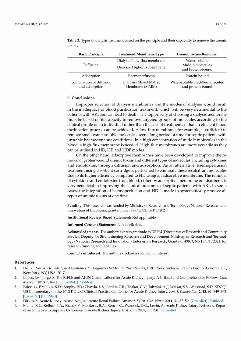
Based on the description above, dialysis treatments can be divided based on theprinciple and their capability to remove uremic toxins, as shown in Table 2. Based on theeconomics in terms of time efficiency and the adsorption principles of haemoperfusion,this treatment takes less time than other treatments. However, it should be stated that theselection of the type of dialysis treatment must be based on the patient’s needs. Basedon the consideration of completeness of uremic toxins adequacy, the treatment using acombination of diffusion and adsorption is very promising in the future. It helps to improvethe quality of life of kidney failure patients.
Membranes 2022, 12, 325 13 of 15
Table 2. Types of dialysis treatment based on the principle and their capability to remove the uremictoxins.
Basic Principle Treatment/Membrane Type Uremic Toxins Removed
DiffusionDialysis/Low-flux membrane Water-soluble
Dialysis/High-flux membrane Middle-moleculesand Protein-bound
Adsorption Haemoperfusion Protein-bound
Combination of diffusionand adsorption
Dialysis/Mixed MatrixMembrane (MMM)
Water-soluble, middle-molecules,and protein-bound
8. Conclusions
Improper selection of dialysis membranes and the modes of dialysis would resultin the inadequacy of blood purification treatment, which will be very detrimental to thepatients with AKI and can lead to death. The top priority of choosing a dialysis membranemust be based on its capacity to remove targeted groups of molecules according to theclinical profile of an individual rather than the cost of treatment so that an efficient bloodpurification process can be achieved. A low-flux membrane, for example, is sufficient toremove small water-soluble molecules over a long period of time for septic patients withunstable haemodynamic conditions. In a high concentration of middle molecules in theblood, a high-flux membrane is needed. High-flux membranes are more versatile as theycan be utilised in HD, HF, and HDF modes.
On the other hand, adsorptive membranes have been developed to improve the re-moval of protein-bound uremic toxins and different types of molecules, including cytokinesand endotoxins, through diffusion and adsorption. As an alternative, haemoperfusiontreatment using a sorbent cartridge is performed to eliminate these recalcitrant moleculesdue to its higher efficiency compared to HD using an adsorptive membrane. The removalof cytokines and endotoxins from blood, either by adsorptive membrane or adsorbent, isvery beneficial in improving the clinical outcomes of septic patients with AKI. In somecases, the integration of haemoperfusion and HD is made to systematically remove alltypes of uremic toxins at one time.
Funding: This research was funded by Ministry of Research and Technology/National Research andInnovation of Indonesia, grant number 498/UN3.15/PT/2021.
Institutional Review Board Statement: Not applicable.
Informed Consent Statement: Not applicable.
Acknowledgments: The authors express gratitude to DRPM (Directorate of Research and CommunityService, Deputy for Strengthening Research and Development, Ministry of Research and Technol-ogy/National Research and Innovation) Indonesia’s Research, Grant no: 498/UN3.15/PT/2021, forresearch funding and facilities.
Conflicts of Interest: The authors declare no conflict of interest.
References1. De, S.; Roy, A. Hemodialysis Membranes, for Engineers to Medical Practitioners; CRC Press Taylor & Francis Group: London, UK;
New York, NY, USA, 2017.2. Lopes, J.A.; Jorge, S. The RIFLE and AKIN Classifications for Acute Kidney Injury: A Critical and Comprehensive Review. Clin.
Kidney J. 2013, 6, 8–14. [CrossRef] [PubMed]3. Palevsky, P.M.; Liu, K.D.; Brophy, P.D.; Chawla, L.S.; Parikh, C.R.; Thakar, C.V.; Tolwani, A.J.; Waikar, S.S.; Weisbord, S.D. KDOQI
US Commentary on the 2012 KDIGO Clinical Practice Guideline for Acute Kidney Injury. Am. J. Kidney Dis. 2013, 61, 649–672.[CrossRef] [PubMed]
4. Dirkes, S. Acute Kidney Injury: Not Just Acute Renal Failure Anymore? Crit. Care Nurse 2011, 31, 37–50. [CrossRef] [PubMed]5. Mehta, R.L.; Kellum, J.A.; Shah, S.V.; Molitoris, B.A.; Ronco, C.; Warnock, D.G.; Levin, A. Acute Kidney Injury Network: Report
of an Initiative to Improve Outcomes in Acute Kidney Injury. Crit. Care 2007, 11, R31. [CrossRef]
Membranes 2022, 12, 325 14 of 15
6. Glorieux, G.; Tattersall, J. Uraemic Toxins and New Methods to Control Their Accumulation: Game Changers for the Concept ofDialysis Adequacy. Clin. Kidney J. 2015, 8, 353–362. [CrossRef]
7. Vanholder, R.; Smet, R.D.; Glorieux, G.; Argiles, A.; Baur eister, U.; Brunet, P.; Clark, W.; Cohen, G.; Deyn, P.P.D.; Deppisch, R.; et al.Review on Uremic Toxins: Classification, Concentration, and Interindividual Variability. Kidney Int. 2003, 63, 1934–1943.[CrossRef]
8. Yamamoto, S.; Kazama, J.J.; Wakamatsu, T.; Takahashi, Y.; Kaneko, Y.; Goto, S.; Narita, I. Removal of Uremic Toxins by RenalReplacement Therapies: A Review of Current Progress and Future Perspectives. Ren. Replace Ther. 2016, 2, 43. [CrossRef]
9. Cui, Z.F.; Muralidhara, H.S. Membrane Technology-A Practical Guide to Membrane Technology and Applications in Food and Bioprocessing;Elsevier: Burlington, MA, USA, 2010.
10. Sakai, K. Determination of Pore Size and Pore Size Distribution: 2. Dialysis Membrane. J. Memb. Sci. 1994, 96, 91–130. [CrossRef]11. Baker, R.W. Membrane Technology and Applications; John Wiley & Sons: West Sussex, UK, 2004.12. Gautham, A.; Muhammed, J.M.; Manavalan, M.; Najeb, M. A Hemodialysis Membranes: Past, Present and Future Trends. Int.
Res. J. Pharm. 2013, 4, 16–19. [CrossRef]13. Hoenich, N.A. Update on the Biocompatibility of Hemodialysis Membranes. Hong Kong J. Nephrol. 2004, 6, 74–78. [CrossRef]14. Zhang, Q.; Lu, X.; Zhao, L. Preparation of Polyvinylidene Fluoride (PVDF) Hollow Fiber Hemodialysis Membranes. Membranes
2014, 4, 81–95. [CrossRef] [PubMed]15. Subramanian, V.; Gupte, O. Functionalization of Poly (Ether Sulfone) (PES) and Polysulfone (PSF) Membrane Case Studies and
Discussion. Bombay Technol. 2014, 64, 22–26.16. Mansur, S.; Othman, M.H.D.; Ismail, A.F.; Sheikh Abdul Kadir, S.H.; Kamal, F.; Goh, P.S.; Hasbullah, H.; Ng, B.C.; Abdullah, M.S.
Investigation on the Effect of Spinning Conditions on the Properties of Hollow Fiber Membrane for Hemodialysis Application.J. Appl. Polym. Sci. 2016, 133, 1–10. [CrossRef]
17. Ronco, C.; Cruz, D. Hemodialysis—From Basic Research to Clinical Trials; Karger: Basel, Switzerland, 2008.18. Hayama, M.; Yamamoto, K.I.; Kohori, F.; Sakai, K. How Polysulfone Dialysis Membranes Containing Polyvinylpyrrolidone
Achieve Excellent Biocompatibility? J. Memb. Sci. 2004, 234, 41–49. [CrossRef]19. Zhao, C.; Xue, J.; Ran, F.; Sun, S. Modification of Polyethersulfone Membranes—A Review of Methods. Prog. Mater. Sci. 2013, 58,
76–150. [CrossRef]20. Dahe, G.J.; Teotia, R.S.; Kadam, S.S.; Bellare, J.R. The Biocompatibility and Separation Performance of Antioxidative Polysul-
fone/Vitamin E TPGS Composite Hollow Fiber Membranes. Biomaterials 2011, 32, 352–365. [CrossRef]21. Hoffman, R.; Nyu, N.; Holubek, W.J.; Hoffman, R.S.; Goldfarb, D.S.; Nelson, L.S. Use of Hemodialysis and Hemoperfusion in
Poisoned Patients Use of Hemodialysis and Hemoperfusion in Poisoned Patients. Kidney Int. 2008, 74, 1327–1334. [CrossRef]22. Raharjo, Y.; Ismail, A.F.; Dzarfan Othman, M.H.; Rosid, S.M.; Azali, M.A.; Santoso, D. Effect of Polymer Loading on Membrane
Properties and Uremic Toxins Removal for Hemodialysis Application. J. Membr. Sci. Res. 2021, 7, 14–19. [CrossRef]23. Waheed, H.; Hussain, A. Effect of Polyvinyl Pyrolidone on Morphology and Performance of Cellulose Acetate Based Dialysis
Membrane. Eng. Technol. Appl. Sci. Res. 2019, 9, 3744–3749. [CrossRef]24. Mansur, S.; Othman, M.H.D.; Ismail, A.F.; Zainol Abidin, M.N.; Said, N.; Sean, G.P.; Hasbullah, H.; Sheikh Abdul Kadir, S.H.;
Kamal, F. Study on the Effect of Spinning Conditions on the Performance of PSf/PVP Ultrafiltration Hollow Fiber Membrane.Malays. J. Fundam. Appl. Sci. 2018, 14, 343–347. [CrossRef]
25. Bergé-Lefranc, D.; Vagner, C.; Schäf, O.; Boulet, P.; Pizzala, H.; Paillaud, J.L.; Denoyel, R. Adsorption of Small Uremic ToxinMolecules onto Zeolites: A First Step towards an Alternative Kidney. Stud. Surf. Sci. Catal. 2007, 170, 1015–1020. [CrossRef]
26. Wernert, V.; Schäf, O.; Ghobarkar, H.; Denoyel, R. Adsorption Properties of Zeolites for Artificial Kidney Applications. MicroporousMesoporous Mater. 2005, 83, 101–113. [CrossRef]
27. Lu, L.; Yeow, J.T.W. An Adsorption Study of Indoxyl Sulfate by Zeolites and Polyethersulfone—Zeolite Composite Membranes.Mater. Des. 2017, 120, 328–335. [CrossRef]
28. Bergé-Lefranc, D.; Pizzala, H.; Paillaud, J.L.; Schäf, O.; Vagner, C.; Boulet, P.; Kuchta, B.; Denoyel, R. Adsorption of Small UremicToxin Molecules on MFI Type Zeolites from Aqueous Solution. Adsorption 2008, 14, 377–387. [CrossRef]
29. Sun, J.; Wu, L. Polyether Sulfone/Hydroxyapatite Mixed Matrix Membranes for Protein Purification. Appl. Surf. Sci. 2014, 308,155–160. [CrossRef]
30. Guo, C.; Zhou, L.; Lv, J. Effects of Expandable Graphite and Modified Ammonium Polyphosphate on the Flame-Retardant andMechanical Properties of Wood Flour-Polypropylene Composites. Polym. Polym. Compos. 2013, 21, 449–456. [CrossRef]
31. Su, B.H.; Fu, P.; Li, Q.; Tao, Y.; Li, Z.; Zao, H.S.; Zhao, C.S. Evaluation of Polyethersulfone Highflux Hemodialysis Membrane inVitro and in Vivo. J. Mater. Sci. Mater. Med. 2008, 19, 745–751. [CrossRef]
32. Clark, W.R.; Gao, D. Properties of Membranes Used for Hemodialysis Therapy. Semin. Dial. 2002, 15, 191–195. [CrossRef]33. Huang, J.; Zhang, K.; Wang, K.; Xie, Z.; Ladewig, B.; Wang, H. Fabrication of Polyethersulfone-Mesoporous Silica Nanocomposite
Ultrafiltration Membranes with Antifouling Properties. J. Memb. Sci. 2012, 423–424, 362–370. [CrossRef]34. Vilar, E.; Farrington, K. Haemodialysis. Medicine 2011, 39, 429–433. [CrossRef]35. Oshvandi, K.; Kavyannejad, R.; Borzuo, S.R.; Gholyaf, M. High-Flux and Low-Flux Membranes: Efficacy in Hemodialysis. Nurs.
Midwifery Stud. 2014, 3, e21764. [CrossRef] [PubMed]36. Saiful, S. Mixed Matrix Membrane Adsorbers for Protein and Blood Purification; University of Twente: Enschede, The Netherlands, 2007.37. Nissenson, A.R.; Fine, R.N. Handbook of Dialysis Therapy, 4th ed.; Elsevier: Philadelphia, PA, USA, 2008. [CrossRef]
Membranes 2022, 12, 325 15 of 15
38. Lonnemann, G. The Quality of Dialysate: An Integrated Approach. Kidney Int. Suppl. 2000, 58, 112–119. [CrossRef] [PubMed]39. Su, B.; Sun, S.; Zhao, C. Polyethersulfone Hollow Fiber Membranes for Hemodialysis. Prog. Hemodial. Emergent Biotechnol. Clin.
Pract. 2011, 1, 65–92. [CrossRef]40. Yu, X.; Shen, L.; Zhu, Y.; Li, X.; Yang, Y.; Wang, X.; Zhu, M.; Hsiao, B.S. High Performance Thin-Film Nanofibrous Composite
Hemodialysis Membranes with Efficient Middle-Molecule Uremic Toxin Removal. J. Memb. Sci. 2017, 523, 173–184. [CrossRef]41. Mortada, W.I.; Nabieh, K.A.; Donia, A.F.; Ismail, A.M.; Kenawy, I.M.M. Impact of Dialyzer Membrane Flux on Metal Clearance in
Hemodialysis Patients. J. Trace Elem. Med. Biol. 2016, 36, 52–56. [CrossRef] [PubMed]42. Eknoyan, G.; Beck, G.J.; Cheung, A.K.; Daugirdas, J.T.; Greene, T.; Kusek, J.W.; Allon, M.; Bailey, J.; Delmez, J.A.; Depner, T.A.; et al.
Effect of Dialysis Dose and Membrane Flux in Maintenance Hemodialysis. N. Engl. J. Med. 2002, 347, 2010–2019. [CrossRef][PubMed]
43. Boure, T. Which Dialyser Membrane to Choose? Nephrol. Dial. Transplant. 2004, 19, 293–296. [CrossRef]44. Tsimihodimos, V.; Mitrogianni, Z.; Elisaf, M. Dyslipidemia Associated with Chronic Kidney Disease. Open Cardiovas. Med. J. 2011,
5, 41–48. [CrossRef]45. Raharjo, Y.; Ismail, A.F.; Othman, M.H.D.; Malek, N.A.N.N.; Santoso, D. Preparation and Characterization of Imprinted Zeolite-Y
for p-Cresol Removal in Haemodialysis. Mater. Sci. Eng. C 2019, 103, 109722. [CrossRef]46. Villa, G.; Zaragoza, J.J.; Sharma, A.; Neri, M.; De Gaudio, A.R.; Ronco, C. Cytokine Removal with High Cut-off Membrane:
Review of Literature. Blood Purif. 2014, 38, 167–173. [CrossRef]47. Tijink, M.S.L.; Wester, M.; Sun, J.; Saris, A.; Bolhuis-Versteeg, L.A.M.; Saiful, S.; Joles, J.A.; Borneman, Z.; Wessling, M.; Stamatialis,
D.F. A Novel Approach for Blood Purification: Mixed-Matrix Membranes Combining Diffusion and Adsorption in One Step.Acta Biomater. 2012, 8, 2279–2287. [CrossRef] [PubMed]
48. Tijink, M.S.L.; Wester, M.; Glorieux, G.; Gerritsen, K.G.F.; Sun, J.; Swart, P.C.; Borneman, Z.; Wessling, M.; Vanholder, R.; Joles, J.A.;et al. Mixed Matrix Hollow Fiber Membranes for Removal of Protein-Bound Toxins from Human Plasma. Biomaterials 2013, 34,7819–7828. [CrossRef] [PubMed]
49. Pavlenko, D.; van Geffen, E.; van Steenbergen, M.J.; Glorieux, G.; Vanholder, R.; Gerritsen, K.G.F.; Stamatialis, D. New Low-FluxMixed Matrix Membranes That Offer Superior Removal of Protein-Bound Toxins from Human Plasma. Sci. Rep. 2016, 6, 34429.[CrossRef] [PubMed]
50. Shimizu, T.; Miyake, T.; Kitamura, N.; Tani, M.; Endo, Y. Endotoxin Adsorption: Direct Hemoperfusion with the PolymyxinB-Immobilized Fiber Column (PMX). Transfus. Apher. Sci. 2017, 56, 682–688. [CrossRef]
51. Saiful; Borneman, Z.; Wessling, M. Double Layer Mixed Matrix Membrane Adsorbers Improving Capacity and Safety Hemodialy-sis. IOP Conf. Ser. Mater. Sci. Eng. 2018, 352, 012048. [CrossRef]
52. Cheah, W.K.; Ishikawa, K.; Othman, R.; Yeoh, F.Y. Nanoporous Biomaterials for Uremic Toxin Adsorption in Artificial KidneySystems: A Review. J. Biomed. Mater. Res.-Part B Appl. Biomater. 2017, 105, 1232–1240. [CrossRef]
53. Ghezzi, P.M.; Sanz-Moreno, C.; Gervasio, R.; Nigrelli, S.; Botella, J. Technical Requirements for Rapid High Efficiency Therapy inUremic Patients. Am. Soc. Artif. Int. Organs 1987, 33, 546–550.
54. Pescatore, N.A. Graphitic Carbon Materials Tailored for the Rapid Adsorption of Biomolecules; Drexel University: Philadelphia, PA,USA, 2016.
55. Lu, L. New Membrane Technologies for Dialysis; University of Waterloo: Waterloo, ON, Canada, 2016.56. Basu, R.; Pathak, S.; Goyal, J.; Chaudhry, R.; Goel, R.B.; Barwal, A. Use of a Novel Hemoadsorption Device for Cytokine Removal
as Adjuvant Therapy in a Patient with Septic Shock with Multi-Organ Dysfunction: A Case Study. Indian J. Crit. Care Med. 2014,18, 822–824. [CrossRef]
57. De Vriese, A.; Colardyn, F.A.; Philippé, J.; Vanholder, R.C.; DE Sutter, J.H.; Lameire, N.H. Cytokine Removal during ContinuousHemofiltration in Septic Patients. J. Am. Soc. Nephrol. 1999, 10, 846–853. [CrossRef]
58. Malard, B.; Lambert, C.; Kellum, J.A. In Vitro Comparison of the Adsorption of Inflammatory Mediators by Blood PurificationDevices. Intensive Care Med. Exp. 2018, 6, 12. [CrossRef]
59. Botella, J.; Ghezzi, P.M.; Sanz-Moreno, C. Adsorption in Hemodialysis. Kidney Int. Suppl. 2000, 58, 60–65. [CrossRef] [PubMed]