development of inductively coupled plasma–mass spectrometry-based protease assays
TRANSCRIPT
Development of inductively coupled plasma-mass spectrometry(ICP-MS) based protease assays
Urja S. Lathia, Olga Ornatsky, Vladimir Baranov, and Mark NitzDepartment of Chemistry, University of Toronto, 80 St. George Street, Toronto, ON, M5S 3H6Urja S. Lathia: ; Olga Ornatsky: ; Vladimir Baranov: ; Mark Nitz: [email protected]
AbstractRapid, sensitive and quantitative assays for proteases are important for drug development and in thediagnosis of disease. Here, an assay for protease activity which uses inductively coupled plasma-mass spectrometry (ICP-MS) detection is described. Peptidic α-chymotrypsin substrates weresynthesized containing a lanthanide ion chelate at the N-terminus to provide a distinct elemental tag.A biotin label was appended to the C-terminus of the peptide allowing separation of uncleaved peptidefrom the enzymatic digestion. The enzyme activity was determined by quantifying the lanthanideion signal of the peptide cleavage products by ICP-MS. Biotinylated substrates synthesized includeLu-DTPA-Asp-Leu-Leu-Val-Tyr∼Asp-Lys(Biotin) and Lu-DTPA-βAla-βAla-βAla-βAla-Gly-Ser-Ala-Tyr∼Gly-Lys-Arg-Lys(biotin)-amide. Parallel assays with a commercially available fluorogenicsubstrate (Suc-AAPF-AMC) for α-chymotrypsin were performed for comparison. Using the ICP-MS assay enzyme concentrations as low as 2 pM could be readily detected which was superior tothe detection limit of an assay using the α-chymotrypsin fluorogenic substrate (Suc-AAPF-AMC).Furthermore, we demonstrated the use of this approach to detect chymotrypsin activity in HeLa celllysates.
KeywordsICP-MS; multiplex; α-chymotrypsin; proteases; lanthanides
IntroductionProteases are known to be involved in a number of pathological conditions including cancer[1], diseases of the central nervous system [2], cardiac diseases [3] and numerous viral andinfectious diseases [4]. The ability of proteases to catalytically and specifically cleave peptidesubstrates makes them ideal biomarkers for the diagnosis of disease [5]. Investigation of thepathological and physiological functions of proteases requires sensitive assays to detect theiractivity in complex tissue samples and other biological matrixes. Commonly protease assaysuse fluorogenic or chromogenic peptide substrates [6-9]. Fluorescence resonance energytransfer (FRET) substrates containing a fluorophore and a quencher attached to opposite endsof the peptide substrate are also a common choice [10,11]. Other methods for detecting proteaseactivity include assays based on calorimetric [12], amperometric [13], radioactive [14,15], andchemiluminescent [16] detection. In addition assays based on analytical instruments such as
Correspondence to: Mark Nitz, [email protected]'s Disclaimer: This is a PDF file of an unedited manuscript that has been accepted for publication. As a service to our customerswe are providing this early version of the manuscript. The manuscript will undergo copyediting, typesetting, and review of the resultingproof before it is published in its final citable form. Please note that during the production process errors may be discovered which couldaffect the content, and all legal disclaimers that apply to the journal pertain.
NIH Public AccessAuthor ManuscriptAnal Biochem. Author manuscript; available in PMC 2011 March 1.
Published in final edited form as:Anal Biochem. 2010 March 1; 398(1): 93–98. doi:10.1016/j.ab.2009.11.010.
NIH
-PA Author Manuscript
NIH
-PA Author Manuscript
NIH
-PA Author Manuscript
HPLC, GC, MS, NMR or IR spectrometers have also been developed [17]. These methodsusually involve the detection of the activity of a single protease or use a single protease substratein each assay.
The development of multiplexed protease assays which are sensitive, reproducible and have alarge dynamic range would be advantageous for applications in medicine and biology[18-21]. The benefit of being able to quantify the activity of numerous proteases in a givensample is that it allows a multiparametric analysis to identify characteristic patterns of proteaseactivity for diagnostic purposes. Multiplexed protease assays would be especially beneficialwhen only small quantities of an analyte solution are available. The optical assays commonlyapplied to detect protease activity are limited in their ability to be used for a multiplex detection.The shortfall of chromogenic and fluorescent methods lie in the broad overlapping absorptionand emission wavelengths of the dyes used in these assays which complicate the use of multiplesubstrates in a single sample. A recent example of a duplex assay employing luminescenceenergy transfer to quantum dots is a good example of the challenges in multiplexing proteaseassays based on chromophores [22].
Using ICP-MS and elemental tagging overcomes the shortcomings of optical detection inmultiplexed biological assays because signals from elemental tags are well resolved andessentially non-overlapping [23]. ICP-MS is a sensitive analytical tool (sub-part per trillion,or attomole/microlitre detection) which offers analyte quantification with high precision, lowdetection limits and a large dynamic range (9 orders of magnitude) [24]. The use of antibodieslabeled with lanthanide metal tags has been demonstrated to be highly effective for themultiplexed detection of cell surface antigens [23] and in standard immunoaffinity assays[25].
Lanthanide metals have been used as tags in proteases activity assays but these methods exploitthe long-lived luminescence of lanthanide ions such as Eu3+ and Tb3+ [26]. There have alsobeen previous reports where elemental tagging has been used to detect proteins [27-30], but tothe best of our knowledge this is the first example of using lanthanide ions in conjugation withICP-MS to develop multiplexed protease assays. α-Chymotrypsin was used as a model enzymeand its activity was measured using a Lu-tagged peptide substrate to demonstrate the potentialof elemental assays to detect protease activity.
Experimental SectionMaterials
The protected amino acids, HOBt [N-hydroxybenzotriazole] and HBTU [2-(1H-benzotriazole-1-yl)-1,1,3,3- tetramethyl uranium hexafluorophosphate] were purchased fromGL Biochem (Shanghai). The biotin labeled lysine [Fmoc-Lys (biotin)-OH] and Fmoc β Ala-OH were purchased from EMD Biosciences. Streptavidin Agarose bead suspension (bindingcapacity: >85 nmol free biotin/mL) was purchased from EMD Biosciences. α-Chymotrypsinfrom bovine pancreas Type II was purchased from Sigma and was used without any furtherpurification. α-Chymotrypsin fluorogenic substrate [Suc-AAPF-AMC] was purchased fromEMD Biosciences. NovaPEG Rink amide resin (capacity 0.67 mmole/g) was purchased fromNova Biochem. ICP-MS grade Baseline Hydrochloric acid (35%) was purchased from SeastarChemicals. Iridium standard 1000 mg/L (1000 ppm) was purchased from SpexCertiprep andthe lutetium plasma standard was purchased from SpecPure. All other reagents, including thelanthanide salts, buffer salts and solvents were bought from Sigma. All buffers were madeusing Milli-Q water (Millipore).
Lathia et al. Page 2
Anal Biochem. Author manuscript; available in PMC 2011 March 1.
NIH
-PA Author Manuscript
NIH
-PA Author Manuscript
NIH
-PA Author Manuscript

Peptide synthesisEnzyme substrates were synthesized using standard Fmoc SPPS (solid phase peptidesynthesis). Peptides were manually synthesized on NovaPEG Rink amide resin (0.67 mmole/g loading, 150 mg). Each amino acid was coupled using HOBt and HBTU to generate the activeester in the presence of DIEA (diisopropylethylamine) using DMF (dimethylformamide) asthe solvent (resin: amino acid: HOBt: HBTU: DIEA- 1:4:4:4:8). Removal of the Fmoc groupwas done using 20% piperidine in DMF (20 minutes). For coupling Fmoc-Lys(biotin) OH,NMP (N- methyl pyrolidone) was used as the solvent. Each residue was coupled for 40 minutes.Deprotection and coupling was confirmed using the TNBS (2,4,6-trinitrobenzene sulfonicacid) test. NHS (N-Hydroxysuccinimide) and HBTU were used to attach DTPA(Diehtylenetriaminepentaacetic acid) to the N-terminus of the peptide. Molar ratios used were,resin: DTPA: DIEA: HBTU: NHS (1: 14: 140:28:28). The DTPA (512 mg) and DIEA (2.15mL) were added to DMF (4 mL) and the suspension was heated to dissolve the DTPA. HBTUand NHS were dissolved in DMF and added to the DTPA solution after it had cooled to roomtemperature. The resulting solution was then added to the resin and allowed to couple overnight.
The protected peptide substrates were globally deprotected using TFA (trifluoroacetic acid)/water/TIS (triisopropyl silane) (95: 2.5: 2.5). Following evaporation of 90% of the solvent thepeptide was precipitated from the TFA solution in cold diethyl ether.
Purification was carried out using reverse phase HPLC (high-performance liquidchromatography). Solvents used were ACN (acetonitrile) + 0.1% TFA and water + 0.1% TFA.The gradient used to elute substrate 1 was 2% ACN for 5 min, 45 min to 50% ACN, and 60min to 100% ACN with a flow rate of 10 mL/min. The gradient used to elute substrate 2 was2% ACN for 2 min, 15 min to 15% ACN, 40 min to 30% ACN, 45 min to 50% ACN, and 50min to 100% ACN with a flow rate of 10 mL/min.
Loading the substrate with a lanthanide metalThe purified peptides were incubated with excess lanthanide salt (approximately 3 mM) inwater at room temperature (10 min). Desalting was done using a C18 column (Bondapak®C18 125 Å, 37-55 μm, 5cc) that was pre-washed with 10 mM ammonium citrate buffer pH 6(100 mL) and water (100 mL). The peptide was loaded on the pre-washed column. It waswashed with 10 mM ammonium citrate buffer pH 6 (100 mL) and water (100 mL), the peptidewas eluted in 50% ACN. The solution was frozen and lyophilized.
The lutetium loading of the peptide was confirmed using MALDI-MS (Matrix-assisted laserdesorption/ionization-mass spectrometry) and the peptide was quantified by ICP-MS using alutetium plasma standard (1 ppb)
α-Chymotrypsin ICP-MS assaysα-Chymotrypsin was reconstituted in 1 mM HCl containing 2 mM CaCl2. Aliquots were keptat -20 °C and were used within a week of reconstitution, according to the manufacturer'sinstructions. The assays were carried out in reaction buffer (100 mM tris pH 8, 10 mMCaCl2, 50 mM NaCl) at 25 °C. The reaction was stopped by diluting the aliquots into 8 M urea(5 μL in 100 μL). Streptavidin agarose bead suspension (70 μL) was added to each sample andwas shaken for 30 min at 25 °C. Samples were centrifuged (13,000 rpm, 25 min) and thesupernatant was diluted according to substrate and enzyme concentrations used. Blanks weretreated identically but did not contain any enzyme. A sample containing the substrate and noenzyme, which was not treated with the streptavidin agarose bead suspension served as apositive control. Triplicate dilutions of the same sample were used to estimate instrumentalerror which was usually less than 3%. Error bars for graphs plotted represent standard deviationfor 3 independent experiments each measured in triplicate.
Lathia et al. Page 3
Anal Biochem. Author manuscript; available in PMC 2011 March 1.
NIH
-PA Author Manuscript
NIH
-PA Author Manuscript
NIH
-PA Author Manuscript
α-Chymotrypsin fluorescence assayα-Chymotrypsin fluorogenic substrate [Suc-AAPF-AMC] was used in control fluorescenceassays. The assay was run using the same protocol as the ICP-MS assay with the eliminationof the streptavidin agarose bead suspension pull down step. The reactions were stopped bydiluting the aliquots in 8 M urea (10 μL in 100 μL). Blank samples were run without the enzyme.The fluorescence was measured using a Tecan safire 2 fluorescence plate reader with anexcitation wavelength of 380 nm, emission wavelength of 460 nm, integration time was 40μs, lag time was 0 μs, and bandwidth was 20 nm.
ICP-MS sample preparation50 μL of diluted assay sample was added to 50 μL Ir standard (1 ppb in 10% HCl). Each samplewas run on the ELAN DRCPlusTM (PerkinElmer SCIEX) in triplicate. The lutetium plasmastandard, diluted to 1 ppb was used to quantify the samples. Full parameters for the ICP-MSexperiments can be found in the supplementary material.
Cell Lysate assaysThe cells (HeLa cells) were plated at 50% confluence in 100 mm tissue culture dishes in regulargrowth media (DMEM, 10% FBS, 2 mM L-glutamine, antibiotics). For Doxorubicin treatedcells, Doxorubicin (1 μg/mL) was added in fresh media after 24 hours. A control set of cellswas also prepared that were not treated with Doxorubicin. The cells were cultured for 48 hours.Cells were then collected with a scraper and pelleted (500 × g, 10 min). Cells were washedwith cold PBS and lysed with cold lysis buffer (20 mM Tris-HCl, 150 mM NaCl, 1% NP-40,0.5% deoxycholate) on ice with occasional trituration. The cell debris was pelleted bycentrifuging (10,000 × g, 10 min). The total protein concentration of the lysates was measured(A280, Nanodrop). Lysates were aliquoted into tubes and stored at -80 °C. Lysates were dilutedin reaction buffer (100 mM tris pH 8, 10 mM CaCl2, 50 mM NaCl) to working proteinconcentrations. Substrate 2 (100 μM) was incubated (37 °C) with lysates (100 μg/mL), thereaction was stopped after 60 min by diluting in 8 M urea (5 μL in 100 μL). Streptavidin agarosebead suspension (70 μL) was added to each sample and was shaken for 30 min at 25 °C. Sampleswere centrifuged (13,000 rpm, 25 min) and the supernatant was diluted (final dilution was 200times). A blank control was run that was treated the identically but did not contain lysate. Asample containing the substrate and no lysate, which was not treated with streptavidin agarosebead suspension served as a positive control.
Results and DiscussionsDesign of the assay
The design of the novel ICP-MS assay for α-chymotrypsin is shown in Figure 1. The assay isbased upon a dual labeled peptide substrate that contains an N-terminal lanthanide chelate anda C-terminal biotin tag. The biotin tag was incorporated into the peptide sequence during thesolid phase synthesis using the commercially available Fmoc-lys(biotin)-OH building block[31,32]. Lanthanides are known to form thermodynamically and kinetically stable complexeswith diethylenetriaminepentaacetic acid (DTPA)-based ligands and this chelate was coupledto the N-terminus of the peptide substrate [33,34]. The sequences of the synthesized α-chymotrypsin substrates are shown in Table 1. Substrate 1 was designed based on acommercially available α-chymotrypsin substrate [35]. The lanthanide metal ions were loadedinto the DTPA labeled substrates after peptide purification. In the study reported here lutetiumions were chosen as the metal tags but DTPA is a general chelator and would allow for any ofthe lanthanide ions to be used as the tagging element.
Lathia et al. Page 4
Anal Biochem. Author manuscript; available in PMC 2011 March 1.
NIH
-PA Author Manuscript
NIH
-PA Author Manuscript
NIH
-PA Author Manuscript
Substrate 2 has a spacer of four β-alanine residues [36] between the DTPA-Lu complex andthe site of enzymatic cleavage. Since β-alanine does not occur in nature, it is unlikely to berecognized by proteases and serves as a spacer without interfering with the substrate specificityof the enzyme.
The enzyme assays were run under the optimized conditions for α-chymotrypsin (100 mM trispH 8, 10 mM CaCl2, 50 mM NaCl at 25 °C). The reactions were quenched with 8 M urea. Thisquench is expected to denature all proteases while not affecting the coordination chemistry ofthe DTPA ligand. 8 M urea does not denature streptavidin, thus biotinylated species can beremoved from the digestion by a streptavidin agarose bead suspension [37,38] The amount ofstreptavidin suspension to effectively remove all of the biotinylated species in the reaction wasoptimized (Figure 2). It was found that a 50-fold excess of streptavidin agarose, based oncommercial loading and total biotin-labeled peptides in the assay, reproducibly removed allthe biotin-labeled peptide. After removing any undigested substrate the supernatant was dilutedand the metal content was analyzed by ICP-MS. The lanthanide ion concentration of thesupernatant is a direct measure of the amount of cleavage product formed by the protease. Forblank controls, samples with no enzyme were treated with streptavidin agarose bead suspensionto pull down all the substrate. For positive controls, samples not treated with streptavidinagarose bead suspension were used, as this would correspond to the maximum signal that couldbe obtained upon complete cleavage of substrate.
Enzymatic studiesThe ICP-MS assay protocol was used to digest substrate 1 with α-chymotrypsin. Unexpectedlyunder a wide range of experimental conditions investigated, less than 10% of substrate 1 wascleaved by the protease. Parallel fluorescence assays with a commonly used commerciallyavailable fluorogenic α-chymotrypsin substrate [Suc-AAPF-AMC] were performed as controlexperiments [39]. A competition assay between substrate 1 and the fluorescent substrate didnot show any inhibition of the cleavage of the fluorescent substrate suggesting α-chymotrypsinhad little affinity for substrate 1 (Supplementary Figure S1). It is likely that the DTPA-Lucomplex was preventing substrate 1 from being cleaved by the enzyme. This could be due toreduced binding of substrate 1 to the enzyme active site or the inhibition of the catalytic stepafter the Michaelis complex has been formed.
Substrate 2 was designed to have a spacer between the lanthanide tag and the site of enzymaticcleavage, and to further simplify the synthesis and handling of the substrate, a water solubleα-chymotrypsin substrate was chosen [40]. When substrate 2 (10 μM) was incubated with 20nM and 200 nM enzyme, complete cleavage of substrate was seen in 30 minutes and 3 minutes,respectively (Figure 3). The Km for substrate 2, calculated using the ICP-MS assay, was foundto be 7.8 μM and kcat was 0.57 s-1 (Figure 4). These values are comparable to those determinedfor the control fluorogenic substrate (Km 15 μM, kcat 1.5 s-1) [6] and to those of otherchymotrypsin substrates [41-43]. A linear relationship was observed between the enzymeconcentration and product formed in the assay showing that the assay can be used quantitatively(Figure 5). At high enzyme concentrations, from 2 nM to 20 nM, both the fluorescence andthe ICP-MS assays showed linear relationships between the product formed and the enzymeconcentration in the assay. However, low enzyme concentrations of 8 pM to 100 pM werebelow the limits of detection for the fluorescence assay and no increase in signal overbackground (reading obtained at 0 pM enzyme concentration) was observed. In this range,however the ICP-MS assay showed a linear relationship between product formation andenzyme concentration. The error bars in Figure 5 represent a standard deviation of threeindependent experiments, each measured in triplicate, showing reproducibility of the method.Thus, the ICP-MS based assay protocol is sensitive and can be used to quantify enzymeconcentrations in the low picomolar range, where the standard fluorescent substrate did not
Lathia et al. Page 5
Anal Biochem. Author manuscript; available in PMC 2011 March 1.
NIH
-PA Author Manuscript
NIH
-PA Author Manuscript
NIH
-PA Author Manuscript
produce a robust signal. With the ICP-MS assay there was a 7 fold increase in signal above thebackground at enzyme concentrations as low as 2 pM, whereas under the same conditions, noα-chymotrypsin activity was detected using the fluorescence assay (Supplementary Figure S2).These results validate the use of ICP-MS as a simple, sensitive and reproducible method todetect enzyme activity.
Cell Lysate experimentsDoxorubicin, which is well known to induce apoptosis, was used to treat Hela cells in culture[18]. Substrate 2 was used to test the response between the doxorubicin-treated and untreatedcells for the expression of α-chymotrypsin activity. Lysates from doxorubicin-treated cellsshow increased α-chymotrypsin activity (Figure 6). This is in agreement with observationsfrom prior studies which suggest that, along with cysteine protease activity such as the caspases,serine protease activity is also increased when apoptosis is induced [18]. The fact that substrate2 was used in cell lysates, suggests that such metal-tagged protease substrates will be effectivefor measuring protease activity in complex biological matrixes.
ConclusionA novel protease assay using ICP-MS as a detection method is presented. The peptide substratessynthesized for α-chymotrypsin showed that the presence of a spacer between the lanthanideion complex and the site of cleavage played a critical role in the affinity of the substrate forthe enzyme. Lu-DTPA-βAla-βAla-βAla-βAla-Gly-Ser-Ala-Tyr∼Gly-Lys-Arg-Lys(biotin)-amide, designed as a α-chymotrypsin substrate, showed high affinity for the enzyme having aKm in the low micromolar range. Futhermore, α-chymotrypsin activity was detected in lysatesof cells treated with doxorubicin showing an increase of serine proteolytic activity during theapoptotic cascade. The results show the ICP-MS assay offers a superior sensitivity to thefluorescence assay which is the most common method used to measure protease activity.
The potential for this assay lies in the ability to multiplex the experiment by adding additionalpeptide substrates synthesized in an analogous manner but tagged with different lanthanidemetals. We have used α-chymotrypsin to develop and illustrate this analytical technique, butmany other orthogonal protease substrates for enzymes such as the caspases, calpains, MMPsand ADAMs are available for multiplexed assays and our lab is continuing research in thisdirection.
Supplementary MaterialRefer to Web version on PubMed Central for supplementary material.
AcknowledgmentsWe gratefully acknowledge financial support from NIH grant #GM076127-01A1 for the support of this work. Wealso thank Professor M. Winnik for helpful discussions.
References1. Stetler-Stevenson WG, Yu AE. Proteases in invasion: matrix metalloproteinases. Seminars in Cancer
biology 2001;11:143–152. [PubMed: 11322833]2. Molinari F. Extracellular proteases and their inhibitors in genetic diseases of the central nervous system.
Hum Mol Genet 2003;12:195–200.3. Singh RB. Role of proteases in the pathophysiology of cardiac disease. Mol Cell Biochem
2004;263:241–256. [PubMed: 15524184]
Lathia et al. Page 6
Anal Biochem. Author manuscript; available in PMC 2011 March 1.
NIH
-PA Author Manuscript
NIH
-PA Author Manuscript
NIH
-PA Author Manuscript
4. Klenk HD, Garten W. Host cell proteases controlling virus pathogenicity. Trends in Microbiology1994;2:39–43. [PubMed: 8162439]
5. Richard I. The genetic and molecular bases of monogenic disorders affecting proteolytic systems. JMed Genet 2005;42:529–539. [PubMed: 15994873]
6. Zimmerman M, Yurewicz E, Patel G. A new fluorogenic substrate for chymotrypsin. Anal Biochem1976;70:258–262. [PubMed: 1259147]
7. Harris JL, Backes BJ, Leonetti F, Mahrus S, Ellman JA, Craik CS. Rapid and general profiling ofprotease specificity by using combinatorial fluorogenic substrate libraries. PNAS 2000;97:7754–7759.[PubMed: 10869434]
8. Gosalia DN. Functional Phenotyping of human plasma using a 361-fluorogenic substrate biosensingmicroarray. Biotechnol Bioeng 2006;94:1099–1110. [PubMed: 16575920]
9. Rlangera BE, Cooper AG, Bendich AJ. On the Heterogeneity of Three-Times-crystallized α-Chymotrypsin. Biochemistry 1964;3:1880–1883. [PubMed: 14269304]
10. Matayoshi ED, Wang GT, Krafft GA, Erickson J. Novel fluorogenic substrates for assaying retroviralproteases by resonance energy transfer. Science 1990;247:754–958.
11. Korkmaz B, Attucci S, Juliano MA, Kalupov T, Jourdan ML, Juliano L, Gauthier F. Measuringelastase, proteinase 3 and cathepsin G activities at the surface of human neutrophils with fluorescenceresonance energy transfer substrates. Nature Protocols 2008;3:991–1000.
12. Williams BA, Toone EJ. Calorimetric evaluation of enzyme kinetic parameters. J Org Chem1993;58:3507–3510.
13. Ionescu RE. Protease amperometric sensor. Anal Chem 2006;78:6327–6331. [PubMed: 16970305]14. Ludwig R, Lucius R, Mentlein R. A radioactive assay for the degradation of neuropeptide. Biochimie
1995;77:739–743. [PubMed: 8789465]15. Wormser U, Zbinden G. Characterization of proteolytic systems in human and rat urine. Biochem
Biophys Res Commun 1985;127:191–197. [PubMed: 3919724]16. Richard JA, Jean Ludovic, Romieu A, Massonneau M, Fraissignes PN, Renard PY. Chemiluminescent
probe for the in vitro detection of protease activity. Org Lett 2007;9:4853–4855. [PubMed: 17939676]17. Wahler D, Reymond JL. Novel Methods for Biocatalyst screening. Current Opinion in Chemical
Biology 2001;5:152–158. [PubMed: 11282341]18. Henares TG, Mizutani F, Sekizawa R, Hisamoto H. Single-drop analysis of various proteases in a
cancer cell lysate using a capillary-assembled microchip. Anal Bioanal Chem 2008;391:2507–2512.[PubMed: 18431562]
19. Kim YP, Oh YH, Oh E, Ko S, Han MK, Kim HS. Energy Transfer-Based Multiplexed Assay ofProteases by Using Gold Nanoparticle and Quantum Dot Conjugates on a Surface. Anal Chem2008;80:4634–4641. [PubMed: 18457412]
20. Medintzi IL, Clapp AR, Brunel FM, Tiefenbrunn T, Uyeda HT, Chang EL, Deschamps JR, DawsonPE, Mattoussi H. Proteolytic activity monitored by fluorescence resonance energy transfer throughquantum-dot–peptide conjugates. Nature Materials 2006;5:581–589.
21. Clapp AR, Medintz IL, Uyeda HT, Fisher BR, Goldman ER, Bawendi MG, Mattoussi H. QuantumDot-Based Multiplexed Fluorescence Resonance Energy Transfer. J Am Chem Soc 2005;127:18212–18221. [PubMed: 16366574]
22. Xia Z, Xing Y, So MK, Koh AL, Sinclair R, Rao J. Multiplex Detection of Protease Activity withQuantum Dot Nanosensors Prepared by Intein-Mediated Specific Bioconjugation. Anal Chem2008;80:8649–8655. [PubMed: 18922019]
23. Lou XD, Zhang GH, Herrera I, Kinach R, Ornatsky O, Baranov V, Nitz M, Winnik MA. Polymer-based elemental tags for sensitive Bioassays. Angew Chem Int Ed 2007;46:6111–6114.
24. Ornatsky OI, Kinach R, Bandura DR, Lou X, Tanner SD, Baranov VI, Nitz M, Winnik MA.Development of analytical methods for multiplex bio-assay with inductively coupled plasma massspectrometry. J Anal At Spectrom 2008;23:463–469. [PubMed: 19122859]
25. Careri M, Elviri L, Mangia A. Element-tagged immunoassay with inductively coupled plasma massspectrometry for multianalyte detection. Anal Bioanal Chem 2009;393:57–61. [PubMed: 18946666]
26. Karvinen J, Laitala V, Makinen ML, Mulari O, Tamminen J, Hermonen J, Hurskainen P, HaemmilaI. Fluorescence quenching- Based assays for hydrolyzing enzymes. Application of time-resolved
Lathia et al. Page 7
Anal Biochem. Author manuscript; available in PMC 2011 March 1.
NIH
-PA Author Manuscript
NIH
-PA Author Manuscript
NIH
-PA Author Manuscript
fluorometry in assays for caspase, helicase, and phosphatase. Anal Chem 2004;76:1429–1436.[PubMed: 14987100]
27. Hu S, Zhang S, Hu Z, Xing Z, Zhang X. Detection of multiple proteins on one spot by laser ablationinductively coupled plasma mass spectrometry and application to immunomicroarray with element-tagged antibodies. Anal Chem 2007;79:923–929. [PubMed: 17263317]
28. Careri M, Elviri L, Mangia A, Mucchino C. ICP-MS as a novel detection system for quantitativeelement-tagged immunoassay of hidden peanut allergens in foods. Anal Bioanal Chem2007;387:1851–1854. [PubMed: 17225106]
29. Razumienko E, Ornatsky O. Element-tagged immunoassay with ICP-MS detection: Evaluation andcomparison to conventional immunoassays. J Immunol Methods 2008;336:56–63. [PubMed:18456275]
30. Bettmer J. Elemental tagging in inorganic mass spectrometric bioanalysis. Anal Bioanal Chem2006;386:7–11. [PubMed: 16924386]
31. Andresen H, Grötzinger C, Zarse K, Birringer M, Hessenius C, Kreuzer OJ, Ehrentreich-Förster E,Bier FF. Peptide microarrays with site-specifically immobilized synthetic peptides for antibodydiagnostics. Sens Actuators, B 2006;113(2):655–663.
32. Brown AM, George SM, Blume AJ, Dushin RG, Jacobsen JS, Sonnenberg-Reines J. Biotinylatedand Cysteine-Modified Peptides as useful reagents for studing the inhibition of Cathepsin G. AnalChem 1994;217:139–147.
33. Edwards WB, Fields CG, Anderson CJ, Pajeau TS, Welch MJ, Fields GB. Generally Applicable,Convenient Solid-Phase Synthesis and Receptor Affinities of Octreotide Analogs. J Med Chem1994;37:3749–3757. [PubMed: 7966134]
34. Moulin C, Amekraz B, Steiner V, Plancque G, Ansoborlo E. Speciation studies on DTPA using thecomplementary nature of electrospray ionization mass spectrometry and time-resolved laser-inducedfluorescence. Appl Spectrosc 2003;57:1151–1161. [PubMed: 14611046]
35. Tsubuki S, Kawasaki H, Saito Y, Miyashita N, Inomata M, Kawashima S. Purification andCharacterization of a Z-Leu-Leu-Leu-MCA Degrading Protease Expected to Regulate NeuriteFormation: A Novel Catalytic Activity in Proteasome. Biochem Biophys Res Commun1993;196:1195–1201. [PubMed: 8250877]
36. Kofoed J, Reymond JL. Identification of protease substrates by combinatorial profiling on TentaGelbeads. Chem Commun 2007:4453–4455.
37. Martin CJ, Frazier AR. The urea denaturation of α-chymotyrpsin. J Biol Chem 1963;238:3869–3875.[PubMed: 14086719]
38. Kurzban GP, Bayer EA, Wilchek M, Horowitz PM. The quaternary structure of streptavidin in urea.J Biol Chem 1991;266:14470–14477. [PubMed: 1860855]
39. Oshima G. Interaction of a-Chymotrypsin with Dimyristoyl Phosphatidylcholine Vesicles. J Biochem1984;95:1131–1136. [PubMed: 6746592]
40. Nishikata M, Yoshimura Y, Deyama Y, Suzuki K. Continuous assay of protein tyrosine phosphatasesbased on fluorescence resonance energy transfer. Biochimie 2006;88:879–886. [PubMed: 16540231]
41. Bausert JH, Wolfbeis OS, Moser R, Koller E. Fluorometric continous kinetic assay of α-chymotrypsinusing new protease substrates possessing long-wave excitation and emission maxima. Anal Biochem1988;171:393–397. [PubMed: 3407938]
42. Bauer CA, Thompson RC, Blout ER. The activity centers of Streptomyces griseus Protease 3. α-chymotrypsin, and elastase: Enzyme-Substrate interactions close to the scissile bond. Biochemistry1976;15:1296–1299. [PubMed: 814925]
43. Case A, Stein RL. Mechanistic origins of the substrate selectivity of serine proteases. Biochemistry2003;42:3335–3348. [PubMed: 12641466]
Lathia et al. Page 8
Anal Biochem. Author manuscript; available in PMC 2011 March 1.
NIH
-PA Author Manuscript
NIH
-PA Author Manuscript
NIH
-PA Author Manuscript
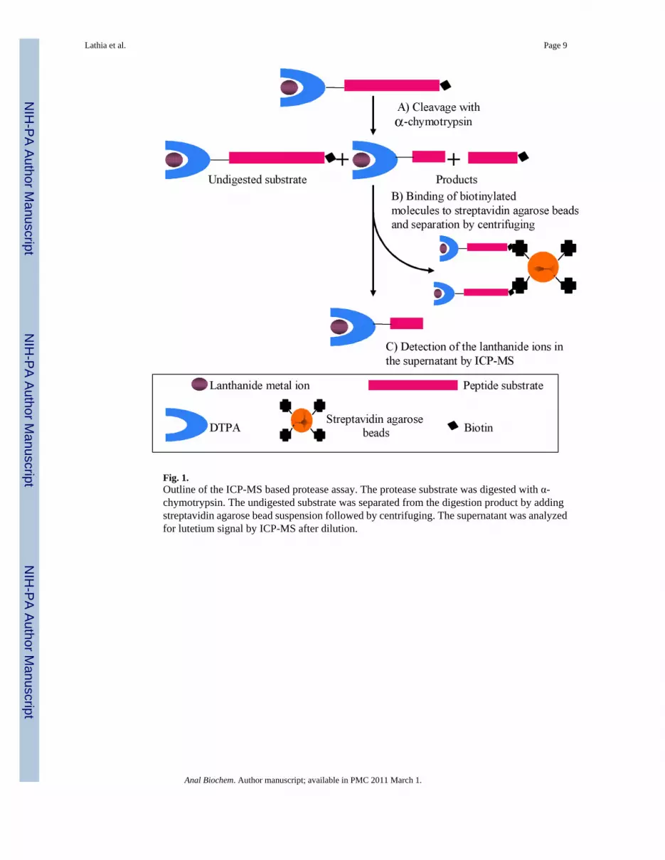
Fig. 1.Outline of the ICP-MS based protease assay. The protease substrate was digested with α-chymotrypsin. The undigested substrate was separated from the digestion product by addingstreptavidin agarose bead suspension followed by centrifuging. The supernatant was analyzedfor lutetium signal by ICP-MS after dilution.
Lathia et al. Page 9
Anal Biochem. Author manuscript; available in PMC 2011 March 1.
NIH
-PA Author Manuscript
NIH
-PA Author Manuscript
NIH
-PA Author Manuscript
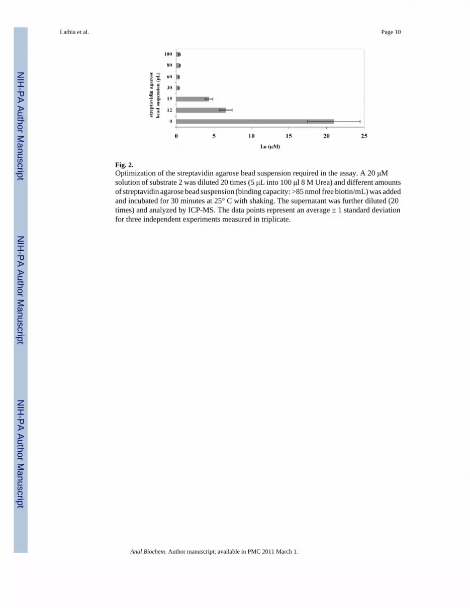
Fig. 2.Optimization of the streptavidin agarose bead suspension required in the assay. A 20 μMsolution of substrate 2 was diluted 20 times (5 μL into 100 μl 8 M Urea) and different amountsof streptavidin agarose bead suspension (binding capacity: >85 nmol free biotin/mL) was addedand incubated for 30 minutes at 25° C with shaking. The supernatant was further diluted (20times) and analyzed by ICP-MS. The data points represent an average ± 1 standard deviationfor three independent experiments measured in triplicate.
Lathia et al. Page 10
Anal Biochem. Author manuscript; available in PMC 2011 March 1.
NIH
-PA Author Manuscript
NIH
-PA Author Manuscript
NIH
-PA Author Manuscript
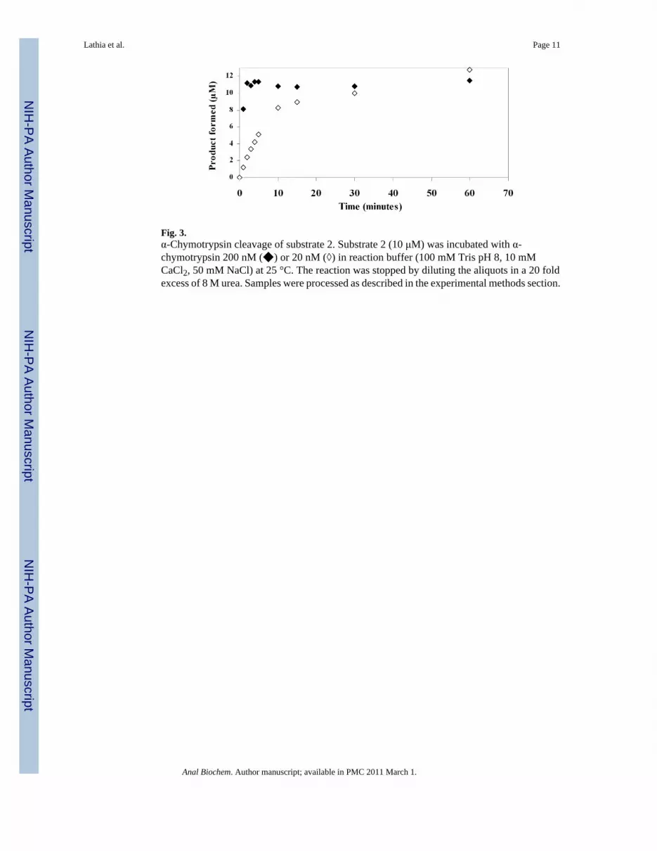
Fig. 3.α-Chymotrypsin cleavage of substrate 2. Substrate 2 (10 μM) was incubated with α-chymotrypsin 200 nM (◆) or 20 nM (◊) in reaction buffer (100 mM Tris pH 8, 10 mMCaCl2, 50 mM NaCl) at 25 °C. The reaction was stopped by diluting the aliquots in a 20 foldexcess of 8 M urea. Samples were processed as described in the experimental methods section.
Lathia et al. Page 11
Anal Biochem. Author manuscript; available in PMC 2011 March 1.
NIH
-PA Author Manuscript
NIH
-PA Author Manuscript
NIH
-PA Author Manuscript
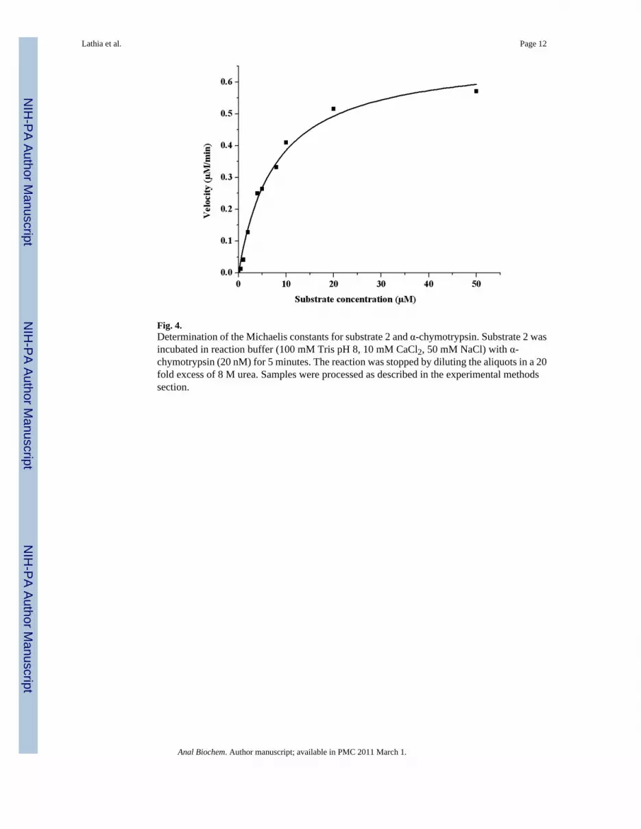
Fig. 4.Determination of the Michaelis constants for substrate 2 and α-chymotrypsin. Substrate 2 wasincubated in reaction buffer (100 mM Tris pH 8, 10 mM CaCl2, 50 mM NaCl) with α-chymotrypsin (20 nM) for 5 minutes. The reaction was stopped by diluting the aliquots in a 20fold excess of 8 M urea. Samples were processed as described in the experimental methodssection.
Lathia et al. Page 12
Anal Biochem. Author manuscript; available in PMC 2011 March 1.
NIH
-PA Author Manuscript
NIH
-PA Author Manuscript
NIH
-PA Author Manuscript
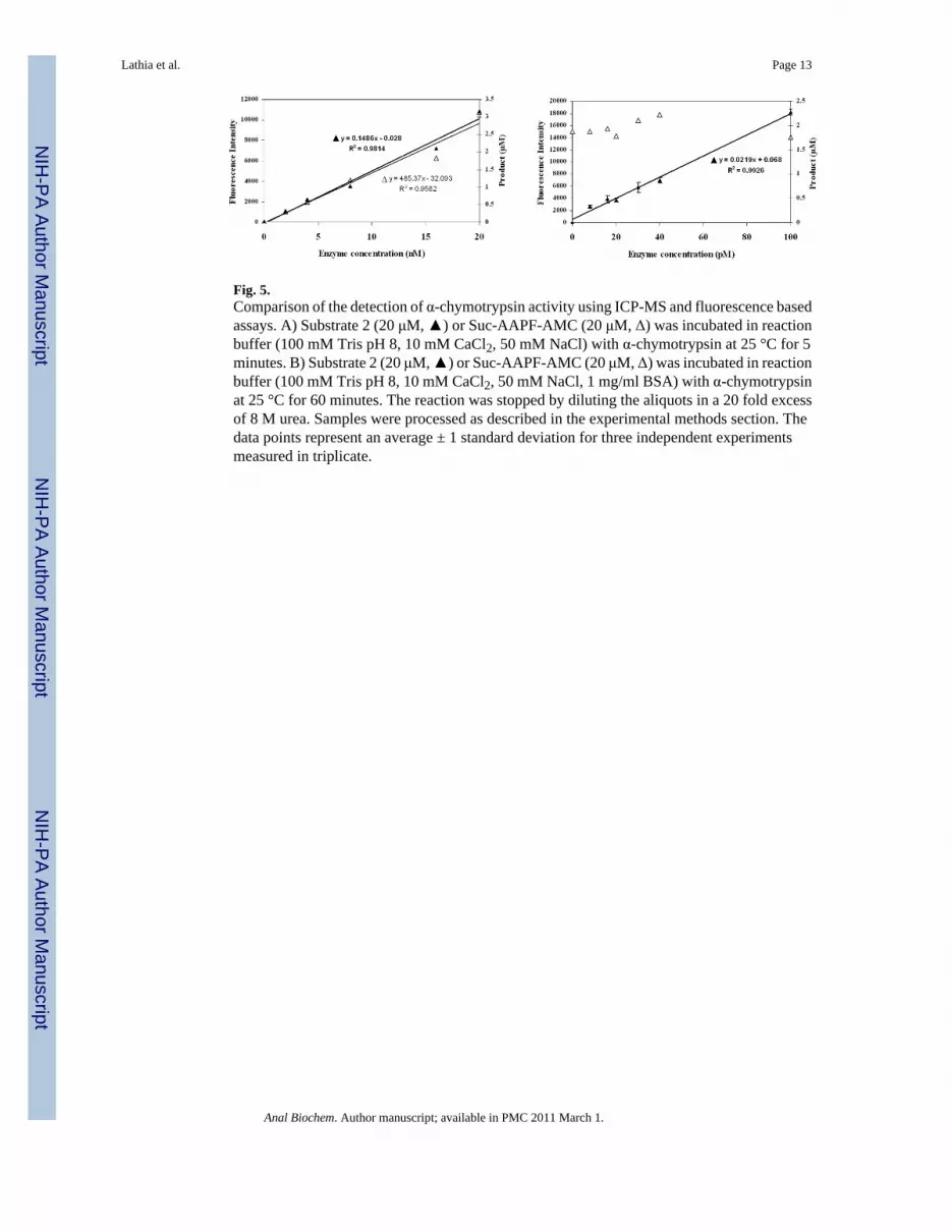
Fig. 5.Comparison of the detection of α-chymotrypsin activity using ICP-MS and fluorescence basedassays. A) Substrate 2 (20 μM, ▲) or Suc-AAPF-AMC (20 μM, Δ) was incubated in reactionbuffer (100 mM Tris pH 8, 10 mM CaCl2, 50 mM NaCl) with α-chymotrypsin at 25 °C for 5minutes. B) Substrate 2 (20 μM, ▲) or Suc-AAPF-AMC (20 μM, Δ) was incubated in reactionbuffer (100 mM Tris pH 8, 10 mM CaCl2, 50 mM NaCl, 1 mg/ml BSA) with α-chymotrypsinat 25 °C for 60 minutes. The reaction was stopped by diluting the aliquots in a 20 fold excessof 8 M urea. Samples were processed as described in the experimental methods section. Thedata points represent an average ± 1 standard deviation for three independent experimentsmeasured in triplicate.
Lathia et al. Page 13
Anal Biochem. Author manuscript; available in PMC 2011 March 1.
NIH
-PA Author Manuscript
NIH
-PA Author Manuscript
NIH
-PA Author Manuscript
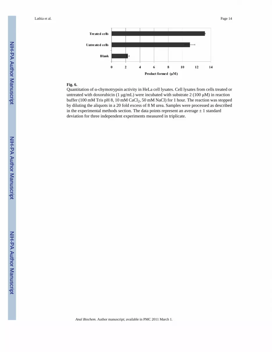
Fig. 6.Quantitation of α-chymotrypsin activity in HeLa cell lysates. Cell lysates from cells treated oruntreated with doxorubicin (1 μg/mL) were incubated with substrate 2 (100 μM) in reactionbuffer (100 mM Tris pH 8, 10 mM CaCl2, 50 mM NaCl) for 1 hour. The reaction was stoppedby diluting the aliquots in a 20 fold excess of 8 M urea. Samples were processed as describedin the experimental methods section. The data points represent an average ± 1 standarddeviation for three independent experiments measured in triplicate.
Lathia et al. Page 14
Anal Biochem. Author manuscript; available in PMC 2011 March 1.
NIH
-PA Author Manuscript
NIH
-PA Author Manuscript
NIH
-PA Author Manuscript

NIH
-PA Author Manuscript
NIH
-PA Author Manuscript
NIH
-PA Author Manuscript
Lathia et al. Page 15
Table 1
Sequences of the α-chymotrypsin substrates used
Substrate 1 DTPA-Asp-Leu-Leu-Val-Tyr∼Asp-Lys(biotin)-amide
Substrate 2 DTPA-βAla- βAla-βAla-βAla-Gly-Ser-Ala-Tyr∼Gly-Lys-Arg-Lys(biotin)-amide
Fluorogenic substrate Suc-Ala-Ala-Pro-Phe-(7-amino-4-methylcoumarin)
Anal Biochem. Author manuscript; available in PMC 2011 March 1.