detection of kiaa1549-braf fusion transcripts in formalin-fixed paraffin-embedded pediatric...
TRANSCRIPT
The Journal of Molecular Diagnostics, Vol. 13, No. 6, November 2011
Copyright © 2011 American Society for Investigative Pathology
and the Association for Molecular Pathology.
Published by Elsevier Inc. All rights reserved.
DOI: 10.1016/j.jmoldx.2011.07.002
Detection of KIAA1549-BRAF Fusion Transcriptsin Formalin-Fixed Paraffin-Embedded Pediatric
Low-Grade GliomasYongji Tian,* Benjamin E. Rich,* Natalie Vena,*†
Justin M. Craig,* Laura E. MacConaill,*‡
Veena Rajaram,§ Stewart Goldman,¶ Hala Taha,�
Madeha Mahmoud,� Memet Ozek,** Aydin Sav,††
Janina A. Longtine,‡‡ Neal I. Lindeman,‡‡
Levi A. Garraway,*‡ Azra H. Ligon,*†
Charles D. Stiles,§§ Sandro Santagata,¶¶��
Jennifer A. Chan,¶¶ Mark W. Kieran,***††† andKeith L. Ligon*†¶¶��
From the Departments of Medical Oncology,* Cancer Biology,§§
and Pediatric Oncology,*** and the Centers for Molecular
Oncologic Pathology,† and Cancer Genome Discovery,‡ Dana-
Farber Cancer Institute, Boston, Massachusetts; the Department
of Pathology and Laboratory Medicine,§ and the Division of
NeuroOncology,¶ Children’s Memorial Hospital, Chicago, Illinois;
the Department of Pediatric Oncology and Pathology,� Children’s
Cancer Hospital, Cairo, Egypt; the Division of Pediatric
Neurosurgery �� and the Department of Pathology,†† Acibadem
University Medical Center, Istanbul, Turkey; the Department of
Pathology,‡‡ and the Division of Neuropathology,¶¶ Brigham and
Women’s Hospital, Boston, Massachusetts; and the Departments
of Pathology,�� and Pediatrics,††† Children’s Hospital Boston,
Boston, Massachusetts
Alterations of BRAF are the most common known ge-netic aberrations in pediatric gliomas. They frequentlyare found in pilocytic astrocytomas, where genomic du-plications involving BRAF and the poorly characterizedgene KIAA1549 create fusion proteins with constitutiveB-Raf kinase activity. BRAF V600E point mutations areless common and generally occur in nonpilocytic tu-mors. The development of BRAF inhibitors as drugs hascreated an urgent need for robust clinical assays to iden-tify activating lesions in BRAF. KIAA1549-BRAF fusiontranscripts have been detected in frozen tissue, how-ever, methods for FFPE tissue have not been reported.We developed a panel of FFPE-compatible quantitativeRT-PCR assays for the most common KIAA1549-BRAFfusion transcripts. Application of these assays to a col-lection of 51 low-grade pediatric gliomas showed 97%sensitivity and 91% specificity compared with fluores-cence in situ hybridization or array comparativegenomic hybridization. In parallel, we assayed samples
for the presence of the BRAF V600E mutation by PCRpyrosequencing. The data further support previous ob-servations that these two alterations of the BRAF,KIAA1549 fusions and V600E point mutations, are asso-ciated primarily with pilocytic astrocytomas and non-pilocytic gliomas, respectively. These results show thatfusion transcripts and mutations can be detected reli-ably in standard FFPE specimens and may be useful forincorporation into future studies of pediatric gliomas inbasic science or clinical trials. (J Mol Diagn 2011, 13:
669–677; DOI: 10.1016/j.jmoldx.2011.07.002)
Brain tumors are the most common solid tumors in chil-dren and are the leading cause of cancer mortality in thisage group (Surveillance Epidemiology and End ResultsCancer Statistics Review, http://seer.cancer.gov/csr/1975_2006/accessed October 21, 2010). Our under-standing of the basic biology of these tumors is not welldeveloped and resources for their diagnosis and man-agement are limited. Most pediatric low-grade gliomasdefined as World Health Organization grade I or II1 areslow growing and have low malignant potential, butthey comprise a heterogeneous group of neoplasmswith diverse behaviors and somewhat unpredictableclinical outcomes. Better understanding of the molec-ular, cellular, and developmental biology of these tu-mors is needed to facilitate improvements in diagnosisand therapy.
Recent studies have highlighted the role of mutationsthat deregulate the activity of RAF family protein kinasesleading to constitutive signaling via the mitogen-activatedprotein kinase pathway.2 Most prominent of these in pe-diatric brain tumors is a class of genomic alterations onchromosome 7q34 that create fusions between a gene ofunknown function, KIAA1549, and the BRAF gene.3–7 Asa result of these fusions, a 2-Mbp region between the twogenes is duplicated in tandem such that the 5= end of the
Supported by the Pediatric Low Grade Astrocytoma Foundation.
Accepted for publication July 6, 2011.
Current address of Y.T., Tiantan Hospital, Beijing, China; of J.A.C.,Departments of Pathology and Laboratory Medicine, Oncology and Clin-ical Neurosciences, University of Calgary, Calgary, Alberta, Canada.
Address reprint requests to Keith Ligon, M.D., Ph.D., Department ofMedical Oncology, Dana Farber Cancer Institute, 450 Brookline Ave.,
Boston, MA 02215. E-mail address: [email protected].669
670 Tian et alJMD November 2011, Vol. 13, No. 6
KIAA1549 gene becomes fused with the 3= end of BRAF.6
Four single instances of similar duplications have beenidentified on chromosome 3 in which the SRGAP3 genebecomes fused to the RAF1 gene.6–8 Three additionalcases of pilocytic astrocytoma (PA) have been identifiedin which a 2.5-Mb deletion results in genetic fusion ofBRAF to the FAM131B gene.7 Each of these chimericgenes encodes a protein in which the C-terminal RAFkinase domain is retained intact, but the N-terminal RAFregulatory domain has been replaced by a polypeptidederived from the N-terminus of the fusion partner,KIAA1549, SRGAP3, or FAM131B. These fusion proteinshave deregulated kinase activity that causes phosphor-ylation of MEK1/27,8 and ERK,6,7 and can transformNIH3T3 cells.3,7,8 The constitutive signaling is attributedto the loss of the RAF regulatory domain, and may beenhanced by membrane association of the N-terminalfusion partner domains.6
The junctions of these fused genes are located atdiverse positions within introns such that splicing ligatesexons with matching reading frames together to createfunctional chimeric mRNAs (Figure 2B and C, of Forshewet al6). Five different configurations of intronic KIAA1549-BRAF fusions have been identified in several studies. Thedistribution of these recently was reviewed by Tatev-ossian et al.9 Seventy-eight percent of the reportedKIAA1549-BRAF fusions (59 of 76) join exon 16 ofKIAA1549 to exon 9 of BRAF (16-9 fusion), 13% of fusions(10 of 76) connect exon 15 of KIAA1549 to exon 9 of BRAF(15-9 fusion), and 7% of fusions (5 of 76) connect exon 16of KIAA1549 to exon 11 of BRAF (16-11 fusion). Singleinstances of two other KIAA1549-BRAF fusion transcriptsalso have been identified. They consist of KIAA1549 exon18:BRAF exon 10 (18-10 fusion) and KIAA1549 exon 19:BRAF exon 9 (19-9 fusion). The FAM131B-BRAF fusionsand SRGAP3-RAF1 fusions are less common and morediverse in structure.6,7
The other BRAF alteration that has been found in pediat-ric low-grade astrocytomas is the T to A transversion atcodon 600 that converts a valine to a glutamic acid (V600E),creating a highly active, constitutive kinase molecule. Acti-vating mutations in BRAF are found in more than half ofmelanomas, about 80% of which are V600E, as well ascarcinomas of the thyroid, colon, and ovary.10,11 The V600Emutation exists in a small number of low-grade pediatricbrain tumors including PAs and diffuse astrocytomas andpleomorphic xanthoastrocytomas,4–6,12–16 a fraction ofhigher-grade astrocytomas (grades 3 and 4),17 and inabout half of gangliogliomas.5,12
Inhibitors of BRAF, some of which already are beingevaluated in adult clinical trials, may be promising ther-apeutics for pediatric gliomas harboring BRAF V600Emutations or BRAF fusions.18,19 It also is possible thatKIAA1549-BRAF fusions contribute to other types of ma-lignancies. As such, there is an emerging need for clini-cally robust technologies to identify tumors with activatedforms of BRAF in individuals who might benefit from thesetargeted agents.
Because they are rare and often small in size, pediatricastrocytoma biopsy samples generally are archived only
as formalin-fixed paraffin-embedded (FFPE) tissue sam-ples in most hospitals. These FFPE tissue samples con-stitute a valuable resource for identifying biomarkers thatmay be useful for diagnosis, determining prognosis, andpredicting response to treatment. The use of archivaltissue samples for molecular genetic studies requiresspecially designed and validated assays that tolerate thedegraded and fragmented nucleic acids extracted fromFFPE tissues. Therefore, to better address the clinicalneed to identify BRAF mutations and to facilitate retro-spective studies on archival tissue samples, we havedeveloped and evaluated novel quantitative reverse tran-scribed PCR (qRT-PCR) methods for detection of multipleKIAA1549-BRAF fusion genes in standard FFPE samplesof pediatric brain tumors. Exon–exon fusion junctions intranscripts from the hybrid KIAA1549-BRAF gene weredetected using a panel of novel qRT-PCR assays involv-ing short amplicons (50–100 bp). The sensitivity, speci-ficity, and accuracy of this method were evaluated bycomparison with cytogenetic data obtained with a clinicalfluorescence in situ hybridization (FISH) assay and arraycomparative genomic hybridization (CGH). In parallel, weidentified mutations at codon 600 of BRAF with a com-mercial PCR pyrosequencing method that is similarly tol-erant of degraded template DNA extracted from FFPEtissue. These assays provide a new strategy for rapid andreliable evaluation of the BRAF status of pediatric gliomasusing FFPE specimens.
Materials and Methods
Pathologic Specimens
Sections of FFPE blocks and unstained slides were ob-tained from the Departments of Pathology at Children’sHospital (Boston, MA), Brigham and Women’s Hospital(Boston, MA), Children’s Cancer Hospital (Cairo, Egypt),Children’s Memorial Hospital (Chicago, IL), and MarmaraUniversity Medical Center (Istanbul, Turkey). All sampleswere obtained under protocols approved by institutionalreview boards at each institution. The FFPE specimensfor which data were available had been stored as paraffinblocks at ambient temperature for up to 14 years, with anaverage time of 3 years. Neuropathology diagnoses weremade by histologic examination according to the criteriaof the World Health Organization classification1 by threeindependent neuropathologists (S.S., Jennifer A. Chan,K.L.L.). PAs were subclassified further as classic (PA_c)if they had a biphasic appearance, Rosenthal fibers, andeosinophilic granular bodies, and nonclassic (PA_nc) ifthey lacked one or more of these specific features but stillwere deemed to be most closely related to PA.
RNA Isolation from FFPE and cDNA Synthesis
Specimens consisting of a total of 40-�m sections ofapproximately 1 cm2 tissue in the form of unmountedscrolls or scrapings from unstained slides were obtainedfrom each FFPE tumor specimen and placed in sterilenuclease-free microcentrifuge tubes. Paraffin was re-moved by extracting the tissue sections twice with 1 mL
xylene. For each extraction the samples were vortexed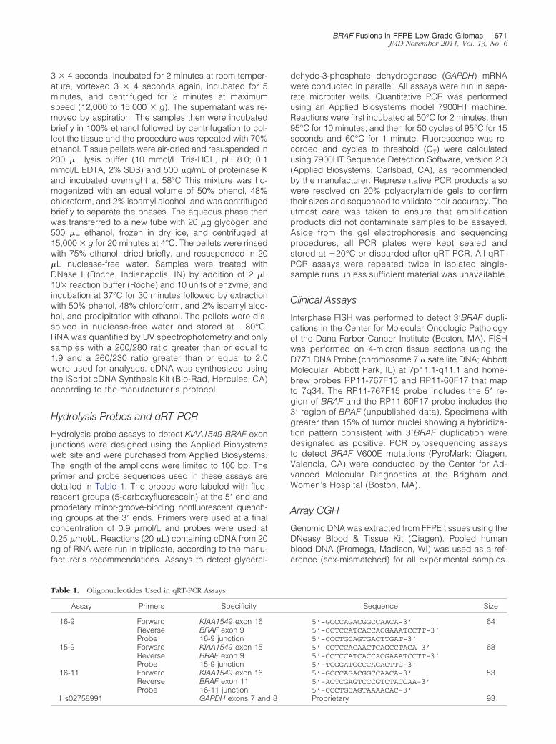
BRAF Fusions in FFPE Low-Grade Gliomas 671JMD November 2011, Vol. 13, No. 6
3 � 4 seconds, incubated for 2 minutes at room temper-ature, vortexed 3 � 4 seconds again, incubated for 5minutes, and centrifuged for 2 minutes at maximumspeed (12,000 to 15,000 � g). The supernatant was re-moved by aspiration. The samples then were incubatedbriefly in 100% ethanol followed by centrifugation to col-lect the tissue and the procedure was repeated with 70%ethanol. Tissue pellets were air-dried and resuspended in200 �L lysis buffer (10 mmol/L Tris-HCL, pH 8.0; 0.1mmol/L EDTA, 2% SDS) and 500 �g/mL of proteinase Kand incubated overnight at 58°C This mixture was ho-mogenized with an equal volume of 50% phenol, 48%chloroform, and 2% isoamyl alcohol, and was centrifugedbriefly to separate the phases. The aqueous phase thenwas transferred to a new tube with 20 �g glycogen and500 �L ethanol, frozen in dry ice, and centrifuged at15,000 � g for 20 minutes at 4°C. The pellets were rinsedwith 75% ethanol, dried briefly, and resuspended in 20�L nuclease-free water. Samples were treated withDNase I (Roche, Indianapolis, IN) by addition of 2 �L10� reaction buffer (Roche) and 10 units of enzyme, andincubation at 37°C for 30 minutes followed by extractionwith 50% phenol, 48% chloroform, and 2% isoamyl alco-hol, and precipitation with ethanol. The pellets were dis-solved in nuclease-free water and stored at �80°C.RNA was quantified by UV spectrophotometry and onlysamples with a 260/280 ratio greater than or equal to1.9 and a 260/230 ratio greater than or equal to 2.0were used for analyses. cDNA was synthesized usingthe iScript cDNA Synthesis Kit (Bio-Rad, Hercules, CA)according to the manufacturer’s protocol.
Hydrolysis Probes and qRT-PCR
Hydrolysis probe assays to detect KIAA1549-BRAF exonjunctions were designed using the Applied Biosystemsweb site and were purchased from Applied Biosystems.The length of the amplicons were limited to 100 bp. Theprimer and probe sequences used in these assays aredetailed in Table 1. The probes were labeled with fluo-rescent groups (5-carboxyfluorescein) at the 5= end andproprietary minor-groove-binding nonfluorescent quench-ing groups at the 3= ends. Primers were used at a finalconcentration of 0.9 �mol/L and probes were used at0.25 �mol/L. Reactions (20 �L) containing cDNA from 20ng of RNA were run in triplicate, according to the manu-facturer’s recommendations. Assays to detect glyceral-
Table 1. Oligonucleotides Used in qRT-PCR Assays
Assay Primers Specificity
16-9 Forward KIAA1549 exon 16Reverse BRAF exon 9Probe 16-9 junction
15-9 Forward KIAA1549 exon 15Reverse BRAF exon 9Probe 15-9 junction
16-11 Forward KIAA1549 exon 16Reverse BRAF exon 11Probe 16-11 junction
Hs02758991 GAPDH exons 7 and 8
dehyde-3-phosphate dehydrogenase (GAPDH) mRNAwere conducted in parallel. All assays were run in sepa-rate microtiter wells. Quantitative PCR was performedusing an Applied Biosystems model 7900HT machine.Reactions were first incubated at 50°C for 2 minutes, then95°C for 10 minutes, and then for 50 cycles of 95°C for 15seconds and 60°C for 1 minute. Fluorescence was re-corded and cycles to threshold (CT) were calculatedusing 7900HT Sequence Detection Software, version 2.3(Applied Biosystems, Carlsbad, CA), as recommendedby the manufacturer. Representative PCR products alsowere resolved on 20% polyacrylamide gels to confirmtheir sizes and sequenced to validate their accuracy. Theutmost care was taken to ensure that amplificationproducts did not contaminate samples to be assayed.Aside from the gel electrophoresis and sequencingprocedures, all PCR plates were kept sealed andstored at �20°C or discarded after qRT-PCR. All qRT-PCR assays were repeated twice in isolated single-sample runs unless sufficient material was unavailable.
Clinical Assays
Interphase FISH was performed to detect 3=BRAF dupli-cations in the Center for Molecular Oncologic Pathologyof the Dana Farber Cancer Institute (Boston, MA). FISHwas performed on 4-micron tissue sections using theD7Z1 DNA Probe (chromosome 7 � satellite DNA; AbbottMolecular, Abbott Park, IL) at 7p11.1-q11.1 and home-brew probes RP11-767F15 and RP11-60F17 that mapto 7q34. The RP11-767F15 probe includes the 5= re-gion of BRAF and the RP11-60F17 probe includes the3= region of BRAF (unpublished data). Specimens withgreater than 15% of tumor nuclei showing a hybridiza-tion pattern consistent with 3=BRAF duplication weredesignated as positive. PCR pyrosequencing assaysto detect BRAF V600E mutations (PyroMark; Qiagen,Valencia, CA) were conducted by the Center for Ad-vanced Molecular Diagnostics at the Brigham andWomen’s Hospital (Boston, MA).
Array CGH
Genomic DNA was extracted from FFPE tissues using theDNeasy Blood & Tissue Kit (Qiagen). Pooled humanblood DNA (Promega, Madison, WI) was used as a ref-erence (sex-mismatched) for all experimental samples.
Sequence Size
5’-GCCCAGACGGCCAACA-3’ 645’-CCTCCATCACCACGAAATCCTT-3’5’-CCCTGCAGTGACTTGAT-3’5’-CGTCCACAACTCAGCCTACA-3’ 685’-CCTCCATCACCACGAAATCCTT-3’5’-TCGGATGCCCAGACTTG-3’5’-GCCCAGACGGCCAACA-3’ 535’-ACTCGAGTCCCGTCTACCAA-3’5’-CCCTGCAGTAAAACAC-3’
Proprietary 93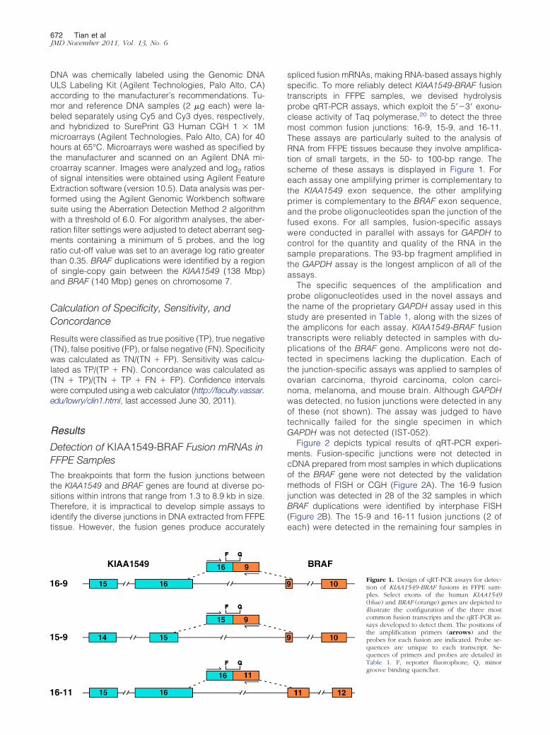
672 Tian et alJMD November 2011, Vol. 13, No. 6
DNA was chemically labeled using the Genomic DNAULS Labeling Kit (Agilent Technologies, Palo Alto, CA)according to the manufacturer’s recommendations. Tu-mor and reference DNA samples (2 �g each) were la-beled separately using Cy5 and Cy3 dyes, respectively,and hybridized to SurePrint G3 Human CGH 1 � 1Mmicroarrays (Agilent Technologies, Palo Alto, CA) for 40hours at 65°C. Microarrays were washed as specified bythe manufacturer and scanned on an Agilent DNA mi-croarray scanner. Images were analyzed and log2 ratiosof signal intensities were obtained using Agilent FeatureExtraction software (version 10.5). Data analysis was per-formed using the Agilent Genomic Workbench softwaresuite using the Aberration Detection Method 2 algorithmwith a threshold of 6.0. For algorithm analyses, the aber-ration filter settings were adjusted to detect aberrant seg-ments containing a minimum of 5 probes, and the logratio cut-off value was set to an average log ratio greaterthan 0.35. BRAF duplications were identified by a regionof single-copy gain between the KIAA1549 (138 Mbp)and BRAF (140 Mbp) genes on chromosome 7.
Calculation of Specificity, Sensitivity, andConcordance
Results were classified as true positive (TP), true negative(TN), false positive (FP), or false negative (FN). Specificitywas calculated as TN/(TN � FP). Sensitivity was calcu-lated as TP/(TP � FN). Concordance was calculated as(TN � TP)/(TN � TP � FN � FP). Confidence intervalswere computed using a web calculator (http://faculty.vassar.edu/lowry/clin1.html, last accessed June 30, 2011).
Results
Detection of KIAA1549-BRAF Fusion mRNAs inFFPE SamplesThe breakpoints that form the fusion junctions betweenthe KIAA1549 and BRAF genes are found at diverse po-sitions within introns that range from 1.3 to 8.9 kb in size.Therefore, it is impractical to develop simple assays toidentify the diverse junctions in DNA extracted from FFPEtissue. However, the fusion genes produce accurately
spliced fusion mRNAs, making RNA-based assays highlyspecific. To more reliably detect KIAA1549-BRAF fusiontranscripts in FFPE samples, we devised hydrolysisprobe qRT-PCR assays, which exploit the 5=�3= exonu-clease activity of Taq polymerase,20 to detect the threemost common fusion junctions: 16-9, 15-9, and 16-11.These assays are particularly suited to the analysis ofRNA from FFPE tissues because they involve amplifica-tion of small targets, in the 50- to 100-bp range. Thescheme of these assays is displayed in Figure 1. Foreach assay one amplifying primer is complementary tothe KIAA1549 exon sequence, the other amplifyingprimer is complementary to the BRAF exon sequence,and the probe oligonucleotides span the junction of thefused exons. For all samples, fusion-specific assayswere conducted in parallel with assays for GAPDH tocontrol for the quantity and quality of the RNA in thesample preparations. The 93-bp fragment amplified inthe GAPDH assay is the longest amplicon of all of theassays.
The specific sequences of the amplification andprobe oligonucleotides used in the novel assays andthe name of the proprietary GAPDH assay used in thisstudy are presented in Table 1, along with the sizes ofthe amplicons for each assay. KIAA1549-BRAF fusiontranscripts were reliably detected in samples with du-plications of the BRAF gene. Amplicons were not de-tected in specimens lacking the duplication. Each ofthe junction-specific assays was applied to samples ofovarian carcinoma, thyroid carcinoma, colon carci-noma, melanoma, and mouse brain. Although GAPDHwas detected, no fusion junctions were detected in anyof these (not shown). The assay was judged to havetechnically failed for the single specimen in whichGAPDH was not detected (IST-052).
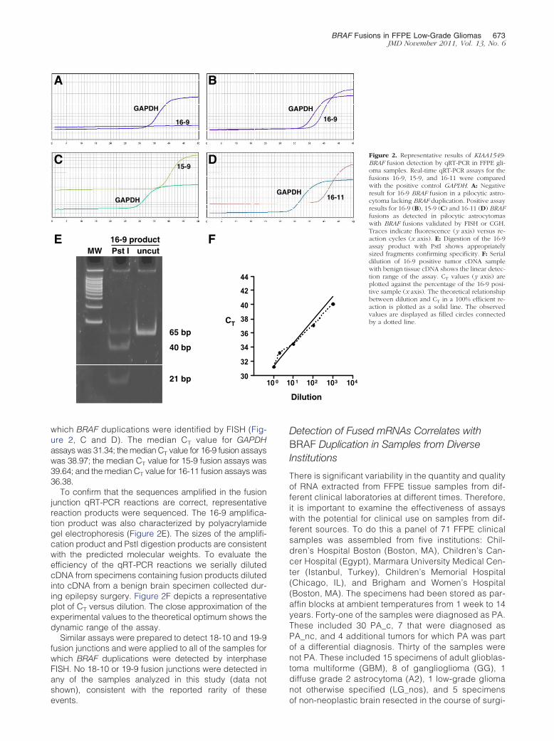
Figure 2 depicts typical results of qRT-PCR experi-ments. Fusion-specific junctions were not detected incDNA prepared from most samples in which duplicationsof the BRAF gene were not detected by the validationmethods of FISH or CGH (Figure 2A). The 16-9 fusionjunction was detected in 28 of the 32 samples in whichBRAF duplications were identified by interphase FISH(Figure 2B). The 15-9 and 16-11 fusion junctions (2 ofeach) were detected in the remaining four samples in
Figure 1. Design of qRT-PCR assays for detec-tion of KIAA1549-BRAF fusions in FFPE sam-ples. Select exons of the human KIAA1549(blue) and BRAF (orange) genes are depicted toillustrate the configuration of the three mostcommon fusion transcripts and the qRT-PCR as-says developed to detect them. The positions ofthe amplification primers (arrows) and theprobes for each fusion are indicated. Probe se-quences are unique to each transcript. Se-quences of primers and probes are detailed inTable 1. F, reporter fluorophore; Q, minorgroove binding quencher.
BRAF Fusions in FFPE Low-Grade Gliomas 673JMD November 2011, Vol. 13, No. 6
which BRAF duplications were identified by FISH (Fig-ure 2, C and D). The median CT value for GAPDHassays was 31.34; the median CT value for 16-9 fusion assayswas 38.97; the median CT value for 15-9 fusion assays was39.64; and the median CT value for 16-11 fusion assays was36.38.
To confirm that the sequences amplified in the fusionjunction qRT-PCR reactions are correct, representativereaction products were sequenced. The 16-9 amplifica-tion product was also characterized by polyacrylamidegel electrophoresis (Figure 2E). The sizes of the amplifi-cation product and PstI digestion products are consistentwith the predicted molecular weights. To evaluate theefficiency of the qRT-PCR reactions we serially dilutedcDNA from specimens containing fusion products dilutedinto cDNA from a benign brain specimen collected dur-ing epilepsy surgery. Figure 2F depicts a representativeplot of CT versus dilution. The close approximation of theexperimental values to the theoretical optimum shows thedynamic range of the assay.
Similar assays were prepared to detect 18-10 and 19-9fusion junctions and were applied to all of the samples forwhich BRAF duplications were detected by interphaseFISH. No 18-10 or 19-9 fusion junctions were detected inany of the samples analyzed in this study (data notshown), consistent with the reported rarity of these
GAPDH
GAPDH
16-9
15-9
A B
C D
MW Pst I uncut16-9 product FE
30
32
34
36
38
40
42
44
CT
65 bp
40 bp
21 bp
events.
Detection of Fused mRNAs Correlates withBRAF Duplication in Samples from DiverseInstitutions
There is significant variability in the quantity and qualityof RNA extracted from FFPE tissue samples from dif-ferent clinical laboratories at different times. Therefore,it is important to examine the effectiveness of assayswith the potential for clinical use on samples from dif-ferent sources. To do this a panel of 71 FFPE clinicalsamples was assembled from five institutions: Chil-dren’s Hospital Boston (Boston, MA), Children’s Can-cer Hospital (Egypt), Marmara University Medical Cen-ter (Istanbul, Turkey), Children’s Memorial Hospital(Chicago, IL), and Brigham and Women’s Hospital(Boston, MA). The specimens had been stored as par-affin blocks at ambient temperatures from 1 week to 14years. Forty-one of the samples were diagnosed as PA.These included 30 PA_c, 7 that were diagnosed asPA_nc, and 4 additional tumors for which PA was partof a differential diagnosis. Thirty of the samples werenot PA. These included 15 specimens of adult glioblas-toma multiforme (GBM), 8 of ganglioglioma (GG), 1diffuse grade 2 astrocytoma (A2), 1 low-grade gliomanot otherwise specified (LG_nos), and 5 specimens
DH
APDH
16-9
16-11
0 1 102 103 104
Dilution
Figure 2. Representative results of KIAA1549-BRAF fusion detection by qRT-PCR in FFPE gli-oma samples. Real-time qRT-PCR assays for thefusions 16-9, 15-9, and 16-11 were comparedwith the positive control GAPDH. A: Negativeresult for 16-9 BRAF fusion in a pilocytic astro-cytoma lacking BRAF duplication. Positive assayresults for 16-9 (B), 15-9 (C) and 16-11 (D) BRAFfusions as detected in pilocytic astrocytomaswith BRAF fusions validated by FISH or CGH.Traces indicate fluorescence (y axis) versus re-action cycles (x axis). E: Digestion of the 16-9assay product with PstI shows appropriatelysized fragments confirming specificity. F: Serialdilution of 16-9 positive tumor cDNA samplewith benign tissue cDNA shows the linear detec-tion range of the assay. CT values (y axis) areplotted against the percentage of the 16-9 posi-tive sample (x axis). The theoretical relationshipbetween dilution and CT in a 100% efficient re-action is plotted as a solid line. The observedvalues are displayed as filled circles connectedby a dotted line.
GAP
G
10 0 1
of non-neoplastic brain resected in the course of surgi-
iagnosis
674 Tian et alJMD November 2011, Vol. 13, No. 6
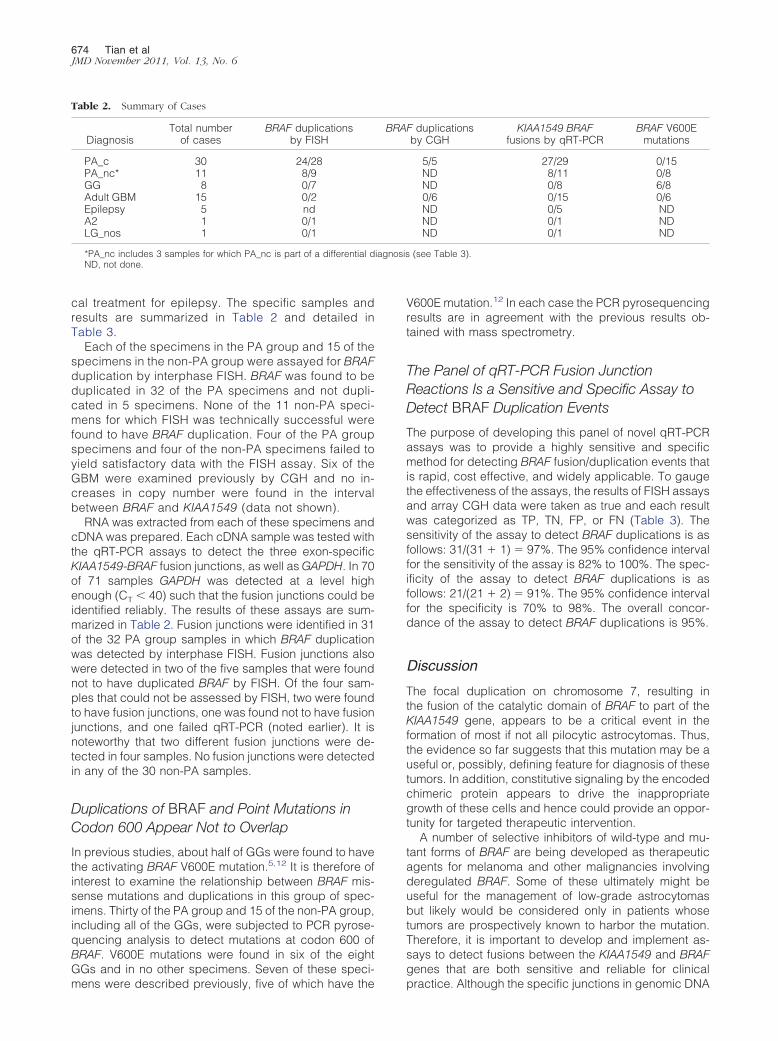
cal treatment for epilepsy. The specific samples andresults are summarized in Table 2 and detailed inTable 3.
Each of the specimens in the PA group and 15 of thespecimens in the non-PA group were assayed for BRAFduplication by interphase FISH. BRAF was found to beduplicated in 32 of the PA specimens and not dupli-cated in 5 specimens. None of the 11 non-PA speci-mens for which FISH was technically successful werefound to have BRAF duplication. Four of the PA groupspecimens and four of the non-PA specimens failed toyield satisfactory data with the FISH assay. Six of theGBM were examined previously by CGH and no in-creases in copy number were found in the intervalbetween BRAF and KIAA1549 (data not shown).
RNA was extracted from each of these specimens andcDNA was prepared. Each cDNA sample was tested withthe qRT-PCR assays to detect the three exon-specificKIAA1549-BRAF fusion junctions, as well as GAPDH. In 70of 71 samples GAPDH was detected at a level highenough (CT � 40) such that the fusion junctions could beidentified reliably. The results of these assays are sum-marized in Table 2. Fusion junctions were identified in 31of the 32 PA group samples in which BRAF duplicationwas detected by interphase FISH. Fusion junctions alsowere detected in two of the five samples that were foundnot to have duplicated BRAF by FISH. Of the four sam-ples that could not be assessed by FISH, two were foundto have fusion junctions, one was found not to have fusionjunctions, and one failed qRT-PCR (noted earlier). It isnoteworthy that two different fusion junctions were de-tected in four samples. No fusion junctions were detectedin any of the 30 non-PA samples.
Duplications of BRAF and Point Mutations inCodon 600 Appear Not to Overlap
In previous studies, about half of GGs were found to havethe activating BRAF V600E mutation.5,12 It is therefore ofinterest to examine the relationship between BRAF mis-sense mutations and duplications in this group of spec-imens. Thirty of the PA group and 15 of the non-PA group,including all of the GGs, were subjected to PCR pyrose-quencing analysis to detect mutations at codon 600 ofBRAF. V600E mutations were found in six of the eightGGs and in no other specimens. Seven of these speci-
Table 2. Summary of Cases
DiagnosisTotal number
of casesBRAF duplications
by FISH
PA_c 30 24/28PA_nc* 11 8/9GG 8 0/7Adult GBM 15 0/2Epilepsy 5 ndA2 1 0/1LG_nos 1 0/1
*PA_nc includes 3 samples for which PA_nc is part of a differential dND, not done.
mens were described previously, five of which have the
V600E mutation.12 In each case the PCR pyrosequencingresults are in agreement with the previous results ob-tained with mass spectrometry.
The Panel of qRT-PCR Fusion JunctionReactions Is a Sensitive and Specific Assay toDetect BRAF Duplication Events
The purpose of developing this panel of novel qRT-PCRassays was to provide a highly sensitive and specificmethod for detecting BRAF fusion/duplication events thatis rapid, cost effective, and widely applicable. To gaugethe effectiveness of the assays, the results of FISH assaysand array CGH data were taken as true and each resultwas categorized as TP, TN, FP, or FN (Table 3). Thesensitivity of the assay to detect BRAF duplications is asfollows: 31/(31 � 1) � 97%. The 95% confidence intervalfor the sensitivity of the assay is 82% to 100%. The spec-ificity of the assay to detect BRAF duplications is asfollows: 21/(21 � 2) � 91%. The 95% confidence intervalfor the specificity is 70% to 98%. The overall concor-dance of the assay to detect BRAF duplications is 95%.
Discussion
The focal duplication on chromosome 7, resulting inthe fusion of the catalytic domain of BRAF to part of theKIAA1549 gene, appears to be a critical event in theformation of most if not all pilocytic astrocytomas. Thus,the evidence so far suggests that this mutation may be auseful or, possibly, defining feature for diagnosis of thesetumors. In addition, constitutive signaling by the encodedchimeric protein appears to drive the inappropriategrowth of these cells and hence could provide an oppor-tunity for targeted therapeutic intervention.
A number of selective inhibitors of wild-type and mu-tant forms of BRAF are being developed as therapeuticagents for melanoma and other malignancies involvingderegulated BRAF. Some of these ultimately might beuseful for the management of low-grade astrocytomasbut likely would be considered only in patients whosetumors are prospectively known to harbor the mutation.Therefore, it is important to develop and implement as-says to detect fusions between the KIAA1549 and BRAFgenes that are both sensitive and reliable for clinical
F duplicationsby CGH
KIAA1549 BRAFfusions by qRT-PCR
BRAF V600Emutations
5/5 27/29 0/15ND 8/11 0/8ND 0/8 6/80/6 0/15 0/6ND 0/5 NDND 0/1 NDND 0/1 ND
(see Table 3).
BRA
practice. Although the specific junctions in genomic DNA
BRAF Fusions in FFPE Low-Grade Gliomas 675JMD November 2011, Vol. 13, No. 6
Table 3. Samples Used in Study
Sample DiagnosisBRAF
Duplication Method NotesBRAF Fusion
qRT-PCRBRAF
Codon 600DuplicationDetection
BOS-001 PA_c YES FISH focal duplication 16-9, 15-9 fail true positiveBOS-004 PA_c YES FISH 16-9 wild type true positiveBOS-006 PA_c YES FISH 16-9 wild type true positiveCAI-003 PA_c YES FISH 16-9 ND true positiveCAI-008 PA_c NO FISH 11% duplicated 16-9 ND false positiveCAI-009 PA_c YES FISH 16-9 ND true positiveCAI-010 PA_c YES FISH 16-9, 15-9 ND true positiveCAI-038 PA_c YES FISH 16-9 ND true positiveCAI-066 PA_c YES FISH 16-9 ND true positiveCAI-072 PA_c YES FISH 16-9 ND true positiveCAI-073 PA_c YES FISH 16-9 ND true positiveCMH-027 PA_c YES FISH 16-9 ND true positiveIST-011 PA_c YES FISH 16-9 wild type true positiveIST-015 PA_c fail FISH looks duplicated 16-9 wild typeIST-017 PA_c NO FISH NONE wild type true negativeIST-018 PA_c NO FISH NONE wild type true negativeIST-019 PA_c NO FISH 8% duplicated 16-9, 16-11 fail false positiveIST-022 PA_c YES FISH 16-9 wild type true positiveIST-023 PA_c YES FISH 16-9 fail true positiveIST-033 PA_c YES FISH 15-9 fail true positiveIST-037 PA_c YES FISH 16-9 wild type true positiveIST-051 PA_c YES FISH 16-9 wild type true positiveIST-052 PA_c fail FISH FAIL NDLGG-073 PA_c YES FISH & CGH 16-9 wild type true positiveLGG-091 PA_c YES FISH & CGH 16-9 wild type true positiveLGG-107 PA_c YES FISH & CGH 16-9 wild type true positiveLGG-172 PA_c YES FISH & CGH 16-9 wild type true positiveLGG-198 PA_c YES FISH 16-11 wild type true positiveLGG-200 PA_c YES FISH & CGH 16-9 wild type true positiveIST-003 PA_c YES FISH 16-9 fail true positiveBOS-002 PA_nc fail FISH 16-9 wild typeIST-006 PA_nc YES FISH NONE fail false negativeIST-013 PA_nc YES FISH 15-9 wild type true positiveIST-025 PA_nc YES FISH 16-9 wild type true positiveIST-032 PA_nc YES FISH 16-9 wild type true positiveIST-034 PA_nc NO FISH NONE wild type true negativeLGG-137 PA_nc YES FISH 16-9 wild type true positiveBOS-007 PA_nc YES FISH 16-11 wild type true positiveIST-020 PA_nc v. A2 YES FISH 16-9 wild type true positiveIST-009 PA_nc v GG_c fail FISH NONE failCMH-010 PA_nc v LG_nos YES FISH 16-9, 15-9 true positiveLGG-058 LG_nos NO FISH NONE wild type true negativeBOS-003 GG_c NO FISH NONE V600E true negativeLGG-097 GG_c NO FISH NONE V600E true negativeLGG-100 GG_c NO FISH NONE wild type true negativeLGG-202 GG_c NO FISH NONE wild type true negativeLGG-267 GG_c NO FISH NONE V600E true negativeLGG-285 GG_c NO FISH NONE V600E true negativeBOS-023 GG_c NO FISH NONE V600E true negativeLGG-132 GG_nc NO FISH NONE V600E true negativeBOS-005 GBM fail FISH NONE wild typeBOS-009 GBM fail FISH NONE wild typeBOS-010 GBM fail FISH NONE wild typeBOS-011 GBM NO FISH NONE wild type true negativeBOS-012 GBM fail FISH NONE wild typeBOS-013 GBM NO FISH NONE wild type true negativeBOS-014 GBM ND NONE NDBOS-015 GBM NO CGH NONE ND true negativeBOS-016 GBM NO CGH NONE ND true negativeBOS-017 GBM NO CGH NONE ND true negativeBOS-018 GBM NO CGH NONE ND true negativeBOS-019 GBM NO CGH NONE ND true negativeBOS-020 GBM NO CGH NONE ND true negativeBOS-021 GBM ND NONE NDBOS-022 GBM ND NONE NDCAI-075 A2 NO FISH NONE ND true negativeBOS-024 seizure ND NONE NDBOS-025 seizure ND NONE NDBOS-026 seizure ND NONE NDBOS-027 seizure ND NONE NDBOS-029 seizure ND NONE NDMUS-001 Mouse brain ND NONE NDMUS-002 Mouse brain ND NONE ND
ND, not done.
676 Tian et alJMD November 2011, Vol. 13, No. 6
formed by these fusions are diverse, the processedmRNAs from the fused genes occur in a small number ofconfigurations. To take advantage of this we have cre-ated a panel of highly sensitive assays to detect each ofthe five splice junctions known to arise in KIAA1549-BRAFfusion mRNAs. The two rarest of these junctions, 18-10and 19-9, have each been identified only once in the 76specimens reported in published studies.9 Neither ofthese rare junctions was detected in this cohort of 40samples with pilocytic features.
Although most of the pilocytic samples were diag-nosed as PA_c or PA_nc, four samples had PA_nc aspart of a differential diagnosis but could not be subclas-sified further. In three of these samples, BRAF was dupli-cated and fusion junctions were detected. Having thiscontributory genetic evidence in a clinical setting wouldallow neuropathologists and neuro-oncologists to con-sider these tumors as being more likely consistent withPA. The fourth of these samples was diagnosed as PA_ncor GG. FISH and BRAF pyrosequencing both failed, likelytechnically owing to the low quality of nucleic acids ex-tracted from this sample. GAPDH amplicons were de-tected, however, and fusion junctions were absent, illus-trating that the qRT-PCR fusion-junction assay may bemore robust and sensitive than FISH and pyrosequenc-ing. In this setting, the qRT-PCR assay then would pro-vide genetic support that this tumor is less likely to rep-resent a pilocytic tumor.
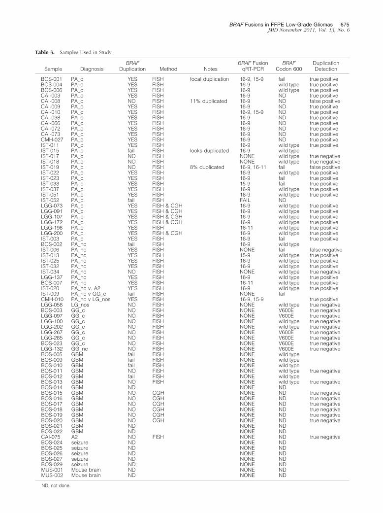
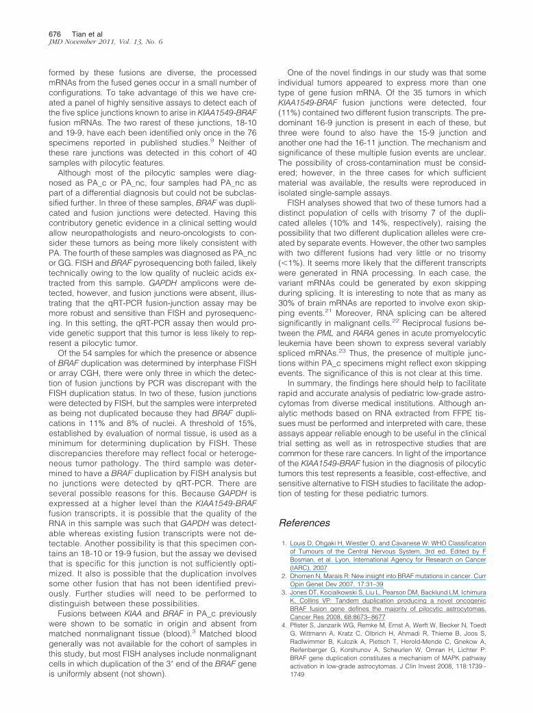
Of the 54 samples for which the presence or absenceof BRAF duplication was determined by interphase FISHor array CGH, there were only three in which the detec-tion of fusion junctions by PCR was discrepant with theFISH duplication status. In two of these, fusion junctionswere detected by FISH, but the samples were interpretedas being not duplicated because they had BRAF dupli-cations in 11% and 8% of nuclei. A threshold of 15%,established by evaluation of normal tissue, is used as aminimum for determining duplication by FISH. Thesediscrepancies therefore may reflect focal or heteroge-neous tumor pathology. The third sample was deter-mined to have a BRAF duplication by FISH analysis butno junctions were detected by qRT-PCR. There areseveral possible reasons for this. Because GAPDH isexpressed at a higher level than the KIAA1549-BRAFfusion transcripts, it is possible that the quality of theRNA in this sample was such that GAPDH was detect-able whereas existing fusion transcripts were not de-tectable. Another possibility is that this specimen con-tains an 18-10 or 19-9 fusion, but the assay we devisedthat is specific for this junction is not sufficiently opti-mized. It also is possible that the duplication involvessome other fusion that has not been identified previ-ously. Further studies will need to be performed todistinguish between these possibilities.
Fusions between KIAA and BRAF in PA_c previouslywere shown to be somatic in origin and absent frommatched nonmalignant tissue (blood).3 Matched bloodgenerally was not available for the cohort of samples inthis study, but most FISH analyses include nonmalignantcells in which duplication of the 3= end of the BRAF gene
is uniformly absent (not shown).One of the novel findings in our study was that someindividual tumors appeared to express more than onetype of gene fusion mRNA. Of the 35 tumors in whichKIAA1549-BRAF fusion junctions were detected, four(11%) contained two different fusion transcripts. The pre-dominant 16-9 junction is present in each of these, butthree were found to also have the 15-9 junction andanother one had the 16-11 junction. The mechanism andsignificance of these multiple fusion events are unclear.The possibility of cross-contamination must be consid-ered; however, in the three cases for which sufficientmaterial was available, the results were reproduced inisolated single-sample assays.
FISH analyses showed that two of these tumors had adistinct population of cells with trisomy 7 of the dupli-cated alleles (10% and 14%, respectively), raising thepossibility that two different duplication alleles were cre-ated by separate events. However, the other two sampleswith two different fusions had very little or no trisomy(�1%). It seems more likely that the different transcriptswere generated in RNA processing. In each case, thevariant mRNAs could be generated by exon skippingduring splicing. It is interesting to note that as many as30% of brain mRNAs are reported to involve exon skip-ping events.21 Moreover, RNA splicing can be alteredsignificantly in malignant cells.22 Reciprocal fusions be-tween the PML and RARA genes in acute promyelocyticleukemia have been shown to express several variablyspliced mRNAs.23 Thus, the presence of multiple junc-tions within PA_c specimens might reflect exon skippingevents. The significance of this is not clear at this time.
In summary, the findings here should help to facilitaterapid and accurate analysis of pediatric low-grade astro-cytomas from diverse medical institutions. Although an-alytic methods based on RNA extracted from FFPE tis-sues must be performed and interpreted with care, theseassays appear reliable enough to be useful in the clinicaltrial setting as well as in retrospective studies that arecommon for these rare cancers. In light of the importanceof the KIAA1549-BRAF fusion in the diagnosis of pilocytictumors this test represents a feasible, cost-effective, andsensitive alternative to FISH studies to facilitate the adop-tion of testing for these pediatric tumors.
References
1. Louis D, Ohgaki H, Wiestler O, and Cavanese W: WHO Classificationof Tumours of the Central Nervous System, 3rd ed. Edited by FBosman, et al. Lyon, International Agency for Research on Cancer(IARC), 2007
2. Dhomen N, Marais R: New insight into BRAF mutations in cancer. CurrOpin Genet Dev 2007, 17:31–39
3. Jones DT, Kocialkowski S, Liu L, Pearson DM, Backlund LM, IchimuraK, Collins VP: Tandem duplication producing a novel oncogenicBRAF fusion gene defines the majority of pilocytic astrocytomas.Cancer Res 2008, 68:8673–8677
4. Pfister S, Janzarik WG, Remke M, Ernst A, Werft W, Becker N, ToedtG, Wittmann A, Kratz C, Olbrich H, Ahmadi R, Thieme B, Joos S,Radlwimmer B, Kulozik A, Pietsch T, Herold-Mende C, Gnekow A,Reifenberger G, Korshunov A, Scheurlen W, Omran H, Lichter P:BRAF gene duplication constitutes a mechanism of MAPK pathway
activation in low-grade astrocytomas. J Clin Invest 2008, 118:1739–1749
BRAF Fusions in FFPE Low-Grade Gliomas 677JMD November 2011, Vol. 13, No. 6
5. Sievert AJ, Jackson EM, Gai X, Hakonarson H, Judkins AR, ResnickAC, Sutton LN, Storm PB, Shaikh TH, Biegel JA: Duplication of 7q34in pediatric low-grade astrocytomas detected by high-density single-nucleotide polymorphism-based genotype arrays results in a novelBRAF fusion gene. Brain Pathol 2009, 19:449–458
6. Forshew T, Tatevossian RG, Lawson AR, Ma J, Neale G, OgunkoladeBW, Jones TA, Aarum J, Dalton J, Bailey S, Chaplin T, Carter RL,Gajjar A, Broniscer A, Young BD, Ellison DW, Sheer D: Activation ofthe ERK/MAPK pathway: a signature genetic defect in posterior fossapilocytic astrocytomas. J Pathol 2009, 218:172–181
7. Cin H, Meyer C, Herr R, Janzarik WG, Lambert S, Jones DT, Jacob K,Benner A, Witt H, Remke M, Bender S, Falkenstein F, Van Anh TN,Olbrich H, von Deimling A, Pekrun A, Kulozik AE, Gnekow A,Scheurlen W, Witt O, Omran H, Jabado N, Collins VP, Brummer T,Marschalek R, Lichter P, Korshunov A, Pfister SM: OncogenicFAM131B-BRAF fusion resulting from 7q34 deletion comprises analternative mechanism of MAPK pathway activation in pilocytic astro-cytoma. Acta Neuropathol 2011, 121:763–774
8. Jones DT, Kocialkowski S, Liu L, Pearson DM, Ichimura K, Collins VP:Oncogenic RAF1 rearrangement and a novel BRAF mutation as al-ternatives to KIAA1549: BRAF fusion in activating the MAPK pathwayin pilocytic astrocytoma. Oncogene 2009, 28:2119–2123
9. Tatevossian RG, Lawson AR, Forshew T, Hindley GF, Ellison DW,Sheer D: MAPK pathway activation and the origins of pediatric low-grade astrocytomas. J Cell Physiol 2010, 222:509–514
10. Davies H, Bignell GR, Cox C, Stephens P, Edkins S, Clegg S, et al:Mutations of the BRAF gene in human cancer. Nature 2002, 417:949–954
11. Michaloglou C, Vredeveld LC, Mooi WJ, Peeper DS: BRAF(E600) inbenign and malignant human tumours. Oncogene 2008, 27:877–895
12. MacConaill LE, Campbell CD, Kehoe SM, Bass AJ, Hatton C, Niu L,Davis M, Yao K, Hanna M, Mondal C, Luongo L, Emery CM, Baker AC,Philips J, Goff DJ, Fiorentino M, Rubin MA, Polyak K, Chan J, Wang Y,Fletcher JA, Santagata S, Corso G, Roviello F, Shivdasani R, KieranMW, Ligon KL, Stiles CD, Hahn WC, Meyerson ML, Garraway LA:Profiling critical cancer gene mutations in clinical tumor samples.PLoS One 2009, 4:e7887
13. Kolb EA, Gorlick R, Houghton PJ, Morton CL, Neale G, Keir ST, Carol
H, Lock R, Phelps D, Kang MH, Reynolds CP, Maris JM, Billups C,Smith MA: Initial testing (stage 1) of AZD6244 (ARRY-142886) by thePediatric Preclinical Testing Program. Pediatr Blood Cancer 2010,55:668–677
14. Eisenhardt AE, Olbrich H, Roring M, Janzarik W, Van Anh TN, Cin H,Remke M, Witt H, Korshunov A, Pfister SM, Omran H, Brummer T:Functional characterization of a BRAF insertion mutant associatedwith pilocytic astrocytoma. Int J Cancer 2010, doi: 10.1002/ijc.25893
15. Dias-Santagata D, Lam Q, Vernovsky K, Vena N, Lennerz JK, BorgerDR, Batchelor TT, Ligon KL, Iafrate AJ, Ligon AH, Louis DN, San-tagata S: BRAF V600E mutations are common in pleomorphicxanthoastrocytoma: diagnostic and therapeutic implications. PLoSOne 2011, 6:e17948
16. Schindler G, Capper D, Meyer J, Janzarik W, Omran H, Herold-Mende C, Schmieder K, Wesseling P, Mawrin C, Hasselblatt M, LouisDN, Korshunov A, Pfister S, Hartmann C, Paulus W, Reifenberger G,von Deimling A: Analysis of BRAF V600E mutation in 1,320 nervoussystem tumors reveals high mutation frequencies in pleomorphicxanthoastrocytoma, ganglioglioma and extra-cerebellar pilocytic as-trocytoma. Acta Neuropathol 2011, 121:397–405
17. Schiffman JD, Hodgson JG, VandenBerg SR, Flaherty P, Polley MY,Yu M, Fisher PG, Rowitch DH, Ford JM, Berger MS, Ji H, GutmannDH, James CD: Oncogenic BRAF mutation with CDKN2A inactivationis characteristic of a subset of pediatric malignant astrocytomas.Cancer Res 2010, 70:512–519
18. Whittaker S, Menard D, Kirk R, Ogilvie L, Hedley D, Zambon A, LopesF, Preece N, Manne H, Rana S, Lambros M, Reis-Filho JS, Marais R,Springer CJ: A novel, selective, and efficacious nanomolar pyridopyr-azinone inhibitor of V600EBRAF. Cancer Res 2010, 70:8036–8044
19. Bollag G, Hirth P, Tsai J, Zhang J, Ibrahim PN, Cho H, et al: Clinicalefficacy of a RAF inhibitor needs broad target blockade in BRAF-mutant melanoma. Nature 2010, 467:596–599
20. Holland PM, Abramson RD, Watson R, Gelfand DH: Detection ofspecific polymerase chain reaction product by utilizing the 5=–3=exonuclease activity of Thermus aquaticus DNA polymerase. ProcNatl Acad Sci U S A 1991, 88:7276–7280
21. Yeo G, Holste D, Kreiman G, Burge CB: Variation in alternativesplicing across human tissues. Genome Biol 2004, 5:R74
22. Ghigna C, Valacca C, Biamonti G: Alternative splicing and tumorprogression. Curr Genomics 2008, 9:556–570
23. Jensen K, Shiels C, Freemont PS: PML protein isoforms and theRBCC/TRIM motif. Oncogene 2001, 20:7223–7233